Controlling Viral Immuno-Inflammatory Lesions by Modulating ...
16
Controlling Viral Immuno-Inflammatory Lesions by Modulating Aryl Hydrocarbon Receptor Signaling Tamara Veiga-Parga 1. , Amol Suryawanshi 2. , Barry T. Rouse 1 * 1 Department of Pathobiology, College of Veterinary Medicine, University of Tennessee, Knoxville, Tennessee, United States of America, 2 Emory Vaccine Center and Yerkes Primate Research Center, Atlanta, Georgia, United States of America Abstract Ocular herpes simplex virus infection can cause a blinding CD4 + T cell orchestrated immuno-inflammatory lesion in the cornea called Stromal Keratitis (SK). A key to controlling the severity of SK lesions is to suppress the activity of T cells that orchestrate lesions and enhance the representation of regulatory cells that inhibit effector cell function. In this report we show that a single administration of TCDD (2, 3, 7, 8- Tetrachlorodibenzo-p-dioxin), a non-physiological ligand for the AhR receptor, was an effective means of reducing the severity of SK lesions. It acted by causing apoptosis of Foxp3 - CD4 + T cells but had no effect on Foxp3 + CD4 + Tregs. TCDD also decreased the proliferation of Foxp3 - CD4 + T cells. The consequence was an increase in the ratio of Tregs to T effectors which likely accounted for the reduced inflammatory responses. In addition, in vitro studies revealed that TCDD addition to anti-CD3/CD28 stimulated naı ¨ve CD4 + T cells caused a significant induction of Tregs, but inhibited the differentiation of Th1 and Th17 cells. Since a single TCDD administration given after the disease process had been initiated generated long lasting anti-inflammatory effects, the approach holds promise as a therapeutic means of controlling virus induced inflammatory lesions. Citation: Veiga-Parga T, Suryawanshi A, Rouse BT (2011) Controlling Viral Immuno-Inflammatory Lesions by Modulating Aryl Hydrocarbon Receptor Signaling. PLoS Pathog 7(12): e1002427. doi:10.1371/journal.ppat.1002427 Editor: E. John Wherry, University of Pennsylvania, United States of America Received April 4, 2011; Accepted October 25, 2011; Published December 8, 2011 Copyright: ß 2011 Veiga-Parga, et al. This is an open-access article distributed under the terms of the Creative Commons Attribution License, which permits unrestricted use, distribution, and reproduction in any medium, provided the original author and source are credited. Funding: This study was supported by National Institute of Allergy and Infectious Diseases Grant AI 063365 and National Institutes of Health Grant EY 005093. The funders had no role in study design, data collection and analysis, decision to publish, or preparation of the manuscript. Competing Interests: The authors have declared that no competing interests exist. * E-mail: [email protected] . These authors contributed equally to this work. Introduction Ocular infection with herpes simplex virus (HSV) can result in a chronic immuno-inflammatory reaction in the cornea which represents a common cause of human blindness [1,2]. The pathogenesis of stromal keratitis (SK) involves numerous events, but studies in murine SK models indicate that lesions are mainly orchestrated by CD4 + T cells that recognize virus derived peptides, or perhaps altered self proteins unmasked in the damaged cornea [1–4]. The severity of SK can be influenced by the balance of CD4 + effector T cells and Foxp3 + regulatory T cells (Treg) [5,6]. Procedures that change this balance represent a promising approach for therapy. This has been achieved either by adoptive transfer with Treg populations [6] or the repeated administration of reagents that can cause naı ¨ve CD4 + T cells to convert to become Treg [7,8]. From a therapeutic angle, procedures that could shift the balance of T effectors and Treg after a single drug administration would represent a convenient maneuver. Recent evidence from studies to control autoimmunity and graft-versus-host disease indicate that the objective might be achieved by the administration of stable agonists of the aryl hydrocarbon receptor (AhR) [9–11]. The AhR is a cytosolic transcription factor that can be activated by different ligands. These include the physiological ligand tryptophan photoproduct 6-formylindolo(2,3-b)carbazole (FICZ), and synthetic molecules such as 2, 3, 7, 8- tetrachlorodibenzo-p-dioxin (TCDD) [10,12]. Signaling through the AhR has consequences that include changes in innate cell function, as well as some modulatory effects on several aspects of T cell immunity [13,14]. For example, Weiner and colleagues showed that TCDD administration could suppress the induction of experimental autoimmune encephalomyelitis (EAE), an effect attributed to a reduction of proinflammatory T cells along with the expansion of Treg [9]. By a similar mechanism, TCDD had suppressive effects in an autoimmune diabetes model [15]. Similarly, the administration of TCDD prior to the induction of colitis led to reduced lesions along with an increase in the Treg population [16]. In graft versus host disease (GVHD) too, the reduced lesions in TCDD treated animals was attributed to the expansion of adaptive Tregs that suppressed allospecific cytotoxic T cell generation [11,17]. Modulating AhR by TCDD has also been shown to control the differentiation of Type 1 regulatory T cells (Tr1) in vitro, which produce IL-10 and are instrumental in the prevention of tissue inflammation, autoimmunity as well as GVHD [18]. Although AhR ligation can result in reduced inflammatory lesions, in some circumstances lesions may be exacerbated. This was noted in the Weiner studies when the physiological ligand FICZ, rather than TCDD, was used for treatment [9]. In this study administration of FICZ boosted Th17 differentiation and increased the severity of EAE. Proinflammatory effects of AhR activation were also noted in a model of rheumatoid arthritis [19], where synoviocytes were exposed to different concentrations of TCDD and shown to produce inflammatory cytokines. Additional PLoS Pathogens | www.plospathogens.org 1 December 2011 | Volume 7 | Issue 12 | e1002427
-
Upload
khangminh22 -
Category
Documents
-
view
0 -
download
0
Transcript of Controlling Viral Immuno-Inflammatory Lesions by Modulating ...

Controlling Viral Immuno-Inflammatory Lesions byModulating Aryl Hydrocarbon Receptor SignalingTamara Veiga-Parga1., Amol Suryawanshi2., Barry T. Rouse1*
1 Department of Pathobiology, College of Veterinary Medicine, University of Tennessee, Knoxville, Tennessee, United States of America, 2 Emory Vaccine Center and
Yerkes Primate Research Center, Atlanta, Georgia, United States of America
Abstract
Ocular herpes simplex virus infection can cause a blinding CD4+ T cell orchestrated immuno-inflammatory lesion in thecornea called Stromal Keratitis (SK). A key to controlling the severity of SK lesions is to suppress the activity of T cells thatorchestrate lesions and enhance the representation of regulatory cells that inhibit effector cell function. In this report weshow that a single administration of TCDD (2, 3, 7, 8- Tetrachlorodibenzo-p-dioxin), a non-physiological ligand for the AhRreceptor, was an effective means of reducing the severity of SK lesions. It acted by causing apoptosis of Foxp3- CD4+ T cellsbut had no effect on Foxp3+ CD4+ Tregs. TCDD also decreased the proliferation of Foxp3- CD4+ T cells. The consequencewas an increase in the ratio of Tregs to T effectors which likely accounted for the reduced inflammatory responses. Inaddition, in vitro studies revealed that TCDD addition to anti-CD3/CD28 stimulated naıve CD4+ T cells caused a significantinduction of Tregs, but inhibited the differentiation of Th1 and Th17 cells. Since a single TCDD administration given after thedisease process had been initiated generated long lasting anti-inflammatory effects, the approach holds promise as atherapeutic means of controlling virus induced inflammatory lesions.
Citation: Veiga-Parga T, Suryawanshi A, Rouse BT (2011) Controlling Viral Immuno-Inflammatory Lesions by Modulating Aryl Hydrocarbon ReceptorSignaling. PLoS Pathog 7(12): e1002427. doi:10.1371/journal.ppat.1002427
Editor: E. John Wherry, University of Pennsylvania, United States of America
Received April 4, 2011; Accepted October 25, 2011; Published December 8, 2011
Copyright: � 2011 Veiga-Parga, et al. This is an open-access article distributed under the terms of the Creative Commons Attribution License, which permitsunrestricted use, distribution, and reproduction in any medium, provided the original author and source are credited.
Funding: This study was supported by National Institute of Allergy and Infectious Diseases Grant AI 063365 and National Institutes of Health Grant EY 005093.The funders had no role in study design, data collection and analysis, decision to publish, or preparation of the manuscript.
Competing Interests: The authors have declared that no competing interests exist.
* E-mail: [email protected]
. These authors contributed equally to this work.
Introduction
Ocular infection with herpes simplex virus (HSV) can result in a
chronic immuno-inflammatory reaction in the cornea which
represents a common cause of human blindness [1,2]. The
pathogenesis of stromal keratitis (SK) involves numerous events,
but studies in murine SK models indicate that lesions are mainly
orchestrated by CD4+ T cells that recognize virus derived
peptides, or perhaps altered self proteins unmasked in the
damaged cornea [1–4]. The severity of SK can be influenced by
the balance of CD4+ effector T cells and Foxp3+ regulatory T cells
(Treg) [5,6]. Procedures that change this balance represent a
promising approach for therapy. This has been achieved either by
adoptive transfer with Treg populations [6] or the repeated
administration of reagents that can cause naıve CD4+ T cells to
convert to become Treg [7,8]. From a therapeutic angle,
procedures that could shift the balance of T effectors and Treg
after a single drug administration would represent a convenient
maneuver.
Recent evidence from studies to control autoimmunity and
graft-versus-host disease indicate that the objective might be
achieved by the administration of stable agonists of the aryl
hydrocarbon receptor (AhR) [9–11]. The AhR is a cytosolic
transcription factor that can be activated by different ligands.
These include the physiological ligand tryptophan photoproduct
6-formylindolo(2,3-b)carbazole (FICZ), and synthetic molecules
such as 2, 3, 7, 8- tetrachlorodibenzo-p-dioxin (TCDD) [10,12].
Signaling through the AhR has consequences that include changes
in innate cell function, as well as some modulatory effects on
several aspects of T cell immunity [13,14]. For example, Weiner
and colleagues showed that TCDD administration could suppress
the induction of experimental autoimmune encephalomyelitis
(EAE), an effect attributed to a reduction of proinflammatory T
cells along with the expansion of Treg [9]. By a similar
mechanism, TCDD had suppressive effects in an autoimmune
diabetes model [15]. Similarly, the administration of TCDD prior
to the induction of colitis led to reduced lesions along with an
increase in the Treg population [16]. In graft versus host disease
(GVHD) too, the reduced lesions in TCDD treated animals was
attributed to the expansion of adaptive Tregs that suppressed
allospecific cytotoxic T cell generation [11,17]. Modulating AhR
by TCDD has also been shown to control the differentiation of
Type 1 regulatory T cells (Tr1) in vitro, which produce IL-10 and
are instrumental in the prevention of tissue inflammation,
autoimmunity as well as GVHD [18].
Although AhR ligation can result in reduced inflammatory
lesions, in some circumstances lesions may be exacerbated. This
was noted in the Weiner studies when the physiological ligand
FICZ, rather than TCDD, was used for treatment [9]. In this
study administration of FICZ boosted Th17 differentiation and
increased the severity of EAE. Proinflammatory effects of AhR
activation were also noted in a model of rheumatoid arthritis [19],
where synoviocytes were exposed to different concentrations of
TCDD and shown to produce inflammatory cytokines. Additional
PLoS Pathogens | www.plospathogens.org 1 December 2011 | Volume 7 | Issue 12 | e1002427

proinflammatory effects of AhR ligation were also associated with
pulmonary neutrophilia [20,21], as well as with the induction and
expansion of IL-17+ secreting CD4+ T cells (Th17) that expressed
high levels of AhR receptors [22,23]. Currently, it is not clear why
AhR activation causes either an increased, or a reduced effect on
inflammatory reactions, but the stability of the ligand used for
AhR stimulation is one suspected explanation [24]. Accordingly,
TCDD is a non-degradable high affinity ligand for AhR and most
studies using this ligand report inhibitory effects on inflammatory
reactions [24,25].
The effects of AhR agonists have not been evaluated in microbe
induced inflammatory lesions. In this report, we show that a single
administration of the stable AhR ligand TCDD was highly
effective at suppressing the severity of ocular immuno-inflamma-
tory lesions caused by HSV. The outcome was attributed to
inhibitory effects on inflammatory IFN-c+ secreting CD4+ T cells
(Th1) and Th17 cells. However, since Foxp3+ regulatory T cell
numbers remained unchanged by the treatment, the balance
between T effectors and Tregs favored the latter population.
TCDD was also shown to cause apoptosis ex vivo of Foxp3- CD4+
T cells and could cause some naıve T cells to convert to Foxp3+
CD4+ T cells. Since a single TCDD administration given after the
disease process had been initiated generated long lasting anti-
inflammatory effects, the approach holds promise as a therapeutic
means of controlling virus induced inflammatory lesions.
Results
Modulation of AhR signaling diminishes HSV-1 mediatedimmunopathology
To evaluate the role of AhR engagement on the outcome of
ocular HSV infection, mice were given a single intraperitoneal (IP)
administration of TCDD on day 1 post-infection (pi), and the
effect on the severity of ocular lesions was compared to untreated
controls. All treated animals developed significantly reduced
lesions compared to controls, but around 40% of the animals
developed clinical signs typical of herpes encephalitis before the
end of the 15 day observation period and had to be terminated
(Figure 1A–D). Ocular viral loads were also increased in the
TCDD treated group (Figure 1E). Accordingly, the drug was
judged to be effective but would not be recommended for use
when virus is present and actively replicating in the cornea. In
other experiments, the physiological AhR ligand FICZ was
administered daily starting at day 1 pi. This drug was without
significant effects on lesion severity (Figure 1F), and none of the
treated animals developed herpetic encephalitis (data not shown).
In additional experiments, TCDD administration was begun on
day 5 pi, a time when levels of replicating virus in the cornea were
barely detectable and inflammatory lesions start to become evident
[3]. This treatment procedure resulted in significantly reduced
lesion severity, as well as the extent of corneal neovascularization,
compared to untreated infected controls (Figure 2A–D), and none
of the treated animals developed encephalitis. The treatment
procedure delayed the time of lesion appearance and average
severity scores were significantly less at most time points over a 15
day observation period. For example, on day 12 pi, whereas 10 of
12 eyes from untreated animals had lesion scores of 3 or above,
only 2 of 14 eyes in the treated group had lesions of such severity
(Figure 2B). An example of comparative severity of control and
treated animals is shown in the histological sections in Figure 2E.
In additional experiments terminated on day 28 pi, the pattern of
results was similar with treated animals showing significantly
diminished lesions compared to untreated controls (Figure 2F). In
conclusion, ligation of the AhR with a single administration of
TCDD given 5 days after virus infection significantly diminished
HSV induced immunopathology.
Modulation of AhR signaling diminishes cell infiltration aswell as cytokines after HSV-1 infection
To measure the effect of TCDD treatment on the cellular
composition of SK lesions collagen digested corneas were analyzed
by FACS and compared to controls at day 15 pi. The combination
of three independent experiments is shown in Figure 3A–D. As
shown in Figure 3D, the average number per cornea of
neutrophils and CD4+ T cells was reduced in the treated group
by 2.03 fold and 4.7 fold respectively when compared to untreated
controls.
In separate experiments of the same design, pools of corneas
were processed to quantify mRNA of selected cytokines (IL-1b,
TNF-a, IL-6, IFN-c, and IL-17) and chemokines (CCL20,
CXCL9, CXCL10, and CXCL11) by quantitative real time
PCR (Q-RTPCR). As shown in Figure 3C, the consequence of
TCDD treatment was a reduction in the levels of several
proinflammatory cytokines and chemokines. However, levels of
the cytokine IL-10 was increased to 1.4 fold in samples from
treated compared to controls. Taken together, our results show
AhR ligation by TCDD significantly reduced the total cellular
infiltration of CD4+ T cells and neutrophils, as well as the amount
of proinflammatory cytokines and chemokines.
AhR signaling changes the balance of effectors and TregTo measure the consequences of TCDD treatment on the T cell
subset composition of SK lesions at day 15 pi, pools of corneas
from treated and control animals were collagen digested to recover
the T cell population. Part of the pool was stimulated in vitro for
4 hours with PMA and ionomycin to enumerate cells that were
either IFN-c or IL-17 producers. The other fraction was used to
enumerate Foxp3+ CD4+ T cells. In the experiment shown, there
was an average 12.3 fold reduction of Th1 cells and a 9.4 fold
reduction of Th17 cells in treated compared to control corneas.
The numbers of Foxp3+ T cells were almost identical in corneal
pools from treated and control animals (Figure 4C). Two
additional experiments provided a similar pattern of results.
Taken together, our results show that a consequence of TCDD
treatment was to increase the ratio of total numbers of Foxp3+
CD4+T cells to both, Th1 and Th17 cells (Figure 4D).
Parallel studies of a similar design were performed with T cells
isolated from the draining lymph nodes (DLN) and spleen
collected from the same animals used for the corneal studies.
The results shown in Figure 5A–D demonstrate that Th1 and
Th17 cell frequencies and total numbers per organ were
significantly reduced in TCDD recipients when compared to
Author Summary
This report describes a novel approach to control ablinding immuno-inflammatory reaction in the eye causedby herpes simplex virus. We showed that a singleadministration of TCDD, a stable agonist of the arylhydrocarbon receptor, significantly reduced the severity ofherpes keratitis lesions. The outcome of the therapy was achange in the balance of effector cells responsible fororchestrating lesions, with regulatory cells able to inhibitthe inflammatory effects of the effectors. Since a singleadministration of TCDD provided effective treatment thatlasted for as long as one month, this approach couldrepresent a valuable therapy for a lesion that is a commoncause of human blindness.
Role of AhR Signaling in HSV-1 Immunopathology
PLoS Pathogens | www.plospathogens.org 2 December 2011 | Volume 7 | Issue 12 | e1002427

Role of AhR Signaling in HSV-1 Immunopathology
PLoS Pathogens | www.plospathogens.org 3 December 2011 | Volume 7 | Issue 12 | e1002427
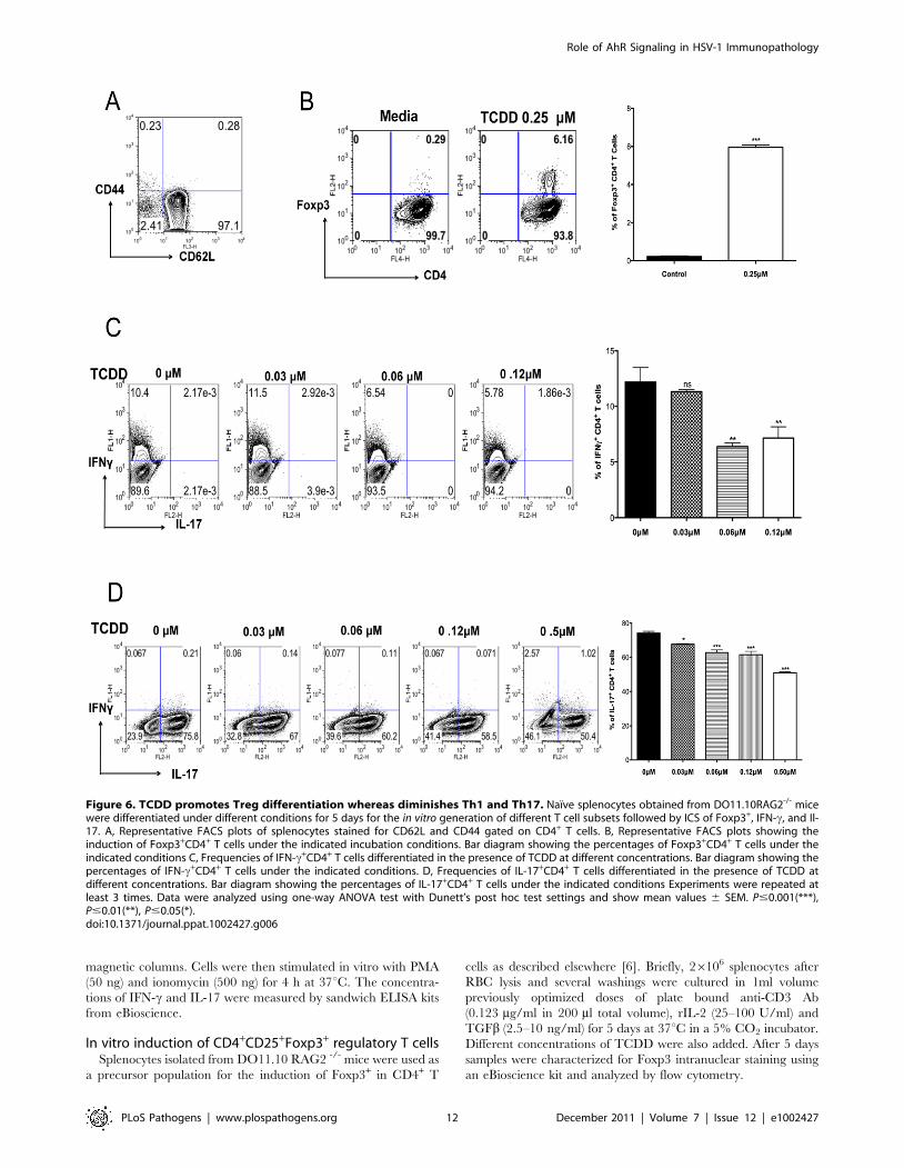
controls. However the frequencies of Foxp3+ Tregs, compared as a
fraction of total CD4+ T cells, were increased in treated animals
when compared to controls. Additionally, when the ratio of total
numbers of Treg per T effectors was compared to controls, a
significant increase in the number of Treg per Th1 or Th17 cells
in the TCDD treated mice was evident (Figure 5E).
To compare levels of IFN-c and IL-17 produced by CD4+ T
cells from infected and treated or untreated mice, sorted CD4+ T
cells were isolated from DLN on day 10 pi and stimulated in vitro
with PMA and ionomycin. When comparing the number of IFN-cand IL-17 secreting CD4+ T cells by ICCS, averages were reduced
for both in TCDD treated animals (Figure 5G). Similarly IFN-csecreting levels measured by ELISA were reduced 2.9 fold as a
consequence of TCDD treatment (Figure 5H).
AhR signaling promotes Treg induction but suppressesTh1 and Th17 cell generation in vitro
Results from the previous section indicated that there was a shift
in the balance between Tregs and T effectors towards Tregs, as
well as a reduction in the production of proinflammatory
cytokines. To further determine how TCDD could change the
balance of Treg to T effectors, naıve splenocytes from
DO11.10RAG2-/- (98% naıve CD4+ T cells) animals were
stimulated in vitro with plate bound anti-CD3 and anti-CD28, in
the presence of IL-2. Cultures were either untreated or treated
with graded amounts of TCDD (from 0.1 mM to 0.25 mM).
Cultures with 0.25 mM of TCDD significantly triggered the
conversion of approximately 6.2% of CD4+ T cells into Foxp3+
CD4+ T cells, as compared to 0.3% in the untreated controls
(Figure 6B).
Other cultures were TCR stimulated in a cytokine cocktail
reported to induce either Th1 or Th17 cells in the additional
presence of different doses of TCDD. The outcome was a
significant decrease in both Th1 and Th17 cell induction
(Figure 6C–D) with the highest TCDD dose studied (0.6 mM)
causing a disappearance of the majority CD4+ T cells from the
cultures (data not shown). Taken together, our results indicate that
activation of AhR signaling by TCDD can induce some CD4+ T
cells to become Foxp3+, but it is inhibitory to the generation of
IFN-c+ CD4+ and IL-17+ CD4+ T cells.
Differential proliferation of Treg over Foxp3- CD4+ T cellsin TCDD treated mice
To determine if TCDD had differential effects on Foxp3+ and
Foxp3- CD4+ T cell proliferation, Foxp3-GFP mice were infected
and some treated with TCDD on day 5 pi. After an injection of 5-
Bromo-2-deoxyuridine (BrdU) on day 8 pi, experiments were
terminated on day 9 pi and proliferation of both Foxp3-CD4+ and
Foxp3+ cells was detected by BrdU incorporation. Our results
show that TCDD treatment significantly reduced the proliferative
response of the Foxp3- CD4+ T cell population in both corneas
and lymphoid tissue, but was without significant inhibitory effects
on the Foxp3+ CD4+ T cell population. Instead, the effect of
TCDD on Foxp3+ cells in the cornea was to cause a modest
increase in proliferation (Figure 7A–B). These effects could explain
in part the balance between Tregs and T effectors in corneal
lesions.
Induction of apoptosis of Foxp3- CD4+ T cells but not ofTreg after TCDD treatment
Prior studies had shown that TCDD administration in vivo
causes thymocytes to undergo apoptosis [26]. We determined if
apoptosis of Foxp3- CD4+ T cells could account for the reduced
numbers of T effectors. We performed experiments with CD4+ T
cells isolated from DLN or spleen on day 8 pi from HSV infected
Foxp3-GFP mice. Cells were cultured ex vivo in the presence of
TCDD for 5 hours and apoptosis of Foxp3- and Foxp3+ CD4+ T
cells was measured using Annexin-V staining. The result showed a
dose dependent increase in the apoptosis of Foxp3-CD4+ T cells,
but no significant apoptosis of Foxp3+CD4+ T cells (Figure 8A and
C). Notably, there was no difference in the frequencies of Tregs
with the addition of different concentrations of TCDD as
compared to media (Figure 8B–C). Taken together, these results
indicate that AhR signaling by TCDD, can promote the apoptosis
of Foxp3- CD4+ T cells in vitro, but did not cause the same effect
in Foxp3+ Treg.
Discussion
SK is a blinding immuno-pathological lesion induced by ocular
infection with HSV [1-3]. Novel treatment procedures are needed
to replace the current long term use of antivirals and corticoste-
roids which have unwanted side effects [27–29]. A key to
controlling the severity of SK lesions is to suppress the activity
of T cells that orchestrate lesions and enhance the representation
of cellular and humoral events that inhibit effector cell function. In
this report, we have evaluated the use of a novel approach to
achieve lesion control in a murine model of SK. We demonstrate
that modulation of AhR signaling with a single dose of a synthetic
stable molecule (TCDD) causes cellular changes in the cornea
after HSV infection that account for significantly reduced SK
lesion severity. The outcome of therapy was reduced effector Th1
and Th17 cells that orchestrate lesions, a reduction of neutrophils
that are mainly responsible for damage to the cornea, as well as an
increase in the representation of Foxp3+ Treg. Accordingly, when
the ratio of Treg per T effectors was compared to controls, a
significant increase in the number of Treg per Th1 as well as Th17
cells in the TCDD treated mice was evident. Foxp3+CD4+ T cells
are assumed to function by inhibiting the inflammatory effects of T
effectors either directly, or by the generation of counter
inflammatory molecules [30]. Since a single administration of
TCDD provided effective treatment that lasted for as long as one
Figure 1. Preventive administration of TCDD diminishes SK severity. C57BL/6 animals infected with 1 x 10⁴ PFU of HSV were given eitherTCDD or vehicle IP on day 1 pi or FICZ or vehicle IP from day 1 to day 11 pi. The disease progression was analyzed throughout time in a blindedmanner using a scale described in materials and methods. A, The progression of SK lesion severity was significantly reduced in the group of micetreated with TCDD as compared with control mice. Kinetics of SK severity is shown. B, The progression of angiogenesis was significantly reduced inthe group of mice treated with TCDD as compared with control mice. Kinetics of angiogenesis lesion severity is shown. C, Individual eye scores of SKlesion severity and angiogenesis on day 14 pi. D, Percentage of encephalitis through time in control and TCDD treated mice. Herpetic encephalitis isan acute inflammation of the brain caused by HSV-1 and in mice gives clinical signs such as lethargy, ruffled fur, and hind limb paralysis [54,55]. E, Eyeswabs were taken from infected corneas from treated and control animals using sterile swabs on day 1, 3 and 6 pi or corneal tissue on day 6 pi andtitration was performed by standard plaque assay as described on materials and methods. Titers were calculated as log10 pfu/ml. F, Kinetics of SKlesion scores in mice treated with FICZ or vehicle. Data are representative of 3 independent experiments and show mean values 6 SEM (n = 10 mice/group). P#0.001(***), P#0.01(**), P#0.05(*).doi:10.1371/journal.ppat.1002427.g001
Role of AhR Signaling in HSV-1 Immunopathology
PLoS Pathogens | www.plospathogens.org 4 December 2011 | Volume 7 | Issue 12 | e1002427

Role of AhR Signaling in HSV-1 Immunopathology
PLoS Pathogens | www.plospathogens.org 5 December 2011 | Volume 7 | Issue 12 | e1002427

month, this approach could represent an effective novel therapy
for a lesion that is a common cause of human blindness.
Aryl hydrocarbon receptors are found in animals in many levels
of the evolutionary scale. They can recognize numerous low
molecular weight synthetic chemicals as well as a list of
endogenous ligands, some of which are photoproducts of
tryptophan breakdown [24,31]. Several cell types express AhR
that includes some, but not all cells, involved in innate and
adaptive immunity [32]. Our own interest in AhR ligands
stemmed from recent reports that synthetic AhR agonists had
anti-inflammatory activity [9,15]. Moreover, the dioxin TCDD
can provide long term activation of the AhR since it is resistant to
metabolic degradation [25]. As a consequence, a single adminis-
tration can result in long term effects on immune mediated
diseases. A recent report using the animal model of multiple
sclerosis, EAE, showed disease suppression when animals were
pretreated with TCDD [9]. The diminished lesions in treated
animals were correlated with expansion of the Foxp3+ CD4+ T cell
population and the levels of some cytokines produced by effector
cells were reduced. The expansion of the Treg population was
explained in part by conversion of naıve T cells to become Treg as
could be demonstrated in vitro. In our studies too, we observed that
a single TCDD administration was an effective means of reducing
HSK ocular lesions, but with our model the outcome appeared to
be more the consequence of suppressed numbers of Th1 and Th17
T cells that orchestrate SK, than any notable effect on the
expansion of Tregs. Accordingly, the cytokine producing cells in
lesions were reduced several fold in treated animals, whereas Treg
numbers remained approximately the same in treated and
controls. We did confirm the Weiner group [9] observations that
TCDD can cause some naıve T cells to convert and become
Foxp3+ in vitro, but in our hands this was a modest effect. This
notwithstanding, it could be that the relative increase in Treg in
the SK lesions of treated animals was the explanation for the
reduced lesions, the Treg acting by inhibiting the functions of
effectors as well as producing anti-inflammatory cytokines such as
IL-10.
Equally possible, however, was that the reduced lesions were the
direct consequence of the fewer numbers and less functional
effectors in the corneas of treated animals. Such effectors would be
less able to recruit inflammatory cells such as neutrophils that are
considered responsible for much of the tissue damage of SK
[33,34]. The reduced numbers of effectors would likely arise
either, or both, from an inhibitory effect of TCDD on effector cell
proliferation and differentiation, or be explained by the drug
causing apoptosis of effectors. The latter effect could readily be
demonstrated by in vitro studies with TCR stimulated CD4+ T cells
cultured with TCDD. In addition, Foxp3- CD4+ T cells from
treated animals proliferated less in vivo than did cells from control
animals. In some reports TCDD was shown to prematurely
terminate the proliferation and decrease the survival of CD4+ T
cell, although differential effects on T cell subsets were not
investigated [35]. Nevertheless, since regulatory T cells may be
more resistant to apoptosis than conventional T cells [32,36]
frequencies of Tregs would be expected to increase when other T
cell populations are depleted.
The reduced number of effector cells present in drug treated
animals in our studies were also functionally impaired in their
ability to mediate inflammatory reactions. Accordingly, ex vivo
stimulation of DLN cells from drug-treated mice produced lower
levels of some proinflammatory cytokines as well as chemokines
responsible for neutrophil recruitment than cells from control
animals. During in vitro studies, AhR ligation was shown to affect
the differentiation of T helper subsets, behaving differently under
identical culture conditions depending on the ligand used. TCDD
for example, was shown to trigger the conversion of Foxp3-CD4+
into Foxp3+CD4+ without the need for TGF-b addition [9,37], to
dampen Th17 differentiation [37] and to increase the frequency of
IL-17 secreting cells induced by TGF-b plus IL-6 [38]. On the
other hand, FICZ under identical culture conditions promoted
Th17 differentiation, but not Treg differentiation [37,38]. Other
ligands too, such as kynurenine (the first tryptophan metabolite of
the IDO pathway), was shown to optimally generate Tregs in the
presence of TGF-b [39]. In our in vitro experiments not only did we
find a conversion of naıve T cells into Tregs, but also provided
support for the notion that the TCDD interfered with the primary
induction of both Th1 and Th17 cells. Thus, in the presence of
TCDD, TCR stimulated naıve CD4+ T cells cultured in
conditions to cause their differentiation into either Th1 or Th17
cells, resulted in significant suppression. As it currently stands, our
mechanistic experiments cannot establish which is the major
explanation for the in vivo anti-inflammatory effects of TCDD
against SK. Further investigations are needed and are underway.
The use of TCDD represents a potentially valuable approach to
control SK since a single injection provided an excellent level of
lesion control for at least a month pi. So far our results can only be
considered as quasi therapeutic since treatment was begun 5 days
after infection, a time when most infectious virus has been
eliminated but clinical lesions are yet to become evident [1–3].
Moreover, we elected to study only the dose shown to be effective
in an autoimmune disease model [9]. Since in some studies the
outcome of treatment has been shown to be dose dependent, [35]
similar dose response studies are warranted in the SK system and
these are planned. Nevertheless, our approach does stand in
contrast to most other investigations where treatment was begun
prior to disease induction, or before natural disease is expected to
occur. With ocular HSV infection in mice, such an early treatment
approach would not be recommended because when TCDD was
given one day after infection up to half of the animals succumbed
to lethal infection of the CNS. Others too have observed that
TCDD administration in viral infections can result in increased
mortality [40–42]. For example, with influenza A virus infection
AhR activation by the administration of TCDD decreased the
survival time to lethal infection and resulted in mortality with a
non lethal dose of the virus [42,43]. The cause of lethality was
unclear since TCDD treated animals cleared the virus from the
lungs as well as a non treated mice [44]. More than likely animals
succumbed to lung pathology associated with increased neutro-
Figure 2. Therapeutic administration of TCDD diminishes SK severity. C57BL/6 animals infected with 16104 PFU of HSV were given eitherTCDD or vehicle IP on day 5 pi. The disease progression was analyzed throughout time. A, The progression of SK lesion severity was significantlyreduced in the group of mice treated with TCDD as compared with control mice. Kinetics of SK severity is shown. B, Individual eye SK scores on day10, 12 and 15 pi. C, The progression of angiogenesis was significantly reduced in the group of mice treated with TCDD as compared with controlmice. Kinetics of angiogenesis lesion severity is shown. D, Individual eye angiogenesis scores on day 10, 12 and 15 pi. E, Eyes were processed for cryo-sections on day 15 pi. Hematoxylin and eosin staining was carried out on 6-mm sections and pictures were taken at different microscopeaugmentations. F, The progression of SK lesion severity was significantly reduced in the group of mice treated with TCDD as compared with controlmice. Kinetics of SK severity is shown up to day 28 pi. Data are representative of 3 independent experiments and show mean values 6 SEM (n = 14mice/group). P#0.001(***), P#0.01(**), P#0.05(*).doi:10.1371/journal.ppat.1002427.g002
Role of AhR Signaling in HSV-1 Immunopathology
PLoS Pathogens | www.plospathogens.org 6 December 2011 | Volume 7 | Issue 12 | e1002427

Figure 3. TCDD treatment reduces cellular infiltration and cytokine levels in corneas of HSV infected animals. Wild-type C57BL/6 micecorneas were scarified and infected with 16104 PFU of HSV-1 in PBS and mice were given either TCDD or vehicle IP on day 5 pi. A, Representative FACSplots of CD45+ cells infiltrated in the corneas of control (left) and day 5 TCDD treated (right panel) mice are shown. B, Representative FACS plots andpercentages of CD4+ T cells gated on total CD45+ cells infiltrated in the corneas of control (left) and day 5 TCDD treated (right panel) mice are shown. C,CD11b+ Ly6G+ polymorphonuclear neutrophils gated on total CD45+ cells infiltrated in the corneas of control (left panel) and day 5 TCDD treated (rightpanel) mice are shown. D, Numbers of total CD45+ cells, neutrophils gated on total CD45+ cells and CD4+ T cells gated on total CD45+ cells infiltrated percornea of control and TCDD treated mice. Data are the combination of 3 independent experiments and show mean values 6 SEM (n = 9 and each sampleis representative of 2 corneas). Total number of CD45+ cells recovered from corneal samples varied and ranged from around 20,000 to 80,000 cells. E,Relative fold change in the mRNA expression of various cytokines (IL-1b, TNF-a, IL-6, IFN-c, IL-17, IL-10, CXCL9, CXCL10, CXCL11, CCL20) was examinedand compared between control and TCDD day 5 treated mice on day 15 pi by Q-RTPCR. mRNA levels for the different cytokines in mock infected micewere set to one and used for the relative fold upregulation. Data represents means 6 SEM from two different independent experiments (n = 2 and eachsample is representative of 8 corneas). Day 15 pi corneal samples were used for all the experiments. P#0.001(***), P#0.01(**), P#0.05(*).doi:10.1371/journal.ppat.1002427.g003
Role of AhR Signaling in HSV-1 Immunopathology
PLoS Pathogens | www.plospathogens.org 7 December 2011 | Volume 7 | Issue 12 | e1002427

Figure 4. TCDD treatment shifts the balance from T effectors to T regulatory cells after HSV infection. C57BL/6 animals infected with16104 PFU of HSV were given either TCDD or vehicle IP on day 5 pi. Mice were sacrificed and corneas were collected on day 15 pi. A, RepresentativeFACS plots gated on CD4+ T cells for IFN-c and IL-17 secreting cells from pooled corneas stimulated with PMA/ionomycin during 4 hours from control
Role of AhR Signaling in HSV-1 Immunopathology
PLoS Pathogens | www.plospathogens.org 8 December 2011 | Volume 7 | Issue 12 | e1002427

philia found in the lungs of TCDD treated mice [21,45]. Curiously
however, in our model TCDD administration reduced the
numbers of neutrophils in the infected corneas.
Aesthetically, the use of TCDD for therapy, a molecule often
castigated as an environmental pollutant, may have minimal
appeal. However, the use of a natural ligand for AhR such as
FICZ that can be metabolized by the body may not represent a
good option. Several studies using FICZ have observed that
inflammatory lesions can be exacerbated by such a treatment
[9,23]. For example, in the studies on EAE by Weiner and
colleagues [9], FICZ treatment resulted in more severe lesions. A
similar outcome was reported too by Stockinger and colleagues
[23]. In our own studies, we observed no beneficial, or in fact
harmful, effects when we treated HSV infected mice with FICZ.
One reason AhR ligation with certain ligands can cause enhanced
inflammatory lesions is that Th17 T cells, mainly responsible for
mediating some inflammatory diseases, express high levels of AhR
[22]17,18]. Consequently, ligation of AhR on Th17 cells can cause
cell expansion and the production of cytokines that contribute to
tissue damage [9,23]. In the SK model, Th17 cells appear to play
only a minor role in SK pathogenesis [46] which may explain our
failure to observe adverse effects of FICZ therapy. It could be
however that using non-toxic ligands such as 2-(19H-indole-39-
carbonyl)-thiazole-4-carboxylic acid methyl ester which induces
Foxp3+ T CD4+ T cells and suppresses EAE [47] could lead to a
more acceptable therapeutic approach for SK.
In conclusion, our results are consistent with the observation
that modulation of AhR signaling through the use of TCDD plays
a role in influencing the expression of SK lesions. The mechanisms
involved to explain the outcome were multiple, and involve a
change in the balance between effector and regulatory T cells. We
anticipate that manipulating AhR signaling, preferable with non-
toxic ligands, could represent a useful approach to control an
important cause of human blindness.
Materials and Methods
Ethics statementThis study was carried out in strict accordance with the
recommendations in the Guide for the Care and Use of
Laboratory Animals of the National Research Council. All
animals were housed in Association for Assessment and Accred-
itation of Laboratory Animal Care (AAALAC)-approved animal
facilities. The protocol was approved by the Institutional Animal
Care and Use Committee of the University of Tennessee (PHS
Assurance number A3668-01). HSV-1 eye infection was per-
formed under anesthesia (avertin), and all efforts were made to
minimize animal suffering.
Mice, virus and cell linesFemale 6 to 8 weeks old C57BL/6 mice were purchased from
Harlan Sprague Dawley (Indianapolis, IN). BALB/c DO11.10
RAG2 -/- mice were purchased from Taconic and kept in our
pathogen free facility where food, water, bedding, and instruments
were autoclaved. All manipulations were done in a laminar flow
hood. All experiment procedures were in complete agreement with
the Association for Research in Vision and Ophthalmology
resolution on the use of animals in research. HSV-1 RE Tumpey
and HSV-RE Hendricks were propagated and titrated on Vero cells
(American Type Culture Collecting no. CCL81) using standard
protocols. The virus was stored in aliquots at 280uC until use.
AbsCD4-allophycocyanin (RM4.5), CD4-FITC (RM4.5), Foxp3-
PE (FJK-16s), anti-IFN-c-FITC (XMG1.2), anti-IL17-PE (TC11-
18H10), CD45-allophycocyanin (30-F11), CD11b-PerCP (M1/
79), Ly6G-PE (1A8).
Corneal HSV-1 infection and clinical observationsCorneal infections of C57BL/6 mice were done under deep
anesthesia induced by IP injection in tribromoethanol (avertin) as
previously described [48]. Mice’s corneas were scarified with a 27-
gauge needle, and a 3 ml drop containing the specific viral dose
was applied to the eye. Eyes were examined on different days pi
(dpi) with a silt-lamp biomicroscope (Kowa Company, Nagoya,
Japan) measuring the progression of SK lesion severity and
angiogenesis of individual mice. The scoring system was as follows:
0, normal cornea; +1, mild corneal haze; +2, moderate corneal
opacity or scarring; +3, severe corneal opacity but iris visible; +4,
opaque cornea and corneal ulcer; +5, corneal rupture and
necrotizing keratitis [49]. The severity of angiogenesis was
recorded as described previously [50]. According to this system,
a grade of 4 for a given quadrant of the circle represents a
centripetal growth of 1.5 mm toward the corneal center. The
score of the four quadrants of the eye were then summed to derive
the neovessel index (range 0–16) for each eye at a given time point.
Treatment of animals with TCDDTCDD (Sigma Aldrich) diluent was evaporated with nitrogen
and reconstituted with DMSO. Female 6 to 8 weeks old C57BL/C
mice were ocularly infected under deep anesthesia with 16104 PFU
of HSV-1 RE Tumpey and divided randomly into groups. Animals
in the treated groups were either treated with TCDD on day 1 pi or
day 5 pi IP, being the dose administered of 1 mg/mice. Animals in
the control groups were treated the same days (either day 1 or day 5
pi) with DMSO IP. Mice were observed for SK and angiogenesis
progression from day 5 until day 15 or 28 as described elsewhere
[49]. Most of the experiments were repeated at least three times.
Treatment of animals with FICZFICZ (Biomol International, L.P., Plymouth Meeting, PA) was
dissolved in DMSO. Female 6 to 8 weeks old C57BL/C mice were
ocularly infected under deep anesthesia with 16104 PFU of HSV-
1 RE Tumpey and divided randomly into groups. Animals in the
treated groups were either treated daily with FICZ from day 1 pi
to day 11 pi (IP), being the dose administered of 1 mg/mice.
Animals in the control groups were treated the same days with
DMSO IP. Mice were observed for HSK and angiogenesis
progression from day 5 until day 15 as described elsewhere [49].
Virus recovery and titrationsEye swabs were taken from infected corneas using sterile swabs
at the indicated time points. Infected corneas were extracted on
day 6 pi and placed on ice sterile 2.0-mL straight-wall ground-glass
tissue homogenizers (Wheaton) with media and homogenized.
and day 5 TCDD treated mice. B, FACS plots of Foxp3+CD4+ cells from pooled corneas from control and day 5 TCDD treated mice. C, Frequencies andtotal numbers of CD4+ T cells, Th1, Th17 and Tregs in infected corneas from control and day 5 TCDD treated animals. D, Cell ratios for total numbersof Tregs per Th1 cell and Tregs per Th17 cell. Data are representative of 3 independent experiments and show mean values 6 SEM (n = 6 and eachsample is representative of 2 corneas). P#0.001(***), P#0.01(**), P#0.05(*). Day 15 pi corneal samples were used for the experiments.doi:10.1371/journal.ppat.1002427.g004
Role of AhR Signaling in HSV-1 Immunopathology
PLoS Pathogens | www.plospathogens.org 9 December 2011 | Volume 7 | Issue 12 | e1002427

Role of AhR Signaling in HSV-1 Immunopathology
PLoS Pathogens | www.plospathogens.org 10 December 2011 | Volume 7 | Issue 12 | e1002427

Homogenates were centrifuged (2,250 g at 4uC) for 5 min, place
on ice, and immediately plated. Titrations were performed by a
standard plaque assay as described previously [51]. Titers were
calculated as log10 pfu/ml per a standard protocol [52].
HistopathologyEyes from control and TCDD treated mice were extirpated on
day 15 pi and snap frozen in OCT compound (Miles, Elkart, IN).
Six micron thick sections were cut, air dried in a desiccation box.
Staining was performed with hematoxylin and eosin (Richard
Allen Scientific, Kalamazoo, MI).
Flow cytometryCell preparation. Single cell suspensions were prepared
from cornea, cervical DLN, and spleen of mice at different time
points pi. Corneas were excised, pooled group wise, and digested
with 60 U/ml Liberase (Roche Diagnostics) for 35 minutes at
37uC in a humified atmosphere of 5% CO2. After incubation, the
corneas were disrupted by grinding with a syringe plunger on a cell
strainer and a single-cell suspension was made in complete RPMI
1640 medium.Staining for flow cytometry. The single cell suspensions
obtained from corneas, DLN, and spleen were stained for different
cell surface molecules for FACS. All steps were performed at 4uC. A
total of 16106 cells were first blocked with an unconjugated anti-
CD32/CD16 mAb for 30 min in FACS buffer. After washing with
FACS buffer, fluorochrome-labeled respective antibodies were
added for 30 min on ice. Finally, the cells were washed three
times and re-suspended in 1% para-formaldehyde. The stained
samples were acquired with a FACS Calibur (BD Biosciences) and
the data were analyzed using the FlowJo software. For corneas, total
cell numbers were calculated by acquiring the totality of the sample
and taking in consideration total number of corneas in the sample.
To enumerate the number of IFN-c and IL-17 producing CD4+
T cells, intracellular cytokine staining was performed as previously
described [5]. In brief, 106 freshly isolated splenocytes, lymph
node and corneal cells were cultured in U bottom 96 well plates.
For in vitro induced cultures, cells were left unstimulated or
stimulated with PMA (50 ng) and ionomycin (500ng) for 4h in the
presence of brefeldin A (10 mg/ml). Subsequently, cell surface
staining was performed, followed by intracellular cytokine staining
using a Cytofix/Cytoperm kit (BD Pharmingen) in accordance
with the manufacturer’s recommendations. The Abs used were
anti-IFN-c FITC and anti-IL-17 PE. The fixed cells were
resuspended in 1% paraformaldehyde. The stained samples were
acquired with a FACS Calibur (BD biosciences), and the data were
analyzed using the FlowJo software.
Real time PCRRNA was extracted from cells and tissue using TRIzol LS
reagent (Invitrogen). Total cDNA was made with 500ng of RNA
using oligo(dT) primer. Quantitative PCR (Q-RTPCR) was
performed using SYBR Green PCR Master Mix (Applied
Biosystem, Foster City, CA) with iQ5 real-time PCR detection
system (Bio Rad, Hercules, CA) using 5 ml of cDNA for 40 cycles.
The expression levels of different molecules were normalized to b-
actin using D threshold cycle method calculation. Relative
expression between mock infected samples and control or day 5
TCDD treated samples from day 15 pi were calculated using the 2-
DDCt formula: DDCt = DCt,sample - DCt,reference. Here, DCt is the
change in cycling threshold between the gene of interest and the
‘housekeeping’ gene b-actin, where DCt,sample was the Ct value for
any day 5 TCDD treated or control samples from day 15 pi
normalized to the b-actin gene and DCt,reference was the Ct value
for the mock infected samples (scratched and infected only with
PBS) also normalized to b-actin. Each of the samples was run in
duplicates to determine sample reproducibility, and a mean Ct
value for each duplicate measurement was calculated. The PCR
primers used were the following: bactin F 59-CCTTCTT-
GGGTATGGAATCCTG-39 and R 59-GGCATAGAGGTCTT-
TACGGATG-39,IL-6 F 59-CGTGGAAATGAGAAAAGAG-
TTGTGC-39 and R 59- ATGCTTAGGCATAACGCACTAG-
GT-39, TNF-a F 59-CAGCCTCTTCTCATTCCTGCTTGTG-
39 and R 59- CTGGAAGACTCCTCCCAGGTATAT-39,IL-1bF 59-GAAATGCCACCTTTTGACAG-39 and R 59- CAAGGC-
CACAGGTATTTTGT-39,IFN-c F 59-GGATGCATTCATGA-
GTATTGC-39 and R 59- GCTTCCTGAGGCTGGATTC-
39,IL-17A F 59-GCTCCAGAAGGCCCTCAG-39 and R 59-
CTTTCCCTCCGCATTGACA-39,IL-10 F 59- CCTTTGACA-
AGCGGACTCTC-39 and R 59- GCCAGCATAAAAACC-
CTTCA-39,CXCL-9 F 59-CAAGCCCCAATTGCAACAAA-39
and R 59- TCC GGA TCT AGG CAG GTT TGA-39,CXCL-10
F 59-TGC TGG GTC TGA AGT GGG ACT-39 and R 59- AAG
CTT CCC TAT GGC CCT CA-39,CXCL-11 F 59-GGTCA-
CAGCCATAGCCCTG-39 and R 59- AGCCTTCATAGTAA-
CAATC-39, CCL-20 F 59-GCCTCTCGTACATACAGACGC-
39 and R 59- CCAGTTCTGCTTTGGATCAGC-39.
Purification of CD4+ T cellsCD4+ T cells were purified from pooled DLN single cell
suspension obtained from HSV-infected mice using a mouse CD4+
T cell isolation kit (Miltenyi Biotec, Auburn, CA). The purity was
achieved at the extent of 90%. Purified CD4+ T cells were
analyzed by Flow cytometry and ELISA after stimulation for the
expression of IFN-c and IL-17.
ELISADLN single cell suspensions from individual mice were
collected at day 15 pi. Cells were stimulated in vitro with anti-
CD3 (2 mg/ml) and anti-CD28 (1 mg/ml) for 48 h at 37uC.
Additionally DLN single cell suspensions from mice were also
collected at day 10 pi and CD4+ T cells were purified using
Figure 5. Differential effect of TCDD treatment on Treg and effector T cells in lymphoid organs of TCDD treated animals. C57BL/6animals infected with 16104 PFU of HSV were given either TCDD or vehicle IP on day 5 pi. A, Representative FACS plots and percentages for IFN-c andIL-17 secreting T cells gated on CD4+ T cells from day 15 DLN stimulated with PMA/ionomycin during 4 hours from control and day 5 TCDD treatedmice. FACS plots of CD4+Foxp3+ cells from DLN from control and day 5 TCDD treated mice. B, Representative FACS plots and percentages for IFN-cand IL-17 secreting T cells gated on CD4+ T cells from day 15 spleens stimulated with PMA/ionomycin during 4 hours from control and day 5 TCDDtreated mice. FACS plots of CD4+Foxp3+ cells from spleens from control and day 5 TCDD treated mice. C, Cell numbers of CD4, Th1, Th17 and Tregs inDLNs stimulated with PMA/ionomycin. D, Cell numbers of CD4, Th1, Th17 and Tregs in spleen stimulated with PMA/ionomycin. E, Cell ratios for totalnumber of Tregs per Th1 cell and Tregs per Th17 cell in DLN and spleen. F, IFN-c and IL-17 protein levels analyzed by ELISA from control and TCDDtreated infected DLN on day 15 pi. G, FACS plots, percentages and cell numbers for IFN-c and IL-17 secreting T cells gated on CD4+ T cells from day 10DLN samples, purified for CD4+ T cells and stimulated with PMA/ionomycin during 4 hours. H, IFN-c protein levels analyzed by ELISA from control andTCDD treated infected DLN at day 10 pi, purified for CD4+ T cells and stimulated with PMA/ionomycin during 4 hours. Data are representative of 3independent experiments and show mean values 6 SEM (n = 12). P#0.001(***), P#0.01(**), P#0.05(*).doi:10.1371/journal.ppat.1002427.g005
Role of AhR Signaling in HSV-1 Immunopathology
PLoS Pathogens | www.plospathogens.org 11 December 2011 | Volume 7 | Issue 12 | e1002427
magnetic columns. Cells were then stimulated in vitro with PMA
(50 ng) and ionomycin (500 ng) for 4 h at 37uC. The concentra-
tions of IFN-c and IL-17 were measured by sandwich ELISA kits
from eBioscience.
In vitro induction of CD4+CD25+Foxp3+ regulatory T cellsSplenocytes isolated from DO11.10 RAG2 -/- mice were used as
a precursor population for the induction of Foxp3+ in CD4+ T
cells as described elsewhere [6]. Briefly, 26106 splenocytes after
RBC lysis and several washings were cultured in 1ml volume
previously optimized doses of plate bound anti-CD3 Ab
(0.123 mg/ml in 200 ml total volume), rIL-2 (25–100 U/ml) and
TGFb (2.5–10 ng/ml) for 5 days at 37uC in a 5% CO2 incubator.
Different concentrations of TCDD were also added. After 5 days
samples were characterized for Foxp3 intranuclear staining using
an eBioscience kit and analyzed by flow cytometry.
Figure 6. TCDD promotes Treg differentiation whereas diminishes Th1 and Th17. Naıve splenocytes obtained from DO11.10RAG2-/- micewere differentiated under different conditions for 5 days for the in vitro generation of different T cell subsets followed by ICS of Foxp3+, IFN-c, and Il-17. A, Representative FACS plots of splenocytes stained for CD62L and CD44 gated on CD4+ T cells. B, Representative FACS plots showing theinduction of Foxp3+CD4+ T cells under the indicated incubation conditions. Bar diagram showing the percentages of Foxp3+CD4+ T cells under theindicated conditions C, Frequencies of IFN-c+CD4+ T cells differentiated in the presence of TCDD at different concentrations. Bar diagram showing thepercentages of IFN-c+CD4+ T cells under the indicated conditions. D, Frequencies of IL-17+CD4+ T cells differentiated in the presence of TCDD atdifferent concentrations. Bar diagram showing the percentages of IL-17+CD4+ T cells under the indicated conditions Experiments were repeated atleast 3 times. Data were analyzed using one-way ANOVA test with Dunett’s post hoc test settings and show mean values 6 SEM. P#0.001(***),P#0.01(**), P#0.05(*).doi:10.1371/journal.ppat.1002427.g006
Role of AhR Signaling in HSV-1 Immunopathology
PLoS Pathogens | www.plospathogens.org 12 December 2011 | Volume 7 | Issue 12 | e1002427

Figure 7. Differential proliferation of Treg over Foxp3-CD4+ T cells in TCDD treated mice after HSV-1 infection. A, Control and day 5TCDD treated GFP-Foxp3+ mice were given BrdU intraperitoneally one day before termination and the next day (day 9 pi) corneas, DLN and spleenswere analyzed for the frequencies of Foxp3+CD4+ and Foxp3- CD4+ T cells that incorporated BrdU; representative FACS plots for cells are shown. B,The bar diagram shows the frequencies of Treg and Foxp3-CD4+ T cells labeled for BrdU in cornea, DLN and spleen. Data are representative of 3independent experiments and show mean values 6 SEM (n = 12). P#0.001(***), P#0.01(**), P#0.05(*).doi:10.1371/journal.ppat.1002427.g007
Role of AhR Signaling in HSV-1 Immunopathology
PLoS Pathogens | www.plospathogens.org 13 December 2011 | Volume 7 | Issue 12 | e1002427

Role of AhR Signaling in HSV-1 Immunopathology
PLoS Pathogens | www.plospathogens.org 14 December 2011 | Volume 7 | Issue 12 | e1002427

Th1 and Th17 differentiation in vitroNaıve CD4+ T cells were stimulated for 4 to 5 days with plate
bound antibody to CD3 (4 mg/ml) and anti CD28 (2 mg/ml). For
Th1 differentiation recombinant mouse IL-12 (10ng/ml) and anti
IL-4 (10 mg/ml) were used. In the case of Th17 differentiation
TGF-b (2.5ng/ml), IL-6 (30 ng/ml), anti IL-4 (10 mg/ml) and anti
IFN-c (10 mg/ml) were added. Concentrations of TCDD were
added into cultures at the beginning of the experiment. After 5
days samples were analyzed by intracellular cytokine staining for
the production of IFN-c and IL-17 using a BD biosciences kit and
then flow cytometry.
The culture mediums used were IMDM (Sigma-Aldrich) for
Th17 differentiation or RPMI 1640 (Sigma-Aldrich) for Th1
differentiation, both supplemented with 261023 M L-glutamine,
100 U/ml penicillin, 100 mg/ml streptomycin, 561025 M b-
mercaptoethanol, and 5% FCS [53].
BrdU incorporation assayFoxp3+-GFP knock-in animals were kindly provided by Dr. M.
Oukka of Seattle Children’s Research Institute. Mice were infected
and divided into two groups: non-treated and TCDD treated
mice. 8 days after ocular HSV 1 infection mice were injected IP
with BrdU (1mg/mouse) and were terminated 12 hours later. 9
dpi, host Foxp3+CD4+ and Foxp3-CD4+ T cells that incorporated
BrdU were analyzed by staining with anti BrdU antibody using an
APC BrdU flow kit from BD Pharmingen as per the manufac-
turer’s instructions. Samples were acquired with a FACSCalibur
(BD biosciences), and the data were analyzed using the FlowJo
software.
Ex vivo apoptosis assayDLN cells and splenocytes isolated from HSV-infected Foxp3-
GFP C57BL/6 mice at 8 days pi were incubated for 5h with
various concentrations of TCDD in 96 well flat-bottom plate in
5% CO2 incubators. After incubation period was over, cells were
stained for annexin V using a kit from BD biosciences.
Additionally cells were costained for CD4. Stained cells were
analyzed immediately by flow cytometry.
Statistical analysisMost of the analyses for determining the level of significance
were performed using unpaired two-tailed Student’s t test. Values
P#0.001(***), P#0.01(**), P#0.05(*) were considered significant.
Results are expressed as means 6SEM. For some experiments, as
mentioned in the figure legends, a one-way ANOVA test was
applied.
Accession numbers for genes and proteinsCD4 (MGI:88335), IFN-c (MGI:107656), Foxp3 (MGI:
1891436), IL-17 (MGI:107364), IL-1b (MGI:96543), TNF-a(MGI:104798), IL-6 (MGI:96559), CCL20 (MGI:1329031),
CXCL9 (MGI:1352449), CXCL10 (MGI:1352450), CXCL11
(MGI:1860203), IL-10 (MGI:96537), b-actin (MGI:87904), CD45
(MGI:97810), CD11b (MGI:96607), Ly6G (MGI:109440), CD3
(MGI:88332), CD28 (MGI:88327), Annexin V (MGI:106008), IL-
12 (MGI:96540), IL-4 (MGI:96556), TGF-b (MGI:98725), IL-6
(MGI:96559).
Acknowledgments
We thank Naveen K. Rajasagi, Pradeep B. J. Reddy, Sachin Mulik, Shalini
Sharma, Fernanda Gimenez and Greg Spencer for assistance during
research and manuscript preparation.
Author Contributions
Conceived and designed the experiments: TVP AS BTR. Performed the
experiments: TVP AS. Analyzed the data: TVP AS. Contributed reagents/
materials/analysis tools: BTR. Wrote the paper: TVP AS BTR.
References
1. Sarangi PP, Rouse BT (2010) Herpetic keratitis. In: Leonard A, Levin DMA,
eds. Ocular Disease Mechanisms and Management. Philadelphia: Saunders.Elsevier. pp 91–97.
2. Streilein JW, Dana MR, Ksander BR (1997) Immunity causing blindness: fivedifferent paths to herpes stromal keratitis. Immunol Today 18: 443–449.
3. Biswas PS, Rouse BT (2005) Early events in HSV keratitis--setting the stage for ablinding disease. Microbes Infect 7: 799–810.
4. Zhao ZS, Granucci F, Yeh L, Schaffer PA, Cantor H (1998) Molecular mimicryby herpes simplex virus-type 1: autoimmune disease after viral infection. Science
279: 1344–1347.
5. Suvas S, Azkur AK, Kim BS, Kumaraguru U, Rouse BT (2004) CD4+CD25+regulatory T cells control the severity of viral immunoinflammatory lesions.J Immunol 172: 4123–4132.
6. Sehrawat S, Suvas S, Sarangi PP, Suryawanshi A, Rouse BT (2008) In vitro-generated antigen-specific CD4+ CD25+ Foxp3+ regulatory T cells control the
severity of herpes simplex virus-induced ocular immunoinflammatory lesions.J Virol 82: 6838–6851.
7. Sehrawat S, Rouse BT (2008) Anti-inflammatory effects of FTY720 againstviral-induced immunopathology: role of drug-induced conversion of T cells to
become Foxp3+ regulators. J Immunol 180: 7636–7647.
8. Sehrawat S, Suryawanshi A, Hirashima M, Rouse BT (2009) Role of Tim-3/
galectin-9 inhibitory interaction in viral-induced immunopathology: shifting the
balance toward regulators. J Immunol 182: 3191–3201.
9. Quintana FJ, Basso AS, Iglesias AH, Korn T, Farez MF, et al. (2008) Control of
T(reg) and T(H)17 cell differentiation by the aryl hydrocarbon receptor. Nature453: 65–71.
10. Stevens EA, Mezrich JD, Bradfield CA (2009) The aryl hydrocarbon receptor: aperspective on potential roles in the immune system. Immunology 127: 299–311.
11. Funatake CJ, Marshall NB, Kerkvliet NI (2008) 2,3,7,8-Tetrachlorodibenzo-p-dioxin alters the differentiation of alloreactive CD8+ T cells toward a regulatory
T cell phenotype by a mechanism that is dependent on aryl hydrocarbon
receptor in CD4+ T cells. J Immunotoxicol 5: 81–91.
12. Denison MS, Nagy SR (2003) Activation of the aryl hydrocarbon receptor bystructurally diverse exogenous and endogenous chemicals. Annu Rev Pharmacol
Toxicol 43: 309–334.
13. Kerkvliet NI (2009) AHR-mediated immunomodulation: the role of altered gene
transcription. Biochem Pharmacol 77: 746–760.
14. Marshall NB, Kerkvliet NI (2010) Dioxin and immune regulation: emerging role
of aryl hydrocarbon receptor in the generation of regulatory T cells.Ann N Y Acad Sci 1183: 25–37.
15. Kerkvliet NI, Steppan LB, Vorachek W, Oda S, Farrer D, et al. (2009) Activationof aryl hydrocarbon receptor by TCDD prevents diabetes in NOD mice and
increases Foxp3+ T cells in pancreatic lymph nodes. Immunotherapy 1: 539–547.
16. Benson JM, Shepherd DM (2011) Aryl hydrocarbon receptor activation by
TCDD reduces inflammation associated with Crohn’s disease. Toxicol Sci 120:
68–78.
Figure 8. TCDD causes apoptosis of Foxp3-CD4+ T cells but not Tregs. Foxp3-GFP mice were HSV-1 infected and sacrificed on day 8 pi. A,Induction of in vitro apoptosis by different concentrations of TCDD on Foxp3-CD4+ T cells but not on Foxp3+CD4+ T cells. Cells isolated from spleensand DLNs of HSV-1 infected mice were cultured ex vivo in the presence of RPMI or RPMI with different concentrations of TCDD for 5 hours andthereafter stained for Annexin-V, representative FACS plots showing Annexin-V expression on gated CD4+ T cells. B, Representative FACS plots ofFoxp3+CD4+ T cells in splenocytes and DLN cells from HSV-1 infected mice treated in vitro with different concentrations of TCDD for 5 hours. C, Bardiagram showing the percentages of Annexin V+Foxp3-CD4+, Annexin V+Foxp3+CD4+ and Foxp3+CD4+ under the indicated conditions in the DLN isshown. Data are representative of 3 independent experiments. Data were analyzed using one-way ANOVA test with Dunett’s post hoc test settingsand show mean values 6 SEM (n = 12). P#0.001(***), P#0.01(**), P#0.05(*).doi:10.1371/journal.ppat.1002427.g008
Role of AhR Signaling in HSV-1 Immunopathology
PLoS Pathogens | www.plospathogens.org 15 December 2011 | Volume 7 | Issue 12 | e1002427

17. Funatake CJ, Marshall NB, Steppan LB, Mourich DV, Kerkvliet NI (2005)
Cutting edge: activation of the aryl hydrocarbon receptor by 2,3,7,8-tetrachlorodibenzo-p-dioxin generates a population of CD4+ CD25+ cells with
characteristics of regulatory T cells. J Immunol 175: 4184–4188.
18. Apetoh L, Quintana FJ, Pot C, Joller N, Xiao S, et al. (2010) The arylhydrocarbon receptor interacts with c-Maf to promote the differentiation of type
1 regulatory T cells induced by IL-27. Nat Immunol 11: 854–861.19. Kobayashi S, Okamoto H, Iwamoto T, Toyama Y, Tomatsu T, et al. (2008) A
role for the aryl hydrocarbon receptor and the dioxin TCDD in rheumatoid
arthritis. Rheumatology 47: 1317–1322.20. Neff-LaFord H, Teske S, Bushnell TP, Lawrence BP (2007) Aryl hydrocarbon
receptor activation during influenza virus infection unveils a novel pathway ofIFN-gamma production by phagocytic cells. J Immunol 179: 247–255.
21. Teske S, Bohn AA, Regal JF, Neumiller JJ, Lawrence BP (2005) Activation of thearyl hydrocarbon receptor increases pulmonary neutrophilia and diminishes host
resistance to influenza A virus. Am J Physiol Lung Cell Mol Physiol 289:
L111–124.22. Stockinger B, Veldhoen M, Hirota K (2009) Modulation of Th17 development
and function by activation of the aryl hydrocarbon receptor--the role ofendogenous ligands. Eur J Immunol 39: 652–654.
23. Veldhoen M, Hirota K, Westendorf AM, Buer J, Dumoutier L, et al. (2008) The
aryl hydrocarbon receptor links TH17-cell-mediated autoimmunity to environ-mental toxins. Nature 453: 106–109.
24. Esser C, Rannug A, Stockinger B (2009) The aryl hydrocarbon receptor inimmunity. Trends Immunol 30: 447–454.
25. Kerkvliet NI (2002) Recent advances in understanding the mechanisms ofTCDD immunotoxicity. Int Immunopharmacol 2: 277–291.
26. Kamath AB, Xu H, Nagarkatti PS, Nagarkatti M (1997) Evidence for the
induction of apoptosis in thymocytes by 2,3,7,8-tetrachlorodibenzo-p-dioxin invivo. Toxicol Appl Pharmacol 142: 367–377.
27. Deshpande S, Banerjee K, Biswas PS, Rouse BT (2004) Herpetic eye disease:immunopathogenesis and therapeutic measures. Expert Rev Mol Med 6: 1–14.
28. Knickelbein JE, Hendricks RL, Charukamnoetkanok P (2009) Management of
herpes simplex virus stromal keratitis: an evidence-based review. SurvOphthalmol 54: 226–234.
29. McGhee CN, Dean S, Danesh-Meyer H (2002) Locally administered ocularcorticosteroids: benefits and risks. Drug Saf 25: 33–55.
30. Belkaid Y, Rouse BT (2005) Natural regulatory T cells in infectious disease. NatImmunol 6: 353–360.
31. Rannug A, Rannug U, Rosenkranz HS, Winqvist L, Westerholm R, et al. (1987)
Certain photooxidized derivatives of tryptophan bind with very high affinity tothe Ah receptor and are likely to be endogenous signal substances. J Biol Chem
262: 15422–15427.32. Stockinger B, Hirota K, Duarte J, Veldhoen M (2011) External influences on the
immune system via activation of the aryl hydrocarbon receptor. Semin Immunol
23: 99–105.33. Thomas J, Gangappa S, Kanangat S, Rouse BT (1997) On the essential
involvement of neutrophils in the immunopathologic disease: herpetic stromalkeratitis. J Immunol 158: 1383–1391.
34. Tumpey TM, Chen SH, Oakes JE, Lausch RN (1996) Neutrophil-mediatedsuppression of virus replication after herpes simplex virus type 1 infection of the
murine cornea. J Virol 70: 898–904.
35. Funatake CJ, Dearstyne EA, Steppan LB, Shepherd DM, Spanjaard ES, et al.(2004) Early consequences of 2,3,7,8-tetrachlorodibenzo-p-dioxin exposure on
the activation and survival of antigen-specific T cells. Toxicol Sci 82: 129–142.36. Taylor SR, Alexander DR, Cooper JC, Higgins CF, Elliott JI (2007) Regulatory
T cells are resistant to apoptosis via TCR but not P2X7. J Immunol 178:
3474–3482.
37. Singh NP, Singh UP, Singh B, Price RL, Nagarkatti M, et al. (2011) Activation
of Aryl Hydrocarbon Receptor (AhR) Leads to Reciprocal EpigeneticRegulation of FoxP3 and IL-17 Expression and Amelioration of Experimental
Colitis. PLoS One 6: e23522.
38. Kimura A, Naka T, Nohara K, Fujii-Kuriyama Y, Kishimoto T (2008) Arylhydrocarbon receptor regulates Stat1 activation and participates in the
development of Th17 cells. Proc Natl Acad Sci U S A 105: 9721–9726.39. Mezrich JD, Fechner JH, Zhang X, Johnson BP, Burlingham WJ, et al. (2010)
An interaction between kynurenine and the aryl hydrocarbon receptor can
generate regulatory T cells. J Immunol 185: 3190–3198.40. Clark DA, Sweeney G, Safe S, Hancock E, Kilburn DG, et al. (1983) Cellular
and genetic basis for suppression of cytotoxic T cell generation by haloaromatichydrocarbons. Immunopharmacology 6: 143–153.
41. Funseth E, Wesslen L, Lindh U, Friman G, Ilback NG (2002) Effect of 2,3,7,8-tetrachlorodibenzo-p-dioxin on trace elements, inflammation and viral clearance
in the myocardium during coxsackievirus B3 infection in mice. Sci Total
Environ 284: 135–147.42. Vorderstrasse BA, Bohn AA, Lawrence BP (2003) Examining the relationship
between impaired host resistance and altered immune function in mice treatedwith TCDD. Toxicology 188: 15–28.
43. Warren TK, Mitchell KA, Lawrence BP (2000) Exposure to 2,3,7,8-
tetrachlorodibenzo-p-dioxin (TCDD) suppresses the humoral and cell-mediatedimmune responses to influenza A virus without affecting cytolytic activity in the
lung. Toxicol Sci 56: 114–123.44. Burleson GR, Lebrec H, Yang YG, Ibanes JD, Pennington KN, et al. (1996)
Effect of 2,3,7,8-tetrachlorodibenzo-p-dioxin (TCDD) on influenza virus hostresistance in mice. Fundam Appl Toxicol 29: 40–47.
45. Head JL, Lawrence BP (2009) The aryl hydrocarbon receptor is a modulator of
anti-viral immunity. Biochem Pharmacol 77: 642–653.46. Suryawanshi A, Veiga-Parga T, Rajasagi NK, Reddy PB, Sehrawat S, et al.
(2011) Role of IL-17 and Th17 Cells in Herpes Simplex Virus-Induced CornealImmunopathology. J Immunol 187: 1919–1930.
47. Quintana FJ, Murugaiyan G, Farez MF, Mitsdoerffer M, Tukpah AM, et al.
(2010) An endogenous aryl hydrocarbon receptor ligand acts on dendritic cellsand T cells to suppress experimental autoimmune encephalomyelitis. Proc Natl
Acad Sci U S A 107: 20768–20773.48. Zheng M, Deshpande S, Lee S, Ferrara N, Rouse BT (2001) Contribution of
vascular endothelial growth factor in the neovascularization process during thepathogenesis of herpetic stromal keratitis. J Virol 75: 9828–9835.
49. Suryawanshi A, Mulik S, Sharma S, Reddy PB, Sehrawat S, et al. (2011) Ocular
Neovascularization Caused by Herpes Simplex Virus Type 1 Infection Resultsfrom Breakdown of Binding between Vascular Endothelial Growth Factor A and
Its Soluble Receptor. J Immunol 186: 3653–3665.50. Dana MR, Zhu SN, Yamada J (1998) Topical modulation of interleukin-1
activity in corneal neovascularization. Cornea 17: 403–409.
51. Babu JS, Thomas J, Kanangat S, Morrison LA, Knipe DM, et al. (1996) Viralreplication is required for induction of ocular immunopathology by herpes
simplex virus. J Virol 70: 101–107.52. Spear PG, Roizman B (1972) Proteins specified by herpes simplex virus. V.
Purification and structural proteins of the herpesvirion. J Virol 9: 143–159.53. Veldhoen M, Hirota K, Christensen J, O’Garra A, Stockinger B (2009) Natural
agonists for aryl hydrocarbon receptor in culture medium are essential for
optimal differentiation of Th17 T cells. J Exp Med 206: 43–49.54. Steiner I (2011) Herpes simplex virus encephalitis: new infection or reactivation?
Curr Opin Neurol 24: 268–274.55. Kurt-Jones EA, Chan M, Zhou S, Wang J, Reed G, et al. (2004) Herpes simplex
virus 1 interaction with Toll-like receptor 2 contributes to lethal encephalitis.
Proc Natl Acad Sci U S A 101: 1315–1320.
Role of AhR Signaling in HSV-1 Immunopathology
PLoS Pathogens | www.plospathogens.org 16 December 2011 | Volume 7 | Issue 12 | e1002427