Protein corona on nanoparticle for modulating cytotoxicity and immunotoxicity
17
© 2015 Lee et al. This work is published by Dove Medical Press Limited, and licensed under Creative Commons Attribution – Non Commercial (unported, v3.0) License. The full terms of the License are available at http://creativecommons.org/licenses/by-nc/3.0/. Non-commercial uses of the work are permitted without any further permission from Dove Medical Press Limited, provided the work is properly attributed. Permissions beyond the scope of the License are administered by Dove Medical Press Limited. Information on how to request permission may be found at: http://www.dovepress.com/permissions.php International Journal of Nanomedicine 2015:10 97–113 International Journal of Nanomedicine Dovepress submit your manuscript | www.dovepress.com Dovepress 97 REVIEW open access to scientific and medical research Open Access Full Text Article http://dx.doi.org/10.2147/IJN.S72998 Effect of the protein corona on nanoparticles for modulating cytotoxicity and immunotoxicity Yeon Kyung Lee 1, * Eun-Ju Choi 2, * Thomas J Webster 3 Sang-Hyun Kim 4 Dongwoo Khang 1 1 Department of Molecular Medicine, School of Medicine, Gachon University, Incheon, South Korea; 2 Division of Sport Science, College of Science and Technology, Konkuk University, Chungju, South Korea; 3 Department of Chemical Engineering and Program in Bioengineering, Northeastern University, Boston, MA, USA; 4 Department of Pharmacology, School of Medicine, Kyungpook National University, Daegu, South Korea *These authors contributed equally to this work Abstract: Although the cytotoxicity of nanoparticles (NPs) is greatly influenced by their interactions with blood proteins, toxic effects resulting from blood interactions are often ignored in the development and use of nanostructured biomaterials for in vivo applications. Protein coronas created during the initial reaction with NPs can determine the subsequent immunological cascade, and protein coronas formed on NPs can either stimulate or mitigate the immune response. Along these lines, the understanding of NP-protein corona formation in terms of physiochemical surface properties of the NPs and NP interactions with the immune system components in blood is an essential step for evaluating NP toxicity for in vivo therapeutics. This article reviews the most recent developments in NP-based protein coronas through the modification of NP surface properties and discusses the associated immune responses. Keywords: nanostructured biomaterials, blood response, cytotoxicity, immunotoxicity, protein corona Introduction The development and rational design of nanoparticles (NPs) has created a new para- digm for overcoming current limitations and concerns in medicine. 1–3 Compared with micron-sized particles, NPs can retain unique material characteristics, such as size, solubility, shape, surface charge, and chemistry, that can be tailored to increase the efficacy of NP-based therapeutics for various biomedical applications. 1,2,4 In drug deliv- ery systems, for example, conventional (micron-sized) particles are quickly cleared by the immune system and can only enter phagocytic cells, whereas NP-based drugs can be delivered to all organs and, thus, all cells. 5,6 In this regard, NPs have been the subject of increasing concern from the medical community because of their immunological toxicity. Once NPs enter various organs and interact with blood, they come into immediate contact with various plasma constituents. These NP-bound blood proteins can influence the immune system by mediating subsequent immune cell responses. 7–10 Protein corona or biological mac- romolecule adsorption is influenced by physiochemical material properties and can affect the in vivo toxicity of the material. For example, a study on polymer-based NPs designed for intravenous administration has demonstrated that NPs can stimu- late and/or suppress immune cell responses, depending on the formation of protein coronas formed in blood. 11 Furthermore, in vivo organ distribution and the clearance rate of nanomaterial-derived drug carriers in blood are significantly influenced by the formation of plasma proteins. 7,11,12 As such, evaluating and understanding the toxicity of nanostructure-based materials coated with protein coronas is a critical step toward manipulating their subsequent immune response and cytotoxic effects. Correspondence: Dongwoo Khang Department of Molecular Medicine, School of Medicine, Gachon University, Incheon 406-840, Republic of Korea Tel/Fax +82 32 883 152 Email [email protected] Sang-Hyun Kim Department of Pharmacology, School of Medicine, Kyungpook National University, Daegu 700-422, Republic of Korea Tel/Fax +82 53 420 4838 Email [email protected]
Transcript of Protein corona on nanoparticle for modulating cytotoxicity and immunotoxicity
© 2015 Lee et al. This work is published by Dove Medical Press Limited, and licensed under Creative Commons Attribution – Non Commercial (unported, v3.0) License. The full terms of the License are available at http://creativecommons.org/licenses/by-nc/3.0/. Non-commercial uses of the work are permitted without any further
permission from Dove Medical Press Limited, provided the work is properly attributed. Permissions beyond the scope of the License are administered by Dove Medical Press Limited. Information on how to request permission may be found at: http://www.dovepress.com/permissions.php
International Journal of Nanomedicine 2015:10 97–113
International Journal of Nanomedicine Dovepress
submit your manuscript | www.dovepress.com
Dovepress 97
R e v I e w
open access to scientific and medical research
Open Access Full Text Article
http://dx.doi.org/10.2147/IJN.S72998
effect of the protein corona on nanoparticles for modulating cytotoxicity and immunotoxicity
Yeon Kyung Lee1,*eun-Ju Choi2,*Thomas J webster3
Sang-Hyun Kim4
Dongwoo Khang1 1Department of Molecular Medicine, School of Medicine, Gachon University, Incheon, South Korea; 2Division of Sport Science, College of Science and Technology, Konkuk University, Chungju, South Korea; 3Department of Chemical engineering and Program in Bioengineering, Northeastern University, Boston, MA, USA; 4Department of Pharmacology, School of Medicine, Kyungpook National University, Daegu, South Korea
*These authors contributed equally to this work
Abstract: Although the cytotoxicity of nanoparticles (NPs) is greatly influenced by their
interactions with blood proteins, toxic effects resulting from blood interactions are often
ignored in the development and use of nanostructured biomaterials for in vivo applications.
Protein coronas created during the initial reaction with NPs can determine the subsequent
immunological cascade, and protein coronas formed on NPs can either stimulate or mitigate the
immune response. Along these lines, the understanding of NP-protein corona formation in terms
of physiochemical surface properties of the NPs and NP interactions with the immune system
components in blood is an essential step for evaluating NP toxicity for in vivo therapeutics.
This article reviews the most recent developments in NP-based protein coronas through the
modification of NP surface properties and discusses the associated immune responses.
Keywords: nanostructured biomaterials, blood response, cytotoxicity, immunotoxicity, protein
corona
IntroductionThe development and rational design of nanoparticles (NPs) has created a new para-
digm for overcoming current limitations and concerns in medicine.1–3 Compared with
micron-sized particles, NPs can retain unique material characteristics, such as size,
solubility, shape, surface charge, and chemistry, that can be tailored to increase the
efficacy of NP-based therapeutics for various biomedical applications.1,2,4 In drug deliv-
ery systems, for example, conventional (micron-sized) particles are quickly cleared
by the immune system and can only enter phagocytic cells, whereas NP-based drugs
can be delivered to all organs and, thus, all cells.5,6
In this regard, NPs have been the subject of increasing concern from the medical
community because of their immunological toxicity. Once NPs enter various organs
and interact with blood, they come into immediate contact with various plasma
constituents. These NP-bound blood proteins can influence the immune system by
mediating subsequent immune cell responses.7–10 Protein corona or biological mac-
romolecule adsorption is influenced by physiochemical material properties and can
affect the in vivo toxicity of the material. For example, a study on polymer-based
NPs designed for intravenous administration has demonstrated that NPs can stimu-
late and/or suppress immune cell responses, depending on the formation of protein
coronas formed in blood.11 Furthermore, in vivo organ distribution and the clearance
rate of nanomaterial-derived drug carriers in blood are significantly influenced by the
formation of plasma proteins.7,11,12 As such, evaluating and understanding the toxicity
of nanostructure-based materials coated with protein coronas is a critical step toward
manipulating their subsequent immune response and cytotoxic effects.
Correspondence: Dongwoo Khang Department of Molecular Medicine, School of Medicine, Gachon University, Incheon 406-840, Republic of KoreaTel/Fax +82 32 883 152email [email protected] Sang-Hyun KimDepartment of Pharmacology, School of Medicine, Kyungpook National University, Daegu 700-422, Republic of KoreaTel/Fax +82 53 420 4838email [email protected]
Journal name: International Journal of NanomedicineArticle Designation: ReviewYear: 2015Volume: 10Running head verso: Lee et alRunning head recto: Protein corona on nanoparticle for modulating cytotoxicity and immunotoxicityDOI: http://dx.doi.org/10.2147/IJN.S72998
International Journal of Nanomedicine 2015:10submit your manuscript | www.dovepress.com
Dovepress
Dovepress
98
Lee et al
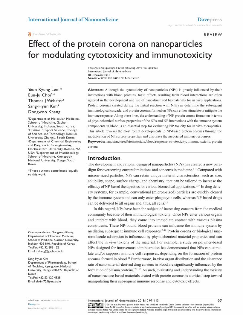
Surprisingly, it was determined that the same NPs can
induce different biological outcomes, depending on the con-
trol presence or absence of a protein corona. For example,
silica NPs in serum conditions showed stronger accumulation
at lysosomes. However, silica NPs without serum proteins
exposed to cells showed a higher degree of attachment to the
cell membrane and greater internalization (both lysosomes
and cytosols; Figure 1).13 In addition, carboxylated polysty-
rene NPs under serum-free conditions exhibit a higher adhe-
sion to the cell membrane than adhesion of the NP surface
to the cell membrane under serum conditions.14
The mechanism of selective uptake of NPs with or without
serum proteins was identified by a recent study that analyzed
this mechanism by a two-step process. The NPs were initially
adhered to the cell membrane at 4°C and internalized by
increasing temperature (37°C). High surface energy of the bare
NPs can cause unspecific interactions and adsorb strongly to
the cell membranes by the process of quite reactive, and chemi-
cally lowering energy. It was interpreted that the formation of
a corona surrounding the NP lowered the energy by unspecific
interactions and, thus, leads to less attachment of NPs at the
cell membrane in the presence of biomolecules.14
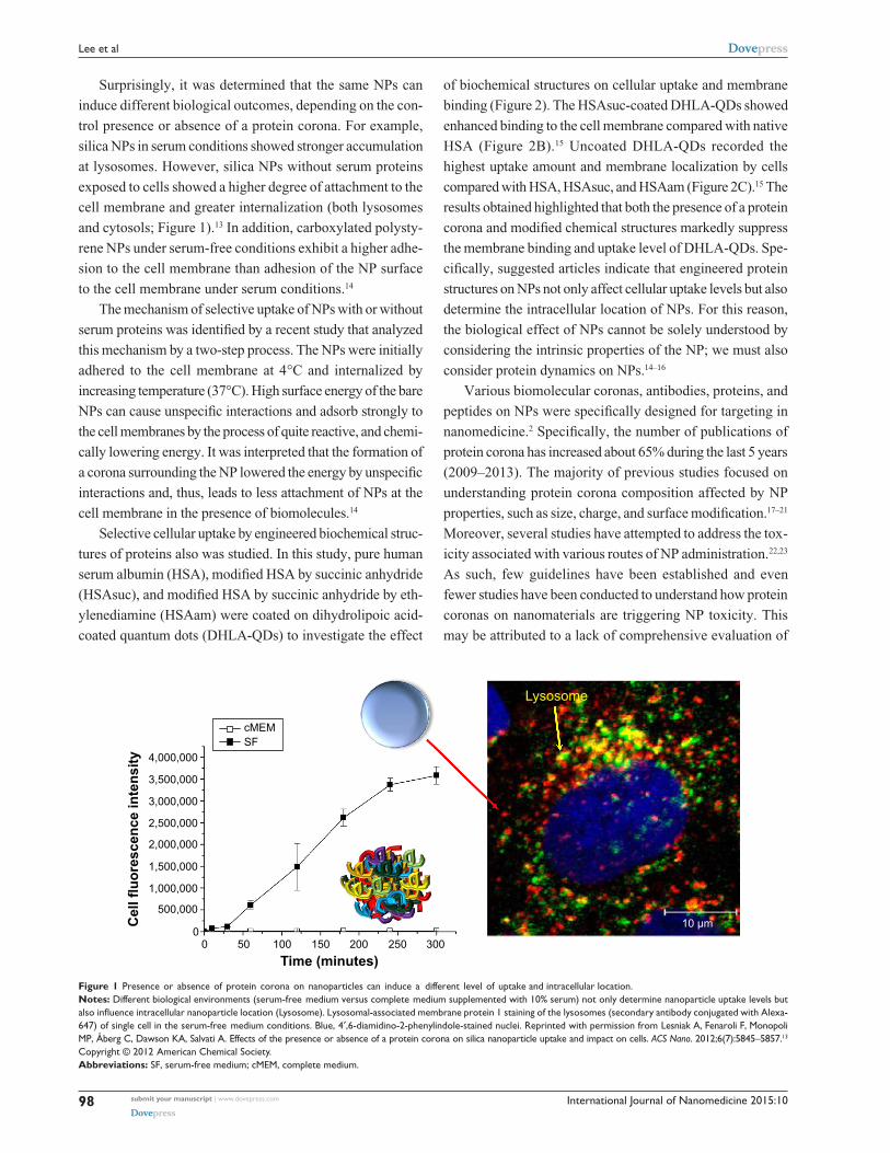
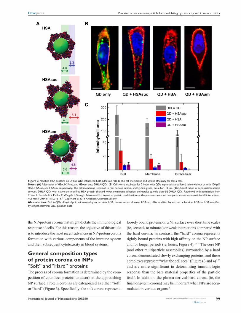
Selective cellular uptake by engineered biochemical struc-
tures of proteins also was studied. In this study, pure human
serum albumin (HSA), modified HSA by succinic anhydride
(HSAsuc), and modified HSA by succinic anhydride by eth-
ylenediamine (HSAam) were coated on dihydrolipoic acid-
coated quantum dots (DHLA-QDs) to investigate the effect
of biochemical structures on cellular uptake and membrane
binding (Figure 2). The HSAsuc-coated DHLA-QDs showed
enhanced binding to the cell membrane compared with native
HSA (Figure 2B).15 Uncoated DHLA-QDs recorded the
highest uptake amount and membrane localization by cells
compared with HSA, HSAsuc, and HSAam (Figure 2C).15 The
results obtained highlighted that both the presence of a protein
corona and modified chemical structures markedly suppress
the membrane binding and uptake level of DHLA-QDs. Spe-
cifically, suggested articles indicate that engineered protein
structures on NPs not only affect cellular uptake levels but also
determine the intracellular location of NPs. For this reason,
the biological effect of NPs cannot be solely understood by
considering the intrinsic properties of the NP; we must also
consider protein dynamics on NPs.14–16
Various biomolecular coronas, antibodies, proteins, and
peptides on NPs were specifically designed for targeting in
nanomedicine.2 Specifically, the number of publications of
protein corona has increased about 65% during the last 5 years
(2009–2013). The majority of previous studies focused on
understanding protein corona composition affected by NP
properties, such as size, charge, and surface modification.17–21
Moreover, several studies have attempted to address the tox-
icity associated with various routes of NP administration.22,23
As such, few guidelines have been established and even
fewer studies have been conducted to understand how protein
coronas on nanomaterials are triggering NP toxicity. This
may be attributed to a lack of comprehensive evaluation of
4,000,000
Lysosome
10 µm
Time (minutes)
Cel
l flu
ores
cenc
e in
tens
ity
3,500,000
3,000,000
2,500,000
2,000,000
1,500,000
1,000,000
500,000
00
50 100 150 200 250 300
cMEMSF
Figure 1 Presence or absence of protein corona on nanoparticles can induce a different level of uptake and intracellular location.Notes: Different biological environments (serum-free medium versus complete medium supplemented with 10% serum) not only determine nanoparticle uptake levels but also influence intracellular nanoparticle location (Lysosome). Lysosomal-associated membrane protein 1 staining of the lysosomes (secondary antibody conjugated with Alexa-647) of single cell in the serum-free medium conditions. Blue, 4′,6-diamidino-2-phenylindole-stained nuclei. Reprinted with permission from Lesniak A, Fenaroli F, Monopoli MP, Åberg C, Dawson KA, Salvati A. effects of the presence or absence of a protein corona on silica nanoparticle uptake and impact on cells. ACS Nano. 2012;6(7):5845–5857.13
Copyright © 2012 American Chemical Society.Abbreviations: SF, serum-free medium; cMeM, complete medium.
International Journal of Nanomedicine 2015:10 submit your manuscript | www.dovepress.com
Dovepress
Dovepress
99
Protein corona on nanoparticle for modulating cytotoxicity and immunotoxicity
the NP-protein corona that might dictate the immunological
response of cells. For this reason, the objective of this article
is to introduce the most recent advances in NP-protein corona
formation with various components of the immune system
and their subsequent cytotoxicity in blood systems.
General composition types of protein corona on NPs“Soft” and “Hard” proteinsThe process of corona formation is determined by the com-
petition of countless proteins to adsorb at the approaching
NP surface. Protein coronas are categorized as either “soft”
or “hard” (Figure 3). Specifically, the soft corona represents
loosely bound proteins on a NP surface over short time scales
(ie, seconds to minutes) or weak interactions compared with
the hard corona. In contrast, the “hard” corona represents
tightly bound proteins with high affinity on the NP surface
and for longer periods (ie, hours; Figure 4).24,25 The core NP
(and other multiparticle assemblies) surrounded by a hard
corona demonstrated slowly exchanging proteins, and these
complexes represent “what the cell sees” (Figures 3 and 4)9,25
and are more significant in determining immunologic
response than the bare material properties of the particle
itself. In addition, the plasma-derived hard corona (ie, the
final long-term corona) may be important when NPs are accu-
mulated in various organs.9
AHSA
QD only
DHLA QD
0
20
40
100
200
300
QD + HSAsuc
QD + HSAsuc
QD + HSA
QD + HSA
QD + HSAam
QD + HSAam
HSAsuc
HSAam
4.8
4.8
5.64.6
8.1
3.3
B
CIn
tens
ity/c
ell a
rea
Total Membrane Intracellular
Figure 2 Modified HSA proteins on DHLA-QDs influenced both adhesion rate to the cell membrane and uptake efficiency for HeLa cells.Notes: (A) Adsorption of HSA, HSAsuc, and HSAam onto DHLA-QDs. (B) Cells were incubated for 2 hours with QDs in phosphate-buffered saline without or with 100 µM HSA, HSAsuc, and HSAam, respectively. The cell membrane is stained in red, nucleus in blue, and QDs in green. Scale bar, 10 µm. (C) Quantification of nanoparticle uptake amount. DHLA-QDs with native and modified HSA protein showed lower membrane adhesion and uptake by cells than did DHLA-QDs. Reprinted with permission from Treuel L, Brandholt S, Maffre P, Wiegele S, Shang L, Nienhaus GU. Impact of protein modification on the protein corona on nanoparticles and nanoparticle-cell interactions. ACS Nano. 2014;8(1):503–513.15 Copyright © 2014 American Chemical Society.Abbreviations: DHLA-QDs, dihydrolipoic acid-coated quantum dots; HSA, human serum albumin; HSAsuc, HSA modified by succinic anhydride; HSAam, HSA modified by ethylenediamine; QD, quantum dots.

International Journal of Nanomedicine 2015:10submit your manuscript | www.dovepress.com
Dovepress
Dovepress
100
Lee et al
Severalminutes
Kd (dissociation constant)9,20,54
Adhesion to hydrophobicity35
Molecular weight25,32
Endosome–Lysosome trafficking13,21
Conformational changes (sheet)35,43
High
High
High
High
Low
Low
Low* Low*,
Low
Low
Nanoparticle
Severalhours
Soft corona
Soft HardCorona
Hard corona conformational changes
High†
Figure 3 Schematic illustration and characteristics of a hard and a soft corona.Notes: The protein corona encompassing the nanoparticles. Hard coronas are characterized by slow exchange (ie, several hours) and lower abundance, with a high affinity of proteins, whereas soft coronas are typified by rapid exchange (ie, several minutes) and lower affinity of proteins with weakly bound outer layers on nanoparticles. There is a different response of cellular and biochemical factors by soft and hard corona formation. *Compared with serum-free condition. †Compared with soft corona.
Cedervall et al determined that the kinetic and equilibrium
binding properties of NP-protein coronas depend not only on
the NP surface and size but also on plasma protein identity,
such as with HSA, fibrinogen, and lipoproteins.24 For exam-
ple, HSA and fibrinogen exhibited higher rates of association
and dissociation than apolipoprotein A-I and other plasma
proteins.24 In addition, it has been demonstrated that HSA
and fibrinogen adsorption dominate on hydrophobic particles
more than on hydrophilic particles for short times.24
However, initially attached proteins were subsequently
replaced by lower-abundance proteins with higher affinity
(ie, slower kinetics), such as lipoproteins, and especially
apolipoprotein A-I.24 Thus, greater proteins with low affin-
ity made up the protein corona during the initial period (soft
corona) but were replaced with proteins of lower abundance
and higher affinity at longer time scales (hard corona).
As a consequence, the hard corona may possess a
greater role than the soft corona in determining the physi-
ological response, and because of the long residence time,
the hard corona on NP experiences further biological process
(ie, endocytosis).9,21 As such, if adsorbed hard coronas with
specific physiochemical nanomaterial properties do not
invoke immune cell responses, they could be advantageous
for reducing NP toxicity.
Protein adsorption on various NPsThe adsorptions of blood proteins on NP have been analyzed
in recent studies.26–32 It was found that proteins (eg, albumin,
apolipoprotein, immunoglobulins [Igs], complement, and
fibrinogen) adsorbed on various NP (polymeric NPs, iron
oxide NPs, gold NPs [Au-NPs], liposomes, QDs, and carbon
nanotubes [CNTs]) surfaces exhibited structural changes in
the bloodstream (ie, bioactivity).33–35 For example, Roach
et al studied the binding events of bovine serum albumin
(BSA) and bovine fibrinogen (BFG) on silica nanospheres
that exhibited both hydrophilic and hydrophobic surface
curvature and performed secondary structure analysis
(ie, infrared spectroscopy on surface-bound proteins).35
Compared with BSA, fibrinogen tended to undergo greater
conformational changes in its secondary structure when pro-
teins were adsorbed on NPs with high surface curvature (ie,
small dimension of radius).35 In addition, surface properties
of NPs, such as charge, size, and the effect of particle coating,
affected the compatibility of NP-protein corona complexes
with the immune system.36
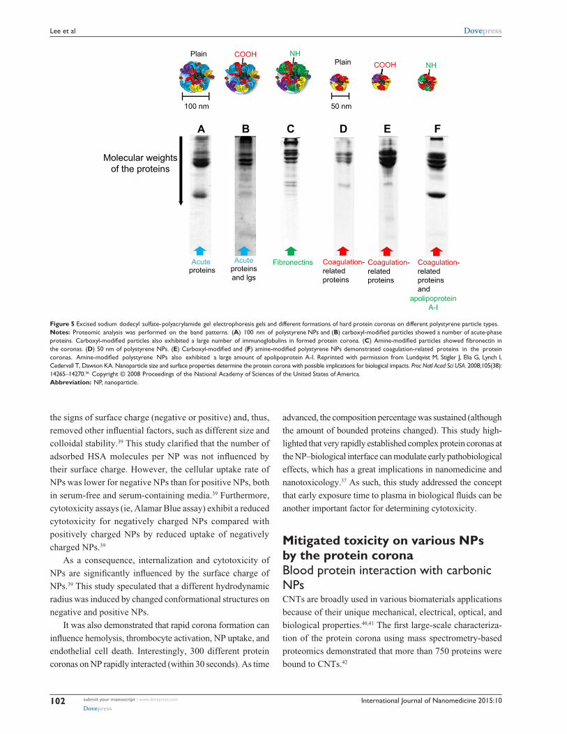
Lundqvist et al categorized different hard corona forma-
tions on NPs by different chemistries (ie, untreated poly-
styrene NP, carboxyl-modified, and amine-modified) with
different sizes (50 and 100 nm) (Figure 5).36 All separated and
International Journal of Nanomedicine 2015:10 submit your manuscript | www.dovepress.com
Dovepress
Dovepress
101
Protein corona on nanoparticle for modulating cytotoxicity and immunotoxicity
10
212223242526272829303132
3334
35
363738394041424344454647
4849
50
17
26
34
435572
130
3% 6% 10% 20% 40% 55% 80%
3% 6% 10% 20% 40% 55% 80%
96
10
12345678
910
1112131415161718
1920
17
26
34435572
13096
Silica NPs
Polystyrene NPs
HSA (60 kDa) and fibrinogencontents
(50–70 kDa)
HSA (60 kDa) and fibrinogencontents
(50–70 kDa)
Increasing plasma concentration
Increasing plasma concentration
Apolipoproteins andcomplement proteins
(30–43 kDa)
Apolipoproteins (26 kDa) andcomplement proteins
(90 kDa)
Soft corona
Hard corona
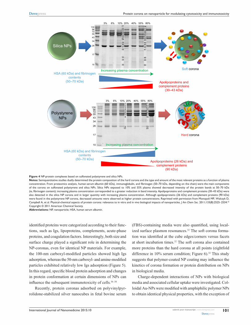
Figure 4 NP-protein complexes based on sulfonated polystyrene and silica NPs.Notes: Semiquantitative studies dually determined the protein composition of the hard corona and the type and amount of the most relevant proteins as a function of plasma concentration. From proteomics analysis, human serum albumin (60 kDa), immunoglobulin, and fibrinogen (50–70 kDa, depending on the chain) were the main components of the corona on sulfonated polystyrene and silica NPs. Silica NPs exposed to 10% and 55% plasma showed decreased intensity of the protein bands at 50–70 kDa (ie, fibrinogen content); increasing plasma concentration corresponded to a greater reduction in band intensity. Apolipoproteins and complement proteins (30–43 kDa) were also detected in the silica NP corona and in larger quantity with increasing plasma concentration. Although apolipoproteins (26 kDa) and complement proteins (90 kDa) were found in the polystyrene NP corona, decreased amounts were observed at higher protein concentrations. Reprinted with permission from Monopoli MP, walczyk D, Campbell A, et al. Physical-chemical aspects of protein corona: relevance to in vitro and in vivo biological impacts of nanoparticles. J Am Chem Soc. 2011;133(8):2525–2534.25
Copyright © 2011 American Chemical Society.Abbreviations: NP, nanoparticle; HSA, human serum albumin.
identified proteins were categorized according to their func-
tions, such as Igs, lipoproteins, complements, acute-phase
proteins, and coagulation factors. Interestingly, both size and
surface charge played a significant role in determining the
NP-coronas, even for identical NP materials. For example,
the 100-nm carboxyl-modified particles showed high Igs
adsorption, whereas the 50-nm carboxyl- and amine-modified
particles exhibited relatively low Igs adsorption (Figure 5).
In this regard, specific blood protein adsorption and changes
in protein conformation at certain dimensions of NPs can
influence the subsequent immunotoxicity of cells.36–38
Recently, protein coronas adsorbed on polyvinylpyr-
rolidone-stabilized silver nanocubes in fetal bovine serum
(FBS)-containing media were also quantified, using local-
ized surface plasmon resonances.32 The soft corona forma-
tion was identified at the cube edges/corners versus facets
at short incubation times.32 The soft corona also contained
more proteins than the hard corona at all points (eightfold
difference in 10% serum condition; Figure 6).32 This study
suggests that polymer-coated NP coating may influence the
kinetics of corona formation or protein distribution on NPs
in biological media.
Charge-dependent interactions of NPs with biological
media and associated cellular uptake were investigated. Col-
loidal Au-NPs were modified with amphiphilic polymer NPs
to obtain identical physical properties, with the exception of
International Journal of Nanomedicine 2015:10submit your manuscript | www.dovepress.com
Dovepress
Dovepress
102
Lee et al
Acuteproteins
Acuteproteinsand lgs
Fibronectins
apolipoproteinA-I
NHNH
Coagulation-relatedproteins
Coagulation-relatedproteins
Coagulation-relatedproteinsand
COOHCOOH
Plain
100 nm 50 nm
Molecular weightsof the proteins
A B C D E F
Plain
Figure 5 excised sodium dodecyl sulfate-polyacrylamide gel electrophoresis gels and different formations of hard protein coronas on different polystyrene particle types.Notes: Proteomic analysis was performed on the band patterns. (A) 100 nm of polystyrene NPs and (B) carboxyl-modified particles showed a number of acute-phase proteins. Carboxyl-modified particles also exhibited a large number of immunoglobulins in formed protein corona. (C) Amine-modified particles showed fibronectin in the coronas. (D) 50 nm of polystyrene NPs. (E) Carboxyl-modified and (F) amine-modified polystyrene NPs demonstrated coagulation-related proteins in the protein coronas. Amine-modified polystyrene NPs also exhibited a large amount of apolipoprotein A-I. Reprinted with permission from Lundqvist M, Stigler J, Elia G, Lynch I, Cedervall T, Dawson KA. Nanoparticle size and surface properties determine the protein corona with possible implications for biological impacts. Proc Natl Acad Sci USA. 2008;105(38): 14265–14270.36
Copyright © 2008 Proceedings of the National Academy of Sciences of the United States of America.
Abbreviation: NP, nanoparticle.
the signs of surface charge (negative or positive) and, thus,
removed other influential factors, such as different size and
colloidal stability.39 This study clarified that the number of
adsorbed HSA molecules per NP was not influenced by
their surface charge. However, the cellular uptake rate of
NPs was lower for negative NPs than for positive NPs, both
in serum-free and serum-containing media.39 Furthermore,
cytotoxicity assays (ie, Alamar Blue assay) exhibit a reduced
cytotoxicity for negatively charged NPs compared with
positively charged NPs by reduced uptake of negatively
charged NPs.39
As a consequence, internalization and cytotoxicity of
NPs are significantly influenced by the surface charge of
NPs.39 This study speculated that a different hydrodynamic
radius was induced by changed conformational structures on
negative and positive NPs.
It was also demonstrated that rapid corona formation can
influence hemolysis, thrombocyte activation, NP uptake, and
endothelial cell death. Interestingly, 300 different protein
coronas on NP rapidly interacted (within 30 seconds). As time
advanced, the composition percentage was sustained (although
the amount of bounded proteins changed). This study high-
lighted that very rapidly established complex protein coronas at
the NP–biological interface can modulate early pathobiological
effects, which has a great implications in nanomedicine and
nanotoxicology.37 As such, this study addressed the concept
that early exposure time to plasma in biological fluids can be
another important factor for determining cytotoxicity.
Mitigated toxicity on various NPs by the protein coronaBlood protein interaction with carbonic NPsCNTs are broadly used in various biomaterials applications
because of their unique mechanical, electrical, optical, and
biological properties.40,41 The first large-scale characteriza-
tion of the protein corona using mass spectrometry-based
proteomics demonstrated that more than 750 proteins were
bound to CNTs.42

International Journal of Nanomedicine 2015:10 submit your manuscript | www.dovepress.com
Dovepress
Dovepress
103
Protein corona on nanoparticle for modulating cytotoxicity and immunotoxicity
tsoft~7 nm
thard~7 nm
Hard corona
1,200
1,000
800
600
400
200
00 120 240 360 480 600 720 840 960 1,080 1,200 1,320 1,440
Soft corona mixed model
Incubation time (minutes)
Prot
ein
mas
s (µ
g/m
2 )
Ag
K1<<k2
K2K1
K1
K2
Figure 6 Quantification of protein corona layers around silver nanocubes; comparison of hard and soft corona.Notes: Schematic cartoon and transmission electron microscopy image of a silver nanocube surrounded by both soft (green) and hard (red) corona in a two-layer model. The soft corona mass was quantified and compared with the mass of hard corona. Data exhibited an eight times greater amount of protein on soft corona than hard corona for all points (ranging from 120 to 1,440 minutes). Reprinted with permission from Miclăuş T, Bochenkov VE, Ogaki R, Howard KA, Sutherland DS. Spatial mapping and quantification of soft and hard protein coronas at silver nanocubes. Nano Lett. 2014;14(4):2086–2093.32 Copyright © 2014 American Chemical Society.
International Journal of Nanomedicine 2015:10submit your manuscript | www.dovepress.com
Dovepress
Dovepress
104
Lee et al
The competitive binding of blood proteins on single-wall
CNTs (SWCNTs) influenced cellular pathways and resulted
in reduced cytotoxicity that depended on the presence of
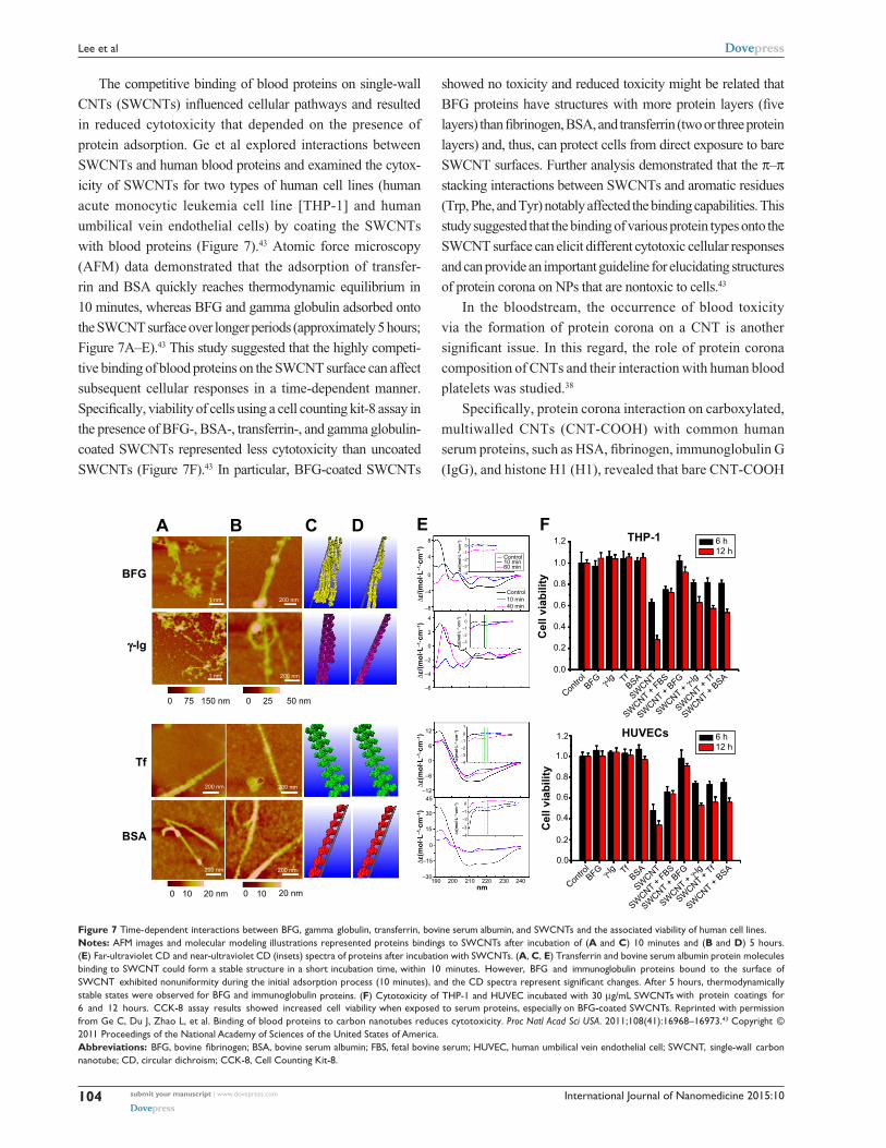
protein adsorption. Ge et al explored interactions between
SWCNTs and human blood proteins and examined the cytox-
icity of SWCNTs for two types of human cell lines (human
acute monocytic leukemia cell line [THP-1] and human
umbilical vein endothelial cells) by coating the SWCNTs
with blood proteins (Figure 7).43 Atomic force microscopy
(AFM) data demonstrated that the adsorption of transfer-
rin and BSA quickly reaches thermodynamic equilibrium in
10 minutes, whereas BFG and gamma globulin adsorbed onto
the SWCNT surface over longer periods (approximately 5 hours;
Figure 7A–E).43 This study suggested that the highly competi-
tive binding of blood proteins on the SWCNT surface can affect
subsequent cellular responses in a time-dependent manner.
Specifically, viability of cells using a cell counting kit-8 assay in
the presence of BFG-, BSA-, transferrin-, and gamma globulin-
coated SWCNTs represented less cytotoxicity than uncoated
SWCNTs (Figure 7F).43 In particular, BFG-coated SWCNTs
showed no toxicity and reduced toxicity might be related that
BFG proteins have structures with more protein layers (five
layers) than fibrinogen, BSA, and transferrin (two or three protein
layers) and, thus, can protect cells from direct exposure to bare
SWCNT surfaces. Further analysis demonstrated that the π–π
stacking interactions between SWCNTs and aromatic residues
(Trp, Phe, and Tyr) notably affected the binding capabilities. This
study suggested that the binding of various protein types onto the
SWCNT surface can elicit different cytotoxic cellular responses
and can provide an important guideline for elucidating structures
of protein corona on NPs that are nontoxic to cells.43
In the bloodstream, the occurrence of blood toxicity
via the formation of protein corona on a CNT is another
significant issue. In this regard, the role of protein corona
composition of CNTs and their interaction with human blood
platelets was studied.38
Specifically, protein corona interaction on carboxylated,
multiwalled CNTs (CNT-COOH) with common human
serum proteins, such as HSA, fibrinogen, immunoglobulin G
(IgG), and histone H1 (H1), revealed that bare CNT-COOH
A B C D E FTHP-1
Cel
l via
bilit
yC
ell v
iabi
lity
∆ε(m
ol∙L
–1∙c
m–1
)∆ε
/(mol
∙L–1
∙cm
–1)
∆ε(m
ol∙L
–1∙c
m–1
)∆ε
(mol
∙L–1
∙cm
–1)
∆ε(m
ol∙L
–1∙c
m–1
)∆ε
/(mol
∙L–1
∙cm
–1)
∆ε/(m
ol∙L
–1∙c
m–1
)
8
4
0
4
2
0
–4
–2
–4
–6
–4
–3–2
–101
–4–3–2–101
–4
–30190 200 210 220 230 240
nm 20 nm 10 0 20 nm
BSA
Tf
γ-lg
BFG
1 nm
1 nm
200 nm
200 nm
200 nm200 nm
200 nm200 nm
10 0
50 nm 25 0 150 nm 75 0
–15
0
15
30
45
12
6
0
–6
–12
–3
–2
–1
0
–4
–3–2
–101
–8
∆ε(m
ol∙L
–1∙c
m–1
)
HUVECs
0.0
0.2
0.4
0.6
0.8
1.0
1.2
0.0
Contro
lBFG γ-l
g TfBSA
SWCNT
SWCNT + FBS
SWCNT + BFG
SWCNT + γ-lg
SWCNT + Tf
SWCNT + BSA
Contro
lBFG γ-l
g TfBSA
SWCNT
SWCNT + FBS
SWCNT + BFG
SWCNT + γ-lg
SWCNT + Tf
SWCNT + BSA
0.2
0.4
0.6
0.8
1.0
1.2
Control10 min60 min
Control10 min40 min
6 h12 h
6 h12 h
Figure 7 Time-dependent interactions between BFG, gamma globulin, transferrin, bovine serum albumin, and SwCNTs and the associated viability of human cell lines.Notes: AFM images and molecular modeling illustrations represented proteins bindings to SwCNTs after incubation of (A and C) 10 minutes and (B and D) 5 hours. (E) Far-ultraviolet CD and near-ultraviolet CD (insets) spectra of proteins after incubation with SwCNTs. (A, C, E) Transferrin and bovine serum albumin protein molecules binding to SwCNT could form a stable structure in a short incubation time, within 10 minutes. However, BFG and immunoglobulin proteins bound to the surface of SWCNT exhibited nonuniformity during the initial adsorption process (10 minutes), and the CD spectra represent significant changes. After 5 hours, thermodynamically stable states were observed for BFG and immunoglobulin proteins. (F) Cytotoxicity of THP-1 and HUveC incubated with 30 µg/mL SwCNTs with protein coatings for 6 and 12 hours. CCK-8 assay results showed increased cell viability when exposed to serum proteins, especially on BFG-coated SwCNTs. Reprinted with permission from Ge C, Du J, Zhao L, et al. Binding of blood proteins to carbon nanotubes reduces cytotoxicity. Proc Natl Acad Sci USA. 2011;108(41):16968–16973.43
Copyright ©
2011 Proceedings of the National Academy of Sciences of the United States of America.Abbreviations: BFG, bovine fibrinogen; BSA, bovine serum albumin; FBS, fetal bovine serum; HUVEC, human umbilical vein endothelial cell; SWCNT, single-wall carbon nanotube; CD, circular dichroism; CCK-8, Cell Counting Kit-8.
International Journal of Nanomedicine 2015:10 submit your manuscript | www.dovepress.com
Dovepress
Dovepress
105
Protein corona on nanoparticle for modulating cytotoxicity and immunotoxicity
and H1 corona induced platelet aggregation and increased
lactate dehydrogenase (one of cytotoxicity markers), whereas
HSA, fibrinogen, and IgG corona on CNT-COOH attenuated
subsequent cytotoxicity.38
The cytotoxicity and serum protein interaction of mul-
tiwalled CNTs with three different grades of carbon blacks
were also investigated.44 The kinetics of various carbon NPs
(CNPs) and serum proteins in the culture medium indicated
that the adsorption of serum proteins (ie, FBS) on CNPs
reached maximum values within 5 minutes.44
AFM studies showed that CNPs were enveloped in serum
proteins (NP-protein coronas) after simple mixing of CNPs
and FBS proteins. It was identified that cellular uptake of
CNPs was greater in serum-free medium than in medium
containing serum (Figure 6A and B). Serum protein adsorp-
tion on CNPs attenuated the inherent cytotoxicity of CNPs
and resulted in decreased cytotoxicity by increasing the
amount of serum proteins adsorbed on the CNPs.44 A possible
mechanism governing this behavior is that the presence of
serum proteins in the medium significantly reduced the intra-
cellular uptake of CNPs. As such, serum proteins adsorbed
on CNPs can inhibit their toxicity by shielding impurities of
metal catalyzers and suppressing the competitive adsorption
of other proteins in the medium.44,45
Graphene is a material composed of either a single layer
or several layers of sp2-bonded carbons that has unique
and highly attractive electrical, mechanical, and thermal
properties.41,46,47
Despite these unique properties, the use of graphene and
its derivatives (eg, graphene oxide [GO]) has raised con-
siderable concerns about human and environmental health.
Recently, the immunotoxicity of hard corona composition
on GO was analyzed.47 Long-term exposure to GO in plasma
conditions (sufficient time to generate hard corona on NP)
decreased reactive oxygen species production and reduced
the cytotoxicity of GO.47
In addition, Hu et al reported a protein corona-mediated
reduction of cytotoxicity of GO.16 In their study, the
authors performed a systematic investigation of the
cellular toxicity of GO nanosheets and identified that
GO interactions with FBS or a protein component in cell
culture medium resulted in a decrease of cytotoxicity
(Figure 8).16 Specifically, at low concentrations of FBS (ie,
1%), cytotoxicity of human lung cancer cells (A549) was
sensitive to the presence of GO and showed concentration-
dependent cytotoxicity.16
However, the cytotoxicity of GO was greatly mitigated
in 10% FBS media and showed no concentration-dependent
toxicity. Specifically, this study addressed the stimulated
cytotoxicity of A549 via the direct interactions with bare GO
nanosheets at the cell membrane that, ultimately, resulted in
physical damage to the membrane.16
Blood protein interaction with polymeric NPs and other inorganic NPsPolymeric NPs [eg, poly(sodium acrylate), poly(ethylene
glycol), chitosan, etc] have been widely used as coating
materials of NPs to avoid activation of the immune system,
to reduce cytotoxicity, and to prolong drug circulation
time in the blood system. Lemarchand et al investigated
the NP–plasma protein interactions with dextran-grafted
poly(e-caprolactone).48 The dextran modification on the NP
surface significantly inhibited the complement system on
human THP-1 and J774.A1 murine macrophage-like cell
lines (phagocytosis). Specifically, dextran-coated poly(e-
caprolactone) showed increased plasma protein adsorption
compared with uncoated poly(e-caprolactone), with the
exception of Ig adsorption.48
The adsorption of proteins to inorganic NPs (eg, Au, Ag,
FeO4, cobalt oxide, and CeO
2) in serum-containing medium
also has been investigated. Casals et al determined that the
production of reactive oxygen species was decreased in THP-1
cells when cobalt oxide NPs were incubated with serum for
48 hours.49 Silver NPs (Ag-NPs) recorded the highest cyto-
toxicity to human cells compared with other metallic NPs,
such as MoO3 and Fe
3O
4.50,51 To improve biocompatibility
of Ag-NPs or Au-NPs, various polymers have been coated
on the NP surfaces. One study examined the cytotoxicity of
spherical Ag-NPs (diameter of 50±20 nm) that were stabilized
with either polyvinylpyrrolidone or citrate and dispersed in
cell culture media (ie, pure media and media containing 10%
BSA or 10% fetal calf serum).52 In this study, the release of Ag
ions from polyvinylpyrrolidone-stabilized Ag-NPs and their
biological effect on human mesenchymal stem cells (hMSCs)
were examined. The formation of Ag-protein coronas with
BSA and fetal calf serum reduced the cytotoxicity of hMSCs
by inhibiting the release of free Ag ions from the silver.
Citrate-coated Au-NPs, in different sizes, were examined
with two types of culture media and cancer cell lines.53 This
study showed that Au-NP-protein coronas formed in Roswell
Park Memorial Institute medium exerted greater cytotoxic
effects compared with Dulbecco modified Eagle’s medium.
This was attributed to the greater abundance and stability of
Au-NP-protein coronas formed in Dulbecco’s medium than
in the Roswell medium. These findings indicated that dif-
ferent compositions of cell culture media can determine the
formation of NP-protein corona and, ultimately, the effect
on cellular toxicity.

International Journal of Nanomedicine 2015:10submit your manuscript | www.dovepress.com
Dovepress
Dovepress
106
Lee et al
A B
C D120
90
60
30
020 µg/mL 100 µg/mL
1% FBS
2 µm2 µm
1 µm
10% FBSFBS-coated GO
Cel
l via
bilit
y (%
)
1 µm
Figure 8 Increased cellular uptake and cytotoxicity were observed when cells were exposed to GO nanosheets in 1% FBS medium, but not in 10% FBS.Notes: (A–C) Transmission electron microscopy images of A549 cells treated with (A and C) 100 µg/mL GO nanosheets and (B) FBS-coated GO nanosheets at 37°C for 2 hours. (C) Magnified images show interactions between GO nanosheets and A549 cells. Red arrows represent nanoparticles in cells. (D) Cell viability of A549 cells treated with GO nanosheets (20 µg/mL, 100 µg/mL) exposed to media containing 1% and 10% FBS for 2 hours. The viability of cells exposed to GO nanosheets in 1% FBS was lower that of cells treated with FBS-coated GO nanosheets. Reprinted with permission from Hu w, Peng C, Lv M, et al. Protein corona-mediated mitigation of cytotoxicity of graphene oxide. ACS Nano. 2011;5(5):3693–3700.16 Copyright © 2011 American Chemical Society.Abbreviations: GO, graphene oxide; FBS, fetal bovine serum.
Activated immune response by protein coronasGeneral recognition of NP-corona by immune cellsWhen NPs are injected into blood systems, they are consid-
ered foreign objects by a series of defense and recognition
mechanisms. Interactions with plasma proteins and plasma
factors can affect clearance and systemic toxicity of the NP
delivery system. Recently, several studies focused on under-
standing the effect of the NP-protein corona on leukocytes
and macrophages.23,54 Synthetic hydroxyapatite particles with
a fine needle shape or hydroxyapatite aggregate can activate
the NLR (nucleotide-binding oligomerization domain recep-
tors) family, pyrin domain-containing 3 inflammasome in
lipopolysaccharide-induced macrophages, and can induce
secretion of proinflammatory cytokines such as interleu-
kin 1β and interleukin 18 in blood.55,56 It has been shown
that the mineral hydroxyapatite NPs exhibit significant
dose-dependent cytotoxicity via human macrophages.55,56
The protein corona can significantly influence NP–cell
interactions through different internalization and path-
way activations.10 Selective cellular uptake of disulfide-
stabilized poly-(methacrylic acid) nanoporous polymer
particles with presence and without presence of protein
corona conditions was recently reported.10 Interestingly,
depending on the corona formation on NP, the uptake
levels of monocytes and macrophages (differentiated
monocyte) were opposites. Specifically, BSA adsorption
on poly-(methacrylic acid) nanoporous polymer particles
International Journal of Nanomedicine 2015:10 submit your manuscript | www.dovepress.com
Dovepress
Dovepress
107
Protein corona on nanoparticle for modulating cytotoxicity and immunotoxicity
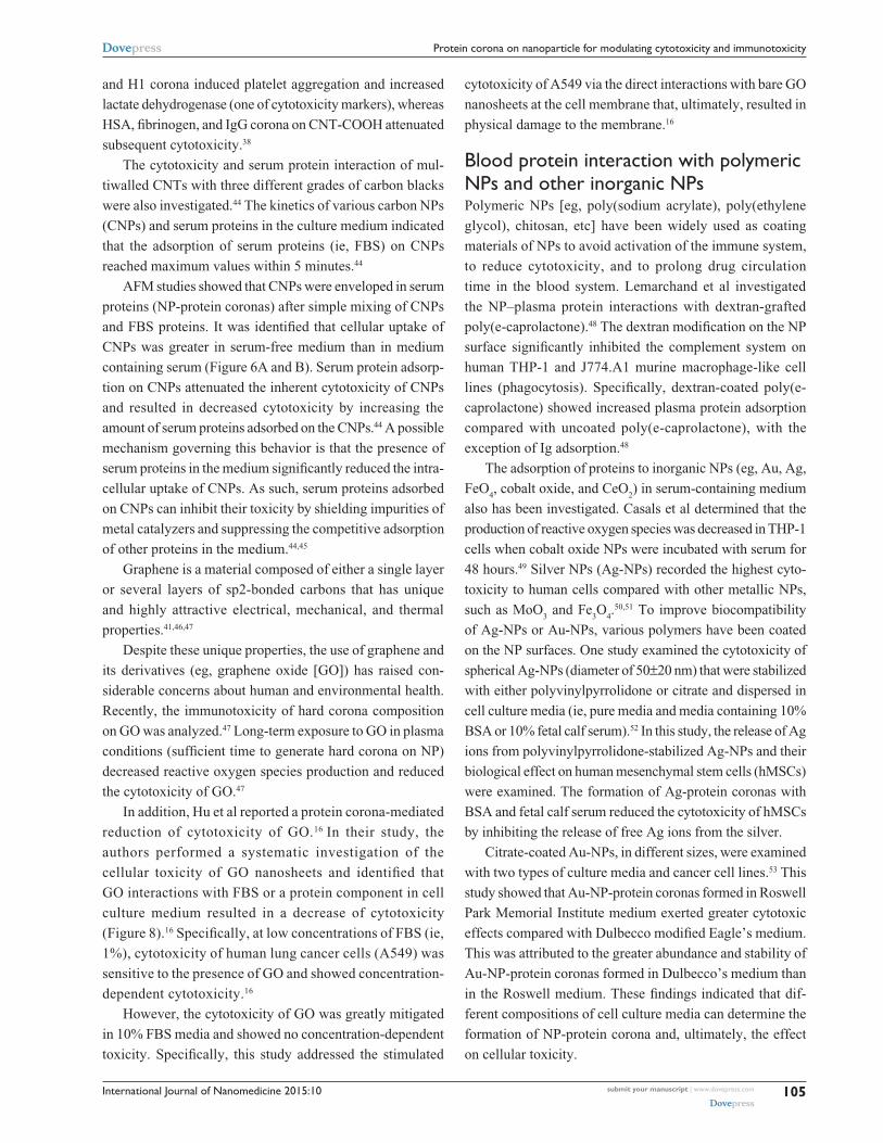
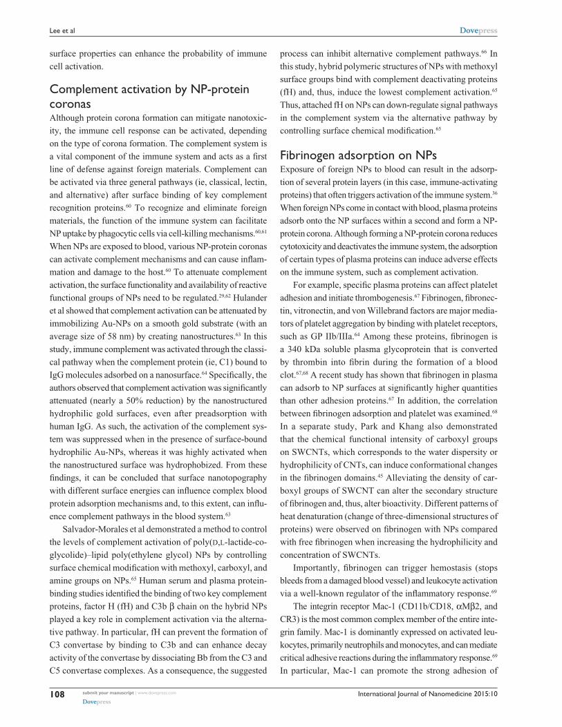
underwent conformational changes and showed decreased
internalization efficiency by THP-1 compared with pure
NPs (Figure 9).10 However, the BSA on nanoporous poly-
mer particles triggered significant internalization and proin-
flammatory cytokine secretion (as a result of phagocytosis
activity) by differentiated macrophage cells (Figure 9).10
This study highlighted selective cell recognition for protein
corona formation on NPs.
The recognition of foreign NPs could be opsonin-
dependent, opsonin-independent, or a combination of both.
An opsonin is any protein (ie, IgG or complement proteins)
that promotes phagocytosis by marking an antigen for an
immune response.57 In addition, molecules that activate
the complement system are considered opsonins.57 In a
series of in vitro experiments, positive correlations between
complement protein (C3) adsorption and uptake/clearance
were observed for liposomes and polylactic acid and poly
(methyl methacrylate) NPs interacting with immune cells.58,59
Along these lines, the physiochemical surface properties of
NPs can mediate the protein corona formation in which NP
Monocyte (THP-1)
100
80
60
40
20
0
100
80
60
40
20
00
cRPMI BSA SF
2 4 6 0 2 4 6
Macrophage (dTHP-1)
Time (hours)Time (hours)
% c
ells
ass
ocia
ted
with
par
ticle
s
% c
ells
ass
ocia
ted
with
par
ticle
s
10 µm 10 µm 10 µm
10 µm 10 µm10 µm
Figure 9 Opposite cellular uptake by monocyte (THP-1) and macrophage (dTHP-1) was observed in the presence and absence of protein corona.Notes: Different cellular uptake of PMAsh NPPs for THP-1 and dTHP-1 cells was measured in cRPMI, BSA-containing medium, and serum-free medium at 6 hours (visualized by fluorescence microscopy). dTHP-1 showed greater uptake for PMAsh NPPs in the presence of serum than the presence of THP-1 cells. Fluorescence images showed the cell membrane (green) and the internalized PMASH particles (red). Scale bars = 10 μm. Reprinted with permission from Yan Y, Gause KT, Kamphuis MM, et al. Differential roles of the protein corona in the cellular uptake of nanoporous polymer particles by monocyte and macrophage cell lines. ACS Nano. 2013;23:7(12): 10960–10970. Copyright ©2013 American Chemical Society.10
Abbreviations: cRPMI, complete Roswell Park Memorial Institute medium; PMAsh NPPs, disulfide-stabilized poly-(methacrylic acid) nanoporous polymer particles; BSA, bovine serum albumin; SF, serum-free medium.
International Journal of Nanomedicine 2015:10submit your manuscript | www.dovepress.com
Dovepress
Dovepress
108
Lee et al
surface properties can enhance the probability of immune
cell activation.
Complement activation by NP-protein coronasAlthough protein corona formation can mitigate nanotoxic-
ity, the immune cell response can be activated, depending
on the type of corona formation. The complement system is
a vital component of the immune system and acts as a first
line of defense against foreign materials. Complement can
be activated via three general pathways (ie, classical, lectin,
and alternative) after surface binding of key complement
recognition proteins.60 To recognize and eliminate foreign
materials, the function of the immune system can facilitate
NP uptake by phagocytic cells via cell-killing mechanisms.60,61
When NPs are exposed to blood, various NP-protein coronas
can activate complement mechanisms and can cause inflam-
mation and damage to the host.60 To attenuate complement
activation, the surface functionality and availability of reactive
functional groups of NPs need to be regulated.29,62 Hulander
et al showed that complement activation can be attenuated by
immobilizing Au-NPs on a smooth gold substrate (with an
average size of 58 nm) by creating nanostructures.63 In this
study, immune complement was activated through the classi-
cal pathway when the complement protein (ie, C1) bound to
IgG molecules adsorbed on a nanosurface.64 Specifically, the
authors observed that complement activation was significantly
attenuated (nearly a 50% reduction) by the nanostructured
hydrophilic gold surfaces, even after preadsorption with
human IgG. As such, the activation of the complement sys-
tem was suppressed when in the presence of surface-bound
hydrophilic Au-NPs, whereas it was highly activated when
the nanostructured surface was hydrophobized. From these
findings, it can be concluded that surface nanotopography
with different surface energies can influence complex blood
protein adsorption mechanisms and, to this extent, can influ-
ence complement pathways in the blood system.63
Salvador-Morales et al demonstrated a method to control
the levels of complement activation of poly(d,l-lactide-co-
glycolide)–lipid poly(ethylene glycol) NPs by controlling
surface chemical modification with methoxyl, carboxyl, and
amine groups on NPs.65 Human serum and plasma protein-
binding studies identified the binding of two key complement
proteins, factor H (fH) and C3b β chain on the hybrid NPs
played a key role in complement activation via the alterna-
tive pathway. In particular, fH can prevent the formation of
C3 convertase by binding to C3b and can enhance decay
activity of the convertase by dissociating Bb from the C3 and
C5 convertase complexes. As a consequence, the suggested
process can inhibit alternative complement pathways.66 In
this study, hybrid polymeric structures of NPs with methoxyl
surface groups bind with complement deactivating proteins
(fH) and, thus, induce the lowest complement activation.65
Thus, attached fH on NPs can down-regulate signal pathways
in the complement system via the alternative pathway by
controlling surface chemical modification.65
Fibrinogen adsorption on NPsExposure of foreign NPs to blood can result in the adsorp-
tion of several protein layers (in this case, immune-activating
proteins) that often triggers activation of the immune system.36
When foreign NPs come in contact with blood, plasma proteins
adsorb onto the NP surfaces within a second and form a NP-
protein corona. Although forming a NP-protein corona reduces
cytotoxicity and deactivates the immune system, the adsorption
of certain types of plasma proteins can induce adverse effects
on the immune system, such as complement activation.
For example, specific plasma proteins can affect platelet
adhesion and initiate thrombogenesis.67 Fibrinogen, fibronec-
tin, vitronectin, and von Willebrand factors are major media-
tors of platelet aggregation by binding with platelet receptors,
such as GP IIb/IIIa.64 Among these proteins, fibrinogen is
a 340 kDa soluble plasma glycoprotein that is converted
by thrombin into fibrin during the formation of a blood
clot.67,68 A recent study has shown that fibrinogen in plasma
can adsorb to NP surfaces at significantly higher quantities
than other adhesion proteins.67 In addition, the correlation
between fibrinogen adsorption and platelet was examined.68
In a separate study, Park and Khang also demonstrated
that the chemical functional intensity of carboxyl groups
on SWCNTs, which corresponds to the water dispersity or
hydrophilicity of CNTs, can induce conformational changes
in the fibrinogen domains.45 Alleviating the density of car-
boxyl groups of SWCNT can alter the secondary structure
of fibrinogen and, thus, alter bioactivity. Different patterns of
heat denaturation (change of three-dimensional structures of
proteins) were observed on fibrinogen with NPs compared
with free fibrinogen when increasing the hydrophilicity and
concentration of SWCNTs.
Importantly, fibrinogen can trigger hemostasis (stops
bleeds from a damaged blood vessel) and leukocyte activation
via a well-known regulator of the inflammatory response.69
The integrin receptor Mac-1 (CD11b/CD18, αMβ2, and
CR3) is the most common complex member of the entire inte-
grin family. Mac-1 is dominantly expressed on activated leu-
kocytes, primarily neutrophils and monocytes, and can mediate
critical adhesive reactions during the inflammatory response.69
In particular, Mac-1 can promote the strong adhesion of
International Journal of Nanomedicine 2015:10 submit your manuscript | www.dovepress.com
Dovepress
Dovepress
109
Protein corona on nanoparticle for modulating cytotoxicity and immunotoxicity
neutrophils to endothelial cells and activate diapedesis, a pro-
cess in which neutrophils migrate through the interstitial matrix
(ie, a type of extracellular matrix found in interstitial connective
tissue).70 The Mac-1 integrin is also involved in other neutro-
phil responses, such as phagocytosis, homotypic aggregation,
degranulation, and adhesion to microorganisms.70
Ligands of Mac-1 can be stimulated by many extracellular
matrix proteins (eg, fibronectin, collagens, thrombospondin,
Cyr61, etc), blood proteins (eg, fibrinogen, iC3b, kininogen,
factor H, factor X, t-PA, and so on), and proteases (eg,
elastase, myeloperoxidase, plasminogen, etc).
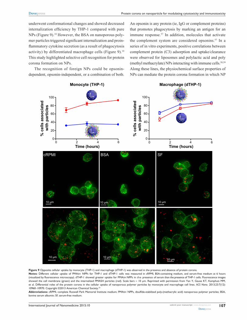
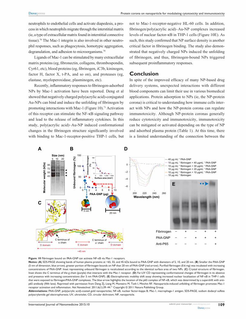
Recently, inflammatory responses to fibrinogen-adsorbed
NPs by Mac-1 activation have been reported. Deng et al
showed that negatively charged poly(acrylic acid)-conjugated
Au-NPs can bind and induce the unfolding of fibrinogen by
promoting interactions with Mac-1 (Figure 10).71 Activation
of this receptor can stimulate the NF-κB signaling pathway
and lead to the release of inflammatory cytokines. In this
study, poly(acrylic acid)–Au-NP induced conformational
changes in the fibrinogen structure significantly involved
with binding to Mac-1-receptor-positive THP-1 cells, but
not to Mac-1-receptor-negative HL-60 cells. In addition,
fibrinogen/poly(acrylic acid)–Au-NP complexes increased
levels of nuclear factor-κB in THP-1 cells (Figure 10E). As
such, this study confirmed that NP surface density is another
critical factor in fibrinogen binding. The study also demon-
strated that negatively charged NPs induced the unfolding
of fibrinogen, and thus, fibrinogen-bound NPs triggered
subsequent proinflammatory responses.
ConclusionIn spite of the improved efficacy of many NP-based drug
delivery systems, unexpected interactions with different
blood components can limit their use in various biomedical
applications. Protein adsorption to NPs (ie, the NP-protein
corona) is critical to understanding how immune cells inter-
act with NPs and how the NP-protein corona can regulate
immunotoxicity. Although NP-protein coronas generally
reduce cytotoxicity and immunotoxicity, immunotoxicity
can be mitigated or activated depending on the type of NP
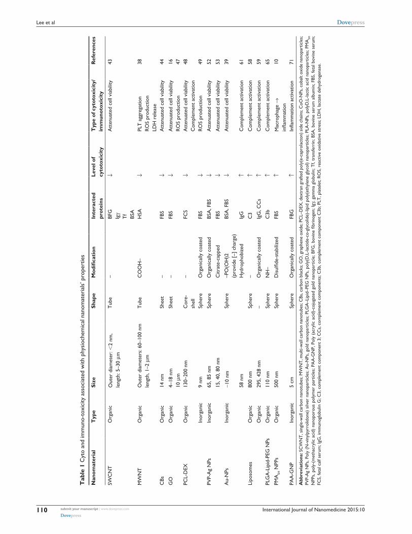
and adsorbed plasma protein (Table 1). At this time, there
is a limited understanding of the connection between the
Size (nm) 150
0
–2
–4
–6
100
50
00
5
A B
C
D
E
10 20
2 4 6
PAA-GNP (µg)
5 nm20 nm
C terminus ofα’ chain
C terminus ofα chain
E domain
~45 nm
D domain D domain
8 10
0Surface area (mm2)
Unb
ound
fibr
inog
en(%
)
Unb
ound
fibr
inog
en (%
)
0
50
100
150
500 1,000 1,500
200 220 240 260Wavelength (nm)
40 µg mL–1 PAA-GNP
10 µg mL–1 fibrinogen + 40 µgmL–1 PAA-GNP
10 µg mL–1 fibrinogen + 30 µgmL–1 PAA-GNP
10 µg mL–1 fibrinogen + 20 µgmL–1 PAA-GNP
10 µg mL–1 fibrinogen + 10 µgmL–1 PAA-GNP
10 µg mL–1 fibrinogen
Fibrinogen
PAA-GNP
Anti-P65
–
–
–
+
–
–
–
+
–
+
+
–
+
+
+
Mol
ecul
ar w
eigh
t (kD
a) 9572
4355
Figure 10 Fibrinogen bound on PAA-GNP can activate NF-κB via Mac-1 receptors.Notes: (A) SDS-PAGe showing bands of human plasma proteins at ~65, 55, and 45 kDa bound to PAA-GNP with diameters of 5, 10, and 20 nm. (B) Smaller the PAA-GNP (5 nm of dimension, blue arrow), greater portion of fibrinogen bounds on NP than 20 nm of PAA-GNP (red arrow). Purified fibrinogen (0.6 mg) was incubated with increasing concentrations of PAA-GNP. Inset representing unbound fibrinogen is recalculated according to the identical surface area of two NPs. (C) Crystal structure of fibrinogen. Inset shows the C terminus of the g chain (purple) that interacts with the Mac-1 receptor. (D) Far-UV CD representing conformational changes of fibrinogen in its absence and presence with increasing concentrations (for 5 nm PAA-GNP). (E) electrophoretic mobility shift assay showing increased nuclear localization of NF-κB in THP-1 cells that were exposed to fibrinogen/PAA-GNP complexes. The blue arrow highlights the location of the p65 complex of NF-κB, which was determined by a supershift with anti-p65 antibody (fifth lane). Reprinted with permission from Deng ZJ, Liang M, Monteiro M, Toth I, Minchin RF. Nanoparticle-induced unfolding of fibrinogen promotes Mac-1 receptor activation and inflammation. Nat Nanotechnol. 2011;6(1):39–44.71 Copyright © 2011 Nature Publishing Group.Abbreviations: PAA-GNP, poly(acrylic acid)-coated gold nanoparticles; NF-κB, nuclear factor-kappa B; Mac-1, macrophage-1 antigen; SDS-PAGe, sodium dodecyl sulfate polyacrylamide gel electrophoresis; Uv, ultraviolet; CD, circular dichroism; NP, nanoparticle.
International Journal of Nanomedicine 2015:10submit your manuscript | www.dovepress.com
Dovepress
Dovepress
110
Lee et al
Tab
le 1
Cyt
o an
d im
mun
o-to
xici
ty a
ssoc
iate
d w
ith p
hysi
oche
mic
al n
anom
ater
ials
’ pro
pert
ies
Nan
omat
eria
lT
ype
Size
Shap
eMod
ification
Inte
ract
ed
prot
eins
Leve
l of
cyto
toxi
city
Typ
e of
cyt
otox
icit
y/im
mun
otox
icit
yR
efer
ence
s
SwC
NT
Org
anic
Out
er d
iam
eter
: 2
nm,
leng
th: 5
–30
µmT
ube
–BF
GIg
γT
fBS
A
↓A
tten
uate
d ce
ll vi
abili
ty43
Mw
NT
Org
anic
Out
er d
iam
eter
s: 6
0–10
0 nm
le
ngth
, 1–2
µm
Tub
eC
OO
H–
HSA
↓PL
T a
ggre
gatio
nR
OS
prod
uctio
nLD
H r
elea
se
38
CBs
Org
anic
14 n
mSh
eet
–FB
S↓
Att
enua
ted
cell
viab
ility
44
GO
Org
anic
4–18
nm
10
µm
Shee
t–
FBS
↓A
tten
uate
d ce
ll vi
abili
tyR
OS
prod
uctio
n16 47
PCL-
DeX
Org
anic
130–
200
nmC
ore-
shel
l–
FCS
↓A
tten
uate
d ce
ll vi
abili
tyC
ompl
emen
t ac
tivat
ion
48
Inor
gani
c9
nmSp
here
Org
anic
ally
coa
ted
FBS
↓R
OS
prod
uctio
n49
PvP-
Ag
NPs
Inor
gani
c65
, 85
nmSp
here
Org
anic
ally
coa
ted
BSA
, FBS
↓A
tten
uate
d ce
ll vi
abili
ty52
15, 4
0, 8
0 nm
Citr
ate-
capp
edFB
S↓
Att
enua
ted
cell
viab
ility
53
Au-
NPs
Inor
gani
c~1
0 nm
Sphe
re–P
O(O
H)2
(p
rovi
de [
–] c
harg
e)BS
A, F
BS↓
Att
enua
ted
cell
viab
ility
39
58 n
mH
ydro
phob
ized
IgG
↑C
ompl
emen
t ac
tivat
ion
61
Lipo
som
esO
rgan
ic80
0 nm
Sphe
re–
C3
↑C
ompl
emen
t ac
tivat
ion
58
Org
anic
295,
428
nm
–O
rgan
ical
ly c
oate
dIg
G, C
Cs
↑C
ompl
emen
t ac
tivat
ion
59
PLG
A-L
ipid
-PeG
NPs
Org
anic
110
nmSp
here
NH
–C
3b↑
Com
plem
ent
activ
atio
n65
PMA
SH N
PPs
Org
anic
500
nmSp
here
Dis
ulfid
e-st
abili
zed
FBS
↑M
acro
phag
e →
in
flam
mat
ion
10
PAA
-GN
PIn
orga
nic
5 cm
Sphe
reO
rgan
ical
ly c
oate
dFB
G↑
Infla
mm
atio
n ac
tivat
ion
71
Abb
revi
atio
ns: S
Cw
NT
, sin
gle-
wal
l car
bon
nano
tube
s; M
wN
T, m
ulti-
wal
l car
bon
nano
tube
s; C
Bs, c
arbo
n bl
acks
; GO
, gra
phen
e ox
ide;
PC
L–D
eX, d
extr
an g
rafte
d po
ly(e
-cap
rola
cton
) sid
e ch
ains
; CoO
-NPs
, cob
alt o
xide
nan
opar
ticle
s;
PvP-
Ag
NPs
, Pol
y (N
-vin
ylpy
rrol
idon
e) s
ilver
nan
opar
ticle
s; A
u-N
Ps, g
old
nano
part
icle
s; P
LGA
–Lip
id–P
eG N
Ps, p
oly(
D,L
-lact
ide-
co-g
lyco
lide)
-lipi
d po
ly(e
thyl
ene
glyc
ol)
nano
part
icle
s; P
LA-N
Ps, p
olyD
,L-la
ctic
aci
d na
nopa
rtic
les;
PM
ASH
N
PPs,
pol
y-(m
etha
cryl
ic a
cid)
nan
opor
ous
poly
mer
par
ticle
s; P
AA
-GN
P, P
oly
(acr
ylic
aci
d)-c
ojug
ated
gol
d na
nopa
rtic
le; B
FG, b
ovin
e fib
rino
gen;
Igγ,
gam
ma
glob
ulin
; Tf,
tran
sfer
rin;
BSA
, bov
ine
seru
m a
lbum
in; F
BS, f
etal
bov
ine
seru
m;
FCS,
feta
l cal
f ser
um; I
gG, i
mm
unog
lobu
lin G
; C3,
com
plem
ent
com
pone
nt 3
; CC
s, c
ompl
emen
t co
mpo
nent
s; C
3b, c
ompl
emen
t co
mpo
nent
C3b
; PLT
, pla
tele
t; R
OS,
rea
ctiv
e ox
idat
ive
stre
ss; L
DH
, lac
tate
deh
ydro
gena
se.
International Journal of Nanomedicine 2015:10 submit your manuscript | www.dovepress.com
Dovepress
Dovepress
111
Protein corona on nanoparticle for modulating cytotoxicity and immunotoxicity
physicochemical properties of NPs and their corresponding
effect on the physiological system. As such, it still remains
unclear how to optimally synthesize and chemically modify
NPs for in vivo application. It is clear, however, that under-
standing NP-protein corona formation and routes of admin-
istration can support the ability to control immune responses
and cytotoxicity. The formation and immunological response
to NP-protein coronas is significantly influenced by the
physiochemical surface properties of the NPs (ie, physical
surface architecture and chemical functionality), and thus,
future works should address the effective physiochemical
properties of NP for determining protein corona and associ-
ated toxicological evaluation by analyzing protein distribu-
tion and examining in vitro and in vivo responses. With an
improved understanding of NP-protein corona interactions in
the immune system, the adverse aspects of NPs will be antici-
pated in advance and inhibited through rational design.
AcknowledgmentsThis research was supported by the National Research
Foundation of Korea funded by the Ministry of Science
(2012R1A1A2041157 and 2014R1A2A1A11052615), Korea
Health technology R&D Project through the KHIDI, funded
by the Ministry of Health & Welfare (HI14C1802) and Korea
Food and Drug Administration (KFDA).
DisclosureThe authors report no conflicts of interest in this work.
References1. De M, Ghosh P, Rotello V. Applications of nanoparticles in biology. Adv
Mater. 2008;20:4225–4241.2. Ferrari M. Cancer nanotechnology: opportunities and challenges. Nat
Rev Cancer. 2005;5(3):161–171.3. Stephan MT, Moon JJ, Um SH, Bershteyn A, Irvine DJ. Therapeutic cell
engineering with surface-conjugated synthetic nanoparticles. Nat Med. 2010;16(9):1035–1041.
4. Zhang L, Gu FX, Chan JM, Wang AZ, Langer RS, Farokhzad OC. Nanoparticles in medicine: therapeutic applications and developments. Clin Pharmacol Ther. 2008;83(5):761–769.
5. Mause SF, Weber C. Microparticles: protagonists of a novel com-munication network for intercellular information exchange. Circ Res. 2010;107(9):1047–1057.
6. Rak J. Microparticles in cancer. Semin Thromb Hemost. 2010;36(8): 888–906.
7. Gref R, Minamitake Y, Peracchia MT, Trubetskoy V, Torchilin V, Langer R. Biodegradable long-circulating polymeric nanospheres. Science. 1994;263(5153):1600–1603.
8. Göppert TM, Müller RH. Polysorbate-stabilized solid lipid nanoparticles as colloidal carriers for intravenous targeting of drugs to the brain: comparison of plasma protein adsorption patterns. J Drug Target. 2005;13(3):179–187.
9. Walczyk D, Bombelli FB, Monopoli MP, Lynch I, Dawson KA. What the cell “sees” in bionanoscience. J Am Chem Soc. 2010; 132(16):5761–5768.
10. Yan Y, Gause KT, Kamphuis MM, et al. Differential roles of the protein corona in the cellular uptake of nanoporous polymer par-ticles by monocyte and macrophage cell lines. ACS Nano. 2013;23: 7(12):10960–10970.
11. Leu D, Manthey B, Kreuter J, Speiser P, DeLuca PP. Distribu-tion and elimination of coated polymethyl [2–14C]methacrylate nanoparticles after intravenous injection in rats. J Pharm Sci. 1984; 73(10):1433–1437.
12. Bala I, Hariharan S, Kumar MN. PLGA nanoparticles in drug delivery: the state of the art. Crit Rev Ther Drug Carrier Syst. 2004;21(5):387–422.
13. Lesniak A, Fenaroli F, Monopoli MP, Åberg C, Dawson KA, Salvati A. Effects of the presence or absence of a protein corona on silica nanopar-ticle uptake and impact on cells. ACS Nano. 2012;6(7):5845–5857.
14. Lesniak A, Salvati A, Santos-Martinez MJ, Radomski MW, Dawson KA, Åberg C. Nanoparticle adhesion to the cell membrane and its effect on nanoparticle uptake efficiency. J Am Chem Soc. 2013;135(4):1438–1444.
15. Treuel L, Brandholt S, Maffre P, Wiegele S, Shang L, Nienhaus GU. Impact of protein modification on the protein corona on nanoparticles and nanoparticle-cell interactions. ACS Nano. 2014;8(1):503–513.
16. Hu W, Peng C, Lv M, et al. Protein corona-mediated mitigation of cytotoxicity of graphene oxide. ACS Nano. 2011;5(5):3693–3700.
17. Yang ST, Liu Y, Wang YW, Cao A. Biosafety and bioapplication of nanomaterials by designing protein-nanoparticle interactions. Small. 2013;9(9–10):1635–1653.
18. Walkey CD, Chan WC. Understanding and controlling the interaction of nanomaterials with proteins in a physiological environment. Chem Soc Rev. 2012;41(7):2780–2799.
19. Fenoglio I, Fubini B, Ghibaudi EM, Turci F. Multiple aspects of the interaction of biomacromolecules with inorganic surfaces. Adv Drug Deliv Rev. 2011;63(13):1186–1209.
20. Monopoli MP, Aberg C, Salvati A, Dawson KA. Biomolecular coronas provide the biological identity of nanosized materials. Nat Nanotechnol. 2012;7(12):779–786.
21. Nel AE, Mädler L, Velegol D, et al. Understanding biophysicochemical interactions at the nano-bio interface. Nat Mater. 2009;8(7):543–557.
22. Wang X, Reece SP, Brown JM. Immunotoxicological impact of engi-neered nanomaterial exposure: mechanisms of immune cell modula-tion. Toxicol Mech Methods. 2013;23(3):168–177.
23. Karmali PP, Simberg D. Interactions of nanoparticles with plasma proteins: implication on clearance and toxicity of drug delivery sys-tems. Expert Opin Drug Deliv. 2011;8(3):343–357.
24. Cedervall T, Lynch I, Lindman S, et al. Understanding the nanoparticle-protein corona using methods to quantify exchange rates and affinities of proteins for nanoparticles. Proc Natl Acad Sci U S A. 2007;104(7):2050–2055.
25. Monopoli MP, Walczyk D, Campbell A, et al. Physical-chemical aspects of protein corona: relevance to in vitro and in vivo biological impacts of nanoparticles. J Am Chem Soc. 2011;133(8):2525–2534.
26. Peracchia MT, Harnisch S, Pinto-Alphandary H, et al. Visualization of in vitro protein-rejecting properties of PEGylated stealth polycyano-acrylate nanoparticles. Biomaterials. 1999;20(14):1269–1275.
27. Gref R, Lück M, Quellec P, et al. ‘Stealth’ corona-core nanoparticles surface modified by polyethylene glycol (PEG): influences of the corona (PEG chain length and surface density) and of the core composition on phagocytic uptake and plasma protein adsorption. Colloids Surf B Biointerfaces. 2000;18(3–4):301–313.
28. Gessner A, Waicz R, Lieske A, Paulke B, Mäder K, Müller RH. Nano-particles with decreasing surface hydrophobicities: influence on plasma protein adsorption. Int J Pharm. 2000;196(2):245–249.
29. Gessner A, Lieske A, Paulke BR, Müller RH. Functional groups on polystyrene model nanoparticles: influence on protein adsorption. J Biomed Mater Res A. 2003;65(3):319–326.
30. Lacerda SH, Park JJ, Meuse C, et al. Interaction of gold nanopar-ticles with common human blood proteins. ACS Nano. 2010;4(1): 365–379.
International Journal of Nanomedicine 2015:10submit your manuscript | www.dovepress.com
Dovepress
Dovepress
112
Lee et al
31. Casals E, Pfaller T, Duschl A, Oostingh GJ, Puntes V. Time evolution of the nanoparticle protein corona. ACS Nano. 2010;4(7):3623–3632.
32. Miclăuş T, Bochenkov VE, Ogaki R, Howard KA, Sutherland DS. Spatial mapping and quantification of soft and hard protein coronas at silver nanocubes. Nano Lett. 2014;14(4):2086–2093.
33. Medintz IL, Konnert JH, Clapp AR, et al. A fluorescence resonance energy transfer-derived structure of a quantum dot-protein bioconjugate nanoassembly. Proc Natl Acad Sci U S A. 2004;101(26):9612–9617.
34. Aubin-Tam ME, Hamad-Schifferli K. Gold nanoparticle-cytochrome C complexes: the effect of nanoparticle ligand charge on protein structure. Langmuir. 2005;21(26):12080–12084.
35. Roach P, Farrar D, Perry CC. Surface tailoring for controlled protein adsorption: effect of topography at the nanometer scale and chemistry. J Am Chem Soc. 2006;128(12):3939–3945.
36. Lundqvist M, Stigler J, Elia G, Lynch I, Cedervall T, Dawson KA. Nanoparticle size and surface properties determine the protein corona with possible implications for biological impacts. Proc Natl Acad Sci U S A. 2008;105(38):14265–14270.
37. Tenzer S, Docter D, Kuharev J, et al. Rapid formation of plasma protein corona critically affects nanoparticle pathophysiology. Nat Nanotech-nol. 2013;8(10):772–781.
38. De Paoli SH, Diduch LL, Tegegn TZ, et al. The effect of protein corona composition on the interaction of carbon nanotubes with human blood platelets. Biomaterials. 2014;35(24):6182–6194.
39. Hühn D, Kantner K, Geidel C, et al. Polymer-coated nanoparticles interacting with proteins and cells: focusing on the sign of the net charge. ACS Nano. 2013;7(4):3253–3263.
40. Lacerda L, Bianco A, Prato M, Kostarelos K. Carbon nanotubes as nanomedicines: from toxicology to pharmacology. Adv Drug Deliv Rev. 2006;58(14):1460–1470.
41. Liu Z, Robinson JT, Tabakman SM, Yang K, Dai H. Carbon materials for drug delivery and cancer therapy. Mater Today. 2011;14(7–8): 316–323.
42. Cai X, Ramalingam R, Wong HS, et al. Characterization of carbon nanotube protein corona by using quantitative proteomics. Nanomedi-cine (Lond). 2013;9(5):583–593.
43. Ge C, Du J, Zhao L, et al. Binding of blood proteins to car-bon nanotubes reduces cytotoxicity. Proc Natl Acad Sci U S A. 2011;108(41):16968–16973.
44. Zhu Y, Li W, Li Q, et al. Effects of serum proteins on intracel-lular uptake and cytotoxicity of carbon nanoparticles. Carbon. 2009;47(5):1351–1358.
45. Park SJ, Khang D. Conformational changes of fibrinogen in dispersed carbon nanotubes. Int J Nanomedicine. 2012;7:4325–4333.
46. Mao HY, Laurent S, Chen W, et al. Graphene: promises, facts, opportunities, and challenges in nanomedicine. Chem Rev. 2013; 113(5):3407–3424.
47. Mao H, Chen W, Laurent S, et al. Hard corona composition and cel-lular toxicities of the graphene sheets. Colloids Surf B Biointerfaces. 2013;109:212–218.
48. Lemarchand C, Gref R, Passirani C, et al. Influence of polysaccharide coating on the interactions of nanoparticles with biological systems. Biomaterials. 2006;27(1):108–118.
49. Casals E, Pfaller T, Duschl A, Oostingh GJ, Puntes VF. Hardening of the nanoparticle-protein corona in metal (Au, Ag) and oxide (Fe
3O
4,
CoO, and CeO2) nanoparticles. Small. 2011;7(24):3479–3486.
50. Braydich-Stolle L, Hussain S, Schlager JJ, Hofmann MC. In vitro cytotoxicity of nanoparticles in mammalian germline stem cells. Toxicol Sci. 2005;88(2):412–419.
51. AshaRani PV, Low Kah Mun G, Hande MP, Valiyaveettil S. Cytotoxic-ity and genotoxicity of silver nanoparticles in human cells. ACS Nano. 2009;3(2):279–290.
52. Kittler S, Greulich C, Diendorf J, Köller M, Epple M. Toxicity of Silver Nanoparticles Increases during Storage Because of Slow Dissolution under Release of Silver Ions. Chem Mater. 2010;22(16):4548–4554.
53. Maiorano G, Sabella S, Sorce B, Brunetti V, Malvindi MA, Cingolani R, Pompa PP. Effects of cell culture media on the dynamic formation of protein-nanoparticle complexes and influence on the cellular response. ACS Nano. 2010;4(12):7481–7491.
54. Lynch I, Salvati A, Dawson KA. Protein-nanoparticle interactions: What does the cell see? Nat Nanotechnol. 2009;4(9):546–547.
55. Müller KH, Motskin M, Philpott AJ, et al. The effect of particle agglomeration on the formation of a surface-connected compartment induced by hydroxyapatite nanoparticles in human monocyte-derived macrophages. Biomaterials. 2014;35(3):1074–1088.
56. Motskin M, Müller KH, Genoud C, Monteith AG, Skepper JN. The sequestration of hydroxyapatite nanoparticles by human monocyte-macrophages in a compartment that allows free diffusion with the extracellular environment. Biomaterials. 2011;32(35):9470–9482.
57. Salmon JE, Kapur S, Kimberly RP. Opsonin-independent ligation of Fc gamma receptors. The 3G8-bearing receptors on neutrophils mediate the phagocytosis of concanavalin A-treated erythrocytes and nonopsonized Escherichia coli. J Exp Med. 1987;166(6):1798–1813.
58. Harashima H, Sakata K, Funato K, Kiwada H. Enhanced hepatic uptake of liposomes through complement activation depending on the size of liposomes. Pharm Res. 1994;11(3):402–406.
59. Leroux JC, De Jaeghere F, Anner B, Doelker E, Gurny R. An investiga-tion on the role of plasma and serum opsonins on the internalization of biodegradable poly(d,l-lactic acid) nanoparticles by human monocytes. Life Sci. 1995;57(7):695–703.
60. Sim RB, Tsiftsoglou SA. Proteases of the complement system. Biochem Soc Trans. 2004;32(Pt 1):21–27.
61. Rybak-Smith MJ, Sim RB; Rybak-Smith MJ. Complement activation by carbon nanotubes. Adv Drug Deliv Rev. 2011;63(12):1031–1041.
62. Bertholon I, Vauthier C, Labarre D. Complement activation by core-shell poly(isobutylcyanoacrylate)-polysaccharide nanoparticles: influ-ences of surface morphology, length, and type of polysaccharide. Pharm Res. 2006;23(6):1313–1323.
63. Hulander M, Lundgren A, Berglin M, Ohrlander M, Lausmaa J, Elwing H. Immune complement activation is attenuated by surface nanotopography. Int J Nanomedicine. 2011;6:2653–2666.
64. Gorbet MB, Sefton MV. Biomaterial-associated thrombosis: roles of coagulation factors, complement, platelets and leukocytes. Biomateri-als. 2004;25(26):5681–5703.
65. Salvador-Morales C, Flahaut E, Sim E, Sloan J, Green ML, Sim RB, Salvador-Morales C. Complement activation and protein adsorption by carbon nanotubes. Mol Immunol. 2006;43(3):193–201.
66. Kouser L, Abdul-Aziz M, Nayak A, Stover CM, Sim RB, Kishore U. Properdin and factor h: opposing players on the alternative complement pathway “see-saw”. Front Immunol. 2013;4:93.
67. Horbett T. Chapter 13 Principles underlying the role of adsorbed plasma proteins in blood interactions with foreign materials. Cardiovasc Pathol. 1993;2(3):137–148.
68. Chinn JA, Horbett TA, Ratner BD. Baboon fibrinogen adsorption and platelet adhesion to polymeric materials. Thromb Haemost. 1991; 65(5):608–617.
69. Lishko VK, Podolnikova NP, Yakubenko VP, et al. Multiple binding sites in fibrinogen for integrin alphaMbeta2 (Mac-1). J Biol Chem. 2004 22;279(43):44897–44906.
70. Ding ZM, Babensee JE, Simon SI, et al. Relative contribution of LFA-1 and Mac-1 to neutrophil adhesion and migration. J Immunol. 1999;163(9):5029–5038.
71. Deng ZJ, Liang M, Monteiro M, Toth I, Minchin RF. Nanoparticle-induced unfolding of fibrinogen promotes Mac-1 receptor activation and inflammation. Nat Nanotechnol. 2011;6(1):39–44.
International Journal of Nanomedicine
Publish your work in this journal
Submit your manuscript here: http://www.dovepress.com/international-journal-of-nanomedicine-journal
The International Journal of Nanomedicine is an international, peer-reviewed journal focusing on the application of nanotechnology in diagnostics, therapeutics, and drug delivery systems throughout the biomedical field. This journal is indexed on PubMed Central, MedLine, CAS, SciSearch®, Current Contents®/Clinical Medicine,
Journal Citation Reports/Science Edition, EMBase, Scopus and the Elsevier Bibliographic databases. The manuscript management system is completely online and includes a very quick and fair peer-review system, which is all easy to use. Visit http://www.dovepress.com/testimonials.php to read real quotes from published authors.
International Journal of Nanomedicine 2015:10 submit your manuscript | www.dovepress.com
Dovepress
Dovepress
Dovepress
113
Protein corona on nanoparticle for modulating cytotoxicity and immunotoxicity