Atomic structure and hierarchical assembly of a cross- amyloid fibril

Fibril Fragmentation Enhances Amyloid Cytotoxicity*□S �
Received for publication, July 28, 2009, and in revised form, September 22, 2009 Published, JBC Papers in Press, October 6, 2009, DOI 10.1074/jbc.M109.049809
Wei-Feng Xue, Andrew L. Hellewell, Walraj S. Gosal1, Steve W. Homans, Eric W. Hewitt, and Sheena E. Radford2
From the Astbury Centre for Structural Molecular Biology, Institute of Molecular and Cellular Biology, University of Leeds,Leeds LS2 9JT, United Kingdom
Fibrils associated with amyloid disease are molecular assem-blies of key biological importance, yet how cells respond to thepresence of amyloid remains unclear. Cellular responses maynot only depend on the chemical composition or molecularproperties of the amyloid fibrils, but their physical attributessuch as length, width, or surface area may also play importantroles. Here, we report a systematic investigation of the effect offragmentation on the structural and biological properties ofamyloid fibrils. In addition to the expected relationship betweenfragmentation and the ability to seed, we show a striking findingthat fibril length correlates with the ability to disrupt mem-branes and to reduce cell viability. Thus, despite otherwiseunchanged molecular architecture, shorter fibrillar samplesshow enhanced cytotoxic potential than their longer counter-parts. The results highlight the importance of fibril length inamyloid disease, with fragmentation not only providing amech-anism by which fibril load can be rapidly increased but also cre-ating fibrillar species of different dimensions that can endownew or enhanced biological properties such as amyloidcytotoxicity.
Amyloid fibril deposits are associated with numerous disor-ders, including type II diabetesmellitus andAlzheimer and Par-kinson diseases (1). These proteinaceous fibrillar aggregates arecommonly regarded as the self-assembly end products of pep-tides or proteins that form by nucleated polymerization (2).Despite sharing a common cross-� molecular architecture,fibrils of different morphologies and/or superstructural fea-tures can be formed, even from the same starting material(3–6). Other types of aggregates, including oligomeric speciesof different sizes (e.g. (25)), typically accumulate during fibrilformation. It has also been shown that mechanical stress canaffect the products of fibril assembly, producing fibrils of differ-ent dimensions and/or molecular structure even under other-wise identical conditions (3, 4, 8).
Because of the enormous complexity and heterogeneity inthe dynamic equilibrium between different species populatedduring amyloid formation, the identity of the culprits of cyto-toxicity associated with amyloid disease remains far from cleardespite a plethora of studies in recent years (for example, Refs.9–15). The species involved inmediating the cytotoxicity asso-ciatedwithmany amyloid disorderswere initially assumed to befibrils and fibril plaques that are abundant in diseased tissues(16, 17). However, numerous recent reports have focused onsoluble prefibrillar oligomers as the primary cytotoxic species(for example, Refs. 9–12). Despite significant evidence sup-porting prefibrillar oligomeric species as toxic agents, examplesof toxicity associated with fibrils persist (e.g. Refs. 13, 15, and18). This raises the possibility that the determinants of cytotox-icitymay not always be associatedwith the same type of species,and for some amyloidogenic proteins, fibrils themselves orfibril-associated species may possess cytotoxic potential (19).Recent studies have shown that A�3 fibrils interacting withsphingolipids, gangliosides, or cholesterol, all of which havebeen shown to associate with amyloid plaques in vivo (20),result in the release of cytotoxic species (14), whereas theassembly process of islet amyloid polypeptide (also known asamylin) fibrils on lipid membranes results in liposome disrup-tion, suggesting fibril-associated toxicity during the fibrilgrowth process (21). Taken together, these studies suggest thatfibrils should perhaps not be dismissed as the inert products ofamyloid assembly but might provide a further source of toxic-ity, either directly by interacting with membranes or indirectlyby acting as a source of cytotoxic entities.How fibrils elicit a biological response may not only depend
on their chemical composition or molecular properties, buttheir physical attributes such as length, width, or surface areamay also play important roles, as found for other nanoscalematerials (22, 23). To investigate this possibility, we report herea detailed analysis of the relation between fibril length, quanti-fied using tapping-mode atomic force microscopy (TM-AFM),and the structural and biological properties of amyloid fibrils.Using long straight (LS) fibrils formed from human �2-micro-globulin (�2m) (3), we show that samples containing thesefibrils can disrupt model liposome membranes and reduce cellviability, whereas prefibrillar oligomeric species formed in thelag phase of assembly and fibrillar aggregates with different
* This work was supported by grants from the Wellcome Trust (Grant 075675)and Biotechnology and Biological Sciences Research Council (GrantBB/526502/1).Author’s Choice—Final version full access.
�This article was selected as a Paper of the Week.□S The on-line version of this article (available at http://www.jbc.org) contains
supplemental Figs. 1 and 2.1 Present address: University of Texas Southwestern Medical Center at
Dallas, 5323 Harry Hines Blvd., Dallas, TX, 75390.2 To whom correspondence should be addressed: Astbury Centre for Struc-
tural Molecular Biology, Garstang Bldg., University of Leeds, Leeds LS2 9JT,UK. Tel.: 44-113-343-3170; Fax: 44-113-343-7486; E-mail [email protected].
3 The abbreviations used are: A�, �-amyloid; �Syn, �-synuclein; �2m, �2-mi-croglobulin; EM, electron microscopy; FTIR, Fourier transform infrared; LS,long-straight �2m fibrils; Lyz, lysozyme; RL, rod-like �2m fibrils; WL, worm-like �2m fibrils; Sh, short form; Ln, long form; MTT, 3-(4,5-dimethylthiazol-2yl)-2,5-diphenyltetrazolium bromide; ThT, thioflavin T; TM-AFM, tappingmode atomic force microscopy; LUV, large unilamellar vesicles.
THE JOURNAL OF BIOLOGICAL CHEMISTRY VOL. 284, NO. 49, pp. 34272–34282, December 4, 2009Author’s Choice © 2009 by The American Society for Biochemistry and Molecular Biology, Inc. Printed in the U.S.A.
34272 JOURNAL OF BIOLOGICAL CHEMISTRY VOLUME 284 • NUMBER 49 • DECEMBER 4, 2009
by guest on April 30, 2016
http://ww
w.jbc.org/
Dow
nloaded from
structural properties (3, 7) do not. Strikingly, we show that thecytotoxicity displayed by the LS fibril samples is enhanced byreducing fibril length, supporting the idea that the physicaldimensions of fibrils can also modulate their cytotoxic poten-tial. The same length-dependent effect is also observed withfibrils formed from lysozyme and �-synuclein, suggesting thatreduction of fibril length by fragmentation presents a genericmechanism by which fibril-associated cytotoxicity, i.e. cytotox-icity caused by fibrils themselves or by species dynamicallyassociated with fibrils through direct exchange, could beenhanced. These results not only demonstrate the cytotoxicpotential associated with fibrillar samples, but more impor-tantly, reveal that fibril breakage can enhance toxic responses incells, even for fibrils that have identical molecular architecture.Fibril fragmentation therefore poses a double threat in amyloiddisease, providing amechanism by which fibril load can be rap-idly increased, as well as a route by which amyloid cytotoxicitymay be enhanced. The results may provide a rationale of thevaried cellular responses to fibrils of apparently identical chem-ical composition and suggest that targeting amyloid fibril sta-bility against breakage may be a powerful strategy for develop-ing therapies against amyloid disease.
EXPERIMENTAL PROCEDURES
Fibril Sample Preparations—All mechanical agitation exper-iments were performed by stirring 500 �l of fibril samples in1.5-ml glass vials, each containing a 3 � 8-mm polytetrafluoro-ethylene-coated magnetic stirring bar. Agitation was per-formed using a custom-made precision stirrer with accuraterpm readout provided by a revolution counter on the rotor axis(custom-built by the workshop of the School of Physics andAstronomy, University of Leeds) at 1,000 rpm, 25 °C.Preparation of �2m fibrils was performed by dissolving
lyophilized protein (wild-type �2m expressed and purified asdescribed previously (3, 24)) into the reaction buffer containing10 mM sodium dihydrogen phosphate and 50 mM NaCl,adjusted to pH 2.0 using HCl. The reaction mixture was imme-diately syringe-filtered (0.2-�m Minisart fast flow, SartoriousStedim Biotech) and split into two equal aliquots. The first ali-quot was stirred as described above for 3 days at 25 °C. Underthis condition, short fragmented fibrils (LSSh) form. Long fibrils(LSLn) were subsequently grown by seeding the second aliquotof �2m monomers with 0.1% (w/w) of LSSh fibrils and incubat-ing the mixture at 25 °C under quiescent conditions for 48 h.The agitation sample series were next prepared by stirring theLSLn fibril sample in a newglass vial. Small sample aliquotsweretaken after different times of stirring with a dead time of 9 minbetween the start of the stirring and the first sample taken.Fibrillar aggregates of �2m with worm-like (WL) or rod-like(RL) morphology (3) were assembled in 150 mM or 250 mM
ammonium formate buffer adjusted to pH 3.6 using HCl,respectively (3, 25). The protein monomer concentration ofsamples used to form the initial fibril stock solutions was 120�M, and under the employed conditions, all fibril solutionsweretranslucent without visible turbidity, indicating that the sam-ples contain well dispersed fibrils.Preparation of lysozyme fibrils was performed by first dis-
solving lyophilized chicken hen egg white lysozyme (Sigma)
into 10mMHCl. The solution was immediately syringe-filtered(0.2-�m MiniSart fast flow) and incubated at 60 °C for 48 hunder quiescent conditions. The solution was next placed at25 °C and left quiescently for �2 months until fibrils (LyzLn)appeared. Fragmented fibrils (LyzSh) were obtained by subse-quently stirring an aliquot of the fibril sample at 1,000 rpm for72 h. The protein monomer concentration was 350 �M.Preparation of �-synuclein fibrils (�Syn) was performed by
first dissolving lyophilized protein powder (wild-type �Synexpressed and purified as described in Ref. 26) into 25 mM
sodium phosphate buffer at pH 7.5. The sample solution(�SynLn) was then incubated under gentle orbital shaking (200rpm in a 1.5-ml Eppendorf tube) at 37 °C for 1 week and subse-quently left quiescently for �2 months. Short fragmentedfibrils (�SynSh) were again obtained by stirring of an aliquot ofthe fibril sample at 1,000 rpm for 72 h. The protein monomerconcentration was 350 �M.Fibril Imaging Using TM-AFM and Negative Stain Elec-
tron Microscopy (EM)—For TM-AFM, the �2m fibrils wereimaged using a Dimension 3100 scanning probe microscope(Veeco Instruments) and PPP-NCLR silicon cantileverprobes (NanoWorld AG, NANOSENSORS, Neuchatel, Swit-zerland) with nominal force constant of 48 newtons/m. ForLS fibril samples, each sample was diluted to 0.4 �M withsterile filtered deionized water and 20 �l incubated on thesurface of freshly cleaved mica for 5 min. The surface wasthen washed with 1 ml of sterile filtered deionized water anddried under a gentle stream of N2 gas. For WL and RL fibrilsamples, the same volume and concentration of the samplematerial was incubated on the mica surface for 1 min butotherwise using the same protocol. Images of 10 � 10 �m at1,024 � 1,024 pixels were collected. Negative stain EM imag-ing was performed as described previously (24).AFM Data Processing—The AFM images were processed
using automated scripts written in MATLAB (MathWorks).For each image, fibril contours were picked out automaticallyby the scripts to ensure objective analysis. The length of theindividual fibrils, as well as the height information of each pixelalong the highest ridge of each fibril, was extracted for fibrilsthat could be unambiguously recognized by the scripts. Theresulting distribution of lengths was biased toward shorterfibrils, because longer fibrils are cut off more frequently byimage boundaries, by overlap with other fibrils, and by length-dependent differences in surface deposition efficiency of fibrilswith different length (27). To correct for this bias, the lengthdistribution was adjusted by factoring in an empirical powerfunction obtained by assuming that the average total length offibrils recognized on each image should be constant (describedin Ref. 27), as expected from the fact that all images are col-lected on surfaces treated with samples formed from an identi-cal protein monomer concentration, using an identical proto-col (27).From the bias-corrected length distributions, weight average
lengths (referred to as the average length throughout thisreport) were calculated. The results from the fibril extensionassay and the liposome dye release assay versus the averagelengths, proportional to the surface area parallel to the fibrilaxis per fibril, were fitted to a power law function (y � axb)
Fibril Fragmentation Enhances Amyloid Cytotoxicity
DECEMBER 4, 2009 • VOLUME 284 • NUMBER 49 JOURNAL OF BIOLOGICAL CHEMISTRY 34273
by guest on April 30, 2016
http://ww
w.jbc.org/
Dow
nloaded from

using the total least squaresmethod (28) that takes into accountvariations in x and y values.Fourier Transform Infrared (FTIR) Spectroscopy—Fibril sam-
ples for FTIRwere prepared as described above but using buffersolutionsmade up using 99.8%D2O.The pDwas adjusted usingDCl. The samples were concentrated 10 times by centrifuging a500-�l sample at 16,300 � g for 2 h and subsequent resuspen-sion to 50 �l using the supernatant. Transmission mode FTIRabsorbance spectra of the samples as well as the buffer and thevapor spectra were collected on a Thermo-Nicolet IR-560(Thermo Scientific) FTIR spectrometer at 25 °C. For each sam-ple, 1,024 scans were obtained at a spectral resolution of 1cm�1, through a sample path length of 0.05mm.The final spec-tra were corrected for the buffer and the vapor signals, andsecondary derivatives of the absorbance spectra were calcu-lated and plotted.Fibril Growth Kinetics Followed by Thioflavin T (ThT)
Fluorescence—The fibril growth kinetics of �2m LSSh fibrilswere monitored using a sample contained within a 1.5-ml glassvial in the same buffer condition and stirred as described aboveat a protein monomer concentration of 60 �M. A 10-�l samplewas removed periodically and diluted into 10mM sodium phos-phate buffer at pH 2.0 with 50 mM NaCl and 10 �M ThT to afinal volume of 200�l. The fluorescence emission of ThT at 480nm was then read at an excitation wavelength of 440 nm on aPTIQuantaMaster spectrometer (Photon Technology Interna-tional). An additional 10 �l of the sample was also taken andused for liposome dye release assays at each time point.For the seeding efficiency assays, the kinetics of 2% (w/w)
seeded fibril growth at 12 �M monomer concentration werefollowed by monitoring the fluorescence of ThT in a 96-wellplate using a BMG LABTECH FLUOStar Optima at 25 °C asdescribed previously (29). The initial slope of the resultinggrowth curves was extracted after normalizing the signals usingthe baseline signal values and the signal values when the reac-tion reaches completion. The initial slopes were subsequentlyused as a measure of the efficiency of fibril samples to seed thegrowth of new fibrils.Liposome Dye Release Assay—Large unilamellar vesicles
(LUV) were prepared using a lipid mixture containing 1.6mg/ml phosphatidylcholine and 0.4 mg/ml phosphatidylglyc-erol purified from chicken egg (Avanti Lipids) to give 20% neg-atively charged head groups (within typical physiological range(30)). The lipid mixture also contained 0.01mg/ml rhodamine-labeled phosphatidylethanolamine (Avanti Lipids) for determi-nation of the lipid concentration. The lipids were dissolved in50mMHEPES, 10mMNaCl, 1mMEDTA, pH 7.4, containing 50mM carboxyfluorescein (Sigma). LUVs were prepared from thelipid mixture by extrusion through a polycarbonate filter with400-nmpore size. The LUVswere nextwashed three timeswitha buffer (liposome buffer) containing 50 mM HEPES, 107 mM
NaCl, 1 mM EDTA, pH 7.4, and resuspended to form a stocksolution with final lipid concentration of 0.5 mM. The finalstock solution containing LUVs with encapsulated carboxy-fluorescein were used within 2 days.For each dye release assay reaction, 2 �l of the LUV stock
solution was diluted with typically 188 �l of liposome buffer.Fibril sample (typically 10 �l) was then added to the solution to
give a final volume of 200 �l, and the fluorescence emission ofcarboxyfluorescein at 513 nm was quantified using an excita-tion wavelength of 492 nm on a PTI QuantaMaster spectrom-eter (Photon Technology International). For the concentrationseries and control experiment with fibril supernatant, fibrilsamples were diluted prior to addition to the liposome solutionwith the fibril growth buffer to the final concentrations inmonomer equivalents between 12�M (highest possible concen-tration for the experiments) and 12 pM for the concentrationseries and 0.12�M for the supernatant control. Triplicates weremeasured for each sample.The dye release kinetics, with typical traces shown in supple-
mental Fig. 1A, are complex and dependent on factors such asmembrane composition. Controls using LUVs composed of100% phosphatidylcholine, phosphatidylglycerol, phosphatidicacid, or phosphatidylserine all showed dye release (supplemen-tal Fig. 1B), demonstrating that disruption of membranes ofdifferent composition occurs upon the addition of fibril sam-ples. The kinetic traces of dye release show that the fluores-cence signal approaches a plateau after 10 min, which showedless than 100% dye release when compared with liposomesincubated with Triton X-100. Why less than complete dyerelease occurs is not clear and could be due to a subpopulationof liposomes that are particularly susceptible to dye release, ordye release may be caused by fusion and/or aggregation of lipo-somes. The fluorescence intensities at 10 min were subse-quently chosen as the reporter parameter value for dye releaseand were recorded and normalized to the signal when lipo-somes are disintegrated by the addition of 5 �l of a solutioncontaining 20% (v/v) Triton X-100. The low level of back-ground response frombuffer was then subtracted from the nor-malized signals. For comparison between fibrils originatingfrom lysozyme, �-synuclein, and �2m (see Fig. 6B), the relativedye release efficiency for short fibrils in each case was obtainedby dividing the dye release efficiency of each samplewith that ofthe corresponding long fibril samples, which had normalizeddye release efficiency of 0.48, 0.13, and 0.11, respectively.Size Exclusion Chromatography—Gel filtration was per-
formed using an analytical Superdex 75 10/300 GL column (GEHealthcare) at 25 °C. All experiments were performed using arunning buffer containing 10 mM sodium phosphate, pH 7.0.After the columnwas equilibrated in the running buffer, 500 �lof a sample (24 �M monomeric �2m, 12 �M monomer equiva-lent of LSSh in liposome buffer, or 12 �M monomer equivalentof LSSh in liposome buffer incubated at 25 °C for 2 h) each thathad been centrifuged at 16,300� g for 30minwas injected. Theflow rate was 0.4 ml/min.3-(4,5-Dimethylthiazol-2yl)-2,5-diphenyltetrazolium Bro-
mide (MTT) Cell Viability Assay—SH-SY5Y, RAW 264.7 andHeLa cells were cultured in Dulbecco’s modified Eagle’smedium supplemented with 10% (v/v) fetal bovine serum at37 °C in 5%CO2. Cells were plated out at 5,000–7,500 cells/wellin 96-well plates (Nunc, Thermo Fisher Scientific or Costar forRAW264.7 cells) and incubated for 24 h at 37 °C and in 5%CO2.The medium was then replaced, and the fibril samples or con-trols (monomeric �2m, buffer alone, or 0.1% (w/v) NaN3) wereincubated with cells for 24 h at 37 °C and in 5% CO2. For the�2mLS fibril samples, the fibrils were concentrated 20-fold into
Fibril Fragmentation Enhances Amyloid Cytotoxicity
34274 JOURNAL OF BIOLOGICAL CHEMISTRY VOLUME 284 • NUMBER 49 • DECEMBER 4, 2009
by guest on April 30, 2016
http://ww
w.jbc.org/
Dow
nloaded from

sterile filtered deionized water through centrifugation at16,300 � g for 2 h to minimize the impact of the low pH bufferon the cells, and 1 �l was added to each well containing 200 �lof medium. For the �2m WL samples, the buffer was neutral-ized using NaOH before addition, and 2 �l was added to eachwell containing 200�l ofmedium. For lysozyme and�Syn sam-ples, 1 �l was added to each well containing 200 of �l medium.After incubation with fibril samples, the mediumwas replaced,and 20 �l of MTT (5 mg/ml) dissolved in cell medium wasincubated with cells for 1.5 h. The medium was discarded, andthe resulting formazan crystals were resuspended in dimethylsulfoxide (DMSO). The absorbance was measured at 570 nmwith the signal of background cell debris (measured at 650 nm)subtracted. For each sample, the experiments were repeated onat least three different days with five replicates generally carriedout during each day to ensure sufficient sampling of the varia-tions of the experiments for accurate statistical analysis. Theresults were normalized using the signal for untreated cells as100% viability and cells treated with NaN3 as 0% viability. Theeffect of the added fibril buffer on viability was also subtractedto give the percentage of cell viability of the sample. For com-parison between fibrils originating from lysozyme,�-synuclein,and �2m (see Fig. 6C), the relative cell viability in each case wasobtained by dividing the viability of cells cultured in each fibril
sample with that of cells culturedwith long fibril samples, which had aviability of 70, 68, and 82%,respectively.Antibody Dot Blots—Dot blots of
the �2m samples using WO1 (31)and A11 (9) antibodies were per-formed according to Ref. 3 exceptthat phosphate-buffered saline(with 0.05% (v/v) Tween 20 for A11and 0.2% (v/v) Tween 20 for WO1)was used as buffer. Antibody bind-ing was visualized using the Super-Signal West Pico chemilumines-cent substrate (Thermo FisherScientific, Perbio) and AmershamBiosciences Hyperfilm ECL.
RESULTS
Effect of Mechanical Agitation onthe Structural Properties of �2mFibrils—To investigate the effects ofagitation on the molecular andsuperstructural properties of amy-loid fibrils and to establish whetheragitation-promoted fragmentationaffects the biological responses ofcells to amyloid fibrils, LS fibrilswith all the characteristics of amy-loid (7) were created in vitro from�2m using well established proce-dures (29). Samples fragmented todifferent extents were then gener-ated from a single stock of quies-
cently grown LS fibrils by mechanical agitation for differentlengths of time using a custom-made precision stirrer (“Exper-imental Procedures”). Each sample was subsequently imagedusing TM-AFM. A total of 46 1,024 � 1,024-pixel images, eachcovering 10 � 10 �m, were collected (Fig. 1A). The distribu-tions of fibril length and height in each sample, quantified fromthe information collected from a total of 5,430 fibrils extractedfrom theAFM image data using automated scripts (27) are plot-ted in Fig. 1, B and C, respectively. The change in average fibrillength as a function of the length of time each sample has beenagitated is shown in Fig. 2A. As shown in Figs. 1B and 2A, thedistribution of the fibril length is shifted toward shorter parti-cles as the duration of time under agitation is increased. Thefibril length distribution of the initial sample is broad and shal-lowwith an average length of 1,401� 88 nm. After 1,800min ofstirring, a narrower distribution results, with an average lengthof only 366 � 12 nm. By contrast, the heights of the fibrils (Fig.1C) remain constant throughout the agitation time (averagemodal height of 5.2 nm, consistent with previously reportedvalues (3)).To investigate whether fragmentation induces structural
changes in the fibril architecture, the initial and final sam-ples in the agitation time series were analyzed by FTIR spec-troscopy (32). The resulting spectra (Fig. 2B) show identical
FIGURE 1. �2m fibrils agitated to different extents characterized by TM-AFM. A, typical AFM height imagesof the fibril samples with scale bars shown below the images. The left column shows whole 1,024 � 1,024 pixel,10 � 10-�m images. The right column shows zoomed in 2 � 2-�m regions of the same images. B, normalizedfrequency (unit area) histograms of the length distribution of each fibril sample. The sample size (Nfibril) isindicated in each histogram. The red text and lines denote the weight average length of each sample with errorscorresponding to one S.E. C, normalized frequency (unit area) histograms of the height distribution of eachfibril sample. The sample size (Npixel) is indicated in each histogram. The red text and lines denote the modalheight values with variations corresponding to one S.D.
Fibril Fragmentation Enhances Amyloid Cytotoxicity
DECEMBER 4, 2009 • VOLUME 284 • NUMBER 49 JOURNAL OF BIOLOGICAL CHEMISTRY 34275
by guest on April 30, 2016
http://ww
w.jbc.org/
Dow
nloaded from

bands, ruling out significant structural perturbations of thefragmented samples when compared with their longer coun-terparts, in agreement with previously reported observationsby other methods (33). The applied mechanical agitationthus extensively alters the physical attributes of the amyloidfibrils by fragmenting them into shorter units, whereas themolecular architecture of the fibrils remains apparentlyunperturbed.Effect of Fibril Fragmentation on the Efficiency of Seeding and
MembraneDisruption—Fibril samples from the agitation serieswere next used in fibril extension assays to probe their ability toseed the growth of new fibrils. In parallel, liposome dye releaseassays were performed on the same samples to explore the abil-ity of fibrils of different length to disrupt membrane bilayerintegrity.To assay the ability of the fibril samples to seed fibril growth,
fresh protein solutions containing 12 �M �2m monomers wereseeded by the addition of 2% (w/w) of each fibril sample fromthe agitation series, and the initial rate of fibril growthwasmon-itored by measuring the fluorescence of the amyloid-specificdye ThT (Fig. 2C). Previous studies have shown that ThT pro-vides an accurate measure of �2m fibril formation (29). Theresults showed, as expected, that samples subjected to extensivefragmentation are significantly more efficient in seeding newfibril growth than their less fragmented counterparts (Fig. 2C).This response is entirely consistent with an assembly mecha-nism involving slow nucleation and rapid elongation, as dem-
onstrated for �2m fibril growth under the conditions employed(29).Because membrane disruption is a potential generic mecha-
nism associatedwith the cytotoxicity of amyloid aggregates (seereview, Ref. 34), including aggregates formed from�2m (35, 36),the ability of the fibril samples to disrupt membranes was alsoassayed by monitoring dye release from LUV formed from 80%phosphatidylcholine and 20% phosphatidylglycerol, withinwhich the fluorescent dye carboxyfluoresceinwas encapsulatedat pH 7.4 (“Experimental Procedures”). When these liposomeswere incubated with each fibrillar sample for 10 min, the fluo-rescence intensity increased considerably (supplemental Fig.1), indicating that a substantial proportion of the liposomeswasdisrupted by the presence of the fibrillar material, releasing theencapsulated dye to the bulk solvent and thereby decreasing theself-quenching of fluorescence of the dye in liposome-encapsu-lated environment. The efficiency by which the fibril samplescause dye releasewas found to be increased in samples that haveexperienced most agitation and consequently have a decreasedaverage length (Fig. 2D). Although the initial fibril sample thatmeasures 1.4 �m in average length released only �10% ofencapsulated dye, the samplewith ameasured average length of366 nm formed by fragmentation for 1,800 min caused a 3-foldincrease in dye release over its unagitated counterpart. Thus,despite being indistinguishable in fibril architecture and iden-tical in protein (monomer equivalent) concentration, the sam-ples containing shorter fibrils are better able to disrupt lipo-some membranes. To further demonstrate the enhancedcapability of shorter fibrils to disrupt LUVmembranes, an addi-tional experiment was performed in which the dye releasecaused by a newly fragmented fibril sample agitated for 1,800min was compared with the dye release capability of the super-natant of exactly the same sample centrifuged at 16,300 � g for30 min. Although the majority of the material in the form oflonger fibrils was pelleted by this procedure (21% of totalprotein material remained in the supernatant after centrifu-gation), the dye release efficiency of the supernatant wasonly reduced to 51 � 2% of the initial sample. This result isconsistent with the long fibrils pelleted by centrifugationbeing less capable of disrupting the liposome membranesthan their shorter counterparts remaining in suspensionafter centrifugation.Taking all the above data into account, the remarkable pic-
ture that emerges is that fibril fragmentation not only increasesthe efficiency of new fibril growth by seeding, but it alsoenhances the ability of fibril samples to disrupt liposomemem-branes. Thus, potentially important newor enhanced biologicalproperties can emerge by shortening amyloid fibrils throughfragmentation without other detectable changes to their struc-tural organization.Cell Viability Reduction in the Presence of Fragmented Fibrils—
Todeterminewhether the enhanced ability to disrupt liposomemembranes associatedwith short fibrils ismirrored in a cellularresponse, the viability of different cell types to the presence offibril samples formedwith or without fragmentationwas deter-mined usingMTT assays (37). Three different cell lines, humanneuroblastoma (SH-SY5Y), mouse macrophage (RAW 264.7),and human cervical carcinoma (HeLa) cells, were each incu-
FIGURE 2. Effect of fibril fragmentation on fibril architecture and the effi-ciency to seed fibril growth or to cause dye release by liposome mem-brane disruption. A, weight average length of �2m fibrils agitated for differ-ent length of time. B, FTIR spectra of �2m fibrils before (gray) and after 1,800min of stirring (black). The second derivative spectra of the amide I region areshown. C, the efficiency of the fibrils to seed new fibril growth characterizedby the initial slopes of the normalized fibril elongation traces. The inset showstypical normalized kinetic traces of fibril growth monitored by ThT fluores-cence at 25 °C with 12 �M �2m monomer seeded with 2% (w/w) of the fibrilsamples. The y axis indicates the reaction progress. D, the efficiency of thefibrils to disrupt liposome membranes of LUVs formed from 80% (w/w) phos-phatidylcholine and 20% (w/w) phosphatidylglycerol encapsulated with 50mM carboxyfluorescein. In A, C, and D, error bars represent one S.E.
Fibril Fragmentation Enhances Amyloid Cytotoxicity
34276 JOURNAL OF BIOLOGICAL CHEMISTRY VOLUME 284 • NUMBER 49 • DECEMBER 4, 2009
by guest on April 30, 2016
http://ww
w.jbc.org/
Dow
nloaded from

bated with �2m fibrils (12 �M initial monomer concentration)formed under quiescent conditions (LSLn, Fig. 3A) or with pre-cisely the same fibril sample fragmented by vigorously stirringfor 3 days (LSSh, Fig. 3A). Cell viability in the presence ofmono-meric �2m, as well as WL, or RL fibrillar aggregates formedfrom �2m that have a structure and morphology distinct fromthe cross-� organization of LS fibrils (3, 38) (Fig. 3A) and wereformed under different solution conditions at the same mono-mer concentration, were also assayed for comparison. All fibril-lar samples were bound by the WO1 antibody (31) (Fig. 3B),which recognizes a generic structural epitope in amyloid fibrils,
confirming the presence of fibrillar material. By contrast, nobinding by the amyloid oligomer-specific antibody A11 (9) wasobserved for any of the samples under the protein (monomerequivalent) concentration employed in theMTTassays (12�M)when compared with strong binding to the A11-positive A�oligomers at �5 �M (Fig. 3B). The exact same samples used forMTTassayswere also used in liposomedye release experimentsfor direct comparison (Fig. 3C). The results of MTT cell viabil-ity assays (Fig. 3, D–F), from data collected on at least threedifferent days with five replicates within each day show that thecell lines respond differently to the presence of fibril samples inthe growth medium, with SH-SY5Y cells showing most sensi-tivity to the fragmented LSSh fibril sample, whereas theRAW264.7 cells also showed the largest reduction in cell viabil-ity in the presence of LSSh fibrils (Fig. 3, D and E). The viabilityof HeLa cells was not affected by any fibril sample (Fig. 3F).Importantly however, for both RAW264.7 and SH-SY5Y cells,cell viability is diminished most markedly when the cells areincubated in the presence of the LSSh fibril sample, whereasconsiderably less cytotoxicity is associatedwith its longer coun-terparts LSLn (Fig. 3,D and E). The dye release experiments alsoshowed a higher level of membrane disruption for the LSShfibril sample when compared with LSLn (Fig. 3C). Incubation inthe presence of monomeric �2m, WL, or RL fibrils resulted inno effect or only a minor decrease in cell viability when com-pared with the effect of the LSSh fibril sample (Fig. 3,D–F) in allof the tested cell lines. In addition, these samples did not resultin major membrane disruption when compared with both theLSSh and the LSLn fibril samples (Fig. 3C).To further confirm the observed cytotoxic effect associated
with the LSSh fibril sample, the dependence of cell viability ofSH-SY5Y cells on the concentration of LSSh fibrils was deter-mined. The resulting data (Fig. 4A) revealed that treatmentwith the LSSh fibril sample resulted in decrease in cell viabilityin a dose-dependent manner down to concentrations in the nMrange (monomer equivalent). Liposome dye release experi-ments performed in parallel on the same samples also showed asimilar dose-dependent response (Fig. 4B). These results cor-roborate the finding that the capacity to disrupt liposomemem-branes and to reduce cell viability is enhanced for the LSSh fibrilsample. Thus, the cytotoxic potential associated with LS fibrilsmay be mediated by disruptive interactions with cellular mem-
FIGURE 3. MTT cell viability of different polymeric forms of �2m. Assayswere performed using monomeric �2m (M) or RL, WL, LSLn, or LSSh fibrilsformed by stirring LSLn for 3 days at 1,000 rpm. A, different fibril samples usedfor the MTT assay imaged using negative stain EM (top images) and TM-AFM(lower images). The scale bar for each method is shown to the left of theimages. B, dot blot analysis of the same samples using WO1 (31) or A11 (9)antibodies. The sample concentration (monomer equivalents) tested for eachsample is shown above each sample series, with a 4-fold dilution betweeneach sample. Immunoreactivity of A11-positive A� oligomers (9) is shown forcomparison. C, liposome dye release assay of the same samples treated iden-tically to those used for the MTT assays. D–F, MTT assays using the samesamples with SH-SY5Y, RAW, or HeLa cell lines. In C–F, a concentration equiv-alent to 12 �M monomer was used. The error bars in C–F indicate one S.E.
FIGURE 4. Disruption of liposome membranes and loss of cell viability asa function of the concentration of fragmented fibrils. A, MTT cell viabilityassay of SH-SY5Y cells incubated with 12 pM to 12 �M monomer equivalent offragmented LSSh fibrils. B, dye release assay using the same samples. The errorbars indicate one S.E.
Fibril Fragmentation Enhances Amyloid Cytotoxicity
DECEMBER 4, 2009 • VOLUME 284 • NUMBER 49 JOURNAL OF BIOLOGICAL CHEMISTRY 34277
by guest on April 30, 2016
http://ww
w.jbc.org/
Dow
nloaded from

branes, which are dependent on the physical length of the fibrilswithin each sample.Analysis of Membrane Disruption by Species Formed during
Fibril Formation and Depolymerization—The results abovesuggest that considerable disruption to membrane bilayers isassociated with LS fibril samples and is enhanced when shortfibrils are predominantly populated. Prefibrillar oligomericspecies, up to tetramers in size, have been shown to be popu-lated during the lag phase of the assembly of LS �2m fibrilsunder the conditions employed (25). It thus remains possiblethat the residual presence of such oligomeric species in solutionat the end of fibril growth that is undetectable by the oligomer-specific antibody A11may contribute to the observed effects offibril samples onbilayer integrity. To investigate this possibility,dye release assays were performed during the course ofunseeded fibril growth. In parallel, to determine whether fibrildepolymerization can generate species capable of membranedisruption, LSSh fibrils were placed under conditions in whichthey are unstable, and dye release assays were performed asfunction of the depolymerization time course.Fig. 5A shows the kinetics of �2m fibril formation at pH 2.0
monitored by ThT fluorescence. As expected for a fibril growthreaction under agitated conditions wherein fibril fragmenta-tion is a significant secondary process (29), a sigmoidal kinetictrace results that has an extended lag phase and a high apparent
rate of elongation. Fig. 5B shows the liposome dye release effi-ciency during the same fibril growth. The data show that thedye release efficiency closelymirrors that of the ThT trace, withsignificant membrane disruption occurring only when fibrilsare formed as judged by WO1 binding, ThT fluorescence, andnegative stain EM.Most importantly, no significant membranedisruption is detected during the lag phase where oligomers aremost populated (up to 25% mass of the initial monomers), asdemonstrated using analytical ultracentrifugation, mass spec-trometry (25), and mechanistic modeling (29). Further experi-ments showed that no fibrils are present during the lag phase,when samples were probed with the amyloid-specific antibodyWO1 (Fig. 5C) and imaged using negative stain EM (Fig. 5D),and noA11 immunoreactivity was observed at any stage duringfibril growth (Fig. 5C). This excludes the possibility that prefi-brillar oligomers are the cause of dye release in this case andinstead attribute the presence of species capable of membranedisruption to the time atwhich LS fibrils first appear. To furtherconfirm that the presence of LS fibrils is responsible for mem-brane disruption, dye release was also measured during thedepolymerization of the LSSh fibrils induced by altering the pHfrom the acidic pH of fibril growth to neutral pH (Fig. 5, E–H).The experiments revealed that the dye release efficiencydecreases (Fig. 5F) during fibril depolymerization in a mannerthat closely mirrors the decrease in the amount of fibrilsremaining as monitored by ThT fluorescence (Fig. 5E), WO1immunoreactivity (Fig. 5G), and negative stain EM (Fig. 5H).No observable A11 immunoreactivity was detected during thetime course, demonstrating that fibril depolymerization doesnot release oligomeric species that are recognized by the A11antibody. To rule out the presence of persistent species that arenot detected by the A11 antibody as the cause of membranedisruption, size exclusion chromatography was performed onsamples containing monomeric �2m, LSSh fibrils, and LSShfibrils depolymerized for 2 h. None of the samples showed anydetectable peaks between the void volume and the peak ofmonomeric �2m and, more importantly, none of the fractionscollected between the void volume and the peak of monomeric�2m were able to cause any dye release (supplemental Fig. 2).Additional control experiments also showed that the presenceof liposomes does not affect the rate of fibril depolymerization.Overall, therefore, the data suggest that the disruption ofmem-brane bilayers is caused by the presence of the short fragmentedLS fibrils, either directed by fibrils themselves or through spe-cies in direct exchange with fibrils that are distinct from prefi-brillar oligomers populated in the lag phase of assembly.Because no persistent oligomers were observed during fibrildepolymerization, such species are also unlikely culprits of themembrane disruption effects observed.Generic Dependence of Cytotoxicity on Fibril Fragmentation—
To investigate whether the fragmentation-dependent cytotox-icity associated with �2m fibrils is observed for amyloid fibrilsformed from different proteins that polymerize under differentconditions, liposome dye release experiments and MTT cellviability assays were performed on fibrils formed from hen eggwhite lysozyme and human �-synuclein (see “ExperimentalProcedures”). Identical to the experiments performed on LS�2m fibrils, precisely the same fibrils before and after extensive
FIGURE 5. Fibril formation and depolymerization time course. A–C, ThTfluorescence (A), dye release assay (B), dot blot analysis using the WO1 (31)and A11 (9) antibodies (C); and negative stain EM of samples taken duringagitated fibril growth at 25 °C, pH 2.0, and 1,000 rpm stirring (D). E–G, ThTfluorescence (E), dye release assay (F), dot blot analysis using the WO1 (31)and A11 (9) antibodies (G), and negative stain EM of samples taken duringfibril depolymerization at 25 °C, pH 7.4, under quiescent conditions (H). ForEM images in D and H, the scale bar is shown in the lower right, and arrowsindicate the location of fibrils in samples scarcely populated with fibrils.
Fibril Fragmentation Enhances Amyloid Cytotoxicity
34278 JOURNAL OF BIOLOGICAL CHEMISTRY VOLUME 284 • NUMBER 49 • DECEMBER 4, 2009
by guest on April 30, 2016
http://ww
w.jbc.org/
Dow
nloaded from

fragmentation by agitation were examined (Fig. 6A). Strikingly,fibril samples of both �-synuclein and lysozyme are able to dis-rupt liposome membranes, and in both cases, the dye releaseefficiency is enhanced for the fragmented fibril samples (Fig.6B). In addition, for all protein fibrils tested, the presence ofshortened fibrils resulted in decreased cell viability when com-pared with their longer counterparts (Fig. 6C). These resultstherefore suggest that fibril fragmentation may provide ageneric mechanism of enhancing cytotoxicity associated withamyloid disease.
DISCUSSION
Role of Physical Dimension in Defining the Biological Proper-ties of Amyloid—The results presented here demonstrate thatthe biological properties of amyloid fibrils depend critically ontheir length, without the need for parallel changes in theirmolecular structure or chemical composition. Through theapplication of carefully controlled fragmentation by mechani-cal agitation, we show a remarkable correlation between thepathogenic potential of amyloid fibrils and their average length,with fibril samples of decreased average length displaying anenhanced ability to disrupt membrane bilayers and to decreasecell viability when compared with identical fibrils of longerlength. Importantly, enhancement of fibril-associated cytotox-icity by fragmentation is not only observed for �2m fibrils but isalso found for fibrils formed from lysozyme and �-synuclein,suggesting that a dependence of cytotoxicity on physical prop-erties such as length is a shared property of these amyloidsamples.The length-dependent fibril-associated cytotoxicity shown
here is consistent with the consideration of amyloid aggregatesas a nanoscale material in which surface properties and biolog-ical availability are critically dependent on their size (22). Akin
to man-made or other natural nanoscale materials, amyloidassemblies may present surfaces not normally encountered invivo despite the fact that they are generated in situ by physio-logically available proteins or peptides. Furthermore, similar toman-made nanomaterials that have been shown to adsorb pro-teins onto their surfaces (e.g. Refs. 39 and 40), amyloid fibrilsmay perturb biological functions through unintended sur-face-mediated interactions with membranes, proteins, orother macromolecules. Because the type and the strength ofsurface-mediated interactions are dependent on the dimen-sions of the aggregates, amyloid species of different size maygive rise to aberrant interactions of different type or biolog-ical responses of different magnitude. This size-dependentcomplexity may contribute to the variability in pathogenicproperties of seemingly identical amyloid aggregates formedby the same polypeptide sequence when assessed in vitro andin disease associated with the aggregation of the same pro-tein sequence in different individuals.Role of Fibril Surfaces in Determining Their Biological
Properties—Wehave demonstrated in the present study that LSfibrils of �2m formed in vitro are associated with cytotoxicity incertain cell types in amanner that is dependent on their averagelength. The fibril samples were further found to be able to dis-ruptmodel membrane bilayers in LUVs in amanner that is alsodependent on their average length. Through detailed kineticanalysis, the possibility that toxicity results from residual pop-ulations of prefibrillar oligomers that are formed during the lagphase of assembly (25, 29) or from persistent oligomers gener-ated during fibril depolymerization could be ruled out. Thecombined results therefore suggest that the observed cytotox-icity of the fibril samples is fibril-associated, i.e. the fibrils them-selves, or species in direct dynamic exchange with fibrils, aretoxic to cells. The observed enhancement in cytotoxic responsefor fibrils of short average length could result from a decrease infibril-fibril interactions and/or an increase in fibril-membraneinteractions.The physical consequences of fibril fragmentation are to
decrease the length of individual fibrils and to increase thenumber of fibrillar particles. These changes lead to a decrease inthe surface area parallel to long axis of each fibril and anincrease in the total surface area of fibril ends. To decouple thecontributions of these different features on the biologicaleffects observed, the scaling relationship between the ability offibril samples to seed fibril growth or to disrupt liposome integ-rity and the average fibril length was analyzed (Fig. 7). Solidblack lines in Fig. 7 show the total least squares fits (28) of apower law function (y � axb) to the initial fibril extension rateor the dye release efficiency versus the average fibril length (pro-portional to the surface area parallel to the fibril axis per fibril).In the case of initial fibril extension rate versus average fibrillength (Fig. 7,A,C, and E), the fit resulted in a scaling exponent,b� �0.95� 0.23. Fitting with the exponent fixed at�1 did notresult in large changes in the quality of the fit. This analysisshows quantitatively that the observed rate of fibril extension isproportional to 1/average fibril length (characterizing the rela-tive number of fibrils, proportional to the total surface areapresented by fibril ends). Fibril extension therefore depends onthe number of specific extension sites located on fibril ends
FIGURE 6. Liposome dye release and MTT assays using fibrils formed fromlysozyme (LyzLn) and �-synuclein (�SynLn) as well as the same fibrilsfragmented by stirring for 3 days at 1,000 rpm (LyzSh and �SynSh). A, neg-ative stain EM images of the fibril samples, with scale bars to the left of theimages. The lower images are 2� magnifications of the upper images. B, lipo-some dye release assays; C, MTT assays using the same fibril samples. In B andC, the relative signal when compared with that of long fibril samples of eachfibril type is plotted to facilitate comparison (see “Experimental Procedures”).Results obtained with fibrils formed by �2m using the same data as shown inFig. 3 are included for comparison. The error bars indicate one S.E.
Fibril Fragmentation Enhances Amyloid Cytotoxicity
DECEMBER 4, 2009 • VOLUME 284 • NUMBER 49 JOURNAL OF BIOLOGICAL CHEMISTRY 34279
by guest on April 30, 2016
http://ww
w.jbc.org/
Dow
nloaded from

(Fig. 7E), precisely as predicted by the nucleated assemblymodel with monomer addition (29). The same type of analysiswith the dye release efficiency data yielded a scaling exponent of�0.57� 0.08, and fittingwith the exponent fixed at�1 resultedin large systematic deviations between the fit and the data (Fig.7, B, D, and F). This result indicates a more complex relation-ship between dye release efficiency and average fibril lengththan can be described solely by the increase in the number offibrils as fragmentation proceeds (Fig. 7F). The complexity ofthe relationship between dye release and the number of fibrils isevident, for example, in that the fibril particle concentration ofthe LSLn sample is less than 10 times lower than the LSSh samplebecause the average length of LSLn is less than 10 times longerthan LSSh (Fig. 7), yet a 10-fold decrease in LSSh concentration(i.e. 1.2 �M instead of 12 �Mmonomer equivalent of LSSh) doesnot lead to a corresponding decrease in membrane disruptionefficiency, as seen in Fig. 4. Thus, a sample with long fibrilsproduces a lower level of membrane disruption under the con-ditions employed than a sample containing the same number ofshort fibrils. The data suggest therefore that the surface pre-sented by fibril ends, increased by fibril fragmentation, isresponsible for promoting fibril extension and may also affectmembrane disruption, whereas the surface along the fibril longaxis, which is decreased within each fibril particle by fragmen-tation, does not notably influence fibril extension but may beinvolved in membrane disruption.Role of Fibril Fragmentation in Amyloid Cytotoxicity and
Implications for Amyloid Disease—Numerous investigationshave reported that soluble prefibrillar oligomers are the pri-
mary cause of toxicity in amyloiddisease, whereas fibrils representthe inert end product of amyloidassembly (16, 17). This hypothesiswas originally pursued because theamount of A� deposits in Alzhei-mer patients does not fully correlatewith cognitive decline (16, 17). Thishas since been supported by the cor-relation found between cognitiveand memory decline in Alzheimerdisease sufferers and the amount ofoligomers present and the demon-stration of oligomer-linked cytotox-icity in various cell lines (9, 11, 12,41).However, by contrastwith theseobservations, other studies haveshown that fibrils may yet causecytotoxicity (13, 15, 18). Recentreports have suggested several dif-ferent mechanisms of fibril-associ-ated cytotoxicity: by fibrils beingcytotoxic themselves (13, 15); by thegrowth process of fibrillation caus-ing toxicity (21, 42); by fibrils being asource of cytotoxic species that arein dynamic equilibrium with fibrilends (43); or by fibrils being a sourceof cytotoxic species as a conse-
quence of their depolymerization caused by interaction withlipids (14). For all these possiblemodes of fibril-associated cyto-toxicity, fibril fragmentation could represent an importantaspect of amyloid disease, with the average length of fibrils pre-senting an important parameter, in defining the extent of cyto-toxicity observed (schematic illustration in Fig. 8). Thus, byenhancing fibril-membrane surface interactions and/or reduc-ing fibril-fibril interactions that may decrease the biologicalavailability and surface activity of fibrils, shorter fibrils coulddisplay enhanced fibril cytotoxicity when compared with lon-ger fibrils. In addition, reducing the overall fibril size couldincrease the internalization of fibrils by cells (44) and enhancediffusion, resulting in increased biological availability. Simi-larly, fragmentation increases the quantity of extension-com-petent surfaces at fibril ends, leading to an increase in the fibrilextension rate, as well as increasing the frequency of exchangewith soluble species (43). In all of these scenarios, decreasingfibril length by fragmentation would have the ultimateresponse of enhancing the capacity to increase fibril load byseed extension, as well as opening the door to new orenhanced biological responses through alteration of thephysical properties of fibril particles.Because physical characteristics such as the average length of
fibrils influence the cytotoxicity of amyloid assemblies, under-standing the mechanistic factors that cause amyloid fragmen-tation in vivo becomes key to understanding amyloidogenicityitself. We have shown previously (29) that fragmentation is theprimary effect of agitation, which subsequently influences therate of fibrillation reactions (29, 45, 46) and can affect the mor-
FIGURE 7. Analyses of the scaling relationships between the ability of the fibril samples agitated fordifferent length of time to seed fibril growth or to disrupt liposome membranes and the average fibrillength. The weight average length is plotted against the initial fibril extension rate (A) or the efficiency of thesample to cause membrane disruption measured by release of carboxyfluorescein from LUVs formed from 80%(w/w) phosphatidylcholine and 20% (w/w) phosphatidylglycerol encapsulated with 50 mM carboxyfluorescein(B). The black lines indicate total least squares fits (28) of a power law function y � axb, and the gray lines indicatethe same fits but with the exponent b fixed to �1, which correspond to the case when the initial fibril extensionrate or the membrane disruption efficiency is proportional to 1/average fibril length. Plots C and D representexactly the same data and fits as A and B, respectively, but are plotted with logarithmic x and y axes to visualizethe exponent parameter values of the fitted power laws as slopes. Plots E and F also represent exactly the samedata and fits as A and B but using 1/average fibril length proportional to the fibril particle concentration (conc.)as the x axis to visualize the relationship between the number of fibrils and the observed seeding or membranedisruption potential of the fibril samples. The error bars indicate one S.E.
Fibril Fragmentation Enhances Amyloid Cytotoxicity
34280 JOURNAL OF BIOLOGICAL CHEMISTRY VOLUME 284 • NUMBER 49 • DECEMBER 4, 2009
by guest on April 30, 2016
http://ww
w.jbc.org/
Dow
nloaded from
phology of the fibrils formed from monomers (3, 4). It has alsobeen reported that the phenotype strength of prion strains isdependent on the brittleness of amyloid fibrils in yeast, furthersupporting the biological importance of fragmentation (47). Invivo, fragmentation could be caused by direct mechanicalstress, thermal motion, or the activity of chaperones such asHsp104, which has a known ability to fragment fibril samples(48). Because the stability toward fragmentation may dependcritically on the cellular environment, chemically identicalamyloid fibrils could provide an array of possibilities in vivo forvaried phenotypic, toxic, and/or pathogenic properties.As we have portrayed here using fibrils formed from three
different proteins as models of intracellular or extracellularamyloid deposits in disease, fragmentation has a marked effecton the progress of amyloidogenesis. First, fibril fragmentation
increases the number of fibril extension sites, hastening the rateof amyloid deposition. Secondly, fragmentation increases thebiological availability of fibrillar material and/or increases itsinteractionwith cellularmembranes, leading to new/enhanced,deleterious, biological responses. Fibril fragmentation couldtherefore be a significant generic factor for amyloidosis, provid-ing a double-edged sword by both increasing the fibril load andenhancing the potential for cytotoxicity in parallel. Under-standing the changes in biological response invoked by fibrilfragmentation will thus become critical in designing therapeu-tic agents targeting amyloid disease.
Acknowledgments—We thank the members of the Radford and theHomans groups for helpful comments.We thankCharlieGlabe for thegenerous gift of A11 antibody and A�-(1–40) oligomers and RonaldWetzel for the generous gift of WO1 antibodies. Construction of thestirrer used in this study by the workshop of School of Physics andAstronomy, University of Leeds, is gratefully acknowledged. We alsothank Simon Connell for technical help with AFM, Carol Ladner forassistancewith FTIR, David Smith for the generous gift of�-synuclein,Toby Tuthill for help with the liposome dye release assay, and KeithAinley for general technical support.
REFERENCES1. Chiti, F., and Dobson, C. M. (2006) Annu. Rev. Biochem. 75, 333–3662. Ferrone, F. A. (1999)Methods Enzymol. 309, 256–2743. Gosal, W. S., Morten, I. J., Hewitt, E. W., Smith, D. A., Thomson, N. H.,
and Radford, S. E. (2005) J. Mol. Biol. 351, 850–8644. Petkova, A. T., Yau, W. M., and Tycko, R. (2006) Biochemistry 45,
498–5125. van der Wel, P. C., Lewandowski, J. R., and Griffin, R. G. (2007) J. Am.
Chem. Soc. 129, 5117–51306. Meinhardt, J., Sachse, C., Hortschansky, P., Grigorieff, N., and Fandrich,
M. (2009) J. Mol. Biol. 386, 869–8777. Smith, D. P., Jones, S., Serpell, L. C., Sunde, M., and Radford, S. E. (2003) J.
Mol. Biol. 330, 943–9548. Lee, S., Fernandez, E. J., and Good, T. A. (2007) Protein Sci. 16, 723–7329. Kayed, R., Head, E., Thompson, J. L., McIntire, T. M., Milton, S. C., Cot-
man, C. W., and Glabe, C. G. (2003) Science 300, 486–48910. Reixach, N., Deechongkit, S., Jiang, X., Kelly, J. W., and Buxbaum, J. N.
(2004) Proc. Natl. Acad. Sci. U.S.A. 101, 2817–282211. Lesne, S., Koh, M. T., Kotilinek, L., Kayed, R., Glabe, C. G., Yang, A.,
Gallagher, M., and Ashe, K. H. (2006) Nature 440, 352–35712. Shankar, G. M., Li, S., Mehta, T. H., Garcia-Munoz, A., Shepardson, N. E.,
Smith, I., Brett, F. M., Farrell, M. A., Rowan, M. J., Lemere, C. A., Regan,C. M., Walsh, D. M., Sabatini, B. L., and Selkoe, D. J. (2008)Nat. Med. 14,837–842
13. Gharibyan, A. L., Zamotin, V., Yanamandra, K., Moskaleva, O. S., Margu-lis, B. A., Kostanyan, I. A., and Morozova-Roche, L. A. (2007) J. Mol. Biol.365, 1337–1349
14. Martins, I. C., Kuperstein, I., Wilkinson, H., Maes, E., Vanbrabant, M.,Jonckheere, W., Van Gelder, P., Hartmann, D., D’Hooge, R., De Strooper,B., Schymkowitz, J., and Rousseau, F. (2008) EMBO J. 27, 224–233
15. Meyer-Luehmann,M., Spires-Jones, T. L., Prada, C., Garcia-Alloza,M., deCalignon, A., Rozkalne, A., Koenigsknecht-Talboo, J., Holtzman, D. M.,Bacskai, B. J., and Hyman, B. T. (2008) Nature 451, 720–724
16. Campbell, A. (2001)Med. Hypotheses 56, 388–39117. Lee, H. G., Casadesus, G., Zhu, X., Joseph, J. A., Perry, G., and Smith,M. A.
(2004) J. Alzheimers Dis. 6, 137–14518. Pieri, L., Bucciantini, M., Nosi, D., Formigli, L., Savistchenko, J., Melki, R.,
and Stefani, M. (2006) J. Biol. Chem. 281, 15337–1534419. Paravastu, A. K., Qahwash, I., Leapman, R. D., Meredith, S. C., and Tycko,
R. (2009) Proc. Natl. Acad. Sci. U.S.A. 106, 7443–744820. Devanathan, S., Salamon, Z., Lindblom, G., Grobner, G., and Tollin, G.
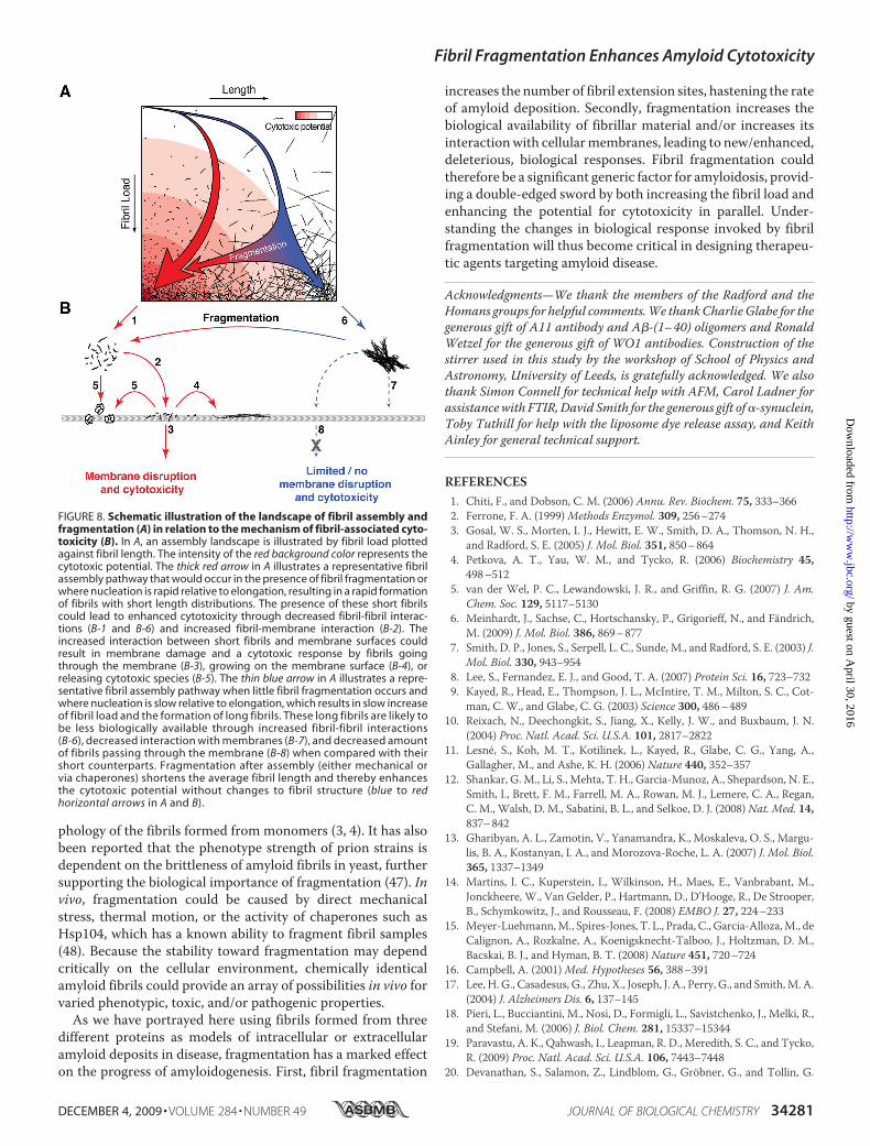
FIGURE 8. Schematic illustration of the landscape of fibril assembly andfragmentation (A) in relation to the mechanism of fibril-associated cyto-toxicity (B). In A, an assembly landscape is illustrated by fibril load plottedagainst fibril length. The intensity of the red background color represents thecytotoxic potential. The thick red arrow in A illustrates a representative fibrilassembly pathway that would occur in the presence of fibril fragmentation orwhere nucleation is rapid relative to elongation, resulting in a rapid formationof fibrils with short length distributions. The presence of these short fibrilscould lead to enhanced cytotoxicity through decreased fibril-fibril interac-tions (B-1 and B-6) and increased fibril-membrane interaction (B-2). Theincreased interaction between short fibrils and membrane surfaces couldresult in membrane damage and a cytotoxic response by fibrils goingthrough the membrane (B-3), growing on the membrane surface (B-4), orreleasing cytotoxic species (B-5). The thin blue arrow in A illustrates a repre-sentative fibril assembly pathway when little fibril fragmentation occurs andwhere nucleation is slow relative to elongation, which results in slow increaseof fibril load and the formation of long fibrils. These long fibrils are likely tobe less biologically available through increased fibril-fibril interactions(B-6), decreased interaction with membranes (B-7), and decreased amountof fibrils passing through the membrane (B-8) when compared with theirshort counterparts. Fragmentation after assembly (either mechanical orvia chaperones) shortens the average fibril length and thereby enhancesthe cytotoxic potential without changes to fibril structure (blue to redhorizontal arrows in A and B).
Fibril Fragmentation Enhances Amyloid Cytotoxicity
DECEMBER 4, 2009 • VOLUME 284 • NUMBER 49 JOURNAL OF BIOLOGICAL CHEMISTRY 34281
by guest on April 30, 2016
http://ww
w.jbc.org/
Dow
nloaded from

(2006) FEBS J. 273, 1389–140221. Engel, M. F., Khemtemourian, L., Kleijer, C. C., Meeldijk, H. J., Jacobs, J.,
Verkleij, A. J., de Kruijff, B., Killian, J. A., and Hoppener, J. W. (2008) Proc.Natl. Acad. Sci. U.S.A. 105, 6033–6038
22. Colvin, V. L. (2003) Nat. Biotechnol. 21, 1166–117023. Lynch, I., Dawson, K. A., and Linse, S. (2006) Sci. STKE 2006, pe1424. Kad, N. M., Thomson, N. H., Smith, D. P., Smith, D. A., and Radford, S. E.
(2001) J. Mol. Biol. 313, 559–57125. Smith, A. M., Jahn, T. R., Ashcroft, A. E., and Radford, S. E. (2006) J. Mol.
Biol. 364, 9–1926. Cappai, R., Leck, S. L., Tew, D. J., Williamson, N. A., Smith, D. P., Galatis,
D., Sharples, R. A., Curtain, C. C., Ali, F. E., Cherny, R. A., Culvenor, J. G.,Bottomley, S. P., Masters, C. L., Barnham, K. J., and Hill, A. F. (2005)FASEB J. 19, 1377–1379
27. Xue,W. F., Homans, S. W., and Radford, S. E. (2009) Protein Eng. Des. Sel.22, 489–496
28. Press,W. H. (2002)Numerical Recipes in C��: The Art of Scientific Com-puting, 2nd Ed., pp. 666–670, Cambridge University Press, Cambridge,UK
29. Xue,W. F., Homans, S.W., and Radford, S. E. (2008) Proc. Natl. Acad. Sci.U.S.A. 105, 8926–8931
30. Knight, J. D., and Miranker, A. D. (2004) J. Mol. Biol. 341, 1175–118731. O’Nuallain, B., and Wetzel, R. (2002) Proc. Natl. Acad. Sci. U.S.A. 99,
1485–149032. Fabian, H., Gast, K., Laue, M., Misselwitz, R., Uchanska-Ziegler, B.,
Ziegler, A., and Naumann, D. (2008) Biochemistry 47, 6895–690633. Lee, Y. H., Chatani, E., Sasahara, K., Naiki, H., and Goto, Y. (2009) J. Biol.
Chem. 284, 2169–217534. Glabe, C. G. (2006) Neurobiol. Aging 27, 570–575
35. Giorgetti, S., Raimondi, S., Cassinelli, S., Bucciantini, M., Stefani, M., Gre-gorini, G., Albonico, G., Moratti, R., Montagna, G., Stoppini, M., andBellotti, V. (2009) Nephrol. Dial. Transplant. 24, 1176–1181
36. Hirakura, Y., and Kagan, B. L. (2001) Amyloid 8, 94–10037. Mosmann, T. (1983) J. Immunol. Methods 65, 55–6338. Jahn, T. R., Tennent, G. A., and Radford, S. E. (2008) J. Biol. Chem. 283,
17279–1728639. Cedervall, T., Lynch, I., Lindman, S., Berggård, T., Thulin, E., Nilsson, H.,
Dawson, K. A., and Linse, S. (2007) Proc. Natl. Acad. Sci. U.S.A. 104,2050–2055
40. Linse, S., Cabaleiro-Lago, C., Xue,W. F., Lynch, I., Lindman, S., Thulin, E.,Radford, S. E., and Dawson, K. A. (2007) Proc. Natl. Acad. Sci. U.S.A. 104,8691–8696
41. Naslund, J., Haroutunian, V., Mohs, R., Davis, K. L., Davies, P., Greengard,P., and Buxbaum, J. D. (2000) JAMA 283, 1571–1577
42. Friedman, R., Pellarin, R., and Caflisch, A. (2009) J. Mol. Biol. 387,407–415
43. Carulla, N., Caddy, G. L., Hall, D. R., Zurdo, J., Gairí, M., Feliz, M., Giralt,E., Robinson, C. V., and Dobson, C. M. (2005) Nature 436, 554–558
44. Morten, I. J., Gosal, W. S., Radford, S. E., and Hewitt, E. W. (2007) J. Biol.Chem. 282, 29691–29700
45. Collins, S. R., Douglass, A., Vale, R. D., and Weissman, J. S. (2004) PLoSBiol. 2, e321
46. Smith, J. F., Knowles, T. P., Dobson, C. M., Macphee, C. E., and Welland,M. E. (2006) Proc. Natl. Acad. Sci. U.S.A. 103, 15806–15811
47. Tanaka, M., Collins, S. R., Toyama, B. H., and Weissman, J. S. (2006)Nature 442, 585–589
48. Shorter, J., and Lindquist, S. (2004) Science 304, 1793–1797
Fibril Fragmentation Enhances Amyloid Cytotoxicity
34282 JOURNAL OF BIOLOGICAL CHEMISTRY VOLUME 284 • NUMBER 49 • DECEMBER 4, 2009
by guest on April 30, 2016
http://ww
w.jbc.org/
Dow
nloaded from

Hewitt and Sheena E. RadfordWei-Feng Xue, Andrew L. Hellewell, Walraj S. Gosal, Steve W. Homans, Eric W.
Fibril Fragmentation Enhances Amyloid Cytotoxicity
doi: 10.1074/jbc.M109.049809 originally published online October 6, 20092009, 284:34272-34282.J. Biol. Chem.
10.1074/jbc.M109.049809Access the most updated version of this article at doi:
Alerts:
When a correction for this article is posted•
When this article is cited•
to choose from all of JBC's e-mail alertsClick here
Supplemental material:
http://www.jbc.org/content/suppl/2009/10/06/M109.049809.DC1.html
http://www.jbc.org/content/suppl/2009/11/24/284.49.34272.DC1.htmlRead an Author Profile for this article at
http://www.jbc.org/content/284/49/34272.full.html#ref-list-1
This article cites 47 references, 18 of which can be accessed free at
by guest on April 30, 2016
http://ww
w.jbc.org/
Dow
nloaded from
Copyright © 2022 FDOKUMEN