The impact of protein disulfide bonds on the amyloid fibril morphology

Journal of Alzheimer’s Disease 37 (2013) 9–18DOI 10.3233/JAD-122389IOS Press
9
Modification of Amyloid-�1-42 FibrilStructure by Methionine-35 Oxidation
Liming Houa,1, Hyoung-gon Leeb,1, Fang Hanc, Johnathan M. Tedescod, George Perryb,e,Mark A. Smithb,2 and Michael G. Zagorskic,∗aAbbott Vascular Inc., Menlo Park, CA, USAbDepartment of Pathology, Case Western Reserve University, Cleveland, OH, USAcDepartment of Chemistry, Case Western Reserve University, Cleveland, OH, USAdDepartment of Chemistry, Lake Erie College, Painesville, OH, USAeCollege of Sciences, University of Texas at San Antonio, San Antonio, TX, USA
Handling Editor: Jesus Avila
Accepted 23 April 2013
Abstract. Oxidative stress and amyloid-� (A�) formation are important processes that occur in Alzheimer’s disease (AD).Amyloid formation is associated with the aggregation and precipitation of the A� peptide, while oxidative stress results from animbalance in pro-oxidant/antioxidant homeostasis that produces harmful reactive oxygen species. The methionine-35 (Met35)residue of the A� peptide plays an important role in AD oxidative stress events and the associated neurotoxicity. We andother research groups previously demonstrated that in vitro oxidation of the Met35 side-chain to the sulfoxide (Met35red −→Met35ox) impedes assembly and aggregation of monomeric A� peptide into protofibrils, the latter being the immediate precursorsof amyloid plaques. Here, we report that Met35 oxidation state affects the stability of preexisting amyloid fibrils and plaques,where the Met35red −→ Met35ox process leads to changes in the morphology of filaments, protofibrils, mature fibrils, and lossof Congo red birefringence in senile plaques isolated from the brains of AD patients. The most notable differences were in fibrilflexibility, as evidenced by changes from straight fibrils to irregularly shaped, rope-like fibrils. These findings suggest that theMet35 oxidation state and amyloid plaque formation may be intimately linked.
Keywords: Alzheimer’s disease, amyloid-�, atomic force microscopy, fibrillization, oxidative stress
INTRODUCTION
The neuropathological characteristics of Alzhei-mer’s disease (AD) are the presence of intraneu-ronal neurofibrillary tangles and extracellular amyloidplaques [1–3]. The amyloid-� (A�) peptide consti-tutes the major protein component of the amyloidplaques and, in vitro, is an extremely sensitive system
1These authors contributed equally to this work.2Deceased.∗Correspondence to: Michael G. Zagorski, Ph.D., Department
of Chemistry, Case Western Reserve University, Cleveland, Ohio44106-7078, USA. Tel.: +1 216 368 3706; Fax: +1 216 368 3006;E-mail: [email protected].
that undergoes environment dependent aggregation oroligomerization [4, 5]. During A� oligomerization,soluble �-sheet aggregates produce protofibrils, whichcoalesce forming larger fibrils and eventually amyloidplaques with distinct tinctorial and structural properties[6–9].
Numerous studies established that A� becomes neu-rotoxic to neuronal cells when aggregated as �-sheetstructures, and in the human brain, neuritic changestake place during both the early and late stages ofA� aggregation and plaque formation [10–13]. Thus,the majority of AD therapeutics in clinical trials aredesigned to prevent the assembly of non-aggregated(monomeric) A� into smaller oligomers and eventuallyamyloid fibrils and plaques [14, 15].
ISSN 1387-2877/13/$27.50 © 2013 – IOS Press and the authors. All rights reserved
10 L. Hou et al. / Oxidation Dissipates A�1-42 Aggregates
Oxidative stress plays an important role in the patho-genesis of AD [16–20] and includes oxidative damageand modification of biological macromolecules such asproteins, carbohydrates, lipids, and nucleic acids. Dueto high oxygen consumption, the human brain is par-ticularly sensitive to the generation of reactive oxygenspecies (ROS) such as H2O2, and increased oxidativestress and H2O2 production has been implicated inthe neurotoxicity of AD [21]. Many studies supportthe view that oxidative stress and amyloid formationare interconnected [16–20], including a study involv-ing doubly transgenic mice that overexpress mutatedhuman amyloid-� protein precursor and do not expressthe manganese superoxide dismutase protein (a scav-enger of ROS). In these mice, there was a dramaticincrease in brain A� levels and A� plaque burden [22].
For proteins, oxidative damage involves numerousposttranslational modifications, such as the oxidationof methionine (Met) side chains to the sulfoxides.Because this represents an early oxidative event andoccurs easily under physiological conditions, it isthought that the Met side chains may function asendogenous antioxidants [23, 24]. Oxidized Met35comprises 10–50% of the total brain A� in postmortemAD plaques [25], and thus may constitute an AD oxida-tive stress event. Interestingly, in vitro oxidation ofthe Met35 side chain to the sulfoxide (Met35red −→Met35ox) significantly retards �-amyloidosis of the 40-and 42-residue A�1-40 and A�1-42 peptides [26–29],and likewise abolishes the neurotoxicity of the unoxi-dized peptides [30].
To date, no studies have addressed the consequencesof the Met35 oxidation state on the stability and struc-ture of preformed A� fibrils. Here, using atomic forcemicroscopy (AFM), electron microscopy (EM), andCongo red staining, we demonstrate that the Met35red
−→ Met35ox process exerts profound effects on thestructures of A� protofibrils and mature fibrils pre-pared in vitro, as well as with purified A� plaquecores isolated from brains of AD patients. The impli-cations of these findings to the pathogenesis of AD arediscussed.
MATERIALS AND METHODS
Materials
The A�1-42 peptide (H3N+-D1-A-E-F-R5-H-D-S-G-Y10-E-V-H-H-Q15-K-L-V-F-F20-A-E-D-V-G25-S-N-K-G-A30-I-I-G-L-M35-V-G-G-V-V40-I-A42-COO−)was synthesized and purified as described previously[29]. Peptide identity was verified by mass spectrome-
try and nuclear magnetic resonance spectroscopy. TheH2O2 was obtained as a 30% commercial solution(Fisher Scientific), and was diluted with water (highperformance liquid chromatography (HPLC) grade,0.2 �m filtered) to the required concentrations. Withthe exception of sample buffers (2×) (Invitrogen),Novex® Tricine Gels (10–20%), tricine SDS run-ning (10×), the prestained SDS-PAGE standards(broad range, MW 6,700 to 208,000 daltons), andthe Coomassie Blue G250 were all purchased fromBio-Rad Laboratories.
Aβ1-42 sample preparation and oxidation
Lyophilized A�1-42 peptide (1.8 mg, 40 �mol) wasdisaggregated by thorough dissolution with sonication(1 min) in dilute NaOH solution (1.9 ml, 10 mM) [31].This solution was combined with cold (5◦C) potas-sium phosphate buffer solution (6.1 ml, 10 mM, pH7.3) to yield a stock solution (8 ml) with a peptideconcentration of 50 �M. The pH of the stock solu-tion was checked and carefully adjusted (if needed) topH 7.3 with dilute NaOH or trifluoroacetic acid solu-tions. The solution was either briefly stored at 5◦ (forless than 1 h) to prevent aggregation, or aged at roomtemperature (∼ 22◦) to encourage aggregation. Dur-ing the aging process, aliquots (1 ml) were removedat 5, 24, 48, and 240 h, and then divided into twoequally sized fractions. One fraction was immediatelyexamined by AFM or EM, while the other was mixedwith dilute H2O2 (6 �l, 7.5% by weight), then allowedto stand at room temperature (3 h) to complete theMet35red −→ Met35ox reaction [26, 32]. After 3 h,the A�1-42Met35ox fraction was examined by AFMor EM. Control experiments established that the H2O2promoted only Met35 oxidation to the sulfoxide [26]and did not promote other possible side-oxidationssuch as to the sulfone or formation of a dityrosineadduct [33, 34].
Atomic force microscopy
The images were generated on a Multimode AFMinstrument equipped with a Nanoscan III controller(Digital Instruments) and an “E” type scanner withxy range 12.5 �m. Imaging was done at 22◦C underthe air-tapping mode with commercially available sili-con NanoProbes (Digital Instruments). In general, thescanning parameters were as follows: 2 V initial root-mean square amplitude, set point at 80% of the freeamplitude, 250–350 kHz tapping frequency, 2 Hz scanrate, and 2 �m sizes (512 × 512 pixel resolution).
L. Hou et al. / Oxidation Dissipates A�1-42 Aggregates 11
Sample preparation on freshly cleaved mica or graphitefollowed previous AFM methods developed for theA� peptides [35], which included diluting the A�1-42aliquots (5–10 �l) with an equal volume of water(HPLC grade, 0.2 �m filtered) immediately beforeapplication to the mica or graphite as required to pro-duce a monolayer. This dilution procedure requiredless than 30 s and did not affect the imaging results [36].Imaging on mica or graphite produced the same results,establishing that the new structures are independent ofhydrophilic or hydrophobic surface interactions [37].
Electron microscopy
Aliquots from the A�1-42 solutions were appliedto Formvar/Carbon coated nickel 400-mesh grids,allowed to settle for approximately 1-2 min, andswabbed gently with KimWipes®. Next, they werestained with 2% uranyl acetate and examined with aJEOL 1200EX electron microscope (SupplementaryFigure 1).
Electrophoretic analysis by SDS-PAGE
A disaggregated A�1-42 stock solution (100 �M)was prepared as described above and allowed to age at22◦C. After 5, 24, and 72 h of aging, fractions (0.2 ml)were removed and split into two equally sized por-tions. One portion was immediately frozen and storedat –80◦C, while the other was mixed with H2O2 (1.5 �l,3%) and aged for 6 h at 22◦C. This latter sample, alongwith the sample stored at –80◦C, were examined bySDS-PAGE, by thoroughly mixing 15 �l aliquots withan equal volume of Novex® Tricine SDS sample buffer,then heating (85◦C, 2 min) and separation with Novex®
Tricine Gels (10–20%) (15 �l per well). Control stud-ies established that the repeated freezing and thawingdid not perturb the A� aggregation state seen by SDS-PAGE, nor the fibril morphology seen by AFM.
Aβ plaque core isolation and Congo red staining
Using modified protocols developed in our lab [38],A� plaque cores were isolated from the cortical graymatter of patients with pathologically confirmed AD(n = 3; 76, 81, and 94 years old). After extraction andpurification, the medium-sized particles (5–35 �m)were separated from the smaller- and larger-sized par-ticles using a fluorescence-activated cell sorter. Themedium-sized particles were pooled and partitionedinto two fractions based on their ability to displaybright or dull auto-fluorescence. One fraction was
mixed with dilute H2O2 (6 �l, 7.5% by weight), thenallowed to stand at room temperature (3 h) to completeMet35ox. Both fractions were analyzed immunocy-tochemically for A� content, which involved dryingon a glass microscope slide, immunostaining usingmonoclonal antibody 498, and developing by standardperoxidase and anti-peroxidase methods [38]. To mea-sure birefringence, aliquots of the fractions were driedon a glass slide, stained with Congo red, and viewedunder cross-polarized illumination.
RESULTS
Influence of the Met35 oxidation state on the Aβ
fibril morphology
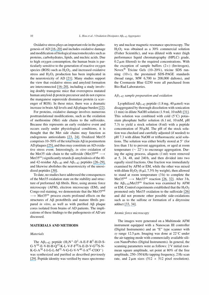
The A�1-42Met35red −→ A�1-42Met35ox conver-sion was performed in a systematic manner (Scheme1), and the fibrils were examined by AFM, a well-accepted tool for analysis of amyloid fibril morphology[34, 39–43]. To produce the fibrils, monomericA�1-42Met35red solutions (50 �M, pH 7.3) were incu-bated (aged) at room temperature, during which smallsized oligomers combine to form filaments, protofib-rils, and eventually large insoluble mature fibrils. Themonomer −→ soluble oligomer −→ filament −→protofibril −→ mature fibril is a generally recognizedpathway associated with plaque formation of the A�and other amyloid forming proteins [44–47].
Aliquots of an A�1-42Met35red stock solution wereremoved after 5, 24, 48, and 240 h of aging and par-titioned into two equally-sized fractions (Scheme 1).
Scheme 1. Protocol for monitoring the effect of Met35 oxidation onthe morphology of A�1-42 (50 �M, 22◦C, pH 7.3) fibril formation.
12 L. Hou et al. / Oxidation Dissipates A�1-42 Aggregates
A D G
B E H
FC I
400 nm
400 nm 400 nm
400 nm 200 nm
200 nm
200 nm400 nm400 nm
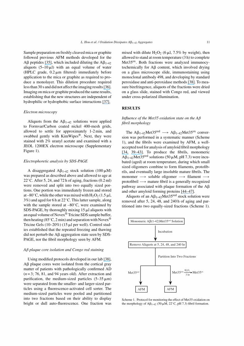
Fig. 1. Representative AFM images of A�1-42Met35red (50 �M, pH 7.3) aged for (A) 5 h, (B) 24 h, and (C) 48 h at 22◦C and after the treatmentwith H2O2 for 3 h (D, E, and F, respectively; image sizes 2 �m2 and Z range 10 nm). Off-line zoomed images (G, H, and I) of A�1-42Met35ox
(image sizes 1 �m2 and Z range 2 nm) that correspond to the boxed-in regions of D, E, and F, respectively.
One fraction was immediately examined by AFM,while the other was mixed with H2O2 to promote theMet35red −→ Met35ox conversion before inspectionby AFM. Mass spectrometric analysis of a week oldA�1-42 and H2O2 solution demonstrated that only theMet35 is affected and becomes oxidized to the sulfox-ide [26], while oxidation to the sulfone or formationof a dityrosine adduct did not occur [33, 34]. Thesemass spectrometry results were confirmed by NMR,in that oxidation does not produce any Met35 sulfone(Supplementary Figure 2). Because the fibril morphol-ogy is sensitive to environmental variables, such asthe A� starting aggregation state, peptide concentra-tion, ionic strength, and temperature [36, 48], single,freshly prepared A�1-42 stock solutions were usedfor all experiments. Thus, any differences betweenthe A�1-42Met35red and A�1-42Met35ox fibrils resultsfrom only the Met35 oxidation state.
After aging for 5 h, the A�1-42Met35red sampleshowed mostly non-fibrillar aggregates with vary-ing shapes (3–4 nm height) (Fig. 1A) that resembledthe individual domains of the protofibrils (Fig. 1B).Thin filaments with no clear periodicity (the shortand faint features in Fig. 1A) were uniformly dis-persed on the mica surface (0.60 ± 0.07 nm height),and constitute early-formed deposits that are the build-ing blocks for the larger protofibrils and mature fibrils[46, 49]. At 24 h, protofibrils and mature fibrils werethe predominant motifs (Fig. 1B). The protofibrils
had diameters and periodicities consistent with pre-viously reported dimensions (3–4 nm average heightor diameter, 20–22 nm periodicity), while the maturefibrils were larger and appeared as two intertwinedprotofibrils [36, 41, 45, 48]. Large bundles or largermature fibrils (12.6 ± 1.0 nm height) were seen after48 h (Fig. 1C), while the amounts of protofibrils andmature fibrils decreased. These results are consistentwith an on-pathway mechanism of A� aggregation andfibril formation.
The AFM images of the A�1-42Met35ox sam-ples were markedly different from those of theA�1-42Met35red, in that there were no well-definedstructures. The heights were much smaller (lessthan 0.8 nm) and the characteristic filament, protofib-ril, and mature fibril morphologies were all absent(Fig. 1D-F). Expanded images revealed variationswith aging (Fig. 1G-I), where the 5 h sample con-tained compactly associated small spheres (0.6–0.8 nmheight) (Fig. 1G), while the 24 h sample containedbright rod-shaped aggregates and globe-like structures(0.6–0.8 nm height) (Fig. 1H) similar to nonfibrillarsheet-like structures seen earlier by EM [50]. The 48 hsample had densely distributed thin filaments (Fig. 1I),comparable in dimensions (0.4–0.6 nm height) to thoseformed during the early stages of aggregation beforeMet35 oxidation.
Nearly identical results were obtained by EM (Sup-plementary Figure 1), where aging of A�1-42Met35red

L. Hou et al. / Oxidation Dissipates A�1-42 Aggregates 13
500 nm
300
100
C D E
HGF
BA
2 µm
400 nm 400 nm 400 nm
200 nm 200 nm
400 nm
Fig. 2. High-resolution AFM images of A�1-42Met35red (50 �M, pH 7.3) aged for 240 h, with the full image (A) (image size 10 �m2 and Zrange 10 nm) and the zoomed image (B) that corresponds to the boxed-in region of (A) (image size 2 �m2 and Z range 10 nm). Additionalimages after allowing the 240 h sample to mix with H2O2 for (C) 3 h, (D) 5 h, and (E) 6 h (image size 2 �m2 and Z range 10 nm). F, G, and Hare the off-line zoomed images taken from the boxed-regions of C, D, and E, respectively. Image F is presented in a different rotation (245◦)and pitch (45◦) angles (image size 0.5 �m2 and Z range 2 nm), while the image sizes of G and H are 1 �m2 and Z range 2 nm.
(before addition of H2O2) at 24, 48, and 96 h pro-duced non-fibrillar aggregates, protofibrils, and maturefibrils, respectively. The heights and lengths were con-sistent with those seen by AFM. After addition ofH2O2, the protofibrils and mature fibrils disappearedgiving globular-like and filament-like structures withsignificantly reduced heights and lengths.
The effect of the A�1-42Met35red −→ A�1-42Met35ox process was explored in greater detail withmore compact mature fibrils, which are the pre-dominant (in vitro) structural motifs formed afterextended incubation times, as well as the predomi-nate (in vivo) fibrils in human brains. These fibrilscontain highly aggregated A� peptide and are veryresistant to standard proteolytic digestion and SDSdenaturation. Shown in Figures 2A and 2B are maturefibrils from A�1-42Met35red samples aged for 240 h.After 3 h incubation with H2O2, the characteristicthin, strand-like mature fibrils (12.6 ± 1.0 nm height)changed to more corpulent, rope-like structures withreduced heights (3–4 nm) and no defined periodici-
ties along the axis (Fig. 2C). These novel structuresare more clearly seen in the high magnification image(Fig. 2F), which shows a fibril at different rotation(245°) and pitch (45°) angles. After 5-h incubationwith H2O2, the rope-like structures were absent andthe major products were thin filaments (0.4–0.6 nmheight) (Figs. 2D, G) that had dimensions similar tothe filaments of the A�1-42Met35red (Fig. 1I). Largeramorphous aggregates were also detected (3–7 nmheight), which could perhaps be intermediates pro-duced during the conversion of the rope-like structuresto the filaments. After 6 h, the fibril or filaments becameglobular-like (0.6–0.8 nm height) (Figs. 2E, H), andwere comparable to those seen during aging of theA�1-42Met35red (Figs. 1D, G). Imaging on mica orgraphite generated identical results (Fig. 3), estab-lishing that the new structures are not dependent onhydrophilic or hydrophobic surface interactions [37].The globular structures were also compactly associatedand appeared like sticky layers on the mica or graphiteimaging surfaces.

14 L. Hou et al. / Oxidation Dissipates A�1-42 Aggregates
A B
Fig. 3. AFM images of A�1-42Met35red (50 �m, pH 7.3) aged for 240 h and treated with H2O2 for 3 h. A) Image was taken on the surface ofmica, while (B) image was taken on the surface of graphite (image sizes 10 �m2 and Z range 10 nm).
Effect of the Met35 oxidation state on Aβ
aggregation
The aggregation state during the A�1-42Met35red
−→ A�1-42Met35ox process was ascertained bySDS-PAGE. Aliquots were removed from anA�1-42Met35red solution after 5, 24, and 72 h ofaging, then partitioned into two fractions (one treatedwith H2O2) and examined by SDS-PAGE. Figure 4shows the SDS-PAGE of the samples with and withoutMet35 oxidation. SDS-stable A� oligomers withmolecular weights ranging from 8 to 20 kDa weredetected in the three unoxidized samples, similar inmass to those seen in conditioned media of transfectedcells [51] and AD brains [52]. By contrast, oligomerswere not detected with the aged oxidized samples,thus the A�1-42Met35red −→ A�1-42Met35ox processremoves the SDS stable aggregates.
Fig. 4. SDS-PAGE of A�1-42 aggregates before and after H2O2 oxi-dation. The A�1-42Met35red (100 �M, pH 7.3) was incubated at 5,24, and 72 h (lanes A, C, and E, respectively, labeled with H2O2 ‘-’).Portions of the 5, 24, and 72 h aged samples were incubated withH2O2 (6 h) (lines B, D, and F, respectively, labeled with H2O2 ‘+’).Molecular mass standards are indicated in the lane on the left.
Effect of the Met35 oxidation state on brainamyloid plaque cores
We further explored oxidation of A� depositsformed in vivo. Isolated A� plaque cores from ADpatients [38] were treated with either buffer alone,or 1.5% H2O2 in buffer for 20 h, followed by treat-ment with Congo red to visualize the A�. As expected,the characteristic birefringence signal from Congo redbound to the A� was seen in the buffer alone sam-ples (Fig. 5A), demonstrating that the majority of theisolated plaque cores contain fibrillized A�. To con-firm this interpretation, the cores were first treated withformic acid (a well-known reagent that converts fibril-lized A� to monomeric A� and does not bind Congored), and inevitably the Congo red positive signals weregreatly diminished (Fig. 5B). The plaque core samplestreated with H2O2 in buffer had no detectable birefrin-gence signals (Fig. 5C), suggesting that the Met35red
−→ Met35ox conversion in human plaques alters theA� fibril structure, consistent with the above in vitrofindings.
DISCUSSION
Relationship between the Aβ structure and Met35oxidation state
The present data demonstrate that the Met35red −→Met35ox conversion disrupts critical structural interac-tions that keep together soluble oligomers, filaments,protofibrils, and mature fibrils, all of which presum-ably form during A� amyloid formation [44–47].

L. Hou et al. / Oxidation Dissipates A�1-42 Aggregates 15
A B C
Fig. 5. H2O2 treatment causes a conformational change of isolated amyloid plaques from AD brain samples. Birefringence by Congo red staindemonstrates fibrillized A� protein is present in isolated amyloid plaques (arrows). After treatment with 35% formic acid (B) and 1.5% H2O2(C), most of the birefringence disappears compared with buffer treated isolated plaques (A).
More importantly, the Met35 oxidation promotes mor-phological changes of plaques from human brains,indicating that the stabilizing interactions of in vitroand in vivo fibril structures are comparable [53, 54].The most notable changes were in fibril flexibility, asevidenced by changes from straight fibrils to irregu-larly shaped, rope-like structures that resemble neitheramyloid fibrils nor any precursor structure. Addition-ally, the inability to detect Met35ox fibrils by SDS gelindicates that they are less stable and more prone todenaturation.
The present results compliment previous workwhere oxidation of the Met35 side chain retards �-aggregation of both the 40- and 42-residue A�1-40and A�1-42 peptides [26–29]. Methionine oxidationmodulates fibril formation and �-aggregation of otheramyloid forming proteins, including transthyretin [55],�-synuclein [56], prion [57], and apolipoprotein C[58]. However, the effect of the Met35 oxidation isexceptional, given that single point mutations do notdestabilize the A� fibril structure [59], apart frommutations in the central hydrophobic cluster (Leu17-Val18-Phe19-Phe20-Ala21) [60, 61].
Several structural models demonstrate that the loca-tions of the Met35red and Met35ox side chains aredifferent and can vary with the aggregation state [62].The NMR solution structure of monomeric A� boundto an engineered affibody protein has the side chainsolvent exposed [63], while NMR-derived models ofearly-formed aggregates (preglobulomer and globu-lomers) [64] and solid state NMR data of the amyloidfibrils has the Met35red packed in a hydrophobic inte-rior [65, 66]. Methionine oxidation promotes increasedbond polarity and dipole moment, plus allows the sul-fur atom to act as a hydrogen bond acceptor [28], allof which can weaken hydrophobic and external quater-nary contacts. When folded as a monomeric �-helical
structure, the Met35 oxidation caused the side chain tobecome repositioned and move from a hydrophobicinterior to an exterior location where it can hydro-gen bond with water [32]. Because �-aggregation islargely driven by hydrophobic interactions, it is pos-sible that the more polar Met35ox favors inclusion ofwater molecules near the hydrophobic interior [67],which in turn breaks apart hydrophobic interactionssuch as the intermolecular Met35-Met35 hydrophobiccontacts among the �-sheets of the fibrils [68]. A simi-lar effect was seen with the apolipoprotein C-II, whereoxidation of a methionine residue in the hydrophobiccore inhibited amyloid fibril assembly [58].
Relevance of the AβMet35 oxidation state to agingand AD
Although the Met35 oxidation state plays a criticalrole in A� aggregation [26–29], controversy stillexists about its role with neurotoxicity. Earlier reportsindicated that Met35 oxidation leads to increasedneurotoxicity, possibly through different pathwaysthan A� containing Met35red [69, 70]. However,subsequent studies using carefully disaggregated A�showed that the Met35ox peptides completely lackedtoxicity in cortical neuronal cultures, opposite tothat seen with the Met35red peptides [30, 71]. It wasthought the Met35 oxidation prevents formation ofsoluble oligomers that are purportedly responsible formost of the A�-induced neurotoxicity. Thus, theseresults together with more recent work [72], suggeststhe A�1-42Met35red −→ A�1-42Met35ox processdoes not promote neurotoxicity.
The oxidation of methionine residues in proteinsplays an important role under physiological conditionsof oxidative stress [73], which constitutes an earlyevent in AD, proximal to the development of hallmark

16 L. Hou et al. / Oxidation Dissipates A�1-42 Aggregates
pathologies [74–76]. Due to its relatively high oxy-gen consumption, generation of ROS such as H2O2is common in the human brain, and increased oxida-tive stress and H2O2 production has been implicatedin the neurotoxicity of AD [21]. It has been suggestedthat monomeric A� may be neuroprotective [77, 78]and prevent copper-, iron-, or zinc-induced neurotoxi-city and related oxidative stress events associated withAD [79, 80], with the Met35 residue playing a criticalrole [81]. Notably, oxidized protein methionines can bereversed in vivo by the action of methionine sulfoxidereductase [82]. Knockout mice lacking the methion-ine sulfoxide reductase gene have elevated levels ofbrain pathologies associated with neurodegenerativediseases [83], plus in humans a decrease in methion-ine sulfoxide reductase activity is associated with thedevelopment of AD [84].
Numerous studies suggest that A� aggregation andamyloid formation are important pathological events inAD. Accordingly, therapeutic strategies have focusedon inhibiting A� aggregation, lowering soluble lev-els, and disassembling existing amyloid plaques [15].Although progress has been made on several fronts,particularly with active and passive A� immunizationtrials [85], numerous obstacles remain and alterna-tive approaches are always sought. Current treatmentfor AD patients involves administration of acetyl-cholinesterase inhibitors, which for some patientsimprove cognition during the first year of use, but donot reduce the amyloid burden or many of the AD-related neuropsychiatric symptoms [86].
ACKNOWLEDGMENTS
Work in the authors’ laboratories is supported bythe National Institutes of Health (NIH R01 AG026151(MAS) and NIH R01 AG027853 (MGZ).
Authors’ disclosures available online (http://www.j-alz.com/disclosures/view.php?id=1769).
SUPPLEMENTARY MATERIAL
Supplementary material is available here: http://dx.doi.org/10.3233/JAD-122389
REFERENCES
[1] Smith MA (1998) Alzheimer disease. Int Rev Neurobiol 42,1-54.
[2] Selkoe DJ (2001) Alzheimer’s disease: Genes, proteins, andtherapy. Physiol Rev 81, 741-766.
[3] Shah RS, Lee HG, Xiongwei Z, Perry G, Smith MA,Castellani RJ (2008) Current approaches in the treatment ofAlzheimer’s disease. Biomed Pharmacother 62, 199-207.
[4] Zagorski MG, Yang J, Shao H, Ma K, Zeng H, Hong A (1999)Methodological and chemical factors affecting amyloid �peptide amyloidogenicity. In Amyloid, Prions, and Other Pro-tein Aggregates, Wetzel R, ed. Academic Press, New York, pp.189-204.
[5] Bitan G, Teplow DB (2005) Preparation of aggregate-free,low molecular weight amyloid-� for assembly and toxicityassays. Methods Mol Biol 299, 3-9.
[6] Inouye H, Kirschner D (1996) Refined fibril structures: Thehydrophobic core in Alzheimer’s �-amyloid and prion asrevealed by X-ray diffraction. In The Nature and Origin ofAmyloid Fibrils, Bock GR, Goode JA, eds. John Wiley &Sons, New York, NY, pp. 22-39.
[7] Harper JD, Wong SS, Lieber CM, Lansbury PT (1997) Obser-vation of metastable A� amyloid protofibrils by atomic forcemicroscopy. Chem Biol 4, 119-125.
[8] Tycko R (2006) Characterization of amyloid structures at themolecular level by solid state nuclear magnetic resonancespectroscopy. Methods Enzymol 413, 103-122.
[9] Kodali R, Wetzel R (2007) Polymorphism in the intermediatesand products of amyloid assembly. Curr Opin Struct Biol 17,48-57.
[10] Simmons LK, May PC, Tomaselli KJ, Rydel RE, Fuson KS,Brigham EF, Wright S, Lieberburg I, Becker GW, BremsDN, Li W (1994) Secondary structure of �-amyloid pep-tide correlates with toxicity in vitro. Mol Pharmacol 45, 373-379.
[11] Yamamoto M, Kiyota T, Walsh SM, Ikezu T (2007) Kineticanalysis of aggregated amyloid-� peptide clearance in adultbone-marrow-derived macrophages from APP and CCL2transgenic mice. J Neuroimmune Pharmacol 2, 213-221.
[12] Selkoe DJ (2008) Soluble oligomers of the amyloid �-proteinimpair synaptic plasticity and behavior. Behav Brain Res 192,106-113.
[13] Meyer-Luehmann M, Spires-Jones TL, Prada C, Garcia-Alloza M, de Calignon A, Rozkalne A, Koenigsknecht-TalbooJ, Holtzman DM, Bacskai BJ, Hyman BT (2008) Rapidappearance and local toxicity of amyloid-� plaques in a mousemodel of Alzheimer’s disease. Nature 451, 720-724.
[14] Ono K, Naiki H, Yamada M (2006) The development of pre-ventives and therapeutics for Alzheimer’s disease that inhibitthe formation of �-amyloid fibrils (fA�), as well as destabilizepreformed fA�. Curr Pharm Des 12, 4357-4375.
[15] Barten DM, Albright CF (2008) Therapeutic strategies forAlzheimer’s disease. Mol Neurobiol 37, 171-186.
[16] Smith MA, Sayre LM, Monnier VM, Perry G (1995) RadicalAGEing in Alzhimer’s disease. Trends Neurosci 18, 172-176.
[17] Markesbery WR (1999) The role of oxidative stress inAlzheimer disease. Arch Neurol 56, 1449-1452.
[18] Sayre LM, Smith MA, Perry G (2001) Chemistry and bio-chemistry of oxidative stress in neurodegenerative disease.Curr Med Chem 8, 721-738.
[19] Nunomura A, Castellani RJ, Zhu X, Moreira PI, PerryG, Smith MA (2006) Involvement of oxidative stress inAlzheimer disease. J Neuropathol Exp Neurol 65, 631-641.
[20] Robinson RA, Lange MB, Sultana R, Galvan V, FombonneJ, Gorostiza O, Zhang J, Warrier G, Cai J, Pierce WM, Bre-desen DE, Butterfield DA (2011) Differential expression andredox proteomics analyses of an Alzheimer disease trans-genic mouse model: Effects of the amyloid-beta peptideof amyloid precursor protein (Xi). Neuroscience 177, 207-222.

L. Hou et al. / Oxidation Dissipates A�1-42 Aggregates 17
[21] Milton NG (2004) Role of hydrogen peroxide in the aetiol-ogy of Alzheimer’s disease: Implications for treatment. DrugsAging 21, 81-100.
[22] Li F, Calingasan NY, Yu F, Mauck WM, Toidze M, AlmeidaCG, Takahashi RH, Carlson GA, Flint Beal M, Lin MT,Gouras GK (2004) Increased plaque burden in brains of APPmutant MnSOD heterozygous knockout mice. J Neurochem89, 1308-1312.
[23] Levine RL, Mosoni L, Berlett BS, Stadtman ER (1996)Methionine residues as endogenous antioxidants in proteins.Proc Natl Acad Sci U S A 93, 15036-15040.
[24] Schoneich C (2005) Methionine oxidation by reactive oxygenspecies: Reaction mechanisms and relevance to Alzheimer’sdisease. Biochim Biophys Acta 1703, 111-119.
[25] Kuo YM, Kokjohn TA, Beach TG, Sue LI, Brune D, Lopez JC,Kalback WM, Abramowski D, Sturchler-Pierrat C, Staufen-biel M, Roher AE (2001) Comparative analysis of A�chemical structure and amyloid plaque morphology of trans-genic mice and Alzheimer disease brains. J Biol Chem 276,12991-12998.
[26] Hou L, Kang I, Marchant RE, Zagorski MG (2002)Methionine-32 oxidation reduces fibril assembly of the amy-loid A�(1-42) peptide of Alzheimer’s disease. J Biol Chem277, 40173-40176.
[27] Palmblad M, Westlind-Danielsson A, Bergquist J (2002)Oxidation of methionine-35 attenuates formation of amy-loid �-peptide 1-40 oligomers. J Biol Chem 277, 19506-19510.
[28] Bitan G, Tarus B, Vollers SS, Lashuel HA, Condron MM,Straub JE, Teplow DB (2003) A molecular switch in amy-loid assembly: Met35 and amyloid �-protein oligomerization.J Am Chem Soc 125, 15359-15365.
[29] Hou L, Shao H, Zhang Y, Li H, Menon NK, Neuhaus EB,Brewer JM, Byeon IJ, Ray DG, Vitek MP, Iwashita T, MakulaRA, Przybyla AB, Zagorski MG (2004) Solution NMR stud-ies of the A�(1-40) and A�(1-42) peptides establish that theMet35 oxidation state affects the mechanism of amyloid for-mation. J Am Chem Soc 126, 1992-2005.
[30] Johansson AS, Bergquist J, Volbracht C, Paivio A, LeistM, Lannfelt L, Westlind-Danielsson A (2007) Attenuatedamyloid-� aggregation and neurotoxicity owing to methio-nine oxidation. Neuroreport 18, 559-563.
[31] Fezoui Y, Hartley DM, Harper JD, Khurana R, Walsh DM,Condron MM, Selkoe DJ, Lansbury PT, Jr Fink AL, TeplowDB (2000) An improved method of preparing the amyloid�-protein for fibrillogenesis and neurotoxicity experiments.Amyloid 7, 166-178.
[32] Watson AA, Fairlie DP, Craik DJ (1998) Solution struc-ture of methionine-oxidized amyloid �-peptide (1-40). Doesoxidation affect conformational switching? Biochemistry 37,12700-12706.
[33] Hensley K, Maidt ML, Yu Z, Sang H, Markesbery WR, FloydRA (1998) Electrochemical analysis of protein nitrotyrosineand dityrosine in the Alzheimer brain indicates region-specificaccumulation. J Neurosci 18, 8126-8132.
[34] Yip CM, McLaurin J (2001) Amyloid-beta peptide assembly:A critical step in fibrillogenesis and membrane disruption.Biophys J 80, 1359-1371.
[35] Ding TT, Harper JD (1999) Analysis of amyloid-� assem-blies using tapping mode atomic force microscopy underambient conditions. In Amyloid, Prions, and Other Pro-tein Aggregates, Wetzel R, ed. Academic Press, New York,pp. 510-525.
[36] Harper JD, Wong SS, Lieber CM, Lansbury PT, Jr (1999)Assembly of A� amyloid protofibrils: An in vitro model for a
possible early event in Alzheimer’s disease. Biochemistry 38,8972-8980.
[37] Kowalewski T, Holtzman DM (1999) In situ atomic forcemicroscopy study of Alzheimer’s �-amyloid peptide on dif-ferent substrates: New insights into mechanism of beta-sheetformation. Proc Natl Acad Sci U S A 96, 3688-3693.
[38] Dong J, Atwood CS, Anderson VE, Siedlak SL, Smith MA,Perry G, Carey PR (2003) Metal binding and oxidation ofamyloid-beta within isolated senile plaque cores: Ramanmicroscopic evidence. Biochemistry 42, 2768-2773.
[39] Chromy BA, Nowak RJ, Lambert MP, Viola KL, Chang L,Velasco PT, Jones BW, Fernandez SJ, Lacor PN, HorowitzP, Finch CE, Krafft GA, Klein WL (2003) Self-assembly ofA�(1-42) into globular neurotoxins. Biochemistry 42, 12749-12760.
[40] Arimon M, Diez-Perez I, Kogan MJ, Durany N, Giralt E, SanzF, Fernandez-Busquets X (2005) Fine structure study of A�1-42 fibrillogenesis with atomic force microscopy. FASEB J 19,1344-1346.
[41] Nichols MR, Moss MA, Reed DK, Hoh JH, Rosenberry TL(2005) Rapid assembly of amyloid-� peptide at a liquid/liquidinterface produces unstable beta-sheet fibers. Biochemistry44, 165-173.
[42] Gosal WS, Myers SL, Radford SE, Thomson NH (2006)Amyloid under the atomic force microscope. Protein PeptLett 13, 261-270.
[43] Mastrangelo IA, Ahmed M, Sato T, Liu W, Wang C, HoughP, Smith SO (2006) High-resolution atomic force microscopyof soluble A�42 oligomers. J Mol Biol 358, 106-119.
[44] Harper JD, Lieber CM, Lansbury PT, Jr (1997) Atomic forcemicroscopic imaging of seeded fibril formation and fibrilbranching by the Alzheimer’s disease amyloid-beta protein.Chem Biol 4, 951-959.
[45] Walsh DM, Hartley DM, Kusumoto Y, Fezoui Y, CondronMM, Lomakin A, Benedek GB, Selkoe DJ, Teplow DB (1999)Amyloid �-protein fibrillogenesis. Structure and biologicalactivity of protofibrillar intermediates. J Biol Chem 274,25945-25952.
[46] Ionescu-Zanetti C, Khurana R, Gillespie JR, Petrick JS,Trabachino LC, Minert LJ, Carter SA, Fink AL (1999) Moni-toring the assembly of Ig light-chain amyloid fibrils by atomicforce microscopy. Proc Natl Acad Sci U S A 96, 13175-13179.
[47] Wetzel R (2006) Kinetics and thermodynamics of amyloidfibril assembly. Acc Chem Res 39, 671-679.
[48] Stine WB, Jr., Dahlgren KN, Krafft GK, LaDu MJ (2003)In vitro characterization of conditions for amyloid-� peptideoligomerization and fibrillogenesis. J Biol Chem 278, 11612-11622.
[49] Stine WB, Snyder SW, Ladror UL, Wade WS, Stewart K,Miller MF, Perun TJ, Holzman TF, Krafft GA (1996) Thenanometer-scale structure of amyloid � visualized by atomicforce microscopy. J Protein Chem 15, 193-203.
[50] Bohrmann B, Adrian M, Dubochet J, Kuner P, Muller F,Huber W, Nordstedt C, Dobeli H (2000) Self-assembly of�-amyloid 42 is retarded by small molecular ligands at thestage of structural intermediates. J Struct Biol 130, 232-246.
[51] Podlisny MB, Ostaszewski BL, Squazzo SL, Koo EH,Rydell RE, Teplow DB, Selkoe DJ (1995) Aggregation ofsecreted amyloid �-protein into sodium dodecyl sulfate-stableoligomers in cell culture. J Biol Chem 270, 9564-9570.
[52] Walsh D, Bertrand T, Kimberly W (2000) Detection ofintraneuronal oligomers of the amyloid �-protein. NeurobiolAging 21, S49.
[53] Kirschner DA, Abraham C, Selkoe DJ (1986) X-raydiffraction from intraneuronal paired helical filaments and

18 L. Hou et al. / Oxidation Dissipates A�1-42 Aggregates
extraneuronal amyloid fibers in Alzheimer’s disease indicatescross-� conformation. Proc Natl Acad Sci U S A 83, 503-507.
[54] Paravastu AK, Qahwash I, Leapman RD, Meredith SC,Tycko R (2009) Seeded growth of �-amyloid fibrils fromAlzheimer’s brain-derived fibrils produces a distinct fibrilstructure. Proc Natl Acad Sci U S A 106, 7443-7448.
[55] Maleknia SD, Reixach N, Buxbaum JN (2006) Oxidationinhibits amyloid fibril formation of transthyretin. FEBS J 273,5400-5406.
[56] Uversky VN, Yamin G, Souillac PO, Goers J, Glaser CB,Fink AL (2002) Methionine oxidation inhibits fibrillation ofhuman �-synuclein in vitro. FEBS Lett 517, 239-244.
[57] Breydo L, Bocharova OV, Makarava N, Salnikov VV,Anderson M, Baskakov IV (2005) Methionine oxidation inter-feres with conversion of the prion protein into the fibrillarproteinase k-resistant conformation. Biochemistry 44, 15534-15543.
[58] Binger KJ, Griffin MD, Howlett GJ (2008) Methionineoxidation inhibits assembly and promotes disassembly ofapolipoprotein C-II amyloid fibrils. Biochemistry 47, 10208-10217.
[59] Wetzel R, Shivaprasad S, Williams AD (2007) Plasticity ofamyloid fibrils. Biochemistry 46, 1-10.
[60] Esler WP, Stimson ER, Ghilardi JR, Lu Y-A, Felix AM,Vinters HV, Mantyh PW, Lee JP, Maggio JE (1996) Pointsubstitution in the central hydrophobic cluster of a human�-amyloid congener disrupts peptide folding and abolishesplaque competence. Biochemistry 35, 13914-13921.
[61] Bernstein SL, Wyttenbach T, Baumketner A, Shea JE, BitanG, Teplow DB, Bowers MT (2005) Amyloid beta-protein:Monomer structure and early aggregation states of A�42 andits Pro19 alloform. J Am Chem Soc 127, 2075-2084.
[62] Triguero L, Singh R, Prabhakar R (2008) Comparative molec-ular dynamics studies of wild-type and oxidized forms offull-length Alzheimer amyloid beta-peptides A�(1-40) andA�(1-42). J Phys Chem B 112, 7123-7131.
[63] Hoyer W, Gronwall C, Jonsson A, Stahl S, Hard T (2008)Stabilization of a beta-hairpin in monomeric Alzheimer’samyloid-� peptide inhibits amyloid formation. Proc NatlAcad Sci U S A 105, 5099-5104.
[64] Yu L, Edalji R, Harlan JE, Holzman TF, Lopez AP, LabkovskyB, Hillen H, Barghorn S, Ebert U, Richardson PL, MiesbauerL, Solomon L, Bartley D, Walter K, Johnson RW, Hajduk PJ,Olejniczak ET (2009) Structural characterization of a solubleamyloid �-peptide oligomer. Biochemistry 48, 1870-1877.
[65] Petkova AT, Yau WM, Tycko R (2006) Experimental con-straints on quaternary structure in Alzheimer’s beta-amyloidfibrils. Biochemistry 45, 498-512.
[66] Paravastu AK, Leapman RD, Yau WM, Tycko R (2008)Molecular structural basis for polymorphism in Alzheimer’sbeta-amyloid fibrils. Proc Natl Acad Sci U S A 105, 18349-18354.
[67] Kim YS, Liu L, Axelsen PH, Hochstrasser RM (2008) Two-dimensional infrared spectra of isotopically diluted amyloidfibrils from A�40. Proc Natl Acad Sci U S A 105, 7720-7725.
[68] Zheng J, Jang H, Ma B, Tsai CJ, Nussinov R (2007) Modelingthe Alzheimer A�17-42 fibril architecture: Tight intermolec-ular sheet-sheet association and intramolecular hydratedcavities. Biophys J 93, 3046-3057.
[69] Seilheimer B, Bohrmann B, Bondolfi L, Muller F, Stuber D,Dobeli H (1997) The toxicity of the Alzheimer’s �-amyloid
peptide correlates with a distinct fiber morphology. J StructBiol 119, 59-71.
[70] Varadarajan S, Kanski J, Aksenova M, Lauderback C, But-terfield DA (2001) Different mechanisms of oxidative stressand neurotoxicity for Alzheimer’s A�(1-42) and A�(25-35).J Am Chem Soc 123, 5625-5631.
[71] Clementi ME, Pezzotti M, Orsini F, Sampaolese B, MezzogoriD, Grassi C, Giardina B, Misiti F (2006) Alzheimer’s amyloid�-peptide (1-42) induces cell death in human neuroblastomavia bax/bcl-2 ratio increase: An intriguing role for methionine35. Biochem Biophys Res Commun 342, 206-213.
[72] Maiti P, Lomakin A, Benedek GB, Bitan G (2010) Despite itsrole in assembly, methionine 35 is not necessary for amyloid�-protein toxicity. J Neurochem 113, 1252-1262.
[73] Vogt W (1995) Oxidation of methionyl residues in proteins:Tools, targets, and reversal. Free Radic Biol Med 18, 93-105.
[74] Zhu X, Moreira PI, Smith MA, Perry G (2005) Alzheimer’sdisease: An intracellular movement disorder?. Trends MolMed 11, 391-393.
[75] Nunomura A, Perry G, Aliev G, Hirai K, Takeda A, BalrajEK, Jones PK, Ghanbari H, Wataya T, Shimohama S, Chiba S,Atwood CS, Petersen RB, Smith MA (2001) Oxidative dam-age is the earliest event in Alzheimer disease. J NeuropatholExp Neurol 60, 759-767.
[76] Nunomura A, Perry G, Pappolla M, Wade R, Hirai K, ChibaS, Smith M (1999) RNA oxidation is a prominent feature ofvulnerable neurons in Alzheimer’s disease. J Neurosci 19,1959-1964.
[77] Plant LD, Boyle JP, Smith IF, Peers C, Pearson HA (2003)The production of amyloid � peptide is a critical requirementfor the viability of central neurons. J Neurosci 23, 5531-5535.
[78] Smith MA, Casadesus G, Joseph JA, Perry G (2002) Amyloid-� and tau serve antioxidant functions in the aging andAlzheimer brain. Free Radic Biol Med 33, 1194-1199.
[79] Kontush A (2001) Amyloid-�: An antioxidant that becomes apro-oxidant and critically contributes to Alzheimer’s disease.Free Radic Biol Med 31, 1120-1131.
[80] Kontush A (2001) Alzheimer’s amyloid-beta as a preventiveantioxidant for brain lipoproteins. Cell Mol Neurobiol 21,299-315.
[81] Butterfield DA, Boyd-Kimball D (2005) The critical roleof methionine 35 in Alzheimer’s amyloid �-peptide (1-42)-induced oxidative stress and neurotoxicity. Biochim BiophysActa 1703, 149-156.
[82] Koc A, Gladyshev VN (2007) Methionine sulfoxide reductionand the aging process. Ann N Y Acad Sci 1100, 383-386.
[83] Pal R, Oien DB, Ersen FY, Moskovitz J (2007) Elevated lev-els of brain-pathologies associated with neurodegenerativediseases in the methionine sulfoxide reductase A knockoutmouse. Exp Brain Res 180, 765-774.
[84] Gabbita SP, Aksenov MY, Lovell MA, Markesbery WR(1999) Decrease in peptide methionine sulfoxide reductasein Alzheimer’s disease brain. J Neurochem 73, 1660-1666.
[85] Foster JK, Verdile G, Bates KA, Martins RN (2008) Immu-nization in Alzheimer’s disease: Naive hope or realisticclinical potential? Mol Psychiatry 14, 239-251.
[86] Howard RJ, Juszczak E, Ballard CG, Bentham P, Brown RG,Bullock R, Burns AS, Holmes C, Jacoby R, Johnson T, KnappM, Lindesay J, O’Brien JT, Wilcock G, Katona C, Jones RW,DeCesare J, Rodger M (2007) Donepezil for the treatment ofagitation in Alzheimer’s disease. N Engl J Med 357, 1382-1392.
Copyright © 2022 FDOKUMEN


![Regional Cerebral L-[14 C-Methyl]Methionine Incorporation into Proteins: Evidence for Methionine Recycling in the Rat Brain](https://static.fdokumen.com/doc/165x107/631b16f4d43f4e176304bcd9/regional-cerebral-l-14-c-methylmethionine-incorporation-into-proteins-evidence.jpg)