Influence of the photoperiod on TGF-β1 and p15 expression in hamster Leydig cells
18
Vol. 12, No. 2 201 Copyright © 2012 by the Society for Biology of Reproduction ORIGINAL RESEARCH Influence of the photoperiod on TGF-β1 and p15 expression in hamster Leydig cells Candela R Gonzalez 2,3 , Ricardo S Calandra 2 , Silvia I Gonzalez-Calvar 1,2,4 2 Institute of Biology and Experimental Medicine, National Council for Scientific and Technical Research, Buenos Aires; 3 Research Center of Biomedical Biotechnology, Environmental and Diagnostic Studies, Maimónides University, Buenos Aires; 4 School of Medicine, Buenos Aires University, Buenos Aires, Argentina Received: 26 August 2011; accepted: 7 December 2011 SUMMARY Adult hamsters exposed to short photoperiods show a marked atrophy of their internal reproductive organs, including a reduction in size, though not number of Leydig cells. Transforming growth factor-β1 (TGF-β1) is involved in the regulation of growth and proliferation of different cell types. The aim of the present study was to examine the influence of photoperiod on the protein and gene expression of TGF-β1 and its receptors as well as gene expression of p15. The effect of TGF-β1 on the expression of p15 in purified Leydig cells from regressed and non- regressed hamster testes was also tested. Protein and gene expression of TGF- β1 was detected in both regressed and non-regressed testes. 1 Corresponding author: Institute of Biology and Experimental Medicine, National Council for Scienti fic and Technical Research, Vuelta de Obligado 2490 (1428), Buenos Aires, Argentina; e-mail: [email protected]
-
Upload
independent -
Category
Documents
-
view
1 -
download
0
Transcript of Influence of the photoperiod on TGF-β1 and p15 expression in hamster Leydig cells

Vol. 12, No. 2 201
Copyright © 2012 by the Society for Biology of Reproduction
ORIGINAL RESEARCH
Infl uence of the photoperiod on TGF-β1 and p15 expression in hamster Leydig
cellsCandela R Gonzalez2,3, Ricardo S Calandra2, Silvia I Gonzalez-Calvar1,2,4
2Institute of Biology and Experimental Medicine, National Council for Scientifi c and Technical Research, Buenos Aires; 3Research Center of Biomedical Biotechnology, Environmental and Diagnostic Studies,
Maimónides University, Buenos Aires; 4School of Medicine, Buenos Aires University, Buenos Aires, Argentina
Received: 26 August 2011; accepted: 7 December 2011
SUMMARY
Adult hamsters exposed to short photoperiods show a marked atrophy of their internal reproductive organs, including a reduction in size, though not number of Leydig cells. Transforming growth factor-β1 (TGF-β1) is involved in the regulation of growth and proliferation of different cell types. The aim of the present study was to examine the infl uence of photoperiod on the protein and gene expression of TGF-β1 and its receptors as well as gene expression of p15. The effect of TGF-β1 on the expression of p15 in purifi ed Leydig cells from regressed and non-regressed hamster testes was also tested. Protein and gene expression of TGF-β1 was detected in both regressed and non-regressed testes.
1Corresponding author: Institute of Biology and Experimental Medicine, National Council for Scientifi c and Technical Research, Vuelta de Obligado 2490 (1428), Buenos Aires, Argentina; e-mail: [email protected]
TGF-β1 and p15 in Leydig cells202
In contrast to the activin receptor-like kinase 1 (ALK-1), the TGF-β1, the activin receptor-like kinase 5 (ALK-5) and the co-receptor endoglin all showed a greater basal expression in regressed than non-regressed hamster testes. Melatonin induced the TGF-β1 mRNA expression in purified Leydig cells from non-regressed testes. The p15 mRNA level was greater in regressed than non-regressed testes. A high dose of TGF-β1 during a short incubation period increased the p15 mRNA level in Leydig cells from non-regressed testes. ALK-5 and mitogen-activated protein kinase (MAPK) p38 might have played a role in this process. In regressed hamster testes, the p15 mRNA level increased due to a low dose of TGF-β1 after short incubation periods and to a high dose after longer incubation periods; in both instances, ALK-5, ERK 1/2 and p38 were involved. Collectively, these results suggest that the alterations in p15 expression, mediated by MAPK, are involved in the shift between the active and inactive states in hamster Leydig cells. Reproductive Biology 2012 12 2: 201–218.Key words: Leydig cells; hamster; TGF-β1; p15; photoperiod; signaling
INTRODUCTION
Golden (Syrian) hamsters (Mesocricetus auratus) breed seasonally and thus show circannual changes in the function of the hypothalamic-pituitary-gonadal axis [1]. The changes in reproductive function are modulated largely by the photic stimuli. During the transition from the breeding to non-breeding state, male hamsters exhibit a profound atrophy of the reproductive system and morphological changes are observed in different cellular populations [2]. Reproductive periodicity can be reproduced under experimental conditions. In adult male hamsters kept under the 14 h light/10 h dark regime (long photoperiod, LD), the secretion of gonadotropins, prolactin and testosterone is maintained at levels conducive to breeding. However, when these animals are exposed to less than 12.5 h of light/day (short photoperiod; i.e., 6 h light/18 h dark, SD) for 14–16 weeks, a profound atrophy of their reproductive organs is observed [1, 22, 29]. Furthermore, under the photoinhibitory conditions, a decrease in Leydig cell size, but not numbers, is observed [2].

Gonzalez et al 203
In addition to the action of gonadotropic hormones on testicular function, several local factors have been implicated in the regulation of cellular changes that occur during the gonadal regression or recrudescence periods [8]. Particularly, transforming growth factor-β1 (TGF-β1) has been identifi ed as an important growth factor modulating gonadal function and is involved in the maintenance of testicular homeostasis [16]. The effects of TGF-β1 are exerted via specifi c type I and II serine/threonine kinase receptors. Two distinct type I receptors (TGFBRI) are activated by TGF-β1: activin receptor-like kinase 1 (ALK-1), which promotes phosphorylation of Smads 1/5, and activin receptor-like kinase 5 (ALK-5), which promotes phosphorylation of Smads 2/3. The activation of the intracellular signaling pathway TGF-β1/ALK-5/Smads 2/3/4 involves the repression of cyclin-dependent protein kinases (CDK; [23]). Signaling pathway triggered by TGF-β1 involves not only Smad proteins (canonical mechanism) but also mitogen-activated protein kinases (MAPK), particularly ERK 1/2 and p38 (non-canonical mechanism; [14]).
The effect of TGF-β1 on cell proliferation can be both inhibitory and stimulatory. Low doses of TGF-β1 stimulate epithelial cell proliferation, whereas high doses inhibit this response in vitro [18]. The co-receptor endoglin plays an essential role in the balance between different subtypes of the TGFBRI [2]. Endoglin is required for an effi cient transduction pathway of ALK-1 since it inhibits the ALK-5 signaling pathway [18]. Recently, we have described the correlation between ALK-1 and endoglin gene expression in human testes with different pathological conditions manifesting in Leydig cell hyperplasia [13], and the role of TGF-β1 in the hyperplasia and hypertrophy of mouse Leydig cells in the presence of progesterone-induced endoglin [11]. In addition, other authors reported that TGF-β1 was involved in the inhibition of growth of several cell types through various mechanisms. Smad proteins are responsible for the activation of the transcription of p15 and p21, which inhibit CDK proteins (CDK-6 and 4; [20, 21]).
The specifi c aim of the present study was to examine the infl uence of photoperiod on the protein and gene expression of TGF-β1 and its receptors, and on p15 gene expression in Leydig cells obtained from golden
TGF-β1 and p15 in Leydig cells204
hamsters exposed to LD or SD. In addition, the effects of TGF-β1 on p15 gene expression and the associated signaling pathway were studied in purifi ed Leydig cells from regressed and non-regressed hamster testes.
MATERIALS AND METHODS
Animals
Male Syrian hamsters (Mesocricetus auratus) were raised in the local Animal Care Unit (Charles River descendants, Animal Care Laboratory, Institute of Biology and Experimental Medicine, National Council for Scientifi c and Technical Research; Buenos Aires, Argentina). Hamsters were kept from birth in LD (14 h light/10 h dark; lights on between 0700–2100 h) at 23±2°C. Animals had free access to water and Purina formula chow. For the present study, separate groups of six adult hamsters each (90–100 days old) were kept under LD or SD conditions (6 h light/18 h dark; lights on between 0900–1500 h) for 16 weeks. Hamsters from our colony reach the maximal gonadal regression after 16 weeks in SD (additional information in: [7]). Hamsters were then euthanized by asphyxiation with CO2 according to protocols for animal use, approved by the Institutional Animal Care Unit and Use Committee (Institute of Biology and Experimental Medicine, National Council for Scientifi c and Technical Research; Buenos Aires, Argentina) and in compliance with the National Institutes of Health guidelines. Testes were removed and used immediately for the isolation of Leydig cells or fi xed for immunohistochemistry.
Purifi cation and incubation of Leydig cells
In all experiments, Leydig cells were isolated from a pool of twelve testes obtained from six adult hamsters (90–100 d of age) kept in LD (non-regressed testes) or exposed to SD for 16 weeks (regressed testes). Leydig cells were isolated under sterile conditions using a discontinuous Percoll density gradient [8]. Briefl y, both testes from each animal were decapsulated, immersed

Gonzalez et al 205
in Medium 199 (Sigma-Aldrich Chemical Co., St. Louis, MO, USA) at pH 7.2 and containing 0.1% bovine serum albumin (Sigma, St. Louis, MO, USA) and 0.2 mg/g tissue collagenase (Worthington Biochemical, Freehold, NY, USA) at a ratio of 1:1 (w/v), and incubated for 10 min at 30–32°C with gentle shaking. The incubation was stopped by addition of 50 ml cold Medium 199 without collagenase. The supernatants were filtered (Cell strainer, BD Falcon, Becton Dickinson and Co., Franklin Lake, MA, USA) and centrifuged at 800×g for 7 min. The pellet was resuspended, subjected to a discontinuous Percoll density gradient, and centrifuged. Cells that migrated to the 1.06–1.12 g/ml density fraction were collected and suspended in Medium 199. An aliquot was incubated for 5 min with 0.4% w/v Trypan blue and used for cell counting and viability assay with a light microscope. Viability of all Leydig cell preparations was 97.5–98.5%. To evaluate purity of Leydig cells, the activity of 3β-hydroxysteroid dehydrogenase was measured [19]. The cell preparations contained 85–90% hamster Leydig cells.
To evaluate TGF-β1 gene expression, the purifi ed Leydig cells from six non-regressed and six regressed testes of adult hamsters were pooled, and 106 cells from each pool were collected in triplicate to extract RNA. In addition, the purifi ed Leydig cells from non-regressed testes of adult hamsters were incubated in 1.5 ml Medium 199 at 37°C with or without melatonin (Sigma, St. Louis, MO, USA; 10–6 M) under a humid atmosphere of 5% CO2/95% air (v/v) for 3 h. In order to analyze the involvement of TGFBRI in p15 gene expression, the purifi ed Leydig cells were incubated with or without SB431542 (ALK-5 inhibitor, 1.3 μM) in the presence or absence of TGF-β1. The incubation periods were 5 min (1 ng/ml of TGF-β1) or 30 min (10 ng/ml TGF-β1) in the experiment with regressed testes and 5 min (10 ng/ml of TGF-β1) in the experiment with non-regressed testes. To examine the effect of the TGF-β1 signaling pathway on p15 gene expression, the purifi ed Leydig cells were incubated in the presence or absence of TGF-β1 with or without inhibitors of MAPK. The inhibitors included: U0126 (inhibitor of the active and inactive forms of MAP kinase 1 and 2; 10 μM), SB203580 (inhibitor of the kinase activity of p38; 40 μM) and SP600125 (inhibitor of JNK; 20 μM). The incubation periods were as described for the previous experiment.

TGF-β1 and p15 in Leydig cells206
Immunohistochemical staining of testicular tissue
Hamster testicular tissue fi xed in Bouin’s fi xative was dehydrated, embedded in paraffin and sectioned at 5 μm. Hematoxylin/eosin stain was used to examine testicular morphology. The sections were deparaffi nized with 100% (v/v) xylene and sequentially rehydrated with 96, 90, 80 and 70% (v/v) ethanol. The slides were then blocked with 3% (v/v) H2O2 in absolute methanol for 5 min, washed twice with phosphate-buffered saline (PBS, pH 7.4) for 5 min, and treated with saponine (0.5% w/v) and proteinase K (20 μg/ml). Non-specifi c binding was blocked with normal serum blocking buffer for 20 min at room temperature (RT). The slides were incubated overnight with the following primary antibodies (all from Santa Cruz Biotechnology, CA, USA): anti-TGF-β1 (sc-146; 1:100); anti-TGFBRI (anti ALK-5; sc-402; 1:100); or anti TGFBRI (anti ALK-1; sc-398; 1:100). After fi ve rinses in PBS, the sections were incubated for 1 h at RT with a biotinylated secondary antibody (Vector Labs, Burlingame, CA, USA, 1:1000). After further washing in PBS, all sections were incubated for 1 h with the streptavidin-peroxidase complex (ABC kit, Vector Labs, Burlingame, CA, USA; 1:100), washed twice in PBS and treated with 0.055% w/v 3, 3’-diaminobenzidine and 0.1% v/v H2O2 in Tris-HCl. Lastly, the sections were washed with distilled water. Negative controls were processed simultaneously, by omitting the primary antibodies, and then treated with preimmune serum.
RNA extraction and Real Time PCR
Total RNA from purified hamster Leydig cells was extracted with Qiagen RNeasy Mini Kit (Qiagen Inc., Valencia, MO, USA) according to the manufacturer’s instructions. Total RNA (0.4 μg) was treated with DNAse I (Invitrogen, Life Technologies, Carlsbad, CA, USA) and used for reverse transcription reaction using M-MLV reverse transcriptase (Promega, Madison, WI, USA, 200 U/μl) and random hexamer primers (Biodynamics, Buenos Aires, Argentina). For quantitative real-time PCR, cDNA was amplifi ed in the ABI PRISM 7700 Sequence Detection System (Applied Biosystems, Life Technologies, Bedford, MA, USA), using SYBR
Gonzalez et al 207
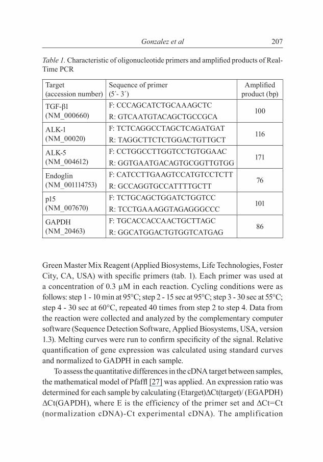
Green Master Mix Reagent (Applied Biosystems, Life Technologies, Foster City, CA, USA) with specifi c primers (tab. 1). Each primer was used at a concentration of 0.3 μM in each reaction. Cycling conditions were as follows: step 1 - 10 min at 95°C; step 2 - 15 sec at 95°C; step 3 - 30 sec at 55°C; step 4 - 30 sec at 60°C, repeated 40 times from step 2 to step 4. Data from the reaction were collected and analyzed by the complementary computer software (Sequence Detection Software, Applied Biosystems, USA, version 1.3). Melting curves were run to confi rm specifi city of the signal. Relative quantifi cation of gene expression was calculated using standard curves and normalized to GADPH in each sample.
To assess the quantitative differences in the cDNA target between samples, the mathematical model of Pfaffl [27] was applied. An expression ratio was determined for each sample by calculating (Etarget)ΔCt(target)/ (EGAPDH)ΔCt(GAPDH), where E is the efficiency of the primer set and ΔCt=Ct (normalization cDNA)-Ct experimental cDNA). The amplification
Table 1. Characteristic of oligonucleotide primers and amplifi ed products of Real-Time PCR
Target (accession number)
Sequence of primer (5´- 3´)
Amplifi ed product (bp)
TGF-β1 (NM_000660)
F: CCCAGCATCTGCAAAGCTC100
R: GTCAATGTACAGCTGCCGCA
ALK-1 (NM_00020)
F: TCTCAGGCCTAGCTCAGATGAT116
R: TAGGCTTCTCTGGACTGTTGCT
ALK-5 (NM_004612)
F: CCTGGCCTTGGTCCTGTGGAAC171
R: GGTGAATGACAGTGCGGTTGTGG
Endoglin (NM_001114753)
F: CATCCTTGAAGTCCATGTCCTCTT76
R: GCCAGGTGCCATTTTGCTT
p15 (NM_007670)
F: TCTGCAGCTGGATCTGGTCC101
R: TCCTGAAAGGTAGAGGGCCC
GAPDH (NM_20463)
F: TGCACCACCAACTGCTTAGC86
R: GGCATGGACTGTGGTCATGAG
TGF-β1 and p15 in Leydig cells208
effi ciency of each set of primers was calculated from the slope of a standard amplifi cation curve of log (ng cDNA) per reaction vs. Ct value (E=10-(1/slope)). Effi ciencies of 2.0±0.1 were considered optimal.
Statistical analysis
Statistical analysis was performed using GraphPad Prism (version 3.00 for Windows, GraphPad software, San Diego, CA, USA). Data are expressed as mean±SEM. Statistical analyses were performed using Student’s t test or one-way ANOVA followed by Tukey’s post hoc tests. A p-value of less than 0.05 was considered signifi cant.
RESULTS
Immunostaining of TGF-β1 was detected in Leydig cells from both the regressed and non-regressed hamster testes (fig. 1AB). In non-regressed hamster testes, the positive immunostaining for TGF-β1 was also detected in germ cells. Type I receptors, ALK-1 and ALK-5 were detected in Leydig cells from regressed and non-regressed testes (fi g. 1DF and fi g. 1CE, respectively). In addition, ALK-5 was detected in germ cells from non-regressed testes, and ALK-1 in regressed testes. Neither Sertoli nor peritubular (myoid) cells contained ALK-1 or ALK-5.
Gene expression of TGF-β1, ALK-5, endoglin and p15 was signifi cantly greater in Leydig cells from regressed as compared with non-regressed testes (p<0.05; fi g. 2ABDE). Gene expression of ALK-1 was greater in Leydig cells from non-regressed than regressed testes (fi g. 2C). Melatonin induced TGF-β1 gene expression in purifi ed Leydig cells from non-regressed hamster testes (fi g. 2F, p<0.05).
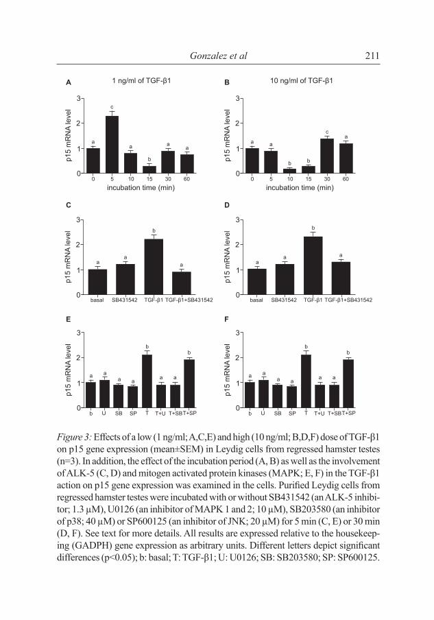
Low doses of TGF-β1 (1 ng/ml) induced p15 gene expression in Leydig cells from regressed testes after 5 min of incubation (fi g. 3A), whereas high doses (10 ng/ml) after 30 min of incubation (p>0.05; fi g. 3B). In cells from non-regressed testes, a high dose ofTGF-β1 (10 ng/ml) induced p15 gene expression after 5, 15 and 30 min of incubation (fi g. 4A), but low doses

Gonzalez et al 209
Figure 1. Immunohistochemical expression of transforming growth factor-β1 (TGF-β1) and its receptors, ALK-1 and ALK-5 in non-regressed (A, C and E) and re-gressed (B, D and F) hamster testes. LC: Leydig cells; ST: seminiferous tubules.
Non-regressed testesTG
F-β1
ALK
-1A
LK-5
Regressed testes
of TGF-β1 (1 ng/ml) did not affect the gene expression (data not shown). SB431542 inhibited (p<0.05) the p15 gene expression induced by TGF-β1 in Leydig cells from both regressed (fi g. 3CD) and non-regressed (fi g. 4B) testes.

TGF-β1 and p15 in Leydig cells210
Figure 2. Basal gene expression (mean±SEM) of TGF-β1 (A), ALK-5 (B), ALK-1 (C), endoglin (EDG; D) and p15 (E) in purifi ed Leydig cells from non-regressed (white bars) and regressed (black bars) hamster testes (n=3). In addi-tion, the in vitro effect of melatonin (Mel) on TGF-β1 expression in Leydig cells from non-regressed testes is demonstrated (F). The Leydig cells were incubated with or without melatonin (10–6 M) for 3 h. All results are expressed relative to the housekeeping gene (GADPH) expression as arbitrary units. Different let-ters depict signifi cant differences (p<0.05).
Gonzalez et al 211
Figure 3: Effects of a low (1 ng/ml; A,C,E) and high (10 ng/ml; B,D,F) dose of TGF-β1 on p15 gene expression (mean±SEM) in Leydig cells from regressed hamster testes (n=3). In addition, the effect of the incubation period (A, B) as well as the involvement of ALK-5 (C, D) and mitogen activated protein kinases (MAPK; E, F) in the TGF-β1 action on p15 gene expression was examined in the cells. Purifi ed Leydig cells from regressed hamster testes were incubated with or without SB431542 (an ALK-5 inhibi-tor; 1.3 µM), U0126 (an inhibitor of MAPK 1 and 2; 10 µM), SB203580 (an inhibitor of p38; 40 µM) or SP600125 (an inhibitor of JNK; 20 µM) for 5 min (C, E) or 30 min (D, F). See text for more details. All results are expressed relative to the housekeep-ing (GADPH) gene expression as arbitrary units. Different letters depict signifi cant differences (p<0.05); b: basal; T: TGF-β1; U: U0126; SB: SB203580; SP: SP600125.

TGF-β1 and p15 in Leydig cells212
Figure 4. Effect of TGF-β1 (10 ng/ml) on p15 gene expression (mean±SEM) in Leydig cells from non-regressed hamster testes (n=3). In addition, the effect of the incubation period (A), the involvement of ALK-5 (B) and the involvement of mitogen activated protein kinases (MAPK; C) in the TGF-β1 action on p15 gene expression was examined in the cells. Purifi ed Leydig cells from regressed hamster testes were incubated with or without SB431542 (an ALK-5 inhibitor; 1.3 µM), U0126 (an inhibitor of MAPK 1 and 2; 10 µM), SB203580 (an inhibitor of p38; 40 µM) or SP600125 (an inhibitor of JNK; 20 µM) for 5 min (B, C). See text for more details. All results are expressed relative to the housekeeping (GADPH) gene expression as arbitrary units. Different letters depict signifi cant differences (p<0.05); b: basal; T: TGF-β1; U: U0126; SB: SB203580; SP: SP600125.

Gonzalez et al 213
To examine the effect of the TGF-β1 signaling pathway on p15 gene expression, the purified Leydig cells were incubated with or without inhibitors of MAPK. In cells derived from regressed testes, U0126 and SB203580 inhibited the p15 gene expression induced by both doses of TGF-β1 (p<0.05; fi g. 3E, F), whereas SP600125 did not affect the p15 gene expression induced by TGF-β1. In cells from non-regressed testes, SB203580 inhibited the p15 gene expression induced by TGF-β1. Neither U0126 nor SP600125 altered the p15 gene expression induced by TGF-β1 (p<0.05; fi g. 4C).
DISCUSSION
Photoperiod-induced testicular atrophy in seasonally breeding Syrian hamsters (Mesocricetus auratus) involves a reduction in gonadal weight but also morphological changes in cellular populations. These changes are not only a consequence of decreased serum levels of gonadotropic hormones but are also due to the action of locally produced factors. TGF-β1 is involved in multiple biological processes including cell differentiation, proliferation, apoptosis, migration, wound healing, immune system regulation and extracellular matrix remodeling. Interestingly, TGF-β1 can exert both inhibitory and stimulatory effects on the regulation of cell homeostasis in physiological and pathological conditions [15, 16]. In the testis, TGF-β1 inhibits steroidogenesis [9], modulates contractility of peritubular cells [10], controls proliferative activity of progenitor Leydig cells [30] and stimulates apoptosis of germ cells [17].
In the present study, TGF-β1 was detected in Leydig cells from both regressed and non-regressed testes of hamsters. These results are in agreement with recent observations in normal and transgenic mice that overexpress α and β subunits of hCG [12]. However, Teerds and Dorrington [30] reported that TGF-β1 was present in fetal and immature Leydig cells from rats but not in Leydig cells from adult hamsters. The positive immunostaining for TGF-β1 was also detected in the germ cells from non-regressed testes. Gene expression of TGF-β1 was greater in Leydig cells

TGF-β1 and p15 in Leydig cells214
from regressed as compared with non-regressed testes. These results are in agreement with the observations in roe deer by Wagener et al. [31] who demonstrated the presence of TGF-β1 mRNA in Leydig cells mainly during the non-breeding season.
Our results confi rm the existence of ALK-5 and ALK-1 in Leydig cells from both regressed and non-regressed hamster testes. These results are in agreement with our previous fi ndings in mice [12]. In addition, ALK-5 was detected in germ cells from non-regressed and ALK-1 in the cells from regressed testes. Neither Sertoli nor peritubular myoid cells exhibited ALK-5 or ALK-1 expression. To the best of our knowledge, there are no previous reports on ALK receptors in the testes of seasonally breeding animals. In adult rat testes, Olaso et al. [24] observed weak ALK-5 protein expression in Leydig cells, strong protein expression in germ cells, and a lack of ALK-5 expression in Sertoli cells. Goddard et al. [10] reported the occurrence of ALK-5 in Leydig cells obtained from immature pigs. The available information on the presence of ALK-1 in the reproductive system is scarce since this receptor has been studied mainly in endothelial cells [3, 18].
The present results show that basal gene expression of TGF-β1 and ALK-5 was greater in regressed than in non-regressed hamster testes. The elevated TGF-β1 gene expression detected at the time when testicular atrophy was maximal leads us to speculate that TGF-β1 might act as a tropic factor inducing the ensuing re-growth of testicular tissue during the recrudescence period. However, the balance between the gene expression of ALK-1 and ALK-5 receptors must also be considered. Since ALK-1-mediated signaling has a stimulatory effect on cell proliferation, the weak expression of this receptor subtype appears to maintain the quiescent state of Leydig cells in the regressed testes. On the other hand, although endoglin gene expression was relatively high in the Leydig cells from regressed testes, its activity might be suppressed as endoglin requires the presence of ALK-1 to generate a proliferative response in these cells.
Hamsters exposed to SD for 14–16 weeks show nearly undetectable levels of LH, FSH, PRL and androgens, and a marked at rophy of the reproductive tract [4, 8, 22, 29]. High melatonin levels have been

Gonzalez et al 215
observed throughout the period of testicular regression [28]. Therefore, we examined the effect of melatonin on the gene expression of TGF-β1 in purifi ed Leydig cells from non-regressed hamster testes and observed that melatonin signifi cantly increased TGF-β1 gene expression. To the best of our knowledge, there are no earlier reports on the regulation of TGF-β1 expression by melatonin in the testis. However, in pig chondrocytes and mammary tumor cells, melatonin is able to stimulate the gene expression or secretion of TGF-β1 [5, 26].
Regressed hamster testes possessed greater p15 gene expression than non-regressed testes. In addition, high doses of TGF-β1 during a short incubation period stimulated p15 expression in Leydig cells from non-regressed testes. Incubation of purifi ed Leydig cells from non-regressed hamster testes with JNK and ERK 1/2 inhibitors did not modify p15 expression induced by TGF-β1. Instead, p38 appears to be the MAPK that participates in this process. The mechanisms governing the regulation of p15 expression by TGF-β1 in testicular tissue remain unknown. Feng et al. [6] proposed that Smad2 and Smad3 proteins induced p15 expression in response to the stimulation with TGF-β1. Wu et al. [32] observed that TGF-β1 activated the p38 pathway leading to cell apoptosis in murine podocytes. On the basis of our present results, there is no direct evidence that p38 induces apoptosis of hamster Leydig cells. Different reports implicated p38 in the apoptosis of germ cells in rodents [25]. However, our present results can be interpreted to suggest that Leydig cells are quiescent at the peak of testicular regression since p15 is a protein involved in the arrest of cellular cycle.
In the present study, the p15 gene expression in regressed hamster testes was stimulated by low doses of TGF-β1 after short incubation periods and by high doses of TGF-β1 after longer incubation periods. In both cases, the ALK-5, ERK 1/2 and p38 were involved. In contrast, ERK 1/2 was not involved in the regulation of p15 gene expression by TGF-β1 in non-regressed testes. Thus, it can be concluded that different MAPKs are activated in the signaling pathway of p15 gene expression (in response to TGF-β1) in regressed and non-regressed hamster testes. Further studies are needed to elucidate whether or not the stimulation of testicular p15 expression by TGF-β1 is dependent on Smad proteins.

TGF-β1 and p15 in Leydig cells216
In summary, the photoperiod modified the expression of TGF-β1 in hamster Leydig cells; TGF-β1 expression was greater in regressed than in non-regressed hamster testes. These changes are likely due to changes in melatonin secretion. The populations of type I TGF-β1 receptors, the co-receptor endoglin and p15 were also infl uenced by photoperiod. The effects of TGF-β1 on p15 gene expression suggest that the alterations in p15 expression, mediated by MAPK, might be involved in the shift between the active and inactive state in hamster Leydig cells.
ACKNOWLEDGEMENTS
This study was supported by grants from Agencia Nacional de Promoción Científi ca y Tecnológica (ANPCYT), Argentina, and Facultad de Medicina, Universidad de Buenos Aires, UBACYT, Argentina.
REFERENCES
1. Bartke A 1985 Male hamster reproductive endocrinology. In: The hamster: Repro-duction and Behavior, pp. 73-98. Ed Siegel HI. Plenum Press, New York, USA.
2. Bartke A, Sinha Hikim AP, Russell LD 1996 Leydig cell structure and function in seasonal breeders. In: The Leydig Cell, pp 431-450. Eds Payne AH, Hardy MP, Russell LD. Cache River Press, USA.
3. Bernabeu C, Conley BA, Vary CP 2007 Novel biochemical pathways of endoglin in vascular cell physiology. Journal of Cellular Biochemistry 102 1375-1388.
4. Bex R, Bartke A, Goldman BD, Dalterios S 1978 Prolactin, growth hormone, lutein-izing hormone receptors, and seasonal changes in testicular activity in the golden hamster. Endocrinology 103 2069-2080.
5. Bizzarri M, Cucina A, Valente MG, Tagliaferri F, Borrelli V, Stipa F, Cavallaro A 2003 Melatonin and vitamin D3 increase TGF-β1 release and induce growth inhibition in breast cancer cell cultures. Journal of Surgical Research 110 332-337.
6. Feng XH, Lin X, Derynck R 2000 Smad2, Smad3 and Smad4 cooperate with Sp1 to induce p15 (Ink4B) transcription in response to TGF-β. The EMBO Journal 19 5178-5193.
7. Frungieri MB, Gonzalez-Calvar SI, Rubio M, Ozu M, Lustig L, Calandra RS 1999 Serotonin in golden hamster testes: testicular levels, immunolocalization and role

Gonzalez et al 217
during sexual development and photoperiodic regression-recrudescence transition. Neuroendocrinology 69 299-308.
8. Frungieri MB, Mayerhofer A, Zitta K, Pignataro OP, Calandra RS, Gonzalez Calvar SI 2005 Direct Effect of Melatonin on Syrian Hamster Testes: Mel1a Receptors, Inhibition of Androgen Production, and Interaction with the Local Corticotropin-Releasing Hormone (CRH) System. Endocrinology 146 1541-1552.
9. Gautier C, Levacher C, Saez JM, Habert R 1997 Transforming growth factor β1 inhibits steroidogenesis in dispersed fetal testicular cells in culture. Molecular and Cellular Endocrinology 131 21-30.
10. Goddard I, Bouras M, Keramidas M, Hendrick JC, Feige JJ, Benahmed M 2000 Transforming growth factor-β receptor types I and II in cultured porcine Leydig cells: expression and hormonal regulation. Endocrinology 141 2068-2074.
11. Gonzalez CR, Gonzalez B, Rulli SB, Dos Santos ML, Mattos Jardim Costa G, França LR, Calandra RS, Gonzalez-Calvar SI 2010 TGF-β1 system in Leydig cells. Part II: TGF-β1 and progesterone, through Smad1/5, are involved in the hy-perplasia/hypertrophy of Leydig cells. Journal of Reproduction and Development 56 400-404.
12. Gonzalez CR, Gonzalez B, Rulli SB, Huhtaniemi I, Calandra RS, Gonzalez-Calvar SI 2010 TGF-β1 system in Leydig cells. Part I: effect of hCG and progesterone. Journal of Reproduction and Development 56 389-395.
13. Gonzalez CR, Matzkin ME, Frungieri MB, Terradas C, Ponzio R, Puigdomenech E, Levalle O, Calandra RS, Gonzalez-Calvar SI 2010 Expression of the TGF-β1 system in human testicular pathologies. Reproductive Biology and Endocrinology 8 148-159.
14. Huang SS, Huang JS 2005 TGF-β control of cell proliferation. Journal of Cellular Biochemistry 96 447-462.
15. Ingman WV, Robertson SA 2007 Transforming growth factor-β1 null mutation causes infertility in male mice associated with testosterone defi ciency and sexual dysfunction. Endocrinology 148 4032-4043.
16. Ingman WV, Robertson SA 2009 The essential roles of TGFB1 in reproduction. Cytokine & Growth Factor Reviews 20 233-239.
17. Konrad L, Keilani MM, Laible L, Nottelmann U, Hofmann R 2006 Effects of TGF-β and a specifi c antagonist on apoptosis of immature rat male germ cells in vitro. Apoptosis 11 739-748.
18. Lebrin F, Deckers M, Bertolino P, Ten Dijke P 2005 TGF-β receptor functions in the endothelium. Cardiovascular Research 65 599-608.
19. Levy H, Deane HW, Rubin BL 1959 Visualization of steroid 3b-ol-dehydrogenase activity in tissues of intact and hypophysectomized rats. Endocrinology 65 932-943.
20. Li X, Zhang YY, Wang Q, Fu SB 2005 Association between endogenous gene expression and growth regulation induced by TGF-beta1 in human gastric cancer cells. World Journal of Gastroenterology 11 61-68.

TGF-β1 and p15 in Leydig cells218
21. Li Z, Chen Y, Cao D, Wang Y, Chen G, Zhang S, Lu J 2006 Glucocorticoid up-regulates transforming growth factor-beta (TGF-beta) type II receptor and en-hances TGF-beta signaling in human prostate cancer PC-3 cell. Endocrinology 147 5259-5267.
22. Mason AO, Duffy S, Zhao S, Ubuka T, Bentley GE, Tsutsui K, Silver R, Kriegs-feld LJ 2010 Photoperiod and reproductive condition are associated with changes in RFamide-related peptide (RFRP) expression in Syrian hamsters (Mesocricetus auratus). Journal of Biological Rhythms. 25 176-85.
23. Massagué J, Seoane J, Wotton D 2005 Smad transcription factors. Genes & De-velopment 19 2783-2810.
24. Olaso R, Pairault C, Habert R 1998 Expression of type I and II receptors for trans-forming growth factor β in the adult rat testis. Histochemistry and Cell Biology 110 613-618.
25. Pastor LM, Zuasti A, Ferrer C, Bernal-Mañas CM, Morales E, Beltrán-Frutos E, Seco-Rovira V 2011 Proliferation and apoptosis in aged and photoregressed mam-malian seminiferous epithelium, with particular attention to rodents and humans. Reproduction in Domestic Animals 46 155-164.
26. Pei M, He F, Wei L, Rawson A 2009 Melatonin enhances cartilage matrix synthesis by porcine articular chondrocytes. Journal Pineal Research 46 181-187.
27. Pfaffl MW 2001 A new mathematical model for relative quantifi cation in real-time RT-PCR. Nucleic Acids Research 29 e45.
28. Reiter RJ 1980 The pineal and its hormones in the control of reproduction in mam-mals. Endocrine Reviews 1 109-131.
29. Simonneaux V, Ansel L, Revel FG, Klosen P, Pévet P, Mikkelsen JD 2009 Kis-speptin and the seasonal control of reproduction in hamsters. Peptides 30 146-153.
30. Teerds KJ, Dorrington JH 1993 Localization of transforming growth factor β1 and β2 during testicular development in the rat. Biology of Reproduction 48 40-45.
31. Wagener A, Fickel J, Schön J, Fritzenkötter A, Göritz F, Blottner S 2005 Seasonal variation in expression and localization of testicular transforming growth factors TGF-β1 and TGF-β3 corresponds with spermatogenic activity in roe deer. Journal of Endocrinology 187 205-215.
32. Wu DT, Bitzer M, Ju W, Mundel P, Böttinger EP. 2005 TGF-β concentration speci-fi es differential signaling profi les of growth arrest/differentiation and apoptosis in podocytes. Journal of the American Society of Nephrology 16 3211-3221.