Activation of FAK and Src are receptor-proximal events required for netrin signaling
Upload
independentCategory
view
1download
0
Combinatorial activation of FAK and AKT by transforming growthfactor-β1 confers an anoikis-resistant phenotype tomyofibroblasts
Jeffrey C. Horowitz, David S. Rogers, Vishal Sharma, Ragini Vittal, Eric S. White, ZongbinCui, and Victor J. Thannickal*Division of Pulmonary and Critical Care Medicine, Department of Internal Medicine, University ofMichigan Medical Center, 6301 MSRB III, 1150 W. Medical Center Dr. Ann Arbor MI 48109, UnitedStates
AbstractTransforming growth factor-β (TGF-β) is a prototypical tumour-suppressor cytokine with cytostaticand pro-apoptotic effects on most target cells; however, mechanisms of its pro-survival/anti-apoptotic signalling in certain cell types and contexts remain unclear. In human lung fibroblasts,TGF-β1 is known to induce myofibroblast differentiation in association with the delayed activationof focal adhesion kinase (FAK) and protein kinase B (PKB/AKT). Here, we demonstrate that FAKand AKT are independently regulated by early activation of SMAD3 and p38 MAPK, respectively.Pharmacologic or genetic approaches that disrupt SMAD3 signalling block TGF-β1-inducedactivation of FAK, but not AKT; in contrast, disruption of early p38 MAPK signalling abrogatesAKT activation, but does not alter FAK activation. TGF-β1 is able to activate AKT in cells expressingmutant FAK or in cells treated with an RGD-containing peptide that interferes with integrinsignalling, inhibits FAK activation and induces anoikis (apoptosis induced by loss of adhesionsignalling). TGF-β1 protects myofibroblasts from anoikis, in part, by activation of the PI3K-AKTpathway. Thus, TGF-β1 co-ordinately and independently activates the FAK and AKT protein kinasepathways to confer an anoikis-resistant phenotype to myofibroblasts. Activation of these pro-survival/anti-anoikis pathways in myofibroblasts likely contributes to essential roles of TGF-β1 intissue fibrosis and tumour-promotion.
KeywordsFocal Adhesion Kinase; Protein kinase B; SMAD proteins; p38 MAPK; Transforming growth factor-beta; Apoptosis; Anoikis; Fibroblasts
1. IntroductionTGF-β1 is a 25 kDa dimeric polypeptide growth factor/ morphogen that functions in a cell-type and context-specific manner to regulate diverse cellular processes such as growth,differentiation, migration and apoptosis [1,2]. Importantly, TGF-β1 mediates divergent effectson epithelial and mesenchymal cells. In epithelial cells, TGF-β1 typically functions as atumour-suppressor through growth-inhibition, cell-cycle arrest and induction of apoptosis[2]. In contrast, TGF-β1 promotes mesenchymal cell proliferation and survival [3–8]. Theseopposing actions on epithelial and mesenchymal cells favour net loss of epithelial cells andaccumulation of myofibroblasts in injured tissues, a hallmark of human fibrotic diseases [9].
*Corresponding author. Tel.: +1 734 936 9371; fax: +1 734 764 2655. E-mail address: [email protected] (V.J. Thannickal)..
NIH Public AccessAuthor ManuscriptCell Signal. Author manuscript; available in PMC 2007 April 1.
Published in final edited form as:Cell Signal. 2007 April ; 19(4): 761–771.
NIH
-PA Author Manuscript
NIH
-PA Author Manuscript
NIH
-PA Author Manuscript
Additionally, myofibroblast activation in the tumour microenvironment is increasinglyrecognized as an important event in cancer progression and metastasis [10,11].
We have previously shown that myofibroblast differentiation by TGF-β1 is dependent onadhesion-mediated signalling through focal adhesion kinase (FAK), a nonreceptor proteintyrosine kinase that is upregulated by TGF-β1 [12]. FAK is also an important regulator of cellsurvival signalling [13]. Apoptosis induced by loss of adhesion or adhesion-mediatedsignalling (termed “anoikis”) is associated with deactivation of FAK [14,15]. A role of FAK-mediated signals in mesenchymal cell survival has been demonstrated in studies showing thatfibroblasts in contractile gels are protected from apoptosis by activating antibodies to α5β1integrins or through constitutive activation of FAK [16]. Other studies have shown, in a varietyof cells, that activation of integrin-FAK signalling pathways can prevent apoptosis due to lossof cell adhesion [17].
Phosphatidylinositol 3′-OH kinase (PI3K) has also been shown to regulate a wide variety ofcellular processes, including proliferation, metabolism, migration and survival; pro-survivalsignalling is primarily mediated through activation of the serine-threonine kinase, proteinkinase B (PKB/AKT) [18]. Activation of the PKB/AKT pathway mediates growth factor-dependent survival of a wide variety of cell types through a number of downstream targets,including caspase-9, BAD, NF– B, fork-head family transcription factors and XIAP (X-linkedinhibitor of apoptosis) [19,20]. In addition to receptor tyrosine kinase(s)-activating growthfactor ligands, PI3K-AKT has also been shown to be activated by adhesion signalling via FAK[16,21,22] and integrin-linked kinase (ILK) [22].
We have previously shown that TGF-β1 protects myofibroblasts from serum deprivation-induced apoptosis through activation of the PI3K-AKT pathway via the p38 MAPK-dependentsecretion of an autocrine growth factor [8]. This study was undertaken to determine theregulatory mechanisms of FAK and AKT by TGF-β1 via SMAD-dependent and -independentpathways, the potential interdependence of these pro-survival protein kinase pathways andtheir role(s) in the protection of myofibroblasts from anoikis.
2. Materials and methods2.1. Cell culture
Normal primary human fetal lung fibroblasts (IMR-90; Institute for Medical Research,Camden, NJ, USA) were cultured as previously described [8] and studies performed at passage5–9. Cells were plated on cell culture dishes or on 96-well cell culture plates and incubatedwith DMEM containing 10% fetal bovine serum (FBS) in 5% CO2–95% air. IMR-90fibroblasts were growth-arrested for 24–48 h in DMEM containing 0.01% FBS prior to varioustreatments.
2.2. ReagentsTGF-β1 derived from porcine platelets was obtained from R and D Systems, Minneapolis, MN.SB203580 was from Calbiochem, La Jolla, CA, USA. LY294002 was obtained from CellSignalling Technology, Beverly MA. SB431542 was from Tocris Bioscience, Ellisville, MO,USA. Rabbit polyclonal antibodies to phospho-Ser473AKT, phospho-Thr308AKT, total AKT,and total FAK were from Cell Signalling Technology. Mouse monoclonal antibody tophosphatidylserine (clone 1H6) was from Upstate Biotechnology, Lake Placid, NY, USA.Mouse monoclonal antibody to β actin and the soluble RGD-containing fibronectin peptide(GRGDS) were from Sigma, St. Louis, MO, USA. The non-RGD-containing peptide(GRADSP) was from Calbiochem. Mouse monoclonal antibody to single-stranded DNA(ssDNA) was from Chemicon International, Temecula, CA, USA. Rabbit polyclonal antibody
Horowitz et al. Page 2
Cell Signal. Author manuscript; available in PMC 2007 April 1.
NIH
-PA Author Manuscript
NIH
-PA Author Manuscript
NIH
-PA Author Manuscript

to phospho-Y397 FAK was from BioSource International Inc, Camarillo, CA, USA. Antibodyto phospho-SMAD3(Ser433/435) was from Cell Signalling Technology. Rabbit polyclonalantibody to total SMAD3 was from Zymed Laboratories, San Francisco, CA, USA. Antibodyto GAPDH was from Abcam, Cambridge, MA, USA. Secondary HRP-conjugated anti-mouseand anti-rabbit antibodies were obtained from Pierce, Rockford, IL, USA.
2.3. Generation of stable cell lines expressing Y397-mutant FAKPlasmid transfections of IMR-90 cells with a FAK mutant construct in which the tyrosine-397phosphorylation site was substituted with phenylalanine (Y397F-FAK) were performed usingthe cationic lipid reagent LipofectAMINE, Invitrogen, Carlsbad, CA, USA, according themanufacturer's instructions. The optimal ratio of DNA (μg) to LipofectAMINE (μl) wasdetermined to be 1:5 for IMR-90 cells. Cells were incubated with DNA-lipid complexes inserum-free Opti-MEM I medium (Invitrogen) for 4–5 h prior to introducing 10% FBS for 16–20 h. The next day, transfection medium was replaced by DMEM supplemented with 10% FBSand geneticin (Invitrogen). Geneticin concentrations were 400 μg/ml for selection transfectedcells and 200 μg/ml for maintenance of stable transfectants prior to treatments.
2.4. siRNA and transfectionsRNA interference was accomplished by transfecting cells with siGENOME SMARTPOOLsiRNA (Dharmacon Inc., Lafayette, CO, USA; Catalogue # 020067). Transfections werecarried out using TransIT-siQUEST lipid reagent according to the manufacturer's instructions(Mirus, Madison WI, USA). Briefly, for each transfection in 35 mm cell culture dishes, 5 μlof lipid reagent was mixed with 250 μl of serum free media for 20 min, followed by the additionpooled siRNA for another 20 min. This lipid–reagent/siRNA mixture was then slowly addedto 750 μl of DMEM supplemented with 5% FBS. IMR-90 cells were then incubated with thismixture for 48–72 h and media changed to serum-free DMEM for another 24 h prior totreatment. For negative controls, cells were cultured, transfected, and treated in a similarmanner using the non-targeting pooled siRNA (siControl) from Dharmacon Inc.
2.5. Western immunoblottingCell lysates were prepared in RIPA buffer and subjected to SDS-PAGE and Western blotanalyses performed as described previously [8].
2.6. Assays for apoptosis/anoikis2.6.1. ELISA for ssDNA—This assay is based on the susceptibility of double-stranded DNAin apoptotic cells to form single strands when exposed to formamide [23] and was performedas previously described [8].
2.6.2. Anti-phosphatidylserine immunofluorescence—Immunofluorescent stainingprocedures were as previously described with minor modifications [8]. IMR-90 cells plated on35 mm tissue culture dishes were grown to 50% confluence and growth-arrested in serum-freemedia for 48 h prior to treatment. Cells were then fixed in 5% formaldehyde and washed threetimes with cold phosphate-buffered saline (PBS), but were not permeabilized. Nonspecificbinding sites were blocked with 1% bovine serum albumin (BSA) for 15 min prior to theaddition of antibody to phosphatidylserine (1:25 dilution) for 1 h followed by fluoresceinisothiocyanate (FITC)-conjugated secondary antibody (1:40 dilution) for 1 h. Nuclearcounterstaining was with DAPI and cells were visualized/photographed using a Zeissfluorescence microscope.
2.6.3. Caspase-3 activation—Caspase-3 activation was assessed using the Caspase-3Fluorometric Assay Kit (Assay Designs, Inc) according to manufacturer's protocol. 50 μl of
Horowitz et al. Page 3
Cell Signal. Author manuscript; available in PMC 2007 April 1.
NIH
-PA Author Manuscript
NIH
-PA Author Manuscript
NIH
-PA Author Manuscript

whole cell lysate for each experimental condition was placed in a 96-well plate with 150 μl offluorometric caspase substrate in reaction buffer and incubated for 3 h. Fluorescence intensitywas read at an excitation wavelength of 360 nm and emission wavelength of 440 nm.
2.7. Induction of cellular anoikisIMR-90 cells were grown to 80–90% confluence in 100 mm tissue culture plastic dishes,growth-arrested for 24–48 h in serum-free media and treated with/without TGF-β1 for 24 h.Cell anoikis was induced by the exogenous addition of RGD-containing peptides, whichinterfere with adhesion signalling, or by placing cells in suspension for defined time-intervals.
2.7.1. RGD-containing peptides—Peptides containing the RGD motif (GRGDS) andcontrol peptide (GRADSP) were reconstituted in DMEM to the final desired concentration.IMR-90 cells were treated with/without TGF-β1 in the presence/absence of synthetic peptidesfor 24 h. Cells were then analyzed by different apoptosis assays as described above.
2.7.2. Cell suspension—IMR-90 cells were trypsinized, pelleted by centrifugation and re-suspended in 10 ml of serum-free DMEM media. Suspended cells were then placed on a rockerin a 37 °C incubator at 5% CO2–95% air for defined time-periods (0–6 h). At the end of theincubation period, cells were again pelleted by centrifugation and seeded onto 96-well platesfor analyses of apoptosis (as described above) or plated onto 35-mm tissue culture dishes forassessment of the ability of cell to re-adhere on tissue culture plastic (described under “cellsurvival studies” below).
2.8. Cell survival (anoikis-resistance) studiesIMR-90 fibroblasts induced to undergo anoikis by either soluble RGD-peptides or by cellsuspension were assessed for their ability to re-adhere to tissue culture plastic. This assay isbased on the principle that non-viable/ apoptotic cells fail to attach to tissue culture plastic.Cells were allowed to re-adhere for 3 h prior to trypsinization of attached cells and cell numberswere measured by Coulter counting (Coulter-Z series, Hialeah, FL, USA).
2.9. Statistical and densitometric analysesStatistical analysis was performed using Student's t test when comparing two groups and one-way analysis of variance (ANOVA) with Bonferroni post-test when comparing three or moreexperimental conditions. This analysis was done using GraphPad Prism version 3.0 forWindows, GraphPad Software, San Diego (www.graphpad.com). Densitometric analyses ofWestern blots were performed using the public domain NIH Image program available on theInternet at rsb.info.nih.gov/nih-image.
3. Results3.1. TGF-β1-induced FAK, but not AKT, activation is dependent on SMAD3
TGF-β1 signalling in fibroblasts involves early receptor(s)-mediated SMAD-dependent and -independent pathways with more delayed activation of the nonreceptor protein kinases, FAKand AKT [8,12]. Our previous studies showed that AKT activation by TGF-β1 in human lungfibroblasts is dependent on early p38-MAPK [8]. Additionally, FAK activation by TGF-β1 ismediated via a transcription-dependent process that is associated with upregulation offibronectin and its integrin receptor subunits [12]. Crosstalk between these two pro-survivalpathways and the specific role(s) of SMAD3, a key regulator of TGF-β1 pro-fibrotic signalling[24], in the delayed activation of FAK and AKT have not been defined.
Horowitz et al. Page 4
Cell Signal. Author manuscript; available in PMC 2007 April 1.
NIH
-PA Author Manuscript
NIH
-PA Author Manuscript
NIH
-PA Author Manuscript

To investigate the role of SMAD3 on the activation of these protein kinases, we used acombination of pharmacologic and genetic approaches to inhibit SMAD3 activity. First, westudied the effects of the type 1 TGF-β receptor (ALK5) inhibitor, SB431542, on the earlyactivation of the SMAD3 and p38 MAPK by TGF-β1 in human lung fibroblasts (IMR-90).IMR-90 fibroblasts were stimulated with TGF-β1 for 45 min in the presence/absence ofincreasing concentrations of SB431542 (0–10 μM). We found that SB431542, at aconcentration of 1 μM, induced a >90% inhibition of the TGF-β1-stimulated phosphorylationof SMAD3 with an IC50 between 0.1 and 0.5 μM (Fig. 1A and B). However, SB431542, at thesame dose (1 μM), reduced TGF-β1-stimulated activation of p38 MAPK by only 40%; theIC50 for p38 MAPK was approximately 5-fold higher than that for the inhibition of SMAD3activation (Fig. 1A and B).
Given the differential inhibition of SMAD3 and p38 MAPK by SB431542, we next examinedthe ability of SB431542 to modulate the delayed activation of FAK and AKT by TGF-β1 inIMR-90 fibroblasts. At 16 h post-TGF-β1 treatment, SB431542 (1 μM) completely inhibits theactivation of FAK, while only partial inhibition of AKT phosphorylation/activation is observed(Fig. 1C). These findings suggest that ALK5/SMAD3 signalling is required for TGF-β1-induced activation of FAK, but that TGF-β1-induced activation of AKT is mediated, in largepart, by a mechanism independent of early SMAD signalling.
To further study the role of SMAD3 signalling in these TGF-β1 responses, IMR-90 fibroblastswere transfected with siRNA targeting SMAD3 (as described in “Materials and methods”).Both phosphorylated and total SMAD3 levels were significantly “knocked-down” in cellstransfected with SMAD3-siRNA compared to cells transfected with non-targeting controlsiRNA (Fig. 2A). However, cells with siRNA knockdown of SMAD3 were able to robustlyactivate p38 MAPK at early time points (45 min) following TGF-β1 stimulation (Fig. 2A),suggesting that SMAD3 is not required for the rapid of activation of p38 MAPK. At 16 hfollowing TGF-β1 stimulation, a slight increase in constitutive FAK phosphorylation wasobserved in SMAD3-knockdown cells; however, TGF-β1 failed to further increase FAKphosphorylation in these cells (Fig. 2A, B). This indicates that early activation of SMAD3 isessential for the delayed activation of FAK induced by TGF-β1. In contrast, under the sameconditions, TGF-β1 maintains its ability to induce AKT phosphorylation at both Ser-473 andThr-308 residues (Fig. 2A, B), indicating that SMAD3 is not required for TGF-β1-inducedAKT activation. These data demonstrate that, although FAK and AKT are co-ordinatelyactivated by TGF-β1, they are independently regulated by SMAD3 and p38 MAPK,respectively.
3.2. TGF-β1-induced FAK is independent of p38 MAPKOur previous studies have shown that activation of the PI3K-AKT pathway is dependent onearly activation of p38 MAPK [8]; however, the potential role of p38 MAPK in TGF-β1-induced activation of FAK is not known. To investigate this possibility, we first assessed theeffect of SB203580 (6 μM; a dose that effectively blocks p38 MAPK activity in these cells[8]) on the induction of FAK autophosphorylation/activation by TGF-β1. Treatment with 6μM SB203580 failed to inhibit TGF-β1-induced FAK autophosphorylation (Fig. 3A); thissuggests that p38 MAPK is not required for activation of FAK. To confirm that p38 MAPK isindeed dispensable for TGF-β1-induced activation of FAK, we employed dominant-negativeover-expression of a kinase-mutant p38 MAPK (p38-KM) which we have shown to inhibitp38 MAPK activity [8]. In support of the pharmacologic studies with SB203580, TGF-β1 iscapable of inducing FAK activation in cells over-expressing p38-KM (Fig. 3B). These resultsindicate that, in contrast to the previously described role of p38 MAPK in TGF-β1-inducedAKTactivation [8], the induction of FAK autophosphorylation/activation does not require theearly activation of the p38 MAPK pathway.
Horowitz et al. Page 5
Cell Signal. Author manuscript; available in PMC 2007 April 1.
NIH
-PA Author Manuscript
NIH
-PA Author Manuscript
NIH
-PA Author Manuscript

3.3. Induction of AKT activation by TGF-β1 is independent of FAK activationActivation of AKT has been shown to be regulated by FAK, primarily in the context of celladhesion signalling [16,21,25]. We next determined if FAK activation by TGF-β1 wasnecessary for the induction of AKT activation in response to this growth factor. To investigatethis possibility, we employed a dominant-negative approach to disrupt FAKautophosphorylation by stably transfecting IMR-90 fibroblasts with a plasmid encoding amutant FAK (Y397F-FAK; tyrosine-397 residue substituted with phenylalanine) or with anempty vector plasmid (pcDNA). A significant reduction in baseline and TGF-β1-stimulatedFAK phosphorylation was confirmed in cells expressing dominant-negative FAK (Fig. 4A,top panels; 4B, open bars). However, AKT phosphorylation was robustly induced in responseto TGF-β1 stimulation in both control and Y397F-FAK mutant cells (Fig. 4A, bottom panels;4B, closed bars). These results indicate that the induction of AKT phosphorylation/ activationin response to TGF-β1 is not dependent on FAK autophosphorylation, further supporting theconcept that TGF-β1 independently regulates the FAK and AKT protein kinase pathways.
The exogenous addition of soluble peptides containing the arginine–glycine–aspartate (RGD)amino acid sequence has been shown to disrupt cell adhesion signalling [26,27]. We firstdetermined if introduction of an RGD-containing peptide modulates FAK autophosphorylationin IMR-90 fibroblasts. Treatment of quiescent IMR-90 fibroblasts with the soluble RGD-containing peptide, GRGDS (0.5 mg/ml), decreased basal levels of FAK autophosphorylationby approximately 50% compared to controls (Fig. 4C, top panel; Fig. 4D, open bars).Stimulation with TGF-β1 was able to augment FAK in cells treated with GRGDS, but only tolevels of the untreated control (Fig. 4C, D). In contrast, treatment with soluble RGD-containingpeptides had no significant effect on either basal or TGF-β1-stimulated levels ofphosphorylated AKT (Fig. 4C, bottom panel; Fig. 4D, closed bars). Treatment of IMR-90 cellswith a control peptide (GRADSP, 0.5 mg/ml) had no effect on basal or TGF-β1-stimulatedactivation of FAK or AKT (data not shown). These studies demonstrate the ability of solubleRGD-containing peptides to suppress constitutive FAK autophosphorylation withoutsignificantly altering either constitutive or inducible AKT phosphorylation. Thus, multiplestrategies/interventions that either block or suppress FAK signalling fail to suppress theinducible activation of AKT by TGF-β1.
3.4. Loss of cell adhesion or exogenous RGD-containing peptides induces fibroblast anoikisAnoikis is a form of programmed cell death induced by the loss of cell adhesion or adhesion-dependent signalling [28]. We utilized two experimental models to induce anoikis of humanlung fibroblasts: (1) by mechanical/enzymatic detachment of cells and maintenance insuspension for specified periods of time; (2) by competitive interruption of integrin-ECMinteractions by addition of soluble RGD-containing peptides [29,30].
Cell suspension studies were performed on quiescent (“growth-arrested”) IMR-90 fibroblastcultures at 80–90% confluence and the measured end-points included apoptosis (by ssDNAELISA) and cell survival (ability of suspended cells to re-adhere to tissue culture plastic —assessed by Coulter counting) as described in “Material and methods”. Cells in suspensiondemonstrated a time-dependent increase in apoptotic rates compared to adherent (“control”)cells, with detectable apoptosis observed within 1 h of suspension and further increases notedat 2 and 6 h of suspension (Fig. 5A). Similarly, the number of cells able to survive and re-adhere to tissue culture plastic decreased as time in suspension was increased (Fig. 5B). Thesestudies indicate that lung fibroblasts subjected to loss of adhesion undergo apoptosis/anoikisand loss of cell viability that is dependent on the time-period for which they are maintained ina non-adherent (suspended) state.
Horowitz et al. Page 6
Cell Signal. Author manuscript; available in PMC 2007 April 1.
NIH
-PA Author Manuscript
NIH
-PA Author Manuscript
NIH
-PA Author Manuscript

Next, we assessed the ability of soluble RGD-containing peptides to induce anoikis/apoptosisof IMR-90 fibroblasts. Treatment with the RGD-containing peptide, GRGDS, inducedmorphological changes, characterized by cell “crowding” and formation of stellate-shapedconglomerates, at 18–24 h in IMR-90 fibroblast cultures; this effect was most marked with the0.5 mg/ml (vs. 0.125 mg/ml) concentration of the GRGDS peptide (Fig. 5C). Thesemorphological changes were not observed in cells treated with a non-RGD containing controlpeptide, GRADSP (Fig. 5C). To determine if treatment with the RGD-containing peptideinduces loss of cell viability, the ability of trypsinized cells to re-adhere to tissue culture plasticwas again assessed. IMR-90 fibroblasts with 0.5 mg/ml of the GRGDS peptide demonstratedsignificant decreases in cell number, suggesting loss of cell viability (Fig. 5D). To confirm thatthis loss of cell viability was related to induction of apoptosis/anoikis, GRGDS-treated cellswere assayed for expression of activated caspase-3 (by ELISA). A significant increase incaspase-3 activity was observed in cells treated with 0.5 mg/ml of the GRGDS peptide (Fig.5E). Together, these results indicate that fibroblast apoptosis/anoikis can be reproduciblyinduced (and detected) in a time- and dose-dependent manner by the disruption of cell adhesionor adhesion-dependent signalling.
3.5. TGF-β1 protects human lung myofibroblasts from anoikisPrevious studies have shown that TGF-β1 protects lung myofibroblasts apoptosis induced byIL-1 [7] or serum deprivation [8]. Whether TGF-β1-differentiated myofibroblasts are resistantto anoikis is not known; moreover, mechanisms for apoptosis/anoikis protection have not beenelucidated. First, to determine whether TGF-β1-differentiated myofibroblasts are protectedfrom anoikis induced by soluble RGD peptides, IMR-90 cells were pre-treated with/withoutTGF-β1 for 16 h and then exposed to two different concentrations of the GRGDS peptide for24 h prior to assessments of apoptosis. Myofibroblasts induced to differentiate in response toTGF-β1 were resistant to anoikis, as evidenced by lower rates of apoptosis, as assessed byssDNA ELISA (Fig. 6A), phosphatidylserine labelling (PS-FITC; Fig. 6B, C) and by theexpression of activated caspase-3 by Western blot analysis (Fig. 6D). No effects on apoptosiswere detected in cells treated with the non-RGD containing peptide (GRADS; data not shown).Together, these studies demonstrate that TGF-β1, simultaneous with the induction ofmyofibroblast differentiation, confers significant protection from anoikis-induced cell death.
3.6. Activation of the AKT pathway contributes to anoikis-resistance in myofibroblastsAdhesion-dependent FAK activation is a critical pathway that protects cells from anoikis[13]; this is supported by our own observations in fibroblasts treated with the RGD-containingpeptide that both inhibited FAK autophosphorylation and induced apoptosis. Based on ourfindings that that AKT activation by TGF-β1 is independent of the SMAD3/FAK pathway, weinvestigated the potential additive role of AKT in protecting myofibroblasts from anoikis-induced cell death. IMR-90 fibroblasts were treated with TGF-β1±LY294002 (10 μM), apharmacologic inhibitor of the PI3K-AKT pathway, with/without GRGDS (0.5 mg/ml) priorto assessment of apoptosis. Blockade of PI3K-AKT significantly attenuated the protectiveeffect of TGF-β1 on myofibroblast anoikis and cell survival (Fig. 7). Thus, myofibroblastsinduced to differentiate in response to TGF-β1 stimulation consistently demonstrate an anti-anoikis phenotype and enhanced viability/survival, an effect that is significantly inhibited byblockade of the PI3K/ AKT pathway. However, blockade of PI3K/AKT activation withLY294002 (10 μM) treatment did not inhibit TGF-β1-induced FAK activation or myofibroblastdifferentiation (data not shown). These results suggest that, in addition to the activation ofadhesion-dependent FAK, the PI3K-AKT pathway contributes to the resistance against anoikisacquired by TGF-β1-differentiated myofibroblasts.
Horowitz et al. Page 7
Cell Signal. Author manuscript; available in PMC 2007 April 1.
NIH
-PA Author Manuscript
NIH
-PA Author Manuscript
NIH
-PA Author Manuscript

4. DiscussionTGF-β1 is a multifunctional growth factor that plays a central role in tissue homeostasis,reparative responses following tissue injury and in the stromal reaction associated with tumourprogression and metastasis [2,11,31]. Effects of TGF-β1 on target cells are highly cell-specificand contextual [1]. Despite the recognition that TGF-β1 functions as an anti-apoptotic signalfor fibroblasts/myofibroblasts [7,8,32], mechanisms for this effect have not been well defined.TGF-β superfamily ligands signal via heterotetrameric complexes of type II and type I receptors(TβR-II and TβR-I, respectively) leading to activation of SMAD proteins which represent thecentral mediators of downstream signalling [1]. There is emerging evidence for non-SMADsignals that mediate specific biological actions of TGF-β [33,34]. Our studies in human lungfibroblasts have demonstrated that early and rapid activation of p38 MAPK by TGF-β1 isessential for the production of an autocrine growth factor that mediates activation of the PI3K-AKT pathway [8]. Additionally, our prior studies have demonstrated delayed, but sustained,activation of FAK that is required for stable induction of myofibroblast differentiation by TGF-β1 [12]. A specific requirement for SMAD3-dependent signalling in the activation of FAK andAKT, potential crosstalk between SMAD-dependent and -independent signalling, or their role(s) in the anoikis-resistance of myofibroblasts have not been previously reported. The currentstudies provide support for a model in which TGF-β1 activates SMAD3 to regulate downstreamactivation of FAK, while p38 MAPK simultaneously mediates the downstream activation ofAKT (schematic in Fig. 8). Interestingly, TGF-β1-induced activation of FAK is independentof p38 MAPK, while TGF-β1-induced activation of AKT is independent of SMAD3.Importantly, TGF-β1-induced activation of PI3K-AKT contributes to the observed anoikis-resistance of myofibroblasts. This is the first study to demonstrate that myofibroblasts inducedto differentiate in response to TGF-β1 are resistant to anoikis-mediated cell death and that theacquisition of an anoikis-resistant myofibroblast phenotype is mediated by the activation oftwo independent, and parallel, protein kinase pathways involving FAK and AKT.
Our studies conclusively demonstrate that the induction of FAK activation by TGF-β1 isdependent on SMAD3 signalling, but is independent of p38 MAPK. A pharmacologic inhibitorof TβR-I/ALK5, SB431542, that is known to inhibit TGF-β1-induced SMAD3 [35], effectivelyblocks TGF-β1-induced FAK activation. That SMAD3 signalling was required for this effectwas confirmed by siRNA targeting of SMAD3. In contrast, pharmacologic and geneticstrategies that disrupt p38 MAPK activation and block activation of the PI3K-AKT pathways[8], were incapable of inhibiting FAK activation induced by TGF-β1. Although studies by ourgroup [12] and others [36] have shown the capacity of TGF-β1 to activate FAK in certain celltypes, a role for SMAD3 in mediating this effect has not been previously demonstrated. Therequirement for SMAD3 in FAK activation is consistent with a role for SMAD3 inmyofibroblast differentiation both in-vitro [37,38] and in-vivo [39–41].
Several lines of evidence support the conclusion that TGF-β1 induces activation of AKT by amechanism independent of the SMAD3-FAK pathway. First, at doses of SB431542, a TβR-I/ALK5 inhibitor, that completely blocks the activation of SMAD3 and FAK, TGF-β1 was ableto significantly induce AKT activation. The relatively small decrease in TGF-β1-induced AKTactivation with 1 μM SB431542 may be accounted for by the observed ~40% inhibition of p38MAPK at this dose. Second, siRNA knockdown of SMAD3 failed to attenuate AKT activationby TGF-β1, while it effectively blocks TGF-β1-induced FAK activation. Third, disruption ofFAK autophosphorylation with expression of mutant FAK does not alter TGF-β1-inducedAKT activation. Finally, introduction of an RGD-containing peptide that suppresses FAKautophosphorylation and induces anoikis does not inhibit activation of AKT in response toTGF-β1 stimulation. A SMAD-independent mechanism for activation of PI3K-AKT by TGF-β1 has been previously demonstrated in a subset of fibroblast cell lines, although this effectoccurred more rapidly and, presumably, in the absence of an autocrine mechanism [42].
Horowitz et al. Page 8
Cell Signal. Author manuscript; available in PMC 2007 April 1.
NIH
-PA Author Manuscript
NIH
-PA Author Manuscript
NIH
-PA Author Manuscript

This is the first study, to our knowledge, that demonstrates a novel effect of TGF-β1 to induceanoikis-resistance in any cell type. Importantly, the activation of PI3K-AKT by TGF-β1 wasfound to be a significant contributor to this anoikis-resistant phenotype of myofibroblasts.There is evidence for a role of FAK and/or ILK signalling in the constitutive activation of AKTin adherent cells [16,22]; however, studies on the role of adhesion-dependent FAK and ILK ininducible AKT activation by growth factors are lacking. Our studies clearly indicate that FAKis not required for the inducible activation of AKT in response to TGF-β1 stimulation; thismay be related to a mechanism whereby soluble autocrine factor(s) secreted in a p38 MAPK-dependent and SMAD3-independent manner are capable of activating the PI3K-AKT pathway[8]. Interestingly, blockade of the PI3K-AKT pathway did not completely inhibit anoikis-resistance conferred by TGF-β1, suggesting that the participation of non-AKT and non-FAKpathways may also contribute to the anoikis-resistant phenotype [43]; alternatively, theadditional protection may be attributed to the relatively small, but significant, increases in FAKactivity observed in cells treated with TGF-β1 despite the presence of RGD-containingpeptides.
Although the precise mechanism(s) of myofibroblast apoptosis in the resolution of woundhealing and tissue repair are not well defined, anoikis is likely a relevant model to studyapoptotic mechanisms since cell adhesion and biomechanical tension unloading appear to playimportant roles in this physiological context [44,45]. Our observations that the PI3K-AKT andintegrin-FAK pathways are activated in a combinatorial manner by TGF-β1 to promotemyofibroblast survival have important implications for broader understanding of dysregulatedtissue repair and fibrotic diseases. Recent studies from our laboratory have shown that bothFAK and AKT activation are increased in areas of active fibrogenesis following acute lunginjury in mice [46]. Treatment with a protein tyrosine kinase inhibitor that blocks TGF-β1-activation of FAK and AKT in lung fibroblasts is protective against the development of fibrosisin this animal model [46]. Additionally, we have shown that alveolar mesenchymal cellsisolated from patients with non-resolving (fibrotic/persistent) acute respiratory distresssyndrome (ARDS) demonstrate high levels of AKT activation and resistance to apoptosiscompared to mesenchymal cells from patients with resolving ARDS [47]. These studies supportan in-vivo role for an apoptosis-resistant mesenchymal cell/myofibroblast phenotype in fibroticlung diseases. Thus, a better understanding of the signalling pathways that regulate thismyofibroblast phenotype may provide novel targets for therapy of these, otherwise, treatment-unresponsive disorders.
5. ConclusionsIn this study, we demonstrate: (1) a novel action of TGF-β1 to protect differentiatedmyofibroblasts from anoikis-mediated cell death; (2) an important role for the inducibleactivation of PI3K-AKT and FAK by TGF-β1 in the acquisition of an anoikis-resistantmyofibroblast phenotype; and (3) independent and coordinate regulation of FAK and AKTactivation by TGF-β1 via SMAD-dependent and -independent mechanisms, respectively. Thesignificance of these findings are that they provide mechanistic insights into TGF-β1 regulationof myofibroblast survival and persistence, a key feature of progressive human fibrotic disorders[9] and of the stromal reaction that supports tumour progression/metastasis [11]. Moreover,these studies provide a novel paradigm for TGF-β1 signalling of cell survival in mesenchymalcells, in contrast to its well recognized anti-proliferative/pro-apoptotic effects on epithelialcells.
Acknowledgements
This work was supported by the National Institutes of Health grants, K08 HL081059 (to J.C.H.), American LungAssociation Dalsemer Award (to J.C.H.), K08 HL070990 (to E.S.W.), R01 HL067967 (to V.J.T.) and P50 HL074024
Horowitz et al. Page 9
Cell Signal. Author manuscript; available in PMC 2007 April 1.
NIH
-PA Author Manuscript
NIH
-PA Author Manuscript
NIH
-PA Author Manuscript

(to V.J.T.). We thank Dr. Kenneth Yamada, National Institute of Dental and Craniofacial Research, National Institutesof Health, Bethesda, MD, for graciously providing the FAK mutant and control plasmids used in these studies.
References1. Massague J. Nat Rev, Mol Cell Biol 2000;1(3):169. [PubMed: 11252892]2. Blobe GC, Schiemann WP, Lodish HF. N Engl J Med 2000;342(18):1350. [PubMed: 10793168]3. Moses HL, Coffey RJ Jr, Leof EB, Lyons RM, Keski-Oja J. J Cell Physiol, Suppl 1987;(Suppl 5):1.
[PubMed: 3316252]4. Tomasek JJ, Gabbiani G, Hinz B, Chaponnier C, Brown RA. Nat Rev Mol Cell Biol 2002;3(5):349.
[PubMed: 11988769]5. Chodon T, Sugihara T, Igawa HH, Funayama E, Furukawa H. Am J Pathol 2000;157(5):1661.
[PubMed: 11073825]6. Thannickal VJ, Aldweib KD, Rajan T, Fanburg BL. Biochem Biophys Res Commun 1998;251(2):437.
[PubMed: 9792792]7. Zhang HY, Phan SH. Am J Respir Cell Mol Biol 1999;21(6):658. [PubMed: 10572062]8. Horowitz JC, Lee DY, Waghray M, Keshamouni VG, Thomas PE, Zhang H, Cui Z, Thannickal VJ. J
Biol Chem 2004;279(2):1359. [PubMed: 14576166]9. Thannickal VJ, Toews GB, White ES, Lynch JP III, Martinez FJ. Annu Rev Med 2004;55:395.
[PubMed: 14746528]10. Desmouliere A, Guyot C, Gabbiani G. Int J Dev Biol 2004;48(5–6):509. [PubMed: 15349825]11. De Wever O, Mareel M. J Pathol 2003;200(4):429. [PubMed: 12845611]12. Thannickal VJ, Lee DY, White ES, Cui Z, Larios JM, Chacon R, Horowitz JC, Day RM, Thomas
PE. J Biol Chem 2003;278(14):12384. [PubMed: 12531888]13. Frisch SM, Vuori K, Ruoslahti E, Chan-Hui PY. J Cell Biol 1996;134(3):793. [PubMed: 8707856]14. Liao YF, Gotwals PJ, Koteliansky VE, Sheppard D, Van De Water L. J Biol Chem 2002;277(17):
14467. [PubMed: 11839764]15. Wen LP, Fahrni JA, Troie S, Guan JL, Orth K, Rosen GD. J Biol Chem 1997;272(41):26056.
[PubMed: 9325343]16. Xia H, Nho RS, Kahm J, Kleidon J, Henke CA. J Biol Chem 2004;279(31):33024. [PubMed:
15166238]17. Zhan M, Zhao H, Han ZC. Histol Histopathol 2004;19(3):973. [PubMed: 15168359]18. Vivanco I, Sawyers CL. Nat Rev Cancer 2002;2(7):489. [PubMed: 12094235]19. Datta SR, Brunet A, Greenberg ME. Genes Dev 1999;13(22):2905. [PubMed: 10579998]20. Dan HC, Sun M, Kaneko S, Feldman RI, Nicosia SV, Wang HG, Tsang BK, Cheng JQ. J Biol Chem
2004;279(7):5405. [PubMed: 14645242]21. Sonoda Y, Watanabe S, Matsumoto Y, Aizu-Yokota E, Kasahara T. J Biol Chem 1999;274(15):10566.
[PubMed: 10187851]22. Persad S, Attwell S, Gray V, Mawji N, Deng JT, Leung D, Yan J, Sanghera J, Walsh MP, Dedhar S.
J Biol Chem 2001;276(29):27462. [PubMed: 11313365]23. Frankfurt OS, Krishan A. J Immunol Methods 2001;253(1–2):133. [PubMed: 11384675]24. Roberts AB, Tian F, Byfield SD, Stuelten C, Ooshima A, Saika S, Flanders KC. Cytokine Growth
Factor Rev 2006;17(1–2):19. [PubMed: 16290023]25. Schaller MD. Biochim Biophys Acta 2001;1540(1):1. [PubMed: 11476890]26. Akiyama SK, Yamada KM. J Biol Chem 1985;260(19):10402. [PubMed: 3161878]27. Hadden HL, Henke CA. Am J Respir Crit Care Med 2000;162(4 Pt 1):1553. [PubMed: 11029376]28. Frisch SM, Screaton RA. Curr Opin Cell Biol 2001;13(5):555. [PubMed: 11544023]29. Michel JB. Arterioscler Thromb Vasc Biol 2003;23(12):2146. [PubMed: 14551156]30. Maubant S, Saint-Dizier D, Boutillon M, Perron-Sierra F, Casara PJ, Hickman JA, Tucker GC, Van
Obberghen-Schilling E. Blood 2006;108(9):3035. [PubMed: 16835373]31. Leask A, Abraham DJ. FASEB J 2004;18(7):816. [PubMed: 15117886]32. Jelaska A, Korn JH. Arthritis Rheum 2000;43(10):2230. [PubMed: 11037882]
Horowitz et al. Page 10
Cell Signal. Author manuscript; available in PMC 2007 April 1.
NIH
-PA Author Manuscript
NIH
-PA Author Manuscript
NIH
-PA Author Manuscript
33. Derynck R, Zhang YE. Nature 2003;425(6958):577. [PubMed: 14534577]34. Moustakas A, Heldin CH. J Cell Sci 2005;118(Pt 16):3573. [PubMed: 16105881]35. Laping NJ, Grygielko E, Mathur A, Butter S, Bomberger J, Tweed C, Martin W, Fornwald J, Lehr
R, Harling J, Gaster L, Callahan JF, Olson BA. Mol Pharmacol 2002;62(1):58. [PubMed: 12065755]36. Wang H, Radjendirane V, Wary KK, Chakrabarty S. Oncogene 2004;23(32):5558. [PubMed:
15133493]37. Hu B, Wu Z, Phan SH. Am J Respir Cell Mol Biol 2003;29(3 Pt 1):397. [PubMed: 12702545]38. Uemura M, Swenson ES, Gaca MD, Giordano FJ, Reiss M, Wells RG. Mol Biol Cell 2005;16(9):
4214. [PubMed: 15987742]39. Bonniaud P, Kolb M, Galt T, Robertson J, Robbins C, Stampfli M, Lavery C, Margetts PJ, Roberts
AB, Gauldie J. J Immunol 2004;173(3):2099. [PubMed: 15265946]40. Lakos G, Takagawa S, Chen SJ, Ferreira AM, Han G, Masuda K, Wang XJ, DiPietro LA, Varga J.
Am J Pathol 2004;165(1):203. [PubMed: 15215176]41. Ramirez AM, Takagawa S, Sekosan M, Jaffe HA, Varga J, Roman J. Am J Pathol 2004;165(4):1223.
[PubMed: 15466388]42. Wilkes MC, Mitchell H, Penheiter SG, Dore JJ, Suzuki K, Edens M, Sharma DK, Pagano RE, Leof
EB. Cancer Res 2005;65(22):10431. [PubMed: 16288034]43. Verderio EA, Telci D, Okoye A, Melino G, Griffin M. J Biol Chem 2003;278(43):42604. [PubMed:
12732629]44. Grinnell F, Zhu M, Carlson MA, Abrams JM. Exp Cell Res 1999;248(2):608. [PubMed: 10222153]45. Desmouliere A, Redard M, Darby I, Gabbiani G. Am J Pathol 1995;146(1):56. [PubMed: 7856739]46. Vittal R, Horowitz JC, Moore BB, Zhang H, Martinez FJ, Toews GB, Standiford TJ, Thannickal VJ.
Am J Pathol 2005;166(2):367. [PubMed: 15681821]47. Horowitz JC, Cui Z, Moore TA, Meier TR, Reddy RC, Toews GB, Standiford TJ, Thannickal VJ.
Am J Physiol Lung Cell Mol Physiol 2006;290(3):L415. [PubMed: 16214815]
Horowitz et al. Page 11
Cell Signal. Author manuscript; available in PMC 2007 April 1.
NIH
-PA Author Manuscript
NIH
-PA Author Manuscript
NIH
-PA Author Manuscript

Fig. 1.Pharmacologic inhibition of SMAD3 phosphorylation blocks activation of FAK, but not AKT,induced by TGF-β1. (A) Quiescent IMR-90 cells were treated with/without TGF-β1 (2 ng/ml)in the presence or absence of increasing concentrations of the TGF-βR1(ALK5) inhibitor,SB431542. Cell lysates were collected after 45 min and assessed by SDS-PAGE followed bywestern immunoblotting for phospho-SMAD3 and phospho-p38 MAPK. The blots were thenstriped and probed for total SMAD3 and p38 MAPK, respectively. (B) Band densities weredetermined for TGF-β1-stimulated phospho-SMAD3 and phospho-p38 MAPK in the absenceand presence of the indicated concentration of SB431542. The band densities for TGF-β1stimulated phospho-SMAD3 and phospho-p38 MAPK in the absence of inhibitor werenormalized to 100%, and the band densities of the respective proteins in the presence of theindicated concentrations of SB431542 were calculated as “% of TGF-β1 stimulated” andplotted on a non-linear scale against the concentration of SB431542. Values are mean±SEM;n=3 independent experiments. (C) Quiescent IMR-90 cells were treated with/without TGF-β1 in the presence/absence of SB431542 (1.0 μM). Cell lysates were collected after 16 h andassessed for Y397-phosphorylated FAK and S473-phosphorylated AKT by SDS-PAGE andwestern immunoblotting. The blots were stripped and probed for total FAK and total AKT,respectively. Results are representative of three separate experiments.
Horowitz et al. Page 12
Cell Signal. Author manuscript; available in PMC 2007 April 1.
NIH
-PA Author Manuscript
NIH
-PA Author Manuscript
NIH
-PA Author Manuscript

Fig. 2.siRNA knockdown of SMAD3 blocks TGF-β1-induced activation of FAK without inhibitingthe inducible activation of AKT. (A) IMR-90 fibroblasts were transfected with siRNA targetingSMAD3 (SMAD3 siRNA) or non-targeting siRNA (control siRNA). Following 48 h oftransfection, cells were treated with TGF-β1 (2 ng/ml) for 45 min or 16 h in the absence ofserum. Cell lysates were collected and subjected to SDS-PAGE and western immunoblotting.The samples obtained at 45 min were assessed for early signalling events of phosphorylatedSMAD3 and p38 MAPK, then stripped and probed for total SMAD3 and p38 MAPK followedby GAPDH. The samples obtained at 16 h following TGF-β1 treatment were assessed forS473-phosphorylated AKT, T308-phosphorylated AKT, and Y397-phosphorylated FAK, then
Horowitz et al. Page 13
Cell Signal. Author manuscript; available in PMC 2007 April 1.
NIH
-PA Author Manuscript
NIH
-PA Author Manuscript
NIH
-PA Author Manuscript

stripped and probed for the corresponding total protein expression. (B) Band-densities weredetermined for western blots in (A) as described in “Materials and methods” and phospho-protein to total protein ratios were calculated for Y397phospho-FAK (open bars), S473phospho-AKT (closed bars) and T308phospho-AKT (crossed bars). Values are mean±SEM; n=3independent experiments.
Horowitz et al. Page 14
Cell Signal. Author manuscript; available in PMC 2007 April 1.
NIH
-PA Author Manuscript
NIH
-PA Author Manuscript
NIH
-PA Author Manuscript

Fig. 3.FAK activation by TGF-β1 is independent of p38 MAPK. (A) Quiescent IMR-90 cells weretreated with TGF-β1 (2 ng/ml) in the presence/absence of the p38 MAPK inhibitor, SB203580(6 μM). Cell lysates collected after 16 h were assessed for Y397-phosphorylated FAK by SDS-PAGE and western immunoblotting. Blots were stripped and probed for total FAK. (B) IMR-90fibroblasts were stably transfected with dominant-negative, kinase-mutant p38 MAPK (p38-KM) or an empty vector control (pcDNA) and treated with TGF-β1 (2 ng/ ml) for 16 h. Wholecell lysates were assessed by western immunoblotting for Y397-phosphorylated FAK.Membranes were stripped and blotted for total FAK.
Horowitz et al. Page 15
Cell Signal. Author manuscript; available in PMC 2007 April 1.
NIH
-PA Author Manuscript
NIH
-PA Author Manuscript
NIH
-PA Author Manuscript

Fig. 4.AKT activation by TGF-β1 is independent of FAK signalling. (A) IMR-90 cells stablyoverexpressing dominant-negative, mutant FAK (Y397F-FAK) or an empty vector control(pcDNA) were treated with/without TGF-β1 (2 ng/ml) for 16 h. Whole cell lysates wereassessed for Y397-phosphorylated FAK or for S473-phosphorylated AKT with westernimmunoblotting. Blots were stripped and blotted for the corresponding total protein and thenfor β-actin to confirm equal loading. A representative blot from three independent experimentsis shown. (B) Densitometric analysis was done to determine changes in phospho-FAK (openbars) and phospho-AKT (closed bars) relative to the expression of each phospho-kinase inempty-vector controls. Data represent relative densitometric changes from five independentexperiments and were analyzed by ANOVA with Bonferroni post-test. *p<0.01 compared tountreated controls. (C) IMR-90 fibroblasts were treated for 16 h with/without TGF-β1 (2 ng/ml) in the presence or absence of soluble peptides containing the RGD amino acid sequence(GRGDS, 0.5 mg/ml). Whole cell lysates were analyzed by SDS-PAGE and westernimmunoblotting for Y397-phosphorylated FAK or for S473-phosphorylated AKT. Blots werestripped and probed for the corresponding total protein. A representative blot from fiveindependent experiments is shown. (D) Relative densitometry of blots in (C), were analyzedas described (B) above. Changes in phospho-FAK (open bars) and phospho-AKT (closed bars)were calculated relative to the expression of the respective phospho-kinase in untreated
Horowitz et al. Page 16
Cell Signal. Author manuscript; available in PMC 2007 April 1.
NIH
-PA Author Manuscript
NIH
-PA Author Manuscript
NIH
-PA Author Manuscript

controls. * p<0.01 compared to expression of the respective phospho-kinase in untreatedcontrols.
Horowitz et al. Page 17
Cell Signal. Author manuscript; available in PMC 2007 April 1.
NIH
-PA Author Manuscript
NIH
-PA Author Manuscript
NIH
-PA Author Manuscript

Fig. 5.Induction of anoikis in IMR-90 fibroblasts. Quiescent IMR-90 fibroblasts were kept insuspension for the indicated time periods as described in “Material and methods”. At the endof the suspension period, cells were: (A) aliquoted into a 96-well plate for analysis of apoptosiswith an ELISA for ssDNA; or, (B) allowed to re-adhere to tissue culture dishes for 3 h andnumbers of attached cells were assessed by Coulter counting. Values are mean±SEM; n=3 foreach time point. * p<0.001 compared to control, # p<0.001 compared to 1 h, ** p<0.001 vs. 2h. (C) Quiescent IMR-90 fibroblasts were treated with soluble RGD-containing peptides(GRGDS, 0.125 mg/ml or 0.5 mg/ml) or a non-RGD containing peptide (GRADSP, 0.5 mg/ml) for 24 h and photographed under light microscopy. (D) Quiescent IMR-90 fibroblasts were
Horowitz et al. Page 18
Cell Signal. Author manuscript; available in PMC 2007 April 1.
NIH
-PA Author Manuscript
NIH
-PA Author Manuscript
NIH
-PA Author Manuscript

treated as in (C), and cell viability was assessed at 24 h as described in Materials and methods.Values are mean±SEM; n=6 per condition and the experiment was done in triplicate. * p<0.05compared to control. (E) IMR-90 cells were treated with soluble RGD-containing fibronectinpeptides (GRGDS, 0.125 mg/ml or 0.5 mg/ml) for 24 h. Whole cell lysates were collected andequal amounts of protein were analyzed for caspase-3 activity as described in “Materials andmethods”. Values are mean±SEM; n=3 per condition and the experiment was repeated 3 timeswith similar results. * p<0.01 compared to control.
Horowitz et al. Page 19
Cell Signal. Author manuscript; available in PMC 2007 April 1.
NIH
-PA Author Manuscript
NIH
-PA Author Manuscript
NIH
-PA Author Manuscript

Fig. 6.Myofibroblasts are protected from anoikis induced by soluble RGD-containing peptides. (A)Quiescent IMR-90 fibroblasts were treated±TGF-β1 (2 ng/ml) for 16 h. The media was thenchanged to DMEM with 0.01% FBS containing 0, 0.125, or 0.5 mg/ml of soluble RGD-containing peptides for 24 h and apoptosis was assessed with an ELISA for ssDNA. Valuesare mean±SEM; n=4 per condition and the experiment was repeated 3 times with similarresults. * p<0.001 compared to untreated control. ** p<0.01 compared to 0.5 mg/ml GRGDStreatment without TGF-β1. (B) Cells treated as in (A) were assessed for apoptosis withimmunofluorescence staining for phosphatidylserine (FITC, green staining) expression.Quantification of apoptosis was determined by examination of 3 random high power fieldsfrom each of three different dishes. % apoptosis represents the total number of FITC+ cells
Horowitz et al. Page 20
Cell Signal. Author manuscript; available in PMC 2007 April 1.
NIH
-PA Author Manuscript
NIH
-PA Author Manuscript
NIH
-PA Author Manuscript

divided by the total number of DAPI+ cells. Results are representative of three independentexperiments with similar results. Values are mean±SEM; * p<0.05 compared to untreatedcontrol. ** p<0.01 compared to 0.5 mg/ml GRGDS treatment without TGF-β1. (C)Representative photographs from one high-power field of the studies in (B) are shown. Nuclearcounterstaining was done with DAPI. (D) IMR-90 fibroblasts were treated with TGF-β1 for16 h prior to treatment with/without soluble peptides containing RGD (0.5 mg/ml) for 24 h.Cell lysates were collected, subjected to SDS-PAGE and western immunoblotting for activated(cleaved) caspase-3 was performed. The blot was then stripped and probed for GAPDH.
Horowitz et al. Page 21
Cell Signal. Author manuscript; available in PMC 2007 April 1.
NIH
-PA Author Manuscript
NIH
-PA Author Manuscript
NIH
-PA Author Manuscript
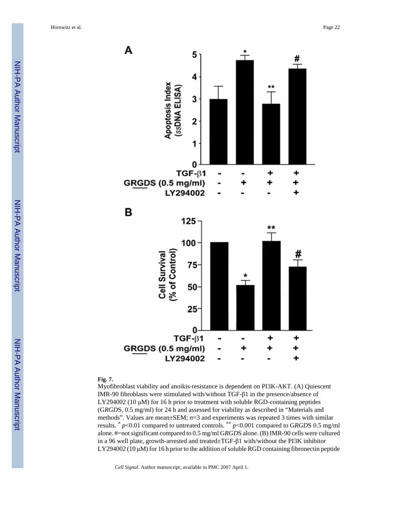
Fig. 7.Myofibroblast viability and anoikis-resistance is dependent on PI3K-AKT. (A) QuiescentIMR-90 fibroblasts were stimulated with/without TGF-β1 in the presence/absence ofLY294002 (10 μM) for 16 h prior to treatment with soluble RGD-containing peptides(GRGDS, 0.5 mg/ml) for 24 h and assessed for viability as described in “Materials andmethods”. Values are mean±SEM; n=3 and experiments was repeated 3 times with similarresults. * p<0.01 compared to untreated controls. ** p<0.001 compared to GRGDS 0.5 mg/mlalone. #=not significant compared to 0.5 mg/ml GRGDS alone. (B) IMR-90 cells were culturedin a 96 well plate, growth-arrested and treated±TGF-β1 with/without the PI3K inhibitorLY294002 (10 μM) for 16 h prior to the addition of soluble RGD containing fibronectin peptide
Horowitz et al. Page 22
Cell Signal. Author manuscript; available in PMC 2007 April 1.
NIH
-PA Author Manuscript
NIH
-PA Author Manuscript
NIH
-PA Author Manuscript

(GRGDS 0.5 mg/ml) for 24 h. Apoptosis was assessed with ELISA for ssDNA. Values aremean±SEM; n=4 per condition, and the experiment was repeated three times with similarresults. * p<0.05 compared to untreated control. ** p<0.05 compared to 0.5 mg/ml GRGDStreatment alone. #=not significant compared to 0.5 mg/ml GRGDS alone.
Horowitz et al. Page 23
Cell Signal. Author manuscript; available in PMC 2007 April 1.
NIH
-PA Author Manuscript
NIH
-PA Author Manuscript
NIH
-PA Author Manuscript

Fig. 8.Model of combinatorial signalling by TGF-β1 of the inducible activation of FAK and AKT inanoikis-resistant myofibroblasts. TGF-β1 activates PKB/ AKT by a p38 MAPK-dependent,SMAD3 independent mechanism; while, activation of FAK is mediated via a SMAD3-dependent, p38 MAPK-independent pathway in human lung fibroblasts. The activation of thesepathways occurs concurrently with the induction of myofibroblast differentiation andacquisition of an anoikis-resistant phenotype. Inducible activation of the PI3K-AKT pathwayby TGF-β1 contributes to anoikis-resistance in myofibroblasts.
Horowitz et al. Page 24
Cell Signal. Author manuscript; available in PMC 2007 April 1.
NIH
-PA Author Manuscript
NIH
-PA Author Manuscript
NIH
-PA Author Manuscript
Copyright © 2022 FDOKUMEN