Tetrathiomolybdate inhibits head and neck cancer metastasis by decreasing tumor cell motility,...
11
RESEARCH Open Access Tetrathiomolybdate inhibits head and neck cancer metastasis by decreasing tumor cell motility, invasiveness and by promoting tumor cell anoikis Pawan Kumar 1* , Arti Yadav 1 , Samip N Patel 2 , Mozaffarul Islam 1 , Quintin Pan 1 , Sofia D Merajver 3 , Theodoros N Teknos 1 Abstract Background: The metastatic spread of solid tumors is directly or indirectly responsible for most cancer-related deaths. Tumor metastasis is very complex and this process requires a tumor cell to acquire enhanced motility, invasiveness and anoikis resistance to successfully establish a tumor at a distal site. Metastatic potential of tumor cells is directly correlated with the expression levels of several angiogenic cytokines. Copper is a mandatory cofactor for the function of many of these angiogenic mediators as well as other proteins that play an important role in tumor cell motility and invasiveness. We have previously shown that tetrathiomolybdate (TM) is a potent chelator of copper and it mediates its anti-tumor effects by suppressing tumor angiogenesis. However, very little is known about the effect of TM on tumor cell function and tumor metastasis. In this study, we explored the mechanisms underlying TM-mediated inhibition of tumor metastasis. Results: We used two in vivo models to examine the effects of TM on tumor metastasis. Animals treated with TM showed a significant decrease in lung metastasis in both in vivo models as compared to the control group. In addition, tumor cells from the lungs of TM treated animals developed significantly smaller colonies and these colonies had significantly fewer tumor cells. TM treatment significantly decreased tumor cell motility and invasiveness by inhibiting lysyl oxidase (LOX) activity, FAK activation and MMP2 levels. Furthermore, TM treatment significantly enhanced tumor cell anoikis by activating p38 MAPK cell death pathway and by downregulating XIAP survival protein expression. Conclusions: Taken together, these results suggest that TM is a potent suppressor of head and neck tumor metastasis by modulating key regulators of tumor cell motility, invasiveness and anoikis resistance. Background Head and neck squamous cell carcinoma (HNSCC) is the sixth most frequent cancer worldwide and five-year survival rates (< 50%) are among the lowest of the major cancers [1,2]. The high mortality associated with advanced head and neck cancers is in large part due to the local spread by primary tumors as well as distal tumor metastasis to vital organs [3,4]. Pulmonary metas- tases are the most frequent in HNSCC, accounting for 66% of distal metastases. Other metastatic sites include bone (22%), liver (10%), skin, mediastinum and bone marrow [4]. HNSCC tumors and their vasculature express numerous angiogenic cytokines including vascu- lar endothelial growth factor (VEGF), interleukin (IL) 1a, IL-6, IL-8, and fibroblast growth factor (FGF) [5-7] which facilitate tumor growth and progression. We and others have demonstrated that VEGF expression directly correlates with poor prognosis in head and neck cancer patients [8-11]. We have recently shown that VEGF, in addition to its pro-angiogenic function, also induces the expression of Bcl-2 in the microvascular endothelial cells [12]. Furthermore, tumor samples from head and * Correspondence: [email protected] 1 Department of Otolaryngology-Head and Neck Surgery and Comprehensive Cancer Center, The Ohio State University, Columbus, OH, USA Kumar et al. Molecular Cancer 2010, 9:206 http://www.molecular-cancer.com/content/9/1/206 © 2010 Kumar et al; licensee BioMed Central Ltd. This is an Open Access article distributed under the terms of the Creative Commons Attribution License (http://creativecommons.org/licenses/by/2.0), which permits unrestricted use, distribution, and reproduction in any medium, provided the original work is properly cited.
Transcript of Tetrathiomolybdate inhibits head and neck cancer metastasis by decreasing tumor cell motility,...

RESEARCH Open Access
Tetrathiomolybdate inhibits head and neckcancer metastasis by decreasing tumor cellmotility, invasiveness and by promoting tumorcell anoikisPawan Kumar1*, Arti Yadav1, Samip N Patel2, Mozaffarul Islam1, Quintin Pan1, Sofia D Merajver3,Theodoros N Teknos1
Abstract
Background: The metastatic spread of solid tumors is directly or indirectly responsible for most cancer-relateddeaths. Tumor metastasis is very complex and this process requires a tumor cell to acquire enhanced motility,invasiveness and anoikis resistance to successfully establish a tumor at a distal site. Metastatic potential of tumorcells is directly correlated with the expression levels of several angiogenic cytokines. Copper is a mandatorycofactor for the function of many of these angiogenic mediators as well as other proteins that play an importantrole in tumor cell motility and invasiveness. We have previously shown that tetrathiomolybdate (TM) is a potentchelator of copper and it mediates its anti-tumor effects by suppressing tumor angiogenesis. However, very little isknown about the effect of TM on tumor cell function and tumor metastasis. In this study, we explored themechanisms underlying TM-mediated inhibition of tumor metastasis.
Results: We used two in vivo models to examine the effects of TM on tumor metastasis. Animals treated with TMshowed a significant decrease in lung metastasis in both in vivo models as compared to the control group. Inaddition, tumor cells from the lungs of TM treated animals developed significantly smaller colonies and thesecolonies had significantly fewer tumor cells. TM treatment significantly decreased tumor cell motility andinvasiveness by inhibiting lysyl oxidase (LOX) activity, FAK activation and MMP2 levels. Furthermore, TM treatmentsignificantly enhanced tumor cell anoikis by activating p38 MAPK cell death pathway and by downregulating XIAPsurvival protein expression.
Conclusions: Taken together, these results suggest that TM is a potent suppressor of head and neck tumormetastasis by modulating key regulators of tumor cell motility, invasiveness and anoikis resistance.
BackgroundHead and neck squamous cell carcinoma (HNSCC) isthe sixth most frequent cancer worldwide and five-yearsurvival rates (< 50%) are among the lowest of themajor cancers [1,2]. The high mortality associated withadvanced head and neck cancers is in large part due tothe local spread by primary tumors as well as distaltumor metastasis to vital organs [3,4]. Pulmonary metas-tases are the most frequent in HNSCC, accounting for
66% of distal metastases. Other metastatic sites includebone (22%), liver (10%), skin, mediastinum and bonemarrow [4]. HNSCC tumors and their vasculatureexpress numerous angiogenic cytokines including vascu-lar endothelial growth factor (VEGF), interleukin (IL)1a, IL-6, IL-8, and fibroblast growth factor (FGF) [5-7]which facilitate tumor growth and progression. We andothers have demonstrated that VEGF expression directlycorrelates with poor prognosis in head and neck cancerpatients [8-11]. We have recently shown that VEGF, inaddition to its pro-angiogenic function, also induces theexpression of Bcl-2 in the microvascular endothelialcells [12]. Furthermore, tumor samples from head and
* Correspondence: [email protected] of Otolaryngology-Head and Neck Surgery and ComprehensiveCancer Center, The Ohio State University, Columbus, OH, USA
Kumar et al. Molecular Cancer 2010, 9:206http://www.molecular-cancer.com/content/9/1/206
© 2010 Kumar et al; licensee BioMed Central Ltd. This is an Open Access article distributed under the terms of the Creative CommonsAttribution License (http://creativecommons.org/licenses/by/2.0), which permits unrestricted use, distribution, and reproduction inany medium, provided the original work is properly cited.

neck cancer patients showed significantly higher Bcl-2expression in tumor blood vessels [13] and thisenhanced Bcl-2 expression in tumor-associated endothe-lial cells was directly correlated with metastatic status ofthese patients [14]. Upregulated Bcl-2 expression intumor-associated endothelial cells was sufficient toenhance tumor angiogenesis, tumor progression andtumor metastasis of oral squamous cell carcinoma in aSCID mouse model [14].Tumor metastasis is a complex process consisting of
multiple individual steps [15]. The metastatic processrequires a tumor cell to acquire the ability to migratethrough the primary tumor mass, intravasate and sur-vive in blood or lymphatic vascular system, and extrava-sate from the vascular system into a secondary organ toform the metastatic nodules. A key process in basic cellmigration is the ability of a cell to form a stable adhe-sion to the extracellular matrix [16]. This process isregulated by two key proteins within cell: Src and focaladhesion kinase (FAK) [17]. Inactivation of either ofthese proteins leads to dramatic loss in the cell motility.FAK activation in squamous cell carcinoma and lungadenocarcinoma has been shown to promote cell inva-sion [18,19]. In addition, FAK signaling alters matrixmetalloproteinases (MMPs, a family of zinc-containingendopeptidases that degrade various components of theextracellular matrix) expression and promotes the gen-eration of an invasive cell phenotype. Overexpression ofMMP-2 in tumor tissue has been linked with tumorinvasion, metastasis and poor survival in many tumortypes including HNSCC [20-22]. FAK gene silencing byRNA interference also inhibited tumor cell metastasis bypromoting tumor cell anoikis (anchorage-dependentcells undergoing cell death due to cell detachment) [23].Recently Payne et al, have demonstrated that lysyl oxi-dase (LOX), a copper dependent kinase, promotestumor cell migration and invasiveness via the activationof FAK [24].Anti-cancer therapy involving copper suppression has
been shown to be an effective inhibitor of angiogenesis[25,26]. Copper is an essential trace element whose dis-tinct angiogenic properties were first discovered in theearly 1980’s [27]. Copper has since shown to be a directstimulator of endothelial cell proliferation and migration[27,28]. Additionally, copper is a required cofactor forthe activity, production and secretion of key angiogeniccytokines up-regulated in head and neck cancers [5,29].Tetrathiomolybdate (TM) is a potent chelator of copperthat has been widely studied for its good oral bioavail-ability, low toxicity profile and its tumor suppressiveeffects [25,30,31]. We have previously shown that TM isa potent angiogenesis inhibitor and inhibits tumorgrowth and tumor metastasis [5,29]. However, the mole-cular mechanisms by which TM inhibits tumor
metastasis are poorly understood. In this study, we haveexamined the role and mechanisms of TM-mediatedinhibition of tumor metastasis. Our results suggest thatTM treatment significantly inhibits HNSCC metastasisby decreasing tumor cell motility by inhibiting lysyl oxi-dase and focal adhesion kinase. In addition, TM treat-ment inhibited tumor cell invasiveness by down-regulating MMP2 levels. Moreover, TM treatment alsosignificantly enhanced tumor cell anoikis by activatingp38 MAPK cell death pathway and downregulatingXIAP survival proteins.
MethodsCell culturesPrimary human dermal microvascular endothelial cells(ECs) were purchased from Lonza (Walkersville, MD).ECs were maintained in Endothelial Cell Basal Medium-2 (EBM-2) containing 5% FBS and growth supplements.Oral squamous carcinoma cells (OSCC-3, a kind giftfrom M. Lingen, University of Chicago) were maintainedin Dulbecco’s Modified Eagle Medium (DMEM) supple-mented with 10% FBS.
Transduction of endothelial cells with Bcl-2Bcl-2 was introduced into human microvascularendothelial cells(ECs) as described previously [32]. TheBcl-2 construct or the vector alone was introduced intoPA317 amphotropic packing cells with Lipofectin. Viralsupernatants were collected after 24 hours, centrifuged,filtered, and stored at -70°C. ECs were transduced witheither Bcl-2 (EC-Bcl-2) or control vector (EC-VC) byovernight incubation with one-tenth dilution of the viralsupernatant in the presence of 6 μg/ml polybrene.Transduced ECs were selected by treating them withG418 (200 μg/ml) for one week. Bcl-2 expression inendothelial cells was confirmed by Western blotanalysis.
Generation of oral squamous cell line stably expressingluciferaseTumor cells (OSCC-3) were transfected with pcDNA3.1plasmid containing the firefly luciferase gene [33] (a giftfrom Dr. Alnawaz Rehemtulla, University of Michigan,Ann Arbor) using Lipofectamine 2000 as described pre-viously [34]. Four μg of each plasmid and 20 μl of Lipo-fectamine 2000 reagent were separately diluted in 200 μlof DMEM and incubated for 45 minutes at room tem-perature. The two solutions were gently mixed andfurther incubated for 15 minutes at room temperatureto prepare the lipid-DNA complexes. Subsequently, 1.6ml of DMEM containing 5% FBS was gently mixed withlipid-DNA complexes. Tumor cells were washed twicewith DMEM and then lipid-DNA complexes were over-laid onto the cells. The cells were incubated for 6 hours
Kumar et al. Molecular Cancer 2010, 9:206http://www.molecular-cancer.com/content/9/1/206
Page 2 of 11

at 37°C in a CO2 incubator. At the end of the incuba-tion, DNA containing medium was removed andreplaced with DMEM. Luciferase transduced OSCC-3(OSCC-3-Luc) cells were selected by incubating inDMEM containing 400 μg/ml G418 for 7 days.
Tumor metastasis modelsWe used two in vivo models (flank xenograft and tailvein injection models) to study the effects of TM ontumor metastasis.SCID mouse flank xenograft modelSix weeks old female SCID mice were used in this study.Animals were randomized to receive either TM or steri-lized water, delivered daily p.o. gavaging. The treatmentwas initiated 2 weeks prior to the tumor inoculation andcontinued until the end of the experiment. Plasma ceru-loplasmin is a good surrogate marker for total body cop-per status therefore ceruloplasmin levels were measuredusing oxidase assay as described before [25]. The base-line levels of plasma ceruloplasmin were determined forthe TM group before initiation of treatment. Subse-quently, ceruloplasmin levels were measured biweeklyover the study periods. TM treatment was started at 0.7mg/day per mouse and then titrated biweekly to main-tain ceruloplasmin suppression at 20% to 30% of base-line. OSCC-3-Luc (1 × 10⁶) and endothelial cells (EC-VC or EC-Bcl-2, 1 × 10⁶) were mixed with 100 μl ofMatrigel and injected subcutaneously in the flanks ofSCID mice [14]. Tumor volume measurements beganon day 3 and continued twice a week until the end ofthe study. The length and width of the tumors weremeasured using a digital caliper and tumor volumeswere calculated using the formula, volume (mm3) = L ×W2/2 (length L, mm; width W, mm). After 3 weeks, pri-mary tumors and lungs were carefully removed and ana-lyzed for tumor growth, tumor angiogenesis and tumormetastasis to lungs. Lungs from each mouse weredivided into two parts. One half of each lung was fixedwith 10% buffered formalin and then processed to formparaffin embedded tissue blocks for immunohistochem-istry. The other half of the lung was used to harvesttumor cells. Lungs were finely minced by scissors,washed with sterile serum free media (DMEM) and trea-ted with collagenase (2.5 mg/ml) for 3 hours at 37°Cwith intermittent shaking. After collagenase treatment,cells were plated in 10 cm culture dishes and treatedwith G418 (400 μg/ml) to select tumor cells (OSCC-3-Luc). After one week, tumor cell colonies were countedusing phase contrast microscope (50×).Tail vein metastasis modelIn this study, SCID mice were treated with TM asdescribed above. OSCC-3-Luc cells (0.5 × 10⁵) in 50 μlvolume were injected in the SCID mice via tail veinusing 30 gauge needles. Tumor metastasis to lungs was
monitored by in vivo bioluminescence imaging. After 3weeks, lungs were carefully removed and tumor meta-stasis to lungs was analyzed as described above.
In vivo Bioluminescence ImagingMice were imaged on a cryogenically cooled imagingsystem (Xenogen, Alameda, CA, USA) coupled to a dataacquisition computer. Mice were anesthetized (1.5% Iso-flurane/air mixture), and injected with luciferin in PBSat a dose of 320 mg/kg body weight. Digital gray imageswere captured and overlaid with pseudocolor images,which represent photon counts emitted from active luci-ferase within viable tumor cells. Luminescence emittedfrom each animal was integrated for one-minute inter-vals, from 5-20 minutes after the injection of Luciferin.Image processing and photon count quantitation wereconducted by means of Living Image software(Xenogen).
Quantitaion of angiogenesis by immunolocalization ofvon Willebrand FactorTissue sections were deparaffinized and antigen retrievalwas achieved by pressure cooking in a Decloakingchamber (Biocare Medical, Walnut Creek, CA) at 120°Cfor 20 minutes [35]. Tissue sections were then treatedwith peroxide block solution for 5 minutes at roomtemperature followed by 1 hour of incubation with pri-mary antibody (anti-von Willebrand Factor, Dako, Car-pinteria, CA) at room temperature. Slides were furtherincubated for 30 minutes with HRP labeled polymer(Dako EnVision+ Kit, Carpinteria, CA) and developedwith AEC+chromagen. Microvessel density was calcu-lated by counting 5 random high power fields (200×).
Tumor cell motility assayRandom cell motility was determined using a motilityassay kit (Cellomics, Pittsburgh, PA) as per manufac-turer’s instructions. In brief, OSCC-3 cells were har-vested, suspended in serum-free medium and plated ontop of a field of microscopic fluorescent beads in thepresence or absence of TM (1 nM). After a 16-hoursincubation period, cells were fixed and areas of clearingin the fluorescent bead field corresponding to phagoki-netic cell tracks were quantified using NIH ScionImager.
Lysyl oxidase (LOX) activity assayLOX activity of whole-cell lysates was measured usingthe Amplex Red fluorescence assay kit (MolecularProbes, Eugene, OR). The assay reaction mixture con-sisted of 50 mM sodium borate (pH 8.2), 1.2 M urea,50 μM Amplex Red, 0.1 units/ml horseradish peroxi-dase, and 10 mM 1,5-diaminopentane substrate. TMtreated or untreated control protein samples were addedto the reaction mixture and incubated at 37°C. The
Kumar et al. Molecular Cancer 2010, 9:206http://www.molecular-cancer.com/content/9/1/206
Page 3 of 11

fluorescence intensity was measured every 30 minutesfor 2 hours using a Synergy 4 multidetection microplatereader (BioTek, Winooski, VT). LOX activity isexpressed as fluorescent units.
Western Blot AnalysisOSCC-3 cells were cultured in 6-well plates and treatedwith TM for different time points. Whole cell lysateswere separated by 4-12% NuPAGE Bis-Tris gels (Invitro-gen, Carlsbad, CA) and transferred onto nitrocellulosemembranes using NuPAGE transfer buffer (Invitrogen,Carlsbad, CA). To block nonspecific binding, membraneswere incubated with 5% non-fat milk in Tris buffered sal-ine containing 0.1% Tween-20 (TBST) for 1 hour atroom temperature. Afterwards, the blots were incubatedin the respective primary antibody in TBST + 5% non-fatmilk at 4°C overnight. After washing with TBST, theblots were incubated with horseradish peroxidase-conju-gated sheep anti-mouse IgG (1:10,000) or with goat anti-rabbit IgG (1:10,000) for 1 hour at room temperature. AnECL-plus detection system (Amersham Life Sciences, Pis-cataway, NJ) was used to detect specific protein bands.Protein loading in all the experiments was normalized bystripping the blots and then re-probing with anti-tubulinantibody.
Tumor cell invasion assaysThe role of TM in tumor cell invasion was investigatedusing a Matrigel invasion assay [14]. 24-well plateinserts (8 μM pore size, Falcon) were coated with 20 μlof Matrigel and incubated at 37°C for 30 minutes to letthe Matrigel polymerize. Next, 50,000 tumor cells(OSCC-3) were carefully layered on top of the Matrigeland the inserts were placed in the 24 well plates in thepresence or absence of TM (1 nM). The plates werefurther incubated for 24 hours at 37°C and the non-invaded cells were carefully removed with a cottonswab. The inserts were then stained with Diff-quicksolution II and mounted on glass slides. The number ofcells that had invaded through the Matrigel was countedin 5 high power fields.
MMP analysis by zymographyOSCC-3 cells (5 × 10⁵) were cultured in a 6-well plate.Separately, EC-VC or EC-Bcl-2 (5 × 10⁵) cells were pla-ted on top of collagen coated inserts and then theseinserts were carefully placed in the 6-well plates con-taining OSCC-3 cells in the presence or absence of TM(1 nM). After 24 hours, OSCC-3 cells were washed andfurther cultured in serum free media (DMEM) for 24hours. At the end of incubation, culture supernatantswere collected and mixed with non-reducing SDS gelsample buffer (3:1) and applied without boiling to a 10%polyacrylamide gelatin gel (Invitrogen, Carlsbad, CA).
After electrophoresis, the gels were washed with ddH2Oand incubated in renaturing buffer (50 mM Tris-HCl,pH 7.5) containing 2% Triton X-100 for 30 minutes atroom temperature, and then incubated in developingbuffer (50 mM Tris-HCl, pH 7.5) containing 5 mMCaCl2 and 1 M ZnCl2 at 37°C for 16 hours. Gels werestained by 0.5% Coomassie Brilliant Blue R-250 solutionto visualize the bands. The band density was measuredusing Alpha Imager software (Alpha Innotech, SanLeandro, CA).
Tumor cell anoikis assayTo evaluate the tumor cell anoikis in non-adherent con-ditions, OSCC-3 cells (5 × 10⁵) were cultured in a 6-well plate over a thick layer of 1% agar in DMEM con-taining 2% serum. Tumor cells cultured in adherentconditions were used as a control. In the TM treatmentexperiments, tumor cells were pre-treated with TM (1nM) for 24 hours and then cultured on top of 1% agar.At the end of incubation, cells were carefully retrievedand analyzed by TUNEL staining for anoikis [12]. Inbrief, tumor cells were fixed with cytofix buffer (BDcytofix, BD Biosciences, San Jose, CA) for 15 minutes at4°C, and then stored overnight in 70% ethanol at -20°C.The percentage of apoptotic cells were then evaluatedusing the APO-BRDU terminal deoxynucleotidyl trans-ferase (TdT)-mediated dUTP-biotin nick end labeling(TUNEL) assay according to the manufacturer’s instruc-tions (Sigma, St. Louis, MO). Apoptotic tumor cellswere quantitated by flow cytometry using an argon laserexcited at 488 nm (BD Biosciences, San Jose, CA).
Statistical analysisData from all the experiments are expressed as mean ±SEM. Statistical differences were determined by two-wayanalysis of variance and Student’s t test. A p value of< 0.05 was considered significant.
ResultsTM treatment significantly inhibited EC-Bcl-2 mediatedtumor growth and angiogenesisWe have previously shown that elevated expression ofBcl-2 in tumor-associated endothelial cells directly cor-relates with tumor metastasis in head and neck cancerpatients [14]. In addition, we also demonstrated that co-implantation of EC-Bcl-2 along with oral squamous car-cinoma cells (OSCC-3) in SCID mice significantlyenhances tumor growth and tumor metastasis to lungs.In this study, we examined if treatment with an anti-angiogenic agent (tetrathiomolybdate, TM) could inhibittumor growth and tumor angiogenesis in this aggressivesquamous cell carcinoma model. The level of plasmaceruloplasmin, which is a good surrogate marker fortotal body copper status, was monitored biweekly and
Kumar et al. Molecular Cancer 2010, 9:206http://www.molecular-cancer.com/content/9/1/206
Page 4 of 11

TM dose was adjusted accordingly to maintain cerulo-plasmin suppression at 20% to 30% of baseline. OSCC-3tumors containing EC-Bcl-2 showed significantly highertumor growth (Fig. 1A) as compared to OSCC-3 tumorscontaining endothelial cells with vector alone (EC-VC).In addition, OSCC-3 tumors containing EC-Bcl-2showed significantly higher tumor weight (Fig. 1B) atthe end of study as compared to OSCC-3 containingEC-VC. TM treatment significantly inhibited tumorgrowth and tumor weights in OSCC-3 tumor containingEC-Bcl-2. In addition, TM treatment significantly inhib-ited blood vessel density in OSCC-3 tumors containingEC-Bcl-2 cells (Fig. 1C-D). TM treatment did not causeany animal mortality or induce significant decrease inbody weight (less than 5% weight loss in TM treatment
as compared control, data not shown). TM treatmentalso did not induce any systemic toxicity such asrespiratory depression or dry scaly skin.
TM treatment significantly inhibited tumor metastasis tolungsWe used two in vivo models (flank xenograft and tailvein injection models) to examine if TM treatmentcould inhibit tumor metastasis. OSCC-3 tumors popu-lated with EC-Bcl-2 showed significantly higher metasta-sis to lungs as compared to OSCC-3 tumors populatedwith EC-VC (Fig. 2). In addition to higher number ofmetastatic nodules, OSCC-3 tumors containing EC-Bcl-2 also had larger lung metastasis nodes (Fig. 2B). Lungsharvested from animals co-implanted with OSCC-3 and
EC-VC EC-Bcl-23)
EC-VC CA
200
300
400
olum
e (m
m3 EC-VC+TM
EC-Bcl-2
EC-Bcl-2+TM*
*
EC-VC+TM EC-Bcl-2+TM
0
100
200
Tum
or v
o *
60
80
sity
**
200
250
(%) *
3 6 9 12 15 18 21Days
DB
20
40
60ro
vess
el d
ens
*
50
100
150
200
mor
wei
ght (
0Mic
0
50
Tum
Figure 1 Tetrathiomolybdate (TM) treatment significantly inhibits HNSCC tumor growth and tumor angiogenesis. OSCC-3 and EC-Bcl-2or EC-VC cells were mixed with 100 μl of Matrigel and injected in the flanks of SCID mice. Tumor volume measurements began on day 3 andcontinued twice a week until the end of the study. Length and width were measured using a digital caliper and tumor volumes were calculatedusing the formula, volume (mm3) = L × W2/2 (length L, mm; width W, mm). A; tumor progression curves for different experimental groups:OSCC-3 plus endothelial cells transduced with vector alone (EC-VC); OSCC-3 plus endothelial cells transduced with vector alone and treated withTM (EC-VC+TM); OSCC-3 plus endothelial cells transduced with Bcl-2 (EC-Bcl-2); OSCC-3 plus endothelial cells transduced with Bcl-2 and treatedwith TM (EC-Bcl-2+TM). B; tumor weights at the end of the study. C-D; Paraffin embedded tumor sections were stained for tumor blood vesselsusing anti-human von Willebrand Factor antibodies. Microvessel density in the tumor samples was calculated by counting 5 random fields (x200). *, represents a significant difference (p < 0.05) as compared to the control group.
Kumar et al. Molecular Cancer 2010, 9:206http://www.molecular-cancer.com/content/9/1/206
Page 5 of 11

EC-Bcl-2 showed significantly higher number of coloniesand contained significantly higher number of cells ascompared to OSCC-3 and EC-VC group. TM treatedanimals showed significantly lower number of tumorcell colonies and cells (Fig. 2C-D).In the tail vein metastasis model, TM treatment mark-
edly inhibited tumor metastasis to lungs at both day 16and 21 (Fig. 3A-B). Similarly, lungs harvested from TMtreated animals showed significantly lower number ofcolonies and tumor cells (Fig. 3C-D).
TM treatment markedly reduced oral squamous cellmotility and invasivenessTumor cells cultured in the presence of serum (S+)exhibited significantly higher motility as compared totumor cells cultured in the absence of serum (S-). TMtreatment significantly inhibited tumor cell motility both
in the presence (S+TM+) and absence (S-TM+) ofserum (Fig. 4A-B). Lysyl oxidase (LOX), a copper-dependent amine oxidase, has been shown to promotetumor cell migration and invasion [24]. We next exam-ined if TM mediated its inhibitory effect on tumor cellmotility by inhibiting LOX. Indeed, TM treatment ofOSCC-3 cells significantly inhibited LOX activity (Fig.4C). In addition, TM also inhibited the activation offocal adhesion kinase (FAK), an important cell migrationmediator (Fig. 4D).In the next set of experiments, we examined if TM
affects OSCC-3 cell invasiveness. TM treatment signifi-cantly inhibited OSCC-3 invasiveness (Fig. 5A-B).Matrix matelloproteinases (MMPs) plays an importantrole in tumor cell invasiveness by degrading extracellularmatrix. We also examined whether EC-Bcl-2 cells couldenhance MMPs production by OSCC-3 cells and if TM
OSCC-3-EC-VC OSCC-3 EC-Bcl-2A B OSCC-3-EC-VC OSCC-3 EC-Bcl-2
OSCC-3 EC-Bcl-2 + TMOSCC-3- EC-VC + TM OSCC-3- EC-VC + TM OSCC-3 EC-Bcl-2 + TM
**
20
25
ies/
field
*
4
5
6
mill
ions
)
DC *
5
10
15
mbe
r of c
olon
i
*
2
3m
ber o
f cel
ls (m
*
0
5
Num
0
1
Num
Figure 2 TM treatment significantly inhibits EC-Bcl-2 mediated HNSCC tumor metastasis. OSCC-3 and EC-Bcl-2 or EC-VC were mixed with100 μl of Matrigel and injected in the flanks of SCID mice. Lungs from SCID mice were carefully removed on day 21. One half of each lung wasfixed and paraffin embedded for immunohistochemical analysis. From the other half of the lung, tumor cells were harvested and selected byG418 treatment. A; representative gross lungs photomicrograph from different experimental groups. B; representative photomicrograph of lungsections from different experimental groups. C; Number of tumor cell colonies present in different groups. D; Number of tumor cell present indifferent groups. *, represents a significant difference (p < 0.05) as compared to the control group.
Kumar et al. Molecular Cancer 2010, 9:206http://www.molecular-cancer.com/content/9/1/206
Page 6 of 11

treatment could inhibit the secretion of MMPs. OSCC-3cells co-cultured with EC-Bcl-2 showed significantincrease in MMP2 secretion as compared to OSCC-3cells co-cultured with EC-VC (Fig. 5A-B). TM treatmentof EC-Bcl-2 and OSCC-3 significantly inhibited MMP2production (Fig. 5C-D).
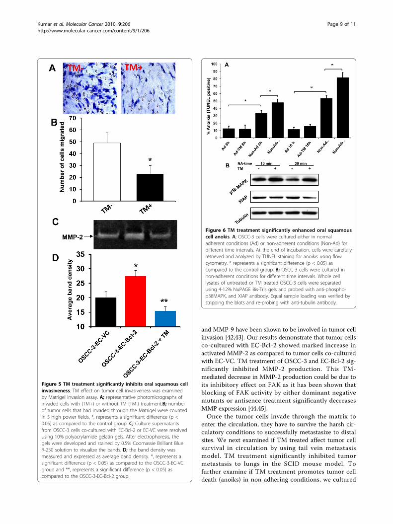
TM treatment significantly inhibited tumor cell anoikisIn our tail vein metastasis model, we observed signifi-cant inhibition of tumor metastasis in TM treated ani-mals. We further examined if TM inhibits tumormetastasis in tail vein model by promoting tumor cellanoikis. TM treatment significantly enhanced tumor cell
anoikis in a time dependent manner (Fig 6A). Interest-ingly, TM had no effect on adherent OSCC-3 cells(Fig 6A). Survival proteins XIAP and Bcl-2 play animportant role in anoikis resistance. We next examinedif TM treatment downregulates the expression of thesesurvival proteins in non-adherent conditions. TM treat-ment markedly downregulated XIAP protein expressionin a time dependent manner (Fig. 6B). However, TMtreatment did not alter the Bcl-2 expression (data notshown). Moreover, TM treatment also activated stressactivated p38 MAPK cell death signaling pathway(Fig. 6B).
DiscussionDistant metastases in head and neck cancer almostinvariably herald a poor prognosis with an average survi-val of 6 months and treatment is usually palliative [36].Five year survival rates for early stage localized head andneck cancers are over 80% but this drop to 40% wheredisease has spread to neck nodes, and to below 20% forpatients with distant metastatic disease [37]. Currentlyused treatment regimens (surgery and/or chemo-radia-tion) are often ineffective in controlling the metastaticspread of disease [3]. It is therefore very important todevelop novel therapies for patients with metastatic dis-ease. Copper is a mandatory cofactor for a number ofproteins that play an important role in angiogenesis,tumor cell migration and survival [38]. We have pre-viously shown that TM, a potent copper chelator, inhi-bits tumor growth and tumor metastasis by suppressingangiogenesis [29]. Recently, Juarez et al, have shownthat TM attenuates angiogenesis and tumor cell prolif-eration by inhibiting superoxide dismutase 1 (SOD1)[30]. However, very little is known about the role of TMon key metastatic processes, particularly tumor cellmigration, invasion and anoikis resistance. In this study,we have examined the mechanisms by which TM inhi-bits tumor cell motility, invasiveness and anoikisresistance.We used two in vivo models (flank xenograft model
and tail vein model) to test the efficacy of TM in inhi-biting tumor metastasis. In the first in vivo model, oralsquamous carcinoma cells (OSCC-3) were co-implantedalong with endothelial cells expressing Bcl-2 (EC-Bcl-2)in the flanks of SCID mice [14]. The rationale for co-implanting EC-Bcl-2 along with OSCC-3 is based onour previous work where we have demonstrated thatco-implantation of EC-Bcl-2 along with tumor cells sig-nificantly enhances tumor metastasis. In addition, theseEC-Bcl-2 containing tumors exhibited highly invasivephenotype and abnormal vascular network as commonlyobserved with aggressive human tumors. Therefore, thishighly aggressive xenograft model is ideally suited totest the efficacy of TM on tumor metastasis. Our results
Day 16 Day 21 TM- TM+ TM- TM+
A
B240
280
(x10
00)
TM-
TM+*
120
160
200
Flux
uni
ts
TM-
TM+
*
C
120Day 16 Day 21
D
**
Figure 3 TM treatment significantly inhibits HNSCC tumormetastasis. OSCC-3-Luc cells were injected in the SCID mice via tailvein. Tumor metastasis to lungs was monitored by in vivobioluminescence imaging. A; representative photomicrograph ofanimals treated with (TM+) or without TM (TM-) at days 16 and 21.B; Quantitative data of bioluminescence imagining as expressed asflux units. C; Tumor cells from lungs were harvested and tumor cellcolonies were selected by G418 treatment (200 μg/ml) for oneweek and counted. D; Tumor cell colonies were then trypsinizedand total number of cells were counted. *, represents a significantdifference (p < 0.05) as compared to the control group.
Kumar et al. Molecular Cancer 2010, 9:206http://www.molecular-cancer.com/content/9/1/206
Page 7 of 11

suggest that TM inhibits tumor metastasis by blockingmultiple steps that are involved in tumor metastasiscascade (tumor cell migration, invasion and survival incirculation) to reach the distal site.Tumor cell motility and invasiveness are key characteris-
tics of aggressive metastatic tumors as they play an impor-tant role in tumor cell release from the primary tumorsinto the circulation. To examine if TM treatment affectstumor cell motility, we used a random cell motility assay.TM treatment significantly inhibited tumor cell motilityboth in the presence and in the absence of serum. Cellmigration is a dynamic process that is regulated by the for-mation or turnover of focal contacts. Focal adhesionkinase (FAK) is the key member of the focal contactassembly and FAK activation is required for optimal cell
motility [39]. TM treatment significantly inhibited FAKactivation as well as the activation of LOX. LOX, a cop-per-dependent amine oxidase, was initially reported asresponsible for the catalysis of collagen and elastin cross-linking within the extracellular matrix [40]. However,recent work has shown that LOX regulates a number ofcellular functions including cell migration via the activa-tion of FAK/Src pathway [41,24]. Therefore, TM may beinhibiting tumor cell migration by inhibiting FAK activa-tion via lysyl oxidase. We further examined the role of TMin tumor cell invasiveness by using Matrigel invasionassay. TM treatment significantly inhibited oral squamouscell invasion through Matrigel. Matrix metalloproteinases(MMPs) play an important role in tumor cell invasivenessby digesting the extracellular matrix. In HNSCC, MMP-2
Figure 4 TM treatment significantly inhibits oral squamous cell motility. A-B; OSCC-3 cells were cultured on top of a field of microscopicfluorescent beads in the following culture conditions: in the absence of serum and TM (S-TM-); in the presence of serum but no TM (S+TM-); inthe absence of serum but presence of TM (S-TM+) or in the presence of serum and TM (S+TM+). After a 16-hours incubation period, cells werefixed and areas of clearing in the fluorescent bead field corresponding to phagokinetic cell tracks were quantified using NIH ScionImager. C; LOXcatalytic activity was measured in whole-cell lysates of untreated (NT) or TM treated OSCC-3 cells (30 minutes). *, represents a significantdifference (p < 0.05) as compared to the control group. D; Whole cell lysates of untreated or TM treated OSCC-3 cells were separated using 4-12% NuPAGE Bis-Tris gels and probed with anti-phospho-FAK antibody. Equal sample loading was verified by stripping the blots and re-probingwith anti-tubulin antibody.
Kumar et al. Molecular Cancer 2010, 9:206http://www.molecular-cancer.com/content/9/1/206
Page 8 of 11
and MMP-9 have been shown to be involved in tumor cellinvasion [42,43]. Our results demonstrate that tumor cellsco-cultured with EC-Bcl-2 showed marked increase inactivated MMP-2 as compared to tumor cells co-culturedwith EC-VC. TM treatment of OSCC-3 and EC-Bcl-2 sig-nificantly inhibited MMP-2 production. This TM-mediated decrease in MMP-2 production could be due toits inhibitory effect on FAK as it has been shown thatblocking of FAK activity by either dominant negativemutants or antisence treatment significantly decreasesMMP expression [44,45].Once the tumor cells invade through the matrix to
enter the circulation, they have to survive the harsh cir-culatory conditions to successfully metastasize to distalsites. We next examined if TM treated affect tumor cellsurvival in circulation by using tail vein metastasismodel. TM treatment significantly inhibited tumormetastasis to lungs in the SCID mouse model. Tofurther examine if TM treatment promotes tumor celldeath (anoiks) in non-adhering conditions, we cultured
Figure 5 TM treatment significantly inhibits oral squamous cellinvasiveness. TM effect on tumor cell invasiveness was examinedby Matrigel invasion assay. A; representative photomicrographs ofinvaded cells with (TM+) or without TM (TM-) treatment.B; numberof tumor cells that had invaded through the Matrigel were countedin 5 high power fields. *, represents a significant difference (p <0.05) as compared to the control group. C; Culture supernatantsfrom OSCC-3 cells co-cultured with EC-Bcl-2 or EC-VC were resolvedusing 10% polyacrylamide gelatin gels. After electrophoresis, thegels were developed and stained by 0.5% Coomassie Brilliant BlueR-250 solution to visualize the bands. D; the band density wasmeasured and expressed as average band density. *, represents asignificant difference (p < 0.05) as compared to the OSCC-3-EC-VCgroup and **, represents a significant difference (p < 0.05) ascompared to the OSCC-3-EC-Bcl-2 group.
Figure 6 TM treatment significantly enhanced oral squamouscell anokis. A; OSCC-3 cells were cultured either in normaladherent conditions (Ad) or non-adherent conditions (Non-Ad) fordifferent time intervals. At the end of incubation, cells were carefullyretrieved and analyzed by TUNEL staining for anoikis using flowcytometry. * represents a significant difference (p < 0.05) ascompared to the control group. B; OSCC-3 cells were cultured innon-adherent conditions for different time intervals. Whole celllysates of untreated or TM treated OSCC-3 cells were separatedusing 4-12% NuPAGE Bis-Tris gels and probed with anti-phospho-p38MAPK, and XIAP antibody. Equal sample loading was verified bystripping the blots and re-probing with anti-tubulin antibody.
Kumar et al. Molecular Cancer 2010, 9:206http://www.molecular-cancer.com/content/9/1/206
Page 9 of 11

OSCC-3 cells on top of 1% agar in the presence orabsence of TM. TM treatment significantly enhancedtumor cell anoikis. Interestingly, TM treatment did notaffect OSCC-3 survival in adherent condition. TM couldbe promoting tumor cell anoikis by increasing oxidativestress (p38 MAPK activation) in the cells by down mod-ulating superoxide dismutase (SOD) and X-linked inhi-bitor of apoptosis (XIAP) , as copper is a cofactor forboth SOD and XIAP proteins [30,46]. SOD protectscells from oxidative stress by catalyzing the dispropor-tionation of superoxide to hydrogen peroxide [47],whereas XIAP protein modulates oxidative stress bybinding to apoptosis-inducing factor (AIF) [46]. In addi-tion, XIAP has been shown to promote cell survival bydirectly binding to caspase 3 and inhibiting its functionby blocking substrate binding [48].
ConclusionsIn conclusion, our results demonstrate that TM signifi-cantly inhibits head and neck tumor metastasis by mod-ulating a number of critical steps involved in tumormetastasis cascade including angiogenesis, tumor cellmotility, invasiveness and tumor cell anoikis. Therefore,TM with its low toxicity profile and good oral bioavail-ability is a potentially novel candidate to control tumormetastasis in head and neck cancer patients.
AbbreviationsHNSCC: head and neck squamous cell carcinoma; OSCC-3: oral squamouscell carcinoma-3; EC: endothelial cell; TM: tetrathiomolybdate; FAK: focaladhesion kinase; LOX: lysyl oxidase; XIAP: X-linked Inhibitor of ApoptosisProtein; VEGF: vascular endothelial cell growth factor; SCID: Severe combinedimmunodeficiency disease; MMP: Matrix metalloproteinases.
AcknowledgementsWe thank Dr. M. Lingen (University of Chicago) for providing OSCC-3 cellline and Dr. A. Rehemtulla (University of Michigan) for providing pcDNA3.1luciferase plasmid. This work was funded by National Cancer Institute SPOREgrants P50CA97248 (PK, TNT and SDM), NCI K-Award CA133250 (PK), NIHCA77612 (SDM) and by the Breast Cancer Research Foundation (SDM).
Author details1Department of Otolaryngology-Head and Neck Surgery and ComprehensiveCancer Center, The Ohio State University, Columbus, OH, USA. 2Departmentof Otolaryngology-Head and Neck Surgery, University of South Florida,Tampa, FL, USA. 3Department of Internal Medicine, Division of Hematologyand Oncology, University of Michigan, Ann Arbor, MI, USA.
Authors’ contributionsPK conceived of the study, participated in the design, carried out some ofthe in vivo, anoikis & invasion studies, and drafted the manuscript. AY carriedout the molecular mechanistic studies. SP carried out in vivo studies. MIcarried out the immunohistochemistry. QP carried out tumor cell motilitystudies, SDM participated in the design and coordination of the studies. TNTparticipated in conceiving, design and coordination of the studies. Allauthors red and approved the final version.
Competing interestsDr. Merajver has equity and was a consultant for Attenuon, LLC, an entitywhich has licensed tetrathiomolybdate for anti-angiogenic applications fromthe University of Michigan.
Received: 27 May 2010 Accepted: 3 August 2010Published: 3 August 2010
References1. Ragin CC, Modugno F, Gollin SM: The epidemiology and risk factors of
head and neck cancer: a focus on human papillomavirus. J Dent Res2007, 86:104-114.
2. Jemal A, Thomas A, Murray T, Thun M: Cancer statistics, 2002. CA Cancer JClin 2002, 52:23-47.
3. Deschamps DR, Spencer HJ, Kokoska MS, Spring PM, Vural EA, Stack BC Jr:Implications of head and neck cancer treatment failure in the neck.Otolaryngol Head Neck Surg 142:722-727.
4. Ferlito A, Shaha AR, Silver CE, Rinaldo A, Mondin V: Incidence and Sites ofDistant Metastases from Head and Neck Cancer. ORL 2001, 63:202-207.
5. Teknos TN, Islam M, Arenberg DA, Pan Q, Carskadon SL, Abarbanell AM,Marcus B, Paul S, Vandenberg CD, Carron M, et al: The effect oftetrathiomolybdate on cytokine expression, angiogenesis, and tumorgrowth in squamous cell carcinoma of the head and neck. ArchOtolaryngol Head Neck Surg 2005, 131:204-211.
6. Lathers DM, Young MR: Increased aberrance of cytokine expression inplasma of patients with more advanced squamous cell carcinoma of thehead and neck. Cytokine 2004, 25:220-228.
7. Chen Z, Malhotra PS, Thomas GR, Ondrey FG, Duffey DC, Smith CW,Enamorado I, Yeh NT, Kroog GS, Rudy S, et al: Expression ofproinflammatory and proangiogenic cytokines in patients with head andneck cancer. Clin Cancer Res 1999, 5:1369-1379.
8. Smith BD, Smith GL, Carter D, Sasaki CT, Haffty BG: Prognostic significanceof vascular endothelial growth factor protein levels in oral andoropharyngeal squamous cell carcinoma. J Clin Oncol 2000, 18:2046-2052.
9. Masuda M, Ruan HY, Ito A, Nakashima T, Toh S, Wakasaki T, Yasumatsu R,Kutratomi Y, Komune S, Weinstein IB: Signal transducers and activators oftranscription 3 up-regulates vascular endothelial growth factorproduction and tumor angiogenesis in head and neck squamous cellcarcinoma. Oral Oncol 2007, 43:785-790.
10. Sauter ER, Nesbit M, Watson JC, Klein-Szanto A, Litwin S, Herlyn M: Vascularendothelial growth factor is a marker of tumor invasion and metastasisin squamous cell carcinomas of the head and neck. Clin Cancer Res 1999,5:775-782.
11. Teknos TN, Cox C, Yoo S, Chepeha DB, Wolf GT, Bradford CR, Carey TE,Fisher SG: Elevated serum vascular endothelial growth factor anddecreased survival in advanced laryngeal carcinoma. Head Neck 2002,24:1004-1011.
12. Kumar P, Miller AI, Polverini PJ: p38 MAPK mediates gamma-irradiation-induced endothelial cell apoptosis, and vascular endothelial growthfactor protects endothelial cells through the phosphoinositide 3-kinase-Akt-Bcl-2 pathway. J Biol Chem 2004, 279:43352-43360.
13. Kumar P, Coltas IK, Kumar B, Chepeha DB, Bradford CR, Polverini PJ: Bcl-2protects endothelial cells against gamma-radiation via a Raf-MEK-ERK-survivin signaling pathway that is independent of cytochrome c release.Cancer Res 2007, 67:1193-1202.
14. Kumar P, Ning Y, Polverini PJ: Endothelial cells expressing Bcl-2 promotestumor metastasis by enhancing tumor angiogenesis, blood vesselleakiness and tumor invasion. Lab Invest 2008, 88:740-749.
15. Ahmad A, Hart IR: Mechanisms of metastasis. Crit Rev Oncol Hematol 1997,26:163-173.
16. Manes S, Mira E, Gomez-Mouton C, Lacalle RA, Martinez C: Cells on themove: a dialogue between polarization and motility. IUBMB Life 2000,49:89-96.
17. Maung K, Easty DJ, Hill SP, Bennett DC: Requirement for focal adhesionkinase in tumor cell adhesion. Oncogene 1999, 18:6824-6828.
18. Schneider GB, Kurago Z, Zaharias R, Gruman LM, Schaller MD, Hendrix MJ:Elevated focal adhesion kinase expression facilitates oral tumor cellinvasion. Cancer 2002, 95:2508-2515.
19. Hauck CR, Sieg DJ, Hsia DA, Loftus JC, Gaarde WA, Monia BP,Schlaepfer DD: Inhibition of focal adhesion kinase expression or activitydisrupts epidermal growth factor-stimulated signaling promoting themigration of invasive human carcinoma cells. Cancer Res 2001,61:7079-7090.
20. Talvensaari-Mattila A, Pääkkö P, Höyhtyä M, Blanco-Sequeiros G,Turpeenniemi-Hujanen T: Matrix metalloproteinase-2 immunoreactiveprotein. Cancer 1998, 83:1153-1162.
Kumar et al. Molecular Cancer 2010, 9:206http://www.molecular-cancer.com/content/9/1/206
Page 10 of 11

21. Väisänen A, Kallioinen M, Taskinen PJ, Turpeenniemi-Hujanen T: Prognosticvalue of MMP-2 immunoreactive protein (72 kD type IV collagenase) inprimary skin melanoma. The Journal of Pathology 1998, 186:51-58.
22. Ruokolainen H, Paakko P, Turpeenniemi-Hujanen T: Tissue and circulatingimmunoreactive protein for MMP-2 and TIMP-2 in head and necksquamous cell carcinoma–tissue immunoreactivity predicts aggressiveclinical course. Mod Pathol 2005, 19:208-217.
23. Duxbury MS, Ito H, Zinner MJ, Ashley SW, Whang EE: Focal adhesionkinase gene silencing promotes anoikis and suppresses metastasis ofhuman pancreatic adenocarcinoma cells. Surgery 2004, 135:555-562.
24. Payne SL, Fogelgren B, Hess AR, Seftor EA, Wiley EL, Fong SF, Csiszar K,Hendrix MJ, Kirschmann DA: Lysyl oxidase regulates breast cancer cellmigration and adhesion through a hydrogen peroxide-mediatedmechanism. Cancer Res 2005, 65:11429-11436.
25. Brewer GJ: Copper control as an antiangiogenic anticancer therapy:lessons from treating Wilson’s disease. Exp Biol Med (Maywood) 2001,226:665-673.
26. Pan Q, Kleer CG, van Golen KL, Irani J, Bottema KM, Bias C, De Carvalho M,Mesri EA, Robins DM, Dick RD, et al: Copper deficiency induced bytetrathiomolybdate suppresses tumor growth and angiogenesis. CancerRes 2002, 62:4854-4859.
27. McAuslan BR, Reilly W: Endothelial cell phagokinesis in response tospecific metal ions. Exp Cell Res 1980, 130:147-157.
28. Hu GF: Copper stimulates proliferation of human endothelial cells underculture. J Cell Biochem 1998, 69:326-335.
29. Hassouneh B, Islam M, Nagel T, Pan Q, Merajver SD, Teknos TN:Tetrathiomolybdate promotes tumor necrosis and prevents distantmetastases by suppressing angiogenesis in head and neck cancer. MolCancer Ther 2007, 6:1039-1045.
30. Juarez JC, Betancourt O Jr, Pirie-Shepherd SR, Guan X, Price ML, Shaw DE,Mazar AP, Donate F: Copper binding by tetrathiomolybdate attenuatesangiogenesis and tumor cell proliferation through the inhibition ofsuperoxide dismutase 1. Clin Cancer Res 2006, 12:4974-4982.
31. Goodman VL, Brewer GJ, Merajver SD: Control of copper status for cancertherapy. Curr Cancer Drug Targets 2005, 5:543-549.
32. Nor JE, Christensen J, Liu J, Peters M, Mooney DJ, Strieter RM, Polverini PJ:Up-Regulation of Bcl-2 in microvascular endothelial cells enhancesintratumoral angiogenesis and accelerates tumor growth. Cancer Res2001, 61:2183-2188.
33. Zhang L, Lee KC, Bhojani MS, Khan AP, Shilman A, Holland EC, Ross BD,Rehemtulla A: Molecular imaging of Akt kinase activity. Nat Med 2007,13:1114-1119.
34. Kumar P, Amin MA, Harlow LA, Polverini PJ, Koch AE: Src andphosphatidylinositol 3-kinase mediate soluble E-selectin-inducedangiogenesis. Blood 2003, 101:3960-3968.
35. Kumar P, Benedict R, Urzua F, Fischbach C, Mooney D, Polverini P:Combination treatment significantly enhances the efficacy of antitumortherapy by preferentially targeting angiogenesis. Lab Invest 2005,85:756-767.
36. Ferlito A, Rinaldo A, Buckley JG, Mondin V: General Considerations onDistant Metastases from Head and Neck Cancer. ORL 2001, 63:189-191.
37. Kalavrezos N, Bhandari R: Current trends and future perspectives in thesurgical management of oral cancer. Oral Oncol 46:429-432.
38. Turski ML, Thiele DJ: New roles for copper metabolism in cellproliferation, signaling, and disease. J Biol Chem 2009, 284:717-721.
39. Schlaepfer DD, Mitra SK: Multiple connections link FAK to cell motilityand invasion. Curr Opin Genet Dev 2004, 14:92-101.
40. Kagan HM, Li W: Lysyl oxidase: properties, specificity, and biological rolesinside and outside of the cell. J Cell Biochem 2003, 88:660-672.
41. Lazarus HM, Cruikshank WW, Narasimhan N, Kagan HM, Center DM:Induction of human monocyte motility by lysyl oxidase. Matrix Biol 1995,14:727-731.
42. Patel BP, Shah SV, Shukla SN, Shah PM, Patel PS: Clinical significance ofMMP-2 and MMP-9 in patients with oral cancer. Head Neck 2007,29:564-572.
43. Kawata R, Shimada T, Maruyama S, Hisa Y, Takenaka H, Murakami Y:Enhanced production of matrix metalloproteinase-2 in human head andneck carcinomas is correlated with lymph node metastasis. ActaOtolaryngol 2002, 122:101-106.
44. Hauck CR, Hsia DA, Puente XS, Cheresh DA, Schlaepfer DD: FRNK blocks v-Src-stimulated invasion and experimental metastases without effects oncell motility or growth. Embo J 2002, 21:6289-6302.
45. Zhang Y, Thant AA, Hiraiwa Y, Naito Y, Sein TT, Sohara Y, Matsuda S,Hamaguchi M: A role for focal adhesion kinase in hyluronan-dependentMMP-2 secretion in a human small-cell lung carcinoma cell line, QG90.Biochem Biophys Res Commun 2002, 290:1123-1127.
46. Wilkinson JC, Wilkinson AS, Galban S, Csomos RA, Duckett CS: Apoptosis-inducing factor is a target for ubiquitination through interaction withXIAP. Mol Cell Biol 2008, 28:237-247.
47. Rhee SG: Cell signaling. H2O2, a necessary evil for cell signaling. Science2006, 312:1882-1883.
48. Riedl SJ, Renatus M, Schwarzenbacher R, Zhou Q, Sun C, Fesik SW,Liddington RC, Salvesen GS: Structural basis for the inhibition of caspase-3 by XIAP. Cell 2001, 104:791-800.
doi:10.1186/1476-4598-9-206Cite this article as: Kumar et al.: Tetrathiomolybdate inhibits head andneck cancer metastasis by decreasing tumor cell motility, invasivenessand by promoting tumor cell anoikis. Molecular Cancer 2010 9:206.
Submit your next manuscript to BioMed Centraland take full advantage of:
• Convenient online submission
• Thorough peer review
• No space constraints or color figure charges
• Immediate publication on acceptance
• Inclusion in PubMed, CAS, Scopus and Google Scholar
• Research which is freely available for redistribution
Submit your manuscript at www.biomedcentral.com/submit
Kumar et al. Molecular Cancer 2010, 9:206http://www.molecular-cancer.com/content/9/1/206
Page 11 of 11