Research Article Enhanced Efficacy of Bleomycin in Bladder ...
Upload
uni-leipzigCategory
view
3download
0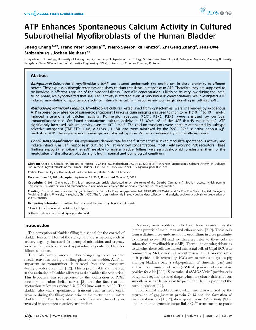
ATP Enhances Spontaneous Calcium Activity in CulturedSuburothelial Myofibroblasts of the Human BladderSheng Cheng1,2., Frank Peter Scigalla1., Pietro Speroni di Fenizio3, Zhi Geng Zhang2, Jens-Uwe
Stolzenburg1, Jochen Neuhaus1*
1 Department of Urology, University of Leipzig, Leipzig, Germany, 2 Department of Urology, Sir Run Run Shaw Hospital, College of Medicine, Zhejiang University,
Hangzhou, China, 3 Department of Informatics Engineering, CISUC, University of Coimbra, Coimbra, Portugal
Abstract
Background: Suburothelial myofibroblasts (sMF) are located underneath the urothelium in close proximity to afferentnerves. They express purinergic receptors and show calcium transients in response to ATP. Therefore they are supposed tobe involved in afferent signaling of the bladder fullness. Since ATP concentration is likely to be very low during the initialfilling phase, we hypothesized that sMF Ca2+ activity is affected even at very low ATP concentrations. We investigated ATPinduced modulation of spontaneous activity, intracellular calcium response and purinergic signaling in cultured sMF.
Methodology/Principal Findings: Myofibroblast cultures, established from cystectomies, were challenged by exogenousATP in presence or absence of purinergic antagonist. Fura-2 calcium imaging was used to monitor ATP (10216 to 1024 mol/l)induced alterations of calcium activity. Purinergic receptors (P2X1, P2X2, P2X3) were analysed by confocalimmunofluorescence. We found spontaneous calcium activity in 55.18%61.65 of the sMF (N = 48 experiments). ATPsignificantly increased calcium activity even at 10216 mol/l. The calcium transients were partially attenuated by subtypeselective antagonist (TNP-ATP, 1 mM; A-317491, 1 mM), and were mimicked by the P2X1, P2X3 selective agonist a,b-methylene ATP. The expression of purinergic receptor subtypes in sMF was confirmed by immunofluorescence.
Conclusions/Significance: Our experiments demonstrate for the first time that ATP can modulate spontaneous activity andinduce intracellular Ca2+ response in cultured sMF at very low concentrations, most likely involving P2X receptors. Thesefindings support the notion that sMF are able to register bladder fullness very sensitively, which predestines them for themodulation of the afferent bladder signaling in normal and pathological conditions.
Citation: Cheng S, Scigalla FP, Speroni di Fenizio P, Zhang ZG, Stolzenburg J-U, et al. (2011) ATP Enhances Spontaneous Calcium Activity in CulturedSuburothelial Myofibroblasts of the Human Bladder. PLoS ONE 6(10): e25769. doi:10.1371/journal.pone.0025769
Editor: David M. Ojcius, University of California Merced, United States of America
Received June 14, 2011; Accepted September 11, 2011; Published October 5, 2011
Copyright: � 2011 Cheng et al. This is an open-access article distributed under the terms of the Creative Commons Attribution License, which permitsunrestricted use, distribution, and reproduction in any medium, provided the original author and source are credited.
Funding: This work was supported by grants from the Deutsche Forschungsgemeinschaft (DFG) (JN:NE425/4-4) and Sir Run Run Shaw Hospital, College ofMedicine, Zhejiang University, Hangzhou, China (SC). The funders had no role in study design, data collection and analysis, decision to publish, or preparation ofthe manuscript.
Competing Interests: The authors have declared that no competing interests exist.
* E-mail: [email protected]
. These authors contributed equally to this work.
Introduction
The perception of bladder filling is essential for the control of
bladder function. Most of the storage urinary symptoms, such as
urinary urgency, increased frequency of micturition and urgency
incontinence can be explained by pathologically enhanced bladder
fullness sensation.
The urothelium releases a number of signaling molecules onto
stretch activation during the filling phase of the bladder. ATP, an
important neurotransmitter, is released from the urothelium
during bladder distension [1,2]. This is presumably the first step
in the excitation of bladder afferents as the bladder fills with urine.
This hypothesis was strengthened by the localization of P2X3
receptors on suburothelial nerves [3] and the fact that the
micturition reflex was reduced in P2X3 knockout mice [4]. The
bladder also elicits spontaneous transient rises in intravesical
pressure during the filling phase prior to the micturition in intact
bladder [5,6]. The details of the mechanisms and the cell types
involved in spontaneous activity are unclear.
Recently, myofibroblastic cells have been identified in the
lamina propria of the human and other species [7–9]. Those cells
form a distinct layer underneath the urothelium in close proximity
to afferent nerves [8] and we therefore refer to these cells as
suburothelial myofibroblasts (sMF). There is an ongoing debate as
to whether these cells are indeed interstitial cells of Cajal (ICCs) as
promoted by McCloskey in a recent review [10]. However, while
c-kit positive cells resembling ICCs are numerous in guinea-pig
and pig bladders only a subpopulation of vimentin (vim) and
alpha-smooth muscle cell actin (aSMCA) positive cells also stain
positive for c-kit [7,11]. Suburothelial aSMCA+/vim+ positive cells
of typical irregular blistered shape, which are clearly different from
smooth muscle cells, are most frequent in the lamina propria of the
human bladder [12].
Suburothelial myofibroblasts, which are characterized by the
expression of gap-junction protein Cx43 and the formation of
functional syncytia [11,12], show spontaneous Ca2+ activity [9,13]
and are able to generate intracellular Ca2+ transients in response
PLoS ONE | www.plosone.org 1 October 2011 | Volume 6 | Issue 10 | e25769
to exogenous ATP application [14]; several purinergic receptors
have been observed in sMF [9,15]. The location of sMF and their
responsiveness to ATP place them in an ideal position to act as
modulators of sensory processes.
Since the physiological ATP concentration during the initial
filling phase is likely to be very low, we hypothesized that the Ca2+
activity of the sMF is affected at very low ATP concentrations.
Furthermore, it was hypothesized that the spontaneous activity of
the sMF is likely to be connected with the generation or
amplification of the afferent signals [8,16]. Thus the autonomous
activity of the detrusor could be ’triggered’ by sMF activity.
In the present study we investigated the ATP induced
modulation of spontaneous activity, intracellular calcium response,
and purinergic signaling in cultured human suburothelial
myofibroblasts.
Materials and Methods
Ethics StatementThe study was approved by the Ethics Committee of the
University of Leipzig (#036—2007) and was conducted according
to the principles expressed in the Declaration of Helsinki. Written
informed consent was obtained from all patients.
Cell culturesWe used tumor free bladder tissue samples from patients
undergoing radical cystectomy due to bladder cancer. For setup of
hsMF we separated mucosa and lamina propria from muscularis
by sharp dissection, ensuring no contamination with detrusor
smooth muscle cells. Thus aSMCA and vimentin positive cells
were the most abundant cell population besides urothelial cells in
this part of the bladder (Fig. S1A,B). CD117 positive interstitial
cells of Cajal (ICC) were only sparse in human bladder lamina
propria and therefore cannot account for the majority of the
cultured suburothelial myofibroblasts (Fig. S1C,D,E). Small tissue
fragments (0.560.560.5 mm) were plated into tissue culture flasks
(TPP AG, Trasadingen, Switzerland) and incubated at 37uCand 5% CO2 in SMC Growth Medium 2 (PromoCell GmbH,
Heidelberg, Germany) and subcultured up to the third passage
(P3). The growing cells showed typical morphological and
immunohistochemical features of myofibroblasts as recently
described [17]. The use of special smooth muscle cell growth
medium was sufficient to avoid growth of urothelial cells as proven
by visual phase contrast (Fig. S2A,B) and lack of cells staining
positive for cytokeratin (monoclonal mouse anti-pan cytokeratin,
Sigma-Aldrich, Steinheim, Germany; data not shown). For
calcium imaging experiments cells were plated onto 13mm glass
coverslips coated with collagen A (Biochrome AG, Berlin,
Germany) and grown to a confluence of about 80% for the
calcium imaging experiments.
Solutions and chemicalsWe used the modified Krebs Ringer solution of the following
composition (mM): CaCl2, 1.9; NaCl, 120.9; NaHCO3, 14.4; KCl,
5.9; MgCl2,1.2; NaH2PO4, 1.55; Hepes, 4.2; Glucose, 11.49;
pH 7.2. All solutions were prepared fresh on the day of use. The
following chemicals were obtained from Sigma-Aldrich: ATP,a,b-
methylene ATP (AMBA), A-317491, DMSO; Fura-2AM and
PluronicH (Invitrogen, Karlsruhe, Germany); TNP-ATP (Torcris
Biosciences, Bristol, UK). All chemicals were diluted from in
Krebs Ringer solution 10 mM stock solutions stored at 220uC on
the day of experiments. ATP and TNP-ATP were kept in the dark.
All drugs were diluted in pure Krebs Ringer solution, which was
also used for control in the different experimental parts.
Intracellular calcium measurementsAfter a brief wash in ringer the coverslips were bulk-loaded with
Fura-2-acetoxymethylester (2.5 mM Fura-2AM solved in DMSO,
2% Pluronic in ringer; 22uC; 40 min.). After 15 minutes wash in
ringer to ensure cleavage of the acetoxymethyl ester by
endogeneous esterases, the cells were superfused for another 10
minutes with carbogenized ringer (pH 7.2) at a constant flow rate
of 0.8 ml/min at 37uC in the recording chamber (WPI, RC-26G,
234 ml volume) before calcium measurements.
Calcium imaging recordings were carried out with a cooled
TILL IMAGO-QE camera, connected to an Olympus IX-71
inverse microscope. TILLvisION 4.5 (TillPhotonics GmbH,
Grafelfing, Germany) was used for camera control and data
acquisition. Image series were recorded every second for 15
minutes with an excitation wavelength of 340 nm and 380 nm.
Fura-2 fluorescence ratios were calculated as FI = F340 nm/
F380 nm*1000 after dynamic background subtraction.
Data analysis with automated Fluorescence analysisWe used a self written ImageJ-script (Rasband WS (1997–2006),
NIH USA, http://rsb.info.nih.gov/ij/) to calculate the fluores-
cence intensity (FI) kinetics of the region of interest (ROIs) (Fig. 1A,
B). The following analysis of the calcium kinetics was performed
with a self programmed automated fluorescence analysis (FA),
which is a self-written script for Python 2.6 (Rossum G (2008)
Python/C, Python Software Foundation, http://python.org/).
The FA was used to detect and characterize the peaks within the
timecourse of the Ca2+ fluorescence. After detection of the signals,
the FA program identifies signal characteristics for the whole
observation interval and for each detected peak separately,
including peak amplitude and number of peaks (Fig. 1C, D).
The experiments consisted of three parts of 5 minutes each
(Fig. 2 Part A, B, C). The suburothelial myofibroblasts were
superfused with Ringer solution in Part A and Part C in order to
assess the spontaneous activity. Cells were defined as ‘spontane-
ously active cells’ (SAC) if they showed spontaneous calcium
transients in Part A of the experimental procedure. In contrast,
inactive cells were denoted IAC.
Confocal ImmunofluorescenceCells were fixed in 4% paraformaldehyde (1 h, 4uC), washed three
times in Tris-buffered saline (TBS; pH 7.2), permeabilized for
10 min with DT-TBS (0.08% Triton-X100 and 0.32% dimethyl
sulfoxide), and blocked with 3% skimmed milk and 1% bovine serum
albumin in TBS. Double labelling experiments were done with
rabbit polyclonal anti-P2X1, P2X2, P2X3 (1:1000, immunoglobulin
G, IgG, Sigma-Aldrich), and mouse monoclonal anti-aSMCA
(1:2000, IgG2a; Sigma-Aldrich) antibodies. Alexa-fluor 488 (A-
488, Goat-anti-Mouse IgG 2a) and A-555 (Goat-anti-Rabbit)
coupled secondary antibodies (Invitrogen) diluted 1:500 in TBS
(1 h, room temperature) were used for visualisation. Nuclei were
stained with 49,6-diamidino-2-phenylindole-dihydrochloride (DAPI;
Roche Diagnostics, Mannheim, Germany). Immunolabelled cells
were examined with a laser-scanning microscope (LSM5 Pascal;
Zeiss, Jena, Germany). The purinergic receptors expression was
analysed with ImageJ and MS-Excel (Microsoft, Redmond, USA).
StatisticsGraphPad Prism (GraphPad 5.0 Software, Inc., San Diego,
USA) was used for presentation and statistical analysis of the data
by Student’s t-test for paired or unpaired data or one-way
ANOVA with Tukey’s multiple comparison test. A P-value,0.05
was regarded as statistically significant.
ATP Enhances Spontaneous Activity in Bladder Cells
PLoS ONE | www.plosone.org 2 October 2011 | Volume 6 | Issue 10 | e25769
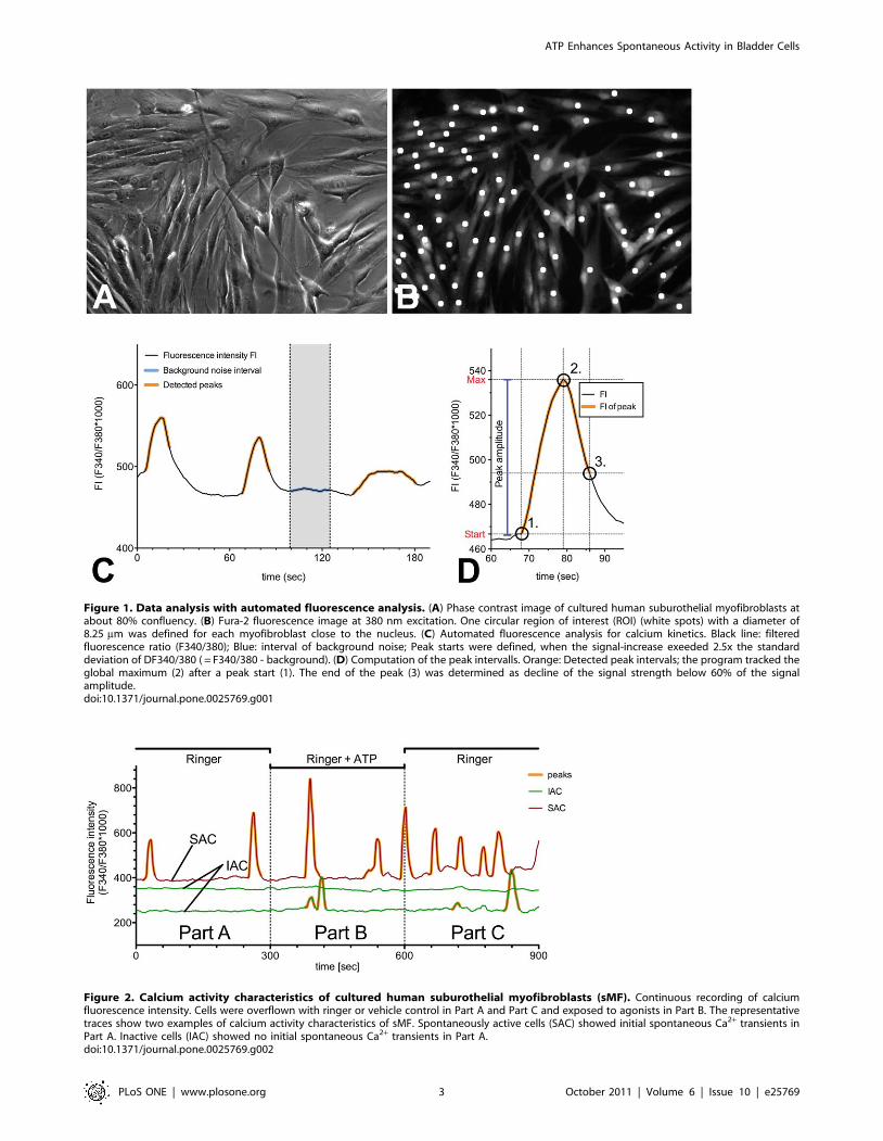
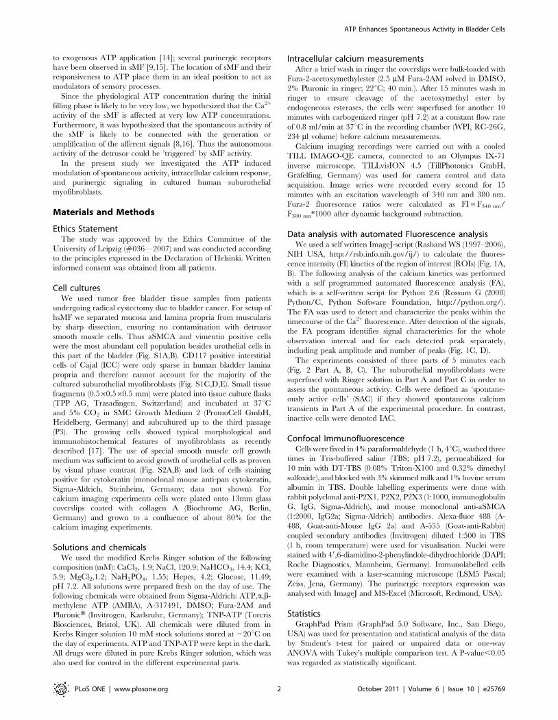
Figure 1. Data analysis with automated fluorescence analysis. (A) Phase contrast image of cultured human suburothelial myofibroblasts atabout 80% confluency. (B) Fura-2 fluorescence image at 380 nm excitation. One circular region of interest (ROI) (white spots) with a diameter of8.25 mm was defined for each myofibroblast close to the nucleus. (C) Automated fluorescence analysis for calcium kinetics. Black line: filteredfluorescence ratio (F340/380); Blue: interval of background noise; Peak starts were defined, when the signal-increase exeeded 2.5x the standarddeviation of DF340/380 ( = F340/380 - background). (D) Computation of the peak intervalls. Orange: Detected peak intervals; the program tracked theglobal maximum (2) after a peak start (1). The end of the peak (3) was determined as decline of the signal strength below 60% of the signalamplitude.doi:10.1371/journal.pone.0025769.g001
Figure 2. Calcium activity characteristics of cultured human suburothelial myofibroblasts (sMF). Continuous recording of calciumfluorescence intensity. Cells were overflown with ringer or vehicle control in Part A and Part C and exposed to agonists in Part B. The representativetraces show two examples of calcium activity characteristics of sMF. Spontaneously active cells (SAC) showed initial spontaneous Ca2+ transients inPart A. Inactive cells (IAC) showed no initial spontaneous Ca2+ transients in Part A.doi:10.1371/journal.pone.0025769.g002
ATP Enhances Spontaneous Activity in Bladder Cells
PLoS ONE | www.plosone.org 3 October 2011 | Volume 6 | Issue 10 | e25769
Results
Spontaneous calcium activity of sMFWe found basic spontaneous calcium activity (Fig. 2, Part A) in
55.18%61.65 of the sMF, with a mean amplitude of
DFI = 243.9610.24 F340 nm/F380 nm*1000 and an average Ca2+
peak frequency of 0.4160.01 min21 (n = 4039; N = 48). After Part
A, we switched the Ringer’s solution to a second solution which
additionally contained the substances (ATP, Antagonists) and
recorded the reaction of the suburothelial myofibroblasts (Fig. 2,
Part B).
ATP effects on calcium response in sMFThe fraction of active cells, the frequency of active cells and
Ca2+ peak amplitude increased with increasing ATP concentra-
tions. However, the ATP dose-response curve showed a bi-phasic
course with an increase from 10216 to 10210 mol/l and from 1026
to 1024 mol/l and a drop to control level at 1028 mol/l (Fig. 3A,
B, C).
The kinetics of the calcium transients in sMF were different at
low and high ATP concentrations. Figure 4 shows representative
traces of sMF (control, 10212 and 1024 mol/l ATP). The
stimulation with a high ATP concentrations (.1028 mol/l)
typically evoked synchronous calcium transients with a rapid
initial steep calcium rise (Fig. 4A). The response to the low ATP-
concentration of 10212 mol/l did not show the characteristic high
fist peak, which we observed at 1024 mol/l ATP. Instead, the first
calcium rise varied considerably from cell to cell (Fig. 4B).
However, the overall profile was similar to the cells in the control
experiments (Fig. 4C), except for higher peak frequency and
amplitude.
We analysed the mean lag time of the first peaks after addition
of ATP and found significant differences between high and low
ATP concentrations (Fig. 5A). Also, the mean amplitude of the
first peaks after high ATP application was significantly higher than
after low ATP application (Fig. 5B). The mean amplitude of the
first peaks was significantly higher than that of the following peaks
at 1024 mol/l ATP, while this was not the case at 10212 mol/l
ATP.
Analysis of purinergic receptors involvedAgonist stimulation. ATP-induced calcium response was
mimicked by a,b-methylene ATP (a,b-meATP), a P2X1 and
P2X3 receptors agonist, at both used concentrations, 1pM
(10212 mol/l) and 100 mM (1024 mol/l). While peak amplitudes
were not different between a,b-meATP and ATP at the same
concentrations (Fig. 6A), area under the curve (AUC), describing
the sum of elevated calcium over time, was significantly different
higher at 100 mM ATP stimulation (Fig. 6B).
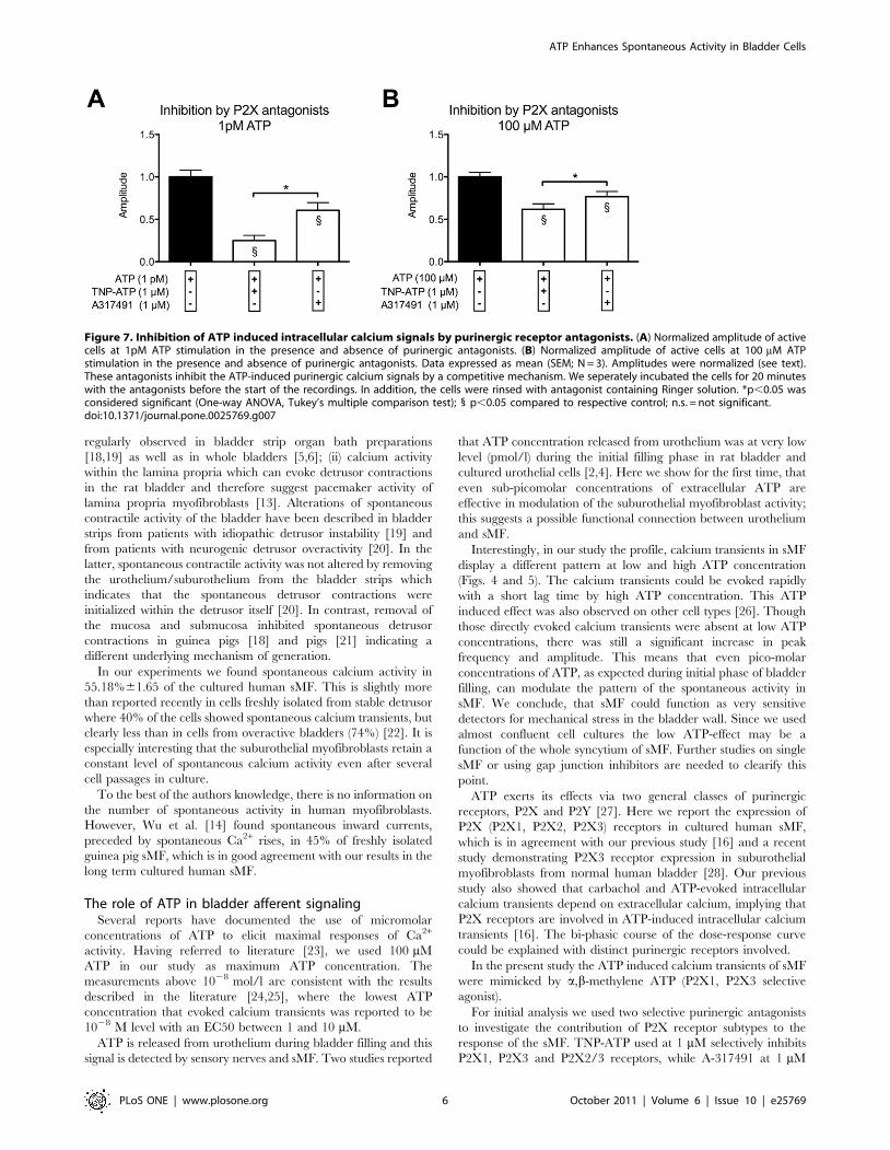
(ii) Signal inhibition by specific antagonists. We used two
different purinergic antagonists to investigate the contribution of
P2X receptor subtypes to the response of the sMF. TNP-ATP
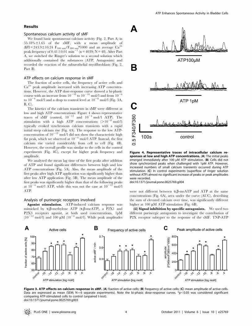
Figure 3. ATP effects on calcium response in sMF. (A) fraction of active cells; (B) frequency of active cells; (C) mean amplitude of active cells.Data are expressed as mean (SEM; N = 6 separate experiments). Note the bi-phasic dose-response curves. *p,0.05 was considered significantcomparing ATP-stimulated cells to control (unpaired t-test).doi:10.1371/journal.pone.0025769.g003
Figure 4. Representative traces of intracellular calcium re-sponses at low and high ATP concentrations. (A) The initial peaksemerged immediately after 100 mM ATP stimulation. (B) Cells did notshow synchronized peaks when challenged with 1pM ATP. However,increased numbers of small calcium transients occurred during ATPstimulation. (C) In control experiments (superflow of ringer solutionwithout ATP) almost no significant increase of peaks or peak amplitudeswere recorded.doi:10.1371/journal.pone.0025769.g004
ATP Enhances Spontaneous Activity in Bladder Cells
PLoS ONE | www.plosone.org 4 October 2011 | Volume 6 | Issue 10 | e25769
(1 mM) selectively inhibits P2X1, P2X3 and P2X2/3 receptors and
A-317491 (1 mM) selectively inhibits P2X3 and P2X2/3 receptors.
ATP evoked calcium transients were partially attenuated by both
purinergic antagonists. Antagonists alone showed no effect (tested
in Part A of the experiments, see Fig. 2). Data were normalized to
agonist stimulation, 10212 mol/l (1pM) and 1024 mol/l (10 mM)
respectively (Fig. 7). The mean amplitude of calcium peaks was
significantly reduced at both ATP concentrations. However, at
1pM ATP stimulation, TNP-ATP (74.98%) showed a significant
higher inhibition than A-317491 (39.29%) (Fig. 7A), while at
100 mM ATP stimulation the inhibition by TNP-ATP and A-
317491 was reduced by 38.1% and 23.4%, respectively (Fig. 7B).
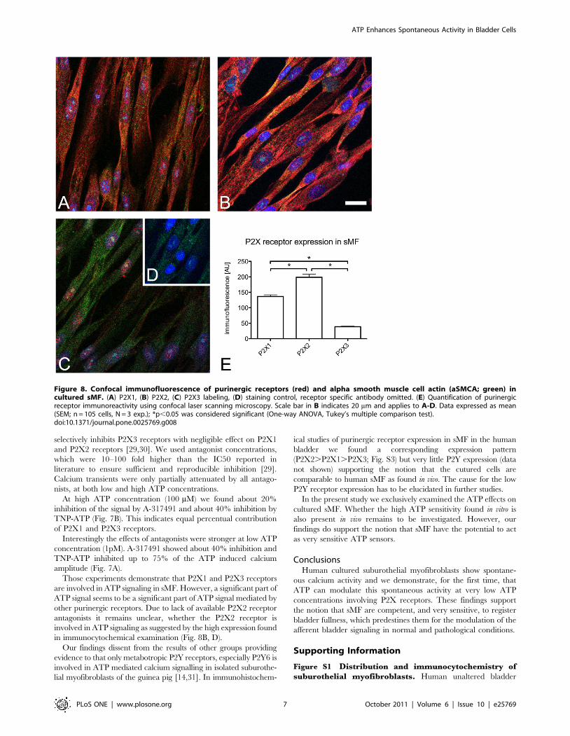
(iii) Confocal immunofluorescence of purinergic
receptors. The expression of purinergic receptor subtypes in sMF
was examined by confocal immunofluorescence (Fig. 8A–E). Strong
P2X1 (Fig. 8A) and P2X2 (Fig. 8B) receptor immunoreactivity (IR) was
seen in cultured suburothelial myofibroblasts of human bladder, while
P2X3 receptor-IR was significantly lower (Fig. 8C, D). The expression
pattern of P2X receptors thereby was the same as measured in sMF in
tissue slices of human bladders (Fig. S3).
Discussion
Two levels of spontaneous activity in the bladder have to be
considered; (i) spontaneous detrusor contractile activity, which is
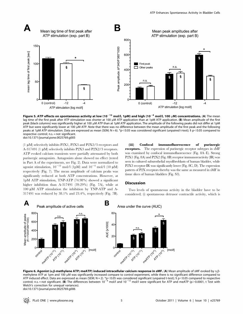
Figure 5. ATP effects on spontaneous activity at low (10212 mol/l, 1pM) and high (1024 mol/l, 100 mM) concentrations. (A) The meanlag time of the first peak after ATP stimulation was shorter at 100 mM ATP application than at 1pM ATP application. (B) Mean amplitude of the firstpeak (black columns) was significantly higher at 100 mM ATP than at 1pM ATP application. The amplitude of the following peaks did not differ at 1pMATP but were significantly lower at 100 mM ATP. Note that there was no difference between the mean amplitude of the first peak and the followingpeaks at 1pM ATP stimulation. Data are expressed as mean (SEM; N = 6). *p,0.05 was considered significant (unpaired t-test); 1 p,0.05 compared torespective control; n.s. = not significant.doi:10.1371/journal.pone.0025769.g005
Figure 6. Agonist (a,b-methylene ATP; meATP) induced intracellular calcium response in sMF. (A) Mean amplitude of sMF evoked by a,b-methylene ATP at 1pm and 100 mM was significantly increased compare to control experiment, while there is no significant difference compared toATP induced effect. Data are expressed as mean (SEM; N = 2). *p,0.05 was considered significant (unpaired t-test); 1 p,0.05 compared to respectivecontrol; n.s. = not significant. (B) The differences between 1024 mol/l and 10212 mol/l were significant for ATP and meATP (p.0.0001, t Test withWelch’s correction for unequal variances).doi:10.1371/journal.pone.0025769.g006
ATP Enhances Spontaneous Activity in Bladder Cells
PLoS ONE | www.plosone.org 5 October 2011 | Volume 6 | Issue 10 | e25769
regularly observed in bladder strip organ bath preparations
[18,19] as well as in whole bladders [5,6]; (ii) calcium activity
within the lamina propria which can evoke detrusor contractions
in the rat bladder and therefore suggest pacemaker activity of
lamina propria myofibroblasts [13]. Alterations of spontaneous
contractile activity of the bladder have been described in bladder
strips from patients with idiopathic detrusor instability [19] and
from patients with neurogenic detrusor overactivity [20]. In the
latter, spontaneous contractile activity was not altered by removing
the urothelium/suburothelium from the bladder strips which
indicates that the spontaneous detrusor contractions were
initialized within the detrusor itself [20]. In contrast, removal of
the mucosa and submucosa inhibited spontaneous detrusor
contractions in guinea pigs [18] and pigs [21] indicating a
different underlying mechanism of generation.
In our experiments we found spontaneous calcium activity in
55.18%61.65 of the cultured human sMF. This is slightly more
than reported recently in cells freshly isolated from stable detrusor
where 40% of the cells showed spontaneous calcium transients, but
clearly less than in cells from overactive bladders (74%) [22]. It is
especially interesting that the suburothelial myofibroblasts retain a
constant level of spontaneous calcium activity even after several
cell passages in culture.
To the best of the authors knowledge, there is no information on
the number of spontaneous activity in human myofibroblasts.
However, Wu et al. [14] found spontaneous inward currents,
preceded by spontaneous Ca2+ rises, in 45% of freshly isolated
guinea pig sMF, which is in good agreement with our results in the
long term cultured human sMF.
The role of ATP in bladder afferent signalingSeveral reports have documented the use of micromolar
concentrations of ATP to elicit maximal responses of Ca2+
activity. Having referred to literature [23], we used 100 mM
ATP in our study as maximum ATP concentration. The
measurements above 1028 mol/l are consistent with the results
described in the literature [24,25], where the lowest ATP
concentration that evoked calcium transients was reported to be
1028 M level with an EC50 between 1 and 10 mM.
ATP is released from urothelium during bladder filling and this
signal is detected by sensory nerves and sMF. Two studies reported
that ATP concentration released from urothelium was at very low
level (pmol/l) during the initial filling phase in rat bladder and
cultured urothelial cells [2,4]. Here we show for the first time, that
even sub-picomolar concentrations of extracellular ATP are
effective in modulation of the suburothelial myofibroblast activity;
this suggests a possible functional connection between urothelium
and sMF.
Interestingly, in our study the profile, calcium transients in sMF
display a different pattern at low and high ATP concentration
(Figs. 4 and 5). The calcium transients could be evoked rapidly
with a short lag time by high ATP concentration. This ATP
induced effect was also observed on other cell types [26]. Though
those directly evoked calcium transients were absent at low ATP
concentrations, there was still a significant increase in peak
frequency and amplitude. This means that even pico-molar
concentrations of ATP, as expected during initial phase of bladder
filling, can modulate the pattern of the spontaneous activity in
sMF. We conclude, that sMF could function as very sensitive
detectors for mechanical stress in the bladder wall. Since we used
almost confluent cell cultures the low ATP-effect may be a
function of the whole syncytium of sMF. Further studies on single
sMF or using gap junction inhibitors are needed to clearify this
point.
ATP exerts its effects via two general classes of purinergic
receptors, P2X and P2Y [27]. Here we report the expression of
P2X (P2X1, P2X2, P2X3) receptors in cultured human sMF,
which is in agreement with our previous study [16] and a recent
study demonstrating P2X3 receptor expression in suburothelial
myofibroblasts from normal human bladder [28]. Our previous
study also showed that carbachol and ATP-evoked intracellular
calcium transients depend on extracellular calcium, implying that
P2X receptors are involved in ATP-induced intracellular calcium
transients [16]. The bi-phasic course of the dose-response curve
could be explained with distinct purinergic receptors involved.
In the present study the ATP induced calcium transients of sMF
were mimicked by a,b-methylene ATP (P2X1, P2X3 selective
agonist).
For initial analysis we used two selective purinergic antagonists
to investigate the contribution of P2X receptor subtypes to the
response of the sMF. TNP-ATP used at 1 mM selectively inhibits
P2X1, P2X3 and P2X2/3 receptors, while A-317491 at 1 mM
Figure 7. Inhibition of ATP induced intracellular calcium signals by purinergic receptor antagonists. (A) Normalized amplitude of activecells at 1pM ATP stimulation in the presence and absence of purinergic antagonists. (B) Normalized amplitude of active cells at 100 mM ATPstimulation in the presence and absence of purinergic antagonists. Data expressed as mean (SEM; N = 3). Amplitudes were normalized (see text).These antagonists inhibit the ATP-induced purinergic calcium signals by a competitive mechanism. We seperately incubated the cells for 20 minuteswith the antagonists before the start of the recordings. In addition, the cells were rinsed with antagonist containing Ringer solution. *p,0.05 wasconsidered significant (One-way ANOVA, Tukey’s multiple comparison test); 1 p,0.05 compared to respective control; n.s. = not significant.doi:10.1371/journal.pone.0025769.g007
ATP Enhances Spontaneous Activity in Bladder Cells
PLoS ONE | www.plosone.org 6 October 2011 | Volume 6 | Issue 10 | e25769
selectively inhibits P2X3 receptors with negligible effect on P2X1
and P2X2 receptors [29,30]. We used antagonist concentrations,
which were 10–100 fold higher than the IC50 reported in
literature to ensure sufficient and reproducible inhibition [29].
Calcium transients were only partially attenuated by all antago-
nists, at both low and high ATP concentrations.
At high ATP concentration (100 mM) we found about 20%
inhibition of the signal by A-317491 and about 40% inhibition by
TNP-ATP (Fig. 7B). This indicates equal percentual contribution
of P2X1 and P2X3 receptors.
Interestingly the effects of antagonists were stronger at low ATP
concentration (1pM). A-317491 showed about 40% inhibition and
TNP-ATP inhibited up to 75% of the ATP induced calcium
amplitude (Fig. 7A).
Those experiments demonstrate that P2X1 and P2X3 receptors
are involved in ATP signaling in sMF. However, a significant part of
ATP signal seems to be a significant part of ATP signal mediated by
other purinergic receptors. Due to lack of available P2X2 receptor
antagonists it remains unclear, whether the P2X2 receptor is
involved in ATP signaling as suggested by the high expression found
in immunocytochemical examination (Fig. 8B, D).
Our findings dissent from the results of other groups providing
evidence to that only metabotropic P2Y receptors, especially P2Y6 is
involved in ATP mediated calcium signalling in isolated suburothe-
lial myofibroblasts of the guinea pig [14,31]. In immunohistochem-
ical studies of purinergic receptor expression in sMF in the human
bladder we found a corresponding expression pattern
(P2X2.P2X1.P2X3; Fig. S3) but very little P2Y expression (data
not shown) supporting the notion that the cutured cells are
comparable to human sMF as found in vivo. The cause for the low
P2Y receptor expression has to be elucidated in further studies.
In the present study we exclusively examined the ATP effects on
cultured sMF. Whether the high ATP sensitivity found in vitro is
also present in vivo remains to be investigated. However, our
findings do support the notion that sMF have the potential to act
as very sensitive ATP sensors.
ConclusionsHuman cultured suburothelial myofibroblasts show spontane-
ous calcium activity and we demonstrate, for the first time, that
ATP can modulate this spontaneous activity at very low ATP
concentrations involving P2X receptors. These findings support
the notion that sMF are competent, and very sensitive, to register
bladder fullness, which predestines them for the modulation of the
afferent bladder signaling in normal and pathological conditions.
Supporting Information
Figure S1 Distribution and immunocytochemistry ofsuburothelial myofibroblasts. Human unaltered bladder
Figure 8. Confocal immunofluorescence of purinergic receptors (red) and alpha smooth muscle cell actin (aSMCA; green) incultured sMF. (A) P2X1, (B) P2X2, (C) P2X3 labeling, (D) staining control, receptor specific antibody omitted. (E) Quantification of purinergicreceptor immunoreactivity using confocal laser scanning microscopy. Scale bar in B indicates 20 mm and applies to A-D. Data expressed as mean(SEM; n = 105 cells, N = 3 exp.); *p,0.05 was considered significant (One-way ANOVA, Tukey’s multiple comparison test).doi:10.1371/journal.pone.0025769.g008
ATP Enhances Spontaneous Activity in Bladder Cells
PLoS ONE | www.plosone.org 7 October 2011 | Volume 6 | Issue 10 | e25769
paraffin sections were incubated over night with monoclonal
mouse antibodies: anti-aSMCA (IgG2a, 1:2000; Sigma-Aldrich),
anti-vimentin (1:100; Sigma-Aldrich) or anti-CD117 (c-Kit, 1:100;
DAKO, Glostrup, Denmark) and visualized with Envision-KitTM
(DAKO) using AEC substrate chromogen (3-Amino-9-ethylcarba-
zole in 2.5% N,N-dimethylformamide; red color). Nuclei were
stained with Mayer’s hematoxylin. (A) Numerous aSMCA positive
cells are present in the lamina propria directly underneath the
urothelium. The staining pattern resembles that of vimentin (B).
CD117 immunoreactivity was confined to only few suburothelial
elongated cells (C, arrowheads) and to cells in-between smooth
muscle cell bundles (D, arrows). Urothelial cells regularly showed
light CD117 immunoreactivity with only few more intensely
stained elongated cells within the urothelial cell layer (arrow inC). (E) Staining control showed no AEC background staining. U –
Urothelium; the scale bar in D indicates 100 mm and applies to A-E.
(TIF)
Figure S2 Cell culture morphology. Phase contrast micro-
graphs. (A) Typical suburothelial myofibroblast culture; note the
characteristic morphology of sMF in this sub-confluent cell
culture; (B) urothelial cell culture demonstrating typical cobble-
stone-like morphology of human urothelial cells. The scale bar in
B indicates 100 mm and applies to A and B.
(TIF)
Figure S3 P2X receptor expression. Confocal immunoflu-
orescence analysis of tissue sections of control bladders. Data
expressed as mean (SEM; n = 105 cells, N = 3 patients); *p,0.05
was considered significant (One-way ANOVA, Tukey’s multiple
comparison test).
(TIF)
Acknowledgments
The authors thank Mrs. A. Weimann and Mrs. M. Berndt for the excellent
technical assistance and Dr. Hasan Qazi for proofreading the manuscript.
Author Contributions
Conceived and designed the experiments: JN SC FPS. Performed the
experiments: SC JN. Analyzed the data: SC FPS JN PSdF. Contributed
reagents/materials/analysis tools: J-US ZGZ. Wrote the paper: SC JN.
Administrative, technical, or material support: ZGZ J-US.
References
1. Ferguson DR, Kennedy I, Burton TJ (1997) ATP is released from rabbit urinary
bladder epithelial cells by hydrostatic pressure changes—a possible sensory
mechanism? J Physiol 505: 503–511.
2. Birder LA, Nakamura Y, Kiss S, Nealen ML, Barrick S, et al. (2002) Altered
urinary bladder function in mice lacking the vanilloid receptor TRPV1. Nat
Neurosci 5: 856–860.
3. Lee HY, Bardini M, Burnstock G (2000) Distribution of P2X receptors in the
urinary bladder and the ureter of the rat. J Urol 163: 2002–2007.
4. Vlaskovska M, Kasakov L, Rong W, Bodin P, Bardini M, et al. (2001) P2X3
knock-out mice reveal a major sensory role for urothelially released ATP.
J Neurosci 21: 5670–5677.
5. Drake MJ, Harvey IJ, Gillespie JI (2003) Autonomous activity in the isolated
guinea pig bladder. Exp Physiol 88: 19–30.
6. Streng T, Hedlund P, Talo A, Andersson KE, Gillespie JI (2006) Phasic non-
micturition contractions in the bladder of the anaesthetized and awake rat. BJU
Int 97: 1094–1101.
7. Sui GP, Rothery S, Dupont E, Fry CH, Severs NJ (2002) Gap junctions and
connexin expression in human suburothelial interstitial cells. BJU Int 90:
118–129.
8. Wiseman OJ, Fowler CJ, Landon DN (2003) The role of the human bladder
lamina propria myofibroblast. BJU Int 91: 89–93.
9. Neuhaus J, Heinrich M, Schlichting N, Oberbach A, Fitzl G, et al. (2007)
[Structure and function of suburothelial myofibroblasts in the human urinary
bladder under normal and pathological conditions.]. Urologe A 46: 1197–1202.
10. McCloskey KD (2010) Interstitial cells in the urinary bladder—localization and
function. Neurourol Urodyn 29: 82–87.
11. Roosen A, Datta SN, Chowdhury RA, Patel PM, Kalsi V, et al. (2009)
Suburothelial Myofibroblasts in the Human Overactive Bladder and the Effect
of Botulinum Neurotoxin Type A Treatment. Eur Urol 55: 1440–1448.
12. Neuhaus J, Pfeiffer F, Wolburg H, Horn LC, Dorschner W (2005) Alterations in
connexin expression in the bladder of patients with urge symptoms. BJU Int 96:
670–676.
13. Ikeda Y, Fry CH, Hayashi F, Stolz DB, Griffiths D, et al. (2007) The Role of
Gap Junctions in Spontaneous Activity of the Rat Bladder. Am J Physiol Renal
Physiol;F1018-25.
14. Wu C, Sui GP, Fry CH (2004) Purinergic regulation of guinea pig suburothelial
myofibroblasts. J Physiol 559: 231–243.
15. Sui GP, Wu C, Fry CH (2006) Characterization of the purinergic receptor
subtype on guinea-pig suburothelial myofibroblasts. BJU Int 97: 1327–1331.
16. Neuhaus J, Scholler U, Freick K, Schwalenberg T, Heinrich M, et al. (2008)
[Myofibroblasts and afferent signalling in the urinary bladder: A concept.].
Urologe A 47: 1085–1090.
17. Heinrich M, Oberbach A, Schlichting N, Stolzenburg JU, Neuhaus J (2011)
Cytokine effects on gap junction communication and connexin expression in
human bladder smooth muscle cells and suburothelial myofibroblasts. PLoSONE 6: e20792.
18. Sui GP, Wu C, Roosen A, Ikeda Y, Kanai AJ, et al. (2008) Modulation ofbladder myofibroblast activity: implications for bladder function. Am J Physiol
Renal Physiol 295: 688–697.
19. Mills IW, Greenland JE, McMurray G, McCoy R, Ho KMT, et al. (2000)Studies of the pathophysiology of idiopathic detrusor instability the physiological
properties of the detrusor smooth muscle and its pattern of innervation. J Urology163: 646–651.
20. Oger S, Behr-Roussel D, Gorny D, Bernabe J, Comperat E, et al. (2010) Effects
of potassium channel modulators on myogenic spontaneous phasic contractileactivity in human detrusor from neurogenic patients. BJU Int.
21. Akino H, Chapple CR, McKay N, Cross RL, Murakami S, et al. (2008)Spontaneous contractions of the pig urinary bladder: the effect of ATP-sensitive
potassium channels and the role of the mucosa. BJU Int 102: 1168–1174.22. Sui G, Fry CH, Malone-Lee J, Wu C (2009) Aberrant Ca2+ oscillations in
smooth muscle cells from overactive human bladders. Cell Calcium 45:
456–464.23. Meng F, To W, Kirkman-Brown J, Kumar P, Gu Y (2007) Calcium oscillations
induced by ATP in human umbilical cord smooth muscle cells. J Cell Physiol213: 79–87.
24. Valera S, Hussy N, Evans RJ, Adami N, North RA, et al. (1994) A new class of
ligand-gated ion channel defined by P2x receptor for extracellular ATP. Nature371: 516–519.
25. Chen CC, Akopian AN, Sivilotti L, Colquhoun D, Burnstock G, et al. (1995) AP2X purinoceptor expressed by a subset of sensory neurons. Nature 377:
428–431.26. Mahoney MG, Slakey LL, Benham CD, Gross DJ (1998) Time course of the
initial [Ca2+]i response to extracellular ATP in smooth muscle depends on
[Ca2+]e and ATP concentration. Biophy J 75: 2050–2058.27. Ralevic V, Burnstock G (1998) Receptors for purines and pyrimidines.
Pharmacol Rev 50: 413–492.28. Liu F, Takahashi N, Yamaguchi O (2009) Expression of P2X3 purinoceptors in
suburothelial myofibroblasts of the normal human urinary bladder. Int J Urol
16: 570–575.29. Jarvis MF, Khakh BS (2009) ATP-gated P2X cation-channels. Neuropharma-
cology 56: 208–215.30. North RA, Surprenant A (2000) Pharmacology of cloned P2X receptors. Annu
Rev Pharmacol Toxicol 40: 563–580.31. Fry CH, Sui GP, Kanai AJ, Wu C (2007) The function of suburothelial
myofibroblasts in the bladder. Neurourol Urodyn 26: 914–919.
ATP Enhances Spontaneous Activity in Bladder Cells
PLoS ONE | www.plosone.org 8 October 2011 | Volume 6 | Issue 10 | e25769
Copyright © 2022 FDOKUMEN