Rabbit Nipple-Search Pheromone Versus Rabbit Mammary Pheromone Revisited
Biomed & Pharmacother (1994) 48, Suppll, 27s-34s© Elsevier, Paris
Molecular control of rabbit urinary bladder hypertrophy
R Buttyan I, MW Chen I, F Monson 2, RM Levin 2
J The Department of Urology, Columbia University College ofPhysicians & Surgeons, 630 W 168th Street, New York, NY 10032;2 The Division of Urology, The University ofPennsylvania School ofMedicine, Veterans Administration Medical Center,
3400 Spruce Street, Philadelphia PA 19104, USA
27s
Summary - Chronic obstruction of the lower genitourinary tract, the major manifestation of BPH, results in charac-teristic pathological changes in the structure and function of the bladder. Here, we describe how an experimental ani-mal model of bladder outlet obstruction involving the rabbit has been used to identify specific genetic markers associ-ated with the response of the bladder to partial outlet obstruction. More important, these types of studies indicate thattwo particular growth factors, basic fibroblast growth factor and transforming growth factor-B. act in a strategic andopposing manner to regulate bladder growth and regression in response to partial outlet obstruction.
urinary bladder hypertrophy I hyperplasia I apoptosis I growth factors
Introduction
Benign prostatic hyperplasia (BPH) is a common andcomplex disease associated with human aging whoseorigin is generally attributed to the anomalous adult-onset reinitiation of growth of the prostate gland. Inspite of the prostatic origin of BPH, the symptomaticpresentation of this disease is entirely related totrauma directed to other organs of the urinary systemwhen the lower urinary tract becomes obstructed bythis abnormal prostate growth. Over time, chronicoutlet obstruction from BPH can result in irreversiblepathological changes to the bladder as well as to thekidney. In fact, many men with BPH will undergopermanent changes in bladder structure and functionso that they will not experience normal urinationeven after the obstruction is relieved. Modern bio-medical research on BPH has tended to focus ondefining those factors associated with the initiationand progression of the abnormal prostate growth.There is increasing interest, however, in research onthe mechanism by which the urinary tract outlet
obstruction associated with BPH causes bladderpathology and the means to prevent or reverse this.
To this end, a variety of animal models (guineapigs, rats and rabbits etc) have already been devel-oped to study obstruction-associated bladder pathol-ogy [9, 13, 16, 18, 20]. The experimental paradigmof partial outlet obstruction in rabbits [5, 12] hasbeen particularly useful in identifying the morpho-logical, physiological and pharmacological parame-ters of obstructive bladder pathology. In this model,outlet obstruction is produced by the surgical place-ment of a ligature [12], cuff [4] or ring [20] around acatheterized rabbit urethra at the base of the bladder.When the catheter is removed, the rabbit recoverswith a partial bladder outlet obstruction estimated todecrease the diameter of the bladder outlet by 20-30%. Although this limited stricture ensures that therabbit can continue to urinate, the partial obstructionleads to rapid and predictable changes in the bladderstructure which can be identified by morphologicaland biochemical criteria. Additionally, the bladderundergoes a number of functional alterations to adapt
28s R Buttyan et at
to the restricted outflow and these changes can bemeasured by urodynamic flow parameters.
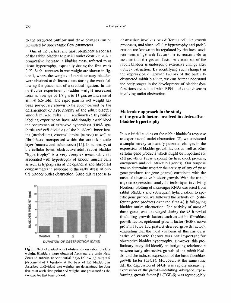
One of the earliest and most prominent responsesof the rabbit bladder to partial outlet obstruction is aprogressive increase in bladder mass, referred to astissue hypertrophy, especially during the first week[12]. Such increases in wet weight are shown in fig-ure 1, where the weights of rabbit urinary bladderswere obtained at different times during the week fol-lowing the placement of a urethral ligation. In thisparticular experiment, bladder weight increasedfrom an average of 1.7 gm to 11 gm, an increase ofalmost 6.5-fold. The rapid gain in wet weight hasbeen previously shown to be accompanied by theenlargement or hypertrophy of the adult bladdersmooth muscle cells [11]. Radioactive thymidinelabeling experiments have additionally establishedthe occurrence of extensive hyperplasia (DNA syn-thesis and cell division) of the bladder's inner lam-ina (urothelium), external lamina (serosa) as well asfibroblasts interspersed within the smooth musclelayer (mucosa and submucosa) [15]. In summary, atthe cellular level, obstructive adult rabbit bladder"hypertrophy" is a very complex event which isassociated with hypertrophy of smooth muscle cellsas well as hyperplasia of the epithelial and fibroblastcompartments in response to the early stress of par-tial bladder outlet obstruction. Since this response to
12 r----------------~
Control 1 3 5 7DURATION OF OBSTRUCTION (DAYS).
Fig 1. Effect of partial outlet obstruction on rabbit bladderweight. Bladders were obtained from mature male NewZealand rabbits at sequential days following surgicalplacement of a ligation at the base of the bladder, asdescribed. Individual wet weights are determined for fourtissues at each time point and weights are presented as theaverage for that time period.
obstruction involves two different cellular growthprocesses, and since cellular hypertrophy and prolif-eration are known to be regulated by the local envi-ronment of growth factors, it is reasonable toassume that the growth factor environment of therabbit bladder is undergoing extensive change afteroutlet obstruction. By identifying such changes inthe expression of growth factors of the partiallyobstructed rabbit bladder, we can better understandthe early stages in the development of bladder dys-functions associated with BPH and other diseasesinvolving outlet obstruction.
Molecular approach to the studyof the growth factors involved in obstructivebladder hypertrophy
In our initial studies on the rabbit bladder's responseto experimental outlet obstruction [2], we conducteda simple survey to identify potential changes in theexpression of bladder growth factors as well as othercellular gene products which might be important forcell growth or stress response (ie heat shock proteins,oncogenes and cell structural genes). Our purposewas to determine whether the activity of any of thesegene products (or gene genres) correlated with theonset of obstructive bladder growth. With the use ofa gene expression analysis technique involvingNorthern blotting of messenger RNAs extracted fromrabbit bladders and subsequent hybridization to spe-cific gene probes, we followed the activity of 15 dif-ferent gene products over the first 48 h followingbladder outlet obstruction. The activity of most ofthese genes was unchanged during the 48-h period(including growth factors such as acidic fibroblastgrowth factor, epidermal growth factor (EGF), nervegrowth factor and platelet-derived growth factor),suggesting that the local synthesis of this particularcadre of growth factors was not important forobstructive bladder hypertrophy. However, this pre-liminary study did identify an intriguing relationshipbetween early obstructive growth of the rabbit blad-der and the induced expression of the basic fibroblastgrowth factor (bFGF). Moreover, at the same timethat the expression of bFGF was rapidly increasing,expression of the growth-inhibiting substance, trans-forming growth factor-Ill (TGF-P) was reproducibly
Molecular control of rabbit urinary bladder hypertrophy 29s
decreased by 50%. Finally, we found that the stress-response gene, hsp 70K, as well as a number of cellu-lar proliferation genes (including c-rnyc, Ha- and N-ras) were highly included corresponding to thehypertrophic growth of the bladder. Based on theseresults, we hypothesized that the significantly alteredgrowth factor environment of the partially obstructedrabbit bladder (increased growth stimulant, bFGF inconjunction with decreased growth inhibitor, TGF-~)provided the appropriate growth factor milieu to initi-ate the complex gene and subsequent cellularresponses (hypertrophy and hyperplasia) leading tobladder hypertrophy.
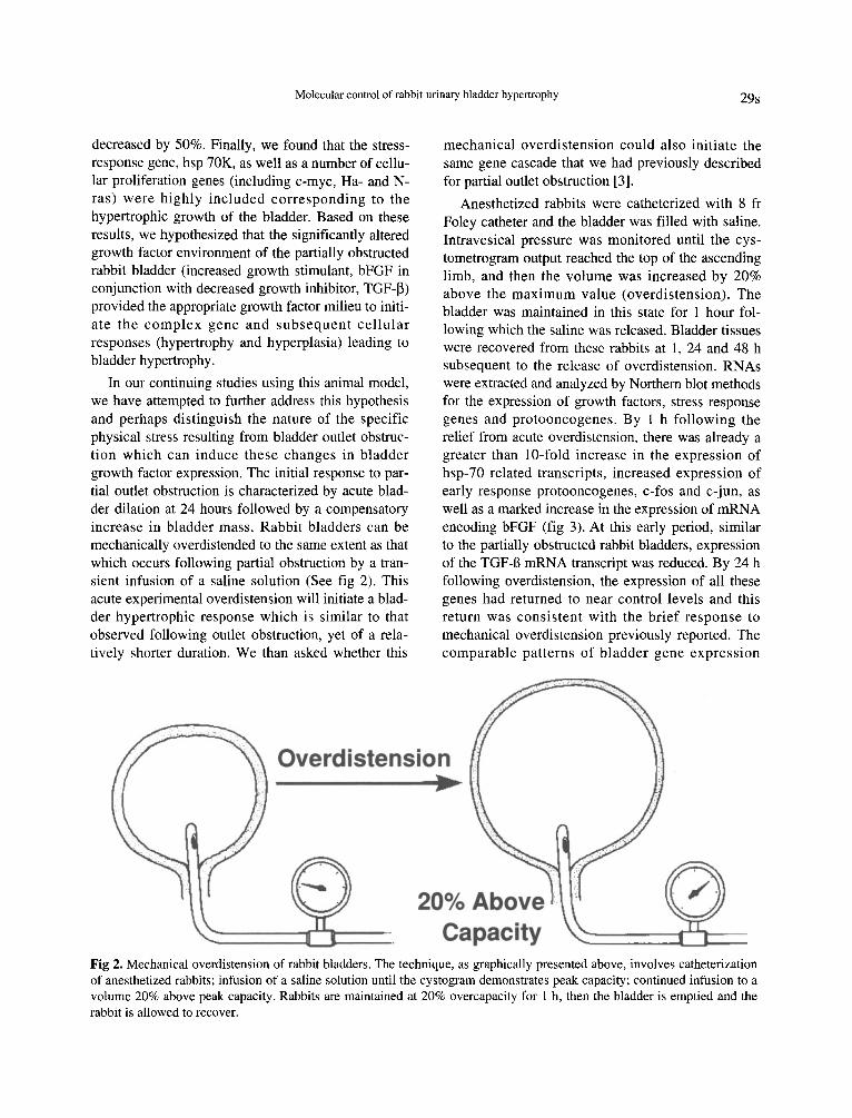
In our continuing studies using this animal model,we have attempted to further address this hypothesisand perhaps distinguish the nature of the specificphysical stress resulting from bladder outlet obstruc-tion which can induce these changes in bladdergrowth factor expression. The initial response to par-tial outlet obstruction is characterized by acute blad-der dilation at 24 hours followed by a compensatoryincrease in bladder mass. Rabbit bladders can bemechanically overdistended to the same extent as thatwhich occurs following partial obstruction by a tran-sient infusion of a saline solution (See fig 2). Thisacute experimental overdistension will initiate a blad-der hypertrophic response which is similar to thatobserved following outlet obstruction, yet of a rela-tively shorter duration. We than asked whether this
mechanical overdistension could also initiate thesame gene cascade that we had previously describedfor partial outlet obstruction [3].
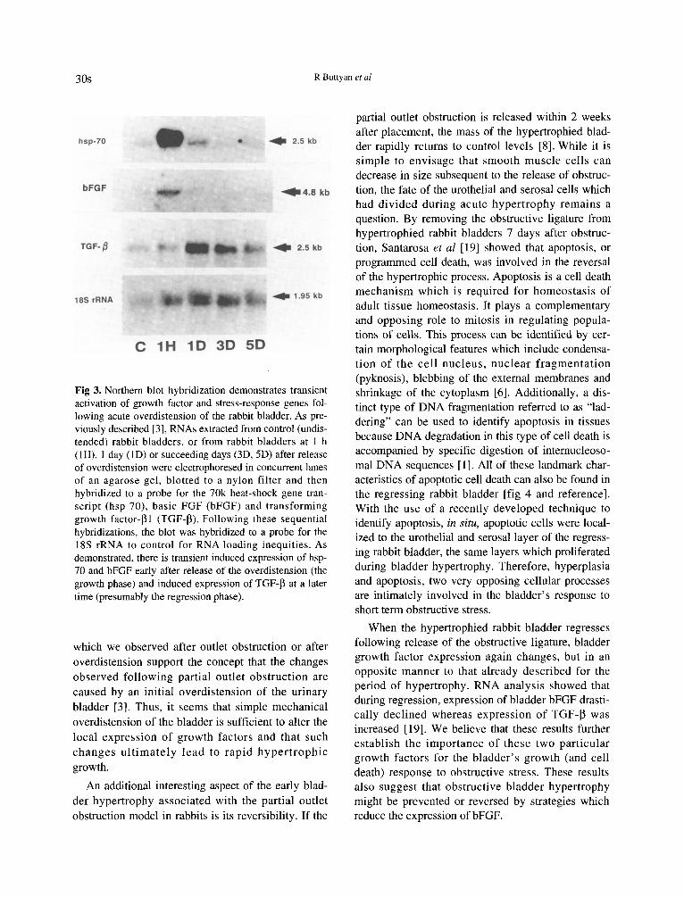
Anesthetized rabbits were catheterized with 8 frFoley catheter and the bladder was filled with saline.Intravesical pressure was monitored until the cys-tometrogram output reached the top of the ascendinglimb, and then the volume was increased by 20%above the maximum value (overdistension). Thebladder was maintained in this state for 1 hour fol-lowing which the saline was released. Bladder tissueswere recovered from these rabbits at 1, 24 and 48 hsubsequent to the release of overdistension. RNAswere extracted and analyzed by Northern blot methodsfor the expression of growth factors, stress responsegenes and protooncogenes. By 1 h following therelief from acute overdistension, there was already agreater than lO-fold increase in the expression ofhsp-70 related transcripts, increased expression ofearly response protooncogenes, c-fos and c-jun, aswell as a marked increase in the expression of mRNAencoding bFGF (fig 3). At this early period, similarto the partially obstructed rabbit bladders, expressionof the TGF-B mRNA transcript was reduced. By 24 hfollowing overdistension, the expression of all thesegenes had returned to near control levels and thisreturn was consistent with the brief response tomechanical overdistension previously reported. Thecomparable patterns of bladder gene expression
Overdistension..20% Above
CapacityFig 2. Mechanical overdistension of rabbit bladders. The technique, as graphically presented above, involves catheterizationof anesthetized rabbits; infusion of a saline solution until the cystogram demonstrates peak capacity; continued infusion to avolume 20% above peak capacity. Rabbits are maintained at 20% overcapacity for I h, then the bladder is emptied and therabbit is allowed to recover.
30s R Bunyan et at
which we observed after outlet obstruction or afteroverdistension support the concept that the changesobserved following partial outlet obstruction arecaused by an initial overdistension of the urinarybladder [3]. Thus, it seems that simple mechanicaloverdistension of the bladder is sufficient to alter thelocal expression of growth factors and that suchchanges ultimately lead to rapid hypertrophicgrowth.
An additional interesting aspect of the early blad-der hypertrophy associated with the partial outletobstruction model in rabbits is its reversibility. If the
Fig 3. Northern blot hybridization demonstrates transientactivation of growth factor and stress-response genes fol-lowing acute overdistension of the rabbit bladder. As pre-viously described [3], RNAs extracted from control (undis-tended) rabbit bladders, or from rabbit bladders at I h(lR), I day (lD) or succeeding days (3D, 5D) after releaseof overdistension were electrophoresed in concurrent lanesof an agarose gel, blotted to a nylon filter and thenhybridized to a probe for the 70k heat-shock gene tran-script (hsp-70), basic FGF (bFGF) and transforminggrowth factor-B) (TGF-P). Following these sequentialhybridizations, the blot was hybridized to a probe for theISS rRNA to control for RNA loading inequities. Asdemonstrated, there is transient induced expression of hsp-70 and bFGF early after release of the overdistension (thegrowth phase) and induced expression of TGF-p at a latertime (presumably the regression phase).
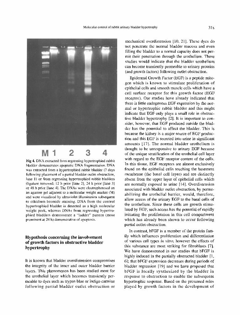
partial outlet obstruction is released within 2 weeksafter placement, the mass of the hypertrophied blad-der rapidly returns to control levels [8]. While it issimple to envisage that smooth muscle cells candecrease in size subsequent to the release of obstruc-tion, the fate of the urothelial and serosal cells whichhad divided during acute hypertrophy remains aquestion. By removing the obstructive ligature fromhypertrophied rabbit bladders 7 days after obstruc-tion, Santarosa et at [19] showed that apoptosis, orprogrammed cell death, was involved in the reversalof the hypertrophic process. Apoptosis is a cell deathmechanism which is required for homeostasis ofadult tissue homeostasis. It plays a complementaryand opposing role to mitosis in regulating popula-tions of cells. This process can be identified by cer-tain morphological features which include condensa-tion of the cell nucleus, nuclea·r fragmentation(pyknosis), blebbing of the external membranes andshrinkage of the cytoplasm [6]. Additionally, a dis-tinct type of DNA fragmentation referred to as "lad-dering" can be used to identify apoptosis in tissuesbecause DNA degradation in this type of cell death isaccompanied by specific digestion of internucleoso-mal DNA sequences [1]. All of these landmark char-acteristics of apoptotic cell death can also be found inthe regressing rabbit bladder [fig 4 and reference].With the use of a recently developed technique toidentify apoptosis, in situ, apoptotic cells were local-ized to the urothelial and serosal layer of the regress-ing rabbit bladder, the same layers which proliferatedduring bladder hypertrophy. Therefore, hyperplasiaand apoptosis, two very opposing cellular processesare intimately involved in the bladder's response toshort term obstructive stress.
When the hypertrophied rabbit bladder regressesfollowing release of the obstructive ligature, bladdergrowth factor expression again changes, but in anopposite manner to that already described for theperiod of hypertrophy. RNA analysis showed thatduring regression, expression of bladder bFGF drasti-cally declined whereas expression of TGF-~ wasincreased [19]. We believe that these results furtherestablish the importance of these two particulargrowth factors for the bladder's growth (and celldeath) response to obstructive stress. These resultsalso suggest that obstructive bladder hypertrophymight be prevented or reversed by strategies whichreduce the expression of bFGF.
~4.8 kb
.. 1.95 kb
.. 2.5 kb
. .. 2.5 kb
•
10 3D 50C 1H
bFGF
TGF- tJ
hsp-70
185 rRNA
Molecular control of rabbit urinary bladder hypertrophy 31s
Hypothesis concerning the involvementof growth factors in obstructive bladderhypertrophy
It is known that bladder overdistension compromisesthe integrity of the inner and outer bladder barrierlayers. This phenomenon has been studied most forthe urothelial layer which becomes transiently per-meable to dyes such as trypan blue or indigo carminefollowing partial bladder outlet obstruction or
Fig 4. DNA extracted fromregressing hypertrophied rabbitbladder demonstrates apoptotic DNA fragmentation. DNAwas extracted from a hypertrophied rabbit bladder (7 daysfollowing placement of a partial bladderoutletobstruction,lane I) or from regressing hypertrophied rabbit bladders(ligature removed; 12 h prior [lane 2], 24 h prior [lane 3]or 48 h prior [lane4]. The DNAs were electrophoresed onan agarose gel adjacent to a molecular weight marker(M)and were visualized by ultraviolet illumination subsequentto ethidium bromide staining. DNA from the controlhypertrophied bladder is detected as a high molecularweight peak, whereas DNAs from regressing hypertro-phied bladders demonstrate a "ladder" pattern (mostprominent at 24 h) demonstrative of apoptosis.
mechanical overdistension [10, 21]. These dyes donot penetrate the normal bladder mucosa and evenfilling the bladder to a normal capacity does not per-mit their penetration through the urothelium. Thesestudies would indicate that the bladder urotheliumcan become transiently permeable to urinary proteins(and growth factors) following outlet obstruction.
Epidermal Growth Factor (EGF) is a peptide mito-gen which is known to stimulate proliferation ofepithelial cells and smooth muscle cells which have acell surface receptor for this growth factor (EGFreceptor). Our studies have already indicated thatthere is little endogenous EGF expression by the nor-mal or hypertrophic rabbit bladder and this mightindicate that EGF only plays a small role in obstruc-tive bladder hypertrophy [2]. It is important to con-sider, however, that EGF produced outside the blad-der has the potential to affect the bladder. This isbecause the kidney is a major source of EGF produc-tion and this EGF is secreted into urine in significantamounts [17]. The normal bladder urothelium isthought to be unresponsive to urinary EGF becauseof the unique stratification of the urothelial cell layerwith regard to the EGF receptor content of the cells.In this tissue, EGF receptors are almost exclusivelyfound on the epithelial cells touching the basementmembrane (the basal cell layer) and are distinctlyabsent from the upper layer of epithelial cells whichare normally exposed to urine [14]. Overdistensionassociated with bladder outlet obstruction, by perme-abilizing the urothelial barrier, would, therefore,allow access of the urinary EGF to the basal cells ofthe urothelium. Since these cells are growth stimu-lated by EGF, such access has the potential of rapidlyinitiating the proliferation in this cell compartmentwhich has already been shown to occur followingpartial outlet obstruction.
In contrast, bFGF is a member of the protein fam-ily which influences proliferation and differentiationof various cell types in vitro, however the effects ofthis substance are most striking for fibroblasts [7].We have demonstrated in our studies that bFGF ishighly induced in the partially obstructed bladder [1,6]; that bFGF expression decreases during periods ofbladder regression [19] and we have proposed thatbFGF is locally synthesized by the bladder inresponse to obstruction to enable the subsequenthypertrophic response. Based on the presumed rolesplayed by growth factors in the development of
432M1
32s R Buttyan et al
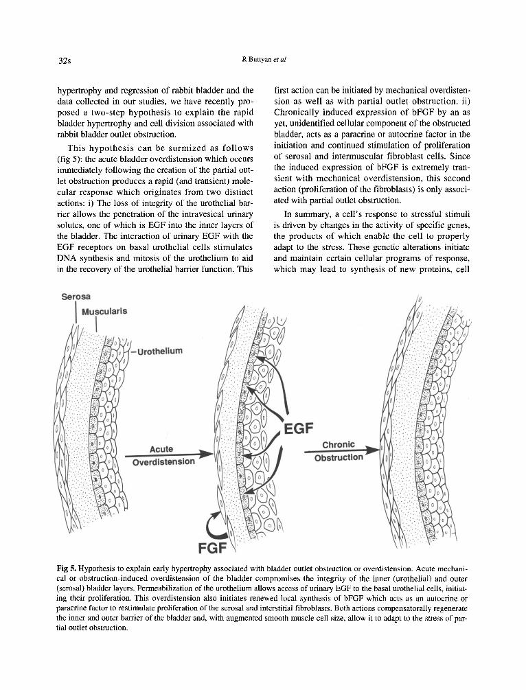
hypertrophy and regression of rabbit bladder and thedata collected in our studies, we have recently pro-posed a two-step hypothesis to explain the rapidbladder hypertrophy and cell division associated withrabbit bladder outlet obstruction.
This hypothesis can be surmized as follows(fig 5): the acute bladder overdistension which occursimmediately following the creation of the partial out-let obstruction produces a rapid (and transient) mole-cular response which originates from two distinctactions: i) The loss of integrity of the urothelial bar-rier allows the penetration of the intravesical urinarysolutes, one of which is EGF into the inner layers ofthe bladder. The interaction of urinary EGF with theEGF receptors on basal urothelial cells stimulatesDNA synthesis and mitosis of the urothelium to aidin the recovery of the urothelial barrier function. This
I
-Urothellum
first action can be initiated by mechanical overdisten-sion as well as with partial outlet obstruction. ii)Chronically induced expression of bFGF by an asyet, unidentified cellular component of the obstructedbladder, acts as a paracrine or autocrine factor in theinitiation and continued stimulation of proliferationof serosal and intermuscular fibroblast cells. Sincethe induced expression of bFGF is extremely tran-sient with mechanical overdistension, this secondaction (proliferation of the fibroblasts) is only associ-ated with partial outlet obstruction.
In summary, a cell's response to stressful stimuliis driven by changes in the activity of specific genes,the products of which enable the cell to properlyadapt to the stress. These genetic alterations initiateand maintain certain cellular programs of response,which may lead to synthesis of new proteins, cell
Fig 5. Hypothesis to explain early hypertrophy associated with bladder outlet obstruction or overdistension. Acute mechani-calor obstruction-induced overdistension of the bladder compromises the integrity of the inner (urothelial) and outer(serosal) bladder layers. Permeabilization of the urothelium allows access of urinary EGF to the basal urothelial cells, initiat-ing their proliferation. This overdistension also initiates renewed local synthesis of bFGF which acts as an autocrine orparacrine factor to restimulate proliferation of the serosal and interstitial fibroblasts. Both actions compensatorally regeneratethe inner and outer barrier of the bladder and, with augmented smooth muscle cell size, allow it to adapt to the stress of par-tial outlet obstruction.

Molecular control of rabbit urinary bladder hypertrophy 33s
Fig 6. The dynamic bladder. Studiesof partialbladderout-let obstruction in the rabbit demonstrate the bladder is adynamic organ with regards to its response to outletobstruction and its subsequent release.The bladderrapidlyhypertrophies when the outlet is obstructed in order tocompensate for the increased urethral resistance and thenregresses againwhenthe outletobstruction is removed.
~.~bstructionu -uR-e-le-a-se....:O
become irreversible to the signals that initiate the celldeath process during chronic obstruction. Our longterm goal, therefore, is to determine the point of irre-versibility and to characterize the molecular markersof irreversibility of this bladder hypertrophic process.We ultimately expect that these markers might proveto be important diagnostic tools for the identificationand treatment of human obstructive bladder pathol-ogy.
Acknowledgments
proliferation, hypertrophy, or to cell death. Manygenes participate in a well defined order in theresponse to stimuli. Our experiments indicate that thebladder is an extremely dynamic tissue whichresponds rapidly to outlet obstruction by initiatedbladder growth, and just as rapidly when the outletobstruction is relieved, by regressing (see fig 6).During the development of obstructive bladderhypertrophy as well as during it reversal, we haveproposed that certain growth factors and their recep-tors, specifically EGF, bFGF, TGF-~ and their recep-tors, seem to play key roles by activating genes andinitiating the cellular programs of hypertrophy, pro-liferation and cell death. By analysing the interactionof EGF, bFGF, TGF-~ with their receptors, we hopewe can start to define the cellular response programsof bladder responding to outlet obstruction.
Irreversible bladder hypertrophy
The partially obstructed rabbit bladder has been anextremely useful model to analyze the mechanism bywhich the bladder initially responds to the stress ofoutlet obstruction, an increasing clinical problem forour aging population. At some point, in a similarmanner to humans, chronic distension in this animalmodel can result in pathological changes that will notbe reversed even if the bladder outlet obstruction isremoved. Since it is demonstrated that apoptosis is aprominent mechanism whereby obstructive bladderchanges are reversed, it becomes important to iden-tify whether the cells of hypertrophied bladder can
This work was supported by research grants providedby the United States Public Health Service (NIH; DK26508, DK 44689) and by the United States VeteransAdministration. MW Chen is supported by theAmerican Foundation for Urological Disease withfunds contributed by the Connaught Foundation.
References
Arends MJ, Morris RG, Wyllie AH. Apoptosis: Therole of the endonuclease. Am J Pathol 1990;136:593-608
2 Buttyan R, Jacobs BZ, Blaivas JG, Levin RM. Earlymolecular response to rabbit bladderoutlet obstruction.Neurourol Urodyn 1992; 11:225-38
3 Chen MW, Krasnapolsky L, Levin RM, Buttyan R. Anearly molecular response induced by acute overdisten-sion of the rabbit urinary bladder. Mol Cell Biochem1994;132:39-44
4 Ghoniem GM, Regnier CH, Biancani P, Johnson L,SussetJG. Effect of vesicaloutlet obstruction on detru-sor contractility and passive properties in rabbits. JUroI1986;135:1284-9
5 Kato K, Wein AJ, Kitada S, Haugaard N, Levin RM.The functional effect of mild outlet obstruction on therabbiturinary bladder. J UroI1988;140:880-4
6 Kerr JFR, WyllieAHR, Currie AR. Apoptosis: A basicbiological phenomenon with wide-ranging implicationsin tissuekinetics. Br J Cancer 1972;26:239-57
7 Lee PL, Johnson DE, CousensLS, Fried VA, WilliamsR. Purification and complementary DNA cloning of areceptor for basic fibroblast growth factor. Science1989;245:57-60
34s R Buttyan et al
8 Levin RM, Malkowicz SB, Wein AJ, Atta MA, 15 Monson FC, Wein AJ, Murphy EM, Levin RM.Elbadawi A. Recovery from short-term obstruction of Stimulation of DNA synthesis in rabbit bladder wallthe rabbit urinary bladder. J UroI1985;134:388-90 after partial outlet obstruction and acute overdistension.
9 Levin R, Kato K, Ruggieri MR, McGuire E, Elbadawi Neurourol Urodym 1994;13:51-62
A, Wein AJ. The physiological effect of outlet obstruc- 16 Mostwin JL, Brooks L. A new guinea pig model of ure-tion in the cats (abstr). J UroI1989;141:335A thral obstruction (abstr). J UroI1989;141-335A
10 Levin RM, Wein AJ, Whitmore K, Monson FC, 17 Nexo E, Jorgensen PE, Hansen MR. Human epidermalMcKenna BAW, Ruggieri R. Trypan blue as an indica- growth factor on molecular forms present in urine andtor of urothelial integrity. Neurourol Urodyn 1990; blood. Regul Pep 1992;42(1-2): 75-849:269-79 18 Rohner TJ, Jr, Hannigan JD, Sanford EJ. Altered in
II Levin RM, Longhurst PA, Monson FC, Haugaard N, vitro adrenergic responses of dog detrusor muscle afterWein AJ. Experimental studies on bladder outlet chronic bladder outlet obstruction. Urology 1978;obstruction. In: Lepor H, Lawson RK, eds, Prostate 11:357-61Diseases. Philadelphia: Sanders Publishing Co, 1993; 19 Santarosa R, Colombell M, Kaplan S, Monson F, Levin119-130 R, Buttyan R. Regression of the hypertrophied rabbit
12 Malkowicz SB, Wein AJ, Elbadawi A, Van Arsdalen bladder involves extensive apoptosis of cells in theK, Ruggieri MR, Levin RM. Acute biochemical and urothelium and serosal lamina. Lab Invest 1994;functional alterations in the partially obstructed rabbit 70:503-10urinary bladder. J Urol 1986;136:1324-9 20 Speakman MJ, Brading AF, Gilpin CJ, Dixon JS,
13 Mattiasson A, Uvelius B. Changes in contractile prop- Gilpin SA, Gosling JA. Bladder outlet obstruction-aerties in hypertrophic rat urinary bladder. J Uro11982; cause of denervation supersensitivity. J Urol 1987;128:1340-2 138:1461-6
14 Messing EM. Clinical implications of the expression of 21 Tammela T, Wein AJ, Monson FC, Levin RM.epidermal growth factor receptors in human transitional Urothelial permeability of the isolated whole bladder.cell carcinoma. Cancer Res 1990;50:2530-7 Neurourol Urodym 1993;12:39-47
Copyright © 2022 FDOKUMEN