Factors Associated with Failure of Bakri Balloon Tamponade ...
Upload
khangminh22Category
view
1download
0
Koniari et al. Lipids in Health and Disease 2014, 13:33http://www.lipidworld.com/content/13/1/33
RESEARCH Open Access
Transauricular balloon angioplasty in rabbitthoracic aorta: a novel model of experimentalrestenosisIoanna Koniari1,8*, Efstratios Apostolakis2, Athanasios Diamantopoulos3, Helen Papadaki4, Evangelia Papadimitriou5,Evangelia Poimenidi5, Dimitrios Karnabatidis3, Anna Karahaliou6, Lena Costaridou6, Apostolos Papalois7,Dimitrios Siablis3, Dimitrios Dougenis1 and Dimitrios Alexopoulos8
Abstract
Background: The aim of this study was to demonstrate a percutaneous transauricular method of balloonangioplasty in high-cholesterol fed rabbits, as an innovative atherosclerosis model.
Methods: Twenty male New Zealand rabbits were randomly divided into two groups of ten animals, as follows:atherogenic diet plus balloon angioplasty (group A) and atherogenic diet alone (group B). Βalloon angioplasty wasperformed in the descending thoracic aorta through percutaneous catheterization of the auricular artery. Eightadditional animals fed regular diet were served as long term control. At the end of 9 week period, rabbits wereeuthanized and thoracic aortas were isolated for histological, immunohistochemical and biochemical analysis.
Results: Atherogenic diet induced severe hypercholesterolemia in both group A and B (2802 ± 188.59 and 4423 ±493.39 mg/dl respectively) compared to the control animals (55.5 ± 11.82 mg/dl; P < 0.001). Group A atheroscleroticlesions appeared to be more advanced histologically (20% type IV and 80% type V) compared to group B lesions(50% type III and 50% type IV). Group A compared to group B atherosclerotic lesions demonstrated similarpercentage of macrophages (79.5 ± 9.56% versus 84 ± 12.2%; P = 0.869), more smooth muscle cells (61 ± 14.10%versus 40.5 ± 17.07; P = 0.027), increased intima/media ratio (1.20 ± 0.50 versus 0.62 ± 0.13; P = 0.015) despite thesimilar degree of intimal hyperplasia (9768 ± 1826.79 μm2 versus 12205 ± 8789.23 μm2; P = 0.796), and furthersignificant lumen deterioration (23722 ± 4508.11 versus 41967 ± 20344.61 μm2; P = 0.05) and total vessel areareduction (42350 ± 5819.70 versus 73190 ± 38902.79 μm2; P = 0.022). Group A and B animals revealed similar nitratedprotein percentage (P = NS), but significantly higher protein nitration compared to control group (P < 0.01; P < 0.01,respectively). No deaths or systemic complications were reported.
Conclusion: Transauricular balloon angioplasty constitutes a safe, minimally invasive and highly successful model ofinduced atherosclerosis in hyperlipidaemic rabbits.
Keywords: Atherosclerosis, Restenosis, Hypercholesterolemic diet, Balloon angioplasty, Oxidative stress,Remodelling, Intima/media ratio
* Correspondence: [email protected] Surgery Department, University Hospital of Patras, Rion Patraszip 25500, Greece8Cardiology Department of Patras University Hospital, Rion Patras, GreeceFull list of author information is available at the end of the article
© 2014 Koniari et al.; licensee BioMed Central Ltd. This is an Open Access article distributed under the terms of the CreativeCommons Attribution License (http://creativecommons.org/licenses/by/2.0), which permits unrestricted use, distribution, andreproduction in any medium, provided the original work is properly cited. The Creative Commons Public Domain Dedicationwaiver (http://creativecommons.org/publicdomain/zero/1.0/) applies to the data made available in this article, unless otherwisestated.

Koniari et al. Lipids in Health and Disease 2014, 13:33 Page 2 of 12http://www.lipidworld.com/content/13/1/33
BackgroundHemodynamic strain promotes the development of athe-rosclerosis, while dietary induced atherosclerosis is ac-celerated and enhanced where mechanical injury hasbeen performed experimentally [1]. Consequently, thefact that endothelial injury and increased tendency to-ward atherosclerotic changes are localized in the sameregions constitutes the basis for the response to injuryhypothesis for atherosclerosis [2].Restenosis, a common complication after angioplasty
represents the arterial wall’s healing response to me-chanical injury and comprises two main processes,neointimal hyperplasia and vascular remodeling [3-6].Although the primary stimulus for restenosis is the
mechanical injury of balloon dilation to the vessel wall, adominant risk factor for the spontaneous developmentof occlusive coronary disease is hypercholesterolemia.Notably, the physical injury caused by balloon dilationinduces intimal hyperplasia independent of blood cho-lesterol levels in angioplasty induced atheroscleroticlesions [7].Traditionally, in experimental atherosclerosis models,
intraarterial access in an animal is achieved throughfemoral artery surgical cut-down. This technique how-ever, may occasionally be followed by severe complica-tions involving bleeding, thrombosis, arterial occlusionand local or systemic infections. The aim of this study isto describe a safe, non-surgical percutaneous method oftransauricular endovascular access and further balloonangioplasty performance in the thoracic aorta of high-cholesterol fed rabbits, as a novel alternative model ofexperimental restenosis.
ResultsTransauricular arterial access for balloon injury of thoracicaortaPercutaneous catheterization of the auricular artery andfurther balloon injury of the descending thoracic aorta
Table 1 Blood assays of control, injured and non- injured hyp
Bloodassays
Group A Group B
5 weeks 9 weeks 5 week
TC 2005 ± 207.20 2802 ± 188.59a 4121 ± 4
TG 197 ± 32.30 324 ± 33.73a 381 ± 54
HDL 295 ± 53.98 384 ± 26.29a 383 ± 40
SGPT 31 ± 7.10 38 ± 6.78 47 ± 5.5
g-GT 5 ± 2.15 7 ± 1.43 6 ± 2.05
Creatinine 1.05 ± 0.14 1.03 ± 0.13 0.79 ± 0
*Baseline biochemical parameters of all groups were into the normal range.aP < 0.001, unpaired t-test vs Control.bP < 0.001,unpaired t- test vs Control.cP ≤0.001, unpaired t-test vs Group A.dP < 0.05, unpaired t-test vs Group A.
was performed successfully in all rabbits of group A (10of 10 animals), with no complication being noted duringangiographic examination. In one case, puncture of therabbit auricular artery resulted in severe vasospasm, withsubsequent inability to infuse contrast medium and in-sert the guide wire. In this rabbit, vascular access wasachieved through the contra lateral central auricularartery. The recovery of all group A rabbits was normalwithout any local or systemic complications. No clinicalsigns of hematoma or local infection were identified.There were no deaths after the intervention and thefollowing 8 week period.After the transauricular injury of descending aorta, the
punctured auricular artery was peripherally destroyedand could not be re- accessed.
Blood chemistryAt the end of the 9-week period mean TC, TG, and HDLlevels (mg/dl) increased significantly in both group A andgroup B hypercholesterolemic animals compared to con-trol group (P < 0.001; Table 1). A statistically significantincrease in TC, TG and HDL levels (P < 0.001; P = 0.001;P = 0.03, respectively) was observed in group B comparedto group A animals.Noteworthily, at the end of 9-week period, both high-
cholesterol fed rabbits subjected to balloon injury andnon-injured hypercholesterolemic rabbits, had normalrenal and liver function, as levels of creatinine and he-patic enzymes remained within normal range (Table 1).Finally, there was no statistically significant difference inbody weight between control, group A and group B rab-bits (3705 ± 140.34 g, 3625 ±88,64 g and and 3600 ±92.58 g, respectively; P = 0.137).
Histological evaluation of atherosclerosisAtherogenic diet resulted in the development of signifi-cant atherosclerotic lesions in all group A and group Banimals compared to the control group, that revealed novisible atherosclerotic lesions (p < 0.001). Advanced type
erlipidemic rabbits
Control
s 9 weeks 5 weeks 9 weeks
14,99 4423 ± 493.39b,c 35 ± 7.25 55.5 ± 11.82
.56 502 ± 96.24b,c 38 ± 2 43.8 ± 9.66
.49 412 ± 15.40b,d 21.8 ± 3.60 34.5 ± 2.88
3 54 ± 5.57 32.5 ± 8 39.7 ± 6
7 ± 1.58 4.25 ± 0.89 5.25 ± 0.89
.11 0.89 ± 0.13 0.78 ± 0.46 0.93 ± 0.18
Koniari et al. Lipids in Health and Disease 2014, 13:33 Page 3 of 12http://www.lipidworld.com/content/13/1/33
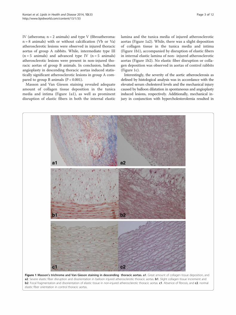
IV (atheroma; n = 2 animals) and type V (fibroatheroma:n = 8 animals) with or without calcification (Vb or Va)atherosclerotic lesions were observed in injured thoracicaortas of group A rabbits. While, intermediate type III(n = 5 animals) and advanced type IV (n = 5 animals)atherosclerotic lesions were present in non-injured tho-racic aortas of group B animals. In conclusion, balloonangioplasty in descending thoracic aortas induced statis-tically significant atherosclerotic lesions in group A com-pared to group B animals (P < 0.001).Masson and Van Gieson staining revealed adequate
amount of collagen tissue deposition in the tunicamedia and intima (Figure 1a1), as well as prominentdisruption of elastic fibers in both the internal elastic
Figure 1 Masson’s trichrome and Van Gieson staining in descendinga2. Severe elastic fiber disruption and disorientation in balloon injured atheb2. Focal fragmentation and disorientation of elastic tissue in non-injured aelastic fiber orientation in control thoracic aortas.
lamina and the tunica media of injured atheroscleroticaortas (Figure 1a2). While, there was a slight depositionof collagen tissue in the tunica media and intima(Figure 1b1), accompanied by disruption of elastic fibersin internal elastic lamina of non- injured atheroscleroticaortas (Figure 1b2). No elastic fiber disruption or colla-gen deposition was observed in aortas of control rabbits(Figure 1c).Interestingly, the severity of the aortic atherosclerosis as
defined by histological analysis was in accordance with theelevated serum cholesterol levels and the mechanical injurycaused by balloon dilatation in spontaneous and angioplastyinduced lesions, respectively. Additionally, mechanical in-jury in conjunction with hypercholesterolemia resulted in
thoracic aortas. a1. Great amount of collagen tissue deposition, androsclerotic thoracic aortas. b1. Slight collagen tissue increment andtherosclerotic thoracic aortas. c1. Absence of fibrosis, and c2. normal

Koniari et al. Lipids in Health and Disease 2014, 13:33 Page 4 of 12http://www.lipidworld.com/content/13/1/33
more prominent atherosclerotic lesions than atherogenicdiet alone.
Immunohistochemical studyThe atherogenic diet was associated with a significantincrease in lipid deposition and foam cell formation,indicated by the increase in RAM-11 immunoreactivity ingroup A and B animals. The aortas of group A rabbitswere mainly strongly positive (n = 8 animals) for RAM-11staining, except two cases that were moderately positive.Similarly, almost all the aortas of group B rabbits revealedstrongly positive (n = 9 animals) RAM-11 immunore-activity. It is prominent that the percentage of RAM-11positive cells had no significant difference between spon-taneous and angioplasty- induced atherosclerotic lesions(79.5 ± 9.56% versus 84 ± 12,2% respectively; P = 0.869), asshown in Figure 2. Also, the relative number of macro-phages was associated with the severity of atheroscleroticlesions, as demonstrated in Figure 3a1 and 3b1.HHF-35 immunostaining for α-actin, demonstrated that
angioplasty- induced atherosclerotic lesions of group Awere mainly moderate positive (n = 7 animals) or stronglypositive in three cases (Figure 3a2). Whereas, spontaneousatherosclerotic lesions of group B, contained either mild(n = 4 animals) or moderate (n = 6 animals) numbers ofHHF-35 immunopositive SMCs (Figure 3b2). There was astatistically significant difference in SMCs percentage bet-ween angioplasty-induced and spontaneous atherosclero-tic lesions (61 ± 14.10% versus 40.5 ± 17.07% respectively;P = 0.027). Notably, the prominent increment of HHF-35positive cells in angioplasty- induced lesions, reflects thekey role of SMCs in restenosis after balloon angioplasty.Control animals demonstrated no RAM-11 or HHF-35
staining (Figure 3c1, 3c2), revealing significant differenceregarding the amount of foam cells and SMCs comparedto group A and group B animals, respectively (P <0.001).
Figure 2 RAM-11 and HHF-35 positive cell distribution in three groupbetween angioplasty induced and spontaneous atherosclerotic lesions: a PP < 0.001, Bonferroni’s post hoc ANOVA analysis vs contol; c P < 0.001, BonfHHF-35 cells was noticed in angioplasty induced atherosclerotic lesions: dTamhane’s post hoc ANOVA analysis vs control; f P < 0.001, Tamhane’s post
Morphometric analysisAtherosclerotic lesions were not present in the thor-acic aorta of control animals. Atherogenic diet inducedsignificant intimal hyperplasia in spontaneous atheros-clerotic lesions in group B compared to control group(12205 ±8789.23 μm2 versus 555 ± 134.75 μm2; P < 0.001),(Figure 4a). Similarly, balloon angioplasty induced signi-ficant intimal hyperplasia in hyperlipidemic animals ofgroup A compared to animals maintained on control diet(9768 ± 1826.79 μm2 versus 555 ± 134.75 μm2; P < 0.001).No significant difference was observed in intimal hy-perpasia between angioplasty- induced and spontaneousatherosclerotic lesions in group A and B, respectively(9768 ± 1826.79 μm2 versus 12205 ± 8789.23 μm2; P =0.796), (Figure 4a). However, angioplasty induced athero-sclerotic lesions demonstrated a significant increase in in-tima/media ratio compared to spontaneous atheroscleroticlesions (1.20 ± 0.50 versus 0.62 ± 0.13, in groups A and Brespectively; P = 0.015). Both angioplasty-induced andspontaneous atherosclerotic lesions revealed a significantincrease in intima/media ratio compared to the control an-imals (P <0.001), (Figure 4b). In addition, balloon dilatationresulted in a significant lumen deterioration in angioplastyinduced lesions compared to spontaneous atherosclerotic lesions (23722 ± 4508.11 versus 41967 ± 20344.61 μm2;P = 0.05). Thus, there was a significant reduction in lumenof angioplasty induced lesions compared to the respectiveof the control group (23722 ± 4508.11 versus 41828 ±1213.53 μm2; P < 0.001), whereas there was no differencein lumen area between group B and control animals(41967 ± 20344.61 μm2 versus 41828 ± 1213.53 μm2; P = 1),(Figure 4c). Finally, a significant reduction in total vesselarea was observed between injured and uninjured thoracicaortas in group A and B, respectively (42350 ± 5819.70versus 73190 ± 38902.79 μm2; P = 0.022), whereas therewas observed a non-significant decrease in total vessel area
s. a. No significant difference in RAM-11 cell percentage was observed= non significant, Bonferroni’s post hoc ANOVA analysis vs Group B; berroni’s post hoc ANOVA analysis vs control. b. Significant increase inP < 0.05, Tamhane’s post hoc ANOVA analysis vs Group B; e P < 0.001,hoc ANOVA analysis vs control.

Figure 3 Ram-11 and HHF-35 staining in thoracic aortas. a1. Plethora RAM-11 positive foam cells, and a2. significant amounts of positive for a-actin SMCs are observed in angioplasty induced atherosclerotic lesions. b1. Significant foam cell formation, and b2. several a-actinpositive SMCs between foam cells in spontaneous atherosclerotic lesions. c1. Lack of foam cell deposition, and c2. Normal presence of HHF-35 positiveSMCs in the media of control aortas.
Koniari et al. Lipids in Health and Disease 2014, 13:33 Page 5 of 12http://www.lipidworld.com/content/13/1/33
between injured and control thoracic aortas (42350 ±5819.70 versus 56298 ± 1346.73 μm2; P = 0.672), (Figure 4d).A non-significant increase was observed in total vessel areabetween hypercholesterolemic and control rabbits (73190 ±38902.79 μm2 versus 56298 ± 1346.73 μm2; P = 0.43).
Biochemical analysisThe effect of the proposed injury model on protein tyro-sine nitration was studied by measuring protein nitroty-rosine levels on aorta samples derived from control andanimals fed with high cholesterol diet, injured and non-injured. High cholesterol diet significantly increased pro-tein nitration levels, while transauricular ballon angioplastydid not cause any further increase (Figure 5).
DiscussionWe used a non-surgical, minimally invasive technique toperform balloon injury in rabbit thoracic aortas bypercutaneous catheterization of the auricular artery. Inexperimental restenosis models, catheterizations are tra-ditionally performed after surgical cutdown of the femoralor carotid arteries and followed by subsequent ballooninjury in femoral, iliac, coronary, carotid arteries and ab-dominal or thoracic aorta, respectively. The peripheralvessels of the rabbit are fragile, while surgical cut-downand catheterization are associated with long proceduraltime periods and deep general anesthesia with intubation[8]. Moreover, a surgically accessed vessel is finally ligated,which frequently renders it vulnerable to iatrogenic

Figure 4 Comparison of neointimal area, intima/media ratio, lumen area and total vessel area between groups. a. Atherogenic diet andballoon angioplasty induced significant intimal hyperplasia in hyperlipidaemic rabbits: a P = non significant, Tamhane’s post hoc ANOVA analysis vsGroup B; b P < 0.001, Tamhane’s post hoc ANOVA analysis vs control; c P < 0.05, Tamhane’s post hoc ANOVA analysis vs control. b. A significantincrease in intima/media ratio was observed in angioplasty induced atherosclerotic lesions: d P < 0.05, Tamhane’s post hoc ANOVA analysis vs GroupA; e P < 0.001, Tamhane’s post hoc ANOVA analysis vs Control; f P < 0.001, Tamhane’s post hoc ANOVA analysis vs Group A. c. Angioplasty resulted ina significant reduction in lumen of injured thoracic aortas: j P = 0.05, Tamhane’s post hoc ANOVA analysis vs Group B; k P < 0.001, Tamhane’s post hocANOVA analysis vs control; l P = non significant, Tamhane’s post hoc ANOVA analysis vs control. d. Injured thoracic aortas demonstrated a significantdecrease in total vessel area consistent with constrictive remodeling occurrence: g P < 0.05, Bonferroni’s post hoc ANOVA analysis vs Group B; hP = non significant, Bonferroni’s post hoc ANOVA analysis vs control; i P = non significant, Bonferroni’s post hoc ANOVA analysis vs control.
Koniari et al. Lipids in Health and Disease 2014, 13:33 Page 6 of 12http://www.lipidworld.com/content/13/1/33
trauma or even thrombosis. On the contrary, our alterna-tive catheterization method is simple, rapid, safe and easilyreproducible, as it is takes advantage of favourable auricu-lar vascular anatomy of the rabbit. The major benefits oftransauricular catheterization technique comprise the ac-celeration of endovascular access, the avoidance of surgi-cal wounds, and, significantly, the preservation of valuablefemoral and cervical vessels. In addition, animals expe-rience less pain, bleeding complications, wound infections,while dissociative anaesthesia is sufficient.Generally, the technique offers the advantages of per-
cutaneous minimally invasive procedures, characterizedby reduced morbidity and mortality as well as minimal
distortion of normal anatomy and physiology. A minordisadvantage of transauricular catheterization methodconstitutes the peripheral auricular artery impairmentthat could be attributed to the cutting of the dermisalong the course of the guide wire and further multipledilatations so as to be achieved the insertion of sheaths.In our model of induced atherosclerosis, angioplasty –
induced atherosclerotic lesions appeared to be signi-ficantly more severe and advanced (type V) compared tospontaneous (type III or type IV) lesions, confirmingstudies with balloon injury through other approaches. Ithas been noticed that in the absence of hypercholeste-rolemia, angioplasty- induced lesions regress and resolve

Figure 5 Tyrosine nitration of proteins is not increased by the transauricular balloon angioplasty. a. Western Blot analysis for3′-nitrotyrosine (upper panel) and β-actin (lower panel) in total protein lysates of descending aortas. b. The protein amounts were quantified bydensitometric analysis of the corresponding bands and the ratio of nitrated proteins/β-actin was calculated for each lane. Results are expressed asmean ± S.E.M. of the percentage change of the amounts of 3′-nitrotyrosine compared with control animals. n.s, not statistically significant.
Koniari et al. Lipids in Health and Disease 2014, 13:33 Page 7 of 12http://www.lipidworld.com/content/13/1/33
spontaneously [9], while, in the presence of hypercholes-terolemia these lesions not only sustain but progress tolarger lesions. Also, plaque size at sites of spontaneouslesions has been reported to increase with increasingblood cholesterol levels [7]. Similarly, in our study, bothspontaneous and angioplasty induced atheroscleroticlesions revealed significant intimal hyperplasia in unin-jured and balloon injured rabbits respectively, after9 weeks of 4% high cholesterol diet.Another difference in pathogenesis between spontan-
eous and angioplasty-induced atherosclerotic lesions con-sists in cell type population. Especially, SMCs have beenfound to be abundant in the intima of balloon- injuredaortas, whereas foam cell-derived macrophages have beenrecognized as the predominant cell type in the intima ofundamaged aortas [10]. In fact, we demonstrated thatthe percentage of macrophages (RAM-11 positive cells)revealed no difference between spontaneous and angio-plasty- induced lesions and was in accordance with thehistological stage of the respective lesions. Whereas, thepercentage of SMCs (HHF-35 positive cells) revealed astatistically significant increase in angioplasty-induced
lesions compared to spontaneous lesions of uninjuredaortas, similarly to previous studies [11,12].Additionally, angioplasty induced lesions demonstrated
a significant increase in intima/media ratio compared tospontaneous atherosclerotic lesions, despite the similardegree of intimal hyperplasia. Furthermore, the increasedintima/media ratio was accompanied with significantlumen deterioration and total vessel reduction in angio-plasty induced lesions, reflecting the artery’s shrinkageor constrictive remodeling. Indeed, the accumulation ofSMCs in combination with ECM reorganization that cha-racterized by increased collagen deposition, contributes tointimal thickening and negative remodeling, leading inrestenosis after balloon angioplasty.It should be noticed the safety of both transauricular
balloon angioplasty and atherogenic diet with regard tothe induction and progression of atherosclerosis. Espe-cially, we used a non-commercial and easy manufacturedatherogenic diet consisting of standard rabbit chowenriched with 4% cholesterol [13], without any ad-ditional atherogenic components, such as palm oil [14],peanut oil [15], high fat coconut oil [16], high fat corn

Koniari et al. Lipids in Health and Disease 2014, 13:33 Page 8 of 12http://www.lipidworld.com/content/13/1/33
oil [17], and lard combined with either yolk powder orpeanut oil [18,19]. Interestingly, the normal renal andliver function of balloon injured hyperlipidaemic animalsconfirms the absence of complications with percuta-neous transauricular angioplasty, suggesting a safe alter-native non-commercial atherogenic diet together with aminimally invasive restenosis model.Generally, hypercholesterolemia has been known to in-
duce the development of atherosclerotic lesions both inhumans and animal models, while the cellular basis forthis action has been largely attributed to the formation ofoxidized LDL (oxLDL) and oxidative injury to endothe-lium [20]. It has been reported that the oxidative modi-fication of LDL correlated with reduction of endothelialnitric oxide synthase (eNOS) expression [21] or modula-tion of eNOS activity and free radical bioavailability [22],resulting in peroxynitrite and 3′-nitrotyrosine formation.Interestingly, application of the transauricular ballooninjury model did not further increase protein nitrationobserved in hyperchoresterolemic animals, suggesting thatballoon angioplasty does not cause peroxynitrite-mediatedoxidative stress.
ConclusionsThis study in high-cholesterol fed rabbits introducestransauricular balloon angioplasty as a novel model ofinduced- atherosclerosis and vascular restenosis. There-fore, transauricular angioplasty constitutes a safe, rapid,minimally invasive, highly successful as well as easilyreproducible restenosis model.
MethodsAnimal modelThe study was conducted in accordance to the Institutional“Guide for the Care and Use of Laboratory Animals” andwas approved by the Institutional Animal Care and UseCommittee of the West Greece Prefecture. All experimentswere performed in the Animal House of the MedicalSchool. Twenty eight male New Zealand White rabbits,weighing 2.5–3 kg, were housed individually at 20 ± 3°Cwith a 12-h: 12-h light/dark cycle and with free access towater. All rabbits were allowed one week, feeding on regu-lar rabbit chow, to acclimate to their environment. Eightanimals were fed regular rabbit chow until the end of thestudy, serving as long term control group (C; n = 8). Afterthe week of acclimation, atherogenic diet consisting ofregular rabbit chow supplemented with 4% cholesterol(ELPEN Pharmaceutical, Athens, Greece) was initiated.Then twenty rabbits were randomly divided into twogroups of ten animals, as following: atherogenic diet plusballoon angioplasty (group A) and atherogenic diet alone(group B). Seven days later, rabbits of group A were anes-thetized with ketamine (50 mg/kg) plus xylazine (10 mg/kg)intramuscularly, and subjected to balloon injury of the
descending thoracic aorta through percutaneous cathe-terization of the auricular artery [8]. At the end of theintervention, antibiotic prophylaxis with cephalosporinwas administered (750 mg cefuroxime intramuscularly;Zinacef; GlaxoSmithKline, Research Triangle Park, NC).The animals were allowed to recover and were maintainedon 4% high- cholesterol diet for additional 8 weeks. At theend of this period, all animals were subsequently anesthe-tized using the above mentioned regimens (ketamine plusxylazine), and euthanized by intravenous injection of a sat-urated KCl solution. The descending aortas were exposed,isolated and harvested for histological, immunohisto-chemical, and biochemical analysis. Rabbit feeding was re-stricted to 120 g/day. Blood samples were collected every4 weeks. The general condition of the rabbits was ob-served daily. Body weights were measured every 4 weeks.
Transauricular approachThe detailed technique of envovascular transauricular ac-cess has been previously described [8]. Here, we describethe balloon angioplasty performance after percutaneouscatheterization of the rabbit auricular artery. In brief,animals were immobilized in the supine position, and bothauricular dorsa (ie, backside surfaces of their ears) wereshaved and scrubbed with a combination of povidoneiodine and an alcohol-based solution to achieve disinfec-tion. Rabbits were placed under a c-arm angiographic unitwith ability to perform road mapping and digital subtrac-tion angiography (Philips DVI-S angiography unit). Car-diovascular monitoring was performed with peripheralpulse oximetry.The central auricular artery was the target vessel for
cannulation and endovascular access of the descendingthoracic aorta respectively. Initially, the auricular arterywas punctured with a 22-gauge intravenous catheter(Helmflon; Helm Pharmaceuticals, Hamburg, Germany)approximately at the distal half of its subcutaneouscourse (Figure 6a). The needle of the catheter was re-moved, and 5 mL of contrast agent diluted with normalsaline (1:1) was infused to obtain roadmap images of theextracranial carotid vasculature. Care was taken to aimat the distal half of the vessel, so as a second more pro-ximal attempt might be performed in case of vasospasmor rupture [8].Secondly, a 0.018-inch guide wire (V-18 control wire;
Boston Scientific, Natick, MA) was carefully advancedinto the external carotid artery (Figure 6d and 6e). Theguide wire was then advanced in the aortic arch and navi-gated into the descending thoracic aorta under fluoro-scopic guidance (Figure 7). Then, the intravenous catheterwas withdrawn, local anesthesia (lidocaine 1%) was ap-plied, and a 2- to 3-cm-long incision of the dermis wasperformed at the point of the initial puncture along thecourse of the guide wire. Finally, a 4-F, 0.018- inch guide

Figure 6 Intraarterial transauricular access in the rabbit thoracic aorta. a. Direct percutaneous catheterization of the central auricular arterywith a 22-gauge intravenous catheter, b. Repeated tract dilations of the peripheral transauricular artery with the introduction of a 4-F vascularsheath, c. Insertion of an 6.0-mm/40- mm (Cordis) peripheral dilatation balloon catheter over the guide wire, d-e Roadmap image of the auricularartery and the common carotid artery.
Koniari et al. Lipids in Health and Disease 2014, 13:33 Page 9 of 12http://www.lipidworld.com/content/13/1/33
wire–compatible vascular sheath (Bolton Medical, Villers-les- Nancy, France) was advanced into the external carotidartery after serial step-by-step dilations with the sheath’sown dilator (Figure 6b), [8]. The repeated over-the wiredilations were necessary to remove the tight and narrowperipheral segment of the arterial endothelium beforesheath insertion. All animals were administered an intra-venous bolus of heparin (100 U/kg) after vascular accesswas established. When transauricular arterial access hadbeen gained, a 6.0-mm/40- mm (Cordis, Abbott Vascular,Santa Clara, USA) peripheral dilatation balloon catheter,preinfused with heparinized saline, was inserted over theguide wire and advanced to the level of descending thoracicaorta below the orifice of subclavian artery (Figures 6c and7). Injury of the descending aorta was performed by infla-ting the balloon twice to 11 atm of pressure for 45 secondsat 30 second intervals. The balloon was then removed fromthe animal and angiographic imaging of the descendingthoracic aorta was performed in order to exclude ruptureor dissection of the vessel. Antibiotic prophylaxis using750 mg of cefuroxime was given intramuscularly. Animals
were monitored during recovery and were closely checkedfor any signs of local hematomas and local or systemicinfection.
Biochemical assaysTotal serum cholesterol (TC), triglyceride (TG), and highdensity lipoprotein (HDL) levels were measured. Renal andliver function were monitored using creatinine and serumhepatic enzymes (SGPTand g-GT) levels, respectively.
Histological analysisBalloon injured descending thoracic aortas were fixed byimmersion in neutrally buffered 10% formalin, followed bydehydration and embedding in paraffin wax using stan-dard procedures. Four-micrometer sections were obtainedfrom each vessel at 5-mm intervals and stained withhematoxylin and eosin for histopathologic analysis.Masson’s trichrome aniline blue and Weigert Van Gieson’selastic stains (Histoline Laboratories, Italy) were used toassay collagen components and the thickness of theintima, respectively.

Figure 7 Balloon angioplasty of descending thoracic aorta. a. Roadmap images of the rabbit’s carotid artery and the thoracic aorta, b. Insertionof the guide wire into the descending thoracic aorta, c. Insertion of a 6.0-mm/40-mm peripheral dilatation balloon catheter over the guide wire andplacement to the level of descending thoracic aorta d. Inflation of the balloon and injury of the descending aorta below the orifice of subclavian artery.
Koniari et al. Lipids in Health and Disease 2014, 13:33 Page 10 of 12http://www.lipidworld.com/content/13/1/33
Spontaneous and angioplasty induced atheroscleroticlesions were classified according to the guidelines of theAmerican Heart Association [23].
ImmunohistochemistryConsecutive 4-μm-thick sections from each aortic speci-men were collected on Superfrost plus glass slides, depa-raffinized, and rehydrated in graded alcohols. Endogenousperoxidase activity was blocked by treatment with 3%hydrogen peroxide for 15 min, followed by incubationwith protein blocking solution to eliminate nonspecificbinding. Immunohistochemistry was performed usingmonoclonal antibodies to detect macrophages (RAM 11,1:200 dilution; Dako Corp, CA, USA), and α-actin inSMCs (HHF-35, 1:100 dilution; DAKO A/S). The EnvisionPlus Detection System kit (DakoCytomation, USA) and 3,3′-diaminobenzidine (DAB) were used to visualize anti-body binding, according to manufacturer’s instructions.
Sections were counterstained with Harris’ hematoxylin,dehydrated, and mounted permanently. For each antibody,all tissues from the different study animals were immuno-stained concurrently. Negative controls were performed inall cases by omitting the primary antibodies.Immunohistochemical staining was graded on a scale
of 0 to 3 based on the percentage of immunopositivecells as follows: 0, <10% positive cells; 1 (mildly positive),10–35% positive cells; 2 (moderately positive), 35–70%positive cells; and 3 (strongly positive), >70% positivecells.
MorphometryCross sectional areas were quantified using IMAGE Jsoftware [24]. For each artery, measurements of luminalarea (LA); area bounded by the internal elastic lamina(IEL; corresponding to the LA in the absence of intimallesions); and area encircled by external elastic lamina

Koniari et al. Lipids in Health and Disease 2014, 13:33 Page 11 of 12http://www.lipidworld.com/content/13/1/33
(EEL; corresponding to overall vessel size) were per-formed. Neo-intimal area (IA) was determined by sub-tracting the LA from the area encircled by the IEL. Themedial area (M) was calculated as the area encircled byEEL minus the area encircled by the IEL. Total vessel area,defined as the area encircled by the EEL, and intima/media (I/M ratio) were determined.
Western blot analysisProtein tyrosine nitration is one of the post-translationalmodifications derived from the reaction of proteins withnitrating agents, such as peroxynitrite, or the activityof myeloperoxidase and eosinophil peroxidase. Proteinnitration can lead to biological function alterations andhas been detected in several pathological situations, suchas cancer, inflammation and atherosclerosis [25].Descending aorta samples were homogenized in RIPA
buffer and total lysates (100 μg/sample) were run on a10% SDS-PAGE gel and transferred to Immobilon Pmembranes. Blocking was performed by incubating themembranes with Tris-buffered saline (TBS), pH 7.4, with0.1% Tween (TBS-T), containing 5% nonfat dry milk.Membranes were incubated with primary antibody for3′-nitrotyrosine (Upstate Biotechnology, #06-284) orβ-actin (Santa Cruiz, #sc58673), in a dilution of 1:1,000 inTBS-T, for 16 h at 4°C under continuous agitation. Themembranes were washed 3 times with TBS-T for 5 minand incubated with HRP- linked anti-rabbit IgG (1:12,500in TBS-T, Sigma #A0545) or anti-mouse IgG (1:5,000 inTBS-T, Sigma #A3682) respectively, for 1 h at roomtemperature. Detection of immunoreactive bands was per-formed using the enhanced chemiluminescence (ECL)detection kit (Pierce Biotechnology, Rockford, IL, USA).The protein levels that corresponded to the immunoreac-tive bands for 3′-nitrotyrosine and β-actin were quantifiedusing the ImagePC image analysis software (Scion Corp.,Frederick, MD, USA) and the ratio of nitrated proteins/β-actin was calculated for each lane.
Statistical analysisSPSS for Windows (release 19. 0. 0 SPSS Inc, Chicago IL,USA) was used for continuous data analysis. All data wereexpressed as the mean ± standard deviation (SD). Com-parison among groups was performed by ANOVA. Com-parison between two specific groups was performed bythe unpaired two-tailed Student’s t-test. A p value < 0.05was regarded as statistically significant.
Competing interestThe authors declare that they have no competing interests.
Authors’ contributionsAll authors participated in the design, interpretation of the studies, analysisof the data and review of the manuscript. IK, EA, AD, DK conducted theexperiments; DK, DS, DD, DA and LC supplied critical reagents; IK wrote themanuscript; HP performed the histological analysis; EP and EP performed the
biochemical assays; AK and IK conducted the morphometric analysis; and IKconducted the statistical analysis. All individuals who made contributions tothis study are included as authors. All authors read and approved the finalmanuscript.
Author details1Cardiothoracic Surgery Department, University Hospital of Patras, Rion Patraszip 25500, Greece. 2Cardiothoracic Surgery Department of IoanninaUniversity Hospital, Ioannina, Greece. 3Department of Diagnostic andInterventional Radiology, Patras University Hospital, School of Medicine, RionPatras, Greece. 4Department of Anatomy, School of Medicine, University ofPatras, Patras, Greece. 5Laboratory of Molecular Pharmacology, Departmentof Pharmacy, University of Patras, Patras, Greece. 6Department of MedicalPhysics, School of Medicine, University of Patras, Patras, Greece.7Experimental-Research Center ELPEN Pharmaceuticals, Athens, Greece.8Cardiology Department of Patras University Hospital, Rion Patras, Greece.
Received: 2 January 2014 Accepted: 5 February 2014Published: 15 February 2014
References1. Björkerud S: Atherosclerosis initiated by mechanical trauma in
normolipidemic rabbits. J Atheroscler Res 1969, 9:209–213.2. Bondjers G, Björnheden T: Experimental atherosclerosis induced by
mechanical trauma in rats. Atherosclerosis 1970, 12:301–306.3. Costa MA, Simon DI: Molecular basis of restenosis and drug-eluting
stents. Circulation 2005, 111:2257–2273.4. Forrester JS, Fishbein M, Helfant R, Fagin J: A paradigm for restenosis
based on cell biology: clues for the development of new preventivetherapies. J Am Coll Cardiol 1991, 17:758–769.
5. Ross R: Atherosclerosis is an inflammatory disease. Am Heart J 1999,138:S419–S420.
6. Labinaz M, Pels K, Hoffert C, Aggarwal S, O’Brien ER: Time course andimportance of neoadventitial formation in arterial remodeling followingballoon angioplasty of porcine coronary arteries. Cardiovasc Res 1999,41:255–266.
7. Kahn MB, Boesze-Battaglia K, Stepp DW, Petrov A, Huang Y, Mason RP,Tulenko TN: Influence of serum cholesterol on atherogenesis and intimalhyperplasia after angioplasty: inhibition by amlodipine. Am J Physiol HeartCirc Physiol 2005, 288(2):H591–H600.
8. Karnabatidis D, Katsanos K, Diamantopoulos A, Kagadis GC, Siablis D:Transauricular arterial or venous access for cardiovascular experimentalprotocols in animals. J Vasc Interv Radiol 2006, 17:1803–1811.
9. Consigny P, Tulenko T, Nicosia R: Acute effects of angioplasty on vascularsmooth muscle. Arteriosclerosis 1986, 6:265–276.
10. Holm P, Stender S, Andersen HO, Hansen B, Nordestgaard B:Antiatherogenic effect of estrogen abolished by balloon catheter injuryin cholesterol-clamped rabbits. Arterioscler Thromb Vasc Biol 1997,17:1504–1511.
11. Schwartz SM, deBlois D, O’Brien ER: The intima. Soil for atherosclerosis andrestenosis. Circ Res 1995, 77:445–465.
12. Coleman KR, Braden GA, Willingham MC, Sane D: Vitaxin, a humanizedmonoclonal antibody to the vitronectin receptor (alphavbeta3), reducesneointimal hyperplasia and total vessel area after balloon injury inhypercholesterolemic rabbits. Circ Res 1999, 84:1268–1276.
13. Koniari I, Maurilas D, Papadaki H, Karanikolas M, Mandellou M, Papalois A,Koletsis E, Dougenis D, Apostolakis E: Structural and biomechanicalalterations in rabbit thoracic aortas are associated with the progressionof atherosclerosis. Lipids Health Dis 2011, 10:125.
14. Calcagno C, Cornily JC, Hyafil F, Rudd JH, Briley-Saebo KC, Mani V, Goldschlager G,Machac J, Fuster V, Fayad ZA: Detection of neovessels in atheroscleroticplaques of rabbits using dynamic contrast enhanced MRI and 18 F-FDG PET.Arterioscler Thromb Vasc Biol 2008, 28:1311–1317.
15. Martin-Ventura JL, Blanco-Colio LM, Aparicio C, Ortega L, Esbrit P, Egido J:LDL induces parathyroid hormone-related protein expression in vascularsmooth muscle cells: modulation by simvastatin. Atherosclerosis 2008,198:264–271.
16. Steen H, Kolmakova A, Stuber M, Rodriguez ER, Gao F, Chatterjee S, Lima JA:MRI visualized neo-intimal dissection and co-localization of novel apoptoticmarkers apolipoprotein C-1, ceramide and caspase-3 in a Watanabehyperlipidemic rabbit model. Atherosclerosis 2007, 191:82–89.

Koniari et al. Lipids in Health and Disease 2014, 13:33 Page 12 of 12http://www.lipidworld.com/content/13/1/33
17. Chang WC, Yu YM, Hsu YM, Wu CH, Yin PL, Chiang SY, Hung JS: Inhibitoryeffect of Magnolia officinalis and lovastatin on aortic oxidative stressand apoptosis in hyperlipidemic rabbits. J Cardiovasc Pharmacol 2006,47:463–468.
18. Zhang H, Sun A, Shen Y, Jia J, Wang S, Wang K, Ge J: Artery interposed tovein did not develop atherosclerosis and underwent atrophicremodeling in cholesterol-fed rabbits. Atherosclerosis 2004, 177:37–41.
19. Lapenna D, Pierdomenico SD, Ciofani G, Giamberardino MA, Cuccurullo F:Aortic glutathione metabolic status: time-dependent alterations infat-fed rabbits. Atherosclerosis 2004, 173:19–25.
20. Steinberg D: Low density lipoprotein oxidation and its pathobiologicalsignificance. J Biol Chem 1997, 272:20963–20966.
21. Liao JK, Shin WS, Lee WY, Clark SL: Oxidized low-density lipoproteindecreases the expression of endothelial nitric oxide synthase.J Biol Chem 1995, 270:319.
22. Kawashima S, Yokoyama M: Dysfunction of endothelial nitric oxidesynthase and atherosclerosis. Arterioscler Thromb Vasc Biol 2004,24:998–1005.
23. Stary HC, Chandler AB, Dinsmore RE, Fuster V, Glagov S, Insull W Jr,Rosenfeld ME, Schwartz CJ, Wagner WD, Wissler RW: A definition ofadvanced types of atherosclerotic lesions and a histological classificationof atherosclerosis. A report from the Committee on Vascular Lesions ofthe Council on Arteriosclerosis, American Heart Association.Arterioscler Thromb Vasc Biol 1995, 15(9):1512–1531.
24. Papadopulos F, Spinelli M, Valente S, Foroni L, Orrico C, Alviano F,Pasquinelli G: Common tasks in microscopic and ultrastructural imageanalysis using ImageJ. Ultrastruct Pathol 2007, 31(6):401–407.
25. Upmacis RK: Atherosclerosis: a link between lipid intake and proteintyrosine nitration. Lipid Insights 2008, 2:75.
doi:10.1186/1476-511X-13-33Cite this article as: Koniari et al.: Transauricular balloon angioplasty inrabbit thoracic aorta: a novel model of experimental restenosis. Lipids inHealth and Disease 2014 13:33.
Submit your next manuscript to BioMed Centraland take full advantage of:
• Convenient online submission
• Thorough peer review
• No space constraints or color figure charges
• Immediate publication on acceptance
• Inclusion in PubMed, CAS, Scopus and Google Scholar
• Research which is freely available for redistribution
Submit your manuscript at www.biomedcentral.com/submit
Copyright © 2022 FDOKUMEN