Carrying out a social autopsy of deaths of persons who are homeless
Upload
independentCategory
view
2download
0
JACC Vol. 22, No . 5November 1, 199.3 :1283-8
Coronary Embolization After Balloon Angioplasty or ThrombolyficTherapy: An Autopsy Study of 32 CasesRASSUL S. SABER, -MD,* WILLIAM D. EDWARDS, MD, FACC, KENT R. BAILEY, PHD,THOMAS W. McGOVERN, MD, ROBERT S . SCHWARTZ, MD, FACC,DAVID R. HOLMES, JR., MD, FACCRochester, Minnesota
Objectives . This study was undertaken to examine the nature,extent and clinical relevance of coronary embolism after balloonangioplasty or thrombolytic therapy, or both .
Background. Histopathologic documentation of postinterven-flonal coronary embolization has been reported in only 10 patientsfrom live studies .
Methods. This retrospective autopsy-based study included 32patients, treated with balloon angioplasty or thrombolysis, orboth, who died within 3 weeks of the proce6ure and underwentautopsy at the Mayo Clinic . Clinical variables included patient ageand gender, artery treated, site and type of obstruction, type ofintervention, success of the procedure, and postproceduralchanges in the electrocardiogram (ECG), cardiac enzymes andhemodynamic status . Histopathologic variables included charac-teristics of treated plaques, acutely infarcted myocardium andcoronary microemboli . Associations between microemboli andclinical and microscopic factors were evaluated by t tests andsimple and multiple linear regression .
Clinically, coronary embolization is considered to representa relatively rare complication of coronary balloon angio-plasty, especially in elective procedures in patients withstable disease (l-3) . The phenomenon is thought to be moreprevalent among patients with acute coronary thrombosisand acute myocardial infarction who undergo balloon angio-plasty or thrombolytic therapy, or both (4-8) . Investigatorshave also reported embolization of thrombotic or atheroma-tous material elsewhere in the systemic circulation afterthese coronary interventions (9-12) .
The clinical consequences of coronary embolization de-pend on both the size and the number of embolic particles . Insome cases, the results may be clinically undetectable,whereas in others the process may be responsible for acutemyocardial ischemia or infarction, ventricular arrhythmias
From the Division of Anatomic Pathology, Section of Biostatistics .Division of Cardiovascular Diseases and Internal Medicine and Mayo MedicalSchool, Mayo Clinic and Mayo Foundation, Rochester, Minnesota . *Dr . Saber isa visiting pathologist from Saulte Sainte Marie, Michigan .
Manuscript received December 8, 1992 ; revised manuscript receivedMarch 26, 1993, accepted April 21, 1993 .
Address for correspondence : William D . Edwards, MD, Hilton (11)Pathology, Mayo Clinic, Rochester, Minnesota 55905 .
©1993 by the American College of Cardiology
1283
Results . Emboli were observed in 26 (81%) of the 32 patients .Among 83 emboli, 95% were thrombotic or atheromatous . Thepresence of microemboli was associated statistically with thedevelopment of postprocedural infarct extension, new myocardialinfarction or new E(% abnormalities . Moreover, the greatestnumber of microemboli were associated with intervention in theleft anterior descending coronary artery, multiple interventionalsites, postpr(wedural medial dissection and plaque rupture orextrusion .
Conclusions. Among patients undergoing balloon angioplastyor thrombolytic therapy who die and undergo autopsy, coronarymicroemboli accur in a substantial percent . The frequency insurvivors is unknown . However, in living patients who developacute myocardial ischemia or new ECG abnormalities after theseinterventions, coronary microemb9lization should be considered apotential cause .
(I Am Coil Cardiol 1993,22 .1283-8)
or sudden death (4,7,13,14) . To our knowledge, histopatho-logic documentation of coronary embolization after balloonangioplasty or thrombolysis, or a combination of the twotherapies, has been reported in only 10 patients from fivestudies (8,15-18) .
With these considerations in mind, the present retrospec-tive autopsy-based study was undertaken to evaluate thenature, extent and clinical relevance of postinterventionalcoronary embolization in a relatively large number of pa-tients from a single institution . This represents an extensionof our previous hisiopathologic study of coronary emboliza-tion after balloon angioplasty of aortocoronary saphenousvein bypass grafts (14) .
Methods
Study group. In the decade between January 1980 andDecember 1989, 32 patients were identified who met thefAlowing criteria : 1) treatment with coronary balloon anglo-plasty or thrombolysis, or both ; 2) death within 3 weeks ofthe intervention ; 3) no coronary surgery during this time ;
and 4) autopsy examination at the Mayo Clinic (with cardiacevaluation by W .D.E .) . The study group included 20 men
0735-1097/93/$6 .00
1284
SABER ET AL .CORONARY EMBOLI ASTER ANGIOPLASTY
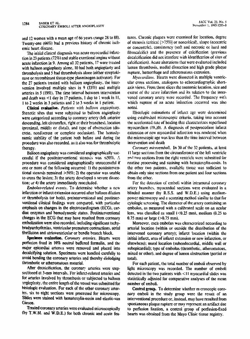
and 12 women with a mean age of 66 years grange 28 to 88) .Twenty-one (66%) had a previous history of chronic isch-emic heart disease .
The initial clinical diagnosis was acute myocardial infarc-tion in 23 patients (72%) and stable exertional angina withoutacute infarction in 9 . Among all 32 patients, 17 were treatedwith balloon angioplasty alone, 10 had both angioplasty andthrombolysis and 5 had thrombolysis alone (either streptoki-nase or recombinant tissue-type plasminogen activator) . Forthe 27 patients treated with balloon angioplasty, the inter-vention involved multiple sites in 9 (33%) and multiplearteries in 5 (19%n) . The time interval between interventionand death was <I day in 17 patients, I day to I week in 11,I to 2 weeks in 3 patients and 2 to 3 weeks in I patient .
Clinical evaluation . Patients with balloon angioplasty .Stenotic sites that were subjected to balloon angioplastywere categorized according to coronary artery (left anteriordescending, left circumflex, right or their branches), location(proximal, middle or distal), and type of obstruction (dis-crete, nondiscrete or complete occlusion) . The hemody-namic stability of the patient both before and during theprocedure was also recorded, a it also was for thrombolytictherapy.
Balloon angioplasty was considered angiographically suc-cessful if the postinterventional stenosis was <50%n . ..procedure was considered angiographically unsuccessful ifone or more of the following occurred: 1) the postinterven-tional stenosis remained >50% ; 2) the operator was unableto cross the lesion; 3) the artery developed a severe dissec-tion ; ur 4) the artery immediately reoccluded .
Embolus-related events. To determine whether a newinfarction or infarct extension occurred after balloon dilationor thrombolysis (or both), preinterventional and postinter-ventional clinical findings were compared, with particularemphasis on changes in the electrocardiogram (ECG), car-diac enzymes and hemodynamic status . Postinterventionalchanges in the ECG that may have resulted from coronaryembolization were also recorded, including significant tachy-bradyarrhythmias, ventricular premature contractions . atria)fibrillation and atrioventricular or bundle branch block .
Specimen evaluation. Coronary arteries . Hearts wereperfusion fixed in 10% neutral buffered formalin, and themajor epicardial arteries were removed and placed intodecalcifying solution . Specimens were handled carefully toavoid bending the coronary arteries and thereby dislodgingthrombotic or atheromatous material .
After decalcification, the coronary arteries were step-sectioned at 3-mm intervals . For infarct-related arteries andfor arteries involved by thrombosis or subjected to balloonangioplasty, the entire length of the vessel was submitted forhistologic evaluation . For each of the other coronary arter-ies, six to eight sections were processed for microscopy .Slides were stained with hematoxylin-eosin and elastic-vanGieson .
Treated coronary arteries were evaluated microscopically(by T.W.M. and W.D.E .) for both chronic and acute fea-
JACC Vol . 22, No .November 1 . 1993:1283--8
tures. Chronic plaques were examined for location, degreeof stenosis (critical [>75%] or noncritical), shape (eccentricor concentric), consistency (soft and necrotic or hard andfibrocalcific) and the presence of calcification (previousdecalcification did not interfere with identification of sites ofcalcification) . Acute alterations that were evaluated includedlumen thrombosis, medial dissection and high grade plaquerupture, hemorrhage and atheromatous extrusion .
Myocardfwn . Hearts were dissected in multiple ventric-ular cross sections, analogous to echocardiographic short-axis views . From these slices the anatomic location, size andextent of the acute infarction and its relation to the inter-vened coronary artery were recorded . The frequency withwhich rupture of an acute infarction occurred was alsonoted .
Histologic estimations of infarct age were determinedusing established microscopic criteria, taking into accountthe accelerated rate of healing that characterizes reperfusedmyocardium (19,20) . A diagnosis of postprocedure infarctextension or new myocardial infarction was rendered whenthe microscopic age was less than the time interval betweenintervention and death
Coronary nrieroentoli. In 30 of the 32 patients, at leastI I large sections from the circumference of the left ventricleand two sections from the right ventricle were submitted forroutine processing and staining with hematoxylin-eosin . Inthe other two patients, available tissue was sufficient toobtain only nine sections from one patient and four sectionsfrom the other .
For the detection of emboli within intramural coronaryartery branches, myocardial sections were evaluated in ablinded manner (by R .S.S. and W.D.E .) using medium-power microscopy and a scanning method similar to that forcytologic screening. The diameter of the artery containing anembolus, as measured with a calibrated scale on an ocularlens, was classified as small (<0 .25 mm), medium (0 .25 to0.75 mm) or large (>0 .75 mm) .
Moreover, each embolus was characterized according toarterial location (within or outside the distribution of theintervened coronary artery); infarct location (within theinitial infarct, area of infarct extension or new infarction, orelsewhere) ; mural location (subendocardial, middle wall orsubepicardial) ; type of embolus (thrombotic, atheromatous,mixed or other), and degree of lumen obstruction (partial ortotal).
For each patient, the total number of emboli observed bylight microscopy was recorded . The number of embolidetected in the two patients with < I I myocardial slides wasstatistically adjusted for comparative analyses of the meannumber of emboli .
Control group. To determine whether m croscopic coro-nary emboli in the study group were the result of aninterventional procedure or, instead, may have resulted fromspontaneous plaque rupture or may represent an artifact dueto perfusion fixation, a control group of perfusion-fixedhearts was obtained from the Mayo Clinic tissue registry .

JACC Vol . 22, No. 5November 1, 1993 :1283-8
This group consisted of specimens from 39 patientsmatched according to age, gender and the presence and ageof acute myocardial infarction with the study group . None ofthe patients had undergone coronary angioplasty or throm-bolytic therapy or had ever had heart surgery of any kind .
Statistical analysis . Data were evaluated by a biomedicalstatistician (K.R.B .) . Comparison of the incidence of coro-nary emboli between study patients and control subjects wasbased on the Pearson chi-square test . Clinicopathologicfactors, patient demographic factors and procedural factorswere compared between patients with and without coronaryemboli either by the Pearson chi-square test for discretevariables or by t tests for continuous variables .
To establish associations between the presence and num-ber of emboli and patient or procedural factors or outcomes,either linear regression analysis or t tests were utilized .Multiple linear regression was used to relate the number ofemboli to multiple baseline or procedural factors . Factorsconsidered in the multiple regression analysis were artery ofintervention, single- or multiple-artery intervention, single-or multiple-site intervention, type or types of interventionand patient age and gender .
A p value < 0 .05 was used to judge statistical signifi-cance, although values between 0 .05 and 0 .20 were noted aspotentially interesting associations .
Results
Control group. The 39 perfusion-fixed control heartswere normal in 12, had coronary atherosclerosis withoutacute infarction in 13, and were involved by acute myocar-dial infarction in 14 . In only one subject, who had a calcifiedaortic valve, was an intramural coronary artery embolusdetected, and it represented a dense calcific nodule (presum-ably of aortic valve origin) .
Thus, in no case, including those with acute coronaryplaque rupture or thrombosis and those with acute myocar-dial infarction, was thrombotic or atheromatous embolicmaterial identified in the coronary circulation . Conse-quently, such emboli within hearts in the study group wereconsidered to be causally related to the interventional ther-apy (either balloon angioplasty or thrombolysis, or both) .
Cause of death. The cause of death was determined foreach of the 32 study patients on the basis of a review ofclinical and autopsy information . In 31 (97%), death was dueto ischemic heart disease . Death in the other patient wasattributable to a cerebrovascular accident .
Features of microemboli. Number of emboli . Of the 32study patients, 26 (81 %) had evidence of one or more emboliwithin the intramural coronary artery branches, comparedwith only I of the 39 control subjects (p < 0 .0001) . In these26 patients, the number of emboli per patient ranged fromI to 6, with a mean of 3 and a total of 83 .
Of the 83 emboli, 67 (81%) were small, I I medium and5 large. Diameters of the involved coronary arteries rangedfrom 0 .045 to 1 .250 mm (mean 0.25). Among the 83 emboli,
SABER ET AL .
1285CORONARY EMBOLI AFTER ANG30PIASTY
Newinfarction
Infarct
Recentextension infarction
Figure 1 . Schematic diagram of postinterventional coronary micro-embolization . Emboli (small circles) within branches of the treatedcoronary artery may involve not only areas of recent infarction,infarct extension or new infarction but also areas of noninfarctedmyocardium .
68 (82%) were identified within branches of the intervenedcoronary artery. Eighteen (22%) were found in areas ofrecent infarction ; 24 (29%) involved areas of new infarctionor infarct extension ; and 41 (49%) involved myocardiumwithout acute infarction (Fig . 1) .
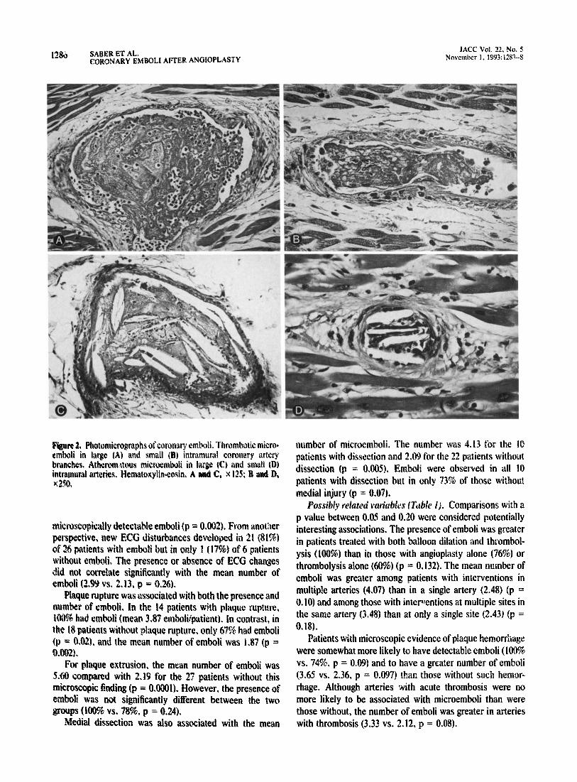
Composition of emboli . Emboli represented thrombus in41 (49%), atlicromatous plaque in 24 (29%), a mixture ofthrombus and atheroma in 14 (17%) and foreign material,presumably derived from the interventional procedure, in 4(5%) (Fig . 2) . In three of the four, the foreign substance wasadmixed with thrombus, and in the fourth case, it occurredalone . The refractile and pularizable foreign material wasconsistent with cotton fibers and talc (glove powder) .
Statistics of microemboli Statistically related variables(Table 1) . Although the left anterior descending coronaryartery was no more likely to be associated with microembolithan were the left circumflex and right coronary arteries (p =0.24), the number of emboli did tend to be greater when theleft anterior descending artery was the artery of intervention(p = 0.035) . In multiple regression analysis of the effects ofage, gender, artery, number of sites and type of intervention,only the artery (left anterior descending vs . other) andnumber of sites (multiple vs . single) showed an independentassociation with the number of emboli .
Infarct extension or new infarction was also related to thepresence of postinterventional coronary microemboli .Among 29 patients with microscopic evidence of infarctextension or new infarction, 26 (90%; had emboli comparedwith none of the 3 patients without these changes (p =0.0002) . Moreover, the mean number of emboli was alsosignificantly higher in the 26 patients Vian in the other 3(3.01 vs . 0, p = 0.01) .
Similarly, important new ECG abnormalities werestrongly associated with the presence of emboli . Of the 22patients with ECG changes, 2! (95%) had emboli, but only 5(50%) of the 10 patients without these ECG changes had
1286
SABER ET AL .CORONARY EMBOLI AFTER ANGIOPLASTY
F%ure 2 . Photomicrographs of coronary emboli . Thrombotic micro-emboli in large (A) and small (R) intramural coronary arterybranches . Atheromitous microemboli in large (C) and small (D)intramural arteries . Hernatoxylin-emin. A mid C, x 125 ;111 and D,x250.
microscopically detectable emboli (p = 0 .002). From anoVierperspective, new ECG disturbances developed in 21 (81%)of 26 patients with emboli but in only 1 (17%) of 6 patientswithout emboli . The presence or absence of ECG changesdid not correlate significantly with the mean number ofemboli (2 .99 vs . 2 . 13, p = 0,26) .
Plaque rupture was associated with both the presence andnumber of emboli . In the 14 patients with plaque rupture'Its had emboli (mean 3 .87 emboli/patient) . In contrast, inthe 18 patients without plaque rupture, only 67% had emboli(p = 0 .02), and the mean number of emboli was 1 .87 (p0-002) .
For plaque extrusion, the mean number of emboli was5.60 compared with 2 .19 for the 27 patients without thismicroscopic finding (p = 0 .0001). However, the presence ofemboli was not significantly different between the twogroups (100% vs. 78%, p = 0.24) .
Medial dissection was also associated with the mean
JACC Vol . 22, No. 5November 1, 1993 :1281-8
number of microemboli . The number was 4 .13 for the 10patients with dissection and 2 .09 for the 22 patients withoutdissection (p = 0 .005) . Emboli were observed in all 10patients with dissection but in only 73% of those withoutmedial injury (p = 0.07) .
Possibly related variables (Table 1) . Comparisons with ap value between 0 .05 and 0.20 were considered potentiallyinteresting associations. The presence of emboli was greaterin patients treated with both balloon dilation and thrombol-ysis (100%) than in those with angioplasty alone (76%) orthrombolysis alone (60%) (p = 0 .132). The mean number ofemboli was greater among patients with interventions inmultiple arteries (4 .07) than in a single artery (2 .48) (p =0.10) and among those with interventions at multiple sites inthe same artery (3 .48) than at only a single site (2 .43) (p0.18) .
Patients with microscopic evidence of plaque hemorrhagewere somewhat more likely to have detectable emboli (100%vs. 74%, p = 0.09) and to have a greater number of emboli(3.65 vs . 2.36, p = 0 .097) than those without such hemor-rhage. Although arteries with acute thrombosis were nomore likely to be associated with microemboli than werethose without, the number of emboli was greater in arterieswith thrombosis (3 .33 vs . 2.12, p = 0 .08) .

JACC Vol . 22, No . 5November 1, 1993 :1283-N
Table 1 . Associations Between Variables and the Presence orNumber of Postinterventional Coronary Microemboli
Statistically related variables (p < 0 .05)Intervention in left anterior descending arteryInfarct extension or new infarctionNew elect rocardiograph ic abnormalitiesPlaque rupturePlaque extrusionMedial dissection
Possibly related variables (p ~ 0 .05 to 0 .20)Both balloon angioplasty and thrombolysisMultiple arteries of interventionMultiple sites of interventionPlaque hemorrhageRecent coronary thrombosis
Unrelated variables (p > 0 .20)Age or gende'History of ischemic heart disease"emodynantic status before or during procedureSuccess of interventionSite or type of arterial obstructionPlaque shape or consistencyPlaque calcificationRupture of acute myocardial infarction
DiscussionAmong patients undergoing coronary balloon angioplasty
or thrombolytic therapy (or both), the pot: ntial exists forembolization of thrombotic or atheromatous material intothe distal coronary artery circulation . However, to ourknowledge, this phenomenon has been previously docu-mented microscopically in only 10 patients from 5 studies(8,15-18) .
In the current retrospective autopsy-based investigation,postinterventional coronary emboLzation was studied in 32patients . Observations that warrant further discussion in-clude 1) the nature of the microemboli, 2) the variablesassociated with the presence or number of microemboli, and3) the limitations of this study .
Nature of microemboli . One or more emboli were identi-fied within the intramural coronary arteries in 81% of the 32patients . Although the number of emboli per patient onlyranged from I to 6, 5 of the 83 emboli obstructed relativelylarge intramural arterial branches .
Microscopically, the emboli were thrombotic or athero ,
matous in 95% of the patients . Four emboli (5%) consisted offoreign material, all of which were refractile or polarizable .These presumably were derived from the catheters, glovepowder, cotton sponges or other materials used during theinterventional procedure . Similar material can occasionallybe identified within small pulmonary arteries after right-sided cardiac catheterization .
Variables associated with emboli . One of the most impor-tant clinical observations in this study was the statisticallysignificant correlation between the presence of coronaryemboli and the development of postprocedure infarct exten-sion, a new myocardial infarction or new ECG abnormali-
SABER ET AL .
1287CORONARY EMBOLI AFTER ANGIOPLASTY
ties . The occurrence of new ECG abnormalities after balloonangioplasty or thrombolysis probably results from a complexinterplay of several factors . among which the effects ofcoronary embolization may be important (21-24) .
In the current study, the greatest number of emboli wasassociated statistically with intervention in the left anteriordescending coronary artery, multiple interventional sites,postinterventional medial dissection, and plaque rupture andextrusion . These findings support intuition that embolizationwould be most extensive among cases in which the degree ofcoronary artery injury is also the greatest . Other investiga-tors have also noted that myocardial infarction due tocoronary embolization is related to the number, size andlocation of the emboli (4,13,18) .
Limitations of the study . This represents a retrospectiveautopsy-based investigation ana ; is subject to the samelimitations inherent in any such study . The results of thecurrent study indicate that co onary microemboli occur in asubstantial number of patients undergoing balloon angio-plasly or thrombolysis (or both) who die and who undergoautopsy . However, the frequency of this postinterventionalphenomenon among the much larger cohort of survivors isunknown. Notwithstanding, in living patients who developacute myocardial ischemia or new ECG changes after theseinterventions, coronary microembolism should be consid-ered as a potential cause .
References
I . Holmes DR Jr . Vlictstra RE . Mock MB, et a] . Angiographic changesproduced by percutaneous transluminal coronary agioplasty . Am JCardiol 1983 :51 :677-83 .
2 . Dorros G . Cowley MJ, Simpson J, et al . Percutaneous transluminalcoronary angioplasty : report of complications from the National Heart .Lung. and Blood Institute PTCA Registry . Circulation 1983*723-30 .
3 . Cowley MJ . Dorros G, Kelsey SF, Van Raden M, Detre KM . Emergencycoronary bypass surgery after coronary angioplasty : the National Heart .Lung . and Blood Institute's Percutaneous Transluminal Coronary Angio-plasty Registry experience . Am J Cardiol 1984 :53 :22C-6C .
4 . MacDonald RG. Feldman RL, Conti CR . Pepine CJ . Thromboemboliccomplications of coronary angioplasty . Am J Cardiol 1984 ;54 :916-7 .
5 . Weyne AE, Heyndrickx GR . Vandekerckhove YR, Clement DL . Embo-lization complicating coronary angioplasty in the presence of an intia-coronary thrombus . Clin Cardiol 1986 :9 :463-5 .
6. Saenz CB . Harrell RR . Sawyer JA III, Hood WP Jr. Acute percutaneoustransluminal coronary angioplasty complicated by embolism to a coro-nary artery remote from the site of infarction . Cathet Cardiova~c Diagn1987:13 :266-8 .
7. Cameron J, Buchbinder M . Wexler L . Ocsvsle SN . Thromboemboliccomplications of percutaneous transluminal coronary angioplasty formyocardial infarction . Cathet Cardiovasc Diagn 19871 ;, :100-6.
8 . Richardson SG . Callen D . Morton P . Murtagh JG, Scott ME, O'KeefeDB. Pathological changes after intravenous streptokinase treatment ineight patients with acute myocardial infarction . Br Heart J 1989 ,.61 :390-5 .
9 . Ueda M, Fujimoto T, Ogawa N, Shoji S . An autopsy case of cholesterolembolism following percutaneous transhiminal coronary angioplasty andaortography . Acta Pathol Jpn 1989,39 :203-6 .
10 . Stafford PJ, Strachan CJL . Vincent R . Chamberlain DA . Multiple microemboli after disintegration of clot during thrombolysis for acute myocar-dial infarction . Br Med J 1990 :299:1310-2 .
H . Zahger D . Weiss AT . Anner H. Waksman R. Systemic embolizaticnfollowing thrombolytic therapy for acute myocardial infarction . Chest1990 :97 :754-6.
1288
SABER ET AL .CORONARY EMBOLI AFTER ANGIOPLASTY
12. Queen M, Biem HJ . Moe GW, Sugar L . Development of cholesterolembolization syndrome after intravenous streptokinase for acute myocar-dial infarction. Am J Cardiol 1990;65 :1042-3 .
13 . Roberts WC. Coronary embolization : a review of causes, consequences,and diagnostic considerations . Cardiovasc Med 1973 ;3 :699-710.
14. Saber RS, Edwards WD, Holmes DR Jr, Vlietstra RE, Reeder GS .Balloon angioplasty of aortocoronary saphenous vein hypass grafts: ahistopathologic study of six grafts in five patients, with emphasis onrestenosis and embolic complications . J Am Coll Cardiol 1988 ;12 :1501-9.
15. Menke DM, Jordan MD, Aust CH, Storer W, Waller BF . Histologicevidence of distal coronary thromboxmbolism : a complication of acuteproximal coronary artery thromhalytic therapy. Chest 1986;90 :614-6 .
16, de Morals CF, Lopes EA, Checchi H . Arie S . Pileggi F. Percutaneoustransluminal coronary angioplasty : histopathologic analysis of nine nec-ropsy cases, Virchows Arch A 1986;410:195-202 .
17 . Colavita PG . ldeker RE, Reimer KA, Hackel DB . Stack RS . Thespectrum of pathology associated with percutaneous transluminal coro-nary angioplasty during acute myocardial infarction . J Am Coll Cardlol1986;8 :855-60.
18 . Walley VM. Higginson LAJ, Marquis J F, Williams WL, Morton BC,
JACC Vol . 22, No. 5November 1, 1993 :1283-8
Beanlands DS . Local morphologic effects of coronary artery balloonangioplasty . Can J Cardiol 1988 :4 :17-24 .
19 . Edwards WD. Pathology of myocardial infarction and reperfusion . In :Gersh BJ, Rahimtoola SH, eds . Acute Myocardial Infarction . New York :Elsevier, 1991 :14-48 .
20. Reichenbach D, Cowan MJ . Healing of myocardial infarction with andwithout reperfusion . In: Virmani R, Atkinson JB, Fenoglio JJ, eds .Cardiovascular Pathology. Philadelphia : WB Saunders, 1990:8699 .
21 . Vlietstra RE, Holmes DR Jr. Percutaneous transluminal coronary ar.gio-plasty. J Cardiac Surg 1988 ;3 :53-66 .
22 . Steffenino G, Meier B, Finci L, et al . Acute complications of electivecoronary angioplasty: a review of 500 consecutive procedures . Br HeartJ 19 ;39:151-8 .
23. Rosen RM. Janse ML Myerburg RJ. Arrhythmia induced by coronaryartery occlusion : what are thw electrophysiologieal mechanisms? In :Hearse D, Manning A, Janse M, eds . Life Threatening Arrhythmia DuringIschemia and Infarction . New York : Raven, 1987 :11-47 .
24. C~anefield PF, Aronson RS. Arrhythmias induced by ischemia andmyocardial infarction . In : Cranefield PF, Aronson RS, eds . CardiacArrhythmias : The Role of Triggered Activity and Other Mechanisms .Mount Kisco, NY: "atura, 1988:481-505 .
Copyright © 2022 FDOKUMEN