Detection of paradoxical cerebral echo contrast embolization by transcranial Doppler ultrasound

Neurosurg. Rev. A new model o f brainstem isehemia by embol izat ion technique in cats 14 (1991) 221-229
Toshinori Nakahara, Shuichi Oki, Zainal Muttaqin, Satoshi Kuwabara, and Tohru Uozumi
Department of Neurosurgery, Hiroshima University School of Medicine, Hiro- shima, Japan
Abstract
An experimental model of brainstem ischemia was de- veloped by embolization technique with cylindrical sili- cone rubber emboli in cats. The embolus reached the basilar artery in 55 cats (58.5%) and stopped in the upper basilar artery (UB) in 32, the middle basilar artery (MB) in 22 and the lower basilar artery (LB) in one animal. When the basilar artery distal to the embolus was not visualized (type 1) by postoperative vertebral angiogram, Evans blue extravasation was observed in the brainstem caudal to the embolus. When only a filling defect of the basilar artery at the site of the embolus was noted (type 2), dye extravazation was observed in the brainstem around the site of the embolus. In UB type l, the regional cerebral blood flow of pons and medulla oblongata decreased immediately after emboli- zation, and six hours after embolization it was 11.4 _+ 5.7 (pons) and 11.7 ___ 4.6 ml/t00g/min (medulla oblongata). In UB type 1 and MB type 1 animals, coma, apnea, tetraplegia, and disturbance of swallowing were noted. These animals died within 50 hours after embol- ization. Animals of UB type 2 and MB type 2 showed neurological deficits, but survived for three days. This paper discusses this method of producing experimental brainstem ischemia, the sites of ischemic lesions, and clinicopathological findings.
Keywords: Basilar artery, brainstem ischemia, cat, em- bolization.
1 Introduction
The incidence of infarctions in the area supplied by the vertebrobasilar arterial system is lower than that of infarctions in the area supplied by the carotid arterial system [13], but the prognosis, however, is worse [1, 13, 27] and there are no effective methods of treatment. Both the internal carotid and the vertebrobasilar arterial systems participate in the circulatory dynamics of the brainstem via the posterior communicating artery.
�9 1991 by Walter de Gruyter & Co. Berlin �9 New York
It is therefore difficult to produce a model of brainstem ischemia. Only a small number of re- ports have been published on such experimental models and the pathophysiological mechanism in- volved has not yet been fully elucidated. In the present study, silicone rubber cylinders were uti- lized to produce an experimental model of brain- stem ischemia. The circulatory dynamics of this model are discussed.
2 Materials and methods
In this study, 94 adult mongrel cats weighing 2 - 4 Kg were used. Arterial blood gas analysis (ABL2, Radiometer, Copenhagen, Denmark) was per- formed throughout the experiment and PaO: was maintained between 80 and 120 mmHg and PaCO2 between 30 and 40 mmHg both being within the physiological range.
2.1 Embolization technique
After intramuscular injection of 0.1 rag/animal of atropine sulfate general anesthesia was induced by intraperitoneal administration of sodium pento- barbital (30 mg/Kg), and followed by immobili- zation through intravenous injection of 0.08 rag/ Kg of pancuronium bromide. After endotracheal intubation, ventilation was controlled with a Har- vard respirator (Bodine Electric Company, Chi- cago, ILL, USA). The animal was fixed in supine position. An incision was made on the right an- terior chest wall to expose the axillary and subcla- vian arteries, and a 4 French size catheter (outer diameter 1.35 ram, inner diameter 0.80 ram) was inserted into the vertebral artery through the sub- clavian artery. Vertebral angiography was then performed, and the diameter at the top of the

222 Nakahara et al., Experimental brainstem ischemia by embolization
basilar artery was estimated based on this angio- gram. The appropriate cylindrical embolus which were made of Xantopren (Bayer, Leverkusen, Ger- many) was selected. The emboli were prepared beforehand and were 5 mm in length and 0.60, 0.65, 0.70 and 0.75 mm in diameter. Embolus of adequate size was injected with 2 ml of NaC1 0.9% (Figure 1). The site whose the embolus stopped was angiographically confirmed 30 minutes after embolization and when the brain was removed for
magnification ft. VA f ~ - - -
e m b o l u s ~
fluid infusion Figure 1. Experimental preparations: The right axillary artery and right subclavian artery were exposed and a 4 Fr. size catheter was inserted into the right vertebral artery. An embolus was injected with NaC1 0.9% through this catheter. (rt. SA.: right subclavian artery, rt. VA.: right vertebral artery, EEG: electroencephalogram, BP: arterial blood pressure, BGA: arterial blood gas analysis)
examination. The second vertebral angiograms obtained 30 minutes after embolization were class- ified into the following types: Type 1 - no visu- alization of the basilar artery distal to the embolus, type 2 - filling defect only around the embolus and type 3 - complete visualization of the em- bolized artery (Figure 2) [23]. Angiograms were taken with a portable X-ray unit (Tanka, Tokyo, Japan) using XTL-2 film (Eastman Kodak Com- pany, Rochester, NY, USA). In the sham operated animals, all the procedures except injection of the embolus were performed.
2.2 Permeability of blood brain barrier (BBB)
In order to ascertain the permeability changes in BBB in response to ischemia, 2% Evans blue (2 ml/Kg) was injected intravenously five hours after embolization. The animals were sacrificed six hours after embolization. The brain was removed and sectioned on the coronal plane at 3 m m inter- vals f rom the mammillary body towards the cau- dal. This allowed examination of the sites of dye permeation and preparation of histological speci- mens for observation under hematoxylin-eosin stain.
2.3 Contact mieroangiograms
Six hours after embolization, the animals were sacrificed by intravenous injection of 10 ml satu- rated KC1. An anterior chest incision was made
Figure 2. Angiographical classification: Vertebral angio- graphy was obtained 30 minutes after embolization. The angiographical findings were divided into three types: type 1: The arteries distal to embolus were not visual-
ized.
type 2: Filling defect in the basilar artery was observed. type 3: Vessels were normally visualized. Control shows the vertebral angiogram before emboli- zation.
Neurosurg. Rev. 14 (1991)

Nakahara et al., Experimental brainstem ischemia by embolization 223
as quickly as possible, the ascending aorta was ligated and a catheter was inserted into the ce- phalad descending aorta. With blood flowing out from the superior vena cava, 2000 ml heparinized NaC1 0.9% was infused via the inserted catheter at a pressure of 150 mmHg for cerebral perfusion, followed by 300 ml of 50% barium solution with 5% gelatin at the same pressure. The animal then was cooled to 4 ~ for 24 hours and the brain was carefully removed. The brain was then sectioned at 3 mm intervals in the coronal plane, caudally from the mammillary body and contact microan- giograms were prepared in order to examine the vascular structure after embolization. Circulex UI4VN-25 X-ray apparatus (Shimadzu Seisaku- sho Ltd., Kyoto, Japan) was used to obtain con- tact microangiograms on X-Omat TL film (East- man Kodak Company, Rochester, NY, USA).
Table I. Neurological deficit score
1. Level of consciousness
Deep coma 4 Semi coma 3 Stupor 2 Somnolence 1 Alert 0
2. Respiration Apnea or respirator 2 Irregular 1 Smooth 0
3. Posture Lying 3 Squatting 2 Standing 1 Able to walk 0
4. Motor disturbance Tetraplegia 4 Tetraparesis 3 Hemiplegia 2 Hemiparesis 1 None 0
5. Tone of muscle Flaccid 2 Spastic 1 Normal 0
6. Response to pain Absent 1 Normal 0
7. Pupils Bilateral mydriatic Bilateral miotic Unilateral mydriatic or miotic
Normal 8. Eye movement
None Deviation Midline Roving Following
9. Reaction to light Absent Sluggish Normal
10. Oculocephalic response
None Dolls Spontaneous movement
11. Oculomotor palsy Present Absent
12. Swallowing Unable Disturbed Able
13. Corneal reflex Absent Normal
Maximal deficit = 35, Normal = 0 Right and left eyes separately
2.4 Neurological findings and vital signs
Electroencephalogram (EEG), mean arterial blood pressure (MABP) and electrocardiogram (ECG) were continuously recorded for six hours after embolization. Active electrodes were placed sub- cutaneously on bilateral parietal regions with ref- erence electrodes on both earlobes for EEG re- cording (EEG Trend Monitor, Model OEE-7102, Nihon Kohden Corp., Tokyo, Japan). The animals were then allowed to survive up to three days after embolization without any treatment except artifi- cial respiration. Neurological findings were eval- uated at 6, 24, 48 and 72 hours after embolization based on the Neurological Deficit Score (Table I) originally prepared by the authors.
2.5 Regional cerebral blood flow (rCBF)
The brainstem was exposed through the transcerv- ical transclival appraoch, rCBF was measured in the pontine reticular formation (PRF) and retic- ular formation of the medulla oblongata (MORF)
3 with the electrolytic hydrogen clearance method 2 (Model RBF-2, Bio-Medical Science, Kanazawa,
Japan) [15]. Using the report of GILLILAN [10] and i the atlas of SNIDER and NIEMER [29] as references, 0 an electrode was inserted at 2.5 mm cephalad from
the inferior cerebellar artery, 2.0 mm laterally from 4 the basilar artery and 5.5 mm inward from the 3 brain surface (P4.0, LI.0, H-5.5) for PRF. It was 2 inserted 1.5 mm caudad from the inferior cerebel- 1 0 lar artery, 1.0 mm laterally from the basilar artery
and 6.5 mm inward from the brain surface (PS.0, 2 LI.0, H-6.5) for M O R E rCBF was measured at 1 30 minute intervals before and up to six hours 0 after embolization.
3 Results
3.1 Location of the embolus (Figure 3)
In 55 of the 94 cats, the embolus stopped in the 1 basilar artery (58.5%). The basilar artery was di- 0 vided into upper basilar artery (UB), middle bas-
ilar artery (MB) and lower basilar artery (LB). 2 The embolus stopped at UB in 32 animals (type 1 1 in 20 animals, type 2 in 8 and type 3 in 4), at 0 MB in 22 animals (type I in 14, type 2 in 6 and 1 type 3 in 2) and at LB in I animal (type 2 in 1). 0 The embolus stopped at the distal portion of the
vertebral artery (VA) in 39 animals (type 1 in 6, type 2 in 9 and type 3 in 24). Among these 55 animals, UB was the most frequent site being
Neurosurg. Rev. 14 (1991)

224 Nakahara et al., Experimental brainstem ischemia by embolization
s~ hffw~'a2gC ~ ~ ~ ~ site liB MB LB VA
number of cases 32 22 1 39
1 20 14 0 6 angiographica[ 2 8 6 1 9
type 3 4 2 0 24
Figure 3. Site of the embolus: In 55 of 94 cats, the em- bolus reached the basilar artery (58.5%). The closed oblong indicates the embolus. UB: upper basilar artery MB: middle basilar artery LB: lower basilar artery VA: vertebral artery
observed in 32 animals (58.1%). There was no evidence of migration of the embolus from the initial site as demonstrated by angiography per- formed 30 minutes after embolization, and by the final site confirmed by the removed brain.
3.2 Blood brain barrier (BBB) and histological findings
Dye extravasation was evaluated in 38 case, eight with the embolus stopping at UB (type 1 in 3, type 2 in 3 and type 3 in 2), five at MB (type I in 4 and type 3 in 1), 20 at VA (type 1 in 2, type 2 in 5 and type 3 in 13) and five sham operated ani- mals. In animals in which the embolus stopped in the basilar artery with angiographies of type 1, extensive extravasation of the dye was noted in the brainstem, caudal to the embolus (Figure 4). In animals in which the embolus stopped at the basilar artery with angiographical type 2, dye leak- age was observed in the brainstem around the site of the embolus, In animals with type 3 and in sham operated animals, no dye leakage was noted. In two of the animals in which the embolus stopped in the vertebral artery with angiographies of type 2 (VA-type 2), dye leakage was seen in the medulla oblongata, but not leakage was noted in the other VA animals. Histological examination of the site of dye leakage revealed vacuolation of neuropils and disappearance of the Nissl body which indicated ischemic lesion (Figure 5).
Figure 4. Representative photomicrograph of dye leakage: The embolus stopped at the middle basilar artery (ar- row). Vertebral angiography 30 minutes after emboli- zation was classified as type 1. Dye leakage was observed at the pons and the medulla oblongata proximal to the embolus.
Figure 5. Histological changes 6 hours after embolization: A. Embolized animal. Representative photomicrograph of a section of brainstem 6 hours after the onset of ischemia showing vacuolation in the neuropil and dis- appearance of the Nissl body. Hematoxylin-eosin stain x200. B. Sham operated animal. No ischemic change was ob- served. Hematoxylin-eosin stain x200.
Neurosurg. Rev. 14 (1991)

Nakahara et al., Experimental brainstem ischemia by embolization 225
3.3 Contact microangiograms
Contact microangiograms were obtained in 28 cats: nine with embolus at UB (type 1 in 5 and type 2 in 4), five with embolus at MB (type i in 4 and type 2 in 1), one with embolus at LB (type 2 in 1), eight with embolus at VA (type 1 in 4, type 2 in 2 and type 3 in 2) and five sham operated animals. In animals with the embolus stopping at the basilar artery with angiographies of type 1 and 2, the wedge shaped avascular area with the bot- tom at the ventral brainstem was noted at the site of the embolus, clearly demarcated from the nor- mal region (Figure 6). In animals with angiogra- phies of type 1, which suggested poor perfusion, ischemic lesions were noted in the brainstem cau- dal to the embolus. In animals with VA and sham operated animals, no abnormal findings were ob- served.
of UB type 2 and MB type 2 showed neurological deficits but survived for three days. In the animal of UB type 2, neurological deficits consisted of coma, respiratory abnormalities and tetraplegia. In the other of MB type 2, apnea, tetraplegia and disturbance of swallowing were noted (Figure 7). Histological examination of the brain of these two animals revealed formation of infarcted lesions at the site of the embolus (Figure 8). The animals of UB type 3, MB type 3, VA and sham operated animals survived for three days without neurolog- ical deficits. The arterial blood pressure decreased and EEG tracings gradually lowered and finally disappeared after six hours in two of the animals of UB type 1. In the others, no changes of arterial blood pressure or EEG were noted. Arrhythmia was not detected by ECG in any animal.
Figure 6. Representative contact microangiogram of cor- onal sections of the pons: A. Sham operated animal. The perforating arteries were well visualized up to their ends. B. The embolus stopped at the upper basilar artery (arrow). Vertebral angiography 30 minutes after embol- ization was classified as type 1. A vascular area was observed in the ventromedian region of pons.
3.4 Neurological findings and vital signs
A total of 26 cats were examined: eight with era- bolus at UB (type 1 in 6, type 2 in 1 and typ 3 in 1), seven with embolus at MB (type I in 5, type 2 in 1 and type 3 in 1), six with embolus at VA (type 2 in 2 and type 3 in 4), and five sham operated animals. In animals of UB-type 1, deep coma and apnea were noted postoperatively. Death occurred in six animals at 6 (four animals), 8 and 9 hours after embolization. In animals of MB-type 1, semi coma, apnea, tetraplegia and disturbance of swal- lowing were noted postoperatively. Death oc- curred in five animals at 13, 34, 37 and 50 (two animals) hours after embolization. Two animals
~D
o
O3 O
03 Z
35
30
25-
20-
15-
10-
5
6hr Day1 Day2 Day3
Figure 7. Summary of neurological deficits: In cases of angiographies of type I, all cats died within 2 days when the basilar artery embolized (open circle and open square). When the angiographical classification was type 2 or 3, all the animals were alive after 3 days and had experienced partial or full recovery. Open circle, open triangle, and open square were the cases with embolus at MB, VA and UB, respectively. One, 2 and 3 were type 1, type 2 and type 3, respectively 30 minutes after em- bolization. The score in maximum deficit was 35 and that in minimum deficit was 0.
Neurosurg. Rev. 14 (i991)

226 Nakahara et al., Experimental brainstem ischemia by embolization
Figure 8. Representative photograph of the coronal sec- tion of upper pons 3 days after embolization: The embolus stopped at the upper basilar artery and angiogram was type 2. Wedge-shaped anemic infarction was observed at the pons. KLI~VER-BARRERA'S stain xl.
3.5 Regional cerebral blood flow (rCBF)
rCBF was measured in 19 cats: six of UB type 1, five of VA type 3 and eight sham operated animals. rCBF of PRF before embolization was 30.7 + 4.5 ml/100 g/rain (mean + S.D.) and that of MORF was 29.0 + 5.6 ml/100 g/rain. In animals of UB type 1, a significant decrease in rCBF of PRF was observed immediately after embolization, reaching 65.5% (20.2 _+ 3.6 ml/100 g/rain) of the pre-em- bolization level in comparison with sham operated animals (p < 0.01, unpaired t-test), rCBF of MORF also decreased immediately after emboli- zation, reaching 70.7% (20.5 + 6.2 ml/100 g/n-fin) of the pre-embolization level, and became signifi- cant three hours after embolization, reaching 60.0% (17.4 + 7.2 ml/100 g/rain) of the pre-em- bolization level compared with sham operated an- imals (13 < 0.01, unpaired t-test), rCBF in PRF and MORF six hours after embolization was 11.4 + 5.7 and 11.7 4- 4.6 ml/100 g/rain, respectively. In the sham operated animals, and those with the embolus at VA, no decline of rCBF was noted, and the value in either PRF or MORF did not change significantly to six hours after emboliza- tion (Figure 9).
4 Discussion
In experimental studies on brainstem ischemia, ischemia has been produced by ligation of the basilar artery after craniotomy [33], electrocoa-
40
35
":'3O E
E 25
g ~. 20
v
15
4 0
35
E 30 E
25
20 g
10
�9 ~ �9 i , i �9 i . J �9 i �9 i
PRE 0 60 120 180 240 300 360
TIME (min.) ( A )
�9 i �9 i , i , i �9 i i ,
PRE 0 60 120 180 240 300 360
TIME (min.) ( B )
Figure 9. Changes in regional cerebral blood flow (rCBF) of pontine reticular formation (PRF) and the reticular formation of the medulla oblongata (MORF): The left figure (A) indicates the changes in rCBF of the PRF and the right one (B) the changes in rCBF of the M O R E rCBFs of the PRF and the M O R F deteriorated immediately after embolization in animals with embolus at UB and angiographies of type 1 (closed circle). In these animals, rCBFs deteriorated significantly imme- diately after emboHzation in the PRF (A) and 3 hours after embolizafion in the M O R F (B) in comparison eith sham operated animals (open circle). No significant change in rCBF was observed between sham operated animals and animals with embolus at the VA exhibiting angiographies of type 3 (open square). A: changes of rCBF in the PRF; B: changes of rCBF on MORF, open circle: sham operated animals (n = 8), closed circle: animals with embolus at the UB exhibiting angiographies of type 1 (n = 6), open square: animals with embolus at VA exhibiting angiographies of type 3 (n = 5), *: p < 0.01, unpaired t-test.
Neurosurg. Rev. 14 (1991)
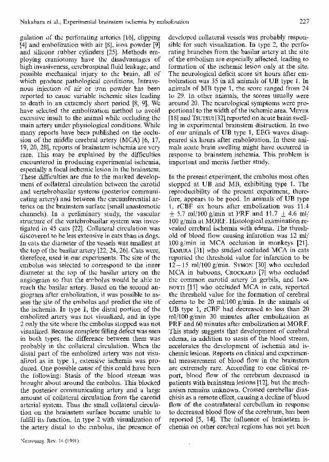
Nakahara et al., Experimental brainstem ischemia by embolization 227
gulation of the perforating arteries [16], clipping [4] and embolization with air [8], iron powder [9] and silicone rubber cylinders [25]. Methods em- ploying craniotomy have the disadvantages of high invasiveness, cerebrospinal fluid leakage, and possible mechanical injury to the brain, all of which produce pathological conditions. Intrave- nous injection of air or iron powder has been reported to cause variable ischemic sites leading to death in an extremely short period [8, 9]. We have selected the embolization method to avoid excessive insult to the animal while occluding the main artery under physiological conditions. While many reports have been published on the occlu- sion of the middle cerebral artery (MCA) [6, 17, t9, 20, 28], reports of brainstem ischemia are very rare. This may be explained by the difficulties encountered in producing experimental ischemia, especially a focal ischemic lesion in the brainstem. These difficulties are due to the marked develop- ment of collateral circulation between the carotid and vertebrobasilar systems (posterior communi- cating artery) and between the circumferential ar- teries on the brainstem surface (small anastomotic channels). In a preliminary study, the vascular structure of the vertebrobasilar system was inves- tigated in 45 cats [22]. Collateral circulation was discovered to be less extensive in cats than in dogs. In cats the diameter of the vessels was smallest at the top of the basilar artery [22, 24, 26]. Cats were, therefore, used in our experiments. The size of the embolus was selected to correspond to the inner diameter at the top of the basilar artery on the angiogram so that the embolus would be able to reach the basilar artery. Based on the second an- giogram after embolization, it was possible to as- sess the site of the embolus and predict the site of the ischemia. In type 1, the distal portion of the embolized artery was not visualized, and in type 2 only the site where the embolus stopped was not visualized. Because complete filling defect was seen in both types, the difference between them was probably in the collateral circulation. When the distal part of the embolized artery was not visu-
a l i zed as in type 1, extensive ischemia was pro- duced. One possible cause of this could have been the following: Stasis of the blood stream was brought about around the embolus. This blocked the posterior communicating artery and a large amount of collateral circulation from the carotid arterial system. Thus the small collateral circula- tion on the brainstem surface became unable to fulfill its function. In type 2 with visualization of the artery distal to the embolus, the presence of
developed collateral vessels was probably respon- sible for such visualization. In type 2, the perfo- rating branches from the basilar artery at the site of the embolism are especially affected, leading to formation of the ischemic lesion only at the site. The neurological deficit score six hours after em- bolization was 35 in all animals of UB type 1. In animals of MB type 1, the score ranged from 24 to 29. In other animals, the scores usually were around 20. The neurological symptoms were pro- portional to the width of the ischemic area. MEYER [18] and TSUTSUX [32] reported on acute brain swell- ing in experimental brainstem distruction. In two of our animals of UB type 1, EEG waves disap- peared six hours after embolization. In these ani- mals acute brain swelling might have occurred in response to brainstem ischemia. This problem is important and merits further study.
In the present experiment, the embolus most often stopped at UB and MB, exhibiting type 1. The reproducibility of the present experiment, there- fore, appears to be good. In animals of UB type 1, rCBF six hours after embolization was 11.4 _+ 5.7 ml/100 g/min at PRF and 11.7 _+ 4.6 ml/ 100 g/min at MORF. Histological examination re- vealed cerebral ischemia with edema. The thresh- old of blood flow causing infarction was 12 ml/ 100 g/rain in MCA occlusion in monkeys [21]. TAMURA [31] who studied occluded MCA in cats reported the threshold value for infarction to be 1 2 - 1 5 ml/100 g/min. SYNON [30] who occluded MCA in baboons, CROCKARD [7] who occluded the common carotid artery in gerbils, and IAN- NOTTI [11] who occluded MCA in cats, reported the threshold value for the formation of cerebral edema to be 20 ml/100 g/min. In the animals of UB type 1, rCBF had decreased to less than 20 ml/100 g/rain 30 minutes after embolization at PRF and 60 minutes after embolization at MORF. This study suggests that development of cerebral edema, in addition to stasis of the blood stream, accelerates the development of ischemia and is- chemic lesions. Reports on clinical and experimen- tal measurement of blood flow in the brainstem are extremely rare. According to one clinical re- port, blood flow of the cerebrum decreased in patients with brainstem lesions [12], but the mech- anism remains unknown. Crossed cerebellar dias- chisis as a remote effect, causing a decline of blood flow of the contralateral cerebellum in response to decreased blood flow of the cerebrum, has been reported [5, 14]. The influence of brainstem is- chemia on other cerebral regions has not yet been
Neurosurg. Rev. 14 (1991)

228 Nakahara et al., Experimental brainstem ischemia by embolization
reported. The present experiment contributes to the unders tanding of the dynamics of cerebral circulation and metabolism during brainstem is-
chemia, the effect of brainstem ischemia on cere- bral blood flow, and studies on the threshold of ischemic penumbra [2, 3] in the brainstem.
References
[1] ARCHER CR, S HORENSTEIN: Basilar artery occlu- sion. Clinical and radiological correlation. Stroke 8 (1977) 383-390
[2] ASTRUP J, L SYMON, NM BRANSTON, NA LASSEN- Cortical evoked potential and extracellular K + and H + at critical levels of brain ischemia. Stroke 8 (1977) 51-57
[3] ASTRUP J, BK SmsJ6, L SYMON: Thresholds in cer- ebral ischemia - The ischemic penumbra. Stroke 12 (1981) 723-726
[4] AYALA GF, WA HIMWICH: Subtemporal approach to the basilar artery in the dog. J Appl Physiol 15 (1960) 1150--1151
[5] BARON JC, MG BOUSSER, D COMAR, N NUQUESNOY, J SASTRE, P CASTA~GNE: "Crossed cerebellar dias- chisis": A remote functional depression secondary to supratentorial infarction of man. J Creb Blood Flow Metab 1 (Suppl. 1)(1981)$500-$501
[6] BREM~R AM, O WATANABE, RS BOLrRKE: Artificial embolization of the middle cerebral artery in pri- mates. Description of an experimental model with extracranial technique. Stroke 6 (1975) 387-390
[7] CROCKARD A, F IAN~!OTTI, AT HUNSTOCK, RD SMITH, RJ HARRIS, L SYMON: Cerebral blood flow and edema following carotid occlusion in the gerbil. Stroke 11 (1980) 494-498
[8] DE LA TORRE E, OC MICHELL, MG NETSKY: The seat of respiratory and cardiovascular responses to cerebral air emboli. Neurology 12 (1962) 140-147
[9] FUJISHIMA M, P SCHE~BERG, OM REINMUTH: Ef- fects of experimental occlusion of the basilar artery by magnetic localization of iron filings on cerebral blood flow and metabolism and cerebrovascular responses to CO2 in the dog. Neurology 20 (1970) 925- 932
[10] GILL1LAN LA: Extra- and intra-cranial blood supply to brains of dog and cat. Am J Anat 146 (1976) 237- 253
[11] IANNOTTI F, JT HOFF, GP SCmELKE: Brain tissue pressure in focal brain ischemia. J Neurosurg 62 (1985) 83--89
[12] INGVAR DH, E HAGGENDAL, NJ NILSSON, P SOUR- ANGER~ I WICKBOM, NA LASSEN: Cerebral circula- tion and metabolism in a comatose patient. Studied with a new method. Arch Neurol 11 (1964) 13-21
[13] JONES HR, CH MILLIKAN, BA SANDOK: Temporal profile (clinical course) of acute vertebrobasilar sys- tem cerebral infarction. Stroke 11 (1980) 173-177
[14] KANAYA H, H ENDO, T SUGIYAMA, K KURODA: "Crossed cerebellar diaschisis" in patients with pu- taminal hemorrhage. J Creb Blood Flow Metab 3 (Suppl. 1) (1983) $27-$28
[15] KosHu K, K KAMIYAMA, N OKA, S ENDO, A TAK- AKU, T SAITO" Meaurement of regional blood flow using hydrogen gas generated by electrolysis. Stroke 13 (1982) 483-487
[16] KUWABARA S, J UNO, S 1SHIKAWA: A new model of brainstem ischemia in dogs. Stroke 19 (1988) 365- 371
[17] LATCHAW JP, JR LITTLE, RM SLUGG, RP LESSER, N STOWE: Treatment of acute focal cerebral is- chemia and recirculation with d-propranolol. Neu- rosurgery 16 (1985) 18-22
[18J MEYER JS, T TERAURA, K SAKAMOTO, A KONDO: Central neurogenie control of cerebral blood flow. Neurologiy 21 (1971) 247-262
[19] MOLINALI GF: Experimental cerebral infarction. I. Selective segmental occlusion of intracranial arter- ies in the dog. Stroke 1 (1970) 224-231
[20] MOLINALI GF, JI MOSEEEY, JP LAURENT: Segmental middle cerebral artery occlusion in primates: An experimental method requiring minimal surgery and anesthesia. Stroke 5 (1974) 334-339
[21] MORAWETZ RB, U DEGIROLAMI, RG OJEMANN, FW MARCOUX, RM CROWELL: Cerebral blood flow de- termined by hydrogen clearance during middle cer- ebral artery occlusion in unanesthctized monkeys. Stroke 9 (1978) 143-149
[22] NAKAHARA T, S OKI, ZAINAL MUTTAQIN, Y TO- KUDA, K EMOTO, S KUWABARA, T UOZUMI: An investigation of vascular anatomy and microvas- cular architecture of the brainstem in cats. HIJM 39 (1990) 83-88
[23] NAKAHARA T, S OKI, ZAINAL MUTTAQIN, Y TO- KUDA, S EMOTO, S KUWABARA, T UOZUMI: Exper- imental bralnstem ischemia in cats using emboli- zation technique. - A new experimental model for brainstem ischemia - (in Japanese). Journal of Clinical and Experimental Medicine (IGAKU NO AYU~I) 155 (1990) 403--404
[24] NISHIMARU K: Arterial anastomosis in the dog brain. (in Japanese) Fukuoka Acta Med 54 (1963) 988-1006
[25] OKI S, T SHIMA, T UOZUMI, H MATSUURA: Exper- imental brainstem infarction in dog. In: BETZ, E, J GROTE, D HEUSER, R W~LLENWEBER (eds): Patho- physiology and pharmacotherapy of cerebrovascu- lar disorders. Gerhard Witzstock, Baden-Baden 1980
[26] OKI S, T SHIMA, T UOZUMI: Experimental brain stem infarction in the dog. - A new segmental embolization method of the canine basilar artery - . (in Japanese) Brain and Nerve 32 (1980) 199-- 207
Neurosurg. Rev. 14 (1991)

Nakahara et al., Experimental brainstem ischemia by embolization 229
[27] PESSIN MS, N DANEAULT, ES KWAN, MA EISEN- GART, LR CAPLAN: Local embolism from vertebral artery occlusion. Stroke 19 (1988) 112 -- 115
[28] PURDY PD, MD DEvous, CL Wm]~ III, HH BATJER, DS SAMSON, K BREWER, K HODGES: Re- versible middle cerebral artery embolization in dogs without intracranial sm-gery. Stroke 20 (1989) 1368-1376
[29] SNtOER RS, WT NIEMER: A stereotaxic atlas of the cat brain. The University of Chicago Press, Chicago 1961
[30] SYMON L, NM BRANSTON, O CHIKOVAM: Ischemic brain edema following middle cerebral artery occlu- sion in baboons: Relationship between regional cer- ebral water content and blood flow at 1 to 2 hours. :Stroke 10 (1979) 184-191
[31] TAMURA A, T ASANO, K SANO: Correlation between rCBF and histological changes following temporary middle cerebral artery occlusion. Stroke 11 (1980) 487-493
[32] TSUTSUI T, Y HONMA, N SUMAMI, F MOMMA, S TSUCHIMOTO, S NAGAO, A NISHIMOTO: The role of brain-stem vasomotor centers on the neurogenic control of vertebrovascular tonus - Part 1: In special reference to cerebral blood volume and in- tracranial pressure - . (in Japanese) Neurological Surgery 12 (1984) 581--589
[33] WHine RJ, DE DONALD: Basilar-artery ligation and cerebral ischemia in dogs. Arch Surg 84 (1962) 470- 475
Submitted November 7, 1990. Accepted January 11, 1991.
Toshinori Nakahara Department of Neurosurgery Hiroshima University School of Medicine 1-2-3 Kasumi, Minami-ku Hiroshima 734 Japan
Neurosurg. Rev. 14 (1991)
Copyright © 2022 FDOKUMEN