Frontal Behavioral Syndromes in Cortical and Subcortical Dementia
Upload
khangminh22Category
view
1download
0
Edorium Journal of Neurology, Vol. 3; 2016.
Edorium J Neurol 2016;3:4–16. www.edoriumjournalofneurology.com
Cuoco et al. 4
REVIEW ARTICLE PEER REVIEWED | OPEN ACCESS
Brainstem vascular syndromes: A practical guide for medical students
Joshua A. Cuoco, Kyle Hitscherich, Christopher L. Hoehmann
ABSTRACT
Current research has indicated an apparent pandemic of fear of the neural sciences, termed neurophobia, among medical students across the world. While the cause of this fear may be multifactorial, we seek to directly address the phenomenon of neurophobia in part by providing medical students as well as healthcare professionals a simplified approach to one of the most challenging topics in neurology; brainstem vascular syndromes. This article will provide an overview of the causes of the various brainstem vascular syndromes emphasizing a simple approach for localizing the artery affected. The rule of 4 is discussed and then applied to each of the brainstem vascular syndromes highlighting the distinguishing features of each specific syndrome. With an abundance of easy to understand illustrations and pictorials, we emphasize the clinical symptomatology, differentiating characteristics, and pertinent neuroanatomy of each brainstem vascular syndrome.
Joshua A. Cuoco1, Kyle Hitscherich2, Christopher L. Hoe-hmann3
Affiliations: 1MS, Third year medical student, Department of Biomedical Sciences, New York Institute of Technology College of Osteopathic Medicine, Old Westbury, New York, USA; 2BA, Third year medical student, Department of Bio-medical Sciences, New York Institute of Technology College of Osteopathic Medicine, Old Westbury, New York, USA; 3BS, Third year medical student, Department of Anatomy, New York Institute of Technology College of Osteopathic Medicine, Old Westbury, New York, USA.Corresponding Author: Joshua A. Cuoco, 6 Manor Place, Glen Cove, New York 11542, USA; E-mail: [email protected]
Received: 04 July 2016Accepted: 20 August 2016Published: 12 September 2016
Keywords: Brainstem, Medical education, Stroke, Vascular syndromes
How to cite this article
Cuoco JA, Hitscherich K, Hoehmann CL. Brainstem vascular syndromes: A practical guide for medical students. Edorium J Neurol 2016;3:4–16.
Article ID: 100008N06JC2016
*********
doi:10.5348/N06-2016-8-RA-2
INTRODUCTION
Recently, it has been reported that medical students and trainee doctors perceive neurology as one of the more difficult disciplines of medicine [1–9]. Furthermore, it has been found that medical students and physician residents feel less confident in the field of neurology than other specialties of medicine [2]. The fear of the neural sciences and clinical neurology, termed “neurophobia”, is a common problem among students and trainee doctors worldwide [1–9]. Traditionally, a difficult topic to grasp in clinical neurology is brainstem vascular syndromes [2, 10–12]. This article provides a brief overview of the etiology of brainstem vascular syndromes and a simplified approach for medical students and trainee doctors to localize the vascular territory affected. We discuss the rule of 4 and then provide a basic overview of each of the major vascular syndromes emphasizing clinical manifestations, distinguishing features, and relevant neuroanatomy for each syndrome. The aim of this article is to be used as a supplement to neuroscience textbooks used by medical students and healthcare professionals.
Edorium Journal of Neurology, Vol. 3; 2016.
Edorium J Neurol 2016;3:4–16. www.edoriumjournalofneurology.com
Cuoco et al. 5
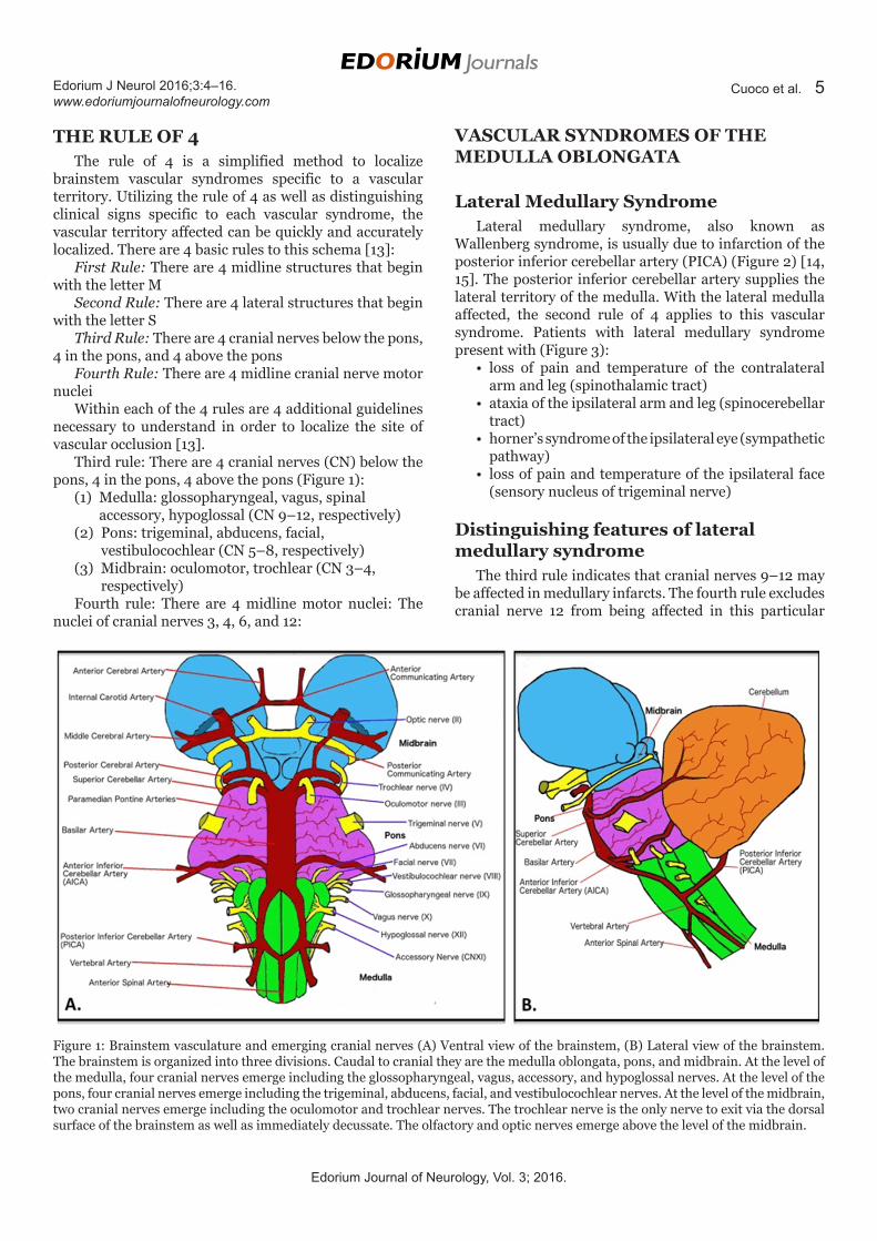
THE RULE OF 4The rule of 4 is a simplified method to localize
brainstem vascular syndromes specific to a vascular territory. Utilizing the rule of 4 as well as distinguishing clinical signs specific to each vascular syndrome, the vascular territory affected can be quickly and accurately localized. There are 4 basic rules to this schema [13]:
First Rule: There are 4 midline structures that begin with the letter M
Second Rule: There are 4 lateral structures that begin with the letter S
Third Rule: There are 4 cranial nerves below the pons, 4 in the pons, and 4 above the pons
Fourth Rule: There are 4 midline cranial nerve motor nuclei
Within each of the 4 rules are 4 additional guidelines necessary to understand in order to localize the site of vascular occlusion [13].
Third rule: There are 4 cranial nerves (CN) below the pons, 4 in the pons, 4 above the pons (Figure 1):
(1) Medulla: glossopharyngeal, vagus, spinal accessory, hypoglossal (CN 9–12, respectively)
(2) Pons: trigeminal, abducens, facial, vestibulocochlear (CN 5–8, respectively)
(3) Midbrain: oculomotor, trochlear (CN 3–4, respectively)
Fourth rule: There are 4 midline motor nuclei: The nuclei of cranial nerves 3, 4, 6, and 12:
Figure 1: Brainstem vasculature and emerging cranial nerves (A) Ventral view of the brainstem, (B) Lateral view of the brainstem. The brainstem is organized into three divisions. Caudal to cranial they are the medulla oblongata, pons, and midbrain. At the level of the medulla, four cranial nerves emerge including the glossopharyngeal, vagus, accessory, and hypoglossal nerves. At the level of the pons, four cranial nerves emerge including the trigeminal, abducens, facial, and vestibulocochlear nerves. At the level of the midbrain, two cranial nerves emerge including the oculomotor and trochlear nerves. The trochlear nerve is the only nerve to exit via the dorsal surface of the brainstem as well as immediately decussate. The olfactory and optic nerves emerge above the level of the midbrain.
VASCULAR SYNDROMES OF THE MEDULLA OBLONGATA
Lateral Medullary SyndromeLateral medullary syndrome, also known as
Wallenberg syndrome, is usually due to infarction of the posterior inferior cerebellar artery (PICA) (Figure 2) [14, 15]. The posterior inferior cerebellar artery supplies the lateral territory of the medulla. With the lateral medulla affected, the second rule of 4 applies to this vascular syndrome. Patients with lateral medullary syndrome present with (Figure 3):
• loss of pain and temperature of the contralateral arm and leg (spinothalamic tract)
• ataxia of the ipsilateral arm and leg (spinocerebellar tract)
• horner’s syndrome of the ipsilateral eye (sympathetic pathway)
• loss of pain and temperature of the ipsilateral face (sensory nucleus of trigeminal nerve)
Distinguishing features of lateral medullary syndrome
The third rule indicates that cranial nerves 9–12 may be affected in medullary infarcts. The fourth rule excludes cranial nerve 12 from being affected in this particular

Edorium Journal of Neurology, Vol. 3; 2016.
Edorium J Neurol 2016;3:4–16. www.edoriumjournalofneurology.com
Cuoco et al. 6
Figure 2: Structures affected in lateral medullary syndrome (A) Transverse view of nuclei affected in the left lateral medullary syndrome. The posterior inferior cerebellar artery (PICA) originates from the vertebral arteries and supplies the lateral most aspect of the medulla. The PICA supplies vestibular nucleus, spinal trigeminal nucleus, and nucleus ambiguus. Furthermore, the inferior cerebellar peduncle, sympathetic tract, and spinothalamic tract can also be affected as these structures extend through the dorsolateral aspect of the medulla, (B) Lateral view of the brainstem level affected in lateral medullary syndrome. The red line indicates the level being described is the medulla in this syndrome. (C) Ventral view of the brainstem level affected in lateral medullary syndrome. The red circle indicates that the PICA is affected in this syndrome.
Figure 3: Clinical manifestations of lateral medullary syndrome (A) Body representation of the clinical manifestations of left lateral medullary syndrome. The green lines indicate loss of pain and temperature of the contralateral arm and leg (spinothalamic tract). The yellow lines represent ataxia of the ipsilateral arm and leg (spinocerebellar tract). The purple lines on the left face indicate loss of pain and temperature of the ipsilateral face (sensory nucleus of trigeminal nerve). The red circle represents Horner’s syndrome of the ipsilateral eye (sympathetic pathway). Furthermore, patients may experience vertigo and nystagmus (vestibular nucleus), loss of the gag reflex (vagus nerve), and dysphagia (nucleus ambiguus and glossopharyngeal nerves), (B) Horner’s syndrome. The left eye exhibits ptosis (drooping of the upper eyelid), anhidrosis (lack of sweat in response to heat), and miosis (constriction of the pupil).

Edorium Journal of Neurology, Vol. 3; 2016.
Edorium J Neurol 2016;3:4–16. www.edoriumjournalofneurology.com
Cuoco et al. 7
syndrome, as cranial nerve 12 is a midline structure. This leaves cranial nerves 9, 10, and 11 as possible cranial nerves affected by a lateral medullary infarct. Cranial nerves 9 and 10 are affected in this particular syndrome as cranial nerve 11 is usually not affected in medullary infarcts. Differentiating features specific to lateral medullary syndrome are loss of function of cranial nerves 9 and 10 (glossopharyngeal and vagus nerve, respectively) resulting in hoarseness and dysphagia [14, 15]. A simple pneumonic to remember these distinguishing features as well as the artery affected is “Never pick a (PICA) horse (hoarseness) that cannot eat (dysphagia) [16].
Medial Medullary SyndromeMedial medullary syndrome, also known as Dejerine
syndrome, is most commonly due to infarction of the vertebral artery (Figure 4) [15, 17]. The vertebral artery supplies the medial territory of the medulla. As the medial medulla is affected, the first rule of 4 applies to this vascular syndrome. Patients with medial medullary syndrome present with (Figure 5):
Figure 4: Structures affected in medial medullary syndrome (A) Transverse view of nuclei affected in left medial medullary syndrome. The anterior spinal artery is a single midline structure that receives blood from the bilateral vertebral arteries. This artery supplies the medial aspect of the medulla. The anterior spinal artery supplies the pyramidal tracts, medial lemniscus, medial longitudinal fasciculus, and hypoglossal nucleus, (B) Lateral view of the brainstem level affected in medial medullary syndrome. The red line indicates the level being described is the medulla in this syndrome, (C) Ventral view of the brainstem level affected in medial medullary syndrome. The red circle indicates that the anterior spinal artery is affected in this syndrome.
• weakness of the contralateral arm and leg (corticospinal tract)
• loss of vibration and proprioception of the contralateral arm and leg (medial lemniscus)
• internuclear ophthalmoplegia of the ipsilateral eye (medial longitudinal fasciculus)
• loss of function of midline ipsilateral cranial nerve 12
Distinguishing features of medial medullary syndrome
The third rule indicates that cranial nerves 9–12 may be affected in medullary infarcts. The fourth rule indicates that cranial nerve 12 is the only cranial nerve affected in this syndrome because it is the only cranial nerve that arises midline and from the medulla. A differentiating feature specific to medial medullary syndrome is loss of function of cranial nerve 12 (hypoglossal nerve) resulting in deviation of the hypoglossal nerve to the ipsilateral side (side of the infarction) [15, 17]. A simple pneumonic to remember that the tongue deviated to the ipsilateral

Edorium Journal of Neurology, Vol. 3; 2016.
Edorium J Neurol 2016;3:4–16. www.edoriumjournalofneurology.com
Cuoco et al. 8
side when the hypoglossal nerve is affected is “lick your wounds” – referring to the tongue deviating towards the side of the “wound” (infarct) [16].
VASCULAR SYNDROMES OF THE PONS
Lateral Pontine SyndromeLateral pontine syndrome usually results from
infarction of the anterior inferior cerebellar artery (AICA) (Figure 6) [15]. The anterior inferior cerebellar artery supplies the lateral territory of the pons. With the lateral pons affected, the second rule of 4 applies to this vascular syndrome. Patients with lateral pontine syndrome present very similar to patients with lateral medullary syndrome, except for different distinguishing cranial nerve features. Patients with lateral pontine syndrome present with (Figure 7):
• loss of pain and temperature of the contralateral arm and leg (spinothalamic tract)
• ataxia of the ipsilateral arm and leg (spinocerebellar tract)
Figure 5: Clinical manifestations of medial medullary syndrome (A) Body representation of the clinical manifestations of left medial medullary syndrome. The red lines represent weakness of the contralateral arm and leg (corticospinal tract). The blue lines indicate loss of vibration and proprioception of the contralateral arm and leg (medial lemniscus). The red circle around the eye represents internuclear ophthalmoplegia of the ipsilateral eye (medial longitudinal fasciculus). The red circle around the mouth indicates deviation of the tongue towards the side of the lesion (hypoglossal nerve), (B) Internuclear ophthalmoplegia. With respect to left medial medullary syndrome, when an attempt is made to look to the right, the left eye will adduct to a minimal extent whereas the right eye will abduct with nystagmus, (C) Deviation of tongue towards side of lesion. With respect to left medial medullary syndrome, infarction of the hypoglossal nucleus will cause the tongue to deviate to the left.
• horner’s syndrome of the ipsilateral eye (sympathetic pathway)
• loss of pain and temperature of the ipsilateral face (sensory nucleus of trigeminal nerve)
Distinguishing features of lateral pontine syndrome
The third rule indicates that cranial nerves 5–8 may be affected in pontine infarcts. The fourth rule excludes cranial nerve 6 from being affected in this particular syndrome, as cranial nerve 6 is a midline structure. This leaves cranial nerves 5, 7, and 8 as possible cranial nerves affected by a lateral pontine infarct. A differentiating feature specific to lateral pontine syndrome is loss of function of cranial nerve 7 (facial nerve) resulting in facial paralysis [15]. A simple pneumonic to remember that facial nucleus effects are specific to AICA lesions is “facial droop means AICA is pooped” [16].
Medial Pontine SyndromeMedial pontine syndrome most commonly results
from infarction of the paramedian branches of the basilar

Edorium Journal of Neurology, Vol. 3; 2016.
Edorium J Neurol 2016;3:4–16. www.edoriumjournalofneurology.com
Cuoco et al. 9
Figure 6: Structures affected in lateral pontine syndrome (A) Transverse view of nuclei affected in left lateral pontine syndrome. The anterior inferior cerebellar artery (AICA) branches from the early basilar artery. This artery supplies the lateral aspect of the pons. The AICA supplies the cochlear nucleus, vestibular nucleus, facial nucleus, spinal trigeminal nucleus, and spinothalamic tract, (B) Lateral view of the brainstem level affected in lateral pontine syndrome. The red line indicates the level being described is the pons in this syndrome, (C) Ventral view of the brainstem level affected in lateral pontine syndrome. The red circle indicates that the AICA is affected in this syndrome.
Figure 7: Clinical manifestations of lateral pontine syndrome (A) Body representation of the clinical manifestations of left lateral pontine syndrome. The green lines indicate loss of pain and temperature of the contralateral arm and leg (spinothalamic tract). The yellow lines represent ataxia of the ipsilateral arm and leg (spinocerebellar tract). The purple lines on the left face indicate loss of pain and temperature of the ipsilateral face (sensory nucleus of trigeminal nerve). The red circle represents the Horner’s syndrome of the ipsilateral eye (sympathetic pathway). Furthermore, patients may experience facial muscle weakness (facial nucleus) as well as vertigo and nystagmus (vestibular nucleus), (B) Horner’s syndrome. The left eye exhibits ptosis (drooping of the upper eyelid), anhidrosis (lack of sweat in response to heat), and miosis (constriction of the pupil).

Edorium Journal of Neurology, Vol. 3; 2016.
Edorium J Neurol 2016;3:4–16. www.edoriumjournalofneurology.com
Cuoco et al. 10
Figure 8: Structures affected in medial pontine syndrome (A) Transverse view of nuclei affected in left medial pontine syndrome. The paramedian pontine arteries are branches originating from the basilar artery. These branches supply the medial aspect of the pons. These branches supply the abducens nucleus, facial nucleus, medial longitudinal fasciculus, medial lemniscus, and corticospinal tracts. However, rarely are all these structures affected with a single infarction due to the numerous branches anastomosing in the pons, (B) Lateral view of the brainstem level affected in medial pontine syndrome. The red line indicates the level being described is the pons in this syndrome, (C) Ventral view of the brainstem level affected in medial pontine syndrome. The red circle indicates that the paramedian pontine arteries are affected in this syndrome.
Figure 9: Clinical manifestations of medial pontine syndrome (A) Body representation of the clinical manifestations of left medial pontine syndrome. The red lines represent weakness of the contralateral arm and leg (corticospinal tract). The blue lines indicate loss of vibration and proprioception of the contralateral arm and leg (medial lemniscus). The red circle around the eye represents internuclear ophthalmoplegia of the ipsilateral eye (medial longitudinal fasciculus), (B) Internuclear ophthalmoplegia. With respect to left medial pontine syndrome, when an attempt is made to look to the right, the left eye will adduct to a minimal extent whereas the right eye will abduct with nystagmus, (C) Abducens nerve palsy. With respect to right medial pontine syndrome, when an attempt is made to look to the right, the right eye will be unable to look to the right due to dysfunction of the right abducens nerve.

Edorium Journal of Neurology, Vol. 3; 2016.
Edorium J Neurol 2016;3:4–16. www.edoriumjournalofneurology.com
Cuoco et al. 11
artery (Figure 8) [15, 18]. The paramedian branches of the basilar artery supply the medial territory of the pons. As the medial pons is affected, the first rule of 4 applies to this vascular syndrome. Patients with medial pontine syndrome present very similar to the patients with medial medullary syndrome, except for different distinguishing cranial nerve features. Patients with medial pontine syndrome present with (Figure 9):
• weakness of the contralateral arm and leg (corticospinal tract)
• loss of vibration and proprioception of the contralateral arm and leg (medial lemniscus)
• internuclear ophthalmoplegia of the ipsilateral eye (medial longitudinal fasciculus)
• loss of function of midline ipsilateral cranial nerve 6
Distinguishing features of medial pontine syndrome
The third rule indicates that cranial nerves 5–8 may be affected in pontine infarcts. The fourth rule indicates that cranial nerve 6 is the major cranial nerve affected in this syndrome, as cranial nerve 6 is a midline structure.
A differentiating feature specific to medial pontine syndrome is loss of function of cranial nerve 6 (abducens nerve) resulting in strabismus [15, 18]. Strabismus is characterized as ipsilateral paralysis of the lateral rectus muscle (the eye that is affected will look inferior and towards the nose).
Ventral Pontine SyndromeVentral pontine syndrome, also known as
cerebromedullospinal disconnection, or locked-in syndrome, is caused by infarction of the basilar artery (Figure 10). The basilar artery supplies the pons. The first and second rule of 4 applies to this vascular syndrome. Ventral pontine syndrome is easy to identify as a collection of bilateral long tract signs (motor and sensory) sometimes supplemented by signs of cranial nerves 5–8 dysfunction. This results in the quadriplegia and aphasia. However, patients with this syndrome usually do not exhibit paralysis of the eyes. Individuals with locked-in syndrome may be able to communicate by moving their eyes or blinking [15, 19].
Figure 10: Structures affected and clinical manifestations in ventral pontine syndrome (A) Transverse view of nuclei affected in ventral pontine syndrome. The basilar artery supplies the pons. Although there is variability in presentation, extensive bilateral destruction is seen in the corticospinal, corticopontine, and corticobulbar tracts. The medial lemniscus may or may not also be affected, (B) Lateral view of the brainstem level affected in ventral pontine syndrome. The red line indicates the level being described is the pons in this syndrome, (C) Ventral view of the brainstem level affected in ventral pontine syndrome. The red circle indicates that the basilar artery is affected in this syndrome, (D) Body representation of the clinical manifestations of ventral pontine syndrome. Black lines represent quadriplegia (complete body paralysis) and numbness. Usually, the only function spared in ventral pontine syndrome is that of eye ovement and blinking.

Edorium Journal of Neurology, Vol. 3; 2016.
Edorium J Neurol 2016;3:4–16. www.edoriumjournalofneurology.com
Cuoco et al. 12
VASCULAR SYNDROMES OF THE MID-BRAIN
Medial Midbrain SyndromeMedial midbrain syndrome, also known as Weber’s
syndrome, is usually due to infarction of the penetrating branches of the posterior cerebral artery (Figure 11) [15, 20]. The penetrating branches of the posterior cerebral artery supply the midbrain [15, 20]. Unlike the medulla and pons, vascular syndromes of the midbrain do not exactly follow the rule of 4. Occlusion of these branches in this syndrome results in (Figure 12):
• ipsilateral eye “down and out” with dilation, and an unresponsive pupil (oculomotor nerve)
• weakness of the contralateral face (corticobulbar tract)
• weakness of the contralateral arm and leg (corticospinal tract)
Lateral Midbrain SyndromeLateral midbrain syndrome, also known as Benedikt
syndrome, is most commonly due to penetrating branches of the posterior cerebral artery (Figure 13) [15, 20]. The penetrating branches of the posterior cerebral artery supply the midbrain [15, 20]. Occlusion of these branches in this syndrome results in (Figure 14):
• ipsilateral eye “down and out” with dilation, and an unresponsive pupil (oculomotor nerve)
• ataxia of the contralateral body (red nucleus)• tremor (dentatorubrothalamic tract)
Dorsal Midbrain SyndromeDorsal midbrain syndrome, also known as Parinaud’s
syndrome and vertical gaze palsy, is most commonly caused by a pinealoma, which compresses the superior colliculus at the rostral interstitial nucleus of medial longitudinal fasciculus (Figure 15) [15]. This syndrome can also result from multiple sclerosis and stroke of the upper brainstem. It should be noted that case reports in
Figure 11: Structures affected in medial midbrain syndrome (A) Transverse view of nuclei affected in left medial midbrain syndrome. The proximal penetrating branches of the posterior cerebral artery supply the most medial aspect of the midbrain. These branches supply the corticospinal and corticobulbar tracts as well as the oculomotor nucleus, (B) Lateral view of the brainstem level affected in medial midbrain syndrome. The red line indicates the level being described is the midbrain in this syndrome, (C) Ventral view of the brainstem level affected in medial midbrain syndrome. The red circle indicates that the posterior cerebral artery (proximal branches) is affected in this syndrome.

Edorium Journal of Neurology, Vol. 3; 2016.
Edorium J Neurol 2016;3:4–16. www.edoriumjournalofneurology.com
Cuoco et al. 13
Figure 12: Clinical manifestations of medial midbrain syndrome (A) Body representation of the clinical manifestations of left medial midbrain syndrome. The red lines represent weakness of the contralateral arm and leg (corticospinal tract) and contralateral face (corticobulbar tract). The red circle around the eye indicates an ipsilateral eye that is “down and out” with dilation and an unresponsive pupil (oculomotor nucleus), (B) Oculomotor nerve palsy. With respect to left medial midbrain syndrome, the left eye will be “down and out” in position, exhibit dilation, and the pupil will be unresponsive to light.
Figure 13: Structures affected in lateral midbrain syndrome (A) Transverse view of nuclei affected in left lateral midbrain syndrome. The distal penetrating branches of the posterior cerebral artery supply the lateral aspect of the midbrain. These branches supply the oculomotor nucleus, medial longitudinal fasciculus, medial lemniscus, red nucleus, and cerebral peduncles, (B) Lateral view of the brainstem level affected in lateral midbrain syndrome. The red line indicates the level being described is the midbrain in this syndrome, (C) Ventral view of the brainstem level affected in lateral midbrain syndrome. The red circle indicates that the posterior cerebral artery (distal branches) is affected in this syndrome.

Edorium Journal of Neurology, Vol. 3; 2016.
Edorium J Neurol 2016;3:4–16. www.edoriumjournalofneurology.com
Cuoco et al. 14
Figure 14: Clinical manifestations of lateral midbrain syndrome (A) Body representation of the clinical manifestations of left medial midbrain syndrome. The orange lines represent contralateral ataxia and tremor (red nucleus). The red circle around the eye indicates an ipsilateral eye that is “down and out” with dilation and an unresponsive pupil (oculomotor nucleus), (B) Oculomotor nerve palsy. With respect to left medial midbrain syndrome, the left eye will be “down and out” in position, exhibit dilation, and the pupil will be unresponsive to light.
Figure 15: Structures affected and clinical manifestations in dorsal midbrain syndrome (A) Transverse view of nuclei affected in dorsal midbrain syndrome. Dorsal midbrain syndrome most commonly results from superior colliculi compression due to mass effect from a pinealoma. Although extremely rare, this syndrome can also be a result of an infarct to the posterior thalamo-subthalamic paramedian artery (not shown), (B) Lateral view of the brainstem level affected in dorsal midbrain syndrome. The red line indicates the level being described is the midbrain in this syndrome, (C) Physical representation of the clinical manifestations of dorsal midbrain syndrome. Compression of the superior colliculus at the rostral interstitial nucleus of medical longitudinal fasciculus results in patients experiencing paralysis of upward gaze.

Edorium Journal of Neurology, Vol. 3; 2016.
Edorium J Neurol 2016;3:4–16. www.edoriumjournalofneurology.com
Cuoco et al. 15
the medical literature have demonstrated that occlusion of the posterior thalamo-subthalamic paramedian artery can also cause this syndrome [21, 22]. Nevertheless, the classic etiology of Parinaud’s syndrome, caused by a pinealoma, results in:
• paralysis of upward gaze (rostral interstitial nucleus of medical longitudinal fasciculus)
• collier’s sign (retraction of the eyelids)
CONCLUSION
In summary, if one can remember the rule of 4 and the distinguishing cranial nerve feature(s) of each vascular syndrome then one will be able to identify the presenting syndrome and localize the stroke to a vascular territory.
*********
Author ContributionsJoshua A. Cuoco – Substantial contributions to conception and design, Acquisition of data, Analysis and interpretation of data, Drafting the article, Revising it critically for important intellectual content, Final approval of the version to be publishedKyle Hitscherich – Analysis and interpretation of data, Revising it critically for important intellectual content, Final approval of the version to be publishedChristopher L. Hoehmann – Analysis and interpretation of data, Revising it critically for important intellectual content, Final approval of the version to be published
GuarantorThe corresponding author is the guarantor of submission.
Conflict of InterestAuthors declare no conflict of interest.
Copyright© 2016 Joshua A. Cuoco et al. This article is distributed under the terms of Creative Commons Attribution License which permits unrestricted use, distribution and reproduction in any medium provided the original author(s) and original publisher are properly credited. Please see the copyright policy on the journal website for more information.
REFERENCES
1. Lim EC, Seet RC. Demystifying neurology: Preventing ‘neurophobia’ among medical students. Nat Clin Pract Neurol 2008 Aug;4(8):462–3.
2. Zinchuk AV, Flanagan EP, Tubridy NJ, Miller WA, McCullough LD. Attitudes of US medical trainees towards neurology education: “Neurophobia” - a global issue. BMC Med Educ 2010 Jun 23;10:49.
3. McColgan P, McKeown PP, Selai C, Doherty-Allan R, McCarron MO. Educational interventions in neurology: A comprehensive systematic review–reply to letter. Eur J Neurol 2013 Oct;20(10):e123.
4. Pakpoor J, Handel AE, Disanto G, Davenport RJ, Giovannoni G, Ramagopalan SV; Association of British Neurologists. National survey of UK medical students on the perception of neurology. BMC Med Educ 2014 Oct 21;14:225.
5. Flanagan E, Walsh C, Tubridy N. ‘Neurophobia’–attitudes of medical students and doctors in Ireland to neurological teaching. Eur J Neurol 2007 Oct;14(10):1109–12.
6. Matthias AT, Nagasingha P, Ranasinghe P, Gunatilake SB. Neurophobia among medical students and non-specialist doctors in Sri Lanka. BMC Med Educ 2013 Dec 9;13:164.
7. Youssef FF. Neurophobia and its implications: Evidence from a Caribbean medical school. BMC Med Educ 2009 Jul 1;9:39.
8. McGee J, Maghzi AH, Minagar A. Neurophobia: A global and under-recognized phenomenon. Clin Neurol Neurosurg 2014 Jul;122:iii–iv.
9. Ramos RL, Cuoco JA, Guercio E, Levitan T. Quantitative Description of Medical Student Interest in Neurology and Psychiatry. J Am Osteopath Assoc 2016 Jul 1;116(7):462–71.
10. Nicholl DJ, Appleton JP. Clinical neurology: why this still matters in the 21st century. J Neurol Neurosurg Psychiatry 2015 Feb;86(2):229–33.
11. Nham B. Graded exposure to neurophobia: Stopping it affect another generation of students. Australian Medical Student Journal 2012;3(1).
12. Eid E, Dastan S, Heckmann JG. Acute dizziness in rural practice: Proposal of a diagnostic procedure. J Neurosci Rural Pract 2015 Apr-Jun;6(2):272–6.
13. Gates P. The rule of 4 of the brainstem: A simplified method for understanding brainstem anatomy and brainstem vascular syndromes for the non-neurologist. Intern Med J 2005 Apr;35(4):263–6.
14. Shetty SR, Anusha R, Thomas PS, Babu SG. Wallenberg’s syndrome. J Neurosci Rural Pract 2012 Jan;3(1):100–2.
15. Longo DL, Fauci AS, Kasper DL, Hauser SL, Jameson JL, Loscalzo J. eds. Harrison’s Principles of Internal Medicine. 18ed. New York, NY: McGraw Hill; 2012. p. 3288–93.
16. Le T, Bhushan V, Sochart M. First Aid for the USMLE Step 1. New York, NY: McGraw Hill; 2016. p. 46.
17. Kim K, Lee HS, Jung YH, et al. Mechanism of medullary infarction based on arterial territory involvement. J Clin Neurol 2012 Jun;8(2):116–22.
18. Ruhland JL, van Kan PL. Medial pontine hemorrhagic stroke. Phys Ther 2003 Jun;83(6):552–66.
19. Laureys S, Pellas F, Van Eeckhout P, et al. The locked-in syndrome: what is it like to be conscious but paralyzed and voiceless? Prog Brain Res 2005;150:495–511.
20. Bailey BJ, Johnson JT, Newlands SD. Head & Neck Surgery—Otolaryngology. 4ed. Philadelphia, PA: Lippincott Williams & Wilkins; 2006. p. 119.
21. Serino J, Martins J, Páris L, Duarte A, Ribeiro I. Parinaud’s syndrome due to an unilateral vascular

Edorium Journal of Neurology, Vol. 3; 2016.
Edorium J Neurol 2016;3:4–16. www.edoriumjournalofneurology.com
Cuoco et al. 16
ischemic lesion. Int Ophthalmol 2015 Apr;35(2):275–9.
22. Clark JM, Albers GW. Vertical gaze palsies from medial thalamic infarctions without midbrain involvement. Stroke 1995 Aug;26(8):1467–70.
Access full text article onother devices
Access PDF of article onother devices
Copyright © 2022 FDOKUMEN


![Syndromes drépanocytaires atypiques : à propos de deux cas [Atypical sickle cell syndromes: A report on two cases]](https://static.fdokumen.com/doc/165x107/6319e3d265e4a6af371005c0/syndromes-drepanocytaires-atypiques-a-propos-de-deux-cas-atypical-sickle-cell.jpg)