A new model of brainstem ischemia by embolization technique in cats
Upload
khangminh22Category
view
6download
0
CASE REPORT Open Access
Crossed brainstem syndrome revealingbleeding brainstem cavernousmalformation: an illustrative caseNathan Beucler1,2* , Sébastien Boissonneau1,3, Aurélia Ruf4, Stéphane Fuentes1, Romain Carron3,5 andHenry Dufour1,6
Abstract
Background: Since the nineteenth century, a great variety of crossed brainstem syndromes (CBS) have beendescribed in the medical literature. A CBS typically combines ipsilateral cranial nerves deficits to contralateral longtracts involvement such as hemiparesis or hemianesthesia. Classical CBS seem in fact not to be so clear-cut entitieswith up to 20% of patients showing different or unnamed combinations of crossed symptoms. In terms ofetiologies, acute brainstem infarction predominates but CBS secondary to hemorrhage, neoplasm, abscess, anddemyelination have been described. The aim of this study was to assess the proportion of CBS caused by ableeding episode arising from a brainstem cavernous malformation (BCM) reported in the literature.
Case presentation: We present the case of a typical Foville syndrome in a 65-year-old man that was caused by apontine BCM with extralesional bleeding. Following the first bleeding episode, a conservative management wasdecided but the patient had eventually to be operated on soon after the second bleeding event.
Discussion: A literature review was conducted focusing on the five most common CBS (Benedikt, Weber, Foville,Millard-Gubler, Wallenberg) on Medline database from inception to 2020. According to the literature, hemorrhagicBCM account for approximately 7 % of CBS. Microsurgical excision may be indicated after the second bleedingepisode but needs to be carefully weighted up against the risks of the surgical procedure and openly discussedwith the patient.
Conclusions: In the setting of a CBS, neuroimaging work-up may not infrequently reveal a BCM requiring complexmultidisciplinary team management including neurosurgical advice.
Keywords: Foville syndrome, Crossed brainstem syndrome, Intracranial hemorrhage, Brainstem cavernousmalformation, Developmental venous anomaly
© The Author(s). 2021 Open Access This article is licensed under a Creative Commons Attribution 4.0 International License,which permits use, sharing, adaptation, distribution and reproduction in any medium or format, as long as you giveappropriate credit to the original author(s) and the source, provide a link to the Creative Commons licence, and indicate ifchanges were made. The images or other third party material in this article are included in the article's Creative Commonslicence, unless indicated otherwise in a credit line to the material. If material is not included in the article's Creative Commonslicence and your intended use is not permitted by statutory regulation or exceeds the permitted use, you will need to obtainpermission directly from the copyright holder. To view a copy of this licence, visit http://creativecommons.org/licenses/by/4.0/.The Creative Commons Public Domain Dedication waiver (http://creativecommons.org/publicdomain/zero/1.0/) applies to thedata made available in this article, unless otherwise stated in a credit line to the data.
* Correspondence: [email protected] of Neurosurgery, Timone University Hospital, APHM, 264 rueSaint-Pierre, 13005 Marseille, France2Ecole du Val-de-Grâce, French Military Health Service Academy, 1 placeAlphonse Laveran, 75230 Paris Cedex 5, FranceFull list of author information is available at the end of the article
Beucler et al. BMC Neurology (2021) 21:204 https://doi.org/10.1186/s12883-021-02223-7

BackgroundThe anatomy of the brainstem is notable for comprisingthe nuclei and fibers of cranial nerves III to XII, longmotor and sensory tracts, and crucial vegetative struc-tures for cardio-respiratory functions and wakefulness.As a consequence, the clinical manifestations of brain-stem injury vary from focal symptoms such as cranialnerves deficits to signs of long tracts involvement withmotor or sensory impairment, and even vegetative stateor death. The most frequent etiology of brainstem dam-age appears to be ischemic stroke [1]. Less frequentcauses include multiple sclerosis, brainstem gliomas,brainstem abscesses, and vascular malformations just tocite a few. Among vascular malformations, a brainstemcavernous malformation (BCM) consists of a mulberry-like assembly of thin-walled vascular sinusoids whichgrowth is self-sustained by repeated intralesional micro-bleed episodes. Yet, BCM may also be responsible forsymptomatic extralesional bleedings which can be lifethreatening. The aim of this report is to present an ori-ginal case of a genuine crossed brainstem syndrome(CBS) that turned out to be the mode of revelation of ableeding BCM, and to discuss its frequency and itsmanagement.
Case presentationA 65-year-old man presented with a 10-day history ofsudden onset binocular diplopia and gait disturbance; he
also complained of tinnitus. His medical history con-sisted in chronic glaucoma treated with latanoprost eyedroplets. The patient was on daily acetylsalicylic acid forprimary prevention of cardiovascular disease. His familymedical history revealed an ischemic stroke in one of hissisters and an unexpected death during her sleep in an-other sister. He also reported a fifty pack-year smokingand admitted chronic alcohol intake. His general practi-tioner introduced candersartan 4 mg daily upon symp-toms onset. Careful neurological examination revealed aleft abducens nerve (CN VI) palsy, a left peripheral facialnerve (CN VII) palsy, and a contralateral face-sparinghemiparesia (Fig. 1, Video 1). Right-sided mild dysesthe-siae were also reported. There was no other cranialnerve deficit, no other focal neurological deficit (FND).There was no headache, no fever, no meningismus. Labtests did not reveal inflammatory reaction. Magnetic res-onance imaging of the brain revealed a BCM located onthe left side of the floor of the fourth ventricle with evi-dence of recent extralesional bleeding. There was noother cerebral cavernous malformation on gradient-echosequences. The BCM was associated with a developmen-tal venous anomaly (DVA) draining both sides of thecerebellum directly into the vein of Galen (Fig. 2). Theco-existence of an ipsilateral deficit of CN VI and VIIand a contralateral face-sparing hemiparesia was highlysuggestive of the inferior medial pontine syndrome, alsoknown as Foville syndrome. The patient was admitted to
Fig. 1 Clinical examination reveals (a) an abducens nerve palsy and (b) a peripheral facial nerve palsy on the left side, associated with (c,d) acontralateral face-sparing hemiparesis. This crossed brainstem syndrome involves the inferior medial pons and was originally described by AchilleLouis Foville in 1859
Beucler et al. BMC Neurology (2021) 21:204 Page 2 of 14
the neurosurgery department for close follow-up. Acetyl-salicylic acid was stopped. Considering this first bleedingepisode, the non-exophytic character of the pontinehemorrhage, and the mild degree of disability of the pa-tient (Glasgow Outcome Scale [GOS] of 5), a conserva-tive management was decided in the first place. Theoption of stereotactic radiosurgery was deemed unneces-sary at the acute phase and in the setting of a first bleed-ing. Five months later, the patient was admitted forrecurrence of the symptoms with a grade V House-Brackman peripheral facial palsy and complete abducensnerve palsy on the left side, associated with contralateralface-sparing paresthesia. The CT scan of the brainshowed evidence of rebleeding. After 2 weeks of closemonitoring in the intensive care unit, surgical excisionof the BCM was performed. The patient was operatedon in a right park-bench position, the head being slightlyrotated on the right to better expose the left side of theposterior fossa. Following a median incision and a me-dian posterior fossa craniotomy, a telovelar approachwas used to gain access to the rhomboid fossa. The exo-phytic hematoma appeared clearly on the left side at thelevel of the striae medullares, thus enabling us to
remove the hematoma and the adjoining cavernomathrough the infrafacial triangle. The DVA was left intact(Figs. 3, 4, Video 2). The postoperative course was com-plicated by a surgical site infection requiring surgical re-vision, placement of a temporary external ventriculardrain and combined antibiotic therapy (meropenem andlinezolid). The patient suffered from a left-sided gradeVI House-Brackmann peripheral facial nerve palsy, fur-ther complicated by a corneal ulcer which was managedwith local treatment. He also presented postoperativelywith a non-pre-existing left-sided glossopharyngealnerve (CN IX) palsy responsible for dysphagia and aspir-ation pneumonia, requiring a temporary gastroplasty.The patient was finally sent to neurological rehabilita-tion 3 months after the procedure.
DiscussionScope of the reviewIn the light of this case report, our aim was to evaluatethe proportion of CBS caused by hemorrhagic BCMs.We purposely chose to restrict the search to the fivemost frequent and widely recognized CBS, namely Bene-dikt (paramedian midbrain syndrome), Weber (superior
Fig. 2 The cerebral MRI shows (a, white arrowhead, post-contrast T1-weighted sequence) a brainstem cavernous malformation (BCM) associatedwith (a,b,c, black arrowheads, post-contrast T1-weighted sequence) a bilateral cerebellar developmental venous anomaly (DVA) prevailing on theright side and draining into the vein of Galen. (b, black arrow, post-contrast T1-weighted sequence and D, back arrow, gradient-echo sequence)The BCM was responsible for a medial pontine hematoma
Fig. 3 Postoperative MRI of the brain in post-contrast T1-weighted sequence. We performed (d) a suboccipital telovelar approach to gain accessto the rhomboid fossa. Then we used (c) the infrafacial triangle as an entry point to the pons to perform microsurgical excision of (a) the BCMand (b,c) the pontine hematoma. a,b,c,d The DVA was left intact
Beucler et al. BMC Neurology (2021) 21:204 Page 3 of 14

alternating hemiplegia), Foville (inferior medial pontinesyndrome), Millard-Gubler (ventral pontine syndrome),and Wallenberg (lateral medullary syndrome)syndromes.
Database researchWe conducted a comprehensive literature review onMedline database (https://pubmed.ncbi.nlm.nih.gov/)from inception to 2020. We used the advanced searchmode with the following Mesh terms in the title or inthe text: Benedikt, Weber, Foville, Millard-Gubler,Wallenberg.
Inclusion and exclusion criteriaIn the first instance, all the articles describing a CBSwere retained regardless of the language and werescreened in a systematic manner. The following informa-tion was extracted as previously planned: author, year,patient’s age, name of the crossed brainstem syndrome,and etiology. When the full text was not available, theabstract was analyzed in search of the same information.Exclusion criteria consisted in articles with no genuineor dubious CBS, no patient’s age, or no clear referenceas to the underlying etiology.
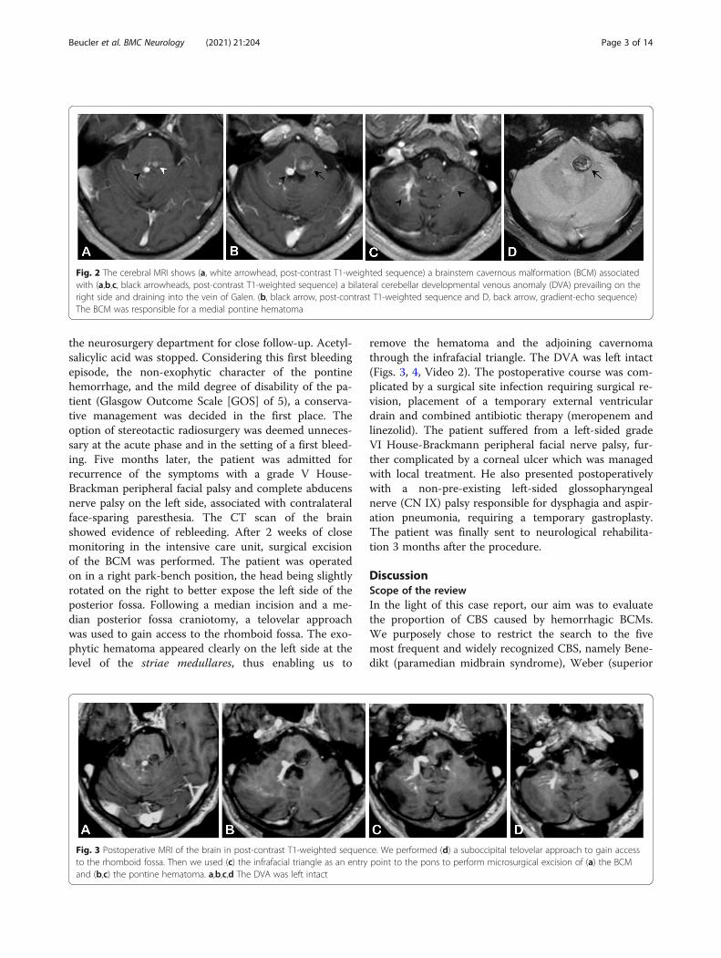
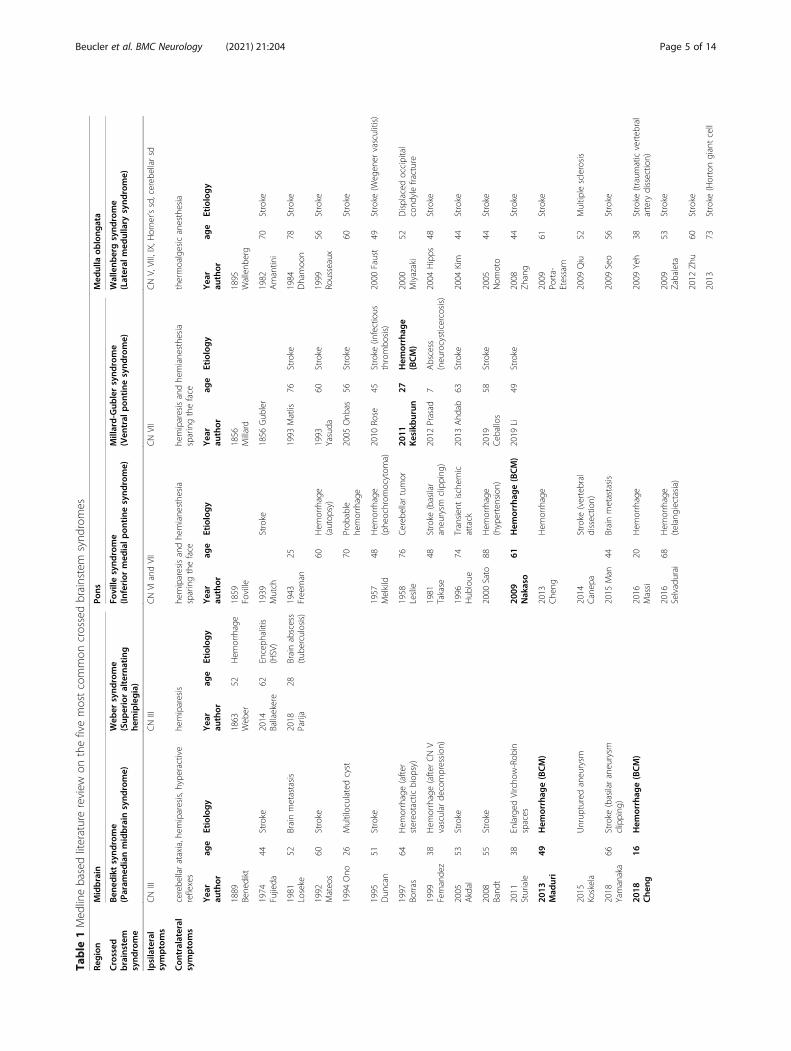
Results of database researchThe primary database research yielded 234 articles,among which 168 met the exclusion criteria after carefulreading of the text or the abstract. Sixty-six articles werefinally retained for a total of 69 patients [2]. There were14 cases of Benedikt syndrome [3], three cases of Webersyndrome [4], 15 cases of Foville syndrome [5], ninecases of Millard-Gubler syndrome [6, 7], and 28 cases ofWallenberg syndrome [8] (Table 1).
Causes of crossed brainstem syndromesAt the level of the midbrain, Benedikt syndrome wasusually caused by ischemic stroke (n = 6/14), followed byhemorrhage (n = 4/14) and direct nervous compression(n = 3/14) [9–22]. Weber syndrome was mainly causedby hemorrhage (n = 1/3) or infectious etiologies (n = 2/3)[4, 23, 24]. At the level of the pons, Foville syndromewas frequently caused by hemorrhage (n = 8/15),followed by ischemic stroke (n = 4/15) and brain metas-tases (n = 2/15) [25–37]. Conversely, Millard-Gubler syn-drome was mostly related to an ischemic stroke (n = 7/9), and rarely brought about by hemorrhage (n = 1/9) orbrain abscess (n = 1/9) [38–46]. At the level of themedulla oblongata, Wallenberg syndrome was
Fig. 4 Important surgical sequences. a Hockey stick fashion opening of the dura mater, discovering the cerebellar notch medially, and the leftsuperior and inferior semilunar lobules. b The floor of the fourth ventricle appears after passing through the inferior medullary velum, anddisplays crucial anatomical landmarks such as the striae medullares (SM), the infrafacial triangle (IFT) just above and the hypoglossal triangle (HT)underneath. The exophytic hematoma appears clearly on the left side at the level of the striae medullares. The cerebellar developmental venousanomaly (DVA) appears in blue under a thin layer of nervous tissue. c Evacuation of the pontine hematoma (H) at the level of the infrafacialtriangle (IFT), just above the striae medullares (SM) The midline is marked with a dotted line. d En-bloc excision of the cavernous malformation(CAV) using a tumor’s clamp. e Resection cavity
Beucler et al. BMC Neurology (2021) 21:204 Page 4 of 14
Table
1Med
linebasedliteraturereview
onthefivemostcommon
crossedbrainstem
synd
romes
Region
Midbrain
Pons
Med
ulla
oblong
ata
Crossed
brainstem
synd
rome
Ben
ediktsynd
rome
(Param
edianmidbrain
synd
rome)
Web
ersynd
rome
(Sup
erioralternating
hemiplegia)
Foville
synd
rome
(Inferior
med
ialp
ontine
synd
rome)
Millard-Gub
lersynd
rome
(Ven
tral
pon
tine
synd
rome)
Wallenb
ergsynd
rome
(Lateral
med
ullary
synd
rome)
Ipsilateral
symptoms
CNIII
CNIII
CNVI
andVII
CNVII
CNV,VIII,IX,H
orne
r’ssd,cereb
ellarsd
Con
tralateral
symptoms
cerebe
llarataxia,hem
iparesis,h
yperactive
reflexes
hemiparesis
hemiparesisandhe
miane
sthe
sia
sparingtheface
hemiparesisandhe
miane
sthe
sia
sparingtheface
thermoalgesicanesthesia
Yea
rau
thor
age
Etiology
Yea
rau
thor
age
Etiology
Yea
rau
thor
age
Etiology
Yea
rau
thor
age
Etiology
Yea
rau
thor
age
Etiology
1889
Bene
dikt
1863
Web
er52
Hem
orrhage
1859
Foville
1856
Millard
1895
Wallenb
erg
1974
Fujieda
44Stroke
2014
Ballaekere
62Enceph
alitis
(HSV)
1939
Mutch
Stroke
1856
Gub
ler
1982
Amantin
i70
Stroke
1981
Loseke
52Brainmetastasis
2018
Parija
28Brainabscess
(tub
erculosis)
1943
Freeman
251993
Matlis
76Stroke
1984
Dhamoo
n78
Stroke
1992
Mateo
s60
Stroke
60Hem
orrhage
(autop
sy)
1993
Yasuda
60Stroke
1999
Rousseaux
56Stroke
1994
Ono
26Multiloculated
cyst
70Prob
able
hemorrhage
2005
Onb
as56
Stroke
60Stroke
1995
Dun
can
51Stroke
1957
Melkild
48Hem
orrhage
(phe
ochrom
ocytom
a)2010
Rose
45Stroke
(infectious
thrombo
sis)
2000
Faust
49Stroke
(Weg
ener
vasculitis)
1997
Borras
64Hem
orrhage(after
stereo
tacticbiop
sy)
1958
Leslie
76Cereb
ellartumor
2011
Kesikburun
27Hem
orrhag
e(BCM)
2000
Miyazaki
52Displaced
occipital
cond
ylefracture
1999
Fernande
z38
Hem
orrhage(after
CNV
vascular
decompression
)1981
Takase
48Stroke
(basilar
aneurysm
clipping
)2012
Prasad
7Abscess
(neurocysticercosis)
2004
Hipps
48Stroke
2005
Akdal
53Stroke
1996
Hub
loue
74Transien
tischem
icattack
2013
Ahd
ab63
Stroke
2004
Kim
44Stroke
2008
Band
t55
Stroke
2000
Sato
88Hem
orrhage
(hypertension)
2019
Ceb
allos
58Stroke
2005
Nom
oto
44Stroke
2011
Sturiale
38Enlarged
Vircho
w-Rob
inspaces
2009
Nakaso
61Hem
orrhag
e(BCM)
2019
Li49
Stroke
2008
Zhang
44Stroke
2013
Mad
uri
49Hem
orrhag
e(BCM)
2013
Che
ngHem
orrhage
2009
Porta-
Etessam
61Stroke
2015
Koskela
Unrup
turedaneurysm
2014
Canep
aStroke
(vertebral
dissectio
n)2009
Qiu
52Multip
lesclerosis
2018
Yamanaka
66Stroke
(basilaraneurysm
clipping
)2015
Man
44Brainmetastasis
2009
Seo
56Stroke
2018
Che
ng16
Hem
orrhag
e(BCM)
2016
Massi
20Hem
orrhage
2009
Yeh
38Stroke
(traum
aticverteb
ral
artery
dissectio
n)
2016
Selvadurai
68Hem
orrhage
(telangiectasia)
2009
Zabaleta
53Stroke
2012
Zhu
60Stroke
2013
73Stroke
(Hortongiantcell
Beucler et al. BMC Neurology (2021) 21:204 Page 5 of 14

Table
1Med
linebasedliteraturereview
onthefivemostcommon
crossedbrainstem
synd
romes
(Con
tinued)
Region
Midbrain
Pons
Med
ulla
oblong
ata
Crossed
brainstem
synd
rome
Ben
ediktsynd
rome
(Param
edianmidbrain
synd
rome)
Web
ersynd
rome
(Sup
erioralternating
hemiplegia)
Foville
synd
rome
(Inferior
med
ialp
ontine
synd
rome)
Millard-Gub
lersynd
rome
(Ven
tral
pon
tine
synd
rome)
Wallenb
ergsynd
rome
(Lateral
med
ullary
synd
rome)
Sten
glarteritis)
2013
Ued
a48
Hem
orrhag
e(m
ultiple
BCM)
72Hem
orrhage(antiplatelet
andanticoagu
lant
therapy)
2014
Wu
43Stroke
2015
Koskela
Unrup
turedaneurysm
2015
Das
86Stroke
2015
Ehresm
ann
7Stroke
2015
Louis
30Stroke
(2weeks
post-
partum
)
2015
Ospino
Quiroz
48Stroke
2018
Kornbluh
14Stroke
2018
Oks
58Stroke
(sarcoidosis)
2018
Sivakumar
62Stroke
(PICAaneurysm
clipping
)
Beucler et al. BMC Neurology (2021) 21:204 Page 6 of 14

predominantly caused by ischemic stroke (n = 23/28),more rarely by hemorrhage (n = 2/28) or multiplesclerosis (n = 1/28) [18, 47–71]. The complete data isprovided in Table 2.Brainstem hemorrhage was responsible for approxi-
mately one quarter of the cases of CBS (n = 15/66). Asfor the underlying condition responsible for the brain-stem bleeding, hypertension was the most frequentlyencountered etiology (n = 6/15), closely followed byBCM (n = 5/15). Extralesional bleeding arising fromBCM was responsible for one-seventh of the cases ofBenedikt syndrome (n = 2/14), one out of ten cases ofMillard-Gubler syndrome (n = 1/9), one-fifteenth of thecases of Foville syndrome (n = 1/15), and approximatelyone out of thirty cases of Wallenberg syndrome (n = 1/28). There was also one case of Foville syndrome causedby a hemorrhage imputed to a telangiectasia.It is to note that posterior circulation aneurysms were
frequently encountered in this review (n = 5/69). Twounruptured aneurysms were responsible for nervouscompression, the first one (probably arising from the
posterior communicating artery) leading to a case ofBenedikt syndrome and the second one (arising from theposterior inferior cerebellar artery) at the origin of aWallenberg syndrome. Three aneurysms clippingresulted in infarction of perforating arteries, causing re-spectively a Benedikt syndrome, a Foville syndrome, anda Wallenberg syndrome.Similarly, two cases of Benedikt syndrome were caused
by a midbrain hematoma which occurred immediatelyafter a neurosurgical procedure: one was secondary to athird ventricle tumor biopsy, and the other one was sec-ondary to microvascular decompression for trigeminalneuralgia.
Physiopathology of cerebral cavernous malformationsCerebral cavernous malformations (CCM) are mulberry-like fragile vascular malformations that are encounteredin the cerebral hemispheres, brainstem and cerebellum,or in the spinal cord. Their structure consists in endo-thelial lined vascular sinusoids with no tight junctionsand even gaps between the endothelial cells, forming
Table 2 Etiologies reported for the five most common crossed brainstem syndromes
Total Benedikt Weber Foville Millard-Gubler Wallenberg
Total 69 14 3 15 9 28
Stroke 40 6 0 4 7 23
Embolic event 30 5 1 6 18
Aneurysm clipping 3 1 1 1
Artery dissection 2 1 1
Transient ischemic attack 1 1
Vasculitis 2 2
Infectious thrombosis 1 1
Sarcoidosis 1 1
Hemorrhage 16 4 1 8 1 2
Hypertension 6 6
Brainstem cavernous malformation 5 2 1 1 1
Telangiectasia 1 1
Post-operative complication 2 2
Anticoagulant therapy 1 1
Compression 5 3 2
Unruptured aneurysm 2 1 1
Cyst / Virchow-Robin spaces 2 2
Occipital fracture 1 1
Brain metastasis 3 1 2
Infection 3 2 1
Brain abscess 2 1 1
Encephalitis 1 1
Multiple sclerosis 1 1
Unknown 1 1
Beucler et al. BMC Neurology (2021) 21:204 Page 7 of 14

caverns within a dense collagen matrix clustered withoutintervening normal parenchyma [72].CCM are often associated with venous drainage anomal-
ies, ranging from solitary trans-cerebral or subpial drain-ing veins to genuine DVAs [73]. DVA constitute anextreme anatomical variation draining normal cerebral tis-sue into an extra-parenchymatous collector; they reflect avariation of the well-known anastomosis between thesuperficial and the deep venous drainage systems of thebrain which respond to a hemodynamic equilibrium [74].The combination of inherently fragile sinusoids walls
in the absence of blood-brain barrier and DVAs withraised venous pressure results in repeated intralesionalmicro hemorrhages which, in turn, leads to neoangio-genesis [75]. This “hemorrhagic angiogenic proliferation”mechanism results over time in the self-sustainedgrowth of CCM, which is why they appear on neuro-imaging as multilobulated vascular and calcified “pop-corn” lesions as the type 2 described by Zabramski [76].Although half of the CCM are discovered incidentally onneuroimaging, the other half may cause seizures relatedto the hemosiderin deposit around the lesion causingcortical irritation (25%), focal neurological symptoms re-lated to mass effect (15%), or intracranial hemorrhage(ICH) (12%) [77].
Specific considerations for brainstem CCMIt comes as no surprise that in the brainstem the mostfeared complication of CCM turns out to be bleedingwhich is also the main indication of excisional surgery[78]. The two main risk factors for the occurrence of anICH ascribable to a CCM are history of a previousbleeding episode and the location in the brainstem [79].Indeed, the estimated 5-year risk of ICH for an un-treated CCM is 3.8% in case of non-brainstem CCMwithout ICH or FND, 8% in case of BCM without ICHor FND, 18.4% for non-brainstem CCM with ICH orFND, and increases up to 30.8% for BCM with ICH orFND [80]. In the brainstem, the estimated annual rate ofextralesional bleeding is 8.7% for asymptomatic CCM,and rises to 12.4% for CCM with asymptomatic ICH,and up to 15.9% for CCM with symptomatic ICH [81].
Relevant surgical anatomy of the ponsAt the middle pons, corticospinal tract fibers are scat-tered anteriorly; motor neurons transit through trans-verse pontine fibers to merge the contralateral pontinenuclei and then join the middle cerebellar peduncle. Thespinothalamic tract is located just posteriorly and lieswithin the medial lemniscus. The floor of the fourthventricle provides a few surface reliefs that constitute
Fig. 5 (Left side) The artistic view of the brainstem shows that the corticospinal tract (red) shares intimate relations with the cranial nerve nucleiand fibers. (Right side) The artistic view of the inferior pons highlights the crossed neurological symptoms observed in the syndrome of Foville.The artistic views were drawn by Dr. Nathan BEUCLER
Beucler et al. BMC Neurology (2021) 21:204 Page 8 of 14

important landmarks for neurosurgeons. The medial sul-cus is bordered by the medial longitudinal fasciculi onboth sides. The nucleus of the facial nerve is located lat-erally at the inferior part of the pons. The fibers of thefuture CN VII loop superiorly and medially around theabducens nerve nucleus. This peculiar anatomical con-figuration creates a bulging within the floor of the fourthventricle known as the facial colliculus. Inferiorly, thestriae medullares define the superior limit of the hypo-glossal (CN XII), ambiguous (CN IX, X, XI) and vagus(CN X) nuclei. Pontine arterial supply is mainly anteriorand lateral; no major artery is to be found near the floorof the fourth ventricle floor (Fig. 5).
Surgical approaches to the ponsThe facial colliculus along with the fibers of future CNVII represent an important surgical landmark within therhomboid fossa. They constitute the inferior limit of thesuprafacial triangle which superior border are the super-ior and the middle cerebellar peduncles. On the sameway, they constitute the superior limit of the infrafacialtriangle which inferior borders are the striae medullares.These two triangles are known to be relatively safe entrycorridors entry corridors for a surgical approach to thefloor of the fourth ventricle as only scarce nerve fibersare encountered there [82, 83].
Surgical considerations for brainstem cavernousmalformationsRecent literature does not provide sufficient evidence re-garding the optimal timing for the surgical excision of abrainstem CCM with symptomatic extralesional bleed-ing, which is still a matter of debate. Zaidi et al. pre-sented a series of 397 patients operated on for brainstemCCM, among which 96% percent presented history ofprior ICH [84]. Thirty-five percent of the patients pre-sented persistent postoperative neurological deficits(mainly CN deficits), and the mean GOS was unchangedat last follow-up compared with the GOS upon admis-sion (4.47 vs 4.46, median follow-up 35.5 months). Theyreported that early surgery within 6 weeks after ICH andsmaller lesion size were associated with improved out-come. Garcia et al. presented a series of 104 patients op-erating on for brainstem CCM, among which 99%presented history of prior ICH [85]. The mean modifiedRankin scale upon admission was 2.23 compared to 1.58at final follow-up. The most frequent perioperativecomplications were cerebrospinal fluid leakage (12.5%),infection (9.6%) and surgical site hematoma (6%). Olderage, large size lesions, lesions crossing the midline, delaybetween last bleeding event and surgery, and the associ-ation with a DVA were associated with a poorerprognosis.
Based on these retrospective series, surgical excision ofa BCM may be deemed reasonable soon after the secondsymptomatic bleeding. In such case, the high operativemorbidity inherent to brainstem surgery is warranted bythe aggressive natural course of the disease.
Surgical considerations for associated developmentalvenous anomaliesUntil the 2000s, there have only been sporadic reportson the treatment of DVA. Some reported cases sup-ported the surgical excision of the DVA [86, 87],whereas intraoperative complications such as brainswelling after DVA coagulation have been reported [88].Campeau et al. neuro-imaging study seemed to confirmthe hypothesis that repeated microbleeding episodes andneoangiogenesis led to the formation of CCM in thevicinity of DVA [89]. In accordance with that theory,Wurm et al. reported a series of 15 patients who bene-fited from microsurgical excision of a CCM [90]. The as-sociated DVA was coagulated in six patients and leftintact in nine of them. Three patients from the groupwith intact DVA presented the recurrence of a CCMand benefited from a second microsurgical excision withsimultaneous coagulation of the DVA. The authors didnot report any venous complication in the patients whobenefited from the treatment of the DVA, with a meanfollow-up of 29 months. Nevertheless, this series, madeup of only 15 patients, lacks long-term follow-up. Be-sides, six patients whose DVA had been left intact didnot present recurrence of CCM. More recent reportscontinue to support the elective microsurgical excisionof symptomatic CCM without touching the associatedDVA [91]. Venous sacrifice in cranial neurosurgical pro-cedures has always been considered hazardous for fearof the potential disastrous consequences of venous in-farction [92, 93], which are very difficult to predict [94].Consequently, we tend to recommend leaving the DVAintact during the microsurgical excision of CCM.
Specific considerations for crossed pontine syndromesThe specific vascular supply of the pons may explain thedifference of etiology that we have observed betweenFoville syndrome (the inferior medial pontine syndrome)and Millard-Gubler syndrome (the ventral pontinesyndrome). Pontine hemorrhage caused by high bloodpressure is usually located more medially and damagesboth CN VI nucleus and CN VII fibers, leading to Fovillesyndrome. By contrast, ischemic stroke involves ratherthe paramedian branches or the short circumferentialbranches of the basilar artery which supply more lateralstructures such as CN VII nucleus, leading thus toMillard-Gubler syndrome [95].If we closely examine the clinical nuances reported
throughout the history concerning Foville syndrome, the
Beucler et al. BMC Neurology (2021) 21:204 Page 9 of 14

Table
3Theinferio
rmed
ialp
ontin
esynd
romeof
Foville:clinicalnu
ancesrepo
rted
sinceits
firstde
scrip
tion
Autho
r-ye
arJourna
lAge
Cau
seVIp
alsy
Ipsilateral
superior
VIIpalsy
Ipsilateral
inferior
VIIpalsy
Lateral
gazepalsy
Face-sparing
hemiparesis/
plegia
Prop
ortion
alhe
miparesis
/plegia
Con
tralateral
hemiane
sthe
sia
Con
tralateral
sympathe
tic
symptoms
Foville
1859
[5]
Gaz
Heb
dMed
Chir
yes
ipsiletaral
contralateral
Mutch
1939
[25]
BritJ
Oph
talm
olog
y56
ipsilateral
yes
yes
ipsilateral
ipsilateral
lower
limb
Freeman
1943
[26]
ArchNeurology
&Psychiatry
25ipsilateral
yes
yes
ipsilateral
contralateral
yes
60po
ntinehe
morrhage
both
side
syes
yes
both
side
scontralateral
uppe
rlim
byes
70po
ntinehe
morrhage
ipsilateral
yes
yes
ipsilateral
contralateral
Melkild
1957
[27]
ActaMed
Scand
48he
morrhage
(phe
ochrom
ocytom
a)yes
yes
ipsilateral
contralateral
Leslie1958
[28]
JAm
GeriatricsSoc
76cerebe
llartumor
ipsilateral
yes
yes
ipsilateral
ipsilateral
pyramidal
Takase
1981
[29]
ShinkeiN
eurolSurg
48basilaraneurysm
clipping
ipsilateral
deviation
yes
Hub
loue
1996
[30]
EurJEm
ergMed
74transien
tischem
icattack
ipsilateral
yes
yes
ipsilateral
contralateral
yes
Sato
2000
[31]
Rinsho
Shinkeigaku
Clin
Neurol
88po
ntinehe
morrhage
ipsilateral
yes
yes
contralateral
prop
ortio
nal
yes
Nakaso2009
[32]
InternalMed
icine
61po
ntinehe
morrhage
(caverno
ma)
ipsilateral
contralateral
Che
ng2013
[33]
Taiwan
Journalo
fOph
talm
olog
ypo
ntinehe
morrhage
ipsilateralinternu
clear
ophtalmop
legia
yes
yes
contralateral
yes
Canep
a-Ragg
io2014
[34]
BMJCaserepo
rts
infarctio
n(vertebrala.
dissecction)
numbn
ess
numbn
ess
contralateral
uppe
rlim
byes
Man
2015
[ 35]
BMJCaserepo
rts
44po
ntinelung
metastasis
yes
yes
ipsilateral
contralateral
Massi2016
[36]
PanAfrMed
J20
pontinehe
morrhage
ipsilateral
yes
yes
ipsilateral
contralateral
Selvadurai
2016
[37]
Neurology
68po
ntinehe
morrhage
(telangiectasia)
ipsilateral
contralateral
Beucler et al. BMC Neurology (2021) 21:204 Page 10 of 14

different forms of oculomotor palsies that were observedled to the distinction between a “superior Foville syn-drome” characterized by the presence of a CN VI palsyand an “inferior Foville syndrome” with lateral conjugategaze palsy due to the involvement of the medial longitu-dinal fasciculus or the paramedian pontine reticular for-mation (Table 3).
Limitations of the studyThis review presents some limits inherent to its retro-spective nature. Purposely or not case reports uncon-sciously select patients with favorable outcome; thus,their compilation may lead to a reporting bias whichmay underestimate the mortality rate. The literature re-view was deliberately restricted to the five most commonCBS which may constitute a limit but still enabled us tocollect a great number of articles. To the best of ourknowledge, this is the first study attempting to provide aclear and updated picture of the proportion of BCMs re-sponsible for or revealed by a genuine CBS.
ConclusionsPure crossed brainstem syndromes are rarely encoun-tered in clinical practice. They remarkably illustrate theanatomical peculiarity of the brainstem, which repre-sents a crossroad between the cranial nerves, the longtracts and key vegetative structures. In the light of thisreview, brainstem cavernous malformations with extrale-sional bleeding appear to account for approximately 7 %of all crossed brainstem syndromes. The indication andtiming of the surgical excision of a symptomaticbrainstem cavernous malformation remains a complexdecision to make and requires multidisciplinary teamexpertise. It has to be discussed openly between neuro-surgeons and their patient, taking into consideration theexisting evidence in favor of surgery but also the sub-stantial risks associated with such a delicate procedure.Multicentric prospective trials will be very difficult toconduct on such rare entities. Robust knowledge inbrainstem anatomy along with thorough neurologicalexamination skills will remain pivotal to the initial man-agement of these patients.
Supplementary InformationThe online version contains supplementary material available at https://doi.org/10.1186/s12883-021-02223-7.
Video 1
Video 2
AcknowledgementsNone.
Authors’ contributionsNB and HD conceptualized the article. NB, SB, AR, SF, RC, HD participated tothe literature review. NB, SB, AR, SF, RC, HD participated to the clinical care ofthe patient. NB, SB, AR, SF, RC, HD participated to the drafting of themanuscript. NB, RC, HD participated to the critical revision of the manuscript.All authors have read and approved the manuscript.
FundingThe authors did not receive any funding for this work. The publicationcharges of this article were gracefully covered by the "neuro-oncology andneuro-endocrinology research group" (Grenone) of Timone University Hos-pital, 13010 Marseille, France.
Availability of data and materialsAll the relevant data is included in the manuscript. There is no data depositfor this work.
Declarations
Ethics approval and consent to participateInformed written consent was obtained from the patient whose case reportis included in the manuscript. He has been given the opportunity to reviewthe manuscript and the attached files. This work was conducted inaccordance with the Declaration of Helsinki of 1964 or its furtheramendments (2013).
Consent for publicationWritten informed consent was obtained from the patient for the publicationof this manuscript and any accompanying figure and video. A copy of thewritten consent is attached to the manuscript.
Competing interestsAll authors certify that they have no affiliations with or involvement in anyorganization or entity with any financial interest (such as honoraria;educational grants; participation in speakers’ bureaus; membership,employment, consultancies, stock ownership, or other equity interest; andexpert testimony or patent-licensing arrangements), or non-financial interest(such as personal or professional relationships, affiliations, knowledge or be-liefs) in the subject matter or materials discussed in this manuscript.
Author details1Department of Neurosurgery, Timone University Hospital, APHM, 264 rueSaint-Pierre, 13005 Marseille, France. 2Ecole du Val-de-Grâce, French MilitaryHealth Service Academy, 1 place Alphonse Laveran, 75230 Paris Cedex 5,France. 3Aix Marseille Univ, INSERM, INS, Inst Neurosci Syst, Marseille, France.4Emergency Department, Timone University Hospital, APHM, 264 RueSaint-Pierre, 13005 Marseille, France. 5Department of Stereotactic andFunctional Neurosurgery, Timone University Hospital, APHM, 264 rueSaint-Pierre, 13005 Marseille, France. 6Aix-Marseille Univ, INSERM, MMG,Marseille, France.
Received: 7 December 2020 Accepted: 4 May 2021
References1. Querol-Pascual MR. Clinical approach to brainstem lesions. Semin
Ultrasound CT MRI. 2010;31(3):220–9. https://doi.org/10.1053/j.sult.2010.03.004.
2. Marx JJ, Thömke F. Classical crossed brain stem syndromes: myth or reality?J Neurol. 2009;256(6):898–903. https://doi.org/10.1007/s00415-009-5037-2.
3. Benedikt M. lecture. 1889;4. Weber H. A contribution to the pathology of the Crura Cerebri. J R Soc
Med. 1863;MCT-46(1):121–39. https://doi.org/10.1177/095952876304600112.5. Foville A. Note sur une paralysie peu connue de certains muscles de l’oeil et
sa liaison avec quelques points de l’anatomie et la physiologie de laprotubérance annulaire. Bull Soc Anat. (Paris). 1858;33:373-405.
6. Cenac H, Millard A. Hémorrhagie de la protubérance annulaire. Bull SocAnat. (Paris). 1856;36:206-21.
7. Gubler A. De l’hémiplégie alterne envisagée comme signe de lésion de laprotubérance annulaire et comme preuve de la décussation. Gaz Hebd MédChir. 1856;3(43):749–54.
Beucler et al. BMC Neurology (2021) 21:204 Page 11 of 14

8. Wallenberg A. Akute Bulbäraffektion (Embolie der Arteria cerebelli post infsinistra). Arch Psychiatry. 1895;27(2):504–40. https://doi.org/10.1007/BF02075799.
9. Fujieda T, Yamauchi T, Takahashi S, Moroji T. Letter: effect of levodopa ontremor in Benedikt’s syndrome. Br Med J. 1974;1(5905):456–7. https://doi.org/10.1136/bmj.1.5905.456-b.
10. Loseke N, Retif J, Noterman J, Flament-Durand J. Inferior red nucleussyndrome (Benedikt’s syndrome) due to a single intramesencephalicmetastasis from a prostatic carcinoma. Case Rep Acta Neurochir (Wien).1981;56(1–2):59–64.
11. Ono Y, Suzuki M, Kayama T, Yoshimoto T. Multilobulated cystic formation inthe brain stem with Benedikt’s syndrome: case report. Neurosurgery. 1994;34(4):726–9 discussion 729.
12. Borrás JM, Salazar FG, Grandas F. Oculomotor palsy and contralateral tremor(Benedikt’s syndrome) following a stereotactic procedure. J Neurol. 1997;244(4):272–4. https://doi.org/10.1007/s004150050085.
13. Fernandez HH, Friedman JH, Centofanti JV. Benedikt’s syndrome with delayed-onset rubral tremor and hemidystonia: a complication of tic douloureuxsurgery. Mov Disord Off J Mov Disord Soc. 1999;14(4):695–7. https://doi.org/10.1002/1531-8257(199907)14:4<695::AID-MDS1024>3.0.CO;2-3.
14. Akdal G, Kutluk K, Men S, Yaka E. Benedikt and “plus–minus lid” syndromesarising from posterior cerebral artery branch occlusion. J Neurol Sci. 2005;228(1):105–7. https://doi.org/10.1016/j.jns.2004.09.029.
15. Bandt SK, Anderson D, Biller J. Deep brain stimulation as an effectivetreatment option for post-midbrain infarction-related tremor as it presentswith Benedikt syndrome. J Neurosurg. 2008;109(4):635–9. https://doi.org/10.3171/JNS/2008/109/10/0635.
16. Sturiale CL, Albanese A, Lofrese G, Frassanito P, Sabatino G, Marchese E,et al. Pathological enlargement of midbrain Virchow–Robin spaces: a rarecause of obstructive hydrocephalus. Br J Neurosurg. 2011;25(1):130–1.https://doi.org/10.3109/02688697.2010.504050.
17. Maduri R, Barbagallo G, Iofrida G, Signorelli M, Signorelli F. Regression ofBenedikt’s syndrome after single-stage removal of mesencephaliccavernoma and temporal meningioma: a case report. Clin NeurolNeurosurg. 2013;115(6):748–50. https://doi.org/10.1016/j.clineuro.2012.06.033.
18. Koskela E, Setälä K, Kivisaari R, Hernesniemi J, Laakso A. Neuro-ophthalmicpresentation and surgical results of unruptured intracranialaneurysms—prospective Helsinki experience of 142 patients. WorldNeurosurg. 2015;83(4):614–9. https://doi.org/10.1016/j.wneu.2014.12.017.
19. Cheng G, Yang Y, Wang Y, Tan H, Zhang S. Deep brain stimulation of thethalamic ventral intermediate nucleus for Benedikt’s syndrome mainlypresent as tremor: a long-term case observation. Acta Neurochir. 2018;160(7):1349–53. https://doi.org/10.1007/s00701-018-3526-8.
20. Mateos V, Campos DM, Colosía VP, Salas-Puig J, Fernández JM, Lahoz CH.Nuclear syndrome of the oculomotor nerve caused by a mesencephalicinfarction confirmed by MRI. Arch Neurobiol (Madr). 1992;55(4):183–7.
21. Duncan GW, Weindling SM. Posterior cerebral artery stenosis with midbraininfarction. Stroke. 1995;26(5):900–2. https://doi.org/10.1161/01.STR.26.5.900.
22. Yamanaka Y, Shinohara T, Kozano I, Yoshizumi T, Kawasaki T. Benediktsyndrome associated with neck clipping of ruptured basilar-superiorcerebellar artery aneurysm:a case report. No Shinkei Geka. 2018;46(11):1007–12. https://doi.org/10.11477/mf.1436203855.
23. Ballaekere JS, Chebbi PP, Sundarmurthy H, Parameshwarappa A. Webersyndrome: herpes simplex virus brainstem encephalitis as an etiology. Am JMed. 2014;127(12):e5–6. https://doi.org/10.1016/j.amjmed.2014.08.005.
24. Parija S, Lalitha CS, Naik S. Weber syndrome secondary to brain stemtuberculoma. Indian J Ophthalmol. 2018;66(7):1036–9. https://doi.org/10.4103/ijo.IJO_1040_17.
25. Mutch JR. FOVILLE’S syndrome record of a CASE. Br J Ophthalmol. 1939;23(4):225–38. https://doi.org/10.1136/bjo.23.4.225.
26. Freeman W, SYNDROMES OF, THE PONTILE TEGMENTUM. FOVILLE’Ssyndrome: report of three cases. Arch Neurol Psychiatr. 1943;50(4):462.https://doi.org/10.1001/archneurpsyc.1943.02290220092008.
27. Melkild A, Mörstad KS. Foville’s syndrome as a complication ofPhaeochromocytoma. Acta Med Scand. 1957;159(6):471–4. https://doi.org/10.1111/j.0954-6820.1957.tb00155.x.
28. Levene LJ. Benign cerebellar tumor presenting a modified Foville syndrome.J Am Geriatr Soc. 1958;6(4):346-51. https://doi.org/10.1111/j.1532-5415.1958.tb00726.x
29. Takase M, Saeki N, Oka N, Satoh A, Otaki M, Yamaura A. Superior Fovillesyndrome after clipping of basilar bifurcation aneurysm--case report(author’s transl). No Shinkei Geka. 1981;9(3):343–7.
30. Hubloue I, Laureys S, Michotte A. A rare case of diplopia: medialinferior pontine syndrome or Foville??s syndrome. Eur J Emerg Med.1996;3(3):194–8.
31. Sato K, Nitta E. Pontine hemorrhage presenting with Foville syndrome andtransient contralateral hyperhidrosis. Rinsho Shinkeigaku. 2000;40(3):271–3.
32. Nakaso K, Sasaki K. Fluctuating Foville’s syndrome caused by a pontineAngioma in a patient with a polycystic kidney. Intern Med. 2009;48(15):1335–6. https://doi.org/10.2169/internalmedicine.48.2314.
33. Cheng H-C, Yen M-Y, Wang A-G. Foville’s syndrome with ipsilateralinternuclear ophthalmoplegia due to spontaneous pontine hemorrhage.Taiwan J Ophtalmol. 2013;3(2):75–7. https://doi.org/10.1016/j.tjo.2012.12.001.
34. Canepa Raggio C, Dasgupta A. Three cases of Spontaneous Vertebral ArteryDissection (SVAD), resulting in two cases of Wallenberg syndrome and onecase of Foville syndrome in young, healthy men. Case Rep. 2014;2014(apr282):bcr2014203945.
35. Man BL, Fu YP. Bronchogenic carcinoma presented as Foville’s syndrome.Case Rep. 2015;2015(mar03 2):bcr2014205025.
36. Massi DG, Nyassinde J, Ndiaye MM. Superior Foville syndrome due topontine hemorrhage: a case report. Pan Afr Med J. 2016;25:215.
37. Selvadurai C, Rondeau MW, Colorado RA, Feske SK, Prasad S. Teaching videoneuro Images : Foville syndrome. Neurology. 2016;86(19):e203. https://doi.org/10.1212/WNL.0000000000002658.
38. Matlis A, Kleinman Y, Korn-Lubetzki I. Millard-Gubler syndrome. AJNR Am JNeuroradiol. 1994;15(1):179–81.
39. Yasuda Y, Matsuda I, Sakagami T, Kobayashi H, Kameyama M. Pontine infarctionwith pure Millard-Gubler syndrome: precise localization with magnetic resonanceimaging. Eur Neurol. 1993;33(4):333–4. https://doi.org/10.1159/000116965.
40. Onbas O, Kantarci M, Alper F, Karaca L, Okur A. Millard-Gublersyndrome: MR findings. Neuroradiology. 2005;47(1):35–7. https://doi.org/10.1007/s00234-004-1312-1.
41. Rose DZ, Parra-Herran C, Petito CK, Post MJD. Restricted diffusion of pus inthe subarachnoid space: MRSA Meningo-Vasculitis and progressivebrainstem ischemic strokes - a case report. Case Rep Neurol. 2010;2(2):101–10. https://doi.org/10.1159/000319691.
42. Kesikburun S, Safaz İ, Alaca R. Pontine cavernoma hemorrhage leading tomillard-gubler syndrome. Am J Phys Med Rehabil. 2011;90(3):263. https://doi.org/10.1097/PHM.0b013e3181e29e8e.
43. Prasad R, Kapoor K, Mishra OP, Srivastava A. Neurocysticercosis presenting asMillard Gubler syndrome. J Neurosci Rural Pract. 2012;03(03):375–7.
44. Ahdab R, Saade HS, Kikano R, Ferzli J, Tarcha W, Riachi N. Pure ipsilateralcentral facial palsy and contralateral hemiparesis secondary to ventro-medial medullary stroke. J Neurol Sci. 2013;332(1–2):154–5. https://doi.org/10.1016/j.jns.2013.06.028.
45. Ceballos-Lizarraga R, Palomino-Díaz C, Romero-Figueroa JÁ. Wall-eyedmonocular Internuclear Ophthalmoplegia (WEMINO) and Millard-Gublersyndromes in a patient with isolated pontine infarction: topographic,oculomotor, and radiological analysis of two very uncommon conditions.Case Rep Neurol. 2019;11(2):230–7. https://doi.org/10.1159/000501794.
46. Li X-T, Yuan J-L, Hu W-L. Vertebrobasilar artery dissection manifesting asMillard-Gubler syndrome in a young ischemic stroke patient: a case report.World J Clin Cases. 2019;7(1):73–8. https://doi.org/10.12998/wjcc.v7.i1.73.
47. Amantini A, Arnetoli G, Rossi L, Fenzi, Salviati A, Rizzuto N, et al. BAEP andautopsy findings in Wallenberg syndrome. Ital J Neurol Sci. 1982;3(3):237–40.https://doi.org/10.1007/BF02043316.
48. Dhamoon SK, Iqbal J, Collins GH. Ipsilateral hemiplegia and the Wallenbergsyndrome. Arch Neurol. 1984;41(2):179–80. https://doi.org/10.1001/archneur.1984.04050140077029.
49. Rousseaux M, Cassim F, Bayle B, Laureau E. Analysis of the perception ofand reactivity to pain and heat in patients with Wallenberg syndrome andsevere spinothalamic tract dysfunction. Stroke. 1999;30(10):2223–9. https://doi.org/10.1161/01.STR.30.10.2223.
50. Faust J, Visbeck A, Fitzek S, et al. Vasculitic wallenberg syndrome withdetection of anti-proteinase 3 antibodies in the cerebrospinal fluid of apatient with severe Wegener’s granulomatosis and only mild kidneyinvolvement. Nephrol Dial Transplant Off Publ Eur Dial Transpl Assoc EurRen Assoc. 2000;15(6):893–6.
51. Miyazaki C, Katsume M, Yamazaki T, Aoki K, Kuroki T, Takasu N. Unusualoccipital condyle fracture with multiple nerve palsies and Wallenberg
Beucler et al. BMC Neurology (2021) 21:204 Page 12 of 14
syndrome. Clin Neurol Neurosurg. 2000;102(4):255–8. https://doi.org/10.1016/S0303-8467(00)00109-8.
52. Hipps WM, Wilhelmus KR. Persistent visual loss from neurotrophic cornealulceration after dorsolateral medullary infarction (Wallenberg syndrome). JNeuroophthalmol. 2004;24(4):345–6. https://doi.org/10.1097/00041327-200412000-00015.
53. Kim JS, Moon SY, Park S-H, Yoon B-W, Roh J-K. Ocular lateropulsion inWallenberg syndrome. Neurology. 2004;62(12):2287. https://doi.org/10.1212/WNL.62.12.2287.
54. Nomoto N, Konno S, Sugimoto H, Kurihara T, Wakata N. Wallenbergsyndrome with SIADH. Eur Neurol. 2005;53(1):35–6. https://doi.org/10.1159/000084260.
55. Zhang S-Q, Liu M-Y, Wan B, Zheng H-M. Contralateral body half Hypalgesiain a patient with lateral medullary infarction: atypical Wallenberg syndrome.Eur Neurol. 2008;59(3–4):211–5. https://doi.org/10.1159/000114050.
56. Porta-Etessam J, Casanova I, Pajuelo B, di Capua D, del Val J, García GarcíaME, et al. See-saw nystagmus in a patient with Wallenberg syndrome. JNeuroophthalmol. 2009;29(1):73–4. https://doi.org/10.1097/WNO.0b013e3181989dc1.
57. Qiu W, Wu J-S, Carroll WM, Mastaglia FL, Kermode AG. Wallenbergsyndrome caused by multiple sclerosis mimicking stroke. J Clin Neurosci.2009;16(12):1700–2. https://doi.org/10.1016/j.jocn.2009.04.008.
58. Seo WK, Kwon DY, Seo SH, Park MH, Park KW. Neuropathic pruritusfollowing Wallenberg syndrome. Neurology. 2009;72(7):676. https://doi.org/10.1212/01.wnl.0000342484.99298.52.
59. Yeh H-F, Seak C-J, Chiu T-F, Chang Y-C. Traumatic vertebral artery dissectionand Wallenberg syndrome after a motorcycle collision. Am J Emerg Med.2009;27(1):131.e1–3.
60. Ayerbe Zabaleta I, Negrevergne M, Ucelay Vicinay I, Catherine M. Vértigoagudo pseudoperiférico y síndrome de Wallenberg. ActaOtorrinolaringológica Esp. 2010;61(4):312–4. https://doi.org/10.1016/j.otorri.2009.06.001.
61. Zhu J, Li J, Liu Y, Han Y, Wang Y. Unilateral headache with visual Aura froma Wallenberg syndrome. CNS Neurosci Ther. 2012;18(7):598–600. https://doi.org/10.1111/j.1755-5949.2012.00330.x.
62. Stengl KL, Buchert R, Bauknecht H, Sobesky J. A hidden giant: Wallenbergsyndrome and aortal wall thickening as an atypical presentation of a giantcell arteritis. Case Rep. 2013;2013(mar01 1):bcr2012006994.
63. Ueda M, Nishiyama Y, Abe A, Katayama Y. Hemorrhagic Wallenbergsyndrome. Intern Med. 2013;52(20):2383–4. https://doi.org/10.2169/internalmedicine.52.1141.
64. Wu S, Li N, Xia F, Sidlauskas K, Lin X, Qian Y, et al. Neurotrophic keratopathydue to dorsolateral medullary infarction (Wallenberg syndrome): case reportand literature review. BMC Neurol. 2014;14(1):231. https://doi.org/10.1186/s12883-014-0231-y.
65. Das P, Chopra A, Rai A, Kuppuswamy PS. Late-onset recurrent mania asa manifestation of Wallenberg syndrome: a case report and review ofthe literature. Bipolar Disord. 2015;17(6):677–82. https://doi.org/10.1111/bdi.12318.
66. Ehresmann A, Van H, Merlini L, Fluss J. Wallenberg syndrome: anexceptional cause of acute Vertigo in children. Neuropediatrics. 2015;47(01):061–3. https://doi.org/10.1055/s-0035-1566731.
67. Louis DW, Dholakia N, Raymond MJ. Wallenberg syndrome with associatedmotor weakness in a two-week-postpartum female. Case Rep Neurol. 2015;7(3):186–90. https://doi.org/10.1159/000440712.
68. Ospino Quiroz JC, Monteagudo CJ. A propósito de un caso de síndrome deWallenberg. SEMERGEN Med Fam. 2016;42(8):e179–80. https://doi.org/10.1016/j.semerg.2015.10.012.
69. Kornbluh A, Twanow J-D. Teaching NeuroImages: adolescent Wallenbergsyndrome with overlooked signs: Ipsipulsion and ipsilateral facial palsy.Neurology. 2018;91(20):e1949–50. https://doi.org/10.1212/WNL.0000000000006513.
70. Oks M, Li A, Makaryus M, Pomeranz HD, Sachdeva M, Pullman J, et al.Sarcoidosis presenting as Wallenberg syndrome and panuveitis. Respir MedCase Rep. 2018;24:16–8. https://doi.org/10.1016/j.rmcr.2018.03.002.
71. Sivakumar K, Garcha M, Mehta D, Leary MC, Yacoub HA. Centralhypoventilation: a rare complication of Wallenberg syndrome. Case RepNeurol Med. 2018;2018:1–3.
72. Clatterbuck RE. Ultrastructural and immunocytochemical evidence that anincompetent blood-brain barrier is related to the pathophysiology of
cavernous malformations. J Neurol Neurosurg Psychiatry. 2001;71(2):188–92.https://doi.org/10.1136/jnnp.71.2.188.
73. Dammann P, Wrede KH, Maderwald S, el Hindy N, Mueller O, Chen B, et al.The venous angioarchitecture of sporadic cerebral cavernousmalformations: a susceptibility weighted imaging study at 7 T MRI. J NeurolNeurosurg Psychiatry. 2013;84(2):194–200. https://doi.org/10.1136/jnnp-2012-302599.
74. Lasjaunias P, Burrows P, Planet C. Developmental venous anomalies (DVA):the so-called venous angioma. Neurosurg Rev. 1986;9(3):233–42. https://doi.org/10.1007/BF01743138.
75. Pozzati E, Acciarri N, Tognetti F, Marliani F, Giangaspero F. Growth,subsequent bleeding, and De novo appearance of cerebral cavernousAngiomas. Neurosurgery. 1996;38(4):662–70. https://doi.org/10.1227/00006123-199604000-00006.
76. Zabramski JM, Wascher TM, Spetzler RF, Johnson B, Golfinos J, Drayer BP,et al. The natural history of familial cavernous malformations: results of anongoing study. J Neurosurg. 1994;80(3):422–32. https://doi.org/10.3171/jns.1994.80.3.0422.
77. Salman RA-S, Hall JM, Horne MA, et al. Untreated clinical course of cerebralcavernous malformations: a prospective, population-based cohort study. LancetNeurol. 2012;11(3):217–24. https://doi.org/10.1016/S1474-4422(12)70004-2.
78. Al-Shahi Salman R, Berg MJ, Morrison L, Awad IA. Hemorrhage fromcavernous malformations of the brain: definition and reporting standards.Stroke. 2008;39(12):3222–30. https://doi.org/10.1161/STROKEAHA.108.515544.
79. Akers A, Al-Shahi Salman R. A. Awad I, et al. synopsis of guidelines for theclinical Management of Cerebral Cavernous Malformations: consensusrecommendations based on systematic literature review by the AngiomaAlliance scientific advisory board clinical experts panel. Neurosurgery. 2017;80(5):665–80. https://doi.org/10.1093/neuros/nyx091.
80. Horne MA, Flemming KD, Su I-C, Stapf C, Jeon JP, Li D, et al. Clinical courseof untreated cerebral cavernous malformations: a meta-analysis of individualpatient data. Lancet Neurol. 2016;15(2):166–73. https://doi.org/10.1016/S1474-4422(15)00303-8.
81. Li D, Hao S-Y, Jia G-J, Wu Z, Zhang L-W, Zhang J-T. Hemorrhage risks andfunctional outcomes of untreated brainstem cavernous malformations. JNeurosurg. 2014;121(1):32–41. https://doi.org/10.3171/2014.3.JNS132537.
82. Kyoshima K, Kobayashi S, Gibo H, Kuroyanagi T. A study of safe entryzones via the floor of the fourth ventricle for brain-stem lesions: reportof three cases. J Neurosurg. 1993;78(6):987–93. https://doi.org/10.3171/jns.1993.78.6.0987.
83. Cavalcanti DD, Preul MC, Kalani MYS, Spetzler RF. Microsurgical anatomy ofsafe entry zones to the brainstem. J Neurosurg. 2016;124(5):1359–76. https://doi.org/10.3171/2015.4.JNS141945.
84. Zaidi HA, Mooney MA, Levitt MR, Dru AB, Abla AA, Spetzler RF. Impactof Timing of Intervention Among 397 Consecutively Treated BrainstemCavernous Malformations. Neurosurgery [Internet] 2017 [cited 2020 May28];Available from: https://academic.oup.com/neurosurgery/article/2981949/Impact
85. Garcia RM, Ivan ME, Lawton MT. Brainstem cavernous malformations.Neurosurgery. 2015;76(3):265–78. https://doi.org/10.1227/NEU.0000000000000602.
86. Chandra PS, Manjari T, Chandramouli BA, Jayakumar PN, Shankar SK.Cavernous-venous malformation of brain stem--report of a case and reviewof literature. Surg Neurol. 1999;52(3):280–5. https://doi.org/10.1016/S0090-3019(99)00079-8.
87. Yamada S, Liwnicz BH, Thompson JR, Colohan AR, Iacono RP, Tran JT.Pericapillary arteriovenous malformations angiographically manifested ascerebral venous malformations. Neurol Res. 2001;23(5):513–21. https://doi.org/10.1179/016164101101198767.
88. Rigamonti D, Spetzler RF. The association of venous and cavernousmalformations. Report of four cases and discussion of thepathophysiological, diagnostic, and therapeutic implications. Acta Neurochir1988;92(1–4):100–105, DOI: https://doi.org/10.1007/BF01401979.
89. Campeau NG, Lane JI. De novo development of a lesion with theappearance of a cavernous malformation adjacent to an existingdevelopmental venous anomaly. AJNR Am J Neuroradiol. 2005;26(1):156–9.
90. Wurm G, Schnizer M, Fellner FA. Cerebral Cavernous MalformationsAssociated with Venous Anomalies: Surgical Considerations. OperNeurosurg. 2005;57(suppl_1):42–58.
Beucler et al. BMC Neurology (2021) 21:204 Page 13 of 14
91. Georgieva VB, Krastev ED. Surgical treatment of brainstem cavernousmalformation with concomitant developmental venous anomaly. Asian JNeurosurg. 2019;14(2):557–60. https://doi.org/10.4103/ajns.AJNS_246_18.
92. Masuoka J, Matsushima T, Hikita T, Inoue E. Cerebellar swelling after sacrificeof the superior petrosal vein during microvascular decompression fortrigeminal neuralgia. J Clin Neurosci Off J Neurosurg Soc Australas. 2009;16(10):1342–4.
93. Piatt JH, Kellogg JX. A hazard of combining the infratentorial supracerebellarand the cerebellomedullary fissure approaches: cerebellar venousinsufficiency. Pediatr Neurosurg. 2000;33(5):243–8. https://doi.org/10.1159/000055962.
94. Ferroli P, Acerbi F, Tringali G, Albanese E, Broggi M, Franzini A, et al. Venoussacrifice in neurosurgery: new insights from venous indocyanine greenvideoangiography: clinical article. J Neurosurg. 2011;115(1):18–23. https://doi.org/10.3171/2011.3.JNS10620.
95. Kumral E, Bayülkem G, Evyapan D. Clinical spectrum of pontine infarction. JNeurol. 2002;249(12):1659–70. https://doi.org/10.1007/s00415-002-0879-x.
Publisher’s NoteSpringer Nature remains neutral with regard to jurisdictional claims inpublished maps and institutional affiliations.
Beucler et al. BMC Neurology (2021) 21:204 Page 14 of 14
Copyright © 2022 FDOKUMEN