seduction or proof? revealing assumptions in the negotiation ...
Upload
khangminh22Category
view
0download
0
15151st Annual Meeting of the ESPE __________________________________________________ _____________________________________________________________________
Horm Res 2012;78(suppl 1)
______________________________________P2-d3-481 Bone, Growth Plate and Mineral Metabolism 3
Neonatal hypocalcaemia revealing maternal calcium metabolism disorderAsmahane Ladjouze; Leila Kedji; Abdeljalil Maoudj; Karima Berkouk; Manoubia Bensmina; Rawda Aboura; Nabila Dahmane; Tahar Anane; Abdenour LarabaCHU Bab el Oued, Pediatrics Department, Algiers, Algeria
Background: During pregnancy, the fetus maintains high blood calcium levels, thanks to an active transport across the placenta. At birth, with the cord clamping, he is abruptly disconnected from the maternal supply of cal-cium and becomes dependant of the intestinal calcium intake and the skel-etal reserves. Besides, the newborn Vitamin D status is directly related to his mother’s Vitamin D status. Thus, the slightest abnormality in the maternal calcium metabolism may have severe consequences in the newborn calcium regulation Objective: To determine the relation between hypocalcaemia in newborns and their mother’s mineral status. Methods: Mother of newborns presenting with symptomatic hypocalcemia were tested to detect either vitamin D deficiency or hyperparathyroidism Results: 7 mother-newborn couples were identified. the mean age was 8.3 days (3-27). There was one female and six males. All the newborns but one were referred for general seizures. All mothers but one wore the veil (3 wore integral veil). None of the mothers had a known calcium metabolism disorder. All newborns but one had severe hypocalcemia (< 60 mg/l). The evaluation of the mothers revealed:-Severe Vitamin D deficiency in 4 mothers, one of them had an asymptomatic hypocalcaemia-Unknown maternal hyperparathyroidism due to parathyroid adenoma in 2 mothers (one had two children). All patients were given calcium supplements and alphacalcidol. Evolution was favorable for all of them. Conclusions: Testing for calcium metabolism and Vitamin D status during pregnancy should be mandatory. It would allow to detect rare cases of asymp-tomatic maternal hyperparathyroidism, and especially to screen for Vitamin D deficiency. Recent publications have reported the increasing frequency of maternal and neonatal vitamin D deficiency in Muslim countries. Therefore, we suggest that vitamin D supplementation should be systematically consid-ered after proper screening in those countries.
______________________________________P2-d3-482 Bone, Growth Plate and Mineral Metabolism 3
Bone mass and quality in juvenile systemic lupus erythematosus (JSLE)Stefano Stagi1; Loredana Cavalli2; Laura Masi2; Maria Luisa Brandi2; Marco Matucci Cerinic2; Maurizio de Martino3; Fernanda Falcini21Mugello’s Hospital, Paediatric Unit, Borgo San Lorenzo, Italy; 2University of Florence, Department of Internal Medicine, Florence, Italy; 3University of Florence, Department of Paediatrics, Florence, Italy
Introduction: There are few data on bone mass and quality in patients with Juvenile Systemic Lupus Erythematosus (JSLE). However, there are few data comparing bone mass and quality determinants using dual energy X-ray ab-sorptiometry (DXA), peripheral quantitative computed tomography (pQCT), and quantitative ultrasound (QUS) in JSLE. Design: To evaluate, through a cross-sectional and longitudinal study, bone mass and quality determinants in JSLE young adolescent and adults, and to assess the prevalence and to identify the main predictors of reduced Bone Mineral Density (BMD) and bone quality using these techniques. Methods: Fifty-six JSLE patients (46 females, 10 males, mean age 21.5 +/- 6.1 years) were evaluated. In all subjects DXA scan at the lumbar spine, radius pQCT, and phalangeal QUS were performed. Of these, forty-six con-secutive females with JSLE were followed-up longitudinally with a second DXA, pQCT and QUS evaluation. The data obtained were compared with 80 ages- and sex- matched healthy subjects. Results: JSLE patients showed a reduced spine BMAD SDS (p < 0.001), TrabBMD (p < 0.0001), Muscle CSA (p < 0.005), SSIp (p < 0.05), AD-SoS (p < 0.005), and QUS z-score (p < 0.005), but not CortBMD and CBA respect to controls. However, fat CSA were significantly increased (p < 0.0001). These data were confirmed in longitudinal evaluation. SSIp longitudinal evaluation showed that JSLE patients presented no more significantly lower levels than controls. Conclusions: Patients with JSLE have a low bone mass, not reaching the
normal condition over time despite the current more effective drugs, with the high risk of osteoporosis in early adulthood. To reduce the risk, a close monitoring of BMD, a better control of disease activity, physical activity, and a dietary intake of calcium and vitamin D are advocated to ameliorate the bone mass.
______________________________________P2-d3-483 Bone, Growth Plate and Mineral Metabolism 3
A novel de-novo mutation of PHEX gene in a patient with X-linked hypophosphataemic rickets with severe bone lesionsAurora Natalia Rossodivita1; Danilo Buonsenso1; Valentina Caturano1; Giampiero Baroncelli21Catholic University of the Sacred Heart, Department of Pediatrics, Rome, Italy; 2University-Hospital, Department of Obstetrics, Gynecology and Pediatrics, Pisa, Italy
Background: X-linked Hypophosphatemic Rickets (XLHR) is the most common familial form of HR, caused by inactivating mutations of the PHEX gene (Phosphate-regulating gene with Homologies to Endopeptidases on the X chromosome), which encodes a zinc-dependent endopeptidase involved in bone mineralization and renal phosphate reabsorption. Objective and hypotheses: Case report of a 2-year old female referred to us for a dwarfism due to XLHR, initially misdiagnosed as impaired growth caused by interventricular defect. Methods: Gene analysis was performed by extraction of genomic DNA and total RNA from leukocytes. The PHEX gene was amplified from DNA by PCR and analyzed by Denaturating High Performance Chromatography (WAVE-MD, Transgenomic) and the amplicons were directly sequenced by 3130x1 Genetic Analyzer (Applied Biosystems). Results: We report on a female with XLHR harboring a novel heterozygous duplication of 5 nucleotides (c.2049_22053dupCTTCT, Ensembl Gene ID ENSG00000102174) at exon 20 of gene PHEX, which leads to a stop co-don and gives rise possibly to a truncated PHEX protein. Physical exami-nation at time of diagnosis showed severe disproportioned short stature, en-larged wrists, knocked knees and bowed legs. Radiographs of wrists and legs showed the classical rickets bone changes. Laboratory evaluation revealed normal serum calcium levels, hypophosphatemia, low 1,25(OH)2D3, low al-caline phosphatase activity and phosphaturia. For 18 months the child has been being treated with phosphate supplements and calcitriol with a slight normalization of biochemical parameters but without improvement in clini-cal signs. DNA analyses of all family members were normal Conclusions: We report a novel nonsense mutation of PHEX gene, gaining an insight into the molecular genetic of this disorder. This case report emphasizes the im-portance of early diagnosis and treatment in order to improve bone growth and skeletal deformities. PHEX gene mutation could be useful for prognosis even if the timing of treatment may have a key role in improving bone lesions
______________________________________P2-d3-484 Bone, Growth Plate and Mineral Metabolism 3
Vitamin D deficiency in children with cerebral palsy: what kind of a problem?Belkis Ipekci1; Filiz Mine Cizmecioglu2; Bulent Kara3; Elif Ozsu2; Sukru Hatun2
1Kocaeli Unversity Mediacl School, Pediatrics, Kocaeli, Turkey; 2Kocaeli Universty Medical School, Pediatric Endocrinology, Kocaeli, Turkey; 3Kocaeli University Medical School, Pediatric Neurology, Kocaeli, Turkey
Background: Children with cerebral palsy (CP) have risks for vitamin D de-ficiency. Aim: Aimed to make the child neurology and emergency unites be more alert in case children with CP are presented with seizures apart from attributed to known CP. Methods: Mean age 10.2 ± 5.0 decimal years %48 girls of 29 patients with CP evaluated as vitamin D deficiency. Vitamin D status defined according to serum 25OHD levels as insufficiency, deficiency and severe deficiency, respectively (25OHDi<20, 15-20 and <5 ng/ml). Rickets diagnosed based on the clinic and biochemical parameters. Results: Mean diagnosis age of CP was 13 ± 1.6 month. Sixty percent of 23 patients who used anti epileptic treatment were given nonenzyme-inducing drugs. Anthropometric data showed many of patients were underweight (75%

152_______________________________________________________________________________________________________________________
Horm Res 2012;78(suppl 1)
of patients weight z score < -2.5 and %50 BMI z score <-2.5). Vitamin D in-sufficiency, deficiency and severe deficiency found in 59% (n 17) of patients, respectively [%17, %38, %4]. Eleven patient (38%) diagnosed as rickets, 7%of them had fracture. Secondary hyperparathyroidism (iPTH>65 pg/ml) was observed in 24%. Two patients with rickets whose weight z score -5.3 and -2,4 had hypocalcemia (serum Ca 7,4 -6,3 mg/dl) and seconder hyperparathy-roidism (iPTH 178,174 pg/ml), although their serum ALP levels were normal and this were explained due to malnutrition. Conclusions: • It is shown that one of five children with CP had rickets and every quarter had seconder hyperparathyroidism in which effects bone health adversely. • We recommend at least 400 U/day and if necessary a higher dose vitamin D supplementation and closely monitored for vitamin D deficiency &hypocal-caemia should not be overlooked.
______________________________________P2-d3-485 Bone, Growth Plate and Mineral Metabolism 3
Vitamin D status in obese children and adolescentsBeril Dilber1; Gülay Karagüzel2; Aysenur Ökten2; Gamze Can3
1Karadeniz Technical University, School of Medicine, Pediatrics, Trabzon, Turkey; 2Karadeniz Technical University, School of Medicine, Pediatric Endocrinology, Trabzon, Turkey; 3Karadeniz Technical University, School of Medicine, Public Health, Trabzon, Turkey
Background: Some reports have indicated that vitamin D level is inversely associated with adiposity. Objective: To assess the association between vitamin D status and obesity in our region (latitude: 40ᵒ N). Population and methods: We measured serum levels of 25-hydroxyvitamin D [25(OH)D3], parathyroid hormone (PTH), alkaline phosphatase (AP), cal-cium, and phosphorus of 66 obese children (girls/boys: 40/26), aged 11-18 years. We classified vitamin D status as deficient [25(OH)D3 levels <10 µg/l], insufficient [25(OH)D3 10-29,9 µg/l], or sufficient [25(OH) D3 ≥30 µg/l]. We obtained anthropometric variables and blood samples at the end of the winter (spring, girls/boys: 22/18) and at the end of the summer (autumn, girls/boys: 18/8). The control group consisted of healthy peers (n: 746). Results: The mean age, body mass index (BMI) and 25(OH)D3 levels of the obese children were 14.3±1,9 yr, 29,5±4,1 kg/m2, and 11,9±6,7 µg/l, respec-tively. There was no difference between the obese girls and boys in terms of the vitamin D levels. In the obese children and the controls, 25(OH) D3 levels were 10,7±7,4 µg/l vs 11,1±5,6 µg/l in spring (p>0.05) and 13,8±5,1 µg/l vs 16,3±7,9 µg/l in autumn (p<0,05). There was a negative correlation between vitamin D and PTH levels in both obese children and controls (p<0,01). Vitamin D levels were significantly higher in autumn (16,2±4,3 µg/l) than spring (11,4±3,9 µg/l) in boys (p<0,05). In the obese children and controls, the frequencies of vitamin D deficiency and insufficiency were 63% vs 49 and 27% vs 51, respectively in spring; and 35% vs 20% and 51% vs 74% in autumn, respectively. Only one obese child was vitamin D sufficient. 25(OH)D3 was not associated with BMI or waist circumference. Conclusion: Vitamin D levels were lower in obese children than those healthy controls. The present study confirmed that obesity was a risk factor for vitamin D deficiency. Our results indicate that vitamin D supplementation is necessary for obese children with a higher dosage in winter time.
______________________________________P2-d1-486 Diabetes and Insulin 3
Renal hemodynamic alteration in early childhood diabetes mellitusSuttipong Wacharasindhu; Rottanat Rugpolmuang; Thapana Roonghiranwat; Thaninee Sahakitrungrueng; Vichit Supornsilchai; Suphab AroonparkmongkolFaculty of Medicine, King Chulalongkorn Memorial Hospital, Chulalongkorn University, Department of Pediatrics, Bangkok, Thailand
Introduction: Diabetes mellitus (DM) has been considered to be associated with an inflammatory process of vascular disease which is the product of a variety of circulating toxins and induces both macro- and micro-vascular dis-eases. The purpose of this study is to assess renal hemodynamics in early childhood DM patients associated with a normal serum creatinine concentra-tion and a normoalbuminuria. Material and method: Eight patients with type 1 DM and 7 patients with
type 2 DM were recruited in this study. All of them had no clinical evidences of microvascular. Five health subjected were recruited as a control group. The intrarenal hemodynamic study by simultaneous assessments of effective renal plasma flow (RPF) using 131I-labeled orthoiodohippuric acid (hippuran) and of glomerular filtration rate (GFR) using 99mTc-labeled diethelene triamine penta-acetic acid (DTPA) was determined by the previously described meth-od. FE Mg which indirectly reflects tubulointerstitial fibrosis was calculated.
Results: The intrarenal hemodynamic study is demonstrated in Table 1.
Control p value VS control Type 1 DM p value
VS control Type 2 DM
FE Mg % 2 ± 0.5 NS 2.9 ± 0.6 NS 2 ± 2
CrClearance ml/min/1.73m2 119 ± 42 NS 137 ± 91 NS 167 ± 69
GFR ml/min/1.73m2 133 ± 30 NS 144± 28 NS 138 ± 40
RPF ml/min/1.73m2 601± 93 NS 503 ± 138 NS 432± 97
PTCF ml/min/1.73m2 484 ± 81 0.05 359 ± 134 0.01 314 ± 94
Filtration fraction % 0.2 ± 0.05 0.09 0.3 ± 0.1 0.1 0.29 ± 0.01
Conclusions: The results show a significant reduction in peritubular capillary flow (PTCF) and a high value of GFR in the presence of reduced renal per-fusion characteristic of glomerular hyperfiltration. These findings imply that renal ischemia has already developed and GFR is over estimated in diabetes mellitus. An implementation of vasodilator treatment in early childhood DM should be considered to enhance the peritubular capillary flow and thereby correct the renal ischemia.
______________________________________P2-d1-487 Diabetes and Insulin 3
Continuous glucose monitoring, oral glucose tolerance, and Insulin – glucose parameters in adolescents with simple obesity Ahmed El Awwa1; Maryam Al Ali2; Ashraf Soliman1; Vincenzo Desanctis3
1Hamad Medical Corporation and Alexandria University, Pediatric Endocrinology and Diabetes, Doha, Qatar; 2Hamad Medical Corporation, Pediatric Endocrinology and Diabetes, Doha, Qatar; 3Quisisana Hospital, Ferrara, Italy, Pediatric and Adolescent Outpatient Clinic, Ferrara, Italy
Background: In obese children pancreatic beta-cells may not be able to cope with insulin resistance leading to hyperglycemia and type2 diabetes (T2DM). Objectives: To assess oral glucose tolerance, 72-h continuous blood glu-cose concentrations (CGM) and calculate homeostatic model assessment (HOMA), and the quantitative insulin sensitivity check index (QUICKI) in 13 children and adolescents with simple obesity (BMISDS = 4± 1.06) Subjects and methods: The study was conducted on thirteen obese ado-lescents with BMISD > 2SD. A standard OGTT was performed (1.75 g of glucose solution/kilogram body weight to a maximum of 75 g). The CGMS sensor was inserted subcutaneously and interstitial fluid (ISF) glucose levels were measured by the glucose oxidase reaction for 24 hours. Insulin resis-tance was estimated by homeostatic model assessment (HOMA) and QUICKI (quantitative insulin sensitivity check index) was calculated. Results: OGTT performed in 13 obese adolescents (13.9 ± 2.2 years) revealed 3 cases (23%) with IFG (> 5.6 mmol/L), 4 cases (30%) with IGT (> 7.8 < 11.1 mmol/L), and none with diabetes. Using the CGMS, IFT was detected in 4 cases, the maximum BG (2h or more after meal) was > 7.8 and < 11.1 mmol/L (IGT) in 9 children (69%) and > 11.1 mmol/L (diabetes) in one case (7.6%). Five cases had a minimum BG recorded of < 2.7 mmol/L (hypoglycemia). No glycemic abnormality was detected using HbA1C (5.7± 0.3 %). 11/13 patients had HOMA values > 2.6 and QUICKI values < 0.35 denoting insulin resistance. Beta cell mass percent (B %) = 200 ± 94.8 % and insulin sensitiv-ity (IS) = 50.4 ± 45.5% denoting insulin resistance with hyper-insulinaemia and preserved beta cell mass. Conclusion: In obese children and adolescents; CGMS is superior to OGTT and HbA1C in detection of the glycemic abnormalities, which appears to be secondary to insulin resistance.
Poster Presentations

15351st Annual Meeting of the ESPE __________________________________________________ _____________________________________________________________________
Horm Res 2012;78(suppl 1)
______________________________________P2-d1-488 Diabetes and Insulin 3
Clinical spectrum and outcome of pandemic influenza A (H1N1) virus in diabetic pediatric populationNancy Elbarbary1; Mohamed Abo El-Asrar1; Rania Amar2; Rania Al-Ghazaly3
1Ain Shams University, Department of pediatrics,Pediatric Diabetes and Endocrinology Unit, Cairo, Egypt; 2Ain Shams University, Department of Clinical Pathology, Cairo, Egypt; 3Ain Shams University, Department of pediatrics, Cairo, Egypt
Background: The 2009 H1N1 Influenza virus was the first infectious pan-demic of the 21st century which spread rapidly throughout the world. Aim: This is observational study of cases admitted in pediatric hospital, Ain Shams University in 2011 with suspected influenza A (H1N1) virus infection. Methods: We investigated clinical characteristics, rate of virus identification and complications with special interest in those developing diabetes and in known diabetic children. Patients were tested for H1N1 virus on pharyngeal brush/nasal aspirates, using a RT-PCR assay to confirm infection. Results: Ninety three cases admitted suffered from influenza H1N1 like symptoms among the diabetic group of which thirty patients(32.2%) H1N1 viral RNA was detected in their nasopharyngeal specimens. The mean age of this group was 9.92+4.06 years, 73.3% were males. The most common symptoms were: flue (57.8%), gastrointestinal (22.2%) and neurological man-ifestation (20%). Overall, 14 patients were newly diagnosed ; five ( 16.6 %) children presented with ketoacidosis(DKA) while nine (30 %) had diabetic ketosis(DK) as the onset manifestation of type 1 diabetes .For the known dia-betic patients 11 ( 68.7%) presented with DK while 2 presented with DKA (12.5%). All patients received oseltamivir, but eventually the mortality rate was 3.3% ( one newly diagnosed) where H1N1 infection was associated with severe DKA at presentation. Abnormalities in chest radiography were detect-ed in 43.9% , Lymphopenia in 65 .6%, and high C-reactive protein in 58.4%. Independent risk factors for RT-PCR positivity included high HbA1c and low lymphocytic count (P=0.001 and 0.021 respectively). Conclusion: Clinicians should be aware of complications of (H1N1) virus infection, particularly in patients with risk factors. H1N1 virus seems in some way involved in the pathogenesis of type 1 diabetes. Rapid diagnosis of influ-enza by RT-PCR and early treatment with oseltamivir should be considered in diabetics and/or DKA patients with flu-like symptoms.
______________________________________P2-d1-489 Diabetes and Insulin 3
Insulin secretion and its association with physical activity, fitness and sedentary behavior in childrenMelanie Henderson1; Katherine Gray-Donald2; Remi Rabasa-Lhoret3; Jean-Philippe Bastard4; Andrea Benedetti5; Marie Lambert6
1McGill University, Epidemiology, Biostatistics and Occupational Health, Montreal, Canada; 2McGill University, Dietetics and Human Nutrition, Montreal, Canada; 3Universite de Montreal, Institut de Recherches Cliniques de Montréal, Montreal, Canada; 4Hôpital Tenon, Service de Biochimie et Hormonologie, Paris, France; 5McGill University, Medicine, Montreal, Canada; 6Universite de Montreal, Pediatrie, Montreal, Canada
Background: Understanding the association of moderate to vigorous physi-cal activity (MVPA), fitness and sedentary behavior (SB) on insulin secre-tion (ISct), beyond their association to insulin sensitivity (IS), is essential to developing effective preventive strategies against type 2 diabetes in youth. Objective and hypotheses: Determine the cross-sectional independent as-sociations of MVPA, fitness and SB to ISct. Methods: Caucasian youth (n= 630) aged 8 to 10 years, with at least one obese biological parent, were studied (QUALITY cohort). Fasting ISct was measured using HOMA2%-beta; 1st-phase ISct by the area under the curve (AUC) of insulin to glucose over the first 30 min (AUC I/Gt30min) of the OGTT; 2nd-phase secretion by AUC I/Gt120min over 2-h. Fitness was measured by VO-2peak, percent fat mass (PFM) by DXA and 7-day MVPA using accelerom-etry. SB indicators included average hours daily of self-report screen time (SBst), and average minutes daily at <100 counts/minute from accelerometry (SBacc). Multivariable linear regression models were adjusted for age, sex, season, puberty and IS. The association between ISct and IS was modeled using non-parametric smoothing splines. Results: In multivariable models, PFM was strongly associated with ISct: for
every 1% increase in PFM, ISct increased from 0.3 to 0.8% across indices. MVPA was negatively associated with HOMA2%-beta (p<0.05), but not with OGTT-derived ISct measures. Fitness was negatively associated with AUC I/Gt120min (p<0.05 with SBst in the model, p=0.07 with SBacc in model). Fit-ness was not associated with HOMA2%-beta or AUC I/Gt30min. SBst showed a trend to be positively associated with HOMA2%-beta in girls (p=0.061). Conclusions: In children with an obese parent, lower ISct requirements are associated with lower adiposity, higher MVPA (fasting ISct), better fitness (2d phase secretion) and possibly reduced SBst (fasting ISct). Strategies target-ing these lifestyle factors might prove beneficial to the prevention of type 2 diabetes in youth.
______________________________________P2-d1-490 Diabetes and Insulin 3
Transition to adult diabetes care - results from the DPV databaseThomas Kapellen1; Wolfgang Marg2; Jürgen Grulich-Henn3; Bernd Schenk4; Otfried Schwab5; Heike Bartelt1; Reinhard W. Holl61University of Leipzig, Hospital for Children and Adolescents, Leipzig, Germany; 2Klinikum Bremen Mitte, Hospital for children and adolescents, Bremen, Germany; 3University of Heidelberg, Hospital for children and adolescents, Heidelberg, Germany; 4Heliosklinikum Schwerin, Hospital for children and adolescents, Schwerin, Germany; 5University of Freiburg, Hospital for children and adolescents, Freiburg, Germany; 6University of Ulm, Institute for Epidemiology and Medical Biometry, Ulm, Germany
Background: Transition from pediatric to adult diabetes care still remains an issue especially regarding drop out of medical treatment. Only few data are available from patients that are followed after transition to adult treatment centers. Controversial data are reported about metabolic control and acute complications like hypoglycemia and ketoacidosis during and after the transi-tion process. Methods: The German DPV database (Diabetes Patienten Verlaufsbeobach-tung) accumulates data form pediatric and adult diabetes treatment centers (370 centers). These data allow to analyse a subgroup of patients that were treated in pediatric centers and were transfered to adult clinics. We analysed all available patients 2 years prior and 2 years after transition regarding meta-bolic control, documented acute complications and microvascular complica-tions. Results: We found 775 patients that where followed at least 2 years prior and 2 years after transition. Mean diabetes duration at transition was 5.6 years. HbA1c 1 year before transition was 8.41±1.86% and 8.51±1.88%. However, the rate of severe hypoglycemia with (5.40±2.5 vs. 7.73±1.6) and without coma (41.5±9.1 vs. 21.5±5.2) was more frequent after transition. There was a more than 20% increase in usage of short and long acting analogues in adult treatment. Patients injected insulin more frequent after transfer to adult centers (5.5±2.2/d vs. 5.1±1.9/d) Lipid lowering drugs and antihypertensive medication also increased after transition. Only a slight increase in microvas-cular complications was noted in the 2 years of follow up. Conclusion: Our data show a large group of patients followed a long time in pediatric and at least two years in adult treatment. 2 years after transition we could find a slight increase in HbA1c and rate of sever hypoglycaemia. Treatment was intensified not only regarding insulin but also regarding com-plications.
______________________________________P2-d1-491 Diabetes and Insulin 3
Incidence of type 1 diabetes mellitus (T1DM) in children in Russian Federation within the years 2001-2010Tatiana Shiryaeva1; Ekaterina Andrianova1; Yury Suntzov2
1Endocrinology Science Center, Pediatric, Moscow, Russian Federation; 2Endocrinology Science Center, Diabetes epidemiology, Moscow, Russian Federation
Background: The incidence of type 1 diabetes mellitus (DM1) annually in-creases in children’s population both in the world and in Russian Federation (RF). The territory of RF is large and submitted by 8 Federal districts (FD) : Northwest (NWD), Central (CD), Volga (VD), North Caucasus (NCD), South (SD), Ural (UD), Siberia (SD), Far East (FED), where live more than 100 nationalities.

154_______________________________________________________________________________________________________________________
Horm Res 2012;78(suppl 1)
Objective and hypotheses: The aim of this study was to estimate the inci-dence of T1DM among the children’s population of RF within 2001-2010 years Methods: The incidence was studied at children with T1DM during the pe-riod 2001-2010 years. The information was received from National Register of DM and annual statistical reports from regional endocrinologists. The inci-dence was calculated on 100.000 the children’s population. The confidential interval was 95%. The age standardized incidence rates were obtained using the direct method with a standard population consisting of equal members of children in each of three subgroups defined by age (0-4, 5-9 and 10-14 years of age). Results: On 01.01.2011 year there were 17519 children with T1DM, 2911 of them were new diagnosed. During 2001-2010 years the average incidence of T1DM among children in RF was 10,74 (95% CI : 10,08 -11,4) on 100000 children’s population. The average annual gain of incidence is 2,7 %. Age standardized incidence rate was 10,86 (95% CI: 10,02-11,6) on 100000 chil-dren’s population. Significant distinctions in incidence level were marked between FD, located in various geographically-defined areas of RF. The max-imum incidence level constantly registered in NW: 15,39/100000 (in 2010 year). Incidence rate was considerably below in SD: 6,89/100000 (in 2010 year). Conclusions: In 2001-2010 years the average and standardized incidence of T1DM are increased in children’s population in RF. Annual incidence gain is 2,7%. The decrease of incidence was observed in a direction from north on a south of RF.
______________________________________P2-d1-492 Diabetes and Insulin 3
Early postnatal growth in children with HLA-conferred susceptibility to type 1 diabetesAleksandr Peet1; Anu-Maaria Hämäläine2; Pille Kool1; Jorma Ilonen3; Mikael Knip4; Vallo Tillmann1
1University of Tartu, Tartu University Hospital, Department of Paediatrics, Tartu, Estonia; 2Jorvi Hospital, Helsinki University Central Hospital, Helsinki, Finland; 3University of Turku, Immunogenetics Laboratory, Turku, Finland; 4Children’s Hospital, University of Helsinki, Helsinki University Central Hospital, Helsinki, Finland
Background: The role of HLA haplotypes conferring risk for type 1 diabetes (T1D) in the association between T1D and increased linear growth is unclear. Objective and hypotheses: To study early linear growth in children with dif-ferent HLA haplotypes conferring risk for T1D in two countries with a three-fold difference in the incidence of T1D. We hypothesize that the association between increased linear growth and HLA risk haplotypes is stronger in the country with higher incidence of T1D. Methods: The linear growth in 425 children with HLA haplotypes confer-ring increased risk for T1D in Estonia and Finland was monitored at the age of 3, 6, 12, 18, and 24 months. Length SD score (SDS) for age and sex was assessed using the WHO growth reference data. According to their HLA haplotypes the participants were divided into three risk groups for T1D. The comparison of mean height SDS between the groups was performed using nonparametric tests and Bonferroni corrections. Results: Mean height SDS at 18 and 24 months was significantly lower in the high-risk group compared to the moderate-risk group (0.01, 95% CI 0.4;0.4 vs. 0.4, 95% CI 0.2;0.5; p=0.001 and -0.2, 95% CI -0.6;0.2 vs. 0.3, 95% CI 0.1;0.5; p=0.002 respectively) and at 24 months also lower than in the low-risk groups (-0.2, 95% CI -0.6;0.2 vs. 0.3, 95% CI 0.2;0.5; p=0.004). When Estonian and Finnish cohorts were analysed separately similar trends were seen, but a statistically significant difference remained only in the Estonian cohort at the age of 24 months. Conclusions: Contradictory to our hypothesis children with HLA haplotypes conferring the highest risk for T1D had the slowest linear growth during their first 24 months. The absence of any difference in growth pattern between these two countries suggests that HLA itself rather than the environment may play the primary role in the observed association.
______________________________________P2-d1-493 Diabetes and Insulin 3
Care of children newly diagnosed with type 1 diabetes: a survey of UK paediatriciansAqeel Farooque; Juliana Chizo AgwuSandwell & West Birmingham Hospitals NHS Trust, Paediatrics, Birmingham, United Kingdom
Background: The DCCT study showed that patients managed with intensive insulin regimens and support achieved better clinical outcomes than those managed on conventional regimen. A UK wide survey in 2005 showed that majority of paediatric diabetes teams (92%) managed children newly diag-nosed with type 1 diabetes on conventional twice daily regimen. No team in that survey offered continuous subcutaneous insulin infusion at diagnosis. In 2011, we carried out a UK wide survey in order to document current practice. Methods: Data was collected in 2011 using email questionnaires sent to Pae-diatricians and paediatric diabetes nurse specialists (PDSN) working in 173 hospitals across the United Kingdom (UK). They were identified from data-base held by the Association of Children’s Diabetes Clinicians (ACDC), UK. Data was analysed using Minitab® (Minitab Inc., USA) and compared to the 2005 survey. Results: 80/173 (46%) teams responded. The most popular insulin initiation dose was 0.5units/kg/day (57.5%). 87.5% of teams now offer basal bolus regi-men at diagnosis with 11.3% offering continuous subcutaneous insulin infu-sion to children aged less than 3 years at diagnosis. There was no significant difference between teams working in tertiary hospitals compared to secondary care hospitals in the choice of insulin regimen or dose of insulin at initiation (p>0.05). Compared to results of 2005 survey, staffing had improved, with mean patients per PDSN of 89 (2005: 103), with 53 (66%) centers having a ratio of < 100 (2005: 45%). Conclusion: Majority of UK teams now offer intensive insulin regimen at di-agnosis. Results of the National Diabetes audit will be used to study whether this has led to improved outcomes.
______________________________________P2-d1-494 Diabetes and Insulin 3
Autoimmune gastropathy in type 1 diabetic patients in childhood: what about Helicobacter pylori?Oleg Malievsky1; Alexander Nijevitch1; Gulnara Yakupova1; Valery Sataev2
1Bashkir State Medical University, Department of Paediatrics, Ufa, Russian Federation; 2Rebublic Children`s Hospital, Department of Endoscopy, Ufa, Russian Federation
Background: Chronic autoagression to the gastric H+/K+ ATPase may result in autoimmune gastropathy in adult diabetic patients. There are few studies on the presence of gastric parietal cell antibodies (PCA) in children with type 1 diabetes mellitus (DM1). Objective and hypotheses: The aim of the study was to establish whether DM1 could be a cause for increased prevalence of PCA in type 1 diabetic pediatric patients. Population and/or methods: 34 pediatric patients with DM1 (13 boys/21 girls), aged from 7 to 17 years (mean age 14,3 years) were enrolled in the study. All of them had dyspeptic complaints and underwent upper gastroin-testinal endoscopy and anti-gastric PCA levels were measured. Antral and fundal biopsies were obtained in all the cases and were examined for the pres-ence of H.pylori and histologic lesions. None of the patients used antibiotics, NSAIDs, PPI of bismuth preparations before the study. Results: Seven patients (20.6%) were PCA positive (5/21 girls and 2/13 boys). 21 patients (61.8%) had H.pylori infection, 13 (38.2%) were H.pylori negative. Six (17.6%) of the H.pylori negative patients were PCA-positive compared with 1(2.9%) patient of the H.pylori-positive children (p=0.021). Corpus atrophy was found in 6 patients (17.6%) but severe atrophic lesions were detected in 2 (5.9%) patients only (1 H.pylori +, 1 H.pylori -). Five of them (14.7%) were PCA-positive, including 2 (5.9%) patients with severe atrophy of corpus mucosa. There was no association between PCA titers and gastric lesion total score (r=0.11). Precise conclusions: Pediatric patients with type 1 diabetes mellitus should be screened for PCA-positive autoimmune gastropathy. Basing on our study results, it is hard to imagine that H.pylori infection can play a key role in au-toimmune gastropathy development in type 1 diabetic patients in childhood. Possibly, infectious factor is just a co-founding pathology in the countries with high prevalence of H.pylori infection.
Poster Presentations

15551st Annual Meeting of the ESPE __________________________________________________ _____________________________________________________________________
Horm Res 2012;78(suppl 1)
______________________________________P2-d1-495 Diabetes and Insulin 3
Evaluation of the quality of life in children and adolescents with type 1 diabetes, coeliac disease and healthy controlsViviana Dora Patianna; Marisa Pugliese; Barbara Predieri; Patrizia Bruzzi; Giulia Vellani; Anna Rita Di Biase; Simona Filomena Madeo; Lorenzo IughettiUniversity of Modena and Reggio Emilia, Pediatrics, Modena, Italy
Background: Health-related quality of life (HRQOL) is an important health outcome and a well-know indicator of the long-term consequences of chronic diseases that affect the quality of life (QOL). Objective and hypotheses: Aim of our study was to investigate general and HRQOL of children with type 1 diabetes (T1DM) and subjects with coeliac disease (CD) compared to healthy controls. Methods: We studied 101 outpatients: 35 children with T1DM (12.8±2.85 yr, duration of T1DM 60.5±33.4 months), 32 subjects with CD (9.60±2.61 yr, duration of CD 52.0±47.9 months), and 34 controls children matched for age and sex. All subjects were assed using the Paediatric Quality of Life Inventory (PedsQL) Generic Core Scales to measure HRQOL with 23 items included in 4 scales. Results: T1DM patients showed a satisfactory metabolic control HbA1c (8.06±0.75%). Twenty-one out of 32 CD subjects showed a strict dietetic control. We demonstrated that social functioning (fx), school fx, psychoso-cial health fx, and total scale were significantly different between groups; the major concern was related to emotional fx (Table. Kruskal-Wallis ANOVA; * vs. controls; ° vs. T1DM).
PedsQL T1DM CD Controls X2 pPhysical Health fx 79.9±12.5 77.4±15.8 85.3±11.2 7.14 0.058
Emotional fx 67.1±19.0 70.3±17.1 77.6±11.6 0.42 0.065
Social fx 89.2±12.6 81.2±12.1*° 90.4±10.6 11.6 0.002
School fx 74.1±15.1 67.6±18.6* 81.6±12.8 5.57 0.005
Psychosocial Health fx 76.8±12.8* 73.0±11.7* 83.2±8.22 9.04 0.001
Total scale 77.9±11.7* 74.5±11.8* 83.9±8.15 14.8 0.002
Conclusions: Our results demonstrate that children and adolescents with chronic disease, despite a good adherence to therapy, have impairment in psy-chosocial health fx. Our data disagree with common opinion that children with CD have a better adaptation and functioning. These findings contribute significant information on the effects of pediatric chronic conditions on ge-neric QOL from the perspectives of children. It is conceivable that a immedi-ate multidisciplinary approach to patients with T1DM can be responsible for this differences.
______________________________________P2-d1-496 Diabetes and Insulin 3
Increased ischaemia-modified albumin levels in children with diabetic ketoacidosisMehmet Emre Atabek1; Nazife Alptekin2; Beray Selver Eklioglu1; Sevil Kurban3
1Necmettin Erbakan University School of Medicine, Division of Pediatric Endocrinology and Diabetes, Konya, Turkey; 2Necmettin Erbakan University School of Medicine, Department of Pediatrics, Konya, Turkey; 3Necmettin Erbakan University School of Medicine, Department of Biochemistry, Konya, Turkey
Background: During acute ischaemic conditions, the metal-binding capacity of albumin to transition metals is reduced, leading to the generation of a meta-bolic variant of the protein, commonly known as ischaemia-modified albumin (IMA). Most studies agree that IMA starts to increase within minutes of the onset of ischaemia. It is known ischaemia and myocardial cell damage occur in diabetic ketoacidosis (DKA). Objective: In our previous study, we have shown that cardiac troponin I, known as a marker of myonecrosis, was elevated in children with DKA. The aim of the present study was to determine the IMA levels, known as a sensi-tive marker for the diagnosis of myocardial ischaemia in children with DKA. Methods: The study included 33 diabetic children with DKA (13 males and 20 females; mean age 8.62± 4.70 years) and 33 healthy controls (10 males and 23 females; mean age 7.92±4.55 years). IMA levels were measured on admission and after 24 hours in diabetic patients and compared with controls. Results: The diabetic children had significantly higher IMA values than the controls on admission and 24 hours later (0.780±0.198 vs 0.425± 0.076;
p<0.001) and (0.485±0.159 vs 0.425 ± 0.076; p=0.01) respectively. Also the difference of IMA values on admission and 24 h later significantly different in diabetic children (0.780±0.198 vs 0.485±0.159; p=0.04). Although IMA levels inversely correlated with blood pH, it wasn’t statistifically significant. Conclusion: According to our findings, IMA levels elevated in children with DKA. We suggested that IMA may be marker of myocardial ischaemia in DKA.
______________________________________P2-d1-497 Diabetes and Insulin 3
Increased sonic hedgehog signaling and regeneration in pancreases of carboxyl-ester lipase MODY patientsHelge Raeder1; Dag Hoem2; Jiang Hu3; Heike Immervoll4; Pal Njolstad5; Anders Molven6; Rohit N Kulkarni31Haukeland University Hospital, Department of Pediatrics, Bergen, Norway; 2Haukeland University Hospital, Department of Surgery, Bergen, Norway; 3Harvard Medical School, Joslin Diabetes Center, Boston, United States; 4Haukeland University Hospital, Section for Pathology, The Gade Institute, Bergen, Norway; 5University of Bergen, Department of Clinical Medicine, Bergen, Norway; 6University of Bergen, Section for Pathology, The Gade Institute, Bergen, Norway
Background: Carboxyl-ester lipase (CEL)-MODY (maturity-onset diabetes of the young type 8, MODY8) is a human monogenic disease model of pan-creatic beta cell failure and exocrine dysfunction characterized by severely defective insulin secretion (Raeder, Nat Gen, 2006 and Raeder, Diabetes, 2007). The mechanism(s) for beta cell failure is however unknown. Sonic hedgehog (SHH) signaling and beta cell dysfunction was recently linked in an animal model (Landsman et al, PNAS, 2011). Objective and hypotheses: Define signaling features involved in the disease mechanism in CEL-MODY. Methods: We used immunohistochemistry to examine pancreatic tissue from two MODY8 patients; one autopsied pancreas and one specimen from from non-tumoral pancreas of a patient undergoing surgery for pancreatic ductal adenocarcinoma. Results: We show that in CEL-MODY patients acinar tissue is replaced by fat and fibrous tissue infiltrated by leucocytes and the remaining acinar cells dis-play features of endoplasmic reticulum stress. The islets have irregular shape but show no evidence of apoptosis or proliferation. The ductal compartment is expanded and contains metaplastic duct cells, which display various regen-erative features including positive staining for Ki67 (proliferation), insulin (neogenesis of beta cells in ducts) and SHH (regeneration). The SHH+ cells surround insulin+ cells suggesting paracrine signaling. Conclusions: In conclusion, our data implicate a pancreatic injury model with regeneration and sonic hedgehog signaling in CEL-MODY patients.

156_______________________________________________________________________________________________________________________
Horm Res 2012;78(suppl 1)
______________________________________P2-d1-498 Diabetes and Insulin 3
Clinical presentation of Maturity-onset diabetes of the young (MODY) 2 in children and adolescents - single centre observationsAnna Wedrychowicz1; Malgorzata Stelmach2; Marta Ciechanowska2; Joanna Nazim1; Jerzy Starzyk1
1Jagiellonian University in Cracow, Medical College, Polish-American Pediatric Institute, Department of Pediatric and Adolescent Endocrinology, Cracow, Poland; 2University Children’s Hospital in Cracow, Department of Pediatric and Adolescent Endocrinology, Cracow, Poland
Background: MODY 2 is the most common monogenic subtype of diabetes mellitus (DM) in Caucasian population characterized by an autosomal domi-nant inheritance with high penetrance. Because of its asymptomatic course and omitting data from family history, MODY 2 is mistakenly diagnosed as Type 1 or Type 2 DM at first. Objective and hypotheses: To analyze clinical features of children and ado-lescents with genetically confirmed mutation of glucokinase (GCK) charac-terized MODY 2. Methods: Twenty one patients (pts): 9 girls and 12 boys 3.8 - 18.3 years old, mean 13.1 years with MODY 2 treated in our diabetic centre were included into the study during 2011 year. Retrospective analysis of clinical data was performed. Results: Pts with MODY 2 constituted 3 % of all DM pts in our center. All were asymptomatic. Before MODY diagnosis 8 pts (38%) had an impaired fasting glucose (IFG), 6 (29%) had DM diagnosed on the basis of an oral glucose tolerance test, 5 (24%) had an IFG and an impaired glucose tolerance, and 2 (10%) had no data. Five pts (24%) were treated with daily insulin < 0.5 U/kg b.w, one of them additionally with metformin, 14 (67%) with diabetic diet, and 4 (19%) did not receive any treatment. Sixteen pts (76%) had de-tected GCK mutation as first members of own families. Only one patient (5%) was obese and had positive autoantibodies typical for DM at diagnosis and negative two years later when his body mass was normalized. All had normal C-peptide levels and never presented diabetic ketoacidosis. All pts had family history of DM, most in previous two generations. After MODY 2 diagnosis diabetes diet and physical activity were recommended to all pts. HbA1c lev-els during the treatment period varied between 5.7–7.5%, (mean 6.3%). Any diabetes complications, autoimmune diseases, and additional abnormalities were observed in presented pts. Conclusions: MODY 2 is a rare type of DM diagnosed mostly in non-obese in children and adolescents with IFG and positive family history. Results of its treatment only with the lifestyle modification are good.
______________________________________P2-d1-499 Diabetes and Insulin 3
Metabolic control with insulin pump therapy in Spanish children and adolescents with type 1 diabetesPatricia Enes; Blanca Cano; M Ángeles Álvarez; Milagros Alonso; María Martín-Frías; Raquel BarrioRamon y Cajal hospital, Pediatric Diabetic Unit, Madrid, Spain
Background: Insulin pump therapy has been shown to be beneficial in pedi-atric patients with type 1 diabetes (T1D). Objective and hypotheses: To report the effect of continuous subcutaneous insulin infusion (CSII) on glycemic control, severe hypoglycemia (SH) and ketoacidosis (KA) rates in prepubertal and pubertal patients with T1D. Methods: Data of 78 patients (29 prepubertal/62% males, 49 pubertal/48% males) treated with CSII in our pediatric diabetic unit from 2001. HbA1c (HPLC-Menarini 5.3±0.4%), insulin requirements (U/kg/day), number of basal rates, SBMG per day, SH and KA rates were collected at baseline, 1/12/24/36 and 48 months. Statistical analysis: SPSS program 20.0. Data ex-pressed in percentage, mean±SDS, median and interquartile range. Results: Mean age at CSII: 11.36±4.9 years. Diabetes duration: 5±3.4 years. Average CSII duration: 3.1±1.6 years. HbA1c at onset of CSII: prepubertal 6.8% (6.4-7.6) and pubertal 7% (6.4-7.5). Both groups improved HbA1c dur-ing first year (prepubertal 0.1% and pubertal 0.2%); this good metabolic con-trol persists in the remaining years. The insulin dose decreased significantly in both groups during the 4 years follow-up, without significant differences be-tween groups. Incidence of SH in prepubertal patients: 6.9 events/100 patient-year previous to CSII, 3.4 in the first year and no event in the follow-up. In pubertal group: 14.3 SH events in the previous year and 6.1, 4, 4 and 0 in the
next four years. In prepubertal patients there were no episodes of KA at any time. In the pubertal group, the incidence of KA was 4 events/100 patient-year previous to CSII, 4 in the 1º year and zero in the follow-up. There is a direct relationship between HbA1c improvement and SBMG/day (p=0.026) but not between number of basal rate and HbA1c. Conclusions: CSII is an effective and safe therapy in children and adoles-cents with T1D. Glycemic targets can be achieved with low incidence of acute complications.
______________________________________P2-d1-500 Diabetes and Insulin 3
The relationship between insulin resistance and IL-6 and TNF-α levels in LGA born children in prepubertal periodCeren Cetin; Firdevs Bas; Banu Aydin Kucukemre; Ahmet Ucar; Ruveyde Bundak; Nurcin Saka; Feyza DarendelilerIstanbul University, Istanbul Faculty of Medicine, Department of Pediatrics, Pediatric Endocrinology Unit, Istanbul, Turkey
Background: It has been shown that there is a risk of insulin resistance in large for gestational age (LGA) born children even in childhood. Adipocyto-kines, that are effecting insulin sensitivity, are thought to be the markers of metabolic impairment in childhood. Objective and hypotheses: We aimed to evaluate the insulin resistance and the relationship between insulin sensitivity and IL-6 and TNF-α in LGA born children in prepubertal ages. Methods: Forty (19 female,21 male) LGA born prepubertal children (mean age 6.1±2.5 years) were evaluated with respect to glucose, insulin, TNF-α, IL-6 levels. Their data were compared to that of prepubertal 49 (25 female,24 male) appropriate for gestational age (AGA) children (mean age 5.4±1.8 year). Results: LGA children were taller, heavier than AGA children but had similar body mass index (BMI) SDS , waist/hip circumference ratio as AGA born children. There were no significant differences in glucose(G), insulin(I) lev-els, G/I ratio, I/G ratio, quantitative insulin sensitivity check index(QUICKI) and homeostasis model assessment-insulin resistance(HOMA-IR) between children born LGA and AGA. TNF-α levels were lower and IL-6 levels were higher in children born LGA than AGA(p=0.000 for both). Univariate vari-ance analysis revealed that being born LGA and having a higher HOMA-IR(higher than 2.5)had significant interaction and was associated with a lower TNF-α level. Conclusions: Increased IL-6 and decreased TNF-α levels in LGA born chil-dren in prepubertal ages points to a decreased insulin sensitivity in this group of children even in the absence of obesity. The close relationship between TNF-α levels and HOMA-IR shows that these cytokines might have a role in the development of insulin resistance.
______________________________________P2-d1-501 Diabetes and Insulin 3
High phenotype variability in Czech patients with MODY5 (Renal Cysts and Diabetes syndrome)Stepanka Pruhova; Petra Dusatkova; Michal Malina; Jiri Dusek; Kveta Blahova; Jan Lebl; Tomas Seeman; Ondrej Cinek2nd Faculty of Medicine, Charles University in Prague and University Hospital Motol, Department of Paediatrics, Prague, Czech Republic
Background: The RCAD (Renal Cysts and Diabetes) syndrome (MODY5) is caused by heterozygous mutations in the HNF1B gene leading to develop-mental kidney damage combined with diabetes mellitus. Patients and methods: The HNF1B gene was investigated by direct sequenc-ing and MLPA in A) 104 Czech patients (median age 16 years) with various cystic kidney diseases B) two patients with RCAD (aged 17 and 53 years) and C) 30 patients with MODYX (without nondiabetic kidney disease, without mutation in classical MODY genes, median age 18 years).Results: The HNF1B gene anomalies were detected in ten patients. In 6 out of 104 children with cystic kidney disease (5,7%) were found point mutations in three patients (V61G, R165H, IVS1-1G>C) and other three had heterozygous whole-gene deletion. All children have chronic renal insufficiency or end-stage renal failure; two underwent kidney transplantation at 2 and 5 years of age. Diabetic phenotypes were detectable in two of these six children: a 16-year old girl manifested with transient diabetes during the course of growth
Poster Presentations

15751st Annual Meeting of the ESPE __________________________________________________ _____________________________________________________________________
Horm Res 2012;78(suppl 1)
hormone therapy, and impaired glucose tolerance was disclosed in a 10-year old girl. Both patients with RCAD have whole-gene deletion and developed serious spontaneous hypomagnesemia as a signs of the RCAD. In a 25-year old patient treated with insulin from 17 years (one out of 30 patients with MODYX) and her mother with diabetes was found novel mutation R235W. Conclusion: Genetic investigation of HNF1B gene identified first Czech patients with various clinical phenotype: from patients with diabetes only, patients having the combination of diabetes with cystic kidney disease and hypomagnesemia to children having end-stage renal disease based on cys-tic kidney disease without diabetes. In contrast to other MODY genes is in HNF1B gene mutations difficult to predict progress of the disease and ideal treatment.
______________________________________P2-d1-502 Diabetes and Insulin 3
Frequency of peripheral neurophaty in children with insulin-dependent diabetes Gabrijela Delonga1; Pavao Jurinoviæ2; Dina Vrkiæ3; Marin Kuzmaniæ4; Tea Miklaušiæ-Šimleša5
1University Hospital Center Split, Pediatrics, Split, Croatia; 2University Hospital Center Split, Neurology, Split, Croatia; 3University Hospital Centre Split, General Practice, Split, Croatia; 4Private Ophtalmic Practice, Ophtalmology, Split, Croatia; 5Sisters of Mercy Clinical Hospital Center, Neurology, Zagreb, Croatia
Background: Aim of prospective research was to establish, during the period of 4 years, the frequency of peripheral neuropathy in 57 children, 8 – 18 year old, who have diabetes mellitus type I. Objective and hypotheses: A possible pathogenetic mechanism of appear-ance of diabetic neuropathy is consequence of insulin and C-peptide deficit which correlates with the earlier influence of oxygen radicals due to nerves ischemia, and with oxidation and phosphorylation disorder which disrupts interaction of build units of axion myelin membrane. Methods: The subjects were divided in two groups, those who perform in-tense physical activity which includes intensified legs work and children who do not train a sport. The obtained results were based on neurological examina-tion and on velocity of nervous implementation of n. peroneus, n. suralis and n. medians, which were conducted at the beginning of the research, and after two or four years. Results: 20.05% children had pathologically changed velocity of nervous im-plementation after two years and 38.61% children had it after four years. Loss of sense of polyneurophatic type (distribution shaped as “gloves and socks”) was established in 8.35% subjects after two years and in 29.40% after four years but with a smaller percentage of damaged postural reflexes. The domi-nant clinical symptom were paresthesias in almost 90% of children. 75.83% of total number of children who showed signs of peripheral neuropathy were from the group of athlete children and only in 2.5% of them appeared syn-drome of painful claudications of upper leg muscles. Conclusions: In those subjects which were under higher body load it is pos-sible that an increased release of damaged products of muscle work occurred; which in turn resulted in toxic effect on myelin membrane. Also, almost 90% of those with signs of peripheral neuropathy have illness duration longer than 7 years, while in only 5.91% of subject retinopathy was proven, nephropathy in 2.33% and higher blood pressure in 1.8% of the subjects examined.
______________________________________P2-d1-503 Diabetes and Insulin 3
Associations between glycaemic control and social family factors in children, adolescents and young adults with type 1 diabetesAngela Galler1; Andrea Ernert2; Klemens Raile1
1Charité - Universitätsmedizin Berlin, Paediatric Endocrinology and Diabetology, Berlin, Germany; 2Charité - Universitätsmedizin Berlin, Institute for Biometrics and Clinical Epidemiology, Berlin, Germany
Background: Achieving good glycaemic control in type 1 diabetes is crucial in order to prevent microvascular and macrovascular complications. Demo-graphic and psychosocial factors are known to have a major impact on gly-caemic control. Objective and hypotheses: Aim was to evaluate the relationship between glycaemic control and social family variables in children, adolescents and young adults with type 1 diabetes.
Methods: A total of 222 children, adolescents and young adults with type 1 diabetes up to the age of 21 years participated in the cross-sectional study. Self-report questionnaires were used to assess social and family variables. Clinical data and HbA1c levels were collected during outpatient clinic visits. Risk factors were analysed by linear regression. Results: Mean age of the study participants was 12.6+/-4.1 years, mean dia-betes duration was 5.8+/-3.3 years, and mean HbA1c level was 8.4+/-1.4%. High socioeconomic status was significantly associated with better glycaemic control whereas middle and low socioeconomic status was associated with poor glycaemic control (HbA1c 8.0+/-1.0% vs 8.5+/-1.4% and 8.9+/-1.7%; p=0.006 and p<0.001). Glycaemic control was significantly better in subjects from one-child families compared to families with more than two children (HbA1c 8.2+/-1.4% vs 9.1+/-1.6%; p=0.011) as well as in subjects from two-parent families compared to single-parent families (HbA1c 8.3+/-1.4% vs 8.9+/-1.5%; p=0.002). In children and adolescents with younger moth-ers HbA1c levels were significantly higher compared to those children with mothers of higher age (p=0.033). Linear regression analysis identified longer diabetes duration, low socioeconomic status, and single-parent status as risk factors for poor glycaemic control. Conclusions: Diabetes duration, socioeconomic status, and family status are significant risk factors for glycaemic control.
______________________________________P2-d1-504 Diabetes and Insulin 3
Thiamine resolved anaemia and improved diabetes control in a child with SLC19A2 (S143F) gene mutationAmal Al-Johani1; Mohamed Adnan Zolaly1; Zakaria Al-hawsawi1; Sian Ellard2; Abdelhadi M Habeb1
1Maternity & Children Hospital, Taibah University, Paediatrics, Al-Madina, Saudi Arabia; 2Institute of Biomedical and Clinical Science, Molecular genetics, Exeter, United Kingdom
Background: Mutations in the thiamine transporter gene SLC19A2 are as-sociated with a triad of early onset diabetes, anaemia and deafness which is referred to as Thiamine Responsive Megaloblastic Anaemia (TRMA) or Roger’s syndrome. Aim: To report a child with neonatal diabetes and deafness in whom anaemia was not megaloblastic and to describe our experience of thiamine therapy in this child. Methods: The child had phenotypic, laboratory assessment including se-quencing of SLC19A2 gene. The response to thiamine therapy was monitored over 18 month period. Results: The girl was referred at 13 months with anaemia started at 4 weeks old and required multiple transfusions. She developed diabetes at 6 weeks old and started on insulin. Iinitial Hb was 6.8 g/l, MCV 88, WBC and plate-lets were normal Bone marrow biopsies were inconclusive. Her development was normal with no concern about hearing. Sequencing of SLC19A2 gene identified a known homozygous missense mutation (S143F) in exon 2 of the SLC19A2 gene, which was inherited from both parents. Subsequently formal hearing test revealed bilateral sensorineural deafness. Oral thiamine 25 mg normalized Hb within 4 weeks and 50 mg over further 2 months reduced insulin requirement by 40% and HbA1c% from 10.2% to 8.4%. Further im-provement in HbA1c to 7.4% was achieved with100mg but not beyond that dose. Hemoglobin and MCV remained normal but hearing test 6 months after treatment showed similar findings. There were no reported side effects of thia-mine therapy over 18 months period. Conclusions: In this child thiamine was safe and effective in resolving anae-mia and improving diabetes control but not deafness. The presence of normo-blastic rather than megaloblastic anaemia in patient with neonatal diabetes and deafness should not preclude from testing for SLC19A2 mutations and the condition would be better named thiamine responsive anaemia with dia-betes.

158_______________________________________________________________________________________________________________________
Horm Res 2012;78(suppl 1)
______________________________________P2-d1-505 Diabetes and Insulin 3
A novel homozygous EIF2AK3 missense mutation in a patient with Wolcott-Rallison syndromeLeyla Akin1; Selim Kurtoglu1; Sarah E. Flanagan2; Mustafa Kendirci1; Nihal Hatipoglu1; Ritika R Kapoor3; Khalid Hussain3
1Erciyes University Faculty of Medicine, Pediatric Endocrinology, Kayseri, Turkey; 2Institute of Biomedical and Clinical Science, Peninsula Medical School, University of Exeter, Exeter, United Kingdom; 3UCL Institute of Child Health and Great Ormond Street Hospital, Clinical and Molecular Genetics Unit, London, United Kingdom
Background: Wolcott-Rallison syndrome (WRS) is a rare autosomal reces-sive disorder characterized by an early-infancy-onset diabetes mellitus asso-ciated with skeletal dysplasia and growth retardation. Other manifestations including frequent episodes of acute liver failure, renal dysfunction, exocrine pancreas insufficiency, intellectual deficit, hypothyroidism, neutropenia and recurrent infections may be seen in these patients. WRS is caused by muta-tions in the gene encoding eukaryotic translation initiation factor 2a kinase 3 (EIF2AK3), also known as PKR-like endoplasmic reticulum kinase (PERK). To date, fewer than 60 cases have been described in the literature. Case: A 3 year-old male presented with diabetic ketoacidosis at the age of three months. Neonatal diabetes was diagnosed and insulin treatment was ini-tiated. On follow up, he had several episodes of acute liver failure. He had also attacks of neutropenia and frequent infections. Growth retardation and skeletal dysplasia was recognized within the second year of life. Molecular genetic testing revealed a homozygous novel EIF2AK3 missense mutation, p.R587L (c.1760G>T). The mutation affects a highly conserved residue and a different mutation at the same position has been previously reported in a patient with WRS, therefore, it is most likely that p.R587L mutation is patho-genic. Conclusions: WRS should be kept in mind for the diagnosis of patients with permanent neonatal diabetes associated with skeletal dysplasia and/or epi-sodes of acute liver failure. Molecular genetic testing is useful for the defini-tive diagnosis.
______________________________________P2-d1-506 Diabetes and Insulin 3
Novel p.Leu795Pro INSR gene mutation causing decreased insulin receptor autophosphorylation in a patient with Donohue syndromeTinka Hovnik1; Katarina Trebusak Podkrajsek2; Jernej Kovac1; Nina Bratanic1; Tadej Battelino1
1University Children’s Hospital, Department of Pediatric Endocrinology, Diabetes and Metabolic Diseases, Ljubljana, Slovenia; 2University Children’s Hospital, Unit for Special Laboratory Diagnostics, Ljubljana, Slovenia
Background: Donohue syndrome (former leprechaunism) is a rare, reces-sively inherited disorder of extreme insulin resistance due to mutation in the insulin receptor gene (INSR). The pathogenesis of insulin resistance is due to a cellular defect in insulin binding, receptor autophosphorylation and tyrosine kinase activity on insulin-stimulated biological activity. Objective and hypotheses: To identify causative INSR gene mutation and to functionally characterize the postbinding defect of the insulin receptor signal-ling in a neonatal patient with Donohue syndrome. Methods: Mutational analysis of the INSR gene was performed by direct sequencing. Functional characterisation of insulin receptor in cultured fibro-blasts from patient, patient’s mother and control individuals was performed with immunochemical analysis of cell insulin receptor autophosphorylation. Results: We have identified novel homozygous missense mutation p.Leu795Pro located in the extracellular portion of the insulin receptor β subunit. Functional characterization showed that the mutation was causing decreased insulin receptor autophosphorylation and therefore resulting in the extreme insulin resistance in patient. Patient had neonatal hyperglycemia (12 – 16,6 mmol/l) with hyperinsulinemia (700 mU/l), characteristic dysmorphic features of leprechaunism, hypertrichosis and acanthosis nigricans, reduced subcutaneous fat, distended abdomen, enlarged external genitalia and severe progressive hypertrophic cardiomyopathy. Conclusions: Novel p.Leu795Pro INSR gene mutation located in insulin receptor β subunit was causing extreme insulin resistance due to decreased insulin receptor autophosphorylation rather than insulin binding impairment.
______________________________________P2-d1-507 Diabetes and Insulin 3
Can kidney injury molecule-1 (KIM-1) and neutrophil gelatinase associated lipocalin (NGAL) be markers for early findings of diabetic nephropathy?Ahmet Ucakturk1; Cengiz Kara2; Bahattin Avci3; Gurkan Genc4; Ozan Özkaya4; Murat Aydin2
1Mersin Children Hospital, Pediatric Endocrinology, Mersin, Turkey; 2Ondokumayis University Faculty of Medicine, Pediatric Endocrinology, Samsun, Turkey; 3Ondokumayis University Faculty of Medicine, Biochemistry, Samsun, Turkey; 4Ondokumayis University Faculty of Medicine, Pediatric Nephrology, Samsun, Turkey
Background: Tubulointerstitial damage is an important factor in the patho-physiology of diabetic nephropathy. KIM-1 and NGAL have been shown to be promising marker of tubular damage in both acute and chronic kidney damage. Objective and hypotheses: We aimed at evaluating urinary levels of NGAL and KIM-1, the relationship between these markers and clinical and labora-tory variables and clinical usefulness of NGAL and KIM-1 in normoalbumin-uric children and adolescents with type 1 diabetes. Methods: The study group consisted of 60 (F/M: 28/32) children and ado-lescents with Type 1 Diabetes (T1D) with a median age 13 (7, 1-17, 9) years and mean HbA1c 8, 6 %. The average period of treatment was 6,8±2,2 years. The control group consisted of 60 (M/F: 32/28; median age 13, 6 (6, 9-17, 9) years] healthy children. NGAL and KIM-1 were measured in second morning spot urine and adjusted for creatinine concentration. Hypertension detected by ABPM in patient group. Results: NGAL and KIM-1 levels were significantly elevated in diabetic group (KIM-1: 0.50 ± 0.34 ng/mg-cr, NGAL 33 ± 31 ng/mg-cr) compared with non diabetic control subjects (KIM-1: 0,26 ± 0.25 ng/mg-cr, NGAL 12,4 ± 13,3 ng/mg-cr) (p<0,001). No significant associations were observed be-tween NGAL or KIM-1 and duration of diabetes, BMI-SDS, GFR, HbA1c, serum lipid levels, albumin excretion rate. NGAL is correlated with KIM-1; however the correlation is weak (r=0,289). We found correlation with 24 hr systolic BP (r=0,343,p=0,02) and KIM-1 level, NGAL levels were not found to be correlated with ABPM results. Conclusions: High NGAL and KIM-1 levels may indicate early diabetic kid-ney injury but we did not find any relationship between these markers and diabetic indices.KIM-1 and NGAL have shown weak correlation. For clinical usefulness of these markers long term studies are required.
______________________________________P2-d1-508 Diabetes and Insulin 3
Investigation of the relation between melatonin and type 1 diabetes mellitusMehmet Keskin1; Iclal Geyikli2; Yilmaz Kor1; Muslum Akan2
1Gaziantep University, Faculty of Medicine, Pediatric Endocrinology and Metabolism, Gaziantep, Turkey; 2Gaziantep University, Faculty of Medicine, Department of Biochemistry and Clinical Biochemistry, Gaziantep, Turkey
Background: Melatonin is an indolamine hormone, synthesized from tryp-tophan in the pineal gland primarily. In humans melatonin is exclusively in-volved in the signaling of “time of day” and “time of year” to all tissues in the body. It may influence other physiological functions. The gland has lost di-rect photosensitivity, but responds to light via multisynaptic pathway. Among other functions, melatonin exerts both antioxidative and immunoregulatory roles but little is known about melatonin secretion in patients with type 1 diabetes mellitus. Objective and hypotheses: The aim of this study was to measure serum mel-atonin levels in patients with T1DM and investigates their relationship with type 1 diabetes mellitus. Methods: Forty children and adolescents with T1DM (18 boys and 22 girls) and thirty healthy control subjects (17 boys and 13 girls) were participated in the study. All patients followed in Pediatric Endocrinology and Metabolism Unit of Gaziantep University Faculty of Medicine and control subjects had no hypertension, obesity, hyperlipidemia, anemia and infection. Blood samples were collected during routine analysis, after overnight fasting. Serum melato-nin levels were analyzed with ELISA. Results: There were no statistically significant differences related with age, sex, BMI distribution between diabetic group and control group. Mean dia-betic duration was 2.89 ± 2.69 years. The variables were in the equation. Mel-
Poster Presentations

15951st Annual Meeting of the ESPE __________________________________________________ _____________________________________________________________________
Horm Res 2012;78(suppl 1)
atonin levels were significantly lower in diabetic group compared to controls (6.75 ± 3.52 pg/ml vs. 11.51 ± 4.74 pg/ml; p< 0.01). Conclusion: Melatonin was associated with type 1 diabetes mellitus signifi-cantly. Melatonin may play role in process of inflammation and treatment.
______________________________________P2-d3-509 Diabetes and Insulin 4
Does insulin pump therapy improve quality of life and satisfaction in children and adolescents with type 1 diabetes?Lydia Lichtenberger-Geslin1; Karine Braun1; Bernard Boudailliez1; Véronique Bach2; Hélène Bony-Trifunovic1
1Chu Amiens Nord, Pediatrie, Amiens, France; 2UPJV Amiens, Laboratoire PERITOX, Amiens, France
Background: Insulin pumps are booming in pediatric diabetology. Objective and hypotheses: The objective is to assess changes for children and adolescents using pump with type 1 diabetes about quality of life (QOL), satisfaction and glycosylated haemoglobine. Methods: A retrospective self-evaluation questionnaire was distributed to 41 patients. It focused on general QOL, diabetes-specific QOL supplemented by specific pump’s questions and satisfaction. Clinical and biological parameters (glycated hemoglobin : HbA1c) were compared before and after pump use. Results: The score for QOL with pump is positive, more if started early after diagnosis of diabetes (p=0.03) and with children under the age of 8 years (p<0.02). These positive results are mainly related to the characteristics of the pump, “insulin management” and “injections”, and also “diabetes manage-ment”, “behaviours”, “schooling”, “family life”, “daily life” and “physicals activities”. On the other hand, the improvement wasn’t significant for the item “life in society and entourage”. Discussion: The injections’ decrease and the flexibility of meals were the most positive points. The HbA1c is improving as the pump is indicated be-fore starting (p=0.005) and is constant during 4 years (p<0.05). Omissions of injections, comments on diabetes and technical problems appeared to be exceptions. The pump changes the body’s perception because of ambivalent feeling with normality (more freedoms) and difference (visibility and remi-niscent of the disease). Conclusions: The benefits of quality of life and glycemic control with pump are indivisible and can only be considered, surrounded by paramedical and medical assistance. Improving QOL in short and long term by reducing the risk of further complications is the daily challenge of families and diabetolo-gists.
______________________________________P2-d3-510 Diabetes and Insulin 4
Effects of treatment with vitamin D on glucose metabolism in subjects with type 1 diabetes mellitusSalvatore Seminara1; Perla Scalini1; Franco Ricci1; Maria Parpagnoli2; Ivana Pela1; Lorenzo Lenzi2; Sonia Toni21University of Florence, Department of Sciences for Woman and Child’s Health, Florence, Italy; 2Anna Meyer Children’s University Hospital, Department of Paediatric Medicine, Florence, Italy
Background: Clinical and experimental data suggest that vitamin D can be one of the important environmental factors responsible for the increased inci-dence of autoimmune diseases in people who have a particular geographical, climatic and ethnic background. Objective and hypotheses: The aim of this study is to demonstrate that, in children with Type 1 Diabetes treated with insulin therapy, vitamin D can positively influence glucose metabolism. Methods: Our study involved 298 children, among these we selected those who presented hypovitaminosis D [25 hydroxiyvitamin D or 25(OH)D < 30 ng/ml]. This group is composed of 124 children (63 M and 61 F, mean age 14.5 ± 5 years). Blood test data including measurement of 25(OH)D levels and HbA1c at the beginning of the study (T0) and after 11 ± 4 mo (T1) were collected. During these months supplementation of vitamin D has been given. Results are presented as mean ± SD. Data were analyzed using 2-tailed Student’s t test and regression coefficient r. Results: The mean serum 25(OH)D concentration was: T0: 19.89 ± 11.21ng/ml; T1: 25.04 ± 15.68; p = 0,001. The mean HbA1c value was: T0: 8.06 ±
1.29 %; T1: 7.77 ± 0.96 %; p = 0,02. In T0 r = - 0.068 and p> 0.05, in T1 r = - 0.20 and p = 0.04. Conclusions: The significant increase of vitamin D levels after therapy is associated with the equally significant decrease of HbA1c. Before therapy with vitamin D between the two parameters analyzed there was no correla-tion, whereas after therapy the correlation is negative and significant. We can conclude that the increased levels of vitamin D, due to therapy, causes the lowering of HbA1c, favoring the improvement of glucose metabolism.
______________________________________P2-d3-511 Diabetes and Insulin 4
The prevalence, epidemiologic and clinical features of type 2 diabetes mellitus in children and adolescent with obesityAyla Güven; Ayþe Nurcan Cebeci; Fatma Dursun; Heves Kirmizibekmez; Metin YildizGöztepe Educational and Research Hospital, Pediatric Endocrinology, Istanbul, Turkey
Objectives: The aim of the study was determining the prevalence of type 2 diabetes mellitus (T2DM) in obese Turkish children and describing the epide-miologic and clinical features of patients. Material and method: Data obtained from hospital records of patients diag-nosed as T2DM between January 2007 and December 2011. 2181 children (2-18 years old) diagnosed as obesity in 2011 in our outpatient clinic. Results: 24 patients diagnosed as T2DM in 2011. The total prevalence of T2DM was 1.1%. Increased prevalence was found in male 15-19 years group (2.43% vs 1.70%). 44 patients (29 F, mean age F:M=13.5±1.7 vs 12.3±2.2) were included. 14 mothers and 10 fathers had T2DM. 31 mothers and 15 fathers were obese. Girls had advanced puberty (F:M=Tanner V n:22 vs Tan-ner II n:7). Vaginal candidiasis was present in 9 girls. At admission 22 pa-tients had hyperglycemia (291±108 mg/dL) and 22 patients were diagnosed with OGTT. Fasting C-peptid (3.7±2.9 vs 6.8±5.5, p=0.027), fasting glucose (131±17 vs 291±10, p=0.000) and HbA1c (6.7±1.3 vs 10.9±2.7, p=0.000) was significantly different. Autoantibodies (ICA, GAD, AIA) were positive in 11 of 37 patients. Hypercholesterolemia was found 11 of 42, hypertrygliceride-mia in 16 of 42, increased LDL in 14 of 40 and decreased HDL in 27 of 40 pa-tients. 4 patients had microalbuminuria. C-peptid was significantly correlated with blood pressure (SBP p=0.025, r=0.362; DBP p=0.004, r=0.452), AST (p=0.004, r=0.488) and ALT (p=0.003, r=0.482). HOMA-IR was significantly correlated with AST (p=0.002, r=0.488) and ALT (p=0.001, r=0.991). Hepat-osteatosis was determined in 87.5% of patients. PCOS was shown in 66.6% of 18 girls by USG. All patients were being treated with metformine (M). Insulin glargine was prescribed in 24 patients and regular insulin in 14. Conclusion: T2DM is increasing in late adolescence with obesity. Hyper-lipidemia, hyperglycemia and obesity are associated with increased risk of cardiovascular disease.
______________________________________P2-d3-512 Diabetes and Insulin 4
Apparatus programmed testing in diagnostics of diabetic vegetative neuropathy in childrenElena Novikova; Nina Bolotova; Elena PerepjolkinaSaratov Medical University, Paediatric endocrinology and diabetology, Saratov, Russian Federation
Background: The diagnostics of diabetic vegetative neuropathy is an actual problem of children diabetology. Objective and hypotheses: To study the early manifestations of DVN by means of apparatus – programmed analysis of SSVP in children with diabetes mellitus (DM) of I type. Methods: There were examined 80 children (64 girls and 16 boys) with DM of 1 type at the age from 7 to 16; 24 of them have the debut of the disease (1 group); 32 children with the duration of the disease from 1 to 5 years (2 group) and 24 children have suffered the disease more than 5 years (3 group). In order to estimate SSVP there was used apparatus programmed complex, to evaluate the state of VNS «VNS – spectrum» (Neurosoft) with VNS block for investigation of SSVP. The basis of this method is the recording of electoder-mal activity as the response to the stimulus. Latent period (LP), amplitudes (A1, A2), duration of ascending phases (S1, S2) were evaluated. Results: The compensation of carbohydrates metabolism (HbA1C 7%) was revealed in all patients of the 1 group; subcompensation (HBA1C7.1-7.5%)

160_______________________________________________________________________________________________________________________
Horm Res 2012;78(suppl 1)
in 6 patients from II group and 5 patients – from III group; decompensation (HbA1C 7.5%) in 10 patients from II group and 7 – from III group. In I group the readings of SSVP were normal. In II and III groups there was noted statistically significant decrease of LP=2.32± 0.09 with (p< 0.05) and 3.19 ±24 with (p< 0.05) correspondingly. More statistically significant decrease of amplitudes was noted in children from III group; A1 = 0.17± 0.08 mwt (p <0.05) and A2=0.28± 0.06 mwt (p< 0.05). Significant increase of duration of ascending phases was recorded in III group: S1 = 1.71 ±0.26 with (p<0.05), S2=40±0.35 with (p<0.05) correspondingly in comparison with the readings in II group (1.4±0.28 and 2.15±0.12) correspondingly. Conclusions: SSVP allows diagnosing DNV in children in preclinical stage. She reading of SSVP becomes worse with the increase of the duration of the disease and degree of carbohydrates metabolism decompensation.
______________________________________P2-d3-513 Diabetes and Insulin 4
Vitamin D status in childhood type 1 diabetes mellitus: analysis of 212 diabetic children in regard to the age onsetErdal Adal1; Hasan Onal1; Atilla Ersen2; Didem Gulcu3; Zerrin Onal3; Ahmet Aydin4
1Ministry of Health Bakirkoy Maternity and Children Education Hospital, Department of Pediatric Endocrinology, Istanbul, Turkey; 2Kasimpasa Military Hospital, Department of Pediatrics, Istanbul, Turkey; 3Ministry of Health Bakirkoy Maternity and Children Education Hospital, Department of Pediatrics, Istanbul, Turkey; 4Istanbul University, Cerrahpasa Medical Faculty, Department of Metabolic Diseases, Istanbul, Turkey
Background: Epidemiological studies convincingly implicate a link between vitamin D and Type 1 diabetes mellitus (T1DM). Furthermore, diabetes with onset before 5 years of age suggests a major role for genetic factors. Objective and hypotheses: To present vitamin D status in diabetic children regarding to age onset and investigate vitamin D effect on insulin treatment of T1DM. Methods: A total of 212 children with T1DM were investigated by screen-ing serum 25(OH)D from December 2009 to April 2010. Daily insulin doses and HbA1c levels were obtained. Two groups were composed; a) patients >5 years of age in DM 1 group (n: 159), b) patients ≤ 5 years of age in DM 2 group (n: 53). Additionally, 52 healthy siblings of diabetic patients were evaluated, due to the similarity of life conditions. Results: Only 32.1% of cases in DM 1, 43.4% of DM2, 36.5% of the siblings were determined to have 25(OH)D levels of ≥30 ng/mL (vitamin D suffi-ciency) (Table 1). There was no statistical difference between these groups about vitamin D status. Although similar daily insulin doses were used in patients with vitamin D deficiency [25(OH)D of <20 ng/mL], HbA1c levels were found significantly higher in DM 1 group (Table 2). Conclusions: Vitamin D deficiency is common in diabetic children indepen-dent from possible genetic and environmental factors. Particularly, vitamin D should be screened especially for insulin therapy in T1DM.
DM1 DM2 Siblings p
Vitamin D status (n=159) (n=53) (n=52) 0.74
Deficiency (<20 ng/ml) 71 (44.7%) 20 (37.7%) 24 (46.2%)
Insufficiency (21-29 ng/ml) 37 (23.3%) 10 (18.9%) 9 (17.3%)
Sufficiency (≥30 ng/ml) 51 (32.1%) 23 (43.4%) 19 (36.5%)
Table 1. Comparison of vitamin D status between groups in regard to vitamin D deficiency, insufficiency, and sufficiency
Vitamin D status Diagnosis age of T1DM N Mean Std.
Deviation p
≤20 ng/ml (n=91) Insulin dose >5 years 71 0.88 0.27 0.71
≤ 5 years 20 0.91 0.21
HbA1c >5 years 71 9.19 2.02 0.007
≤ 5 years 20 7.85 1.49
21-29 ng/ml (n=47) Insulin dose >5 years 37 0.79 0.26 0.79
≤ 5 years 10 0.82 0.20
HbA1c >5 years 37 8.81 2.14 0.59
≤ 5 years 10 8.54 1.12
≥30 ng/ml (n=74) Insulin dose >5 years 51 0.86 0.29 0.36
≤ 5 years 23 0.93 0.30
HbA1c >5 years 51 9.14 2.37 0.62
≤ 5 years 23 8.86 2.08
Table 2. Association of vitamin D status with insulin dosage and HbA1c levels in diabetic children in regard to age onset
______________________________________P2-d3-514 Diabetes and Insulin 4
Investigation of relation between three adipokines (adiponectin, leptin, resistin) and type 1 diabetes mellitusMehmet Keskin1; Iclal Geyikli2; Yilmaz Kor1; Muslum Akan2
1Gaziantep University, Faculty of Medicine, Pediatric Endocrinology and Metabolism, Gaziantep, Turkey; 2Gaziantep University, Faculty of Medicine, Department of Biochemistry and Clinical Biochemistry, Gaziantep, Turkey
Background: Adipose tissue is not only a storage organ for lipids because it is also hormonally active tissue, producing adipocytokines which may in-fluence activity of other tissues. Adiponectin, leptin and resistin are popular adipokines and they play a significant role in regulation of lipid and carbohy-drate metabolism especially in patients with type 2 diabetes but little is known about the relation with type 1 diabetes mellitus. Objective and hypotheses: The aim of this study was to measure serum adiponectin, leptin, and resistin levels in patients with T1DM and investigate their relationship with type 1 diabetes mellitus. Methods: Fifty children and adolescents with T1DM (21 boys and 29 girls) and thirty three healthy control subjects (18 boys and 15 girls) were par-ticipated in the study. All patients followed in Pediatric Endocrinology and Metabolism Unit of Gaziantep University Faculty of Medicine and control subjects had no hypertension, obesity, hyperlipidemia, anemia and infection. Adiponectin, leptin and resistin levels were analyzed with ELISA. Results: There were no statistically significant differences related with age, sex, BMI distribution between diabetic group and control group. The vari-ables were in the equation. Resistin levels were significantly higher in dia-betic group compared to controls (5.26 ± 3.15 ng/ml vs. 3.50 ± 1.26 ng/ml; p< 0.01). There were no significant differences in mean adiponectin and median leptin levels in diabetic group and control group. Conclusions: Only resistin was associated with type 1 diabetes mellitus be-tween three adipokines. Therefore, resistin may play role in process of inflam-mation and also pathophysiology of T1DM.
______________________________________P2-d3-515 Diabetes and Insulin 4
Partial remission period in pediatric patients with type 1 diabetes mellitus: which is the best definition?María Martín-Frías; Blanca Cano; Patricia Enes; Rosa Yelmo; Milagros Alonso; Raquel BarrioRamón y Cajal Hospital, Alcalá University, Pediatric Diabetes Unit, Madrid, Spain
Background: Type 1 diabetic (T1D) patients with partial remission (PR) have easier metabolic control and protection for chronic complications. The definition of PR has varied from the conventional definitions based on stimu-lated C-peptide levels and insulin doses to the new one based on insulin dose and HbA1c levels (Mortensen 2009). It would be useful to have an easy clini-cal measure of PR.
Poster Presentations

16151st Annual Meeting of the ESPE __________________________________________________ _____________________________________________________________________
Horm Res 2012;78(suppl 1)
Objective and hypotheses: To compare different definitions of PR in T1D pediatric population. Methods: Prospective study of 45 patients with T1D from diagnosis. We ana-lyzed: 1) at diagnosis: age and HbA1c [HPLC-Menarini, nv 5.31±0.31%]; 2) after one-two months: C-peptide response to intravenous glucagon stim-ulation test (GST, 15µg/kg, maximum 1mg) (PR >0,9ng/ml; 3) after 6 and 12 months: insulin dose and HbA1c (PR ≤0.5u/kg/day, HbA1c ≤7.5% or HbA1c+(4*insulin-dose) ≤9)]. Statistical analysis: SPSS program, version 15.0, non parametric tests; data expressed in percentage, median and range (percentile 25-75). Results: Mean age at T1D diagnosis: 9.7 years (4.0-13.1), 51% males. Mean HbA1c: at diagnosis 10.8% (9.6-11.9), 6 months 6.2% (5.9-6.9) and 12 months 6.5% (6.2-6.7). Different definitions of PR were analyzed:
C-peptide response
Insulin dose
Insulin dose
HbA1c levels
HbA1c levels
HbA1c+(4*insulin-dose)
HbA1c+(4*insulin-dose)
6 months 12 months 6 months 12 months 6 months 12 months
40% 53% 31% 93% 89% 62% 44%
There was good relation between C-peptide response, insulin dose and HbA1c+(4*insulin-dose) levels 6 and 12 months after diagnosis (p<0.05), not with HbA1c alone. Six and 12 months after diagnosis, patients with positive C-peptide response had lower insulin dose (0.32 vs 0.6 and 0.44 vs 0.72u/kg/day, respectively) and lower HbA1c+(4*insulin-dose) levels (7.3 vs 9.4 and 8.2 vs 9.1, respectively) (p<0.05). Moreover, they had lower HbA1c levels at 6 months (5.9 vs 6.5%, p<0.05), not at diagnosis neither 12 months later. Conclusions: The new definition of partial remission in T1D is simple and has a good correlation with conventional definitions.
______________________________________P2-d3-516 Diabetes and Insulin 4
Wolfram syndrome epidemiology: a hot-spot area in north-eastern SicilyGiuseppina Salzano; Filippo De Luca; Luciana Rigoli; Chiara Di Bella; Luca Alessi; Francesca Pugliatti; Fortunato LombardoUniversity of Messina, Department of Pediatrics, Messina, Italy
Background: According to the few available epidemiological studies Wol-fram syndrome (WS) prevalence ranges from 1:100 000 in North-America to 1:805 000 in North India. Objective and hypotheses: The main purpose of this study was to ascertain WS prevalence in a district of North-eastern Sicily, i.e. a geographic area where consanguineous unions are not very unusual, particularly in the moun-tain villages of the hinterland. Methods: Prevalence rates were calculated by considering both the total population of our district (653 737) and the populations included within the 0-30 year age range (202 681). Furthermore, we also estimated the relative prevalence of WS among patients with youth-onset insulin-dependent DM of the Messina district who are currently aged under 30 years (256). Results: According to our findings, the global WS prevalence in our district is 1: 54 478, whereas its prevalence among individuals under 30 is 1: 16 890 and its relative prevalence among patients with juvenile-onset insulin-depen-dent diabetes mellitus is 1: 22.3. Seven of the 12 WS patients included in our study population came from the same small town in the Nebrodi Mountains and descended from two common ancestors. These 7 patients were born to 3 first cousin couples and exhibited the same homozygous frameshift/trunca-tion mutation (Y454_L459del_fsX454). This same mutation was detected in other 2 sisters belonging to another, unrelated family, whereas the remaining 3 patients came from other 3 unrelated families and were heterozygotes for other 3 different mutations. The proportion of affected siblings in the context of their families was 0.50. Conclusions: WS prevalence may significantly change in different ethnic groups, as in the case of other genetically inherited diseases and it is pos-sible to find hot-spot areas in regions where consanguineous unions are not infrequent.
______________________________________P2-d3-517 Diabetes and Insulin 4
A 10 year review of annual screening for autoimmune disease in children with type 1 diabetesLorraine Stallard1; Robyn Moxley2; Stephen O’ Riordan1
1Cork University Hospital, Paediatric Diabetes and Endocrinology, Cork, Ireland; 2University College Cork, Department of Medicine, Cork, Ireland
Background: Type 1 diabetes mellitus (DM1) is strongly associated with oth-er autoimmune diseases such as coeliac disease (CD) and thyroid disorders. Objective and hypotheses: To estimate the prevalence of autoimmune dis-ease including: autoantibodies for DM1, coeliac and thyroid disease and com-pare current screening practises with International Society for Pediatric & Adolescent Diabetes (ISPAD) Guidelines. Methods: A retrospective chart review over the past 10 years was undertaken to determine the prevalence of autoimmune disease and rate of annual screen-ing of 393 children in Cork University Hospital (CUH). DM1 was confirmed by screening with the following autoantobodies: GAD65, Islet cell Ab and Insulin Ab. Thyroid disease was defined as an elevated TSH >10mmol/L with a low T4. Coeliac disease was defined as positive endomyseal Ab and TTG >30U/ml. Results: From 393 children with DM1, 340 (160 male) were age eligible (<18years) with a mean age 12.67 +/- 4.12. Over a 10 year period, screening for GAD Ab was undertaken in 93 (27.1%), Islet Cell ab 42 (12.4%) and Insu-lin Ab 29 (8.4%); 31.1% had been screened for at least one antibody. Coeliac screening was undertaken in 300 (88%); 28 (7.8%) were diagnosed with CD and commenced on a gluten free diet. Thyroid screening was undertaken in 301 (88%) children and 25 (4.5%) revealed high TSH. Eighty five of 340 (25%) children were screened for anti TPO antibodies and 18 (20%) were positive. Annual screening in the last 12 months revealed 224 (66%) were screened for CD and 231 (68%) were screened for thyroid disease. Conclusions: In this DM1 population there is a high prevalence of both coeliac disease (7.8%) and autoimmune thyroid disease (4.5%). These are consistent with internationally quoted figures: 2.5-7% coeliac and 4-12% thy-roid disease. Current practice in the Paediatric Department in CUH is in line with ISPAD guidelines; however the introduction of a monthly annual review clinic is warranted.
______________________________________P2-d3-518 Diabetes and Insulin 4
Heparin treatment for severe hypertriglyceridaemia in a child with new-onset type 1 diabetes mellitus presenting with diabetic ketoacidosis Alma Toromanovic1; Husref Tahirovic2
1University Clinical Center Tuzla, Department of Pediatrics, Tuzla, Bosnia and Herzegovina; 2Academy of Sciences and Arts of Bosnia and Herzegovina, Department of Medical Science, Sarajevo, Bosnia and Herzegovina
Background: Diabetic ketoacidosis (DKA) is severe complication of type 1 diabetes and can be associated with severe hypertriglyceridemia, exposing the patient to the risk of pancreatitis. However, diabetic ketoacidosis-induced severe hypertriglyceridemia and its treatment in children have been rarely reported in the literature. Objective: To report a patient with severe hypertriglyceridemia in DKA treat-ed successfully with heparin. Methods: We report on a 10-year-old male patient with new-onset type 1 dia-betes mellitus presenting with severe DKA and severe hypertriglyceridemia. Results: On admission, physical examination revealed signs of severe dehy-dration, with no cutaneous signs of hyperlipidemia, nor an abdominal ten-derness on abdominal examination. Blood drawn for investigation appeared milky which suggested a lipemic state. Findings from laboratory tests showed a severe ketoacidosis (pH 7.03, bicarbonate level of 5.0 mmol/l), glucose lev-el of 26.7 mmol/l, normal almylase level. Hemoglobin A1c level was 14.6%. Lipid levels could not be measured due to the milky serum. The patient was admitted to the intensive care unit and treated with intravenous fluids and insulin infusion. Over the next 30 hours resolution of diabetic ketoacidosis was noted. However, at 30 hours of treatment, his blood still showed milky appearance and triglyceride (TG) level measured for the first time was 190.3 mmol/l, and total cholesterol was 22.9 mmol/l. Although insulin drip was con-

162_______________________________________________________________________________________________________________________
Horm Res 2012;78(suppl 1)
tinued even after DKA resolution, there was not the improvement of severe hyperlipidemia and intravenous regular heparin therapy was started at 500 U/h, 30 hours after initiatin of insulin treatment. At 6 hours of heparin treat-ment decline in TG level to 81.9 mmol/l was noted. Heparin was stopped in 3 days with TG dropped to 14.9 mmol/l. The patient had normal lipid profile on outpatient follow-up. Conclusion: Heparin can be considered a safe treatment modality for rapidly reducing TG levels in children with severe hypertriglyceridemia and diabetic ketoacidosis.
______________________________________P2-d3-519 Diabetes and Insulin 4
Structure of paediatric diabetes in LatviaIveta Dzivite-Krisane; Zane Eglite; Una Lauga; Inara KirillovaChildren’s University Hospital, Endocrinology Centre, Riga, Latvia
Background: In 1993 the Latvian childhood diabetes register was estab-lished. All newly diagnosed diabetic patients <18 years have enrolled since 1993. Objective: To describe the Diabetes incidence during the time period since 1993 to 2011 years. Methods: Since 1 January 1993, all hospitalized incident cases have been reported on a special form.Childhood diabetes register data analysis. Popula-tion data were obtained from Latvian Statistics Agency. Results: A total of 1003 children with type-1-diabetes < 18 years were diag-nosed 1993 – 2011. There were 589 boys and 414 girls. The mean increase in incidence per year was 2.74% (CI 0.8 – 4.1%). A total 25 children with type-2-diabetes < 18 years were diagnosed 2002 – 2011, there were 16 girls and 9 boys. The age of the patients at the moment of diagnosis ranges from 9.5 years to 17 years. 24 of 25 patients have BMI to sex and age above 95th percentile. 2 patients diagnosed PNDM with clinical manifestation before 6 mounths of age and with detected heterozygos mutation R89C INS gene exon 3. We have a family with two children with TNDM (transient neonatal diabetes mellitus), who have not typical chromosome 6 abnormalities. The family will be tested for chromosome 15 mutations. We suspect DIADMOADs by one girl with atrophy of the optic nerve, deafness and diabetes mellitus, but the genetic tests are not done. In our register we have 8 patients with MODY and 1 patient with double diabetes. Conclusions: There is a continuous increase of type-1-diabetes in Latvia. In-cidence of type-2-diabetes is increasing in Latvia, but is still much lower us expected. The main risk factors of type-2-diabetes in our patients are: Over-weight – 91%, overweight in the family – 55%, diabetes in the family – 72%. We suspect, that not all children with type-2-diabetes are diagnosed, and fur-ther screening programs need to be work out. Sometimes it is not easy to de-termine the type of diabetes – type1, type2, MODY or maybe double diabetes, what ia a really challenge for doctors to choose the best treatment.
______________________________________P2-d3-520 Diabetes and Insulin 4
Epidemiology of the type 1 diabetes debut in children and people under the age of 15 in NavarreMirentxu Oyarzabal Irigoyen1; Nora Lecumberri Garcia1; David Mozas Ruiz2; Sara Berrade Zubiri1; Maria Chueca Guindulain1; Alberto Sola1
1Complejo Hospitalario de Navarra, Pediatric endocrinology, Pamplona, Spain; 2Complejo Hospitalario de Navarra, Diabetes, Pamplona, Spain
Background: There has been a clear increase in the incidence of type 1 dia-betes in recent years, particularly among children under the age of 5 years. Objective: To find out the epidemiological data and characteristics of type 1 diabetic patients under the age of 15 years at the time of onset who are cared for at a tertiary reference center in the Community of Navarre (641.293 inhab-itants), between January 1990 and December 2011. Methods: Retrospective study of children and young people diagnosed with type 1 diabetes between 1990-2011. Analyze the DKA at diagnosis after pre-vention campaign underway at the healthcare centers since 2009. Capture-recapture methodology was applied using various sources: hospitals, Primary Care, and the Navarre Diabetes Association (confidence interval > 95%) Results: From 1990 to 2011, 311 children and adolescents under the age of 15 years debuted in Navarre (171 males, 140 females) with a mean age of 8.65 years ± 3.8 SD (range: 1-15 years). No significant seasonal variation at the time of debut. Mean number of cases was 14.14/ year. The highest incidence
is in the 10-14 year old age group (49.8%). The analysis per 5-year periods reveals that the mean incidence in young people was 13.5/ 100,000 from years 1990-1995 and 20.5/ 100,000 between 2006-2011, with an evident increase in recent years.34% of the patients were diagnosed with DKA, most commonly (39.7%) in the 0-4 year old age group. There were no significant differences per 5-year periods. Although there is a clear decrease in DKA incidence after the prevention campaign (21.4% in 2011).Conclusion: The incidence of type 1 diabetes in the pediatric population has clearly increased in recent years in Navarre.The frequency of CAD at the time of diagnosis was unacceptably high in our population before DKA prevention campaign.
______________________________________P2-d3-521 Diabetes and Insulin 4
Adipokine serum levels are affected by weight loss during short-term intervention in obese childrenJulia Bielitz1; Isabel Wagner1; Kathrin Dittrich1; Julia Gesing1; Denise Rockstroh1; Kathrin Landgraf1; Daniela Friebe1; Antje Berthold1; Firooz Ahmadi2; Wieland Kiess1; Antje Körner1
1University of Leipzig, University Hospital for Children and Adolescents, Leipzig, Germany; 2Rehabilitation Centre Bad Frankenhausen, Rehabilitation Centre for Children and Adolescents, Bad Frankenhausen, Germany
Background: Adipokines are secreted by adipose tissue in relation to adi-pose tissue mass and may be a link between obesity, inflammation and car-diovascular dysfunction. Only limited data are available on how adipokines and inflammatory markers are affected by short-term intervention focusing on dietetic treatment and therapeutic exercises. Objective and hypotheses: The aim of this study was to analyze the effects of short-term intervention in obese children on adipokine patterns (chemerin, resistin, progranulin, FGF-21). We hypothesized that weight loss influences adipokine serum levels and positively affects cardiovascular risk profile. Methods: Blood samples and anthropometric data of 58 obese children and adolescents were assessed before and after up to six weeks of dietetic treat-ment and therapeutic exercises. Serum levels of the adipokines were mea-sured by ELISAs (VC intra-assay 1.8-11.2%, VC inter-assay 0.7-2.8%) . Results: The intervention had positive effects on weight status (BMI-SDS -0.26±0.015, p<0.0001) and metabolic parameters. Concomitantly, serum chemerin (-18.44±3.02[ng/ml], p<0.0001) and resistin (-0.69±0.20[ng/ml], p=0.0011) levels decreased significantly. Furthermore, we observed significantly increased FGF-21 (+0.034±0.01[ng/ml], p=0.0007) serum levels after short-term intervention. In contrast, there was no difference in serum progranulin levels detectable. Conclusions: Cardiovascular risk factors such as adipokines can be affected positively by dietary change and a higher level of physical activity in obese children. We conclude that already short-term intervention is beneficial in terms of improving the metabolic state and circulating adipokines as a sur-rogate marker for cardiovascular and metabolic health.
______________________________________P2-d3-522 Diabetes and Insulin 4
Maturity onset diabetes of the young in a child with Prader-Willi syndromeBenedetta Mariani1; Bruna Cammarata2; Elena Grechi2; Stefania Di Candia2; Giuseppe Chiumello2
1IRCCSSanRaffaeleScientificInstitute,DepartmentofPediatrics,Endocrine Unit, Milan, Italy; 2IRCCSSanRaffaeleScientificInstitute,Vita-Salute San Raffaele University, Department of Pediatrics, Endocrine Unit, Milan, Italy
Background: Prader-Willi syndrome (PWS) is a relatively common mul-tisystem disorder with a prevalence estimated in several studies to be in a range of 1 in 10,000 to 30,000 individuals. The main beneficial effect of GH therapy in PWS is promoting growth and muscular trophism, with consequent improvement in body composition, strength, agility, physical activity and car-diorespiratory function. Case: We describe the case of a female, born at term from a pregnancy com-plicated by gestational diabetes mellitus, treated with insulin therapy from the 34th week of gestation. At the age of three years, for persistence of hypotonic-
Poster Presentations

16351st Annual Meeting of the ESPE __________________________________________________ _____________________________________________________________________
Horm Res 2012;78(suppl 1)
ity, genetic test confirmed the diagnosis of PWS (deletion). At the age of five years, after performing a sleep study and blood tests, including oral glucose tolerance test (OGTT) and HbA1c which resulted normal, the child started GH therapy. Blood tests performed 4 months after the beginning of GH thera-py showed a normal OGTT, with HbA1C in the upper normal range (6%). The following control showed an increase in HbA1C (6,3%). Consequently, GH therapy was reduced; for impaired glucose tolerance and persistent high val-ues of HbA1C, GH therapy was therefore interrupted after 14 months. Despite two years of treatment interruption, the child still presents impaired glucose tolerance and HbA1C levels of 6,2%. Patient’s impaired glucose tolerance and history in first degree relatives for impaired glucose metabolism, sug-gested a genetic analysis of Maturity Onset Diabetes of the Young (MODY) 2, which resulted positive. Conclusions: Coexistence of PWS and MODY, presented here for the first time to our knowledge, is a peculiar situation with managing difficulties aris-ing from the fact that the only treatment available for the former is potentially dangerous for the latter. Moreover, the lack of studies on the effects of GH on glucose tolerance in patients affected by MODY make it difficult to determine whether the risks of GH are balanced by benefits of the same therapy on PWS.
______________________________________P2-d3-523 Diabetes and Insulin 4
Carotid intima media thickness (CIMT) of children with type 1 diabetes mellitus before and after four yearsGeorgios Kafalidis; Nikolaos Liasis; Efrosini Patsourou; Paraskevi Zosi; Dionisios Karakaidos; Zlatka Nikitaki; Christos KarisGeneral Hospital Nikaia- Piraeus “Agios Panteleimon”, Department of Pediatrics, Nikaia- Piraeus, Greece
Background: Cardiovascular disease and the development of coronary artery atherosclerosis hold a key role in increasing mortality among patients with diabetes. Objective and hypotheses: The aim of the present study was to determine the possible presence of subclinical atherosclerosis (evaluated as carotid in-tima media thickness) (CIMT) in children with type 1 diabetes mellitus before and after 4 years. Methods: Out of 10 children with a mean age 14,37 ± 7,09 years who had type 1 diabetes mellitus participated in the study. Their average disease dura-tion was 9,83± 4,95 yrs. Lipid profile and HbA1c were also assayed. 10 nor-mal children of similar age and sex served as our control group. Carotid artery media thickness (CIMT) was measured using coloured Doppler ultrasound. Patients were being treated with three or more daily subcutaneous insulin in-jections. The pearson correlation coefficient and Mann - Whitney test were employed for purposes of comparison. Results: The mean CIMT was significantly high in diabetic children than controls (0,57 ±0,08 mm VS 0,43±0,098 P=0,004) However CIMT was not found to positively correlate with either age (r=0,420 p=0,153) duration of disease (r=0,387 p=240) and HbA1c (r=0,110, p=0,725). No correlation was also identified between the lipid profile and CIMT. (TGS r=0,262 p=0,413, LDL r=0,155, p=0,632 , HDL r=0,149 p=640) No significant differences ex-isted in all measure parameters before and 4 years after therapy in diabetic children. Conclusions: CIMT is definitely higher in diabetics than controls and can prove a very useful index monitoring early vascular changes certain high risk groups. This can help reduce cardiovascular even in those patients.
______________________________________P2-d3-524 Diabetes and Insulin 4
Comparative effects of oral honey vs. glucose tolerance test solutions on circulating glucose and insulin concentrations in obese prepubertal girlsIoanna Farakla1; Eleni Koui1; Jessica Arditi1; Paraskevi Moutsatsou2; Maria Dracopoulou1; Ioannis Papassotiriou3; George P. Chrousos1; Evangelia Charmandari11University of Athens Medical School, Division of Endocrinology, Metabolism and Diabetes, First Department of Pediatrics, Aghia Sophia’ Children’s Hospital, Athens, Greece; 2University of Athens Medical School, Department of Biological Chemistry, Athens, Greece; 3University of Athens Medical School, Department of Clinical Biochemistry, First Department of Pediatrics, ‚Aghia Sophia’ Children’s Hospital, Athens, Greece
Background: Honey is a composite biological carbohydrate solution that is regularly used as a natural sweetener and a traditional medicinal agent. Honey contains sugars (75%), protein (3%) and water (19%), as well as sub-stances from the bees’ hypopharyngeal glands. Recent studies performed in adult healthy subjects suggested that honey has a beneficial effect on plasma glucose and serum insulin concentrations compared with monosaccharides and disaccharides. Objective and hypotheses: To compare the effects of Oral Honey (OHTT) and Glucose Tolerance Test (OGTT) solutions on plasma glucose and serum insulin concentrations in obese prepubertal girls. Methods: Seventeen healthy obese prepubertal girls aged 10.6 (± SE: 0.4) years with a body mass index above the 90th centile for age (26.8 ± 0.7 kg/m2) were recruited to participate in the study. All subjects underwent initially a standard OGTT and two weeks later an OHTT. Both solutions contained 75 gr of carbohydrate. Plasma glucose and serum insulin concentrations were de-termined before the solution administration and at 30 min intervals thereafter for a total of 3 hours. Baseline biochemical and endocrine investigations were performed at time 0 min. Results: Fasting plasma glucose (76.9 ± 1.7 mg/dL vs. 77.2 ± 1.4 mg/dL) and insulin (53.6 ± 19.9 ìIU/mL vs. 34.9 ± 14.5 ìIU/mL) concentrations did not differ between OGTT and OHTT. However, plasma glucose concentrations at 120 min were significantly lower following the OHTT (88.2 ± 2.1 mg/dL) than the OGTT (100.7 ± 3.5 mg/dL, P = 0.028). Similarly, serum insulin con-centrations at 120 min were significantly lower following the OHTT (55.7 ± 8.0 ìIU/mL) than those of the OGTT (113.8 ± 21.2 ìIU/mL, P < 0.01). Conclusions: Honey had a beneficial effect on stimulated plasma glucose and serum insulin concentrations compared with the standard OGTT solution. These data indicate that honey contains substances that might delay or pre-vent the development of insulin resistance, impaired glucose tolerance and/or diabetes in obese children.
______________________________________P2-d3-525 Diabetes and Insulin 4
Severe lipoatrophy complicating insulin analogue treatment: first reported case of lipoatrophy complicating the administration of insulin aspart via continuous subcutaneous insulin infusion (CSII)Jahnaky Suththanantha; Vijith Reddy Puthi; Sarah WaltonPeterborough City Hospital, Paediatrics, Peterborough, United Kingdom
Background: Lipoatrophy is a rare complication of insulin administration. It is thought to be immunologically mediated and is associated with glycaemic flux and can be disfiguring. Lipoatrophy has been reported in only a small number of patients using insulin analogues. We would like report 2 cases with severe lipoatrophy with Insulin Aspart. To our knowledge this is the first re-ported case of lipoatrophy complicating the administration of Insulin Aspart via continuous subcutaneous insulin infusion (CSII). Objective and hypotheses: To illustrate lipoatrophy complicating Insulin Aspart administration Method: Two case reports: - The first, a 7-year old girl with a 2 ½ year his-tory of Insulin Dependant Diabetes Mellitus (IDDM) in whom lipoatrophy complicated the administration of Insulin Aspart via CSII. Her Glycosylated Haemoglobin (Hba1c) has worsened and a Continuous Glucose Sensor Moni-toring (CGMS) showed worsening variability in glucose reading. In the litera-

164_______________________________________________________________________________________________________________________
Horm Res 2012;78(suppl 1)
ture only three cases report lipoatrophy in patients using Insulin Aspart, all of whom were on a Multiple Daily Injection (MDI). - The second, a particularly severe localized lipoatrophy measuring 7x10cm at an injection site in a 17-year old boy with IDDM, Hypothyroidism and Addison’s disease, who is on MDI involving Insulin Aspart at mealtimes and Insulin Glargine at night. His HBa1C had also worsened with recurrent hypoglycaemic episodes. His dose of hydrocortisone was adequate, and his lipoatrophy still continues to be a problem despite cessation of injection in the site. Results: Lipoatrophy is seen to complicate two cases of Insulin Aspart ad-ministration. Conclusions: Insulin analogue treatment via CSII or MDI is commonplace amongst diabetic paediatric patients. Our cases highlight the need for vigi-lance for lipoatrophy when using CSII or MDI, regardless of which insulin is used. They also promote cautious interpretation of evidence in the literature for recovery of lipoatrophy following the use of CSII and/or short-acting in-sulin analogues.
______________________________________P2-d3-526 Diabetes and Insulin 4
Combined therapy with insulin and rGH in thirteen Italian patients with type 1 diabetes (T1DM) and growth disordersStefano Zucchini1; Gabriella Pozzobon2; Riccardo Bonfanti2; Silvia Vannelli3; Ivana Rabbone3; Mohammad Maghnie4; Carla Bizzarri5; Stefano Tumini6; Lorenzo Lenzi7; Maria Cristina Maggio8; Dario Iafusco9; Marco Marigliano10; Valentino Cherubini11; Lorenzo Iughetti12
1S. Orsola-Malpighi Hospital, University of Bologna, Pediatric Clinic, Endocrine Unit, Bologna, Italy; 2Vita-Salute San Raffaele University, SanRaffaeleScientificInstitute,DepartmentofPediatrics,MIlan,Italy; 3Regina Margherita Children’s Hospital, University of Torino, Department of Pediatric Endocrinology and Diabetology, Turin, Italy; 4IRCCS G. Gaslini, University of Genova, Department of Paediatrics, Genoa, Italy; 5Bambino Gesù Children’s Hospital, IRCCS, Unit of Endocrinology and Diabetes, Rome, Italy; 6University of Chieti, Department of Pediatrics, Chieti, Italy; 7Meyer Children Hospital, Department of Pediatrics, Florence, Italy; 8University of Palermo, Dipartimento Universitario Materno-Infantile, di Andrologia e di Urologia, Palermo, Italy; 9Second University of Naples, Department of Pediatrics, Naples, Italy; 10University of Verona, Department of Science of Life & Reproduction, Verona, Italy; 11Polytechnic University of Marche, Pediatric Endocrinology Centre, Ancona, Italy; 12University of Modena and Reggio Emilia, Department of Pediatrics, Modena, Italy
Background: Combined GH and insulin therapy are rarely prescribed in pe-diatric pts because the association of GHD and T1DM is rare and maybe for the difficulties in managing a double therapy with opposite effects on glucose metabolism. Objective and hypotheses: To investigate on the attitude of pediatric endo-diabetologists in treating these pts. Methods: Data were collected from over 50 centres belonging to the ISPED. The inclusion criterion was based on the double therapy for at least 6 months with insulin due to T1DM, and rGH, due to growth impairment. Results: Most centres stated that the use of combined therapy was considered uncomfortable and frequently avoided, whereas 10 centres reported the treat-ment of 13 pts (7M, 6F). In 7 pts T1DM was the first diagnosis (age at onset from 1.5 to 9.5 yrs) and they were treated with insulin (group 1) and with rGH subsequently (after 0.5-9.75 yrs) due to idiopathic GHD in 4 pts, Turner s. in 1 pt, Leri-Weill s. in 1 pt and bone dysplasia in 1 pt. In 6 pts rGH therapy was started first (age at start 2.5-12 yrs) due to idiopathic GHD in 4 pts, organic GHD in 1 pt and Turner s. in 1 pt. Height SDS at the start of rGH therapy ranged from -2.5 to -3.9. Longest duration of rGH therapy was 7 yrs and 5 pts are still treated. Insulin schedule was with MDI in 10 pts and with CSII in the remaining 3. In the 7 pts of group 1, mean insulin dose increased during the first 6 months after rGH start from 0.68 to 1.06 U/kg (p=0.03). HbA1c was not modified after 6 months compared to the baseline value (7.62±0.8 vs 7.76±0.57). In the pt with Leri-Weill s. rGH therapy was stopped due to impaired metabolic control. No significant side-effects during the treatment were reported. Conclusions: Double therapy with insulin and GH is uncommonly performed in pediatric patients. Despite a higher insulin requirement, metabolic control in patients with T1DM was not impaired significantly by the simultaneous treatment. Our data suggest that GH is not an absolute contraindication for treatment of children with T1DM and growth disorders.
______________________________________P2-d3-527 Diabetes and Insulin 4
Is documented stress a factor in the development of type 1 diabetes?Elif Ozsu1; Gul Yesiltepe Mutlu1; Aysegul Yuksel1; Asuman Bayhan2; Filiz Mine Cizmecioglu1; Sukru Hatun1
1Kocaeli Universty Medical School, Pediatric Endocrinology, Kocaeli, Turkey; 2Kocaeli Universty Medical School, Psychologist, Kocaeli, Turkey
Background/aim: Emotional stress is considered as one of the precipitat-ing factors in the development of type 1 diabetes. We aimed to evaluate the relationship between the stressful life events and development of diabetes in this study. Methods: The study group was comprised of the 56 children aged 0.8-16 years, diagnosed with type 1 diabetes in the last year. Participants were clas-sified into 3 groups according to their age (0-4, 5-9, 10-16). Participants and their parents completed modified Coddington life change values (LCU) ques-tionarre consisted of 45 events. A control group was comprised of 51 healthy children whose age, gender, ethnicity and social status are smilar. The par-ticipants were asked to answer the questions considering the last one year of diagnosis. The scoring was made according to age groups as predicted in LCU. Scores above 50 were considered significant. In addition, the number of stressful life events was determined. Results: The mean age of study group was 8.4±4.2 years, 53% (n:30) of the participants were girls. LCU score of the study group was higher than control group (78±86, 51±71, respectively), but the difference was not statistically significant. Sixteen percent (n:9) of the study group reported one life event during the last year. The mean number of experienced life event of diabetic children was 2.8 for 0-4 years group (n:14), 2.4 for 5-9 years group (n:20) and 2.3 for 10-16 years group (n:22). The frequency of life events in diabetic children was higher than control group. The experience of stressful life events was associated with a 1.6 fold increased the risk of developing diabetes. Conclusion: Stressful life events may be precipitating factor for developing diabetes. Large-scale studies are required to reveal the relationship between psychological stress and developing type 1 diabetes.
______________________________________P2-d3-528 Diabetes and Insulin 4
Atypical progeorid syndrome - case reportSladjana Todorovic1; Tatjana Milenkovic1; Katarina Mitrovic1; Dragan Zdravkovic1; Natasa Stajic2; Rade Vukovic1
1Institute for mother and child healthcare of Serbia “Dr Vukan Cupic”, Endocrinology, Belgrade, Serbia; 2Institute for mother and child healthcare of Serbia “Dr Vukan Cupic”, Nephrology, Belgrade, Serbia
Background: A typical progeorid syndrome is a very rare desease, from the group of congenital lipodystrophies. Until 2009 only 13 cases were described. The spectrum of clinical features is very wide. Methods: We describe a girl, who was born with a normal weight and lenght. At the age of six months, when “cafe au late” spots was noticed, she was di-agnosed with neurofibromatosis type 1. Because of limited range of motions of the knees and ankles, at the age of seven years she was diagnosed with scleromyositis. She was initialy treated with methotrexate, prednisolone, folic acid and alphaD3. At the age of 11 years, she was examened by nephrologist because of the dysuric problems and abdominal pain. She had mandibular hy-poplasia, hyperthelorism, prominent eyes, beaked nose. Skin showed mottled hyper- and hypopigmentations on the extremities, teleangiectasies on the legs. There was generalized loss of subcutaneous fat, with prominent muscle. Ac-anthosis nigricans at the neck, hyperkeratosis on the knees and elbows were noticed. Cardiac examination revealed a grade 3/6 mesosystolic murmor. The liver was palpabile 8 cm below the right costal margin. At that time, there was assumption that girl had some kind of congenital lipodystrophy. Because of the high morning blood glucose levels, oral glucose tolerance test was per-formed. Test revealed diabetes (glucose 13.2 mmol/l at the 120 min), with a wery high insulin concentration (1408 mU/l). Genetic testing showed muta-tion in LMNA gene (c.139hetG>T; p.D47Y). There were several atemptions to determine the concentration of leptin without success. Initially she was treated with a diet. After getting the genetic testing results, metformin was introduced to the therapy. She has no other therapy now. Conclusions: Atypical progeorid syndrome is the example that one gene mu-tations can cause many clinical manifestations, which were described as a diferent deseases. This is a extrealmy rare form of diabetes and therapy is only empiric.
Poster Presentations

16551st Annual Meeting of the ESPE __________________________________________________ _____________________________________________________________________
Horm Res 2012;78(suppl 1)
______________________________________P2-d3-529 Diabetes and Insulin 4
Extremely elevated triglyceride blood concentration as a presentation of type 2 diabetes mellitus (T2DM) in childhood obesityPietro Lazzeroni; Chiara Sartori; Chiara Franzini; A.M. Cangelosi; Silvia Cesari; Silvia Merli; Giovanni Chiari; Sergio Bernasconi; Maria E StreetUniversity Hospital of Parma, Department of Paediatrics, Parma, Italy
Background: T2DM is a chronic disease, tightly related to obesity character-ised by a gradual increase in insulin insensitivity associated with a subsequent decline in insulin secretion. Treatment milestones of T2DM are diet, exercise and oral hypoglycaemic agents. Objective: This case presentation shows that besides insulin insensitivity, in-sulin deficiency can be severe also in childhood T2DM, can cause extremely high serum lipid concentrations, and might require insulin treatment at onset. Results: At the age 14 yr, a child was admitted for investigations for high TG and cholesterol concentrations (4294 mg/dL and 487 mg/dL, respectively) noticed during routine follow-up blood tests. Born at term, normal pregnancy, vaginal delivery, BW 4165 gr. Family history significant for hypothyroidism, T1DM, obesity and CVD. The patient was obese since age 2yr and was diagnosed of primary non auto-immune hypothyroidism at age 3 yr. He had already developed stigma of the Metabolic Syndrome (hypertension, dyslipidemia, hepatic steathosis, insulin insensitivity and impaired glucose tolerance). Compliance with dieting and exercise had always been poor. On admission, based on standard criteria, the diagnosis of DM was immediately made, and the conditions were such that the protocol for diabetic ketoacidosis had to be applied. Weight loss had occurred over the past 2 months. Intravenous insulin treat-ment was required for 24 hr with a reduction in TG concentrations from 4294 mg/dL to 533 mg/dL. Then subcutaneous insulin treatment was given for over 3 months, when the child was gradually switched to oral Metformin treatment. TG concentrations were normalized after 40 days of insulin treat-ment. The child is now in good glycaemic control on oral medication only. All markers for T1DM were negative, C-peptide was normal, and a diagnosis of T2DM was made. Conclusions: T2DM onset in childhood obesity may present with extreme-ly elevated triglyceride blood concentrations that require prolonged insulin treatment before oral hypoglycaemic drugs can be used alone.
______________________________________P2-d3-530 Diabetes and Insulin 4
Use of metformin in the management of insulin resistance and metabolic syndrome in obese childrenTin Hla; Kirn SandhuPrincess Alexandra Hospital, Paediatrics, Harlow, United Kingdom
Background: The prevalence of obesity in childhood is increasing, including its complications such as metabolic syndrome. There is a high incidence of in-sulin resistance in obese children, which is a precursor to diabetes mellitus. As well as adopting a healthy lifestyle, drugs including metformin, have shown some benefit in improving metabolic syndrome. Objective and hypotheses: To assess efficacy of metformin in children with insulin resistance and metabolic syndrome. Methods: 31 children with metabolic syndrome were randomly selected from outpatient clinics. Patients were reviewed after >6months. These children had a BMI assessment before and after treatment, assessment of diet and physical activity, blood tests to analyse insulin resistance (HOMA), cholesterol, USS of liver, questionnaires to assess energy levels, concentration after treatment with metformin. Results: 30 children were assessed, 13 were male and 17 female with a mean age of 12.4yrs (4-17). BMI was assessed in 21 patients(70%) after treatment with metformin. 12 (60%) had a reduction in BMI after 1-year and 9 (43%) had an increased BMI after 1-year. 22 patients had a liver USS at diagnosis of which 10 (45%) had fatty infiltration of the liver. 21 patients (91%) reported improvements in energy levels only 1 still felt tired whilst taking metformin. 18 patients (78%) felt happier and more positive and 10 (43%) reported to have better concentration. Insulin resistance was measured by the homeosta-sis model assessment of insulin resistance (HOMA) in 19 patients. HOMA scores improved in 9 (47%), worsened in 8 (42%) and remained the same in 2 (11%). There was a reduction in cholesterol levels in 10 patients (67%) on metformin.
Conclusions: Metformin therapy causes an improvement in BMI and insulin levels in those with insulin resistance. Hence metformin can be used for the prevention of typeII diabetes in obese children with insulin resistance.
______________________________________P2-d3-531 Diabetes and Insulin 4
IPEX syndrome: a Moroccan caseZineb Imane1; Fadoua El midaoui1; Samah Amhager1; Naima Bennani1; Asmae Alaoui Mdaghri2; Amina Balafrej1; Zineb Imane1; Sian Ellard3; Andrew Hatterslay3
1Children hospital of Rabat, Unit of Pediatric Endocrinology /Diabetology, Rabat, Morocco; 2Children hospital of Rabat, Unit of neonatal medicine, Rabat, Morocco; 3Royal Devon and Exeter Hospital NHS, Department of Molecular Genetics, Exeter, United Kingdom
Introduction: IPEX is a rare pediatric syndrome, poor prognosis, secondary to a mutation in the FOXP3 gene on chromosome X (Xp 23.11 region-q 13.3). We report the case of an infant aged 8 months suffering from IPEX syndrome confirmed by the presence of a mutation in the FOXP3 gene. Observation: This is YD from a non consanguineous marriage, who was hos-pitalized at the age of 5 days with acute dehydration and metabolic acidosis, and whose diagnosis of diabetes mellitus was made at day 11 to life after a misdiagnosis of neonatal infection and then congenital adrenal hyperplasia. Insulin therapy was then started. Laboratory tests found a C-peptide levels undetectable anti-GAD negative, normal liver function tests and eosino-philia. The initial genetic study has excluded common mutations responsible for neonatal diabetes (KCJN11 mutation, mutation of ABCC8). The patient subsequently developed atopic eczema at the age of 3 months, then was hos-pitalized twice for dehydration in acute diarrhea mucous at the age of five and seven months. The child died at the age of 8 months in an array of se-vere growth restriction and dehydration. The review of his case,suggested the IPEX syndrome. The search of the FOXP3 gene mutation has returned positive, and the mother is a carrier of this mutation. Discuusion: IPEX is a Deregulation immune polyendocrinopathies, autoimmune enteropathy X-linked, which often manifests itself in a boy in the early neonatal period or before the age of 4 months for insulin-dependent diabetes, a profuse secretory diarrhea responsible for a significant failure to thrive, eczema, thrombocyto-penia, anemia, thyroid dysfunction and recurrent infections. The evolution is often fatal, children with rarely exceed the first year of life. Conclusion: IPEX is rare but one should think before a boy with neonatal diabetes that develops later any autoimmune disorder.
______________________________________P2-d3-532 Endocrine Oncology 2
The development of endocrine dysfunction after allogeneic HSCT in childrenIwona Ben-Skowronek1; Maria Klatka1; Katarzyna Drabko2; Beata Wojcik2; Agnieszka Zaucha-Prazmo2; Jerzy Kowalczyk2
1Medical University, Paediatric Endocrinology and Diabetology, Lublin, Poland; 2Medical University, Pediatric Oncology and Hematology, Lublin, Poland
Background: The mechanisms of endocrine dysfunction developing af-ter hematopoietic stem cell transplantation (HSCT) in children remain not completely recognized. Toxicity of the conditioning regimen, especially total body irradiation, and immunologic mechanisms are most commonly postu-lated cause of endocrine abnormalities. Objective and hypotheses: The aim of the study is evaluation of endocrine dysfunction after HSTC. Methods: We prospectively evaluated the function of thyroid, gonads, pitu-itary and adrenals in 51 children, aged 1-18 years, who survived after trans-plantation. The reasons for bone marrow transplantation were: severe aplastic anemia, acute lymphoblastic anemia, inborn errors, acute myeloblastic leuke-mia and others. The conditioning regimen included chemotherapy in 27 chil-dren and TBI in 8 patients. Every year we evaluated high velocity and sexual maturation of the children TSH, fT4, anti-thyroglobulin and anti-peroxidase antibodies, FSH, LH, prolactin, estradiol and testosterone, GH, glycaemia, lipids, insulin and glucose levels and circadian rhythm of cortisol secretion. Results: In 8 children, autoimmune hypothyroidism was diagnosed. In 3 pa-tients this feature was transient. One patient was diagnosed as having Graves’ disease. Transient delay of growth velocity was observed in all the children in the first year after HSCT, in one girl was observed growth hormone deficien-cy, in 2 boys they were short stature observed; however the growth hormone

166_______________________________________________________________________________________________________________________
Horm Res 2012;78(suppl 1)
secretion was normal. In in 4 hypergonadotropic hypogonadism and in 2 hy-pogonadotropic hypogonadism. The 3 children were treated for obesity with-out adrenal and pancreatic disorders. The endocrine disorders increased with age of the patients from 20% in first year to 48% in five years after HSTC. Conclusions: 1. Autoimmune thyroid diseases are a most frequent complication in children after HSCT. 2. The frequent complication is hyper- and hypogodotropic gonad disorders and short stature. 3. The children after HSCT need permanent observation in
______________________________________P2-d3-533 Endocrine Oncology 2
Long-term endocrine sequelae after therapy for acute lymphoblastic leukemia in Thai children Chansuda Bongsebandhu-phubhakdi; Sutthipong WacharasindhuChulalongkorn, Pediatrics, Bangkok, Thailand
Background: The survival rate of acute lymphoblastic leukemia (ALL) has improved substantially over the last few decades. However after the therapy ALL survivors are at risk of metabolic and endocrine complications including growth hormone deficiency (GHD), obesity, gonodal dysfunction, abnormal thyroid function, cortisol deficiency, and osteoporosis. Objective and hypotheses: To determine the prevalence of hormonal ab-normalities, metabolic disturbances and low bone mass density in childhood ALL survivors in Thailand and to provide risk factors relevant to those ab-normalities. Methods: Thirty ALL survivors were determined for metabolic and endo-crine abnormalities. Testing including growth hormone (GH) and cortisol by insulin tolerance test (ITT), IGF-I, IGFBP-3, thyroid function test, LH, FSH, estradiol or testosterone, FPG, lipid profiles and bone mass density(BMD). Results: Thirty patients of childhood onset ALL survivors were recruited in this study. Eight patients were over than 18 years old. Twenty-five patients (83.33%) received cranial radiotherapy. In accordance with complete endo-crine evaluation, almost patients (90%) had endocrine abnormalities. Adult height was correlated to their genetic potential (mid-parental height, MPH) and was more deteriorated in female group (p=0.091). Ten patients with growth hormone deficiency (GHD) were detected; however, their IGF-I and IGFBP-3 were between -2SD to 2SD. Early onset of ALL was the poten-tial risk factor (p=0.033). We found 20% with subnormal cortisol responses. Gonadal failure was found in one case experienced testicular irradiation. No diabetes insipidus was detected. Among 10 obese patients, 2 patients were probable metabolic syndrome and 1 was diagnosed diabetes mellitus. Low BMD was detected in the most of patients. Conclusions: This study shows various endocrine and metabolic sequelae oc-curring in childhood onset ALL after completion of their therapy. Biochemi-cal and hormonal abnormalities should be regularly aware and monitor for prompt treatment.
______________________________________P2-d3-534 Endocrine Oncology 2
Frequency and risk factors of endocrine complications in Turkish children and adolescents with sickle cell anemiaSamim Ozen1; Selma Unal2; Neslihan Ercetin3; Bahar Tasdelen4
1Mersin Maternity and Children’s Hospital, Department of Pediatric Endocrinolgy, Mersin, Turkey; 2Mersin University, Pediatric Hematology, Mersin, Turkey; 3Mersin Maternity and Children’s Hospital, Biochemistry, Mersin, Turkey; 4Mersin University, Biostatistics, Mersin, Turkey
Objective: To define frequency and risk factors of abnormalities in for growth, puberty, thyroid, bone and carbohydrate metabolisms in children and adolescents with sickle cell disease (SCD). Methods: Endocrine problems including height, puberty, thyroid, carbohy-drate, bone metabolisms and body measurements in 50 Turkish children and adolescents with SCD have been evaluated. Relationships between gender, disease type, blood transfusions, exchange and exacerbation frequency, fer-ritin levels and endocrine pathologies have been investigated. Results: Age mean of study group was 13.1± 2.9 years. Weights and heights of 12 participants (24%) were below - 2 standard deviations (SD), and 4 par-ticipants (8%) had malnutrition. Endocrine complication frequency was 50%: Hypergonadotropic hypogonadism in 3 patients (6%); hypogonadotropic hy-pogonadism in 1 female patient (2%), and small testicular volume in respect
to age in 3 male (8.5%) patients. Growth hormone deficiency (GHD) was de-tected in 1 (2%) female patient, hypothyroidism was diagnosed in 3 patients (6%; 1 case with central, 2 cases with primary hypothyroidism). At vertebral level, 5 patients (11.1%) had osteopenia, 1 patient (2.2%) had osteoporosis, and 5 patients (11.1%) had osteopenia at femur neck level. 25-hydroxyvita-min D [25 (OH) D] was deficient in 63.2%, and was insufficient in 18.4% of patients. Gender, disease type, blood transfusion frequency, exacerbation frequency, and ferritin levels were not related to endocrine pathologies. As the age was increased, SDS of femur neck bone mineral density (BMD) was decreased (r=-0.56; p<0.05). Vitamin D was lower in patient, whose weights and/or heights were below – 2 SD (p<0.05). Conclusion: Endocrine organ dysfunctions are commonly detected in chil-dren and adolescents with SCD, and vitamin D deficiency is the most com-monly encountered endocrine disorder. Regular follow- ups of patients for endocrine complications starting from early ages of patients, and initiation of appropriate treatments will elongate expectancy and quality of life.
______________________________________P2-d3-535 Endocrine Oncology 2
Growth hormone treatment in survivors of childhood brain tumorsNadezhda Mazerkina1; Sergey Gorelyshev1; Olga Geludkova2
1Burdenko Neurosurgery Institute, 1st, Moscow, Russian Federation; 2Research Institute for Pediatric Hematology, Neurooncology, Moscow, Russian Federation
Background: GH deficiency is the most common endocrine abnormality in childhood brain tumours survivors. Objective. To evaluate the effectiveness and safety of GH treatment in patients with childhood brain tumours. Populations and methods: 68 patients (35 craniopharyngioma, 18 medullo-blastoma, 15 with germ cell tumours) received hGH (manufactured in Russia) in the dose 0.03-0.034 mg/kg/day for 1.4±0.7 yrs. The mean chronological age at the start of the treatment was 13.2±3.2 yrs, bone age 10.8±2.4 yrs, remission time 2.8±1.4 yrs, height SDS -2.8±1.2. Results: During the 1st year of treatment growth velocity increased in patients with craniopharyngioma from 2,3±1,6 to 9,4±1,9 cm/yr (p<0,001), in patients with germ cell tumours from 1,2±0,9 to 7,4±2,6 cm/yr (p=0,01), in patients with medulloblastoma from 2,3±1,5 to 6,2±2,6 cm/yr (p<0,01). Height SDS increment after the 1st year of treatment was +0.7 SD±0.2 in patients with craniopharyngioma and germ cell tumors and +0.1±0.1 SD in medulloblas-toma patients. Growth velocity was significantly (p<0.001) lower in patients received spinal irradiation (6.0±2.3 cm/yr) then in patients who didn’t receive spinal irradiation (9.2±2.1 cm/yr). Tumour recurrence occurred in 8/35 (23%) patients with craniopharyngioma, which was similar to untreated group. The were no tumour recurrence in patients with medulloblastoma and germ cell tumours during GH treatment. No significant adverse events were registered. Conclusion: This data suggest that hGH treatment is effective and save treat-ment of GH deficiency in paediatric brain tumours survivors.
______________________________________P2-d3-536 Endocrine Oncology 2
Induction of puberty by autotransplantation of frozen/thawed ovarian tissue in a girl with ovarian failure after treatment for Ewing sarcomaNiels H Birkebaek1; Mimi Kjaersgaard1; Niels Clausen1; Kurt Kristensen1; Claus Y Andersen2; Erik Ernst3
1Aarhus University Hospital, Department of Pediatrics, Aarhus, Denmark; 2Copenhagen University Hospital, Laboratory of Reproductive Biology, Rigshospitalet, Copenhagen, Denmark; 3Aarhus University Hospital, Department of Gynecology and Obstetrics, Aarhus, Denmark
Background: Aggressive cancer treatment in children with high-dose che-motherapy and/or radiation often leads to sterility. Many parents now request cryopreservation of their childrens ovarian tissue prior to potential gonado-toxic treatment. Several children have been born after autotransplantation of ovarian tissue. Cryopreserved ovarian tissue may also have the potential to induce puberty. Objective: To describe how cryopreserved ovarian tissue from a pre-pubertal girl with cancer has the capacity to induce puberty after autotransplantation years after gonadotoxic treatment.
Poster Presentations

16751st Annual Meeting of the ESPE __________________________________________________ _____________________________________________________________________
Horm Res 2012;78(suppl 1)
Case story: A nine-year old girl was diagnosed with Ewing sarcoma of the pelvis. Prior to therapy, one ovary was removed and cryopreserved. The che-motherapy protocol included vincristine, ifosfamide, doxorubicin, etoposide, actinomycin, and cyclofosfamide. Subsequently, she underwent total surgical tumor resection. Postoperatively, she received a radiation dose of 41.17 Gy to the pelvis in 23 fractions. At the age of 13.4 years, the girl showed no signs of relapse. She had consistent high levels of follicle stimulating hormone (FSH) of more than 80 IU/L, and showed no clinical signs of puberty. Two pieces of ovarian cortex with histologically verified high density of primordial follicles were autotransplanted to the remaining small atrophic ovary. Three months after autotransplantation, FSH was reduced to 14 IU/L, luteinizing hormone (LH) was reduced from 19 IU/L to 8.6 IU/L, and the level of estradiol in-creased from undetectable to 70 pmol/l. She went into puberty stage B 3-4 and P-3 during the next year; at that time she had menarche. The next months she had six cycles. Up to 19 months after the graft, the gonadotrophins were suppressed, but then gradually increased. Conclusion: This study showed that small amounts of frozen/thawed ovarian tissue from a pre-pubertal girl has the capacity to induce puberty and restore ovarian function. However, the ovarian function vanished after 19 months.
______________________________________P2-d3-537 Endocrine Oncology 2
Fertility preservation in paediatric cancer patients, a multidisciplinary approach Céline Girardin1; Franziska Phan-Hug2; Michal Yaron3; Maja Beck-Popovic2; Rita Turello2; Dorothea Wunder4; Marie-Pierre Primi4; Marina Bellavia4; Victoria Ibecheole5; Alexandra Ambrosetti4; Fabienne Gumy-Pause1; Valérie Schwitzgebel11Geneva University Hospital, Pediatrics, Geneva, Switzerland; 2University Hospital of Canton de Vaud, Pediatrics, Lausanne, Switzerland; 3Geneva University Hospital, Gynecology-Obstetrics, Geneva, Switzerland; 4University Hospital of Canton de Vaud, Gynecology-Obstetrics, Lausanne, Switzerland; 5University Hospital of Geneva, Gynecology-Obstetrics, Geneva, Switzerland
Background: Almost 80% of children diagnosed with cancer are alive five years after diagnosis and 1/650 young adults is a childhood cancer survivor. Due to cancer treatments, 23% suffer from gonadal insufficiency. Different fertility preservation options (FPO) are available for post-pubertal patients (embryo/oocytes and sperm cryopreservation (CP)). Only recently experi-mental options can be offered to pre-pubertal patients such as ovarian/tes-ticular tissue CP. Once the patient is ready to conceive, two possibilities are available: auto-transplantation of the cryopreserved tissue (already performed in older women, potentially allowing a recovery of endocrine function/not reported for men) or still experimental in-vitro (iv) maturation of primordial follicles/spermatids followed by iv fertilization. Objective: To offer FPO to pre-pubertal patients. Method: In 2010, a multidisciplinary team was formed in two university hospitals of Switzerland, including specialists in pediatric oncology, endocri-nology, gynecology, surgery, reproductive medicine, ethics and law. Monthly meetings take place to present new medical cases, debate recommendations and literature, complete the patient registry and later on analyse the long-term outcome of patients. The indication for FPO is discussed for every newly diagnosed oncological female patient with potentially gonadotoxic treatment. When indicated, the chosen FPO is proposed to the girl and her family. Before proceeding, FSH, LH, E2 and AMH are measured and antral follicles counted. The procedure for boys is still under elaboration. Results: To date FPO indication was discussed for 20 girls, and 9 were real-ized (ages 3-17 years). Conclusions: Fertility preservation techniques are nowadays proposed to both pre- and postpubertal pediatric cancer patients. As the methods avail-able for fertility restoration are still experimental, accurate information should be provided to patients and their parents. In view of the paucity of cases, a multidisciplinary approach and the follow-up using a patient registry are indispensable.
______________________________________P2-d3-538 Endocrine Oncology 2
Pre-cancerous thyroid lesions in a 14-years old girl with Cowden syndrome: should thyroid ultrasound screening start earlier than recommended?Céline Girardin1; Mirjam Dirlewanger1; Patrick Meyer2; Armand Bottani3; Valérie Schwitzgebel11Geneva University Hospital, Pediatrics, Geneva, Switzerland; 2Geneva University Hospital, Medical Specialties, Geneva, Switzerland; 3Geneva University Hospital, Genetics, Geneva, Switzerland
Background: Cowden syndrome (CS) is an autosomal dominant disease caused by germline mutations in the PTEN tumor suppressor gene. It is char-acterized by multiple hamartomatous lesions and an increased risk of benign and malignant tumors of the thyroid, breast and endometrium. Lifetime risk of thyroid cancer is estimated to be 10%. Recommendations suggest starting thyroid ultrasound screening at the age of 18 years or five years before the earliest age at thyroid cancer diagnosis in the family. Hypothesis: Cases of thyroid cancer can be missed following these recom-mendations. Method: We report the case of a girl addressed to our clinic at age 12 years. She had macrocephaly, an arterio-venous malformation at the ankle and a positive familial history for CS. As her father, brother and dizygotic twin sis-ter, she also carried a PTEN mutation (c.697C>T, p.Arg233X). Her father underwent a hemi-thyroidectomy at the age of 34 years because of a multi-nodular goiter. Histopathology was benign. Her brother, now 24 years old, was followed for infracentimetric thyroid nodules starting at the age of 15 years. Thyroid ultrasound performed at the age of 12 years in our patient showed a multinodular thyroid, the largest nodule measuring 8x6x11 mm. Given the growth of the nodules upon follow-up, a fine needle aspiration was performed showing hypercellular and follicular lesions. Thyroid function was normal with the presence of anti-TPO and anti-TG antibodies. Subsequent ultrasounds showed a further significant growth of the nodules (maximum 17x15x13mm), leading to a thyroidectomy at the age of 14 years. Results: Histopathology of the thyroid revealed a macro- and microfollicular multinodular goiter with pre-cancerous lesions such as capsular invasion. Conclusions: Precancerous nodules of the thyroid can be present in pediatric patients with CS even in the absence of thyroid cancer in affected family members. We therefore propose to start ultrasound screening earlier than rec-ommended, that is at the age of 12-14 years.
______________________________________P2-d3-539 Endocrine Oncology 2
Papillary thyroid carcinoma in a 15 month old childBanu Kucukemre Aydin1; Feryal Gun2; Nurcin Saka1; Firdevs Bas1; Ahmet Ucar1; Alaaddin Celik2
1Istanbul University, Istanbul Faculty of Medicine, Department of Pediatrics, Pediatric Endocrinology Unit, Istanbul, Turkey; 2Istanbul University, Istanbul Faculty of Medicine, Pediatric Surgery Unit, Istanbul, Turkey
Background: Differentiated thyroid carcinomas constitutes 0.4% of all child-hood malignancies. More than 70% of these cases are present between the ages of 11 to 17 and 85% to 90% of them are papillary thyroid carcinomas. Papillary thyroid carcinoma is very rare before the age of five and no case before the age of two is reported previously in the literature. Results: Case: A 15 months old boy with no other previous medical condi-tions presented with a cervical mass. His parents noticed the mass when he was 3 months old but did not seek medical advice until four months ago, when the mass became enlarged. Ultrasonoraphic examinations showed a solid mass with dimensions of 50x27 mm on his right cervical region and he was operated in pediatric surgery department. Pathological examination of surgical specimen revealed a papillary thyroid carcinoma and the patient was consulted with pediatric endocrinology unit. The case is evaluated by a multidiciplinary team and after total thyroidectomy replacement therapy with L-thyroxin is commenced. The patient has been followed up at our pediatric endocrinology outpatient clinic. Conclusions: We present a rare and possibly the youngest case of papillary thyroid carcinoma in a 15-month-old child. Rare malignancies can be seen in patients presenting with servical masses. A multidisciplinary approach in experienced centers is necessary to deal with such cases.

168_______________________________________________________________________________________________________________________
Horm Res 2012;78(suppl 1)
______________________________________P2-d3-540 Endocrine Oncology 2
Spindle epithelial tumour with thymus-like differentiation (SETTLE) in a 12 years-old malePaulo Cesar Silva; Genoir Simoni; Edson Cechinel; Rose Marie Linhares; Juliana Lee; Paulo Roberto Nascimento; Marilza NascimentoHospital Infantil Joana de Gusmão, Pediatric Endocrine Division, Florianópolis, Brazil
Background: Spindle Epithelial Tumor with Thymus-like Differentiation is a rare malignant neoplasm occurring predominantly in children, adolescents and young adults. It was first described as a cervical tumor derived from ec-topic thymic tissue or remnants of the branquial pouch. SETTLE seems to behave as a low-grade malignancy, with potential of sending late metastasis. Therewith patients should receive special attention in the initial diagnosis and long term follow up after resection of the lesion Objective and hypotheses: To describe a case of SETTLE in a 12-year-old male with a right thyroid nodule and cervical lymphadenopathy Methods: A retrospective descriptive study involving medical record review of a patient accompanied by a pediatric endocrinology service in southern Brazil. Ethical approval was obtained Results: Laboratory testing revealed normal thyroid function. Neck ultra-sound showed a hypoechoic nodule, with irregular borders and gross calci-fications within, measuring 2.2 x 1.4 x 1.8 cm in the right lobe of the thyroid and bilateral cervical lymph nodes. The patient subsequently underwent fine needle aspiration biopsy of the thyroid nodule, with pathologic criteria for medullary thyroid carcinoma, spindle cell variant. Total thyroidectomy and resection of cervical lymph nodes were performed, in which confirmed by histological findings and immunohistochemistry study the diagnosis of SET-TLE. Conclusions: A long term follow-up is recommended since these patients may present with delayed metastasis. With early diagnosis and prompt surgi-cal intervention, this patient would be expected to have a favorable prognosis. Key Words: SETTLE; thyroid; metastasis.
______________________________________P2-d3-541 Endocrine Oncology 2
Medullary thyroid carcinoma in a 9 year old boy with Multiple Endocrine Neoplasia type 2B (MEN2B)Mette Madsen1; Kirsten Lau Baggesen2; Mariane Rix1
1Aalborg Hospital, Aarhus University Hospital, Department of Pediatrics, Aalborg, Denmark; 2Aalborg Hospital, Aarhus University Hospital, Department of Ophthalmology, Aalborg, Denmark
Background: MEN2B is the rarest and most aggressive form of MEN syn-drome. MEN 2B patients manifest characteristic marfanoid body habitus, besides the neural crest cell-derived tumors including medullary thyroid car-cinoma (MTC), pheochromocytoma, mucosal neuroma, and ganglioneuroma-tosis of the gut. MTC develops early in life and is described as early as the first year of life. Objective and hypotheses: A 9 year old boy was referred to the depart-ment of pediatrics from the ophthalmological department, because corneal fibers had been demonstrated by a slit lamp examination and so MEN2B was concidered. The boy appeared with big bumpy lips, gingiva and tongue with numerous mucosal neuromas, high-arch palate and marfanoid habitus. Dur-ing early childhood he had been examinated because of suspicion of Ehlers-Danlos, hypermobile joints and tendency to massive constipation without conclusive diagnosis. Results: MEN2B was confirmed with genetic test showing a M918T mu-tation in the RET oncogene. The pheochromocytoma was ruled out on the laboratory test results. Calcitonin 5.0 pmol/l (normal < 3,8 pmol/l) was slight-ly elevated. Ultrasound of the thyroid gland was normal. CT-scan of upper thorax was without signs of metastases. Thyroidectomy was made without complication. Two foci of MTC were found, each 4 mm in size, respectively in the right and the left lobe. Lymph nodes were without metastases. After surgery PTH and calcium remained in normal levels. Thyroid substitution was initiated. One year follow up with normal calcitonin and no signs of pheo-chromocytome. Conclusions: Ophthalmological and oral manifestations led to the diagnosis in our case. Because of its rareness and de novo occurrence, early diagnosis of MEN 2B is still difficult, but is crucial for curative thyroid surgery.
______________________________________P2-d2-542 Fat Metabolism and Obesity 3
Vitamin D in obese children and adolescents. Relationship with insulin resistance parameters and adipocyte cytokinesDiego Yeste1; Maria Patricia Mesa1; Roser Ferrer2; Roberto Catalan2; Antonio Carrascosa1; Laura Audi1; Monica Fernandez1
1Hospital Vall d’Hebron, Paediatric Endocrinology, Barcelona, Spain; 2Hospital Vall d’Hebron, Hormone Laboratory, Barcelona, Spain
Introduction: Different experimental and clinical studies have associated vitamin D with insulin secretion regulation and sensibility. Low vitamin D levels have been related with the development of insulin resistance, glucose intolerance and the metabolic syndrome. Aims: 1. To establish vitamin D levels in obese children and adolescents. 2. To determine the relationship of plasma vitamin D levels with BMI, insulin resistance indexes, adipocyte cytokines and metabolic syndrome (MS). Patients and methods: Cross-sectional study of 272 obese Caucasian pa-tients (137 males) aged from 8 to 18 years (mean: 12.5 ± 2.3), and the fol-lowing BMI distribution: between +2 and +3 SD: 45.0%; between +3 and +4 SD: 33.6% and > +4 SD: 21.4%. All patients underwent an OTTG and WHO, IDF-2007 classification criteria. Vitamin D and PTH were determined by RIA, Adpt by ELISA, RBP4 by nephelometry and IL-6 by immunoassay. Results: Mean vitamin D concentrations were 20.4 ± 7.3 ng/dl; 48.9% of pa-tients presented deficient vitamin D values (<20 ng/dl) without season-related differences. Glucose intolerance prevalence was 8.7% (n=25). MS prevalence was 15% (n=43). Plasma 25OHD concentrations showed no statistically-sig-nificant differences in relation to glucose intolerance and MS. 25OHD cor-related negatively and statistically with BMI (r=-0.16, p<0.001), waist perim-eter (r=-0.20, p=0.001), insulin (r=-0.12, p=0.04), HOMA (r=-0.13, p=0.05) and PTH (r=-0.26, p<0.0001), and positively with HDL-c (r=0.20, p=0.01). Conclusions: A significant proportion of obese children and adolescents (87.8%) present vitamin D deficiency and insufficiency. Vitamin D deficiency in obese patients is associated with a greater degree of adiposity and its central distribution, a greater degree of insulin resistance and a more atherogenic plasma lipid profile. The absence of relationship between plasma adipocyto-kine concentrations and circulating vitamin D levels does not support a role of vitamin D in the regulation of proinflammatory activity present in obese children and adolescents.
______________________________________P2-d2-543 Fat Metabolism and Obesity 3
No long-term weight reduction after gastric banding (LAGB) in obese patients with craniopharyngioma involving hypothalamic structures – Experiences from kraniopharyngeom 2000Hermann L. Müller1; Ursel Gebhardt1; Jörn Maroske2; Ernst Hanisch3
1Klinikum Oldenburg, Department of Pediatrics, Oldenburg, Germany; 2Verbundklinikum Rothenburg o.d. Tauber, Department of Surgery, Rothenburg, Germany; 3Asklepios Klinik, Department of General, Visceral and Endocrine Surgery, Langen, Germany
Background: Craniopharyngiomas are embryogenic malformations which lead to eating disorders and morbid obesity due to hypothalamic involvement. Objective and hypotheses: The experience with laparoscopic adjustable gastric banding (LAGB) in obese craniopharyngioma patients is limited espe-cially in regard to long-term effects and tolerability. Methods: We are reporting on four patients with childhood craniopharyn-gioma diagnosed at age 2, 13, 12, and 20 years. Results: Body mass index (BMI-SDS) at diagnosis was -0.9, +4.5, +4.7 and +0.23 SD. All patients developed morbid obesity (BMI-SDS: +10.8, +10.3, +11.4, +6.2) so that 11, 5, 9 and 3 years after diagnosis LAGB were per-formed. LAGB were well tolerated. During long-term follow-up, the nadir BMI SDS (+6.9, +9.5, +7.8, +4.9) were reached 2.0, 0.5, 1.0, 0.8 years after LAGB. At last evaluation 9.1, 5.3, 7.1, 7.1 years after LAGB, the patients BMI (BMI SDS at last evaluation: +10.2, +13.9, +10.2, +6.3) had increased again but remained at a constant level comparable with baseline BMI SDS at the time of LABG. Quality of life was not decreased due to LAGB and toler-ability was sufficient. Conclusions: We conclude that LAGB is feasible and could have clinical relevant effects on long-term weight stabilization of obese craniopharyn-gioma patients with hypothalamic syndrome. However, a significant weight
Poster Presentations

16951st Annual Meeting of the ESPE __________________________________________________ _____________________________________________________________________
Horm Res 2012;78(suppl 1)
reduction was not achieved after LAGB in patients with childhood craniopha-ryngioma. Non-reversible bariatric procedures such as gastric bypass are not recommended for treatment of obese children and adolescents with cranio-pharyngioma due to ethical considerations.
______________________________________P2-d2-544 Fat Metabolism and Obesity 3
Rapid-onset obesity with hypothalamic dysfunction, hypoventilation, and autonomic dysregulation (ROHHAD) syndrome: two case reportsMariella Valenzise1; Malgorzata Wasniewska1; Vincenzo Ramistella1; Giuseppina Salzano1; Giuseppina Zirilli1; Silvio Mazziotti2; Filippo De Luca1
1University of Messina, Department of Pediatrics, Messina, Italy; 2University of Messina, Department of Radiology, Messina, Italy
Background: Rapid-onset Obesity with Hypothalamic Dysfunction, Hy-poventilation, and Autonomic Dysregulation (ROHHAD) is a rare and com-plex pediatric disorder. Children typically show ROHHAD after the first years of life with rapid weight gain as the initial sign. Subsequently they develop autonomic nervous system dysregulation including altered pain perception, pupillary dysfunction, hypothermia and profound bradycardia; alveolar hy-poventilation with risk of cardiorespiratory arrest and hypothalamic dysfunc-tion (central diabetes insipidus, hypothyroidism, growth hormone and/or cor-ticotrophin deficiency). Tumours of neural crest origin, such as ganglioneuro-blastoma and ganglioneuronoma, are also reported in 33% of the patients and may be found in the chest and/or abdomen. Case reports: Here we describe two girls who presented with rapid weight gain, at the age of 5 and 11 years respectively. The first girl was admitted due to obesity and hypothyroidism. After two months she rapidly developed a clinical picture characterized by thermal dysregulation, hypodipsia and severe hypernatriemia, hypertrigliceridemia, alveolar hypoventilation that was sup-ported by mechanical ventilation. The second girl presented with rapid-onset obesity and a mild hyperprolac-tinemia. After three months of follow-up she was admitted due to a clinical picture of severe hypothermia, seizures and hyponatremia. Subsequentely she developed altered water balance (severe hypernatremia) and severe hypoven-tilation. Chest CT and MR imaging showed a posterior mediastinal mass that measured approximately 4,5 x 2 cm. Endocrinological investigation showed corticotrophin deficiency and central hypothyroidism that were treated with specific therapies. Conclusions: On the basis of our experiences we can infer that: 1) it is nec-essary to identify all the children with rapid onset obesity in order to early prevent the catastrophic consequences that may occur; 2) a vigilant screening for tumours of neural crest origin should also be a part of ongoing care for children with ROHHAD.
______________________________________P2-d2-545 Fat Metabolism and Obesity 3
The content of cadmium, cobalt and nickel in children with obesityMaria Klatka1; Anna Blazewicz2; Iwona Ben- Skowronek1; Ryszard Kocjan2
1Medical University of Lublin, Department of Pediatric Endocrinology and Diabetology, Lublin, Poland; 2Medical University of Lublin, Department of Analytical Chemistry, Lublin, Poland
Background: Obesity is a long term disease associated with the presence of too much body fat. Obesity occurs as a result of a combination of genetic, environmental and psychological factors. Many trace elements affect the me-tabolism of the body. Objective and hypotheses: The aim of the study was to determine the con-tent of cadmium (Cd), cobalt (Co) and nickel (Ni) in the blood, serum and urine in children with obesity in comparison with level of these elements in the blood, serum and urine of healthy controls. Methods: The study was conducted on 55 patients with obesity aged 3- 17. The levels of Cd, Co, and Ni in children with obesity and in healthy controls were determined using Ion Chromatography (IC) method preceded by micro-wave mineralization of the samples. Results: Significant increases of Cd, Ni were found in blood of children with obesity. Significant decreases of Co was found in blood obese children. No
significant differences were found between the levels of the Cd, Co and Ni in urine of obese children. Conclusions: The imbalance in the level of nickel, cadmium and cobalt may be due to a changed cellular metabolism in the obese children. However, the results of our study reveal the significant differences in the concentration of these metals between patients with obesity and healthy children, which sug-gest that this fact may be related to environmental or occupational factors and therefore it requires further study.
______________________________________P2-d2-546 Fat Metabolism and Obesity 3
Follow-up of a children’s cohort from age 3-4 to 7-8 years: evaluation of overweight risk factorsFlorence Marchal1; Melaine Rose1; Anne-Marie Bertrand2; Dominique Arnould3; Pierre Rohrlich2; Veronique Negre1
1RePPOP-FC, CHU, Besancon, France; 2CHU, Pediatrics, Besancon, France; 3Conseil General, PMI, Vesoul, France
Background: In 2008, 1043 children of 3-4 years were examined during school health assessment in Haute-Saône, France. 6.7% of the children were overweight according to the French national cut off (including 2,1% obese). Prevalence of early adiposity rebound (EAR) reached 35.5%.Objective and hypotheses: In 2011, at age 7-8 years, this cohort was reas-sessed.Methods: Evaluation included Body Mass Index (BMI) and EAR survey through individual analysis of the BMI curve. Parents filled a questionnaire specifying their anthropometric data, characterizing food intake and family lifestyle.Results: 88.3% (n=921, male=52%) of the initial cohort could be seen again at the age of 7-8y. 15% of these children were overweight, including 5.1% of obese. The following risk factors of being overweight at the age of 7-8y were found: a high BMI in parents (p<0,0001), lack of sleep (p=0,012) and of daily physical activity (p=0,033), an early adiposity rebound at 3-4y and being overweight at 3-4y (p<0,0001). When focusing on EAR, we noticed that 74.3% of the overweight children at 7-8y had had an EAR at 3-4y. This confirms the importance of EAR diagnosis at age 3-4y to identify a popula-tion at risk, but 25.7% of overweight children at 7-8y didn’t present an EAR at 3-4y suggesting that AR occurred later. On the other hand, 73.8% of the children with EAR at 3-4y had a normal corpulence at 7-8y. In those children, there might have been a physiological variation of the BMI curve rather than a real AR. That might also suggests that their family changed habits in order to normalize child’s BMI. Finally, a longer follow-up is required to verify that these children won’t become overweight when older than 7-8y.Conclusions: These data confirm the importance of BMI curve supervision in all the children, especially from age 3-4y that appears adequate to identify children at risk. Finding an EAR at 3-4y thus warrants a subsequent individu-alized follow-up, without moralizing nor alarming the parents. The follow up of this cohort will be further continued.
______________________________________P2-d2-547 Fat Metabolism and Obesity 3
Distributions of serum lipid levels, prevalence of dyslipidemia, and prevalence of potentially qualified for pharmacological treatment among korean children and adolescents ages 10—18 yearsJung Sub Lim1; Seung Yang2; Hong Kyu Park3; Hae Sang Lee3; Jin Soon Hwang3; Hae Soon Kim4; Eun Young Kim5
1Korea Cancer Center Hospital, Pediatrics, Seoul, Republic of Korea; 2Hallym University College of Medicine, Pediatrics, Seoul, Republic of Korea; 3Ajou University School of Medicine, Pediatrics, Suwon, Republic of Korea; 4Ewha womans University School of Medicine, Pediatrics, Seoul, Republic of Korea; 5College of Medicine Chosun University, Pediatrics, Gwangju, Republic of Korea
Background: Dyslipidemia is one of the important risk factors for cardio-vascular disease. The reduction of dyslipidemia might reduce cardiovascular morbidity and mortality in adulthood. Recently, American Academy of Pe-diatrics (AAP) updated guideline on lipid screening in childhood. Thus, it is important to know the lipid distributions and prevalence of dyslipidemia among Korean children and adolescents based on other risk factors such as obesity, hypertension, smoking, and diabetes.

170_______________________________________________________________________________________________________________________
Horm Res 2012;78(suppl 1)
Methods and results: Data from 2,438 examinees aged 10 to 18 years in Korea National Health and Nutrition Examination Survey IV (2007—2009) were used. The reference values of each lipid profile were made according to age and sex. The mean serum level for total cholesterol (TC), low-density lipoprotein cho-lesterol (LDL-C), triglycerides (TGs), and high-density lipoprotein choles-terol (HDL-C) were 158 mg/dL, 91 mg/dL, 90 mg/dL, and 49 mg/dL, respec-tively. The 95th percentile for TC, LDL-C, and TGs were 204 mg/dL, 129 mg/dL, and 186 mg/dL. The 5th percentile for HDL-C was 36 mg/dL. Depending on the cut points used, the prevalence of dyslipidemia of TC, LDL-C, TGs, and HDL-C were 6.7%, 4.8%, 10.1%, and 7.1%, respectively. Considering risk factors, approximately 0.95% of children and adolescents were potentially eligible for pharmacological treatment according to AAP guideline. Conclusion: These findings provide the lipid distribution, prevalence of dys-lipidemia, and prevalence of pharmacological treatment among Korean chil-dren and adolescents. This information might be useful not only Korean but also Asian in planning programs for the prevention of cardiovascular disease through lipid control from childhood.
______________________________________P2-d2-548 Fat Metabolism and Obesity 3
Body composition analysis in girls with central precocious puberty and early pubertyHyung Joong Kim1; Min Kyung Song1; Yong Hyuk Kim2; Sochung Chung3
1Konkuk University Medical Center, Departments of Pediatrics, Seoul, Republic of Korea; 2Yonsei University College of Medicine, Departments of Pediatrics, Seoul, Republic of Korea; 3Konkuk University Medical Center, Research Institute of Biomedical Science, Konkuk University School of Medicine, Departments of Pediatrics, Seoul, Republic of Korea
Background: Earlier onset of puberty is observed in association with increas-ing the prevalence of obesity. Objective and hypotheses: The purpose of this study was to analyze the body composition and growth state in central precocious puberty (CPP) and early puberty (EP) girls. Our hypothesis was EP girls have higher fat com-ponent in body composition compare to CPP girls with similar height and weight. Methods: Data of 105 girls were obtained, who visited our hospital with early onset of breast budding and performed GnRH stimulation test. We defined subjects in two-groups by the level of peak leuteinizing hormone (LH), peak LH level ≥5 mIU/L is CPP (n=49), peak LH level <5 mIU/L is EP (n=56). Height and weight were measured and body mass index (BMI) was calculat-ed. For body composition component, fat mass (FM) and fat-free mass (FFM) were measured by bioelectric impedance analysis and fat mass index (FMI), fat-free mass index (FFMI) and percent of body fat (PBF) were obtained. Results: There is no difference of height, weight, height z-score between CPP and EP groups. But weight z-score (p=0.045), BMI (p=0.015), BMI z-score (p=0.006), PBF (p=0.018), FM (p=0.047), FMI (p=0.017) were significantly higher in the EP group than CPP group. Conclusions: In EP girls, increased BMI were attributed to increased FMI. Body composition analysis is useful to identify early puberty girls with high FM component. It should be applied in monitoring life style modification in-tervention effect in EP girls to prevent obesity complications.
______________________________________P2-d2-549 Fat Metabolism and Obesity 3
Bariatric surgery in severely obese adolescents- a single centre experiencePooja Sachdev1; Taffy Makaya2; Roger Ackroyd3; Sean Marven4; Jerry Wales1; Neil Wright2
1UniversityofSheffield,DepartmentofHumanMetabolism,Sheffield,United Kingdom; 2SheffieldChildren’sHospitalNHSFoundationTrust,DepartmentofPaediatricEndocrinology,Sheffield,UnitedKingdom;3SheffieldTeachingHospitals,DepartmentofSurgery,Sheffield,United Kingdom; 4SheffieldChildren’sHospitalNHSFoundationTrust,DepartmentofPaediatricSurgery,Sheffield,UnitedKingdom
Background: Increasing numbers of severely obese young people undergo bariatric surgery in the USA with reports of 58-73 % of excess weight lost after one year. In 2006, NICE suggested considering surgery for young people
in ‘exceptional circumstances’. We present 5 patients operated upon 2005-2011 at our centre. Case series and methods: Five patients (4 male) aged 14-16 years (mean age 15.25) underwent bariatric surgery. Mean pre-operative BMI was 60.3 kg/m2 and BMI SDS + 4.3. Comorbidities included hypertension, insulin resistance, obstructive sleep apnoea, limited mobility and psychosocial issues. All 5 pa-tients had prior involvement with local weight management services.Three patients had tried drug treatment with orlistat and/or sibutramine. Three lapa-roscopic gastric bypass procedures, 1 laparoscopic gastric banding (patient had a gastric balloon prior to band) and 1 laparoscopic sleeve gastrectomy was performed. No major post operative procedural complications were noted (one patient had a port rotation). Results: Mean percentage of weight loss as a percentage of total body weight at 3 months and 6 months was 11% and 13.25% respectively. Three year data for 1 patient showed 15% weight loss but disappointingly at 5 years, this has not been sustained. Mean BMI at 3 months post procedure was 53 kg/m2 and BMI SDS +4.1. Mean BMI and BMI SDS at 6 months was 51 and +4.0. Reso-lution of hypertension, increased physical activity and improved school at-tendance were some of the other benefits noted. No patient developed T2DM. Conclusion: Recent systematic reviews and meta-analyses suggest that bar-iatric surgery results in sustained and clinically significant reduction in BMI in adolescents. There were no significant changes recommended in the 2011 review of the previous NICE guidance. Short term follow up data on our co-hort of patient’s shows initial significant weight loss but less than suggested from previous data with longer term outcomes awaited.The surgical option should continue to be exercised with extreme caution and only in severely obese adolescents.
______________________________________P2-d2-550 Fat Metabolism and Obesity 3
Body fat in children evaluated by BMI, DXA and skinfoldsChristine Wohlfahrt-Veje; Jeanette Tinggaard; Annette Mouritsen; Casper Hagen; Mikkel Grunnet Mieritz; Katrine Tefre de Renzy-Martin; Malene Boas; Katharina Maria MainRigshospitalet, Department of Growth and Reproduction 5064, Copenhagen, Denmark
Background: Body mass index (BMI) may not be a very good measure of body fat in children if the aim is to predict future disease risk. Body fat per-centage derived from DXA scans is widely recognized as a better measure when evaluating fatness; however this method is often not available for clini-cal purposes. Objective and hypotheses: We aimed to provide a reference material for fat percentages of healthy Danish children, and to investigate correlations between body fat percentages calculated from skinfold-measurements, DXA scans and BMI in children. Methods: Height, weight, and skinfolds were measured in a large longitu-dinal cohort of Danish children (n=2500) at age 0, 3, 18, 36 months, once at age 4-9 years, and again twice at age 6-14 years (12850 examinations). DXA scans were performed once at age 6-14 (n=1116). We calculated body fat percentages from skinfold-measurements (Slaugther et al) and gender/ age specific BMI Z-scores. Results: Reference curves for fat percentage were constructed. BMI and BMI Z-score correlated positively with fat percentages (r= 0.776, r=0.765, P< 0.001). Fat percentage calculated from skinfolds correlated strongly with DXA fat %, (n=1078, r=0.872, p<0.001). Fat percentage from skinfolds was generally 15% lower than fat % from DXA. A child with a normal BMI for gender and age (BMI Z-score between -1 and 1) had body fat between 6-35 % (boys) and 9-37 % (girls) measured by DXA and between 6-28 % and 9-27 % respectively when evaluated with skinfolds. Conclusions: Fat percentages derived from skinfolds appear to reflect “true” fatness in school children similar to DXA scans although with lower values. Corresponding to the well known clinical entity of “skinny fat” individuals, we found that a given normal BMI could reflect very large differences in body fat percent.
Poster Presentations

17151st Annual Meeting of the ESPE __________________________________________________ _____________________________________________________________________
Horm Res 2012;78(suppl 1)
______________________________________P2-d2-551 Fat Metabolism and Obesity 3
SGBS adipocytes and THP-1 macrophages – a new human in vitro model system of inflamed adipose tissuePamela Fischer-Posovszky; Michaela Keuper; Ivana Zagotta; Primoz Kotnik; Martin WabitschUlm University Medical Center, Division of Pediatric Endocrinology and Diabetes, Ulm, Germany
Background: Obesity is associated with an accumulation of macrophages in adipose tissue. This inflammation of adipose tissue is a key event in the patho-genesis of several obesity-related disorders, particularly insulin resistance. Objective and hypotheses: We sought to develop an in vitro model of in-flamed adipose tissue to study the interaction of adipocytes and macrophages. Methods: Human THP-1 monocytes were differentiated into macrophages by incubation with 125 ng/ml phorbol 12-myristate 13-acetate for 48 h. Mac-rophage differentiation was controlled by flow cytometry. Macrophage-con-ditioned media (MacCM) were collected after 48 h. Human SGBS preadipo-cytes and adipocytes were either cultivated with MacCM or directly cultured with THP-1 macrophages. Adipogenic differentiation, insulin-stimulated glucose uptake as well as lipogenesis, and the secretion of adipokines was studied. Results: Incubation with 10 % MacCM or co-culture of SGBS cells and mac-rophages at a ratio of 1:1 completely blocked adipogenic differentiation as seen by inhibition of triglyceride formation. Insulin-stimulated glucose up-take was robustly inhibited by MacCM (inhibition by 50 % at insulin 10 nM and MacCM 10 %). Consequently insulin-stimulated de novo lipogenesis was blocked to a similar extent accompanied by reduced phosphorylation of Akt. Treatment with MacCM shifts the expression of adipokines towards a pro-inflammatory profile with increased expression of IL-6 and IL-8 and reduced expression of adiponectin. Conclusions: Perfectly mimicking the biology of inflamed adipose tissue in vivo, this in vitro model is a useful tool to study adipose inflammation in vitro. It can be easily extended for usage with human primary macrophages and fat cells.
______________________________________P2-d2-552 Fat Metabolism and Obesity 3
Fatty liver index as a predictor of hepatic steatosis in obese childrenMaria Bellizzi1; Vittoria Cauvin1; Roberto Franceschi1; Federica Sodini1; Elisa Andreatta1; Riccarda Failo2; Silvia Longhi3; Annunziata Di Palma1; Giorgio Radetti31S.Chiara Hospital of Trento, Pediatrics, Trento, Italy; 2S.Chiara Hospital of Trento, Diagnostic Imaging Unit, Trento, Italy; 3Regional Hospital of Bolzano, Pediatrics, Bolzano, Italy
Background: The prevalence of obesity and its complications in child-hood and adolescence is rapidly increasing. Nonalcoholic fatty liver disease (NAFLD) can be detected by ultrasonography (US), but is not routinely recommended as screening in obese children. The fatty liver index (FLI) is an algorithm based on body mass index (BMI), waist circumference (WC), triglycerides (TG), and gamma-glutamyl transferase (GGT) and might serve as a simple and accurate predictor of hepatic steatosis in the general adult population. A FLI < 30 ruled out and FLI >60 ruled in hepatic steatosis as detected by US. Potential clinical use of FLI includes the selection of subjects to be referred for US and the identification of patients for intensified lifestyle counseling. FLI has never been tested before in a paediatric population. Objective and hypotheses: The aim of this study was to analyze whether FLI was an accurate predictor of hepatic steatosis in a paediatric population of obese children. Methods: We studied 38 (20 M, 18 F) obese children aged 4.55-15.08 years. All the subjects underwent anthropometric data, laboratory evaluation and liver ultrasonography. Results: The prevalence of steatosis as diagnosed by US was 52.6% in our cohort. The prevalence of FLI > 60 was 42.1% with sensitivity for steatosis that was 45% and specificity 28%. The prevalence of FLI < 30 was 26,3%, sensitivity for “non steatosis” was 28% and specificity 25%. Conclusions: FLI utilized with adults’ cut-off seems useless in selecting pae-diatric subjects for liver ultrasonography, due to low sensitivity and specifici-ty. In order to exploit this simple predictor of hepatic steatosis also in children, validation of a different FLI cut-off in a larger population is highly desirable.
______________________________________P2-d2-553 Fat Metabolism and Obesity 3
Prenatal and perinatal risk factors of overweight and obesity in childhoodVira Yakovenko1; Nataliya Zelinska2; Igor Lebid1; Tatiana Kobez1; Galina Solovyova3
1Crimean State Medical University, Periatric Department, Simferopol, Ukraine; 2Ukrainian Center of Endocrine Surgery and Transplantation of Endocrine Organs and Tissues Ministry of Healths of Ukraine, Department of Children Endocrinology, Kiev, Ukraine; 3Ukrainian Children Specialized Hospital “OHMATDIT”, Endocrinology, Kiev, Ukraine
Background: The WHO predicts that by 2015 approximately 2.3 billion adults will be overweight (OW) and more than 700 million will be obese (O). In 2005, at least 20 million children (Ch) under the age of 5 were OW. O is determined by a complex interaction of prenatal, perinatal, genetic, and other factors. Objective and hypotheses: The aim of the study is to investigate different maternal risk factors in pregnancy (P) and delivery (D) for development OW and O in childhood. Method: Target group: elder Ch and adolescents 10-18 y.o.: Gr.1 - 30 OW Ch, Gr.2 - 65 O Ch. Diagnostic criteria: Body mass index (BMI) over the 85-th percentile for OW Ch, BMI over 95-th - for O Ch for age and sex (CDC, 2000). Control group (C.Gr) - 35 healthy Ch with normal BMI. The associa-tion between medical history of P and D, maternal risk factors and childhood OW and O was investigated. Results: Non-physiological P was observed in 54.17% of Gr.1 and in 72.73% Gr.2 compared to 24.14% patients from C.Gr. In many cases first trimester gestosis (Gr.1 – 25.00%, Gr.2 – 29.55%) and risk of miscarriage (Gr.1 – 29.17%, and Gr.2 – 25.00%) were observed. Excessive increase in a body weight during P is a big risk factor for OW or O in a child. Mothers of O and OW Ch during P were more likely to have edema (Gr1 -8.33%, Gr2 – 22.73%), P hypertension (Gr1 4.17%, Gr2 9.09%), gestational pyelonephritis (Gr1 0.00%, Gr2 9.09%), intrauterine fetus hypoxia (Gr1 0.00%, Gr2 9.09%), iron-deficiency anemia (Gr1 16.67%, Gr2 27.27%) compared to mothers of Ch from CGr.
Mothers of OW and O Ch more often have premature D or to deliver after 42 gestational weeks, and have pathological labor period ether an accelerated or powerless labor.
Gr.1 (n=30) Gr.2 (n=65) C.Gr. (n=35)
Terminated pregnancy 75.06 74.91 96.55
Cesarean sections 8.34 2.27 10.34
Preterm delivery 12.51 13.62 3.45
Delivery after 42 gestational weeks 8.34 13.62 -
Accelerated labor 25.00 20.45 -
Powerless labor 8.34 18.18 -
Conclusions: Non-physiological pregnancy and pathological delivery period are both risk factors for development of OW and O in Ch and adolescents. In-vestigation of P and medical history should be taken into consideration during investigation of Ch with normal BMI as a predicting factor of development of excessive weight.

172_______________________________________________________________________________________________________________________
Horm Res 2012;78(suppl 1)
______________________________________P2-d2-554 Fat Metabolism and Obesity 3
Evaluation of obese children in the last nine years: clinical and metabolic parametersEsra Celik Kuzaytepe1; Nursel Muratoglu Sahin2; Ayse Canan Yazici3; Sibel Tulgar Kinik2
1Baskent University, Pediatrics, Ankara, Turkey; 2Baskent University, pediatric endocrinology, Ankara, Turkey; 3Baskent University, Biostatistics, Ankara, Turkey
Background: The prevalence of obesity in children is increasing all over the world. Objective and hypotheses: To investigate the changes of clinic and labora-tory variables of obese children between 2002-2010 years, in our center. Methods: 1054 patients who presented to Pediatric Endocrinology Clinic for obesity between 2002-2010 years were included to the study. Patients were evaluated according to admission date (2002-2005 and 2006-2010). Labora-tory and clinical data of the patients were obtained from the hospital records. Results: 1054 children (577 female, 477 male) aged between 5.6 to18 years were included the study. The mean age was 11.8 years, body mass index(BMI) was 27.8 kg/m2, relative BMI was 147.9%. Patients who had family history of obesity were more obese and had higher waist and hip circumferences and fasting glucose levels. The frequencies of dyslipidaemia, hepatosteatosis, hy-pertension, were 38.1%, 32.9%, and 69.3% respectively. Metabolic syndrome rate in children above 10-year-old was 7.0%. Dyslipidemic patients had higher age, relative BMI, systolic and diastolic blood pressure, waist and hip circumferences, insulin, ALT levels and HOMA-IR score. Patients with hepa-tosteatosis had higher age, relative BMI, systolic and diastolic blood pres-sure, waist and hip circumferences, fasting insulin, triglyceride, ALT levels, HOMA-IR score; lower HDL cholesterol level. The frequency of hyperten-sion was higher and the frequency of hepatosteatosis was lower in girls. The patients who admitted the last four years although didn’t have any differences in age and relative BMI, they had higher waist and hip circumferences, waist/hip ratio, ALT levels; lower HDL cholesterol levels than the first five years. Hepatosteatosis and dyslipidaemia had seen increased rates in the last four years. Conclusions: Our results suggested that, obesity-related metabolic disorders are getting worse, although obesity severity and admission age didn’t change. We thought that childhood obesity will be more problematic in the future.
______________________________________P2-d2-555 Fat Metabolism and Obesity 3
Factors affecting the timing of adiposity reboundKoyama Satomi1; Kariya Katsura1; Ichikawa Go1; Shimura Naoto2; Sairenchi Toshimi3; Arisaka Osamu2
1Dokkyo Medical University, Pediatrics, Mibu, Tochigi, Japan; 2Dokkyo Medical University, Pediatrics, Tochigi, Japan; 3Dokkyo Medical University, Public Health, Tochigi, Japan
Background: The age of adiposity rebound (AR), when body mass index (BMI) starts to rise after infancy, is thought to be an origin of obesity in later life. We have already reported that children who exhibited an earlier AR were associated with the higher BMI value and atherogenic metabolic status at 12 years of age. We investigated which factors influenced on an earlier AR, birth weight, initial feeding, family history, meals or exercise. Methods: A total of 533 children in the community were enrolled in the study. Serial measurements of BMI from 4 months to 12 years were carried out prospectively. We calculated the age of AR, defined as the age which the lowest BMI occurred during this period. The subjects were divided into 2 groups according to BMI at 3 years is bigger than at 1.5 years (earlier AR group) or not (later AR group). We asked the answering to the question sheet about weight at birth, initial feeding (breast-feeding, bottle-feeding or mixed feeding), family history, meals, and exercises of their parents when children were at 3 years old. We also analyzed which BMI predicted the obesity at 12 years old, 4, 8, 12, 18 month or 2, 3, 4, 5 or 6 years by using ROC analysis. Results: Weight at birth was associated with earlier AR if birth weight was over 3500g, but was not associated with the timing of AR if it was between 1500g and 3000g. Initial feeding was not related to the timing of AR and the frequency of obesity at 2 years old. None of the breast-feeding subjects showed severe obesity at 12 years old. The factors as follows were associated with later AR; eating breakfast everyday, not eating snacks, non-obese father, the first baby, going to kindergarten. Contrary to expectation the habits of drinking sweet beverages and playing outside were not related to earlier AR.
BMI at over 2 years old predicted to the obesity at 12 years old, but BMI in the infantile periods did not. Conclusions: This study showed that obesity at 12 years old was associated with weight gain over 2 years old, but not with the weight gain during infan-tile period.
______________________________________P2-d2-556 Fat Metabolism and Obesity 3
Adipocyte aquaglyceroporin 7 (AQP7) protein expression: a comparative study of lean and obese children, adolescents and adultsRosa Mourelatou1; Eleni Oikonomou1; Alexia Karvela1; Ioanna Logoviti1; Dimitris Tassopoulos1; Ioannis Kehagias2; Fotios Kalfarentzos2; George Georgiou3; Bessie E. Spiliotis1
1University of Patras, School of Medicine, Research Laboratory of the Division of Pediatric Endocrinology and Diabetes, Department of Pediatrics, Patras, Greece; 2University of Patras, School of Medicine, Department of Surgery, Patras, Greece; 3Karamandaneio Childrens Hospital, Department of Pediatric Surgery, Patras, Greece
Background: Adipocyte AQP7,expressed in three isoforms, 41,37 and 34kDa, plays a pivotal role in adipocyte glycerol efflux.Objective and hypotheses: To compare AQP7expression of lean and obese children, adolescents and adults to determine:a)its physiological changes with increasing age and b)its involvement in obesity.Methods: AQP7 protein expression was investigated by Western Immu-noblotting in primary adipocyte cultures from surgical biopsies of subcuta-neous adipose tissue from 41 obese (BMI>95%) and 61 lean (BMI<85%) prepubertal children and adolescents, and 9 morbidly obese (BMI>45) and 6 lean(BMI<27) adults. The children were divided into two prepubertal groups,Group A:2mos-7yrs and Group B:8-12yrs and into pubertal Group C(10-15yrs).Results: AQP7 in the mature adipocytes showed that:1)the 34kDa isoform was significantly increased in lean and obese adults (p≤0.05), whereas the 37kDa isoform was significantly increased in the lean adults vs. the lean children of Groups A and B(p=0.016,p=0.005) respectively), 2)its 34kDa isoform decreased significantly(p=0.008) in the obese adults vs their lean, 3)the 41kDa isoform was more frequently expressed in the obese children than in the obese adults, and 4)its 34kDa isoform was equally present in the lean adults and children. Most of the morbidly obese adults lacked the 41kDa isoform of AQP7.Conclusions: In the lean groups, the AQP7 37 and 34kDa isoforms appear to increase with age, especially in the lean adults,whereas the 34kDa isoform decreases in the obese, possibly resulting in glycerol accumulation in the lipid droplet, contributing to their adipocyte hypertrophy. On the contrary, in the pubertal children, the 41kDa isoform shows a higher frequency of expression and is decreased in the obese adolescents possibly indicating its importance in the glycerol metabolism of the developing child. The lack of the highly gly-cosylated 41kDa AQP7 in the morbidly obese adults, may reflect their higher risk for developing the comorbidities of central obesity.
______________________________________P2-d2-557 Fat Metabolism and Obesity 3
Rapid-onset obesity, hypoventilation, hypothalamic dysfunction, autonomic dysregulation, and neural tumour (ROHHADNET) syndrome in two Italian patients: clinical characterization and exome sequencing analysisAnnalisa Gallizia1; Flavia Napoli1; Isabella Ceccherini2; Giancarlo Ottonello3; Natascia Di Iorgi1; Annalisa Calcagno1; Tiziana Bachetti2; Sara Parodi2; Kristina Andreakos2; Giorgia Brigati1; Irene Olivieri1; Enrica Bertelli1; Mohamad Maghnie1; Annalisa Gallizia1
1G. Gaslini, Pediatrics, Genova, Italy; 2G. Gaslini, Lab. of Molecular Genetics, Genova, Italy; 3G. Gaslini, Consultant, Genova, Italy
Background: ROHHADNET syndrome is often misdiagnosed. Finding the gene responsible for ROHHADNET pathogenesis will allow molecular diag-nosis and genetic counselling. Objective and hypotheses: We present our preliminary results of exome se-quencing analysis in two patients with ROHHADNET syndrome.
Poster Presentations

17351st Annual Meeting of the ESPE __________________________________________________ _____________________________________________________________________
Horm Res 2012;78(suppl 1)
Results: VSM, female, developed rapid weight gain, fatigue, polydipsia, syncope episodes, strabismus, behavioral problems at 3 years of age. At 3.5 years mechanical ventilation was started for sleep apnea. Blood tests showed normal thyroid function, low cortisol, high prolactin. Low dose and standard dose ACTH tests confirmed adrenal insufficiency. GH deficiency was diag-nosed at 4.5 years. PA, female, had a positive history for mild perinatal as-phyxia. History was otherwise not significant until 3 years of age, when she developed rapid weight gain, polyuria, sleep apnea. Blood tests showed hy-perprolactinemia, normal thyroid and adrenal function. She developed central precocious puberty at 6.5 years and GH deficiency at 7.5 years. Brain MRI was normal in both patients. No evidence of neural tumor has been found in either patient yet. After excluding mutations of PHOX2B (responsible for CCHS), an exome sequencing procedure has been commissioned to ©Ge-nomnia srl (www.genomnia.it). A list of all the single nucleotide, indels and copy number changes found in these patients was provided, including a total of 13822 and 14085 single nucleotide substitutions respectively. Of these, 409 and 388 resulted to be still unreported in the dbSNP nor in in-house controls and around 100 from each patient are potentially damaging (www.ensembl.org). After checking 14 genes carrying variants in both patients, and verifying whether these had been inherited from one of the parents, we have started to test different hypothesis, among which the recessive inheritance of homozy-gous variants for different genes in the two patients and the possible presence of severe mutations affecting a same pathway. Conclusions: The analysis and validation of results is in progress.
______________________________________P2-d2-558 Fat Metabolism and Obesity 3
Decreased circulating 25-(OH) D3 concentrations in obese female children and adolescents: associations with RBP-4 and lipocalin-2Panagiota Pervanidou; Dimitra Metheniti; Sophia Sakka; Maria Dracopoulou; Christina Kanaka-Gantenbein; George ChrousosUniversity of Athens Medical School, First Department of Pediatrics, Athens, Greece
Background: Hypovitaminosis D has been associated with adult obesity, while recent data have also shown low vitamin D concentrations in obese children and adolescents. Retinol-binding protein-4 and lipocalin-2 are al-tered in obese individuals. Objectives: The aim of this study was to examine circulating 25-(OH) D3 concentrations in female children and adolescents, according to BMI status, and to associate 25-(OH) D3 with RBP-4 and lipocalin-2 concentrations.Methods: Seventy-nine (79) female children and adolescents, aged 8-16 years, were studied. The children were divided into four groups according to their obesity status: 19 lean (mean BMI z-score <1.24), 20 overweight (mean BMI z-score 1.92 0.58), 20 obese (mean BMI z-score 2.625 ± 0.225) and 20 morbidly obese (mean BMI z-score >3). Overweight and obese children were derived from the Obesity Clinic of our Department. Plasma 25-(OH) D3, RBP-4 and lipocalin-2 concentrations were measured with specific assays.Results: Plasma 25-(OH) D3 concentrations were decreased significantly in the morbidly obese (p=0.005) and marginally in the obese group (p=0.05). In the entire BMI range, Spearman correlations revealed positive associations between 25-(OH) D3 and RBP-4 (r=0.349, p=0.002) and between 25-(OH) D3 and lipocalin-2 (r=0.338, p=0.003).Conclusions: Decreased 25-(OH) D3 concentrations are present in female obese children and adolescents. 25-(OH) D3 concentrations are associated with other markers of childhood obesity, such as RBP-4 and lipocalin-2. These associations may reveal new nutritional interventions in childhood and adolescent obesity.
______________________________________P2-d2-559 Fat Metabolism and Obesity 3
Neck circumference and waist to height index are good predictors of blood pressure levels in school childrenNayara Paula Bermudes Giovaninni1; Jeanne Teixeira Bessa Fuly1; Daniele Gasparini Marcato2; Eduardo Roberty Badiani Alves2; Leonardo Iezzi de Moraes2; Jéssica Dutra Sampaio2; Everlayny Fiorot Costalonga1
1Vila Velha University, Post Graduation Program in Pharmaceutical Sciences, Vila Velha-ES, Brazil; 2Vila Velha University, Graduation in Medicine, Vila Velha-ES, Brazil
Background: Hypertension is a major health problem, under-diagnosed in children. Early identification of children at risk for high blood pressure levels is important to prevent cardiovascular complications. Excess body fat, par-ticularly central adiposity, are recognized as important predictors of hyperten-sion. Considering that body mass index (BMI) does not adequately describe regional adiposity, other indices of body fatness have being explored. Objective and hypotheses: To investigate the ability of different anthropo-metric parameters (BMI, neck circumference, waist circumference and waist to height index) in predicting blood pressure (BP) levels in children from a middle city in southeast of Brazil. Methods: A total of 320 children aged 6 to 13 years were evaluated. Weight, height and waist and neck circumferences were measured. BP levels were measured three times and converted to standard deviation scores (SDS) ad-justed for sex, age and height. Hypertension was defined following the crite-ria of the 2004 Task Force Report on High Blood Pressure in Children and Adolescents. Results: The prevalence of high BP was 6% (3% pre hypertensive and 3% hy-pertensive children). Among those who were obese, this prevalence increased to 11%. Systolic blood pressure (SBP) SDS were significantly related to BMI SDS (p = 0,003) and waist to height index (W/H-I) (p < 0,001). Diastolic blood pressure (DBP) SDS showed linear association with BMI SDS (p = 0,005), W/H-I (p = 0,008) and neck circumference (p = 0,004). The best in-dividual predictor of SBP was the W/H-I (R2 = 0,037). Neck circumference was o superior to both BMI and W/H-I in predicting DBP, explaining 2,6% of observed varialility. Conclusions: BMI, waist to height and neck circumference are useful tools to predict BP levels in children. Neck circumference is a simple measure-ment which has been undervalorized, and may be used as a valuable screening method for diastolic blood pressure.
______________________________________P2-d2-560 Fat Metabolism and Obesity 3
Increase in maximal isometric grip force (MIGF) is associated with increase in insulin sensitivity in obese children and adolescents during lifestyle interventionRoland Schweizer1; Sebastian Schwalbe1; Regina Braun1; Stefan Ehehalt2; Gerhard Binder1
1University Children’s Hospital, Pediatric Endocrinology and Diabetology, Tübingen, Germany; 2Public Health Department, Children’s Health, Stuttgart, Germany
Background: Obesity is a risk factor for impaired insulin sensitivity. Physical activity increases muscle force and should increase insulin sensitivity. Objective: Aim of the study was to investigate muscle force and insulin sen-sitivity in obese children and adolescents during a short term intervention. Patients and methods: We investigated 86 obese children and adolescents (43 females) before and after 6 months of a short term uncontrolled lifestyle intervention (increase in physical activity, e.g. strength training, nutritional recommendations). Mean (SD) age of patients was 12.3 (2.8) yrs and BMI SDSLMS was 2.9 (0.5). In all patients serum insulin levels were measured with a commercial assay. MIGF was measured with a hand dynamometer (JAMAR ®) at start and after 6 months. Values were transformed in age and gender related SDS. Results: Initial mean MIGF SDS and insulin levels were 1.2 (1.4), and 146 (81) pmol/L. All patients had normal fasting glucose levels. After 6 months mean MIGF SDS and mean insulin level did not change significantly. Patients with the lowest initial MIGF SDS had the highest initial insulin levels (R= -0.32, p=0.0016). After six months, this group of patients (n=22) showed a significant increase of their MIGF SDS. In patients with highest MIGF at

174_______________________________________________________________________________________________________________________
Horm Res 2012;78(suppl 1)
start of intervention, we observed the opposite (R=-0.50, p<0.001). Changes in MIGF were correlated negative with changes of insulin levels (R=-0.24, p=0.015). Conclusions: Mean MIGF SDS and mean insulin levels did not change in obese children and adolescents after an uncontrolled intervention. However, a subgroup of patients, who had initially high insulin levels and low MIGF SDS, increased their MIGF SDS due to increase in physical activity (strength training). This change was associated with a decrease of insulin levels. The measurement of MIGF is a cheap and easy tool to measure the adherence to physical activity interventions (strength training) in obese children and adolescents.
______________________________________P2-d2-561 Fat Metabolism and Obesity 3
Effects of growth hormone treatment on dietary energy intake, body composition and resting energy expenditure in children with Prader-Willi Syndrome Nienke Bakker; Elbrich Siemensma; Carla Koopman; Anita Hokken-KoelegaDutch Growth and Research Foundation, Pediatric endocrinology, Rotterdam, Netherlands
Background: Diet management is important in children with Prader-Willi Syndrome (PWS). Objective and hypotheses: To investigate the effect of growth hormone (GH) treatment on energy intake in children with PWS, in relation with body composition and resting energy expenditure (REE). Methods: We evaluated energy intake with use of 5-day dietary records in 47 children with PWS, in a randomized controlled GH trial. Body composi-tion was measured by DXA. REE was calculated by the equation of Müller. Baseline characteristics were expressed as mean ± SD. Results were analyzed with repeated measures ANOVA. Results: Infants (age 2.4 ± 0.9 yr, n=19) treated with GH demonstrated sig-nificantly higher increase in energy intake after 12 months compared to un-treated infants (230 ± 40 vs. 76 ± 48 kcal/d, P=0.02), whereas no significant changes were observed between GH treated and untreated pre-pubertal chil-dren (age 7.0 ± 2.4 yr, n=28). Change in energy intake after 12 months was inversely related with change in body fat percentage SDS in all infants (r= -0.6, P=0.03) and all pre-pubertal children (r= -0.6, P<0.01), especially during GH treatment in pre pubertal children (r= -0.8, P=0.01). Adiponectin levels after 12 and 24 months were positively correlated with the increase in energy intake(r=0.6, P<0.01, n=10 and r=0.8, P=<0.01, n=12). Increases in energy intake by GH treatment did not show a significant correlation in REE changes (r=0.1, P=0.66). Conclusions: In this study we found a protective role of GH treatment in rela-tion to obesity in infants and children with PWS. Infants treated with growth hormone have a significant increase in energy intake with a significant de-crease in body fat percentage. During GH treatment, changes in energy intake are inversely related to the fat mass and positively correlated with adiponec-tin. Increasing adiponectin levels are in line with high insulin sensitivity in PWS.
______________________________________P2-d2-562 Fat Metabolism and Obesity 3
Macrophage infiltration and BAT distribution in adipose tissue of children Denise Rockstroh1; Isabel Wagner1; Kathrin Landgraf1; Roy Tauscher1; Julia Gesing1; Sebastian Weise1; Matthias Blüher2; Wieland Kiess1; Holger Till3; Magdalena Wojan4; Antje Körner1
1University of Leipzig, Center for Pediatric Research, Dept. of Women’s & Child Health, Leipzig, Germany; 2University of Leipzig, University Hospital for Endocrinology and Nephrology, Leipzig, Germany; 3University of Leipzig, University Hospital for Pediatric Surgery, Leipzig, Germany; 4University of Leipzig, University Hospital for Orthopaedic Surgery, Leipzig, Germany
Background: The role of adipose tissue in the development of obesity in children has not been sufficiently investigated. In adults it has recently been shown that obesity is accompanied by an increased infiltration of macrophages. Compared to adults, children are hypothesized to have considerably more brown adipose tissue (BAT), which decreases within the first years of life.
Objective and hypotheses: In this study we aimed to characterize the infiltra-tion of macrophages in adipose tissue and the distribution of BAT in children. We analyzed 150 adipose tissue samples of lean and obese children across dif-ferent ages (0 to 18 years) for macrophage infiltration and BAT distribution. Results: Immunohistochemical analyses of the macrophage-specific marker CD68 indicated a positive correlation of macrophage infiltration with BMI-SDS and adipocyte size in subcutaneous depots. We detected crown-like structures in 40% of the overweight/obese, but only in 5% of the lean children. Altogether, 12 of the 150 tissue samples presented a positive UCP1 staining, which corresponded with a significantly increased UCP1 mRNA expression compared to WAT. Exclusively lean children aged 0-10 showed prevalence for BAT. Morphometrically, brown adipocytes of lean children were signifi-cantly smaller (1477±346µm2) than white adipocytes of lean (4316±254µm2) and overweight/obese children (6251±313µm2); (P<0.001). Conclusions: We show that an increased macrophage infiltration into adipose tissue occurs already in obese children. In addition, we present evidence that BAT is present in subcutaneous adipose tissue of lean children.
______________________________________P2-d2-563 Fat Metabolism and Obesity 3
Prevelance of non-alcoholic fatty liver disease (NAFLD) in obese children and the role of uric acid on the development of insulin resistance and hepatosteatosisHuseyin Demirbilek1; Mehmet Nuri Ozbek1; Semra Saygi2; Aysen Turedi Yildirim3; Bedri Aldudak4
1Diyarbakir Children’s Diseases Hospital, Pediatric Endocrinology, Diyarbakir, Turkey; 2Diyarbakir Children’s Diseases Hospital, Pediatric Neurology, Diyarbakir, Turkey; 3Diyarbakir Children’s Diseases Hospital, Pediatric Hematology, Diyarbakir, Turkey; 4Diyarbakir Children’s Diseases Hospital, Pediatric Cardiology, Diyarbakir, Turkey
Background: In children and adolescent, the frequency of obesity and meta-bolic syndrome is increasing in parallel. Nonalcoholic fatty liver disease (NAFLD) is a common liver disease that often coexists with other features of the metabolic syndrome. Uric acid level is a simple, cheap and widely available routine biochemical parameter. Hyperuricemia increases the risk of NAFLD and several metabolic disorders such as insulin resistance in obese subjects. Objective and hypotheses: To determine the prevalance of NAFLD in obese children and to investigate the role of uric acid on insulin resistance and de-velopment of NAFLD. Methods: Study included 203 exogenous obese children and 69 healthy age-matched controls. Serum uric acid level, lipid profile, fasting glucose and in-sulin level were measured in all patients and control. A standart oral glucose tolerans test was performed to obese subjects to evaluate glucose metabo-lism. Hepatosteatozis was graded as grade-I, grade-II and grade-III accord-ing to hepatobiliary ultrasound findings. Patients was divided into 3 groups; control(group A), obese NAFLD(-) (group B) and obese NAFLD(+) (group C).
Results: Mean age of obese patients was 12.1±2.1 and mean age of controls was 12.2±2.0 (p=0.28). NAFLD was detected in 94 (46.3%) of 203 obese
Poster Presentations
control obese NAFLD (-) obese NAFLD (+)
patient group
8,00
6,00
4,00
2,00
0,00
uric
aci
d le
vel (
mg/
dl)

17551st Annual Meeting of the ESPE __________________________________________________ _____________________________________________________________________
Horm Res 2012;78(suppl 1)
children (grade-I in 53, grade-II in 38 and grade-III in 3 patients respectively). Serum uric acid level was higher in obese NAFLD(+) patients than in obese NAFLD(-) and controls (Figure) (p<0.0001). Patients in group C had higher HOMA-IR than of group A and group B (HOMA-IR were 1.3±0.7; 3.2±2.1; 4.8±2.6 in group A,B and C respectively, p<0.0001). There was positive cor-relation between uric acid level and HOMA-IR (r=0.50; p<0.0001). Conclusions: In present study in a large cohort we showed that NAFLD affects approximately half of obese children. Uric acid level is a valuable marker for NAFLD and insulin resistance in obese children.
______________________________________P2-d2-564 Fat Metabolism and Obesity 3
Gastric banding procedure for morbid obesity in adolescents: results of 12 months follow-up Gianpaolo De Filippo1; Iva Gueorguieva1; Catherine Piquard1; Naziha Khen-Dunlop2; Yann Révillon2; Pierre Bougnères1
1Hôpital Bicêtre, Assistance Publique - Hôpitaux de Paris, Service d’Endocrinologie et Diabétologie Pédiatrique, Le Kremlin-Bicêtre, France; 2Hôpital Necker Enfants Malades, Université René Descartes, Service de Chirurgie Viscérale Pédiatrique, Paris, France
Background: Last year we presented the first results of a pilot study con-cerning the chirurgical approach (gastric banding procedure - GBP) to severe obesity in young adolescents. Preliminary results supported that GBP may be an alternative to the classical approach in young obese. Nevertheless, no definitive conclusion can be stated before an adequate period of follow-up (1). Objective and hypotheses: Aim of the study was to evaluate the efficacy, safety and quality of life in our cohort of obese patients after a 12 months follow-up. Methods: From the initial cohort of 14 patients (10 F/4 M), 12 were followed up to 12 months. All patients had a tightening of gastric banding between month 3 and month 6 in case of insufficient weight loss, according to study protocol. Results: Two patients (two girls) were lost at follow up (at month 1 and 6, respectively) and one of them had her gastric banding removed due to slip-ping of the band. All but one patient presented a significant weight loss, ame-liorating their BMI. Mean weight at the time of the surgery was 106.29 ± 9.76 kg versus 87.53 ± 13 after 12 months, representing a variation of BMI from 38.95 ± 3.52 to 31.15 ± 4.36 (from 4.28 ± 0.54 to 3.061 ± 0.98 in terms of z-score, p < 0.001). A 14.5 years-old girl achieved only a stabilisation of her weight without a significant loss. One patient showed uncomplicated coleli-thiasis at 6 months, after the loss of 18 kilos. Two patients needed a release of tightening because dilatation of gastric pouch due to bulimic behaviour. No predictive factor for long term outcome was detected. Conclusions: GBP procedure confirms its reliability after a 12 months follow-up. Results after a long-term follow up (i.e. five years) will allow a definitive analysis of the efficacy of this approach in preventing a morbid obesity status in adulthood. 1. Gueorguieva I et al. Gastric banding in early adolescence: preliminary results of a pilot study (50th annual ESPE meeting, P2-d2-579).
______________________________________P2-d2-565 Fat Metabolism and Obesity 3
The role of sleeping efficacy and sedentary time on obese children’s healthRita Capanna1; Andrea Di Blasio2; Marika Berchicchi2; Francesco Di Donato2; Elena Di Pietro1; Pascal Izzicupo2; Elisabetta Modestini1; Mario Di Pietro1
1Regional Center of Auxology and Pediatric Nutrition, of Pediatric, Atri, Italy; 2Faculty of Human Movement Sciences, G D’Annunzio University of Chieti-Pescara, Chieti, Italy
Background: Children obesity is a growing disorder leading to a reduced quality of life during childhood and adolescence and to an increase of met-abolic-related diseases. Primary intervention requires improving our knowl-edge about its causes. Objective and hypotheses: The aim of the study was to observe the relation-ship of spontaneous physical activity and aerobic fitness with some metabolic indicators of health in obese children. Methods: Twenty-seven prepubertal overweight-obese children (9.73±1.57 yrs, 26.86±3.71 BMI) were recruited by the Regional Center of Auxology and Pediatric Nutrition at the “S.Liberatore” Hospital of Atri (Italy). Body composition, blood pressure, plasma values of glucose, insulin, glutamic oxa-lacetic transaminase (GOT), glutamate pyruvate transaminase (GPT), total
cholesterol, high-density lipoprotein cholesterol and tryglicerides were inves-tigated. Insulin sensitivity was measured throught the quantitative insulin sen-sitivity check index (QUICKI). Spontaneous physical activity of participants was measured throught a multisensor device (i.e. Sensewear Armband) for 7 consecutive days, and 6-min walking test (6MWT) was used to measure aerobic fitness. Results: Linear regression model showed that sleeping efficacy (i.e. noctur-nal sleeping to nocturnal lying down) is related, throught an inverse relation-ship, to BMI (p=0.01) and mean arterial blood pressure (p=0.006), and also to QUICKI (p=0.02), through a direct relationship, whereas it was inversely correlated with sedentary time (r=-.965, p<.001). Linear regression analysis also showed that 6MWT (p=.02) and sedentary time (p=.03) predicted plasma values of GPT, throught negative and positive relationship respectively.Conclusions: Our preliminary data suggest the importance of increasing aer-obic capacity for its relation with GPT and reducing the children’s sedentary time, given its relation with sleeping efficacy, which was in turn correlated with BMI, blood pressure and QUICKI.
______________________________________P2-d2-566 Fat Metabolism and Obesity 3
Laboratory and imaging studies in obese children: is there a correlation with a family history of obesity, cardiovascular disease and/or type 2 diabetes?Zacharoula Karabouta; Dimitrios Papandreou; Fani Athanasiadou-Piperopoulou; Israel RoussoAHEPA General University Hospital of Thessaloniki, 2nd paediatric Department, thessaloniki, Greece
Background: Excess body weight in children predisposes to cardiovascular disease, dyslipidaemia, impaired glucose metabolism. Parental obesity is an important predictor of childhood obesity. Objective and hypotheses: The aim of this study was to determine the rela-tionship between biochemical and imaging characteristics of obese children and a family history (FH) of cardiovascular disease (CVS) and/or type 2 dia-betes (TD2). Methods: 48 obese children (22 boys, 26 girls) underwent auxology (weight (Wt), height (Ht), aist circumference (WC), Body Mass Index (BMI), blood pressure (BP)). An oral glucose tolerance test (OGTT) was performed, fasting blood samples were obtained for lipid and liver profile. Insulin resistance (IR) was determined by the Homeostasis Model Assessment of Insulin Resistance (HOMA-IR). Non-alcoholic fatty liver disease (NAFLD) was assessed by liver ultrasound and an alanine aminotransferase (ALT). FH of CVS disease and/or T2DM,parental BMIs were recorded. Results: Childrens’s mean age was 8.98 years old (range 2.2-13.5). All had BMI≥97th percentile, WC>90th percentile for age and sex. The children with systolic BP (SBP)≥90th centile (27%), higher low-density lipoprotein cho-lesterol (LDL)≥130mg/dl (6.2%), lower high-density lipoprotein cholesterol (HDL)≤40mg/dl (37.5%), high triglycerides (Tg) ≥110mg/dl(31.2%), fatty infiltration of the liver (18.7%) and dysglycaemia (29.1%) were more likely to have a positive FH of CVS disease and/or T2DM (p <0.05). 4 children had impaired fasting glucose>100mg/dl,10 impaired OGTT. 20.8% of all children showed insulin resistance (HOMA_IR index≥2.5), 53.8% a positive FH of T2DM(p<0.05). FH of CVS disease alone and/or T2DM, were positive in 67.3%, 50% and 32.6% of the children respectively. Parents were overweight in 23.3%, obese in 44.3% (class I 27.7%, II 11.1%, III 5.5%). One child had a raised ALT (>40U/L) and fatty infiltration of the liver. Conclusions: Obese children with positive FH history of overweight/obesity, CVS disease and/or T2DM are at more risk to develop higher SBP, impaired glucose tolerance and abnormal lipid profiles than those with negative family history.

176_______________________________________________________________________________________________________________________
Horm Res 2012;78(suppl 1)
______________________________________P2-d2-567 Fat Metabolism and Obesity 3
Evidence of impaired endothelial and oxidative stress markers across rising categories of “normal” 2hr glucose levels in obese youthsValentina Chiavaroli; Cosimo Giannini; Tommaso de Giorgis; Stefania De Marco; Francesco Chiarelli; Angelika MohnUniversity of Chieti, Department of Paediatrics, Chieti, Italy
Background: Obese youths with high normal 2-hour (2hr) post OGTT glu-cose levels already display defects in both insulin secretion and sensitivity. Objective and hypotheses: We sought to determine whether obese youths with high “normal” 2hr post OGTT glucose levels also display impaired en-dothelial and oxidative stress markers, well known risk factors for cardiovas-cular disease (CVD). Methods: A group of 94 obese youths and 37 healthy (12.1±3.1 and 11.4±3.3, years) matched peers were recruited. Fasting blood and urine samples were obtained for the evaluation of insulin, blood glucose and lipid profile. Asym-metric Dimethylarginine (ADMA), serum and urinary Nitric Oxide (s- and u-NO) were determined as markers of endothelial dysfunction and hs-CRP, esRAGE, sRAGE, and urinary PGF-2alpha as oxidative stress indexes. HOMA-IR was calculated as index of insulin sensitivity. An OGTT was per-formed, in all obese subjects, and according to the 2hr glucose levels, subjects were divided into three groups (<100; 100-119; 120-139, mg/dl). Differences across controls and 2hr categories were evaluated by ANCOVA adjusting for age, gender and pubertal stage. Results: BMI and BMI-SDS were higher in obese compared to controls (all P<0.001), while no difference was documented across the 2hr glucose lev-els groups (all P>0.05). HOMA-IR values significantly increased across the four groups (P for trend <0.001). ADMA levels significantly increased (P for trend <0.001) while s-NO (P for trend 0<.001) and u-NO (P for trend <0.04) decreased across the four groups. Similarly, hs-CRP (P for trend <0.001) and PGF-2alpha (P for trend <0.001) concentration significantly increased while esRAGE (P for trend <0.001) and sRAGE (P for trend <0.001) decreased across the control and 2hr glucose levels groups (P for trend <0.001). Conclusions: Across rising categories of normal 2hr glucose levels, obese youths exhibit already progressive impairment of markers of endothelial dys-function and oxidative stress, defining an increased risk for adulthood CVD.
______________________________________P2-d2-568 Fat Metabolism and Obesity 3
Serum concentration of adiponectin, resistin, retinol binding protein 4 and leptin in obese children with non-alcoholic fatty liver diseaseMehmet Boyraz1; Baki Karaoglu2; Erkan Sari3; Nihal Hatipoglu4; Aysun Bideci5; Peyami Cinaz5
1Þiþli Etfal Training and Resarch Hospital, Pediatric Endocrinology, Istanbul, Turkey; 2GATA, Pediatri, Ankara, Turkey; 3GATA, Pediatric Endocrinology, Ankara, Turkey; 4Erciyes University Medical Faculty, Pediatric Endocrinology, Kayseri, Turkey; 5Gazi University Medical Faculty, Pediatric Endocrinology, Ankara, Turkey
Objective and hypotheses: The aim of the study was to evaluate the se-rum concentration of selected adipokines: adiponectin, leptin, resistin, retinol binding protein 4(RBP-4) in obese children with special regard to correlations between their levels and the ultrasound grade of the liver steatosis. Methods: In this study, 148 pubertal children aged between 8-18 years ( 12.23±2.25 ) with a BMI of over 95th persentile regarding sex and age were evaluated. The control group included 63 sex-, age- and pubertal stage-matched non-obese healthy subjects without liver steatosis. After a minimum of 12-14 hours fasting samples for fasting blood glucose, lipid profile (TG, LDL, VLDL, HDL, total cholesterol) leptin,adiponektin, resistin, RBP-4 and insulin levels were taken. Ultrasonographic examination of liver was per-formed by an experienced radiologist. Results: Sixty three children (%42.5) had a liver steatosis: 44 of them(69.8%) score1- 2 (mild liver steatosis) and 19 children (32.2%) had an advanced ste-atosis (score 3). The concentration of adiponectin and resistin was significant-ly lower in children with advanced liver steatosis (p<0.05). The concentration of RBP-4 was significantly higher in children with advanced liver steatosis (p<0.0001). Adiponectin, resistin and RBP-4 ability to differentiate the chil-dren with an advanced and liver steatosis from those with mild steatosis was found significant (AUC=0.809 ± 0.0573, p<0.0001; 0.661 ± 0.0765, p<0.05; 0.782 ± 0.0557, p<0.0001, respectively). A serum adiponectin level (cut-off) above 2.56 µg/ml had sensitivity of 84.21% and specifity of 63.64%, serum
resistin level (cut-off) above 5.2 ng/ml had sensitivity of 36.8% and specifity of 95.5%, and serum RBP-4 level (cut-off) above 35 µg/ml had sensitivity of 84.20% and specifity of 68.20%. Leptin was not good predictor of the ultra-sonografically diagnosed liver steatosis. Conclusions: In conclusion, these data suggest a role of both adiponectin, resistin RBP-4 and leptin in the pathogenesis of NAFLD in obese children.
______________________________________P2-d3-569 Fat Metabolism and Obesity 4
Health consequences of obesity and outcome of group-based treatment among obese children and adolescentsJeerunda Santiprabhob1; Chanin Leewanun2; Pornpimol Kiattisakthavee1; Renu Wong-arn1; Kawewan Limprayoon1; Prapun Aanpreung1; Supawadee Likitmaskul11Siriraj Hospital Mahidol University, Pediatrics, Bangkok, Thailand; 2Siriraj Hospital Mahidol University, Rehabilitation Medicine, Bangkok, Thailand
Background: The increasing prevalence of obesity and its complications have been a concern around the world. Objective and hypotheses: A 1-year prospective study was conducted to de-termine the prevalence of adverse health outcomes of obesity among obese children and adolescents and to evaluate the effectiveness of group-based treatment focusing on a healthy lifestyle and parental involvement on weight control and obesity complications. Methods: Participants aged 8-18 years old with percent weight for height ≥120% were recruited and evaluated for metabolic disorders, abnormal liver function presumptive of non-alcoholic steatohepatitis (NASH), obstructive sleep apnea (OSA), microalbuminuria, and orthopedic complications. Partici-pants and their parents were provided knowledge on healthy diet, appropriate exercise, and healthy lifestyle as a group-based session at 1, 2, 3, 6, and 9 months. Participants were reevaluated at 12 months after intervention. Results: One hundred and twenty six participants (67 male and 59 female, mean age 12.3±2.1 years, percent weight for height 181.9±38.8%, BMI 33.9±7.2 kg/m2) attended the study. Dyslipidemia (57.9%) was the most common adverse health outcomes, followed by NASH (28.6%), and OSA (26.2%). Type 2 diabetes, pre-diabetes, and metabolic syndrome were found in 2 (1.6%), 24 (19.0%), and 22 (17.5%) participants, respectively. Four par-ticipants had orthopedic complication and one participant had microalbumin-uria. One hundred and fifteen participants completed a 1-year intervention program in which their percent weight for height and percent body fat de-creased significantly (both p<0.001). Improvement in insulin resistance, lipid levels, and liver function were seen (all p<0.05). Numbers of participants with pre-diabetes, dyslipidemia, and NASH reduced significantly from baseline (all p<0.05). Conclusions: Obese children and adolescents are at risk of developing com-plications of obesity. Group-based treatment was effective and resulted in im-provement of obesity complications
______________________________________P2-d3-570 Fat Metabolism and Obesity 4
Does obesity have an influence on intellectual capacity?Maria Lundgren; Ellen Morgården; Jan GustafssonWomen’s and Children’s Health, Uppsala University, Uppsala, Sweden
Background: Overweight and obesity may be associated with impaired men-tal function in the elderly. However, little information is available on whether there is an association between BMI and cognition in children and adoles-cents. Hypothesis: Overweight and obesity may lead to metabolic alterations which influence cognition. Objective: To study if there is an association between BMI and intellectual performance and to analyse whether birth characteristics will have modifying effect. Methods: The study was performed as a retrospective cohort study of young males (n= 612 994) born 1973-1988 and conscripted for military service in 1991-2006. Birth characteristics were collected from the Swedish Medical Birth Registry and data on intellectual performance and BMI were obtained from the Swedish Conscript Register. Results: A BMI above 30 kg/m2 was associated with an odds ratio (OR) of 1.95 (CI 95%, 1.89-2.01) for subnormal intellectual capacity at conscription.
Poster Presentations

17751st Annual Meeting of the ESPE __________________________________________________ _____________________________________________________________________
Horm Res 2012;78(suppl 1)
In addition, ORs for subnormal intellectual capacity were consistently higher among obese subjects, regardless of birth characteristics. However, for those with low birth weight and obesity at conscription, the risk was even more pronounced [OR 3.04 (CI 95%, 2.40-3.85)]. Conclusions: A high BMI at conscription is associated with an increased risk of subnormal intellectual capacity. The risk is even more pronounced for obese subjects, who were born with a low weight for gestational age.
______________________________________P2-d3-571 Fat Metabolism and Obesity 4
Severe obesity and cardiovascular risk factors in a cohort of italian children and adolescentsG. Valerio1; A. Balsamo2; P. Brambilla3; C. Brufani4; N. Corciulo5; S. Di Candia6; M. Di Pietro7; M. Don8; A. Franzese9; G. Grugni10; P. Iaccarino Idelson9; M.R. Licenziati11; C. Maffeis12; M. Manco13; E. Miraglia Del Giudice14; G. Morino15; B. Moro16; E. Spada17; R. Tanas18
1Parthenope University, Dipartimento di Studi delle Istituzioni e Sistemi Territoriali, Napoli, Italy; 2University of Bologna, Department of Gynecologic, Obstetric & Pediatric Sciences, Bologna, Italy; 3ASL Milano 2, ASL Milano 2, Milano, Italy; 4Endocrinology & Diabetology Unit, Bambin Gesu’ Pediatric Hospital, Roma, Italy; 5Pediatrics Unit, Gallipoli Hospital, Gallipoli (LE), Italy; 6Centre of Endocrinology for Children & Adolescents, S.Raffaele Hospital, Milano, Italy; 7Pediatric Unit, S.Liberatore Hospital, Atri (TE), Italy; 8Pediatric SOC, S.Antonio Hospital, S.Daniele del Friuli (UD), Italy; 9Federico II University, Department of Pediatrics, Napoli, Italy; 10Italian Auxologic Institute, Auxologic Division, Piancavallo (Verbania), Italy; 11Santobono Pausillipon AORN, Department of Pediatrics, Napoli, Italy; 12University of Verona, Pediatric U. for Diabetes, Clinical Nutrition & Obesity, Verona, Italy; 13Bambin Gesu’ Pediatric Hospital, Research Unit on Multifactor Diseases, Roma, Italy; 142nd University of Napoli, Department of Pediatrics, Napoli, Italy; 15Bambin Gesu’ Pediatric Hospital, Department of Clinical Dietology, Roma, Italy; 16ULSS 16, ULSS 16, Padova, Italy; 17University of Milano, Inst. of Medical Statistics & Biometry G.A.Maccacaro, Milano, Italy; 18University/Hospital of Ferrara, Pediatric Unit, Ferrara, Italy
Background: The frequence of children with severe obesity (OBsev) is rais-ing and has a high risk of tracking to adulthood. Objective and hypotheses: We evaluated the frequency of OBsev among the obese patients referring to specialist paediatric outpatient clinics and analyzed the association with cardiovascular risk factors (RF). Methods: 2943 obese subjects (1502 males), aged 5-18 years and followed at 15 care centers for obesity affiliated to ISPED/SIEDP were enrolled. Moder-ate obese (OBmod) were defined the subjects with BMI >95th percentile for age and gender according to the curves ISPED/SIEDP and OBsev people with BMI > 99th percentile. The following conditions were considered indicative of cardiovascular risk: waist circumference > 90 percentile, systolic (SBP) and diastolic (DBP) blood pressure > 95th percentile for age, sex and height, triglycerides > 95th percentile, HDL cholesterol < 5th percentile, fasting glu-cose > 100 mg/dl. Results: OBsev was present in 59.9% of patients, without gender difference (males 58.8%, females 61.1%). The age distribution was highest at 5 (83%) and 16 years (86.9%) and lowest at 11 years (45.5%). All cardiovascular RF, except hypertriglyceridemia, were significantly higher in patients with OBsev compared to OBmod Conclusions: OBsev represents the 60% of cases of obesity followed at spe-cialist centres and presents more cardiovascular RF than OBmod. Since the management of children and adolescents with OBsev requires significant pro-fessional contact time with a trained multidisciplinary team, it is necessary and urgent to enhance the specialized centers dedicated to the care of Pediatric obesity in order to deal with the load relief that this disease causes and prevent or reduce the associated co-morbidities.
OBmod N=1179 OBsev N=1764 PAge (years) 10,9±2,4 11,4 ±3,2 0,001Waist circumference > 90° percentile (%) 99,8 100 0,296SBP > 95° percentile (%) 23,4 36,1 0,001DBP > 95° percentile (%) 10,4 17,6 0,001Tryglicerids > 95° percentile (%) 34,4 36,2 0,313HDL- Cholesterol < 5° percentile (%) 25,4 32,4 0,001Glycaemia > 100 mg/dl (%) 2,3 3,6 0,048At least 2 cardiovascular RF (%) 23,0 32,5 0,001
______________________________________P2-d3-572 Fat Metabolism and Obesity 4
Obesity and asthma in iranian adolescentsHeshmat Moayeri1; Delaram Vafaeinejad1; Katayoon Bidad2; Heshmat Moayeri11Tehran university of medical sciences, Pediatric endocrinology, Tehran, Islamic Republic of Iran; 2Tehran university of medical sciences, Immunology and Alltergy, Tehran, Islamic Republic of Iran
Background: Both asthma and obesity have become more common in west-ern societies during the recent decades and several studies have shown a cor-relation between presence of asthma and obesity. Objective and hypotheses: Many studies show that increased obesity is a risk factor for asthma however, there have been few studies on this subject in developing countries such as Iran. Methods: The aim of this study was to evaluate the frequency of allergy and asthma in overweight and obese students. In this study, 2900 pupils (1700 females and 1200 males) attending 20 secondary schools, distributed all over Tehran, were questioned about asthma and allergy. Height and weight were measured using standard procedures and the BMI of each student was deter-mined and adjusted for age- and sex-specific tables. The students were classi-fied to normal, overweight, and obese. Results: Prevalence of asthma in obese students was 14.7%, while it was 6.1% in normal students. 8.8% of overweight students reported asthma. Therefore asthma was significantly more frequent in obese and overweight students (P Value<0.05). The prevalence of allergy symptoms was not signifi-cantly different in overweight and obese students. Conclusion: This study confirms earlier findings of an increased prevalence of asthma in obese and overweight patients. Increased obesity is thus a risk factor for asthma, which probably contributes to the high prevalence of asth-ma in recent years. Further research is needed on the associations between asthma and both obesity and being underweight, and on the benefits of weight loss in asthmatics.
______________________________________P2-d3-573 Fat Metabolism and Obesity 4
Clinical observations, molecular genetic analysis, and treatment of homozygous LDL receptor defect in childrenDau-Ming Niu; Yu-Hsiu Huang; Ming-Yu Lo; Tina Jui-Ting WuTaipei Veterans General Hospital, Pediatrics, Taipei, Taiwan
Background: The clinical observation and treatment of children with homo-zygous LDL receptor defect has rarely been reported. Objective, hypotheses and method: In this paper, we report the clini-cal observation, biochemical, and molecular genetic analysis and treat-ment outcomes of 8 Chinese children (each came from different families), with homozygous LDL receptor defect, in whom two novel (c.64del G and exon 3-5 duplication) and 9 known (p.D96N, p.C308Y, p.R395W, p.A410T, p.G457R,W462X, p.H562Y, D679G and c.1953-1954 delTA) mutations were found in the LDL receptor gene. Results: The p.C308Y mutation was the most common mutation and was found in 31% of studied alleles. Four patients whose pretreatment serum total cholesterol levels were around 500 mg/dl had a fair response to high-dose HMG-CoA reductase inhibitor therapy (total cholesterol levels decrease to lower than 240 mg/dl). The other patients who had higher pretreatment total cholesterol levels had poor response to HMG-CoA reductase inhibitor ther-apy. Two patients of the poor responders received liver transplantation with good outcomes. Conclusions: The relationship between genotypes and phenotypes, clinical manifestations and responses to different types of treatments in these patients are presented in this report.

178_______________________________________________________________________________________________________________________
Horm Res 2012;78(suppl 1)
______________________________________P2-d3-574 Fat Metabolism and Obesity 4
Vitamin D levels in a paediatric population of normal weight and obese subjectsSimonetta Bellone; Valentina Agarla; Giulia Genoni; Roberta Ricotti; Antonella Petri; Silvia Parlamento; Alice Monzani; Flavia Prodam; Gianni BonaDivision of Pediatrics, Department of Health Medicine, University of Piemonte Orientale, Novara, Italy
Introduction: Vitamin D plays an important role on musculoskeletal com-position, but new evidences highlight others possible pleiotropic effects on many tissues and also metabolic functions. Vitamin D insufficiency should be associated with all-cause mortality, in particular with cardiovascular disease and metabolic syndrome. International studies suggested that 25(OH)D level sufficiency should be established at 30 ng/ml, insufficiency between 30 and 20 ng/ml and deficiency lower than 20 ng/ml. Methods: To evaluate vitamin D status, we studied vitamin D levels in a population of normal weight (NW) and obese (OB) children: 113 were NW children, 105 M and 8 F, 46 prepubertal and 67 pubertal children and 444 were OB, 219 M and 225 F, 299 prepubertal and 145 pubertal children. Results: Only 28.3% of NW children showed normal levels of vitamin D, 49.6 % showed vitamin D insufficiency while 22.1 % showed a clear vita-min D deficiency. Among vitamin D deficient children, 8.8 % demonstrated vitamin D levels lower than 14.5 ng/ml. Obese children showed 18.9 % of subjects with normal levels of vitamin D, 36.7 % of subjects with vitamin D insufficiency and 44.4 % of subjects a status of vitamin D deficiency. Among these, 23.2 % showed vitamin D levels lower than 14.5 ng/ml. Mean vitamin D levels in NW children (27.3 ± 1.2 ng/ml) resulted higher than in OB chil-dren (21.8 ± 0.6 ng/ml). No differences have been found between prepubertal and pubertal children in terms of vitamin D levels. Conclusions: Our pediatric population demonstrates a low percentage of vi-tamin D levels sufficiency. In particular obese children show only 19% of subjects with normal levels while almost half of this population show a clear insufficiency. Further studies are needed to support these results and to evalu-ate the possible metabolic consequences.
______________________________________P2-d3-575 Fat Metabolism and Obesity 4
How reliable are HOMA-IR and fasting insulin in determining the insulin resistance in obese adolescents?Nursel Muratoglu Sahin; Sibel Tulgar KinikBaskent University, pediatric endocrinology, Ankara, Turkey
Background: In adolescents, HOMA-IR is frequently used to define insulin sensitivity. Objective and hypotheses: The aim of the present study was to investigate IR in obese adolescents who have normal fasting insulin and HOMA-IR. Patients: A total of 97 obese adolescents (mean ages14.0±2.2 years) who had values of HOMA-IR less than 3.16 and insulin levels less than 18 µU /mL were included in the study. Methods: A standard OGTT was performed for all subjects. We divided the subjects into two groups: subjects with and without IR by using an insulin peak of ≥150 µU/ml and/or ≥75 µU/ml in 120 min after glucose charge. IR risk factors were defined as family history of diabetes mellitus, AN, and HS. Results: IR was detected in 61 patients (62.9%). There was no significant dif-ference in the median values of HOMA-IR and mean levels of fasting insulin between IR and non-IR group. The IR group had significantly more frequent AN when compared to the non-IR group (p: 0.0001). Although the frequency of IR was higher in obese adolescents with HS or with family history of type 2 diabetes mellitus, that difference was not statistically significant. As the num-ber of risk factors increased, the frequency of IR also increased (from %28.6 up to %80). These differences were found statistically significant (p: 0.01). Conclusions: Our results suggest that HOMA-IR is not reliable in determin-ing IR. OGTT should be performed in order to decide the presence of IR, es-pecially in obese adolescents with the above mentioned risk factors of IR. IR: Insulin resistance AN: Acanthosis nigricans HS: Hepatic steatosis HOMA-IR: homeostasis model assessment for insulin resistance.
______________________________________P2-d3-576 Fat Metabolism and Obesity 4
Effect of weight reduction on leptin, total ghrelin and obestatin concentrations in prepubertal children Teresa Arrigo; Caterina Munafò; Valeria Chirico; Valeria Ferraù; Carmelo SalpietroUniversity of Messina, Department of Pediatrics, Messina, Italy
Introduction: There have been several studies reporting the role of insulin (I), leptin (L), ghrelin(Ghr) and obestatin(Obe) on energy homeostasis through control of appetite and energy expenditure. The published data concerning the possible relationships between childhood obesity and levels of Ghr , Obe and L at baseline and after an intervention program are conflicting. The aim of the study was to evaluate fasting levels of glucose, I, L, total Ghr and Obe in a group of prepubescent obese children before and after weight loss. Subjects and methods: We enrolled 64 prepubescent obese children, but only 35 completed the study (mean age 7.6 ± 0.9 years, 19 females; BMI +2.5±0.3 SDS) and 20 normal-weight prepubescent children as controls (BMI 0.1± 1.0 SDS). Fasting plasma concentration of glucose, I, HOMA-IR,L, Ghre and Obe levels were measured at baseline and after a 6 month lifestyle intervention (i.e. improved nutrition and increased physical activity). Hormone concentrations were assessed for all children by commercial Kits. Statistical analysis was performed. Results: At baseline, obese children showed significantly (p<0.001) higher L (44.6±6.7 vs 9.1±3.4 ng/ml) and Obe levels (134.9±10.6 vs 93.3±12.6 ng/ml), and lower Ghrelin concentrations(408.0 ±38.9 vs 672.2 ±106.0 ng/ml) than control subjects. Weight loss significantly (p<0.001) diminished plasma L (19.1±5.5 ng/ml) and I levels (7.1 ±4.4 µU/ml)and increased Ghr (438.4 ±47.2 ng/ml) and Obe concentrations (191.8±21.5ng/ml). At the end of follow-up, waist circumference correlated positively with T6 HOMA-IR (rS=0.380; p=0.021) and at regression analysis there was a significant depen-dence on basal HOMA-IR ( β=2.768; p=0.025). Conclusions: Weight loss in prepubescent children is associated with a sig-nificant change in L, Ghre and Obe concentrations. These results confirm the hypothesis that levels of these hormones are closely associated with obesity in childhood and might take part, as consequence but not as a cause, in glucose, fat, and energy metabolism.
______________________________________P2-d3-577 Fat Metabolism and Obesity 4
Endocrine effects of valproate versus carbamazepine in male children and adolescents with epilepsyLiat de Vries1; Tomer Itzhaki2; Hadassa Goldberg-Stern3
1Schneider Children’s Medical Center of Israel and Sackler Faculty of Medicine, Tel Aviv University, The Jesse Z and Sara Lea Shafer Institute for Endocrinology and Diabetes, Petah Tikva, Israel; 2Dana Children’s Hospital and Sackler Faculty of Medicine, Tel Aviv University, Department of Pediatrics, Tel-Aviv, Israel; 3Schneider Children’s Medical Center of Israel and Sackler Faculty of Medicine, Tel Aviv University, Epilepsy Center and Department of Neurology, Petah Tikva, Israel
Background: Both epilepsy and antiepileptic drugs (AEDs) have been as-sociated with endocrine abnormalities. Previous studies focused mainly on female patients. Objective and hypotheses: To prospectively evaluate endocrine effects of valproic acid (VPA) and carbamezapine (CBZ) in young male patients with epilepsy. Methods: The study group included 37 boys (14 prepubertal, 15 pubertal, 8 postpubertal) with epilepsy (generalized in 22, partial in 14, unclassified seizures in 1), of whom 24 were treated with VPA (mean dose, 13.9mg/kg/day; mean duration, 7.1 years) and 13 with CBZ (mean dose, 13.4mg/kg/day; mean duration, 6.1 years).The control group included 48 age-matched male patients with adequately treated hypothyroidism (all euthyroid). Metabolic, auxological, and hormonal parameters were compared between the study and control groups and between the VPA and CBZ subgroups before and 6 months after treatment initiation and at the last follow-up. Results: In the study group, mean weight-standard deviation score signifi-cantly increased from baseline to 6 months (P<0.001) and was significantly lower at the last visit than at the first (P=0.01). In the CBZ subgroup, mean free-T4 level significantly decreased between the first and last visit (P=0.006), though it remained within normal range, with no change in mean levels of
Poster Presentations

17951st Annual Meeting of the ESPE __________________________________________________ _____________________________________________________________________
Horm Res 2012;78(suppl 1)
thyroid-stimulating hormone. There were no significant changes over time in insulin resistance or lipid profile in the whole study group or by treatment or type of epilepsy, and no correlation between mean dose or treatment duration and auxological characteristics and fasting metabolic profile. All patients had a normal pattern of puberty and linear growth. Conclusions: Long-term therapy with VPA or CBZ has no significant clinical or endocrinological effect on male children and adolescents with epilepsy, except for an increase in body weight during the first 6 months of treatment, with a decline thereafter. Further, larger prospective studies are required to corroborate our findings.
______________________________________P2-d3-578 Fat Metabolism and Obesity 4
Prevalence of elevated TSH and autoimmune thyroiditis in obsese children and adolescentsFeneli Karachaliou1; Elpis Vlachopapadopoulou1; Antonis Togias1; Aspasia Fotinou2; Irini Paraskaki2; Stefanos Michalacos1
1P A Kyriakou Children’s Hospital, Endocrinology Dept, Athens, Greece; 2P A Kyriakou Children’s Hospital, Microbiology Dept, Athens, Greece
Background: The association between obesity and thyroid function is still under evaluation. Aim of the study was to assess the prevalence of elevated TSH levels in obese children and adolescents and to determine the prevalence of positive antithyroid antibodies among obese children and adolescents with raised TSH. Patients and methods: Data from 230 childen and adolescents with BMI>97th centile, aged 6-16 years, were examined retrospectively and com-pared to data from 230 age and sex matched controls with BMI within normal ranges. All childen had their thyroid function assessed (fT4 or ô4 and TSH). In the group of children who had TSH >5 ìU/ml, antithyroglobulin and anti-hyperoxidase antibodies were determined. Results: Elevated TSH >5 ìU/ml levels, were found in 21 (9%) of obese chil-dren and adolescents, whereas only in two (0.95%) in the control group. In the group of 21 obese with raised TSH, only five were prepubertal, the rest of them being Tanner II-V. Among the pubertal obese children with raised TSH, four (25%) had positive antithyroid antibodies. Mean TSH levels of these four children (9.5 ìU/ml), was higher than the mean value of the rest 17 of the group of obese children with raised TSH (5.7 ìU/m)l. Conclusion: An increased prevalence of raised TSH levels was observed in obese children and adolescents. In some of them, especially during puberty, this finding can be attributed to autoimmune thyroiditis. However, in the ma-jority of them it may be due to the obesity
______________________________________P2-d3-579 Fat Metabolism and Obesity 4
Overweight and obesity prevalence in children and adolescents between 2 and 16 years oldEmilio García-García1; Icíar García-Escobar2; Patricia Oliva3; José-Luis Gómez-Llorente3; Jerónimo Momblan3; María-Angeles Vázquez4; Antonio Bonillo4
1Virgen del Rocio Hospital, Pediatric Endocrinology, Seville, Spain; 2Punta de Europa Hospital, Pediatric Endocrinology, Algeciras, Spain; 3Torrecárdenas Hospital, Pediatric Endocrinology, Almería, Spain; 4Torrecárdenas Hospital, Pediatrics, Almería, Spain
Background: Obesity and overweight in children are public health problems. It is of crucial importance to identify their exact prevalence in order to plan for the resources needed to deal with them. Objective and hypotheses: To calculate obesity and overweight prevalence in children and adolescents in our city and to research into associated factors. Methods: Cross-sectional study. 1317 children and adolescents aged between 2 and 16. By means of a multistage probability sampling 3 groups of subjects were selected: 411 including individuals aged 12 to 16, 504 aged 6 to 12, and 402 aged 2 to 6. Body Mass Index was calculated and obesity and overweight were diagnosed using the threshold levels by International Obesity Task Force for children and adolescents. Parents were asked about eating habits, health, social and demographic aspects. Results are shown using percentages (95% confidence interval). The relation between obesity and overweight and the different variables was studied using multiple logistic regression. Adjusted Odd Ratio (OR) was calculated. Results: 9.5% (8.0%-11.0%) of individuals aged 2 to 16 are obese and 22.4% (23.3%-24.6%) are overweight. In the group including individuals aged 12 to
16, 8.5% (5.9%-11.2%) are obese and 20.5% (16.7%-24.3%) are overweight. In the group of age between 6 and 12, 11.6% (8.9% -14.3%) and 31.0% (27.0-35.0) and in the group aged between 2 and 6, 8.0% (5.4%-10.6%) and 13.6% (10.3%-16.9%) respectively. Being obese or overweight is associated with age (OR 1.21; p<0.001), mother’s obesity (OR 10.99; p 0.008), weight at birth higher than 4 kg (OR 2.91; p 0.002) and formula feeding (OR 1.82; p 0.005). Conclusions: Obesity and overweight in children and adolescents are highly prevalent problems in our city.
______________________________________P2-d3-580 Fat Metabolism and Obesity 4
The association of overweight and obesity with kidney stone disease in childrenHanna Borysewicz-Sanczyk1; Tadeusz Porowski2; Andrzej Hryniewicz1; Marcin Baran1; Aneta Zasim1; Artur Bossowski11Children’s Teaching Hospital, Department of Paediatrics, Endocrinology, Diabetology with the Cardiology Division, Bialystok, Poland; 2Children’s Teaching Hospital, Department of Paediatrics and Nephrology, Bialystok, Poland
Background: The prevalence of obesity and overweight has risen dramati-cally in the past decade in parallel with the increase in kidney stones in chil-dren. Health consequences of excess weight such as hypertension, diabetes mellitus, cardiovascular disease and chronic kidney disease are well known. Findings from few studies in adults show that obesity might increase the risk of kidney stone disease. Objective: The purpose of this study was to evaluate the relationship between overweight and obesity and urolithiasis risk factors in children. Population and methods: The main kidney stones risk factors in urine such as calcium concentration, oxalate concentration, pH of urine as well as BMI (body mass index) and lipid profile in blood were analyzed in 249 overweight and obese children (study group) and in 281 children with normal weight (control). Results: In the study group the mean oxalate concentration was signifi-cantly higher than in control (0.52±0.48 vs 0.26±0.12; p˂0.05). The mean calcium concentration of overweight/obese patients was higher than those of normal body weight and the difference was close to statistically significant (3.23±2.55 vs 2.58±1.59; p=0.0537). The mean urine pH in the study group was 6.28±0.46 and was significantly lower (p˂0.05) than the mean urine pH in control witch was 6.40±0.47. We observed correlation between BMI and urine calcium concentration (r=0.29; p<0.05) in overweight/obese individu-als. Urine pH was inversely related to BMI (r=-0.16; p<0.05) and weight (r=-0.18; p<0.05) as well as LDL-cholesterol in blood (r=-0.22; p<0.05) among overweight/obese patients. Conclusions: Our results suggest that obesity or overweight at young age is associated with an increased risk of kidney stones. Weight loss might be explored as a potential treatment to prevent kidney stone formation.
______________________________________P2-d3-581 Fat Metabolism and Obesity 4
The association of serum lipocalin-2 levels with metabolic and clinical parameters in obese childrenAhmet Zülfikar Akelma1; Abaci Ayhan2; Osman Ozdemir3; Aydin Celik3; Zekai Avci3; Cem Hasan Razi3; Samil Hizli41Fatih University, Faculty of Medicine, Pediatrics, Ankara, Turkey; 2Dokuz Eylul University, Faculty of Medicine, Pediatric Endocrinology, Izmir, Turkey; 3Kecioren Training Research Hospital, Pediatrics, Ankara, Turkey; 4Gaziantep University, Faculty of Medicine, Pediatrics Gastroenterology, Hepatology and Nutrition, Ankara, Turkey
Background: In vivo and in vitro studies have shown that lipocalin-2 is an dipokine secreted from adipose tissue and it is associated with insulin resis-tance in obesity. It has been reported that lipocalin-2 also play a role in body fat mass distribution, lipid metabolism and thermoregulation in experimental rat and adult studies. In obese children only two studies have been reported on lipocalin-2 and their results are conflicting Objective: We aimed to evaluate the association between serum lipocalin-2 level and clinical and metabolic parameters in obese children. Methods: The study included obese children with a body mass index (BMI) greater than 95 p who applied to Kecioren Teaching and Research Hospital with the com-plaint of weight gain and healthy children with a BMI under 85p. The height and weight of the patients were measured for compartment of anthropometric

180_______________________________________________________________________________________________________________________
Horm Res 2012;78(suppl 1)
data. Fasting blood glucose, insulin, lipid profile and serum lipocalin-2 level were measured to evaluate the laboratory parameters. Results: The study was included 33 obese and 34 healthy non-obese children. In the comparisons of the data on the obese subjects with those of the healthly subjects, the differences between BMI, BMI-SDS, TG, insulin and HOMA-IR levels of the two groups were statistically significant (p<0.05), while the serum lipocalin-2 was not statistically significant (p>0.05). There was not statistically significant difference in serum lipocalin-2 levels when obese and control group were reclassified as prepubertal and pubertal (p>0.05). Conclusions: In our study serum lipocalin-2 level was not detected statisti-cally significant in obese children than those of healthy children. As there was not statistically significant correlation between serum lipocalin-2 level and clinic and metabolic parameters in obese children, we suggest that serum lipocalin level could not be used as a predictor of obesity complications.
______________________________________P2-d3-582 Fat Metabolism and Obesity 4
Hypovitaminosis D and nocturnal hypertension in obese children: an interesting linkClaudio Maffeis1; Evelina Maines1; Paolo Cavarzere1; Rossella Gaudino1; Cristiano Fava2; Pietro Minuz2; Attilio Boner1; Franco Antoniazzi1; Claudia Banzato1
1University of Verona, Department of Life and Reproduction, Verona, Italy; 2University of Verona, Department of medicine, Verona, Italy
Background: Hypovitaminosis D is an independent risk factor for cardiovas-cular morbidity. In adults, low levels of vitamin D are associated with hyper-tension. The prevalence of hypertension is increasing in childhood especially in obese children. Objective: The aim of this study was to evaluate the relationship between 24hour-blood pressure patterns and vitamin D levels in obese children. Out-come Measures and Subjects. We recorded anthropometric parameters, took blood samples for 25-hydroxivitamin D measurements and monitored am-bulatory blood pressure (ABP) in 32 obese children (M/F: 21/11, age 7-16 years). Results: Hypovitaminosis D was diagnosed in 84.4% of the study group chil-dren. Subjects in the lower tertiles had higher HOMAIR, nighttime systolic and diastolic ABP, nighttime systolic and diastolic ABP load, 24-h ABP index and nighttime systolic and diastolic ABP index than those in the higher tertile. Vitamin D correlated negatively with 24-hour and nighttime systolic ABP, 24-h systolic ABP load, nighttime systolic and diastolic ABP load, 24-h sys-tolic ABP index and nighttime systolic ABP index. The percentage of subjects with pathological 24-h SBP load, nighttime SBP load, nighttime DBP load, nighttime SBP index and nighttime DBP index increased progressively as the vitamin deficiency categories increased (÷2 10.26, p < 0.05; ÷2 16.34, p < 0.01; ÷2 10.23, p < 0.05; ÷2 10.38, p <0.01; ÷2 10.06, p <0.01). Conclusions: Low levels of vitamin D in obese children were associated with a higher BP burden, especially at night. Prospective studies and vitamin D supplementation trials could confirm a cause-effect relationship between vita-min D and BP also in children/adolescents.
______________________________________P2-d3-583 Fat Metabolism and Obesity 4
Influence of obesity on growth rate, bone maturation and adult height prediction in children and adolescentsFrancisco Javier Caballero-Mora; Gabriel Ángel Martos-Moreno; Jesús Pozo-Román; Jesús ArgenteHospital Infantil Universitario Nino Jesús, Endocrinology, Madrid, Spain
Background: Obesity influences growth rate in children. However, the pre-cise role of gender, race, age at onset and severity of obesity and hyperinsu-linemia on adult height is insufficiently characterized. Objective and hypotheses: To study the pattern of growth and the reliability of the Bailey&Pinneau (B&P) method for adult height prediction (based on Greulich & Pyle´s [G&P] bone age) in obese children according to their age, sex, race, pubertal stage and serum insulin levels. Methods: A retrospective study of 673 obese children [BMI > +2 SDS (3.9±1.4), age: 10.7±3.2 years (0.5-17.5), 49% girls, 51% boys; 51.3% pre-pubertal, 48.7% pubertal; 77.3% Caucasians, 18.3% Latinos] was carried out. Parameters studied: Chronological and bone age (G&P), sex, race, pu-bertal stage (Tanner), height, BMI, target height (mid-parental height +/-6.5
cm boys/girls, respectively), adult height prediction (B&P), and adult height (growth < 1cm/year; n=114) were recorded. Results: Obese children showed advanced bone age (+0.76±1.19 years; p<0.001) that positively correlated with BMI-SDS and insulin levels (both p<0.001) and was influenced by sex (females: 1.0±1.2 vs. males: 0.6±1.2; p<0.001); puberty (prepubertal: 0.9±1.2 vs. pubertal: 0.6±1.12; p<0.01) and race (Latinos: 0.9±1.1 vs. Caucasians: 0.7±1.2; p<0.05). Standardized height at diagnosis exceeded target height (+0.94±1.09 vs. -0.43±0.97 SDS; p<0.001), with the B&P prediction overestimating the target height (+0.23±1.26 DE) over the final height reached by the patients (-0.35±1.02 SDS; p<0.001), al-though the later was still above the target height (-0.56±1.07 SDS; p<0.001). These findings were most evident in male and Latino patients (p<0.001), al-though the later showed no gain in final height. Conclusions: Obesity is associated with an acceleration of skeletal matura-tion in children that is influenced by sex and race. This results in transient overgrowth but has minimal impact on adult height, which leads to the over-estimation of final height by B &P.
______________________________________P2-d3-584 Fat Metabolism and Obesity 4
Advantages of oral glucose tolerance test (OGTT) derived indexes over HbA1c or HOMA as predictors of metabolic derangement in obese childrenGabriel Ángel Martos-Moreno; Francisco Javier Caballero-Mora; María Teresa Muñoz-Calvo; Jesús ArgenteHospital Infantil Universitario Nino Jesús, Endocrinology, Madrid, Spain
Background: Insulin resistance (IR) is the basis of metabolic derangement in obesity. The best clinical approach for its estimation is under discussion. Objective and hypotheses: To compare HOMA and HbA1c with insulin secretion during the OGTT as predictors of metabolic impairment in obese children. Methods: Fasting glucose, insulin, HbA1c, lipid profile and uric acid were studied in 673 patients (49% girls/51% boys); BMI >+2SDS (3.9±1.4), age: 10.7±3.2 years (0.5-17.5); 51.3% prepubertal, with 405 undergo-ing an OGTT (1.75 g/kg; maximum 75g). Parameters: HOMA; Impaired fasting glucose (IFG): glycemia>100mg/dl; impaired glucose tolerance: glycemia-120´>140mg/dl. IR: fasting insulin>15, 30/60´>150 and/or 120´>75µUml (0 to 4 alterations). Area under the curve (AUC): 0.25×fast-ing+0.5×30´+0.75×60´+0.5×120´. Results: HOMA and HbA1c differed among patients with IGT (n=42), IFG (n=74), IR (n=185) or with no alteration (Table1A). HbA1c correlated with HOMA (rho=0.10; p<0.05), but not with AUCs (glucose/insulin). When HbA1c was ≥ 5.7%, total, LDL, and VLDL cholesterol and triglyceride levels were increased (p<0.05), increasing in parallel with insulin-AUC (Table 1B), particularly in those patients combining fasting and 120´ hyperinsulinemia.
A: No alteration IR IFG IGT
HbA1c (%) 5.48±0.30
5.50±0.30
5.59±0.38
5.60±0.38 p<0.05
HOMA 1.94±0.80
4.26±1.82
3.75±1.97
5.20±5.69 p<0.001
B:
Points of IR in the OGTT 0 1 2 3 4 points
Insulin-AUC 98.4±31.1
151.7±50.2
198.5±45.5
268.0±51.7
435.6±119.9 p<0.001
Glucose-AUC 233.2±25.5
251.0±34.3
258.5±34.1
258.5±38.2
277.7±40.2 p<0.001
Uric acid (mg/dl) 4.9±1.1
5.0±1.0
5.3±1.4
5.1±1.3
5.4±0.9 p<0.05
VLDL (mg/dl) 13.6±7.9
15.6±7.6
22.2±17.4
21.3±14.2
23.4±12.7 p<0.001
Triglycerides (mg/dl)
68.4±39.8
79.7±39.2
101.6±62.4
106.3±71.1
116.7±63.5 p<0.001
HDL (mg/dl) 44.6±9.5
43.3±10.5
40.2±10.7
41.3±10.0
40.4±10.9 p<0.05
Conclusions: Insulin-AUC shows wider variations than HbA1c and is more sensitive in the detection of metabolic impairment in obese children.
Poster Presentations

18151st Annual Meeting of the ESPE __________________________________________________ _____________________________________________________________________
Horm Res 2012;78(suppl 1)
______________________________________P2-d3-585 Fat Metabolism and Obesity 4
The evaluation of the waist-to-height ratio in screening the cardio metabolic risk in 6 to 10 year old childrenValesca Mansur Kuba1; Cláudio Leone2; Durval Damiani31IEDE, Endocrinology, Campos dos Goytacazes, Brazil; 2São Paulo University, Department of Mother and Child Health School of Public Health, São Paulo, Brazil; 3São Paulo University Medical School, Pediatric Endocrinology Unit, Instituto da Criança, São Paulo, Brazil
Background: Childhood obesity is a worldwide problem and followed by an increased risk of cardiovascular and metabolic disturbances. Objective and hypotheses: This study aims to compare the performances of waist-to-height ratio (WHtR) and the 2007 World Health Organization (WHO) body mass index (BMI) in screening the cardio metabolic and inflammatory disturbances, in 6-10-year-old children. Methods: A cross-sectional study was undertaken including 175 subjects, selected from an outpatient Reference Center for Treatment of Children and Adolescents. The subjects were classified according to 2007 OMS reference as non obese (BMI z score > -1 and < 1) and overweight/obese ones (BMI z score > 1). The analyzed cardio metabolic variables were systolic (SBP) and diastolic blood pressure (DBP), fasting glycemia, low (LDL) and high-density lipoproteins (HDL), triglyceride (TG), ‘homeostatic model assessment’ (HOMA-IR), leukocyte count and ultra-sensitive C–reactive protein (CRP). Results: There were correlations between the WHtR and BMI z score (r = 0.88, p < 0.0001), SBP (r = 0.51, p < 0.0001), DBP (r = 0.49, p < 0.0001), LDL (r= 0.25, p < 0.0008, HDL (r = -0.28, p < 0.0002), TG (0.26, p < 0.0006), HOMA-IR (r= 0.83, p <0.0001) and CRP (r= 0.51, p< 0.0001). The WHtR area under the curve was equivalent to that of the BMI in the diagnosis of all cardio metabolic variables. The WHtR cut-off higher than 0.47 was sensitive to screen insulin resistance and any one of the cardio metabolic disturbances. Conclusions: The WHtR was as sensitive as the 2007 OMS BMI in screening the cardio metabolic and inflammatory risk in 6-10 year old children, even in the normal weight ones. The message ‘keep your waist to less than your height’ is effective in preventing the cardio metabolic disturbances in primary pediatric care.
______________________________________P2-d3-586 Fat Metabolism and Obesity 4
Screening marker for obesity complications in children and adolescents: using haemoglobin A1CMin Kyung Song1; Yong Hyuk Kim2; Jae Wook Bae1; Sochung Chung3
1Konkuk University Medical Center, Department of Pediatrics, Seoul, Republic of Korea; 2Yonsei University College of Medicine, Department of Pediatrics, Seoul, Republic of Korea; 3Konkuk University Medical Center, Research Institute of Biomedical Science, Konkuk University School of Medicine, Department of Pediatrics, Seoul, Republic of Korea
Background: Hemoglobin A1c (A1C) is recommended to diagnose and to identify subjects at risk for developing diabetes in the future. Objective and hypotheses: The aim of this study was to investigate the dif-ference of the A1C according to the degree of obesity, difference in clinical features between the groups by A1C 5.7% and to evaluated the contributing factors to A1C in obese children and adolescent. Methods: Data from 168 children and adolescents (M/F 93/75, age 10.2±2.6) who visited our hospital for obesity screening were included. Body mass in-dex (BMI), percent weight for height (PWH), height z score (HTZ), weight z score (WTZ) and BMI z score (BMIZ) were calculated by the measured height and weight. Glucose, insulin, total cholesterol, triglyceride, HDL cho-lesterol, AST, ALT, IGF-I and IGFBP-3 were analyzed. Confirmed cases of diabetes and endocrine disease were excluded. We analyzed the difference of the A1C between the groups based on the BMIZ 2.0 and PWH 20, and the clinical findings according to the A1C 5.7%. Correlation analysis between A1C and metabolic parameters were conducted and contributing factors for A1C were evaluated with regression analysis. Results: A1C was greater in subjects with impaired fasting glucose. A1C and HOMA-IR were not significantly different between the groups based on BMIZ 2.0. Based on PWH 20, HOMA-IR were significantly different, how-ever A1C were not significantly different. TG, HDL, AST and ALT levels showed significant difference between the groups divided by the A1C 5.7%. There were positive correlations between A1C with height, weight, BMI,
AST, ALT, glucose and HOMA-IR, however no significant correlation with HTZ, WTZ, BMIZ. The contributing factors for A1C were gender, BMI and IGFBP-3. Conclusions: A1C level is associated with metabolic syndrome parameters, however, not correlated with obesity degree. Along to A1C and other factors should be considered to evaluate the risk of obesity complication in obese children and adolescents.
______________________________________P2-d3-587 Fat Metabolism and Obesity 4
Increased body weight, fat mass and leptin levels due to neonatal over-nutrition in rats is associated with decreased circulating levels and adipose mRNA levels of interleukin 1β at 10 days of lifePilar Argente-Arizón; Esther Fuente-Martín; David Castro-González; Francisca Díaz; Vicente Barrios; Laura M. Frago; Jesús Argente; Julie A. ChowenHospital Infantil Universitario Niño Jesús, Universidad Autónoma de Madrid, CIBERobn, Endocrinology, Madrid, Spain
Background: Early over-nutrition has long-term effects on metabolism. However, little is known regarding the mechanisms underlying these long-lasting modifications. Objective and hypotheses: We hypothesized that early onset obesity modi-fies cytokine levels, both systemically and centrally, that could induce these long-term effects. Methods: At birth, Wistar rats were organized into litters of 4 (neonatal over-nutrition; NON) or 12 (control; Ct) pups and sacrificed on day 10. Serum levels of leptin, insulin, interleukins (IL) 6 and 1β and TNFα were measured with a multiplexed bead immunoassay and mRNA levels in adipose tissue, liver and the hypothalamus by real-time PCR. Results: Neonatal over-nutrition increased body weight and subcutaneous fat mass (SC), with SC increasing more in females. Serum glucose, leptin and insulin levels were increased in NON rats, but serum IL1α levels were de-creased, with no change in IL6 or TNFα. The increased leptin and decreased IL1β in serum corresponded to modifications in their mRNA levels in SC adipose tissue (see table), with no modification in IL1β mRNA in the hypo-thalamus or liver.
MCt MNON FCt FNON ANOVA
Weight (g) 18.0±0.3 25.6±1.2* 18.0±0.3 25.3±0.5+ P<0.0001
Adipose (mg) 193.2±30.4 680.4±49.5* 248.8±32.1 975.0±32.1+# P<0.0001
Glycemia (mg/dl) 114.4±4.1 130.9±5.9* 121.6±5.1 135.8±4.5+ P<0.02
Insulin (pg/ml) 440.4±66.0 918.4±103.4 335.9±22.0* 600.6±56.1+ P<0.0001
Leptin (ng/ml) 1.3 ±0.3 2.5±0.4* 1.4±0.2 2.1±0.3+ P<0.03
IL1β (pg/ml) 14.9±3.1 5.1±1.2* 15.2±5.1 7.9±2.0 P<0.05
Leptin mRNA 100±7.6 242.9±62.2 101.2±9.4 347.7±62.8+ P<0.005
IL1β mRNA 100±94 69.9±23.3 114.3±22.9 33.6±7.9+ P<0.01
M: male, F: female * vs MCt, + vs FCt, # vs MNON
Conclusions: Neonatal over-nutrition induces rapid modifications in body composition associated with a state of insulin resistance and hyperleptinemia, even at a very early age, but not systemic inflammation. Rapid modifications in cytokines such as IL1β and leptin could be involved in the long-term ef-fects reported on the development of both adipose tissue and hypothalamic metabolic circuits.

182_______________________________________________________________________________________________________________________
Horm Res 2012;78(suppl 1)
______________________________________P2-d3-588 Fat Metabolism and Obesity 4
Anthropometric parameters and sex steroids – the impact on insulin sensitivity during preschool ageAnn-Katrine Karlsson; Carina Ankarberg-Lindgren; Kerstin Allvin; Jovanna DahlgrenInstitute of Clinical Sciences, The Sahlgrenska Academy at University of Gothenburg, Department of Pediatrics, Gothenburg, Sweden
Background: Adiponectin is an indirect marker of insulin sensitivity (IS) and levels are decreased in obesity. An oral glucose tolerance test (OGTT) mea-suring glucose at 120-minutes is a simple and recognized method to evaluate IS. Little is known about the longitudinal changes in adiponectin and IS dur-ing preschool years. Objective and hypotheses: To investigate if IS changes over time and if it is dependent of body composition or sex steroids during preschool age. Hypoth-esis: Sex steroids and anthropometry influence IS already at preschool age. Methods: A longitudinal study was conducted in healthy children (57 boys/48 girls) at 5 and 7 years of age. Body mass index (BMI) ranged between 12.8 and 21.5. Variables examined included height, weight, BMI, waist-to-height ratio (WHtR), plasma-glucose at 120 minutes with OGTT, testosterone, es-tradiol, adiponectin. Truncal fat percentage (%) was measured by DXA. The Pearson’s correlation coefficients were calculated to assess the associations between variables. Results: In boys, mean ±SD adiponectin was 9.62 ±3.4 and 9.37 ±3.7 at 5 and 7 years, and in girls 9.88 ±4.4 and 9.50 ±4.0, respectively. Mean 120-min-utes glucose was 5.8 ±1.0 and 6.1 ±1.5 in boys, and 6.0 ±1.0 and 6.0 ±0.9 in girls. Adiponectin levels correlated negatively to 120-minutes glucose dur-ing OGTT at 7 years in girls (r = -0.35, p<0.05 respectively) and in boys (r = -0.38, p<0.05). Neither adiponectin nor glucose levels during OGTT did correlate to anthropometry in boys. In 7-year old girls, adiponectin correlated to WHtR (r = -0.33, p<0.05). In 7-year old boys, a correlation was found between estradiol and 120-minutes glucose (r = 0.52, p<0.001), but not sig-nificant in girls (r = 0.34, p = 0,088). Conclusions: In 7 year old children, IS correlated to sex steroids only in boys, whereas IS correlated to anthropometry only in girls.
______________________________________P2-d3-589 Fat Metabolism and Obesity 4
Usefulness of triglyceride to high density lipoprotein cholesterol ratio to identify endothelial dysfunction in obese pre-pubertal childrenTommaso de Giorgis; Valentina Chiavaroli; Cosimo Giannini; M. Loredana Marcovecchio; Ebe D’Adamo; Francesco Chiarelli; Angelika MohnUniversity of Chieti, Department of Paediatrics, Chieti, Italy
Background: Childhood obesity represents an important risk factor for the development of cardiovascular disease. Studies in adults have demonstrated that triglyceride (TG)-to-high density lipoprotein (HDL)-cholesterol ratio represents a useful marker able to predict cardiovascular risk. However, no data are available on the potential relationship between TG-to-HDL ratio and early signs of atherosclerosis in youths. Objective and hypotheses: Our aim was to assess the relationship between TG-to-HDL ratio and carotid intima media thickness (cIMT) in pre-pubertal children. Methods: In 50 obese (27 boys, age 7.8±1.5yrs) and 37 (20 boys; age 7.3±1.5 yrs) healthy pre-pubertal children, anthropometric measurements, oxidative stress markers (urinary prostaglandin F2α [PGF-2α], endogenous secretory receptor for advanced glycation end products [esRAGE] and soluble RAGE [sRAGE]) and insulin resistance indexes (HOMA-IR and WBISI) were eval-uated. Lipids profile was assessed and TG/HDL ratio was calculated. In addi-tion, high-resolution ultrasound was performed to assess cIMT. Results: Obese children showed significantly higher values of TG-to-HDL ratio and cIMT (0.42±0.06 vs 0.31±0.07, p=0.001and 2.01±1.24 vs1.2±0.59, p=0.002, respectively) compared to controls. By dividing the study popu-lation in tertiles of TG-to-HDL ratio (<1.04, 1.04-1.67, >1.67), cIMT (0.35±0.08, 0.37±0.08, 0.43±0.06, p=0.009), HOMA-IR (p=0.001) and PGF-2α (p=0.002) progressively increased from the lower to the upper tertile, whereas WBISI (p=0.003) and sRAGE (p=0.05) progressively decreased. In a multiple regression model, TG-to-HDL ratio was directly associated with
cIMT (r2=0.758, beta= 0.432, p= 0.001), independently of IR, oxidative stress indexes and SDS-BMI. Conclusions: In conclusion, TG-to-HDL ratio represents an independent marker of cardiovascular risk and endothelial dysfunction even in obese pre-pubertal children. Thus, TG-to-HDL ratio could be considered a useful marker to detect early signs of atherosclerosis in clinical practice.
______________________________________P2-d3-590 Fat Metabolism and Obesity 4
Differences between prepubertal obese children with and without fatty liver using two ultrasonographic methodsBetul Ersoy1; Bayram Ozhan1; Mine Ozkol2; Fatma Taneli31Celal Bayar University, School of Medicine, Division of Pediatric Endocrinology and Metabolism, Manisa, Turkey; 2Celal Bayar University, School of Medicine, Department of Radiodiagnostic, Manisa, Turkey; 3Celal Bayar University, School of Medicine, Department of Clinical Biochemistry, Manisa, Turkey
Aims: We aimed to determine clinical, metabolic and anthropometric differ-ences between obese prepubertal children with and without fatty liver (FL) using hepatic vein Doppler ultrasonography (USG) and B-mode USG. Addi-tionally, our aim is to investigate the gender differences in prepubertal obese children between two groups using two methods. Subjects and methods: Ninety seven prepubertal obese children aged be-tween 7-11 years were included in this study. Hepatic vein Doppler USG and B-mode USG were performed to all children to identify FL. Results: When evaluating all children, among all parameters, only triglycer-ide levels was significantly higher in obese with FL identifying Doppler USG than those in obese without FL (p=0.009). In girls, Body Mass Index (BMI) SDS, triglyceride levels and homeostasis model assessment of insulin resis-tance (HOMA-IR) were found significantly difference between two groups (p=0.028, 0.013, 0.025, respectively). In boys, no significant difference was found (p>0.05). In all children, Alanine aminotranferase (ALT), triglyceride levels and BMI SDS circumference were significantly higher in obese with FL identifying B-mode USG than those in obese without FL (p=0.05, 0.13, 0.05, respectively). In girls, waist hip ratio was significantly higher in obese with FL than those in other groups (p<0.04). In boys, no significant difference was found between in obese with and without FL identifying B-mode USG (p>0.05). Conclusions: In obese children, FL can be identified by two ultrasonographic methods even prepubertal period. Although there are some markers suggest-ing the presence of FL in prepubertal obese girls, no markers were found in prepubertal obese boys.
______________________________________P2-d3-591 Fat Metabolism and Obesity 4
The metabolic parameters of insulin sensitivity relative to Body Mass Index (BMI) in Prader-Willi patients in Russian populationElena Bogova; Natalya Volevodz; Tatiana Bakanova; Olga ScheglovaEndocrinology Research Centre, Pediatric endocrinology, Moscow, Russian Federation
Background: Prader-Willi syndrome (PWS) is the most frequent cause of syndromic obesity and occurs in 1 in 15,000-25,000 live births. The syndrome is characterized by hyperphagia and weight gain between the ages of 1 and 6 years, leading most PWS subjects to develop morbid obesity and therefore premature mortality from its complications. Obesity plays an important role in the development of insulin resistance and hyperinsulinemia, that is why the metabolic profile investigation in such patients can be useful for achieving possible clinical benefits. Objective: Evaluate metabolic parameters in chil-dren with Prader-Willi syndrome (PWS) Methods: We studied 25 patients with PWS nontreated with GH and 20 subjects with age, sex and BMI-mathed non-PWS controls. Anthropometric measurements consisted of weight, BMI, waist and hip circumferences. Bio-chemical measurements included serum glucose, insulin, C-reactive protein (CRP), leptin, ALT, AST. The standart oral glucose tolerance test (OGTT) was performed using 1,75 mg/kg (maximum 75 g) oral glucose. Data are reported as mean values with standart deviations; Kolmogorov-Smirnov test was used for between-group comparisons. Results: There were no significant differences in AST, ALT, leptin, CRP lev-
Poster Presentations

18351st Annual Meeting of the ESPE __________________________________________________ _____________________________________________________________________
Horm Res 2012;78(suppl 1)
els. Waist circumference, fasting glucose, insulin levels (fasting and 30, 60 min response to an oral glucose), HOMA-IR, Matsuda, Karo in PWS sub-jects were lower (p<0.05) than in obese children (OC). Significantly different (p<0.05) clinical and metabolic characteristics of two groups are shown in Table 1. Our results are similar to those of other researchers who found a lower degree of insulin resistance and higher insulin sensitivity in PWS chil-dren than in equally obese children.
PWS (n=25) OC (n=20)
Age(years) 10 (2-18,0) 11 (2-17,6)
Males/females 10/15 11/9
BMI Z-score 2,5(±1,5) 3,1 (±0,46)
Waist circumference(cm) 80,7(±2,37) 99,6(±1,2)
Glucose 0 min (mg/dl) 4,56(±0,83) 4,96(±0,29)
Insulin 0 min (mIU/ml) 7,6(±5,4) 12,86±5,96
Insulin 30 min (mIU/ml) 62,68(±4,79) 88,63(±6,19)
Insulin 60 min (mIU/ml) 66,7(±7,22) 94,49(±5,615)
HOMA-IR 1,9(±2,13) 3,59(±3,5)
Matsuda 11,98(±2,13) 3,98(±3,65)
Karo 1,9(±4,4) 0,44 (±0,269)
Conclusions: Glucoregulatory mechanisms are different in obese PWS ver-sus non-PWS subjects. This could suggest a different role of insulin in the pathogenesis of metabolic alterations in PWS as compared to simple obesity and further long-term studies are needed.
______________________________________P2-d3-592 Fat Metabolism and Obesity 4
Are patients born small for gestational age at additional risk of developing metabolic syndrome? Ramona Stroescu1; Ioana Micle1; Monica Marazan1; Teofana Bizerea2
1V. Babes University of Medicine and Pharmacology, Emergency Hospital for Children “Louis Turcanu”, Pediatrics, Timisoara, Romania; 2V. Babes University of Medicine and Pharmacology, Pediatrics, Timisoara, Romania
Background: Approximately 3-5% of all infants are born SGA (small for gestational age). SGA and rapid increase in weight during early childhood and infancy has been strongly linked with metabolic syndrome. However, it is unknown whether it is obesity or being born SGA that leads to metabolic syndrome in these children. Objective and hypotheses: We set out to investigate whether SGA consti-tutes an additional risk factor in the development of metabolic syndrome. Methods: A retrospective study was carried out on long-term metabolic com-plications in children born SGA, which were admitted to our hospital over a 5 year period from 2006 to 2010. A number of 187 subjects (mean age 12 years ±6, aged between 6 - 18 years) were divided in two study groups, following the statistical processing of data sheets, as follows: 150 obese patients that were born AGA appropriate for gestational age)( (80,21% of the total) and 37 obese patients that were born SGA (19,78% of the total). Blood pressure, lipid, glucose and insulin levels of the patients were measured. Oral glucose tolerance tests (oGTT) were performed on all patients. Results: The metabolic syndrome prevalence (according to the IDF defini-tion) was more than double in obese patients born SGA (8 patients, account-ing for 21,62%) compared to obese patients born AGA (9,33%, representing 14 subjects). The remaining subjects of the SGA group had all developed one of the components of metabolic syndrome, besides obesity (dyslipidemia, hypertension or impaired glucose tolerance). Conclusions: Increased prevalence of metabolic syndrome in patients born SGA compared to obese patients born AGA indicates that being born SGA appears to be an additional risk factor in the developement of metabolic syn-drome. Monitoring, periodic evaluation and appropriate dietary therapy in the case of obese children born SGA is crucial in preventing or reversing meta-bolic syndrome. Prophylactic treatment of obesity in patients born SGA is of paramount importance.
______________________________________P2-d3-593 Fat Metabolism and Obesity 4
Correlation between segmental body fat and anthropometric parametersNihal Hatipoglu1; Mustafa Mumtaz Mazicioglu2; Ahmet Ozturk3; Betul Cicek4; Demet Unalan5; Demet Unalan5; Vesile Senol5; Meral Bayat6; Ferhan Elmali3; Selim Kurtoglu1; Hasan Basri Ustunbas2
1Erciyes University, Medical Faculty, Pediatric Endocrinology, Kayseri, Turkey; 2Erciyes University, Medical Faculty, Family Medicine, Kayseri, Turkey; 3Erciyes University, Medical Faculty, Biostatistics, Kayseri, Turkey; 4Erciyes University, Ataturk Health School, Nutrition and Dietetics, Kayseri, Turkey; 5Erciyes Univesity, Halil Bayraktar Health Services Vocational College, Public Health, Kayseri, Turkey; 6Erciyes University, Faculty of Health Science, Pediatric Nursing Departman, Kayseri, Turkey
Background: It is now known that fat distribution is important than total body fat. Bioelectric impedance analysis (BIA) is well-known method of body composition analysis. Anthropometric measurements are also used to determine the distribution of the body fat. Objective and hypotheses: Aim of this study is to determine the relationship between anthropometric measure-ments (AM) and regional body composition by using BIA. Methods: A total of 4,151 (2,297 girls, 1,854 boys) children and adolescents aged 6–17 years were recruited for this study. Regional body fat percent (BF%), fat mass (FM) and fat free mass (FFM) distribution were evaluat-ed using BIA. The measurements were made from total body, upper limbs, trunk, and lower limbs. We examined the growth patterns of these parameters according to gender and age. Body mass index (BMI), waist circumference (WC), mid-upper arm circumference (MUAC), triceps skin-fold (TS) and waist-height ratio (WHR) were used as anthropometric parameters. Results: Among all the anthropometric parameters, BMI showed best cor-relation with total and regional BF% and FM. The other anthropometric pa-rameters showed also strong correlation with both BF% and FM. The leg fat percent showed the best correlation with total BF%, while arm fat percent showed the best correlation with trunk fat percent. Conclusions: BMI is the best indicator of both total and regional fat percent and fat mass. WC, WHR and triceps skin fold well show correlation with especially total, trunk and leg fat percent and FM.
Table 1: Partial correlation between segmental body fat and anthropometric parameters
Fat% Fat-mass
Ffm (kg) Tfat%
Tfat-mass (kg)
Tffm (kg)
Arm-fat%
armfat-mass (kg)
Arm-ffm (kg)
Leg-fat%
Legfat-mass (kg)
Leg-ffm (kg)
BMI 0.854 0.773 0.651 0.813 0.840 0.580 0.687 0.797 0.576 0.846 0.835 0.654
WC 0.784 0.732 0.643 0.748 0.789 0.584 0.660 0.765 0.553 0.755 0.785 0.635
MUAC 0.685 0.660 0.705 0.641 0.697 0.641 0.521 0.680 0.650 0.697 0.791 0.688
triceps 0.803 0.686 0.364 0.785 0.741 0.323 0.724 0.706 0.279 0.740 0.590 0.376
whr 0.817 0.678 0.388 0.784 0.751 0.350 0.701 0.684 0.310 0.799 0.601 0.387
Fat% 0.777 0.329 0.980 0.878 0.303 0.916 0.800 0.215 0.939 0.615 0.324
fatmass 0.777 0.476 0.760 0.788 0.433 0.678 0.735 0.384 0.735 0.677 0.472
ffm 0.329 0.476 0.302 0.490 0.900 0.162 0.509 0.936 0.355 0.860 0.956
Tfat% 0.980 0.760 0.302 0.887 0.272 0.912 0.778 0.185 0.875 0.584 0.308
tfat-mass
0.878 0.788 0.490 0.887 0.438 0.776 0.803 0.381 0.805 0.712 0.484
tffm 0.303 0.433 0.900 0.272 0.438 0.141 0.447 0.853 0.356 0.759 0.836
Arm-fat%
0.916 0.678 0.162 0.912 0.776 0.141 0.739 0.005 0.800 0.468 0.169
armfat-mass
0.800 0.735 0.509 0.778 0.803 0.447 0.739 0.400 0.737 0.725 0.519
armffm 0.215 0.384 0.936 0.185 0.381 0.853 0.005 0.400 0.267 0.787 0.936
Legfat% 0.936 0.735 0.355 0.875 0.805 0.356 0.800 0.737 0.267 0.605 0.317
legfat-mass
0.615 0.677 0.860 0.584 0.712 0.759 0.468 0.725 0.787 0.605 0.883
legffm 0.324 0.472 0.956 0.308 0.484 0.836 0.169 0.519 0.936 0.317 0.883
p<0.001, ffm: fat free mass, tfat%: trunk fat percent, tfatmass: trunk fat mass, tffm:trunk fat mass, armff:arm fat free mass,

184_______________________________________________________________________________________________________________________
Horm Res 2012;78(suppl 1)
______________________________________P2-d3-594 Fat Metabolism and Obesity 4
Endothelial dysfunction in children and adolescents with common obesityIuliana Gherlan1; Cristina Patricia Dumitrescu1; Andreea Cristiana Brehar1; Andra Caragheorgheopol2; Florin Alexiu2; Suzana Vladoiu2; Camelia Procopiuc1
1”C.I.Parhon” National Institute of Endocrinology, Pediatric Endocrinology, Bucharest, Romania; 2”C.I.Parhon” National Institute of Endocrinology, Research Laboratory, Bucharest, Romania
Background: Flow mediated vasodilatation (FMD), a simple and noninva-sive method of endothelial dysfunction assessment, is largely used in adult studies as a precocious marker of cardiovascular risk. The correlation between endothelial dysfunction and metabolic syndrome in children is under debate. Objective and hypotheses: To assess the endothelial dysfunction in obese children and adolescents and to identify possible correlations with metabolic syndrome (MS) components. Methods: A case controlled study comparing FMD in 40 obese children and adolescents aged 10 to 18 years to normal weight age-matched children; be-side ultrasound assessment (FMD) of endothelial dysfunction, in each group we analyzed the possible presence of the MS or its components. Results: The absolute variation of brachial artery diameter in obese chil-dren was 0.2±0.13 mm (6.54±4% compared to basic lumen), significantly (p<0.01) lower than the absolute variation of the brachial artery diameter in normal weight children: 0.49±0.18 mm (18.33±7.25% from the preocclu-sion diameter). The magnitude of endothelial dysfunction did not correlate independently with anthropometrical parameters (BMI, waist circumference) nor with biochemical markers (insulinemia, HOMA-IR, cholesterol, tryglic-erides). Obese children with MS had lower absolute variations of brachial artery diameter compared to obese children without MS, but the difference didn’t reach statistical significance. Conclusions: Childhood obesity per se is an independent risk factor for future cardiovascular events; the association of metabolic syndrome exacerbates the endothelial dysfunction in obese children and adolescents.
______________________________________P2-d3-595 Fat Metabolism and Obesity 4
The relationship between changes in anthropometric parameters and cardio-metabolic risk factorsNihal Hatipoglu1; Selim Kurtoglu1; Mustafa Mumtaz Mazicioglu1; Mehmet Boyraz2
1Erciyes University, Medical Faculty, Pediatric Endocrinology, Kayseri, Turkey; 2Sisli Etfal Training and Research Hospital, Pediatric Endocrinology, Istanbul, Turkey
Background: Body mass index (BMI), waist circumference (WC), neck circumference (NC) and mid-upper arm circumference are simple screening methods to determine obese children and adolescence. It is known that these parameters are associated with cardio-metabolic risk factors. Objective and hypotheses: Aim of this study was to determine the relation-ship between changes in these anthropometric parameters and changes in cardio-metabolic risk factors. Methods: This study is longitudinal cohort study. One hundred and twenty seven obese children (60 boys and 67 girls), aged 9-18 years were assessed for 6 months. Metabolic risk factors were identified based on blood pressure, fast-ing blood sugar, insulin, total cholesterol, triglyceride, HDL-cholesterol and HOMA-IR. Anthropometric parameters included BMI, WC, NC and MUAC. Diet and exercises programs were recommended to the patients. The values were recorded in the first time and 6 months laters. Partial pearson correlation analysis was applied after adjusting age and gender. Results: Changes in NC showed a significant correlation with changes in BMI (r: 330, p<0.001), changes in MUAC (r: 0.407, p<0.001), changes to-tal cholesterol (r: 0.223, p: 0.027) and changes TG (r:0.226, p:0.008). The changes of BMI showed significant correlation with changes in WC (r: 0.330, p<0.001), changes in MUAC (r: 0.363, p<0.001), changes in systolic blood pressure (r:0.189, p:0.036), changes in insulin (r:0.329, p<0.001), changes in total cholesterol (r:0.215, p: 0.021), changes in TG (r: 0.349, p<0.001) and changes in HOMA (r:0.275, p:0.003). The changes of WC were only cor-related with changes in BMI. The changes of MUAC were correlated with changes in BMI and NC, changes in total cholesterol (r: 0.223, p:0.027) and changes in TG (r:0.201, p: 0.047). Conclusions: Changes in NC were best correlated with BMI. Changes
in BMI were best correlated with change of cardio metabolic risk factors. Change in WC didn’t show good correlation with changes in anthropometric and metabolic risk factor as expected.
______________________________________P2-d3-596 GH and IGF Physiology 2
Insulin like growth factor-I (IGF-I) levels and metabolic parameters in a population of obese children and adolescentsFlavia Prodam1; Simonetta Bellone1; Roberta Ricotti1; Erica Pozzi1; Caterina Balossini1; Valentina Agarla1; Giulia Genoni1; Gillian Walker1; Gianluca Aimaretti2; Gianni Bona1
1Division of Pediatrics, Department of Health Medicine, University of Piemonte Orientale, Novara, Italy; 2Endocrinology, Dept of Traslational Medicine, University of Piemonte Orientale, Novara, Italy
Introduction: The risk association between the insulin like growth factor-I (IGF-I) and cardiovascular risk is inconclusive in adults and under-explored in the pediatric population. We aimed to investigate the associations between serum concentrations of IGF-I and cardiovascular risk factors in obese chil-dren and adolescents. Methods: Cross-sectional study. Clinical and metabolic evaluations includ-ing an oral glucose tolerance test (OGTT) were performed at fasting in 594 overweight an obese children and adolescents (295 males and 299 females). IGF-I levels were measured by Immulite and IGF-SDS for each age and gen-der subgroup was calculated and, then, divided into quartiles. Results: 239 subjects were in Tanner 1, 206 in Tanner 2 or 3, 149 in Tanner 4 or 5 stages, respectively. Subjects in the lowest quartile of IGF-I SDS were older (p<0.01) and with higher BMI (p<0.03) respect to the highest quartile. After correction for age, gender and pubertal stages, subjects in the high-est quartile presented higher insulin levels at fasting (p<0.01), post-OGTT (p<0.03) and higher HOMA-index (p<0.01). No significance was detected for glucose, lipids or pressure with exception of higher triglycerides in the lowest quartiles of IGF-SDS in the crude (p<0.03) but not in the corrected models. Continuous IGF-I levels maintained the same associations observed for IGF SDS. Acanthosis index did not correlate with IGF-I levels. Conclusions: IGF-I levels were directly associated with insulin levels and insulin resistance in obese children and adolescent irrespective of gender and puberty. The association with other cardiovascular risk factor observed in adults could be modulated by age.
______________________________________P2-d3-597 GH and IGF Physiology 2
Evaluation of total and active ghrelin concentration in children with growth hormone deficiencyMonika Obara-Moszynska; Hanna Mikos; Barbara Rabska-Pietrzak; Marek NiedzielaPoznan University of Medical Sciences, Department of Pediatric Endocrinology and Rheumatology, Poznan, Poland
Background: Ghrelin is considered as a hormone, which has a substantial ef-fect on regulation of growth hormone (GH) secretion and energy homeostasis of the body. However, the role of ghrelin in the regulation of GH secretion in physiological conditions and its involvement in the etiology of short stature have not been fully explained. The aim of the study was to assess the con-centration of total and active ghrelin levels in children with GH deficiency (GHD). Methods: The study group consisted of 20 prepubertal children, aged 4-16 years with short stature and GHD and 14 prepubertal, healthy children, aged 6-12 years, who were the control group. In each child serum concentrations of total and active (after octanoylation) ghrelin were measured. Results: In the group with GHD the mean concentration of total ghrelin was significantly higher (701 pg/ml ± 445) compared to the control group (303 pg/ml ± 131). The difference between concentrations of total ghrelin in the group with GHD and control group was statistically significant (p=0.001). The average concentration of active ghrelin in the group with GHD was 42.8 pg/ml ± 38.3, in control group 25.1 pg/ml ± 0.334. There was no significant statistical difference between both groups. A statistically significant correla-tion was found between the concentration of total ghrelin and BMI (r = -0.54, p<0.05), the concentration of total ghrelin and IGF-1 (r = - 0.43, p<0.05) in the GHD group. There was no statistically significant correlation between the
Poster Presentations

18551st Annual Meeting of the ESPE __________________________________________________ _____________________________________________________________________
Horm Res 2012;78(suppl 1)
concentration of total ghrelin and the maximal secretion of GH in stimulation tests in the GHD group. Conclusions: These results may suggest that there is a negative feedback loop between GH and ghrelin. Ghrelin concentrations were associated with the metabolic state of the body and were related with the nutritional status. Whether high levels of ghrelin (a potent stimulator of appetite and energy balance) in hypopituitarism compensates the metabolic effects of GHD needs further clinical and biochemical prospective analysis.
______________________________________P2-d3-598 GH and IGF Physiology 2
Clonidine and glucagon provocative tests for growth hormone deficiency: can we reliably perform them with fewer samples?Athanasios Christoforidis1; Panagiota Triantafyllou1; Slavakis Aris2; George Katzos1
1Aristotle University, 1st Paediatric Department, Thessaloniki, Greece; 2Ippokratio General Hospital, Hormone Assay Laboratory, Department of Biochemistry, Thessaloniki, Greece
Background: The diagnosis of Growth Hormone Deficiency (GHD) in childen is based on a number of tests that determine GH response to provoca-tive agents during standarized protocols requiring serial serum GH analyses. Objective and hypotheses: Our objective was to evaluate the possibility to reduce the number of GH analyses during clonidine and glucagon GH provo-cation test without compromising the diagnostic specificity. Methods: Two hundrend and fourty-five tests (158 clonidine and 87 gluca-gon) were perfomed in a total of 188 children and adolescents (104 boys and 84 girls) with a mean age of 9.93 ± 2.88 years in a single center during the last five years. Results: Ninty-one out of 158 (57.59%) clonidine tests and 47/87 (54.02%) glucagon tests had at least one sample above 10 mg/ml and were character-ized negative for GH sufficient. For clonidine tests, not measuring GH at 30 minutes would have missed only one sufficient case (0.63% false positive results), whereas not measuring both at 0 and 30 minutes would increased the false positive percentage to 2.53%. Ending the clonidine test at 90 min-utes would resulted in 7 GH-sufficient cases missed (4.43% false positive results). For glucagon tests, more than half of the tests peaked at 120 minutes (56.32%). Skipping sampling at 0, 60 and 180 minutes provided a false posi-tive rate of 5.75%. Conclusions: We can perform provocation tests for GH secretion with fewer samples analyzed (60, 90’and 120’ for clonidine and 90’, 120’ and 150’ for glucagon test) without sacrificing the validity of the method but significantly reducing the cost.
______________________________________P2-d3-599 GH and IGF Physiology 2
Diagnosis of growth hormone deficiency in the transition periodNatascia Di Iorgi1; Marco Cappa2; Lucia Ghizzoni3; Sandro Loche4; Giorgio Radetti5; Maria Carolina Salerno6; Donatella Capaldo6; Grazia Maria Ubertini2; Ofelia Bianca Iovovich7; Annalisa Calcagno1; Annalisa Gallizia1; Mohamad Maghnie1
1IRCCS Giannina Gaslini, University of Genova, Pediatrics, Genova, Italy; 2IRCCS Bambino Gesù Pediatric Hospital, Endocrinology, Roma, Italy; 3San Giovanni Battista-Molinette Hospital, University of Torino, Division of Endocrinology and Metabolism, Department of Internal Medicine, Torino, Italy; 4Microcitemic Hospital, ASL Cagliari, Division of Pediatric Endocrinology, Cagliari, Italy; 5Regional Hospital of Bolzano, Pediatrics, Bolzano, Italy; 6Federico II Hospital, University of Napoli, Pediatrics, Napoli, Italy; 7IRCCS Giannina Gaslini, University of Genova, Genova, Biochemical Laboratory, Genova, Italy
Objective and hypotheses: To reassess GH status in young adults with child-hood-onset GHD. Methods: We present preliminary data of 69 subjects (30F, 39M) recruited from a multicenter cross-sectional study, in whom anthropometrics, ITT (n=53), GHRH-arginine (n=67), IGF-1 evaluations were undertaken at a mean age of 17.3±1.8yrs. Thirty-three subjects had idiopathic GHD (iGHD), 36 secondary GHD (SGHD); 45 isolated GHD (IGHD; n=29 iGHD, n=16 SGHD) and 23 MPHD (n=4 iGHD, n=19 SGHD). Peak GH values >6µg/L
for ITT and >19µg/L for GHRH-arginine were considered normal. Results: Peak GH responses to ITT (P=0.002) and GHRH-arginine (P=0.0003) were lower in SGHD (5,2±6,0 and 14,2±12,0µg/L, respectively) compared to iGHD subjects (13,0±11,6 and 19,8±14,4µg/L, respectively); there were no differences in mean IGF-1 values. Peak responses to ITT and to GHRH-arginine were lower in SGHD whit IGHD compared to iGHD with IGHD (P=0.0039 and P=0.03, respectively), while patients with MPHD did not dis-play any differences after both tests. Patients with SGHD and IGHD showed higher GH responses after GHRH-arginine compared to MPHD (P<0,005). In iGHD IGF-1 values were significantly lower in MPHD compared to IGHD (P=0.02), while there was no significant differences in SGHD subjects either they had IGHD or MPHD. A GH peak <6µg/L for ITT was found in 28/53 of whom 22 SGHD and 6 iGHD; a GH peak <19µg/L after GHRH-arginine was obtained in 35/67 of whom 26 SGHD and 9 IGHD. Mean BMI SDS was higher in patients with SGHD compared to iGHD (P=0.01). A negative correlation was found between BMI SDS and GH peak to ITT (P=0,01 for iGHD, P=0.07 for SGHD) or GH peak to GHRH-arginine (P=0,0008 for iGHD, P=0.01 for SGHD). Conclusions: Patients with childhood onset SGHD are at higher risk of per-manent GHD compared to iGHD. ITT confirms to be reliable in the identifica-tion of patients who may need rhGH treatment in adult life. BMI affects more profoundly GH response after GHRH-arginine.
______________________________________P2-d3-600 GH and IGF Physiology 2
Effects of GH treatment on oxygen-transporting properties of the erythrocytes and blood antioxidant system in GHD childrenMaria Pankratova1; Maria Vorontsova1; Tatyana Shiryaeva1; Elena Nagaeva1; Valentina Peterkova1; Svetlana Kovalenko2; Adil Baizhumanov2; Evgenia Parshina2; Alexander Yusipovich2; Georgy Maksimov2
1Endocrinology Research Center, Department of Paediatric Endocrinology, Moscow, Russian Federation; 2Moscow State University, Biophysics Department, Faculty of Biology, Moscow, Russian Federation
Background: Beneficial effects of growth hormone (GH) on vascular reactiv-ity and oxidative stress in GH-deficient (GHD) adults have been previously described. Objective and hypotheses: We suggest that GHD children have a period of adjustment that influences on morphofunctional properties of the blood cell in the first months of GH therapy. The aim of our study is to examine effects of GH treatment on oxygen-transporting properties of erythrocytes and blood antioxidant system in GHD children. Methods: 11 prepubertal children (2 girls, 9 boys; aged 4-9 yr; median 7.6 yr) with GHD (peak GH in response to stimuli <10 ng/mL) were included in the study. All of them have not been treated with GH before. The data of 9 prepubertal children (2 girls, 7 boys; aged 5-7 yr; median 5.9 yr) without GHD used as control. The properties of hemoglobin were examined using Raman spectroscopy. The shape of erythrocytes was examined using laser in-terference microscopy. Blood antioxidant system was examined using activity of superoxide dismutase (SOD), catalase and ceruloplasmin level. Complete blood count, IGF-1 and IGFBP-3 were also measured. All parameters were measured before and after 3 month of standart GH treatment (0.033 mg/kg/day). Results: The oxyhemoglobin (oxy-Hb) fraction of GHD children was signifi-cantly higher than control data and increased about 10% as the average after GH treatment. There were increase of SOD and decrease of catalase activity after GH treatment that could be interpreted as adaptation to mild oxidative stress. Also direct correlation between oxy-Hb and height velocity (HV) SDS (r = 0.64, p <0.05), inverse correlation between MCHC (mean cell hemoglo-bin concentration) and HV SDS (r = -0.69, p <0.05) and inverse correlation between catalase and IGFBP-3 (r = -0,67, p<0,05) were observed. Conclusions: This study suggests possible interaction between GH/IGF-1 axis and morphofunctional properties of erythrocytes and indicates devel-opment of adaptive mechanisms in GHD children following 3-month GH-therapy.

186_______________________________________________________________________________________________________________________
Horm Res 2012;78(suppl 1)
______________________________________P2-d3-601 GH and IGF Physiology 2
IGF-I response to growth hormone replacement therapy during childhood- adult transition: prominent effects of sex hormonesAjay Thankamony1; Donatella Capalbo1; James Heywood1; Rachel Williams1; Ken Ong2; David Dunger1; Helen Simpson3
1University of Cambridge, University Department of Paediatrics, Cambridge, United Kingdom; 2Institute of Metabolic Science, MRC Epidemiology Unit, Cambridge, United Kingdom; 3Cambridge University Hospitals Foundation Trust, Wolfson Adult Diabetes and Endocrine Clinic, Institute of Metabolic Science, Cambridge, United Kingdom
Background: GH replacement therapy during the childhood-adult transition period is important for somatic maturation. Objective and hypotheses: The aim of the study was to explore the factors influencing IGF-I responses to GH during transition. Methods: From the KIMS (Pfizer International Metabolic Study) UK database, 99 patients with childhood-onset GH deficiency (GHD), who had been transitioned to an adult GH dose (at age 15-26 yrs) were identified. IGF-I standard deviation scores (SDS) were calculated using age and gender-specific references. ‚IGF-I response’ was calculated from IGF-I levels while off therapy, and mean IGF-I levels and GH dose after dose titration, using the formula: D IGF-I SDS/ GH dose (mg/surface area). Results: IGF-I SDS (±SD) increased from -2.90±1.96 to 0.05±1.79 during 12.1±3.5 months of GH therapy. However, on-treatment IGF-I levels were suboptimal (<0 SDS) in 43 patients (44%) despite dose titration. Compared with men, women had lower baseline IGF-I levels (p=0.006), required higher GH doses (p<0.001) and had lower IGF-I responses (p=0.025).
Mean (SD) Median (range) Mean (SD) Median (range) Women ( n=44) Men (n=55) p
Age (years) 19.7 (18.1-21.3) 20.73 (18.6-22.4) 0.173
Height SDS -1.18 (1.5) -1.02 (1.2) 0.56
BMI (kg/m2) 30.06 (8.34) 26.19(6.70) 0.016
Baseline IGF-I SDS -3.28 (2.07) -2.43 (1.67) 0.005
IGF-I SDS on GH -0.26 (181) 0.26 (1.75) 0.151
GH dose (mg/m2/day) 0.33 (0.12) 0.21 (0.09) <0.0001
IGF-I Response (SDS/ mg/m2) 10.87 (5.79) 13.46 (7.63) 0.025
IGF-I responses were also positively associated with age (r=0.29, p=0.003). Among women, oral oestrogen therapy (n=27/44) was associated with higher GH doses (0.35±0.13 vs 0.24±0.09 mg/m2, p=0.005), but lower on-treatment IGF-I SDS (-0.72±1.84 vs 0.45±1.36, p=0.036) and thus reduced IGF-I responses (9.7±4.7 vs 13.7±6.7 mg/m2, p=0.001). In contrast, testosterone therapy in men (n=30/55) was associated with higher IGF-I responses (16.6±8.2 vs 9.9±5.8 mg/m2, p=0.016). BMI, GHD aetiology (idiopathic vs organic), and the number or type of other hormonal deficiencies did not affect IGF-I responses to GH therapy. Conclusions: We identified apparent effects of age, gender and sex hormone therapy on GH sensitivity during transition. Modifying the route of oestrogen administration may improve the IGF-I responses in women. Further studies should explore the potential effects of these factors on other clinical outcomes relevant to GHD.
______________________________________P2-d1-602 GH and IGF Treatment 2
Care of patients with childhood onset growth hormone deficiency (CO-GHD) before and after the transition periodCarine Courtillot1; Roselyne Baudoin1; Tatiana DuSouich1; Jean-Louis Golmard2; Lucile Saatdjian1; Philippe Touraine1
1GH Pitié-Salpêtrière, Endocrinology and Reproductive Medicine, Paris, France; 2GH Pitié-Salpêtrière, Clinical Research Unit, Paris, France
Objectives: Little is known about long term adult follow-up of patients with CO-GHD, whether GH is continued or not. We investigated how these pa-tients were taken care of in paediatrics, during the transition period and in adulthood. Methods: This is a retrospective cohort study of CO-GHD patients, trans-ferred in our adult department between 1994 and 2011. Paediatric charts were
available for all patients. For the adult follow-up, parameters of the metabolic, bone and cardiovascular status were recorded at several hospitalisations: the first one (V0) and at 1 (V1), 3 (V3) and 5 (V5) years after transition. Three groups of patients were studied: treated and untreated persistent GHD and resolutive GHD. Results: We present a cohort of 113 patients with a median age at transition of 19.5 years. Aetiologies of GHD were acquired in 54%, congenital in 34% and idiopathic in 12% of cases. GHD was complete in 72% of patients (mostly congenital GHD) and otherwise partial (mostly idiopathic GHD). Other pi-tuitary deficits were often associated in congenital (70%) or acquired GHD (78%), but idiopathic GHD were generally isolated (64%). In childhood, GH was started at a mean dose of 34.6mg/kg/d and a mean age of 9.9 years. Mean difference between final and target heights was -0.4SD. At transition, 14% of patients had a normal GH axis. Forty eight patients completed V1, among whom 30 were under GH (mean dose 0.62 mg/d). At V3 and V5, 32 and 22 patients were studied respectively, 18 and 5 respectively being under GH (mean doses 0.66 and 0.54 mg/d). Other data for each group at each visit are under current investigation. Conclusion: This is the largest cohort CO-GHD patients followed until adult-hood. It allows us to evaluate our medical practices, in a time when GH sup-plementation in adults remains a matter of debate.
______________________________________P2-d1-603 GH and IGF Treatment 2
Two-year growth hormone treatment is only effective in pre-pubertal compared to pubertal patients in short children with X-linked hypophosphatemic rickets Anya Rothenbuhler1; Laure Esterle1; Iva Gueorguieva1; Jean-Pierre Salles2; Brigitte Mignot3; Pierre Bougneres1; Agnes Linglart1
1Bicêtre Hospital, Pediatric Endocrinology, Le Kremlin Bicêtre, France; 2Hôpital des Enfants, Pediatric Endocrinology, Toulouse, France; 3CHU Besancon, Pediatric Endocrinology, Besancon, France
Background: Twenty-five to 30% of patients with well-controlled X-linked hypophosphatemic rickets (XLHR) show linear growth failure despite optimal treatment with calcitriol and phosphate supplements and have a final height under -2 SDS. Previous studies have shown that growth hormone treatment improves linear growth in short children with XLHR. Objective: To determine if pubertal status plays a role on the effectiveness of GH treatment in children with XLHR. Population and methods: 20 patients with XLHR and a mutation in the PHEX gene were treated for 2 years with 80 µg/kg/day of recombinant GH. Six male and 6 female Tanner stage 1 patients (age 5,7 ± 2 years) and 5 male and 3 female Tanner stage 2 patients (age 13,1 ± 1 years). At inclusion, pre-pubertal and pubertal patients respectively had a height SDS of -2.3 ± 0.8 and -2.4 ± 1. Results: Pre-pubertal patients height SDS improved compared to baseline height SDS after one (-1.5 ± 0.7, p<0.03) and two (-0.96 ± 1, p<0.03) years of GH treatment. In pubertal patients there was no improvement in height SDS after one year (-1.75 ± 1) and after two years (-1.7 ± 0.8) of GH treatment. Conclusion: Two-year GH treatment is effective to treat short stature in pre-pubertal children with XLHR. However, follow-up until final height is needed to evaluate the long-term benefit of GH treatment on final height. We were not able to show a benefit of GH treatment on height SDS in XLHR patients when initiated after puberty had started.
Poster Presentations

18751st Annual Meeting of the ESPE __________________________________________________ _____________________________________________________________________
Horm Res 2012;78(suppl 1)
______________________________________P2-d1-604 GH and IGF Treatment 2
Impaired energy expenditure and physical activity in children affected by GH deficiency measured by SenseWear Armband: preliminary resultsDanilo Fintini1; Francesca Crea2; Maria Cristina Maggio3; Giulia Cafiero4; Rossana Fiori2; Attilio Turchetta4; Ugo Giordano1; Claudia Brufani2; Armando Calzolari4; Marco Cappa2
1Bambino Gesù Children Hospital, Cardiorespiratory and Sport Medicine Unit, Rome, Italy; 2Bambino Gesù Children Hospital, Endocrinology and Diabetology Unit, Rome, Italy; 3Palermo University, Mother and Child University Department, Palermo, Italy; 4Bambino Gesù Children’s Hospital-IRCCS, Cardiorespiratory and Sport Medicine Unit, Rome, Italy
Background: Influence of growth hormone on energy expenditure and physi-cal performance is clearly known. Objective and hypotheses: The aim of our study is to evaluate the energy ex-penditure (EE) during physical (PA) and sedentary activities (SA), in a group of children/adolescents affected by growth hormone deficiency (GHD) com-pared to healthy subjects, using an objective measure as SenseWear Armband (SWA-BodyMedia). Patients, methods and results: These preliminary data included 13 un-treated, consecutive GHD children and adolescents (6 males) (GH peak <10 ng/ml; IGF1 SDS -2.0±0.3) and 10 controls (6 males), age and sex matched. As expected, the GHD group showed statistically lower height (-2.7±0.9vs0.4±0.5 SDS), weight (-1.5±1.2vs1.1±0.6 SDS) and Body Mass Index (BMI) (-0.1±1.2vs0.6±0.2 SDS). The use of SWA demonstrated that the GHD children showed lower Energy expenditure, total (1007±458vs1337±125 cal/d;1.7±0.2vs1.9±0.2 Mets/d) and active (214±136vs435±88 cal/d;7±3vs13±4 cal/Kg/d) and spent statisti-cally less time in physical activity (>3 Mets) (1.5±0.8vs2.3±1 h/d), especially moderate (3-6 Mets)(1.4±0.8vs2.2±0.9 h/d; 5.8±3.1vs9.2±3.9% of daily hours) compared with healthy subjects. A tendency to spend more time in sedentary activities was found in GHD group (16±4vs14±3 h/d), although not statistically significant. In multivariate regression IGF1 and BMISD resulted positive predictors of EE/daily (cal/daily) in GHD children. Conclusions: In conclusion our preliminary results seem to confirm that chil-dren affected by growth hormone deficiency showed lower energy expen-diture as calories/daily and spent less time in physical activities compared to normal children. This result seems correlate to IGF1 values indicating a possible role of GH in physical performance. Further evaluations on greater number of patients, before and after GH therapy, are ongoing to confirm our findings.
______________________________________P2-d1-605 GH and IGF Treatment 2
A puzzling case of growth hormone deficiency in the neonatal periodWalter Bonfig1; Warncke Katharina1; Köhle Julia1; Hempel Maja2; Lohse Peter3; Oexle Konrad2
1Children´s Hospital TU Munich, Pediatric Endocrinology, München, Germany; 2Institute of Human Genetics, TU Munich, Department of Genetics, München, Germany; 3Institute of Genetics LMU, Department of Clinical Chemistry, München, Germany
Background: Hypoglycemia may be caused by neonatal GHD and/or ACTH-deficiency. Objective and hypotheses: To report a puzzling case of neonatal GHD. Method and results: A twin preterm infant, born appropriate for gestation-al age presented with recurrent hypoglycaemia (blood glucose <45 mg/dl). Diagnostic work-up during hypoglycaemia revealed GHD (glucose 40 mg/dl: hGH 2.7 µg/l, cortisol 22 µg/dl, insulin <2 µU/ml, C-peptide 0.2 nmol/l, free fatty acids 0.21 mmol/l, beta-hydroxybutyrat 0.03 mmol/l, IGF-1 <25 µg/l, IGF-BP3 0.5 mg/l). There was no evidence for TSH deficiency or other metabolic diseases. Cerebral MRI showed a normally located and structured pituitary gland. GH therapy was started at a conventional dose (25-30µg/kg/d) and blood sugars rose slightly, but intermittent hypoglycaemia reap-peared. Simulatneously hepatopathy with elevated liver transaminases and hypogammaglobulinemia occurred (GOT 123 U/l, GPT 86 U/l, GGT 345 U/l, AP 640 U/l, IgG <40 mg/dl). The coincidence of GHD and hypogam-maglobulinemia led to the diagnosis of suspicion of X-chromosomal reces-sive isolated GHD type 3, but no mutation was found in the ELF4 and BTK
gene. Searching for an X-inactivation because of the X-chromosomal reces-sive inheritance, surprisingly Turner´s Syndrome (TS) was diagnosed with a karyotype 45,X[75]/46,X,i(Xq)[21]/47,X,i(Xq)x2[4]. GH dose was therefore further increased (50-55 µg/kg/d) and subsequently blood sugars completely normalized. Clinically no signs of TS were present in the patient. Initially FSH was not significantly elevated (LH 0.6 U/l, FSH 2.5 U/l) but rose to 80 U/l at the age of three months. Within three months the hepatopathy resolved and increase of immunoglobulins was observed. During follow-up, thyroid and adrenal function remained normal so far. Conclusions: If GHD induced hypoglycaemia does not respond to GH treat-ment as expected, GH resistant states should be considered. The pathophysi-ology of the associated hepatopathy in the absence of cortisol deficiency is poorly understood so far.
______________________________________P2-d1-606 GH and IGF Treatment 2
Insulin sensitivity and glucose tolerance in a large cohort of GHD children: results from a 4-years prospective study Donatella Capalbo; Iolanda Di Donato; Nicola Improda; Ida D’Acunzo; Carla Ungaro; Mariacarolina SalernoUniversity Federico II of Naples, Pediatrics, Naples, Italy
Background: The effects of Growth Hormone (GH) replacement therapy on insulin sensitivity and glucose metabolism in GH deficient (GHD) subjects are still debated. Only a few studies investigated glucose homeostasis in chil-dren with GHD before and after GH treatment. Objective and hypotheses: To evaluate the effects of GHD and GH treatment on glucose metabolism in a large cohort of GHD children before and after GH replacement therapy. Methods: Fasting glucose, insulin, HbA1c, and HOMA were assessed in 60 GHD children, aged 9.8±0.3 years, before and after 1, 2 and 4 years of GH replacement treatment. 60 healthy, age-, sex- and BMI-matched healthy con-trols were enrolled. Results: In GHD children at baseline, fasting glucose (77.8±1 vs 77.9±1.2, mg/dl), insulin (4.7±0.4 vs 4.6±0.4 µU/ml), HOMA (1.13±0.2 vs 1.12±0.1) and HbA1c (5.29±0.1 vs 5.29±0.04%) levels were comparable to healthy con-trols. One year of GH therapy (33µg/kg/d) was associated with significant increase in insulin (7.69±0.6, p <0.0001) and HOMA (1.64±0.15, p = 0.009), without significant changes in fasting glucose and HbA1c levels. Insulin and HOMA levels did not further increase after 2 (7.56±0.6 and 1.85±0.2, respec-tively) and 4 years (7.65±0.7 and 1.5±0.1, respectively) of treatment, remain-ing significantly elevated compared to pre-treatment levels. Fasting glucose and HbA1c did not change after 2 and 4 years of GH therapy. Conclusions: In our cohort of GH deficient children, untreated GHD was not associated with significant alterations of insulin sensitivity and glucose ho-meostasis. A slight impairment in insulin sensitivity occurred after one year of GH treatment but then remained stable during long-term replacement therapy. Neither GHD nor GH treatment were associated with impaired glucose toler-ance.
______________________________________P2-d1-607 GH and IGF Treatment 2
Initiation of growth hormone therapy in idiopathic short stature: do gender differences exist?Tal Ben-Ari; Yael Lebenthal; Moshe Phillip; Liora LazarSchneider Children’s Medical Center of Israel, The Jesse Z and Sara Lea Shafer Institute for Endocrinology and Diabetes, Petach Tikva, Israel
Background: Growth hormone (GH) registries indicate that boys receive preferential GH treatment for idiopathic short stature (ISS). However, data comparing the clinical characteristics of boys and girls at initiation of treat-ment remain scarce. Objective and hypotheses: To determine whether age, auxological param-eters, pubertal status and target height differ between genders at initiation of GH therapy for ISS. Methods: Review of the computerized files of the endocrine department in a tertiary pediatric medical center identified 184 patients who started GH ther-apy for ISS in 2003-2011. Data on auxological parameters, predicted height, and parental height were collected and compared between boys and girls.

188_______________________________________________________________________________________________________________________
Horm Res 2012;78(suppl 1)
Results: Boys accounted for a significantly higher percentage of the study group (n=121, 65.8%) than girls (p<0.001). At onset of GH therapy, there were no significant differences between boys and girls in age (10.2±3.1 vs. 9.9±2.4 years, p=0.518), height-standard deviation score (SDS) (-2.64±0.5 vs. -2.79±0.5, p=0.071), body mass index-SDS (-0.65±1.01 vs. -0.80±1.13, p=0.349), or pubertal status (66% vs. 63.5% prepubertal, p=0.917). Predict-ed height-SDS was significantly higher among boys than girls (-1.48±1.01 vs. -2.22±0.75, p<0.001). Target height SDS (-1.10±0.77 vs. -1.01±0.08, p=0.482) as well as paternal (169.5±7.7 vs. 169.7±7.8 cm, p=0.889) and ma-ternal (155.4±6.3 vs. 156.9±6.2 cm, p=0.112) stature were also similar in boys and girls. Conclusions: Despite the male predominance among patients treated with GH for ISS, there appear to be no gender differences in auxological charac-teristics at initiation of therapy. The present study shows that male and female patients with ISS start therapy at the same age, with a similar height deficit, pubertal status, and target height.
______________________________________P2-d1-608 GH and IGF Treatment 2
Adult height in short children born SGA treated with growth hormone and gonadotropin releasing hormone analogue: results of a randomized, dose-response GH trialAnnemieke Lem1; Danielle Van der Kaay2; Maria De Ridder3; Anita Hokken-Koelega1
1Dutch Growth Research Foundation / Erasmus MC Sophia, Paediatric Endocrinology, Rotterdam, Netherlands; 2Erasmus MC Sophia, Paediatric Endocrinology, Rotterdam, Netherlands; 3Erasmus University, Biostatistics, Rotterdam, Netherlands
Background: Growth hormone (GH) treatment is effective in improving height in short children born small for gestational age (SGA). GH is thought to have limited effect when started during adolescence. Objective and hypotheses: To investigate efficacy of GH treatment in short SGA children above 8 years of age. To assess whether GH 2mg/m²∙day during puberty improves adult height (AH), compared with GH 1mg/m²∙day. Also, to assess whether additional postponement of puberty by 2 years gonadotropin releasing hormone analogue (GnRHa) improves AH in children with a poor AH prediction at start of puberty. Methods: In this longitudinal, randomized, dose-response GH trial we in-cluded 121 short SGA children (60 boys). Height gain and adult height stan-dard deviation scores (SDS) were analysed. We performed intention-to-treat analysis on all 121 children, and per-protocol analysis on 84 children who reached AH. Results: Short SGA children started treatment at a median age of 11.2 years, when 46% of the children already entered puberty. Median height increased from -2.9 at start to -1.7 standard deviation score (SDS) at AH (Figure, p<0.001). Height gain ranged from -0.7 to +3.3 SDS. GH 2mg/m²∙day dur-ing puberty resulted in 0.6 SDS better AH (p=0.002). Children who started puberty and had a poor AH prediction were able to gain a median height of 23.5 cm (girls GH 1mg), 26.3 cm (girls GH 2mg), 31.8 cm (boys GH 1mg) and 34.7 cm (boys GH 2mg) during GnRHa/GH treatment. Conclusions: Also when started in adolescence, GH treatment significantly improves AH in short SGA children, especially with GH 2mg/m²∙day during puberty. When AH prediction at start of puberty is poor, children can benefit from combined GnRHa/GH treatment.
______________________________________P2-d1-609 GH and IGF Treatment 2
Effect of short term growth hormone (GH) replacement on whole body bone mineral measures of children with GH deficiencyRajesh Khadgawat1; Monita Gahlot2; Rekha Ramot1; V Singh1; AC Ammini11AIIMS, Endocrinology, New Delhi, India; 2AIIMS, Dietitics, New Delhi, India
Introduction: GH has a well recognized role in bone elongation and skeletal maturation. However a direct role of GH for bone mineral accrual and bone density is controversial. Objective: The objective of this study was to assess effect of one year GH re-placement on whole body (WB) bone measures corrected for size in children suffering from GHD. Methods: Whole body bone mineral content (WB BMC), WB Bone area (BA), and lean body mass (LBM) were measured in 35 GHD children (aged 5-12 years) and 80 controls (aged 4-12 years) by using dual energy X-ray absortiometry (DXA). Multiple linear regression model used calculate size corrected (Sc) WB BASc and WB BMCSc using control population. Muscle and bone relationship was studied by first assessing LBM for height (LBMHt) and then determining WB BMC for LBM (WB BMCLbm). All values were converted to Z score and compared with control at baseline as well as one year after GH replacement. Z- Score value -2 SD was chosen to represent the cut-off between normal and abnormal. Out of total, 20 (M=11. F=9) could complete one year of replacement with GH (dose- 0.02-0.03 mg/Kg/d). Results: At diagnosis Z-score for size corrected WB BMC was not signifi-cantly different from control whereas WB BASc (-0.55+1.15, p<.02), and LBMHt (-0.57+1.75, p<.04) was significantly reduced compared to con-trol. The mean increase in height SDS (1.25, p<.0001), weight SDS (1.06, p<.0001), WB BASc (0.7, p<.02), LBMHt (1.68, p<.0001), and WB BM-CLbm SDS (0.98, p<.05) after one year GH replacement was significantly different f compared to baseline. Conclusion: One year GH replacement did not significantly increase WB BMCsc of GHD children. However bone area and muscle mass was signifi-cantly increased. Short term growth hormone therapy has primarily beneficial effect on bone area, bone size, geometry, and muscle mass rather than mass of the bone.
______________________________________P2-d1-610 GH and IGF Treatment 2
Influence of cytosine-adenine (CA) repeat polymorphism of the IGF-1 promotor gene on growth hormone effect in children with growth hormone deficiencyJi Woong Lee1; Sun Hee Lee1; Seung Hwan Oh2; Cheol Woo Ko3; Woo Yeong Chung1
1Busan Paik Hospital, Pediatrics, Busan, Republic of Korea; 2Busan Paik Hospital, Laboratory Medicine, Busan, Republic of Korea; 3Kyungpook National university Hospital, Pediatrics, Daegu, Republic of Korea
Objective and hypothesis: The objectives of the present study was to inves-tigate the effect of polymorphic cytosine-adenine(CA) repeat of the IGF-I promotor gene in children with idiopathic growth hormone deficiency(iGHD) during first 1 year GH therapy. Population and method: 70 iGHD patient ( 43 boys 27 girls), aged 3-15 yr, were involved in this study. The diagnosis of iGHD was based on the short stature, low annual growth rate (< 4 cm/yr), and failure to show serum GH levels > 10ng/mL after at least 2 provocation test. 53 iGHD (32 boys and 21 girls), aged 5-12 yr, who remained in prepubertal status after 1 year GH thera-py were finally analyzed for the evaluation of the effect of 1 yr GH therapy on growth. The mean dose of GH was 0.28±0.08 (0.25-0.30) mg/kg/wk. Results: Deletion of 2 bp(G,A) following 3 end of CA repeat were observed in all Korean children. The CA repeat sequences ranged from 16 to 22, and 19CA repeat were the most common with an allele frequency of 28.6%. Con-sidering genotype, homozygote for 19 CA repeat was 5.7%, heterozygote for 19 CA repeat was 45.7%, and 19 CA non carrier was 48.6%. Height standard deviation score(SDS) revealed -2.39±0.65 in 19 CA carrier and -2.29±0.66 in 19CA non carrier (P>0.05). Serum IGF-I levels showed 180.42±107.65 ng/mL in 19CA repeat and 170.08±88.12 ng/mL in 19 CA non-carrier (P >0.05). The mean height velocity during first 1 yr GH therapy in iGHD patient was
Poster Presentations

18951st Annual Meeting of the ESPE __________________________________________________ _____________________________________________________________________
Horm Res 2012;78(suppl 1)
8.71±1.62 cm. Analyzing according to genotype, the mean Ht. velocity in 19 CA carrier was 8.42±1.33 cm, and 9.1±1.97 cm in 19 CA non-carrier, there was also no significant differences between two groups (P>0.05). Height SDS after GH therapy and changes of height SDS before and after 1 yr GH theray were -1.80±0.62, 0.65±0.33 in 19 CA repeat and -1.26±0.69, 0.83±0.47 in 19CA non-carrier, respectively. Also there were no significant differences (P>0.05, P>0.05) between two groups. Conclusion: There was no significant effect of polymorphic CA repeat of IGF-I promotor gene in children with iGHD during 1 yr GH therapy on growth.
______________________________________P2-d1-611 GH and IGF Treatment 2
Intra-uterine and early life events in short children born small for gestational age (SGA): relationship to first year response to recombinant human growth hormone (rhGH)Gianluca Tornese1; Leena Patel2; Indi Banerjee2; Raja Padidela2; Sarah Ehtisham2; Mars Skae2; Julie Jones2; Elaine O’Shea2; Peter E. Clayton2
1Institute for Maternal and Child Health - IRCCS “Burlo Garofolo” and University of Trieste, Department of Paediatrics, Trieste, Italy; 2Royal Manchester Children’s Hospital, Department of Paediatric Endocrinology, Manchester, United Kingdom
Background: rhGH can be an effective treatment for the short stature associated with SGA children without catch-up. There is however significant variability in first year growth response, of which 52% can be explained by baseline auxology and GH dose used. Objective and hypotheses: To assess whether intra-uterine and early life events experienced by SGA patients influenced first year response to rhGH treatment. Methods: Retrospective review of clinical data in 61 SGA children receiving rhGH for SGA indication from one tertiary centre. Variables were tested for their effects on birth size and first year growth response. Results: Data are reported in the Table.
n %CAUSE OF SGAKnown 44 72.1- 1 cause 30 49.2- 2 causes 14 23.0Unknown 17 27.9MATERNAL FACTORS (maternal age, use of drugs, problems in pregnancy, medications)No 37 60.7Yes 24 39.3PLACENTAL FACTORSNo 52 85.2Yes 9 14.8FETAL FACTORS (including identified genetic/malformation conditions)No 40 67.2Yes 21 32.8PERINATAL PROBLEMSNo 37 60.7Neonatal care 15 24.6Intensive neonatal care 9 14.7FEEDING DIFFICULTIESNo 33 54.1Some minor 17 27.9Major 11 18.0FETAL GROWTHNo IUGR 12 19.7IUGR 38 62.3Severe IUGR 11 18.0CONSANGUINITYNo 52 85.2Yes 9 14.8DYSMORPHIC FEATURESNo 10 16.4Minor 21 34.4Major 30 49.2GENETIC OPINION SOUGHTNo 19 31.1Yes 42 68.9DEVELOPMENTAL DELAYNo 34 55.7Yes 27 44.3FAMILIAL SHORT STATURENo 39 63.9Yes 22 36.1
The presence of developmental delay was the only variable inversely cor-related with the degree of SGA (birth weight SDS) (Spearman’s rho=-0.25, p=0.05) and with the first year response to rhGH (annualized Δ height SDS) (r=-0.31, p=0.01). A stepwise multivariate analysis confirmed developmental delay as the only significant variable related to first year response to rhGH (ANOVA, p=0.01, adjusted R2 0.10). Developmental delay was correlated with the presence of dysmorphic features (r=0.30, p=0.02) and inversely cor-related with familial short stature (r=-0.33, p=0.01). Conclusions: SGA children receiving rhGH treatment have a high incidence of co-morbidity, including 40% with developmental delay and 83% with dys-morphic features. Searching for genetic aetiologies should be an important part of the management plan. Intra-uterine and early life events do not how-ever have a major impact on first year response to rhGH, although those with developmental delay do have a better response to rhGH.
______________________________________P2-d1-612 GH and IGF Treatment 2
Simultaneous assessment of nocturnal GH secretion and IGF-I concentration as a screening test in diagnosing GHD in childrenMaciej Hilczer1; Joanna Smyczynska1; Renata Stawerska1; Andrzej Lewinski21Medical University of Lodz, Department of Pediatric Endocrinology, Lodz, Poland; 2Polish Mother’s Memorial Hospital - Research Institute, Department of Endocrinology and Metabolic Diseases, Lodz, Poland
Background: In diagnosing growth hormone (GH) deficiency (GHD) in children, the procedures, allowing to avoid unnecessary GH stimulating tests (GHST) are of great importance. It has been proposed that combination of low height velocity (HV) and IGF-I SDS <-1.0 suggests GHD, while normal re-sults of these tests exclude GHD. In Poland, the assessment of GH peak after falling asleep is currently a screening test in diagnosing GHD. Objective and hypotheses: The aim of the study was to compare the ac-curacy of the above-mentioned screening procedures either separately or in combination (with normal results of both tests required to exclude GHD). Methods: Analysis comprised 500 children (324 boys, 176 girls), age 11.1±3.4 years (mean±SD), with height SDS <-2.0 and HV SDS <-1.0 for (with excluded concomitant diseases). In all of them, basal IGF-I concentra-tion was expressed as IGF-I SDS, and GH secretion was assessed every 30 minutes during 2 hours after falling asleep and in 2 GHST (with clonidine and with glucagon), with the cut-off value of 10.0 ng/ml. The following indices of accuracy of screening tests were calculated: sensitivity, specificity, positive (PPV) and negative (NPV) predictive value. Results: The sensitivity of both the procedures, analysed separately, was too low with respect to the recommendations for screening tests. However, only 6 patients with decreased GH peak in GHST presented with the combination of nocturnal GH peak >10.0 ng/ml and IGF-I SDS >-1.0 (only 2 of them had IGF-I SDS >0). Even so, the specificity of any variant of screening test was poor. The detailed data are presented in the Table. Conclusions: Normal GH peak after falling asleep together with normal IGF-I SDS seem to exclude GHD with no need for performing GHST in such patients. However, all the analysed variants of screening tests present with unsatisfactory specificity.
nocturnal GH peak IGF-I SDS both tests positive
sensitivity 69.1% 68.3% 94.2%
specificity 59.2% 45.9% 29.7%
PPV 35.6% 29.4% 29.7%
NPV 85.4% 82.0% 94.9%

190_______________________________________________________________________________________________________________________
Horm Res 2012;78(suppl 1)
______________________________________P2-d1-613 GH and IGF Treatment 2
Normal IGF-I secretion as a prognostic factor of transient growth hormone deficiency in childrenJoanna Smyczynska1; Renata Stawerska1; Andrzej Lewinski2; Maciej Hilczer1
1Medical University of Lodz, Department of Pediatric Endocrinology, Lodz, Poland; 2Polish Mother’s Memorial Hospital - Research Institute, Department of Endocrinology and Metabolic Diseases, Lodz, Poland
Background: In the majority of the patients with childhood-onset isolated, non-acquired growth hormone (GH) deficiency (GHD) a normalization of GH secretion at the attainment of final height (FH) is observed. Objective and hypotheses: The aim of the study was to find out the prognostic factors of persistent and transient GHD in the patients with isolated, non-acquired childhood-onset GHD, available at therapy onset. Methods: Analysis comprised 100 patients (74 boys, age 17.9±0.9 years and 26 girls, age 15.7±0.9 years) with isolated, non-acquired childhood-onset GHD (diagnosed on the basis of GH peak <10 ng/ml in 2 stimulating tests: with clonidine and with glucagon), who attained near-FH (height velocity <2 cm/year, bone age >16 years in boys and >14 years in girls) and completed GH therapy. In all the patients 2 GH stimulating tests (with insulin and with clonidine) were performed with the cut-off level of 6 ng/ml for transient and persistent GHD. Results: Before treatment, there was no significant difference in both patients’ height SDS (HoSDS) and GH peak in stimulating tests, while IGF-I SDS for age and sex presented significantly lower in the patients with persistent GHD than in those with normalised GH secretion at FH. For detailed data see the Table. Conclusions: It seems that in children with isolated, non-acquired GHD, only the severe IGF-I deficiency is associated with persistent GHD, while normal IGF-I secretion before GH therapy onset may be a prognostic factor of transient GHD.
data before treatment persistent GHD transient GHD p
age [years] 12.1±1.8 12.5±2.4 0.46
HoSDS -2.04±0.34 -2.15±0.62 0.41
GH peak [ng/ml] 6.4±3.9 7.8±3.1 0.92
IGF-I SDS -3.32±2.21 -0.85±1.66 0.04
______________________________________P2-d1-614 GH and IGF Treatment 2
Are there any predictive parameters for successful response to growth hormone therapy in patients born small for gestational age?Amaya Rodriguez Estevez1; Concepcion Fernandez Ramos2; Lorea Martinez-Indart3; Isabel Rios Orbañanos1; Beatriz Pacho del Castaño1; Itxaso Rica Etxebarria1; Amaya Vela Desojo1; Gema Grau Bolado1; Francisco Javier Nuñez Rodriguez2; Ignacio Diez Lopez4; Ainhoa Sarasua Miranda4; Elena Artola Aizalde5; V. Cancela Muñiz5; E. Blarduni Cardon6; Andere Eguireun Rodriguez7
1Hospital Universitario de Cruces, Paediatric Endocrinology, Barakaldo, Spain; 2Hospital Universitario de Basurto, Paediatric Endocrinology, Bilbao, Spain; 3Hospital Universitario de Cruces, Epidemiology, Barakaldo, Spain; 4Hospital Universitario de Alava, Paediatric Endocrinology, Vitoria, Spain; 5Hospital Universitario de Donosti, Paediatric Endocrinology, Donosti, Spain; 6Hospital Universitario de Zumarraga, Paediatric Endocrinology, Zumarraga, Spain; 7Hospital Universitario de Mendaro, Paediatric Endocrinology, Mendaro, Spain
Background: Most children who were born small for gestational age (SGA) have adequate catch-up growth without pharmacologic intervention. How-ever, for a minority, growth hormone can augment growth parameters. Objectives: 1-Describe SGA children in treatment with GH in our region. 2-Evaluate treatment response according to different variables. Methods: Retrospective review of SGA children in treatment with GH in our Community, using as data sources the GH Committee Register. Description
of categorical (%) and continuous (mean±SD) variables. Comparative study of different parameters and growth rate (GR) (Kruskal-Wallis non-parametric test). Results: 87 SGA patients (43M, 44F). Birth height (BH) 42.9±4.3cm (-2.8±1.2) and birth weight (BW) 2067.96±38g (-2.1±0.7). No one had phe-notypic anomalies, 5 (5.74%) had psychomotor retardation and 27 (31%) were premature. Target height in girls was 154.7±4.7cm (-1.5±0.8SD) and in boys 170.8±4.5cm (-0.9±0.8SD). 14 patients (16%) had family history of delayed puberty. Evolution is showed on table:
Beginning First year Fourth year N=20
Chronological age (CA) (years) 7.0±2.4 8.3±2.4 11.1±2.3
Bone age (BA) (years) 5.4±2.6 6.8±2.5 10.3±2.3
SD length -3.1±0.6 -2.6±0.7 -1.9±0.7
SD weight -2.1±0.6 -1.8±0.5 -1.5±0.5
SD growth rate -1.4±0.8 2.5±1.5 1.6±1.7
GH doses (µg/kg/day) --- 34.0±5.8 33.0±4.4
We established 3 groups depending on the growth rate in the first year of treat-ment: <1SD: n=17; 1.1-2SD: n=14; >2.1SD: n=56. Comparative study be-tween the 3 groups showed no statistical association with paternal and mater-nal size, BH and BW, gestational age (preterm / term), initial parameters (size, CA and BA, GR) or the dose of GH, similar in the 3 groups (34.5/35.1/33.6 µg/kg/d). Familial short stature individuals (28%) had lower height at base-line (-3.3±0.6 versus -3.0±0.5) (p 0.040). Conclusions: 1-Treatment response to GH was favorable with a height gain of 0.5 and 1.2 SD after 1 and 4 years, respectively. 2-We have not found any predictive parameters for successful response to GH therapy.
______________________________________P2-d1-615 GH and IGF Treatment 2
A dose-dependent effect of growth hormone on growth velocity in Chinese children with idiopathic short stature Feng Xiong1; Wen-ke Dong2; Wei Wang2; Min Zhu1; Pei-yu Lei11Children’s Hospital of Chongqing Medical University, Endocrinolgy, Chongqing, China; 2Ruijin Hospital of Shanghai Jiaotong University, Pediatric, Shanghai, China
Background: What dose of growth hormone is more effect on hight for idio-pathic short stature continue(ISS) to puzzle endocrine doctor. Objective: To assess the dose-dependent effect of recombinant human growth hormone(rhGH) in Chinese children with ISS. Methods: A total 106 ISS patients(52 males and 54 females)are enrolled from endocrine clinic of two centers. 75 cases are Tanner stage I and 31 stage II. They are divided into 3 dose groups according to GH doses: low-dose group (0.26mg/kg.w), the middle-dose group (0.35 mg / kg.w) and high-dose group (0.41 mg / kg.w) with 39, 23 and 44 patients, respectively. They were given rhGH 7 days per week for 1-2 years.Observed the changes of Ht, GV, HtSDS, bone age, PAH and side effects. Results: ( 1) GV of 3 groups after 1 year treatment were enhanced to 8.22 plus/minus1.93cm , 9.97 plus/minus 1.95cm and 11.48 plus/minus 1.63cm comparison to 4.59 plus/minus 1.45cm , 3.65 plus/minus 1.81cm and 4.5 plus/minus 0.67cm of basic GV, respectively, P <0.01. High dose group gained bet-ter growth than mild and middle dose group,P<0.01. GV of the 3 groups after 2 years, were 7.33 plus/minus 3.18cm, 7.73 plus/minus 1.11cm and 8.39 plus/minus 1.48cm,no difference in three groups,P> 0.05. (2)Bone age progress of three groups after one and two year treatment were 1.31 plus/minus 0.81,1.07 plus/minus 0.4,1.16 plus/minus 0.49 and1.43 plus/minus 0.69,1.27 plus/mi-nus 0.26,1.31 plus/minus 0.37 respectively P> 0.05. (3) 6 cases of high-dose group had mild high fasting blood glucose after 3 months of treatment with 5.7, 6, 6, 5.89, 6.62, 6.05mmol/L,respectively, but came back to normal after short period stoped GH and kept normal during continuing treatment. Knee pain occurred in occasional individual, no bone and biochemical abnormali-ties. Conclusion: A more obvious dose-response relationship for the effect of GH treatment on GV appeared during catch-up growth period of first year. There were no significant differences among the three dose groups in the rate of bone age progression.Only occasional side effects happened in high-doses group.
Poster Presentations

19151st Annual Meeting of the ESPE __________________________________________________ _____________________________________________________________________
Horm Res 2012;78(suppl 1)
______________________________________P2-d1-616 GH and IGF Treatment 2
Influence of sex gland function on final adult height in growth hormone deficiency childrenSinian Pan1; Huamei Ma2; Shunye Zhu1; Zhe Su2; Yanhong Li2; Minlian Du2
1ThirdAffiliatedHospitalofSunYat-senUniversity,Pediatrics,Guangzhou, China; 2FirstAffiliatedHospitalofSunYat-senUniversity,Pediatrics, Guangzhou, China
Background: Growth hormone(GH) and sex hormone are cooperated in the pubertal growth of children. Objective and hypotheses: To evaluate the influence of sexual gonadal func-tion on the final adult height (FAH) in GH-treated growth hormone deficiency (GHD) children. Methods: 30 children with GHD who reached final adult height (FAH) after receiving recombinant human growth hormone (rhGH) treatment were in-cluded in this study. They were divided into two groups, spontaneous puberty development group (n=18, 10 boys and 8 girls) and induced puberty group (n=12, 6 boys and 6 girls). All cases of induced puberty group had gonado-tropin deficiency. Results: Standard deviation score (SDS) of FAH were (-1.13+/-0.54) in spon-taneous puberty development group and (-1.00+/-0.47) in induced puberty group. A total of 77.8% (14/18) of spontaneous puberty development group and all induced puberty group achieved final adult height which was com-parable to or above their target height. There was no statistic significance difference of FAH-SDS and THt-SDS between two groups (P >0.05). Age at puberty of spontaneous puberty development group were (12.98+/-0.59) years old for boys and (10.75+/-0.84) years old for girls, and the puberty age in induced puberty group were (21.92+/-5.81) years old for boys and (19.44+/-0.68) years old for girls. Puberty height gain of spontaneous puberty development group were (24.38+/-1.86) cm for boys and (22.47+/-2.65) cm for girls, which accounted for (14.91+/-1.15)% and (15.21+/-1.43)% of final adult height, respectively. Puberty height gain of induced puberty group were (3.07+/-2.64) cm for boys and (3.63+/-2.18) cm for girls, which accounted for (1.86+/-1.61)% and (2.35+/-1.40)%, respectively. Conclusions: GH could improve FAH of GHD children. Delayed induction of puberty can improve FAH in GHD children with gonadal deficiency who start rhGH treatment late and height significantly behind normal children at the beginning of the therapy.
______________________________________P2-d1-617 GH and IGF Treatment 2
Growth hormone deficiency associated with pituitary abnormalities in 14q microdeletion syndrome. A new phenotype-genotype correlation?Claudio Giacomozzi1; Stefania Pedicelli2; Annalisa Deodati2; Valentina Pampanini2; Emanuela Peschiaroli2; Gian Luigi Spadoni2; Giuseppe Sciré2; Stefano Cianfarani21Belcolle Hospital, Center of Pediatric Endocrinology, U.O.C. Pediatria, Viterbo, Italy; 2Tor Vergata University, ‚Rina Balducci’ Center of Pediatric Endocrinology, D.P.U.O. ‚Bambino Gesù’ Children’s Hospital, Rome, Italy
Background: 14q Microdeletion syndrome is a rare congenital disease char-acterized by dysmorphic features, developmental delay, mild-to-moderate mental retardation, and variable degree of postnatal growth retardation. Objective and hypotheses: Growth hormone deficiency (GHD) has not been reported as cause of growth impairment in 14q microdeletion syndrome. To date, no candidate gene for short stature has been hypothesized to map in region 14q32.31q32.33. Case presentation: A 12 2/12 year old boy was referred to us because of short stature. The child was born at term from two unrelated and healthy par-ents. The boy was 136,5 cm tall (-2.5 SDS) and his face was characterized by micrognatia, hypotelorism, blefarophimosis, epicantal folds, long broad phil-trum. The pubertal stage was Tanner I. Due to his dysmorphic features, devel-opmental delay and growth retardation, he was investigated cytogenetically. Results: Karyotype was normal, but array-CGH showed a de novo 14q32.31q32.33 microdeletion. Thyroid function and screening for celiac dis-ease were normal. IGF-I was 146 ng/ml (-1.86 SDS). The highest GH peak in two different provocative tests was 3.16 ng/ml. Brain MRI showed hypopla-sia of corpus callosum, hypoplasia of anterior pituitary and ectopic posterior pituitary. The other pituitary hormones were in the normal range. Patient was
started on rhGH therapy at the dose of 27 ìg/kg/day with an improvement of growth velocity after 6 months. To date, no candidate gene associated with pituitary alterations has been mapped in the microdeleted region. Conclusions: Here we first describe a child with GHD and pituitary abnor-malities associated with 14q microdeletion syndrome. This case illustrates the importance to assess pituitary function in short patients with this syndrome in order to start replacement therapy. Furthermore, this case suggests the pres-ence in 14q32.31q32.33 locus of gene(s) potentially involved in pituitary de-velopment.
______________________________________P2-d1-618 GH and IGF Treatment 2
Continuum of GH and IGF-1 sensitivities in subjects from GH deficiency to small for gestational age and Turner syndromeKlaus K.P. Hartmann1; Martin O. Savage2
1Inst. Pediatric Endocrinology & Diabetology, Endocinology, Frankfurt, Germany; 2William Harvey Research Institute, Barts and the Royal London School of Medicine & Dentistry, Department of Endocrinology, London, United Kingdom
Background: Continuum of GH sensitivity and secretion in patients with short stature (Savage et al. 2010) under GH therapy. Objective and hypotheses: Auxological and biochemical responses of 640 patients to GH treatment due to individual GH sensitivity. Method: All patients were treated with 0.031 ± 0.005 mg/kg/day GH. Patients were divided into the following diagnostic categories: severe GH deficiency (sGHD) [stimulated GH max <3 ng/ml, n= 36], partial GH deficiency (pGHD) [stimulated GH max 3-8 ng/ml, n= 191], idiopathic short stature (ISS) [stimu-lated GH max > 8 ng/ml, n=320], small for gestational age (SGA) [n= 56] and Turner syndrome (TS) [n= 37]. Results: After 1 year of treatment patients with sGHD had the highest in-crease of growth velocity: 4.7 ± 3.2cm/yr, compared with pGHD; 3.7± 2.4cm/yr (NS); ISS 3.1± 2.5cm/yr, (P< 0.02); SGA 2.5 ± 2.3 cm/yr (P< 0.001) and TS; 1.8 ± 2.5 cm/yr (P< 0.001). Within the continuum of sGHD to pGHD and ISS there was a parallel trend in IGF-1 SDS increase: greatest in the sGHD group and least in the TS group (P< 0.05). For SGA patients, the IGF-1 SDS increase was intermediate between sGHD and ISS. In SGA patients, the growth response was even lower than in ISS but the IGF-1 response was not, which suggests a degree of IGF-1 resistance. TS girls showed the lowest growth and IGF-1 responses, consistent with decreased sensitivity to GH as described in the continuum model. Change in growth velocity showed no correlation with IGF-1 or IGF-1 SDS before and after the first year of GH therapy. However, overall BMI and IGF-1 SDS values showed a significant positive correlation (R2= 0.45). Conclusions: Severe primary IGF-I deficiency (height < -3 SDS, IGF-1 < 2.5 percentile for age and sex, GH normal) fulfilling the EMA criteria for rhIGF-I (Increlex®) treatment was present in 14.9 and 19 % of the ISS and SGA groups respectively. Increlex could therefore be a therapeutic alternative for these patients who do not respond well to GH.
______________________________________P2-d1-619 GH and IGF Treatment 2
Growth targets for prepubertal children with idiopathic growth hormone deficiency during the first year of growth hormone therapySaartje Straetemans1; Mathieu Roelants2; Muriel Thomas1; Raoul Rooman3; Jean De Schepper4
1Belgian Study Group for Pediatric Endocrinology, Pediatric Endocrinology, Brussels, Belgium; 2Vrije Universiteit Brussel, Laboratory of Anthropogenetics, Brussels, Belgium; 3University Hospital Antwerp, Pediatric Endocrinology, Antwerp, Belgium; 4University Hospital Brussels, Pediatric Endocrinology, Brussels, Belgium
Background: Mathematical models for evaluating the growth response dur-ing the first year of treatment with growth hormone (GH) have been devel-oped for several growth disorders. However, they are rarely used in clinical practice because they are time consuming and rely on parameters that are not always available. Objective and hypotheses: To generate age-specific targets for growth re-sponse after the first year of GH treatment in short prepubertal children with

192_______________________________________________________________________________________________________________________
Horm Res 2012;78(suppl 1)
idiopathic growth hormone deficiency (iGHD). Methods: Height data at start and after one year of GH therapy were retrieved from the Belgian Registry for growth and pubertal disorders on 358 (240 male and 118 female) children with iGHD, who were prepubertal (age between 1.2 and 14 years) at start and remained prepubertal during the first year of GH therapy (20-35 µg/kg bodyweight/day). One year growth velocity percentile and SD curves were constructed with the LMS method. Results: The growth velocities were log-normal distributed by age and de-creased significantly (p<0.001) with age: median growth velocity decreased from 12.0 cm/yr at 2 years to 8.2 cm/yr at 12 years. The mean growth velocity SDS was not different between boys (0.01 ± 0.98) and girls (-0.08 ± 1.31) (p = 0.5). Children with isolated GHD (n=274) showed a significantly lower mean response than those with multiple pituitary hormone deficiencies (GV SDS -0.13±0.95 vs. 0.44±1.04; p< 0.0001). Children with severe GHD (peak GH value < 3 µg/L; n=102) had a higher mean response than those with a less severe form (GV SDS 0.64± 0.93 vs -0.25 ±0.92; p < 0.0001). Conclusions: Disease (type and severity of the GHD) specific charts were developed to evaluate the first-year growth response to GH treatment in pre-pubertal children with iGHD in Belgium. They have the advantage to be easy to use in daily clinical practice, enabling the rapid identification of poor re-sponders to GH treatment.
______________________________________P2-d1-620 GH and IGF Treatment 2
Predictors of height gain in children born small for gestational age under treatment with recombinant human growth hormoneLuciana Felipe Férrer; Lygia Spassapan Oliveira; Camila Mascaretti Dias; Caroline de G. Buff Passone; Karina Rinaldo; Rachel M. Lana Obata Giroto; Hamilton Cabral Menezes-Filho; Hilton Kuperman; Thais Della Manna; Nuvarte Setian; Durval DamianiInstituto da Criança - São Paulo University Medical School, Pediatric Endocrinology Unit, São Paulo, Brazil
Background: Small for gestational age(SGA) children whose height did not attain the percentile related to the genetic growth potential until 24-36 months of age may benefit from treatment with rhGH. Objective and hypotheses: To identify in SGA children the predictors asso-ciated to growth response after two years of treatment with rhGH. Methods: We evaluated 25 SGA children(16 boys) with short stature treated with rhGH. The criterion for SGA was birth weight-SDS for gestational age lower than -2.0. Children under puberty, aged over 11 years-old, with ge-netic syndromes or other causes of short stature were excluded. The values of the height-SDS at the beginning of treatment(BT) with rhGH(HSDS0), one year(HSDS1) and two years(HSDS2) after it were compared through ANOVA. The comparison between target-height SDS(THSDS), HSDS0, HSDS1 and HSDS2 values was performed through Student’s t-test. We used Pearson’s correlation coefficient to study the correlation of the difference between HSDS1 and HSDS0(HSDS1-HSDS0) and between HSDS2 and HSDS0(HSDS2-HSDS0) and the variables:chronological age at the BT(CA), rhGH dose, THSDS, bone age and chronological age ratio at the BT(BA/CA) and change of IGF-I plasma levels after one and two years of treatment. Results: The mean age at the BT was 6.8years. The mean of THSDS values(-1.1) was significantly higher than the mean of HSDS0(-2.59), HSDS1(-2.10) and HSDS2(-1.94) values (p<0.001). There was a signifi-cant linear trend for higher values of HSDS during the treatment with rhGH (p=0.0003). We found a significant correlation between HSDS1-HSDS0 and: THSDS(R=0.407;p=0.049) and BA/CA(R=-0.507;p=0.014). Conclusions: In our prepubertal SGA patients with short stature there was a significant increase in height velocity during the first two years of treatment with rhGH. The height gain after the first year of treatment was mainly influ-enced by TH and bone age delay. On the other hand the two-years treatment with rhGH did not allow our patients to attain their genetic growth potential.
______________________________________P2-d1-621 GH and IGF Treatment 2
Influence of growth hormone secretory status on growth and on insulin sensitivity in small for gestational age (SGA) short children after one year of GH treatmentMaria Korpal-SzczyrskaMedical University of Gdansk, Clinic of Pediatrics, Hematology, Oncology and Endocrinology, Gdansk, Poland
Background: In short SGA children evaluation of growth hormone (GH)se-cretory status is not necessary before putting them on GH treatment because their growth response seems to be independent of this factor. Objective and hypotheses: The aim of the study was to compare growth re-sponse and insulin sensitivity after GH treatment in short children born SGA depending on the GH secretory status. Methods: I n 38 short SGA prepubertal children (14 SGA with GH deficien-cy: SGA-GHD and 24 with normal GH level: SGA-GHN) aged 6,46 ± 2,5 years fasting blood glucose, insulin, HbA1c, IGF-1 was assessed at baseline and after 1 year of GH treatment. Insulin sensitivity was calculated with the homeostasis model assessment (HOMA). Results: After one year treatment mean height increased from -3,47 to -2,21 SD in the whole SGA group. At the baseline the mean height in SGA-GHN group was -3,73 ± 0,79 SD, in SGA-GHD -3,02 ±0,36 SD (p<0,05). After one year no significant differences in height between SGA-GHN (-2,41 ±0,84 SD) and SGA-GHD (-2,12 ±0,92 SD) were observed. There was no significant difference between IGF-1 SD’s in both groups before treatment , but after one year IGF-1 SD was higher in SGA-GHN than in SGA-GHD group (0,1± 1,25 v -0,67±0,58). In all 38 SGA children HOMA increased from 0,8± 0,4 to 1,48 ± 0,76 (p<0,05) after one year of treatment. No significant differ-ences between HOMA in SGA-GHN and in SGA-GHD group at the baseline (0,77± 0,39 v 0,84 ±0,42) and after one year (1,57 ±0,78 v 1,27 ± 0,67) were observed. HbA1c levels did not change significantly. Conclusions: After 1 year of GH treatment SGA children experienced signifi-cant height increase; SGA-GHN and SGA-GHD group showed comparable height. Insulin sensitivity decreases in SGA children after GH treatment ir-respective of the GH secretory status.
______________________________________P2-d1-622 GH and IGF Treatment 2
Positive outcome of GH therapy in a patient carrying a mutation in the CHD7 gene (CHARGE Syndrome)Laura Guazzarotti1; Mariangela Petruzzi2; Sonia Coletto2; Pilar Nannini2; Silvana Ungari3; Antonella Maffè3; Maddalena Macedoni2; Gian Vincenzo Zuccotti21University of Milan, Luigi Sacco Hospital, Milan, Paediatric Unit, Department of Clinical Sciences, Milano, Italy; 2University of Milan, Luigi Sacco Hospital, Paediatric Unit, Department of Clinical Sciences, Milano, Italy; 3Croce e Carle Hospital, Laboratory Department of Genetics and Molecular Biology, Cuneo, Italy
Background: CHARGE syndrome (Coloboma, Heart defects, choanal Atre-sia, Retarded growth and development, Genital and/or urinary abnormalities, Ear abnormalities) is a genetic disorder characterized by a specific pattern of anomalies. De novo mutations in the gene encoding chromodomain helicase DNA binding protein 7 (CHD7) are the major cause of this syndrome. Genital hypoplasia, delayed puberty, and retarded growth are common, but GH defi-ciency have been documented in rare cases in literature. Objective and hypotheses: The aim of this study is to report the clinical outcome of GH therapy in a child affected by CHARGE syndrome. Methods: We describe a child presented a severe phenotype of the CHARGE association. All the major criteria of the syndrome were present, moreover, bilateral surgery for cryptorchidism was performed and a severe scoliosis of the thoracic and lumbar spinal regions was present. We performed the mo-lecular analysis of the CHD7 gene that revealed a novel de novo heterozygous mutation in intron 32 (c.6936+1 G>A), with a splicing effect. At the age of 4 he showed a severe delayed growth with height at -3 SDS and weight at–2.5 SDS. The secretion study of the growth hormone (GH) showed an insufficient response to two subsequent stimulation tests. IGF1 and IGFBP3 levels were below the normal limits for age. The IGF1 generation test showed an increase of GH secretion more than 15 ng/ml. At the age of 5 years he started therapy with recombinant somatotropin at the dose of 0.033 mg/Kg/die.
Poster Presentations

19351st Annual Meeting of the ESPE __________________________________________________ _____________________________________________________________________
Horm Res 2012;78(suppl 1)
Results: After 2 years of therapy he showed an height gain of +1 SDS (12 cm) and a weight gain of +0.5 SDS. A significant improvement of the muscle mass was also evident while the parameters of the spinal defects were stable. Conclusions: Our clinical report confirmed that GH deficiency can be present in patients affected by CHARGE syndrome. In this observation the GH treat-ment showed a significative increase of growth velocity after 2 years of ther-apy without adverse effects, in particular on the severe scoliosis of the child.
______________________________________P2-d1-623 GH and IGF Treatment 2
Restrictive cardiomyopathy during rhGH treatment – coincidence or consequence?Raluca Petri1; Ionela Pascanu1; Bianca Moja2
1University of Medicine and Pharmacy Tirgu Mures, Endocrinology, Tirgu Mures, Romania; 2Mures Clinical County Hospital, Endocrinology, Tirgu Mures, Romania
Background: Prepubertal growth hormone (GH) deficiency is characterized by cardiac atrophy with a significant reduction in left ventricular mass that can lead to the development of dilated cardiomyopathy (DCM) and ultimately to heart failure. Objective and hypotheses: Presentation of a case of pituitary dwarfism with restrictive cardiomyopathy (RCM) developed during the growth hormone re-placement treatment. Hypothese - the restrictive cardiomyopathy is a conse-quence of the treatment with recombined growth hormone (rGH). Methods: Clinical evaluation, assessment of the GH reserve, cardiological exam. Results: Male patient, 11 years old, without a documented history of cardio-vascular pathology, diagnosed with GH deficiency at the age of 6 years. Treat-ment with rGH was initiated with gradually increasing doses from 0.6 mg/day to 1 mg/day, with positive response. Since January 2011, the patient presents fatigue, dyspnea with medium effort, palpitations with a sudden onset. The cardiologic exam confirmed the presence of restrictive cardiomyopathy, with the characteristic “dip and plateau” pressure type during catheterization. rGH therapy was stopped. The reassessment of the GH reserve proved the com-plete GH deficiency – ITT (hGH: 2.57-2.99-2.45-3.27 ng/ml, with a basal level of 2.22 ng/ml). Conclusions: rGH therapy is beneficial for improving cardiac contractility and hemodynamic, but may be associated with transient side effects such as induction of left ventricular hypertrophy with worsening diastolic function. Restrictive cardiomyopathy hasn’t been described so far as a side effect of somatropin treatment. We may conclude that the association found in our case is just a coincidence. Nevertheless, rGH therapy wasn’t restarted.
______________________________________P2-d1-624 GH and IGF Treatment 2
Efficacy and safety of growth hormone in the treatment of children with hypochondroplasia (HCH): comparison with a historical cohort of untreated children with HCHGraziella Pinto1; Valérie Cormier2; Martine Le Merrer2; Dinane Samara-Boustani1; Laurence Fresneau3; Magali Viaud1; Jean Claude Souberbielle4; Jean Claude Pineau5; Michel Polak6
1Hopital Necker Enfants Malades, Pediatric Endocrinology and Diabetes, Paris, France; 2Hopital Necker Enfants Malades, Medical Genetics Department, Paris, France; 3Merck Serono s.a.s, Endocrinology, Lyon, France; 4Hopital Necker Enfants Malades, Physiology Laboratory, Paris, France; 5CNRS, UPR 2147, Paris, France; 6Université Paris Descartes, Hopital Necker Enfants Malades, Pediatric Endocrinology and Diabetes, Paris, France
Background: Hypochondroplasia (HCH) is a skeletal dysplasia, mainly caused by mutations in the fibroblastgrowth factor receptor 3 (FGFR3) gene and characterized by disproportionate variable short stature. Objective and hypotheses: To determine the efficacy of growth hormone (GH) therapy in 19 children with HCH, compared with a historical cohort of 40 untreated children with HCH. Methods: HCH subjects were diagnosed on specific skeletal abnormalities and confirmed by two experienced physicians of Bone Dysplasia Center. From the historical cohort data, growth charts were derived and height stan-dard deviation score (SDS) calculated. Nineteen patients (9 males) with initial height ≤-2DS were included and treated at a mean age of 9.0 (3.0)yrs (range
3-14yrs) with a mean GH (Saizen®, Merck Serono)dose of 0.053(0.005) mg/kg/day (dose ajusted with IGF-I levels) over 3 yrs. Results:
Baseline 1st yr treatment
2nd yr treatment
3rd treatment
Totalgain during 3yrs treatment
Height velocity (cm/yr) 8.1±1.9 6.2±1.7 4.8±1.6
Height (SDS)/Sempe1 -2.8±0.83 -2.38±0.98* -2.23±1.38* -2.18±1.52* 0.62±0.81*
BMI (SDS)/ Sempe1 1.50±1.22 1.38±0.90 1.59±1.25 1.89±1.33 0.39±1.02
Height (SDS)/ HCH2 -0.53±1.09 0.22±1.3** 0.90±1.35** 0.79±1.47** 1.32±1.05**
BMI (SDS)/HCH2 -0.38±1.45 -0.38±1.13 -0.03±1.8 0.72±1.96 1.11±1.36
Upper segment (SDS)/Sempe1 -0.90±1.14 -0.48±1.15* 0.23±1.49* 0.92±1.4* 1.83±0.96*
% total fat mass (SDS)/Sempe1 1.13±0.81 -0.46±0.71* 0.54±0.83* -2.34±4.77* -1.16±1.61*
IGF-1 (Zscore) -1.00±1.04 1.19±0.72 1.21±0.96 1.47±1.17
*p<0.05 **p<0.01 1vs Sempe table values-2vs values of a non treated histori-cal cohort of patients with HCH Height gain was +0.62 (0.81)SDS compared with a standard population but it was +1.32 (1.05)SDS (i.e7.4cm) with the historical cohort.There is a negative correlation between height gain (SDS) and initiation of treatment (p=0.04). There was no significant change in re-sponse between patients with FGFR3 mutation (n=11) and the patients with-out mutation. No serious adverse events occured. Conclusions: These results suggest that GH is effective and well tolerated in improving growth in patients with HCH particularly when treatment is started early. Supported by Merck Serono s.a.s, Lyon, France
______________________________________P2-d1-625 GH and IGF Treatment 2
Patient perspectives on a new injection pen for growth hormone: results of a multinational clinical study in children and adultsAndreas M. Pleil1; Feyza Darendeliler2; Hartmut Wollmann3
1PfizerInc,MedicinesDevelopmentGroup,SanDiego,UnitedStates;2Istanbul University Hospital, Department of Endocrinology, Istanbul, Turkey; 3PfizerInc,EndocrineCare,WaltonOaks,UnitedKingdom
Background: To achieve optimal therapeutic results with growth hormone therapy, continuous, long-term adherence is essential. To enhance treatment compliance it is important that the device used for drug administration is con-venient and acceptable to the subject. In this study [NCT01112865], the ease of use and preference for a new injection pen versus an existing pen were evaluated in a population of treatment naïve children and adults in seven Eu-ropean countries. Objective and hypotheses: The study was designed to assess the ease of use/convenience characteristics of two injection pens individually and compara-tively. Patient preference based on self-report and observed behavior, when possible, was also assessed at the conclusion of the study. Methods: Treatment naïve subjects primarily with a diagnosis of growth hor-mone deficiency or short for gestational age [and their caregivers in the case of children] were included in the study. Three study groups were identified; 1) self-treating adults; 2) children between the ages of 8 and 18 and their care-giver (dyads); and 3) caregivers of patients ages 4 to 7 years. The study was an open-label randomized crossover design and the endpoint was measured using the Injection Pen Assessment Questionnaire (IPAQ), a validated self-report measure of ease of use and preference. Results: Overall, 67.2% (95% CI - 59% – 76%) and 64.2% (95% CI - 56% – 73%) reported the new pen as easy or easier to use and equal or preferred, respectively. Conclusions: Both pens were considered easy to use with the majority find-ing the new pen as easy or easier to use than the current pen. Among those ex-pressing a preference, the majority preferred the new pen. Adult participants selected the new pen more often than child/parent dyads as did participants from the Eastern as opposed to Western parts of Europe.

194_______________________________________________________________________________________________________________________
Horm Res 2012;78(suppl 1)
______________________________________P2-d1-626 GH and IGF Treatment 2
Hallerman-Streiff Syndrome with growth hormone (GH) deficiency: is GH therapy beneficial?Isolina Riano-Galan1; Cristina Rodriguez-Dehli1; David Perez Solis1; Jose Ignacio Suarez Tomas1; Joaquin Fernandez Toral21Hospital San Agustin (Sespa), Pediatrics, Aviles- Asturias, Spain; 2Hospitak Universitario Central Aasturias, Genetics, Oviedo, Spain
Background: Hallermann-Streiff syndrome (HSS) is a rare genetic disorder which involves multiple congenital abnormalities affecting head and face: malformations of craniofacial region (bird face) and ocular abnormalities. Short stature is a common feature. All cases are sporadic with an unknown pattern of inheritance. Objective: To report a patient with HSS and growth hormone (GH) deficien-cy with very low levels of IGF-1. Methods: Case report and evolution. Results: A 22 months old girl diagnosed of HSS was referred to our center because extreme growth failure (height –6.6 SD). She was the second child of healthy non-consanguineous parents. Family history was unremarkable. Pregnancy was uncomplicated and delivery at term (38 weeks) with normal birth weight (2800 g) and length (48 cm). Physical examination showed brachycephaly with a prominent forehead, a thin tapering nose, maxillary and mandibular hypoplasia, sparse hair, hypodontia, and hypoplastic thorax with protuding abdomen. She was operated for bilateral congenital cataract. It was not possible to implant intraocular lens due to microphthalmia. She suffered multiple respiratory infections. Intellect is normal. Repeated IGF-1 blood determinations disclosed levels under 25 ng/mL and IGFBP3 also very low (1.64 mcg/mL). The GH response to pharmacological stimuli reached maximal levels of 1.52 ng/mL after glucagon stimulus and 8.7 ng/mL after clonidine stimulus. Other laboratory tests, including extensive metabolic studies and karyotipe were normal. Nutrition status was good with an intake over 175 kcal/ kg/day. Growth velocity was very low (-3.95 SD). Bone age was retarded two years. She initiated GH therapy at 0.028 mg/kg/day (off label indication) with initial good auxological answer, remarkable general improvement, and not adverse effects. IGF-1 has raised soon to 79 ng/mL. Conclusions: GH therapy seems to beneficiate this patient. Further investiga-tion is needed in view of the possibility of realizing a therapeutic clinical trial in this rare syndrome.
______________________________________P2-d1-627 GH and IGF Treatment 2
Effects of recombinant human growth hormone treatment on myocardial geometry and functions in children with growth hormone deficiencyIhsan Esen1; Ilker Cetin2; Fatma Demirel1; Filiz Ekici21Ankara Child Health and Hematology Oncology Hospital, Pediatric Endocrinology, Ankara, Turkey; 2Ankara Child Health and Hematology Oncology Hospital, Pediatric Cardiology, Ankara, Turkey
Aim: This study was designed to investigate the effects of recombinant hu-man growth hormone (rhGH) treatment on myocardial geometry and func-tions in children with idiopathic isolated growth hormone deficiency (GHD) by conventional echocardiography and tissue Doppler imaging. Patients and method: Thirty patients (19 boys and 11 girls) who were diag-nosed as having idiopathic isolated GHD between December 2010 and July 2011 in our hospital were followed for 6 months. The mean age of patients were 11.0 ± 2.6 (6.3 - 15.5) years. At baseline, 3rd and 6th month of treatment, the structure of left ventricle (LV) was assessed by conventional echocardiog-raphy, and myocardial rates and time intervals were assessed by tissue Dop-pler imaging. By using these data; LV mass indexes (LVMI) by two different methods (LVMI1: g/m2 and LVMI2: g/m2.7), relative wall thickness indexes (RWTI) and myocardial performance indexes (MPI) for LV, IVS and RV were calculated. Patients who have co-morbidities and who have taken any other medication were excluded. Results: There were no significant differences for LVMI1 (59.5±13.3 and 64.0±13.9), LVMI2 (31.3±5.7 and 32.4±6.8) and RWTI (0.40±0.0.3 and 0.42±0.03) at 3rd month. However, the differences for LVMI1 (68.6±17.4, p<0.05) and LVMI2 (34.0±7.3, p<0.05) were significant, except for RWTI (0.43±0.03) at 6th month. There were also no significant differences for LV MPI (0.51±0.07, 0.52±0.07 and 0.51±0.07), IVS MPI (0.51±0.07, 0.50±0.05 and 0.53±0.04) and RV MPI (0.48±0.65, 0.48±0.05 and 0.49±0.04) at both
3rd and 6th months according to baseline measurements. Conclusion: The results of this study showed that the rhGH treatment in-creases the LV mass; however it does not affect the LV geometry and also both systolic and diastolic functions of the myocardium.
______________________________________P2-d1-628 GH and IGF Treatment 2
Italian survey on GHD management in transitional age (the ANTARES project)Flavia Prodam1; Gianluca Aimaretti1; Marco Cappa2; Carolina Di Somma3; Sandro Loche4; Mohamad Maghnie5; Lucia Ghizzoni61University of Piemonte Orientale;on behalf of Working Group on Growth Factors and Puberty, Endocrinology, Department of Translational Medicine,, Novara, Italy; 2Bambino Gesù Children’s Hospital-Tor Vergata University;on behalf of Working Group on Growth Factors and Puberty, Department University-Hospital, Endocrinology Unit, Roma, Italy; 3IRCCS SDN Foundation Naples;on behalf of Working Group on Growth Factors and Puberty, IRCCS SDN Foundation Naples, Napoli, Italy; 4Ospedale Microcitemico;on behalf of Working Group on Growth Factors and Puberty, ASL Cagliari, Cagliari, Italy; 5IRCCS G Gaslini, University of Genova;on behalf of Working Group on Growth Factors and Puberty, Department of Pediatrics, Genova, Italy; 6Dept. of Internal Medicine, University of Turin;on behalf of Working Group on Growth Factors and Puberty, Division of Endocrinology, Diabetology and Metabolism, Torino, Italy
Background: There is no general consensus among endocrinologist on the management of GH deficiency during the transitional age. Objective and hypotheses: Aim of the present survey is to analyse the at-titude of Italian endocrinologists in this context. Methods: A questionnaire on GHD management in the transitional age de-signed by the Italian Society of Paediatric Endocrinology and Diabetology (ISPED) was filled by 78/98 centres involved (paediatrics and endocrinol-ogy). The questionnaire was divided in 4 sections with 28 total questions. Results: There was no agreement on the definition of transitional age. Mean age at rhGH discontinuation was 15.7 y. Retesting was performed on average 13 weeks after stopping therapy. GH secretory status was retested by mea-surement of IGF-I in 7.5% of the centres, and by GH stimulation tests in the remaining cases. Among them 68.8% used the GHRH + arginine and 28.1% the ITT. 30.7% of the centres did not follow a structured follow-up program for GHD patients after rhGH discontinuation. 97.1% of the centres measured patients’ height, 95.7% weight and only 49.3% of them evaluated bone me-tabolism and peak bone mass. However, bone mineral density and peak bone mass were considered the most relevant clinical outcome to be pursued for 61.5% of the centres. Conclusions: The present survey indicates that there are a number of dif-ferences among several Italian Paediatric and adult endocrinology centres in the management of GHD in the transitional age. This observation highlights the actual need of a consensus guideline in the diagnostic and therapeutic ap-proach of GHD in this particular age.
______________________________________P2-d1-629 GH and IGF Treatment 2
Elevation of serum creatine phospokinase during growth hormone treatmentNesibe Andiran; Ayse Derya BulusKeçiören Training and Education Hospital, Pediatric Endocrinology, Ankara, Turkey
Background: Repeated intramuscular or subcutaneus (s.c) injections of re-combinat human growth hormone (rGH) in GH deficient patients have been believed to be safe for muscle tissues. However we recently observed an el-evation of serum creatinine phosphokinase (CPK) during GH therapy in some patients with GH deficiency so we evaluated the CPK levels of GH deficiency patients receiving rGH. Patients and methods: 47 patients (26 female, 21 male) receiving rGH in the last 2 years were taken into the study. CPK levels were measured every 3 months during subcutaneus rGH treatment. If CPK elevated, GH treatment was stopped for ten or fifteen days, later CPK level was reappraised. Results: Mean age of patients at diagnosis was 11.8±2.9 (7-16) years. Four patients had multiple pituitary hormone deficiency with complete GH, TSH and gonadotropin deficiency. The others were isolated GH deficiency. Mean
Poster Presentations

19551st Annual Meeting of the ESPE __________________________________________________ _____________________________________________________________________
Horm Res 2012;78(suppl 1)
duration of treatment was 12.11± 5.3 (6-24) months. In eight patients CPK level increased to over 200 IU/L. We stopped rGH treatment until CPK levels decreased to normal values . Mean duration was 16.4±4.9 (10-30) days. In one patient CPK level was increased up to 2000 U/L after three months of therapy who had multiple pituitary hormone deficiency after brain travma. CPK levels are shown on the table 1 p: CPK levels were compared with initial values Discussion: CPK levels showed a gradual and significant increase during treatment in spite of all patients administered rGH subcutaneusly not intra-musculary. Although mechanism is not clear, we recommend the measure-ment of CPK levels closely.
CPK p
Initial 92.5 ±21.8 U/L (48-153)
At 6 months of therapy 122 ±31.9 U/L (72-197) p=0.05
At one year‘s of therapy 154,7±54.3 U/L (108-239) p<0.001
At two years’ of therapy 163±43.8 U/L (113-258) p<0.001
______________________________________P2-d1-630 GH and IGF Treatment 2
Adherence to growth hormone treatment before and during puberty assessed with an electronic injection recording deviceKlaus Hartmann1; Rene Ramseger2
1Medical Centre Frankfurt, Pediatric Endocrinology and Diabetology, Frankfurt am Main, Germany; 2Merck Serono GmbH, Business Unit Endocrinology, Darmstadt, Germany
Background: The safety and efficacy of recombinant human growth hor-mone (rhGH) has been demonstrated. However, accurate data about the ad-herence to GH are lacking. easypod™ is the only automated electronic injec-tion device for growth hormone (Saizen®) delivery which accurately records dose and injection time. Objective and hypotheses: To evaluate adherence to rhGH treatment under everyday conditions through the use of easypod™. Methods: This is a prospective, observational, open-label, non-controlled, multicentre study. Patients with growth hormone deficiency (GHD), small for gestational age (SGA), Turner Syndrome (TS) and chronic renal insufficiency (CRI) were included. Differentiation between pre-pubertal and pubertal chil-dren was performed by collecting clinical signs of puberty. Results of the first interim analysis are reported here. Results: Data from 75 patients (46 male, 29 female), mean age 12.5 ± 3.5 years, treated with GH over a mean period of 343 (± 201) days were analyzed. Overall male and female children showed a similar mean adherence of 90.5 % and 92.2 % respectively, pubertal children had a lower mean adherence (89.2 %) in comparison to pre-pubertal children (96.5 %). Conclusions: The first results of this study demonstrate that especially pu-bertal children face difficulties in sticking to rhGH treatment properly which may lead to suboptimal growth. Appropriate measures such as personalized trainings should be taken envisaged in order to foster the child’s adherence to the treatment regimen.
______________________________________P2-d1-631 GH and IGF Treatment 2
A comparison of factors that influence choice of growth hormone device in those children commencing and already established on growth hormoneStephanie How Yaw; Sabah Alvi; Jenny Walker; Amanda Whitehead; Talat MushtaqLeedsGeneralInfirmary,Departmentofpaediatricendocrinology,Leeds, United Kingdom
Background: Several devices are available for administration of recombinant growth hormone (GH). A prospective study was undertaken to look at those attributes of GH delivery device most important to patients when making their choice. Objective and hypotheses: •To understand which features of a GH delivery device are considered most important to patients when choosing a device.
•Comparison of the patient device preferences at start of GH treatment and after two years of treatment.•Correlation of these factors to the actual GH device chosen after demonstra-tion of device. Methods: Children attending a large tertiary paediatric endocrine centre were enrolled in the study. Ethical approval was obtained. The parent/childs prefer-ence for various device characteristics was evaluated through two question-naires including a pictorial chart. The eight delivery devices currently avail-able were then demonstrated. Children were divided into two groups:•Treatment-naive (Group I)•Treatment-established (Group II). Results: There were 33 children (aged 1-16years) in Group 1 and 27 children (aged 4-17years) in Group II. The option of prefilled cartridges was ranked as the top most desirable characteristic in both groups (85% in Group I, 44% in Group II). The Easypod™ device was the most commonly chosen device in both groups (42% in Group I , 37% in Group II). 36% of Group I and 70% of Group II picked the device identified as their top preference. 79% of Group I and 96% of Group II picked a device among their top three choices. In Group II, 70% of subjects chose the same device they are currently using. Conclusions: The patient device preference was similar at the start of treat-ment and after 2 years of treatment, but 30% of those established on treatment would consider choosing an alternative device. These questionnaires can be used to streamline the devices shown to patients.
______________________________________P2-d1-632 GH and IGF Treatment 2
Successful growth hormone replacement therapy in Costello syndromePanagiota Triantafyllou; Athanasios Christoforidis; Euthimia Vargiami; Dimitrios ZafeiriouAristotle University of Thessaloniki, 1st Paediatric Department, Thessaloniki, Greece
Background: Costello syndrome(CS) is considered an overgrowth disorder given the macrosomia that is present at birth in 89% of the cases. However, shortly after birth the weight drops dramatically and the patients are usually referred for failure to thrive. Subsequently, affected patients develop the dis-tinctive coarse facial appearance and are at risk for cardiac anomalies and sol-id tumor malignancies. Various endocrine disorders, although not very often, have been reported in patients with CS, including growth hormone deficiency, hypoglycemia, ACTH deficiency, cryptorchidism and hypothyroidism. Objective and hypotheses: We report a case of Costello syndrome and growth hormone deficiency and we evaluate the long-term safety and efficacy of growth hormone replacement therapy.Methods: The index patient is a male born macrosomic at 35 weeks’ gesta-tional age by phenotypically healthy, non-consanguineous parents. Shortly after birth he was diagnosed with congenital hypothyroidism due to thyroid hypoplasia and put on thyroxine. At the age of 3.5 years’ he was diagnosed with Costello syndrome based on clinical presentation and the diagnosis was confirmed with sequencing analysis of DNA sample indentified a heterozy-gous mutation, c.34G>A (p.Gly12Ser), in exon 2 of HRAS. Both weight and height were 5 SD below the mean and he found to have cryptorchidirm on the right. Serial two-dimensional echocardiograms showed very mild thickening of the intraventricular septum. He was diagnosed with growth hormone defi-ciency and put on growth hormone replacement therapy. Results: On follow-up the patient’s height gradually improved without any signs of worsening cardiomyopathy or malignancies. He is currently 14-year-old and his height is at 10th percentile. Conclusions: Given the limited literature on growth hormone deficiency in patients with CS the current case adds useful information on this field. Since patients with CS are at increased risk for cardiac myopathy and tumor devel-opment they deserve close monitoring during treatment.
______________________________________P2-d1-633 GH and IGF Treatment 2
Treatment with GH is more effective than IGF-I to improve growth in normal female ratsKatja Sundström; Cecilia Camacho-Hübner; Lars SävendahlKarolinska Institutet, Women’s and Children’s Health, Stockholm, Sweden
Background: The growth-promoting effect of rhIGF-I is well established in GH-deficient hypophysectomised rats. However, in normal rats the effects of rhIGF-I or rhIGF-I+rhGH are poorly documented.

196_______________________________________________________________________________________________________________________
Horm Res 2012;78(suppl 1)
Objective and hypotheses: The objective was to compare the growth pro-moting effects between rhIGF-I and rhGH and also evaluate the combination of rhIGF-I+rhGH in normal female rats. Methods: Female, prepubertal Sprague-Dawley rats (23 days of age) were treated with vehicle, rhIGF-I (2.2 or 4.4 mg/kg/day using Alzet osmotic mini-pumps) and/or rhGH (5 mg/kg/day; daily subcutaneous injections) for 4 weeks. Longitudinal bone growth was monitored by weekly X-ray. Blood samples were obtained weekly. Serum IGF-I levels were determined by radio-immunoassay detecting both rat and human IGF-I. Results:
Vehicles rhIGF-I (2.2 mg/kg/day)
rhIGF-I (4.4 mg/kg/day)
rhIGF-I (2.2 mg/kg/day)
plus rhGH (5 mg/kg/day)
rhIGF-I (4.4 mg/kg/day)
plus rhGH (5 mg/kg/day)
rhGH (5 mg/kg/day)
Delta body weight (g)
129.1±4.9 144.0±4.0 174.3±4.2a 213.7±8.5a 230.3±6.8a 203.8±3.5a, b
Delta nose-anus length (cm)
7.3±0.2 7.6±0.1 8.3±0.2a 9.2±0.2a, d 9.5±0.2a, d 9.4±0.1 a, c
Delta tibia length (mm)
12.0±0.3 12.0±0.1 12.8±0.3 13.9±0.2a, d 13.8±0.2a, d 14.0±0.2 a, b
Serum IGF-I (ng/mL)
521 (447-721)
941.5 (829-1592)
1622.5 (1224-1870)e
1328.5 (1067-1585)e
1414 (1097-2273)e
665 (495-887)
a) p<0.001 vs. vehicles; b) p<0.01 vs. rhIGF-I 4.4 mg/kg/day; c) p<0.001 vs. rhIGF-I 4.4 mg/kg/day; d) n.s vs. rhGH 5 mg/kg/day; e) p<0.05 vs. vehicles or rhGH 5 mg/kg/day rhGH monotherapy was more effective than rhIGF-I to increase body weight, nose-anus length, and tibiae length. Nose-anus length and tibia length were similar in rats treated with rhGH alone compared to those receiving rhIGF-I+rhGH in combination. Conclusions: Therapy with rhGH is more effective than rhIGF-I to increase body weight and bone growth in normal rats. Despite markedly increasing serum IGF-I levels, rhIGF-I treatment does not further improve bone growth in GH treated normal rats.
______________________________________P2-d3-634 Gonads and Gynaecology 2
Caspase-3 activity in human chorionic gonadotropin treated descended and undescended rat testesErkan Sari1; Ediz Yesilkaya1; Murat Zor2; Cem Irkilata2; Yusuf Kibar1; Cigdem Elmas3; Murat Dayanc2
1Gulhane Military Medical Academy, Pediatric Endocrinology, Ankara, Turkey; 2Gulhane Military Medical Academy, Urology, Ankara, Turkey; 3Gazi University, Physiology, Ankara, Turkey
Background: Apoptosis is a selective process for deletion of cells in various biological systems. Enzyme systems including the asparate-specific cysteinyl proteases or caspases play role in the apoptosis pathway. A member of this family, caspase-3 has been identified as being a key mediator of apoptosis of mammalian cells. Objective and hypotheses: We aimed to evaluate the caspase-3 activity in the descended and undescended rat testes after human chorionic gonadotro-phin (hCG) treatment. Methods: 30 Sprague-Dawley rats weighing 300 to 350 g were allocated ran-domly into 5 groups consisting of 6 animals each and evaluated at 7 groups: A; Sham operated (control), B and D; Unilateral undescended testicle-1500 U/m2 hCG treatment descended and undescended one, C and E; Unilateral undescended testicle-5000 U/m2 hCG treatment descended and undescended one, F; Bilateral undescended testicle-1500 U/m2 hCG treatment, G; Bilat-eral undescended testicle-5000 U/m2 hCG treatment. hCG treatment were performed subcutaneously once daily for 35 days. At the end of the treatment rats in each group were sacrified and their testes removed. The testes were im-munohistochemically examined to evaluate the caspase 3 activity. Results: Immunohistochemical stainings with caspase-3 antibody were per-formed to all collected tissues. H scores were calculated for each tissue acord-ing to density and persentage of the staining in the tissues. Caspase-3 activity was higher in high doses, bilateral and unilateral-undescended one when com-pared with low doses, unilateral and unilateral-descended one, respectively (Kruskal-Wallis test, p=0,0001). Conclusions: This study indicates that HCG treatment lead higher caspase-3 activity depending on dose, laterality and severity of cryptorchidism.
______________________________________P2-d3-635 Gonads and Gynaecology 2
Oxidative stress in rat testis tissues exposed to plant growth regulator (4-chlorophenoxy acetic acid)Ediz Yesilkaya1; Erkan Sari1; Cigdem Ozer2; Aysun Bideci3; Peyami Cinaz3
1Gulhane Military Medical Academy, Pediatric Endocrinology, Ankara, Turkey; 2Gazi University, Physiology, Ankara, Turkey; 3Gazi University, Pediatric Endocrinology, Ankara, Turkey
Background: Plant Growth Regulators are generally used in greenhouses to obtain maximum yield; 4-Chlorophenoxy acetic acid being the most widely used one. Plant Growth Regulators remnants may lead to adverse effects at inappropriate or high doses. Hepatocellular necrosis and/or increased apop-tosis in genital organs have been reported in rats exposed to this substance. Methods: We aimed to investigate the possible role of oxidative mechanisms in rat testis exposed to 4-chlorophenoxy acetic acid. Objective and hypotheses: The study was implemented on 20 day-old Wis-tar albino rats. Forty rats were randomized into five groups (a control group, a saline group and three 4-Chlorophenoxy acetic acid groups that received 25-50-100 mg/kg/day until 50 days of age respectively). Results: There was no statistical significant difference between saline and control group in terms of in terms of oxidative stress markers (MDA, GSH and NOx levels) whereas there was a significant difference between 4-Chlo-rophenoxy acetic acid received group and control group in terms of MDA and NOx levels. Conclusions: 4-Chlorophenoxy acetic acid may have an impact on rat testis at tissue level by oxidative stress and may eventually lead to infertility.
______________________________________P2-d3-636 Gonads and Gynaecology 2
Sertoli cell hyperfunction with low FSH in a pubertal boy with bilateral macroorchidismRomina Grinspon; Paula Scaglia; Débora Braslavsky; Stella Campo; Ignacio Bergadá; Horacio Domené; Mirta Gryngarten; Rodolfo ReyHospital de Niños Ricardo Gutiérrez, Centro de Investigaciones Endocrinológicas. División de Endocrinología, Buenos Aires, Argentina
Background: Bilateral macroorchidism has been described with no testicu-lar hyperfunction in Fragile X syndrome, adrenal rest tumors or infiltrative disorders, and with testicular hyperfunction, due to increased gonadotropin signaling activity, in gonadotropin-secreting adenomas, McCune-Albright syndrome, aromatase deficiency and severe hypothyroidism. Testis size is mainly dependent on Sertoli cell number, whose proliferation is regulated primarily by FSH. Clinical case: A 10-yr-old boy presented with a remarkable discordance be-tween enlarged testes (20 ml) and penis and pubic hair corresponding only to incipient Tanner stage 3. He had bilateral mild gynecomastia. No signs of Fragile X, McCune-Albright, infiltrative disorders, CAH, aromatase defi-ciency or hypothyroidism were present. Inhibin B was abnormally high at 464 pg/ml (Tanner 3 range: 126-257), and basal FSH was low at 0.76 IU/L (1.43-7.44) with no response to GnRH (0.97 IU/L). Discordantly, T was 263 ng/dl (12-368) and basal LH 2.23 IU/L (0.67-4.65) with a normal response to GnRH (15.8 IU/L). E2 was 14 pg/mL (10-35). During 2-yr follow-up, testicular size increased to >25 ml and penis and pubic hair progressed to Tanner stage 5. Gynecomastia regressed. Inhibin B persisted high at 628 pg/ml (118-340) with FSH low at 0.82 (1.14-6.99) and normal T and LH. With suspicion of FSH-R constitutive activation, we sequenced FSHR gene (exons 1-10 and flanking intronic regions) and found 3 SNPs in heterozygosis in noncoding regions and 2 in homozygosis in coding regions, but no alleg-edly activating mutation. Conclusion: Macroorchidism may be a normal variant, or a sign of gonadal hyperfunction. Increased testicular activity may involve both tubular and in-terstitial compartments or be dissociated. A dissociated primary testicular hyperfunction restricted to Sertoli cells is likely the underlying cause of macroorchidism in this patient. An activating mutation of the FSH-R could not be evidenced.
Poster Presentations

19751st Annual Meeting of the ESPE __________________________________________________ _____________________________________________________________________
Horm Res 2012;78(suppl 1)
______________________________________P2-d3-637 Gonads and Gynaecology 2
Primary clear cell adenocarcinoma of the uterine cervix in a 8-year-old: a transgenerational effect of DES?Laura Gaspari; Francoise Paris; Charles SultanService de Pédiatrie 1, Hôpital Arnaud-de-Villeneuve, CHU Montpellier et Université Montpellier 1, Unité d’Endocrinologie-Gynécologie Pédiatrique, Montpellier, France
Background: Primary clear cell adenocarcinoma (CCAC) of the uterine cer-vix and vagina are tumors occurring in adolescent girls and young women prenatally exposed to diethylstilbestrol (DES). Conversely, before the clini-cal use of DES, CCAC was rare and affected exclusively post-menopausal women. Based on the wide range of reproductive adverse health outcomes in human female and male offspring, DES has been considered as a prototype of endocrine-disruptor chemicals and is thus used experimentally. Prenatal DES-exposed mice have raised the suspicion of a transgenerational effect on repro-ductive health outcomes in offspring and some human reports have shown an increased risk of hypospadias in sons of DES daughters. We report here for the first time a case of CCAC of the uterine cervix in a 8-year-old girl whose grandmother received DES therapy. Case report: A 8-year-old girl was admitted to the pediatric emergency de-partment after 2 days of severe vaginal bleeding, with no sign of abuse or pre-cocious puberty, and no pain or mass at abdominal palpation. Vaginal bleed-ing continued to occur frequently. Pelvic ultrasonography was unremarkable. A vaginoscopy was performed and showed a friable burgeoning cervix mass. Pathology examination of tissue samples raised the hypothesis of CCAC. Abdominal and pelvic MRI evidenced no abnormality. She underwent sur-gery and staging that revealed CCAC confined to the uterine cervix. Medical records established unequivocally that her grandmother had received DES therapy before becoming pregnant with the patient’s mother, that the patient’s mother underwent surgical removal of left ovary for a rapidly increasing cyst early in life, and that the patient’s brother presented severe micropenis. Conclusions: Despite no molecular demonstration of direct relationship, this case raises for the first time the hypothesis of transgenerational effects of DES in girls also. It strongly suggests the need to follow the grandchildren of DES-treated women.
______________________________________P2-d3-638 Gonads and Gynaecology 2
Gonadal failure in children with acute lymphoblastic leukaemia treated by bone marrow transplantation: prevalence and risk factorsHuda Burrani1; M. Guftar Shaikh2
1Al Jala Children Hospital, Paediatrics, Tripoli, Libyan Arab Jamahiriya; 2Royal Hospital of Sick Children, Paediatrics Endocrinology and Diabetes, Glasgow, United Kingdom
Background: Gonadal failure is a well-recognised long-term complication of bone marrow transplantation (BMT) in children with acute lymphoblastic leu-kaemia (ALL). Identifying key risk factors is helpful in planning and counsel-ling for hormon replacement therapy (HRT) and in targeting future research. Objective and hypotheses: To determine the prevalence and risk factors for primary gonadal failure (PGF) in childhood ALL treated with BMT in a single centre. Methods: Retrospective study of 108 patients treated from 1989-2009. Of 54 survivors, 40 (25 males and 15 females) aged 22.6 (range 11-32) years were analysed after excluding patients with insufficient data (4), and BMT carried out <2 years previously (10). BMT was performed at 9.4 (2.3-17.3) years, using high dose chemotherapy with alkylating agents, mainly cyclophospha-mide and total body irrediation (1440 cGY in 8 fractions). PGF was defined as FSH and LH >10 IU/L in pre-pubertal, and FSH > 16 and/or LH > 18 IU/L in pubertal/post-pubertal patients. Results: The overall prevalence of PGF is 83%. All females (8 pre-pubertal, 7 pubertal/post-pubertal were affected irrespective of pubertal status at the time of BMT. 18/25 (72%) of males were affected (11/17 pre-pubertal, 7/8 puber-tal/postpubertal). In females, both FSH and LH began rising 6 months after transplantation at 11.7±3.6 years reaching a peak of 58.1±52.6 IU/L(1-155), and 26.7±21.8 IU/L(0.1-68.2) respectively. In males, FSH elevation began 3 years post-BMT at 14.2 (10.1-19.4) years reaching a peak of 22 IU/L (0.3-76.9); LH rose after 5 years at 16.9 (14.8-19.4) years to reach 24.3 IU/L (20.2-
32.4). All 15 females but only 5 males required HRT at 15.7±2.5 (12-20.8). Conclusions: There is a high prevalence of PGF in Paediatric ALL requiring BMT. the increased risk in all females and in pubertal/post-pubertal males may be explained by the limited follicular reserve in girls and increased Ser-toli/Leydig cells vulnerability in older boys.
______________________________________P2-d3-639 Gonads and Gynaecology 2
Relationship between self-reported genital sensitivity and female sexual functionNina Callens1; Guy Bronselaer2; Griet De Cuypere3; Guy T’Sjoen4; Piet Hoebeke2; Martine Cools1
1University Hospital Ghent and Ghent University, Pediatric Endocrinology, Ghent, Belgium; 2University Hospital Ghent and Ghent University, Urology, Ghent, Belgium; 3University Hospital Ghent and Ghent University, Centre for Sexology and Gender Problems, Ghent, Belgium; 4University Hospital Ghent and Ghent University, Endocrinology and Centre for Sexology and Gender Problems, Ghent, Belgium
Background: Perceived genital sensitivity represents a potentially important component of sexual well-being. Objective and hypotheses: Clarify the implications of self-perceived genital sensitivity of the clitoris and vagina for women’s sexuality, and particularly the experience of orgasm, as the latter appears to be closely related to genital sensation. Methods: 302 genitally unoperated, sexually active women (18-67 yrs, me-dian 22 yrs) rated the sexual pleasure, discomfort, orgasm intensity and effort to attain orgasm in specified areas around the clitoris and within the vagina through an online survey (validated Dutch translation of the Self-Assessment of Genital Anatomy and Sexual Function). Psychosexual functioning was as-sessed with the Female Sexual Function Index and Female Sexual Distress Scale. Results: Sexual pleasure and orgasm intensity was strongest and effort and discomfort lowest when stimulating the clitoris and sides of the clitoris. For vaginal sensitivity, scores for sexual pleasure, discomfort, orgasm intensity and effort increased with increasing vaginal depth. 14% of the women had a sexual dysfunction and experienced sexual distress. Women with a sexual dysfunction also reported to be most genitally sensitive on and around the clitoris and within the deep vagina, but experienced significantly less sexual pleasure and orgasm intensity and more effort and discomfort in these same areas as compared to women without a sexual dysfunction(p<0.001). Conclusions: Our findings suggest that self-perceived genital sensitivity and sexual well-being are interrelated. Further elucidation of the causal relation-ship is necessary. Although some genital cosmetic surgeries are sought to im-prove women’s genital functionality, most are undertaken in order to address perceived flaws in appearance or enhance psychosexual well-being; however, our results do not suggest that these procedures would alleviate this distress. Genital surgery risks disruption of nerves, which may impair sensation to the genital area and affect future capacity for sexual pleasure.
______________________________________P2-d3-640 Gonads and Gynaecology 2
Diminished ovarian reserve in young survivors of childhood cancer after treatment with alkylating agentsCecile Thomas-Teinturier1; Florent De Vathaire2; Anne-Elodie Millischer3; Sylvie Epelboin4; Helene Pacquement5; Marie-Dominique Tabone6; Helene Sudour7; André Baruchel8; Isao Kobayashi2; Najiba Lahlou9; Odile Oberlin10
1Hopital Bicetre, Pediatric Endocrinology, Le Kremlin Bicetre, France; 2INSERM, CESP Unit 1018, Villejuif, France; 3Hopital Necker, Pediatric Radiology, Paris, France; 4Hopital Bichat, Gynecology, Paris, France; 5Institut Curie, Pediatric Oncology, Paris, France; 6Hopital Trousseau, Pediatric Hematology, Paris, France; 7Centre Oscar Lambret, Pediatric Oncology, Lille, France; 8Hopital Robert Debre, Pediatric Hematology, Paris, France; 9Hopital Cochin, Hormonal Biology, Paris, France; 10Institut Gustave Roussy, Pediatric Oncology, Villejuif, France
Background: A few studies have demonstrated partial loss of the ovarian reserve in survivors with various combination therapy. The aim of this study was to evaluate ovarian reserve in survivors of childhood cancer who received

198_______________________________________________________________________________________________________________________
Horm Res 2012;78(suppl 1)
alkylating agents without pelvic radiation therapy in order to assess the proper role of these agents on ovarian damage. Methods: Ovarian function was evaluated in 115 survivors aged 18 to 40 years old (median: 25 yrs). 57 patients were investigated on days 2-5 of a menstrual cycle and 58 on day 7 of the combined oral contraceptive (OC) pill-free interval. Ovarian volume and total number of antral follicles (AFC) were evaluated with transvaginal ultrasonography. Serum levels of FSH, LH, Oestradiol, inhibin B and AMH were measured. Results: Young survivors had a reduced ovarian reserve compared to normal values, even if only 12 of them (10%) had FSH levels higher than 15 UI/l. Survivors of Hodgkin disease had the smallest mean ovarian volume (2.1 ml, median age 24.3 yrs), median total AFC (9) and median AMH level (3.6 pmol/l). Ovarian reserve was better in survivors who received only alkylating agents than in those who received alkylating agents and subdiaphragmatic ra-diation therapy or high-dose chemotherapy before bone marrow transplanta-tion (Table 1). A multiple linear regression analysis adjusted on age at evalua-tion and OC taking showed a reduced ovarian reserve with exposure to higher Procarbazine doses, high-dose chemotherapy before bone marrow transplan-tation, chemotherapy received after onset of puberty and subdiaphragmatic radiation therapy. Conclusions: Young women, who have been treated with alkylating agents during childhood, have evidence of diminished ovarian reserve, especially after treatment for Hodgkin’disease or high-dose chemotherapy before bone marrow transplantation and should be advised not to postpone childbearing after the age of 30-35 years.
Treatments
Group 1 Alkylating
agents alone (n=81)
Group 2 Alkylating agents+ subdiaphragmatic
radiotherapy (n=16)
Group 3 High-dose
chemotherapy for bone marrow transplantation
(n=18)
P (Wilcoxon)
FSH (n=113) (UI/l) 7.5±0.8 9.3±3 16.7±5.7
0.004 1-2 p=0.005 1-3 p=0.02 2-3
p=0.7
AMH (n=112) (pmol/l)
19.7±2.3 12.8±5.3 4.7±0.9
0.0003 1-2 p=0.06 1-3
p=0.0002 2-3 p=0.09
Total AFC (n=107) 14.8±0.9 10.4±1.5 5.5±0.8
<0.0001 1-2 p=0.01 1-3
p<0.0001 2-3 p=0.005
______________________________________P2-d3-641 Gonads and Gynaecology 2
Genetic analysis of atypical cases of polycystic ovary syndromeMartin Scholten1; Claudia Vilser1; Anja Weise2; Aria Baniahmad2
1Friedrich Schiller University, Pediatrics, Jena, Germany; 2Friedrich Schiller University, Human Genetics, Jena, Germany
Background: Although polycystic ovary syndrome (PCOS) is a very com-mon endocrinopathy the pathogenesis is not yet entirely understood. In most cases women with high testosterone levels show a high grade of virilization. We report two groups of patients in which testosterone levels are either un-expectedly high despite low grade of virilization or low despite a high grade of virilization. Objective and hypotheses: Several mutations have been described that lead to more severe PCOS, e.g. changes in the poly-(CAG)-region in the androgen receptor coding sequence. In this study, the androgen receptor of six patients with untypical PCOS was examined for alterations that could potentially ex-plain unexpected androgen action. When possible we also included in our analyses the parents of the affected patients. Methods: In a first step, GTG banding of the metaphase chromosomes were analyzed for aberrations, in a second step we screened for altered length of the poly-(CAG)-region of the androgen receptor gene and in a third step, the entire androgen receptor gene of the patients was sequenced. Results: No changes of the androgen receptor gene sequence were detect-ed. To our knowledge this is the first report of combining the chromosomal analysis of PCOS patients with full sequencing of the entire human androgen receptor gene.
Conclusions: We postulate that other causes for untypical PCOS exist beyond mutation of the androgen receptor gene. Further examinations including e.g. expression analysis and epigenetic changes are required to reveal the mecha-nism of altered androgen receptor activity.
______________________________________P2-d3-642 Gonads and Gynaecology 2
Bilateral macroorchidism with extremely high AMH and inhibine B secretion: which underlying defect?Patricia Bretones1; Michel David1; Daniela Gorduza2; Sndrine Giscard d’Estaing3; Ingrid Plotton4; Juan-Pablo Llano1; Delphine Mallet4; Jean-Pierre Pracros5; Marie-Pierre Cordier6; Vincent Jaccard1; Claire-Lise Gay1; Bénédicte Laurent-Atthalin7; Pierre Chatelain1; Pierre Mouriquand2; Yves Morel4; Marc Nicolino1
1Hôpital Femme Mère Enfant, Pediatric Endocrinology, Lyon-Bron, France; 2Hôpital Femme Mère Enfant, Pediatric Urology, Lyon-Bron, France; 3Hôpital Femme Mère Enfant, Human Reproduction, Lyon-Bron, France; 4Hôpital Femme Mère Enfant, Molecular Endocrinology, Lyon-Bron, France; 5Hôpital Femme Mère Enfant, Pediatric Radiology, Lyon-Bron, France; 6Hôpital Femme Mère Enfant, Medical Genetics, Lyon-Bron, France; 7Centre hospitalier des Chanaux, Pediatrics, Macon, France
Background: Bilateral macroorchidsm is a rare condition described in frag-ile X syndrome, hypothyroidism, amyloidosis, adrenal rest tumors, McCune-Albright syndrome (MAS), activating mutation of FSH-receptor and FSH-secreting pituitary adenomas. We report a puzzling case in whom both Sertoli-cell secretion and FSH are increased. Case report: A healthy boy referred for bilateral embarrassing macroor-chidism at the age of 12. There is no gonad pathology in the family. He is the only male from non-consanguineous Algerian parents. Macroorchidism was noticed at the age of 6 and increased dramatically with puberty. The clinical exam confirmed bilateral enlarged 9x5 cm smooth testis and puberty A2P3G3. No organomegaly, dimorphic features or mental retardation were retained. Growth is normal. Testicular ultrasonography (US) confirmed high volume (Table) and revealed homogeneous tissue and few microlithiasis. Hormone assays showed high FSH, AMH and inhibine B but normal LH and testosterone for the pubertal stage (Table). Thyroid hormones, prolactin and tumoral markers are normal. Karyotype is 46, XY. DNA-analysis for Fragile X syndrome and MAS R201H mutation are negative. No adenoma was seen in consecutive pituitary MRI (interval of 2 years). Methods: 1) GnRH-agonist treatment was initiated: triptorelin 3 mg in-tramuscular every 4 weeks. 2) Surgical testicular biopsies were realized 2 months after Rx break. Results: 1) GnRH-agonist Rx leads to: a) testicular volume reduction, b) LH, FSH and testosterone suppression, c) but no modification in Sertoli cell secre-tions.
Age 13 yrs 8/12 14 yrs 10/12
Treatment No Triptorelin since 9 months
Testis volume (US) R= 75 ml, L= 58 ml R= 52 ml, L= 54 ml
LH Basal/Peak (UI/L) 0.6/ 7.2 <0.2
FSH Basal/Peak (UI/L) 15.0/ 35.6 2.3
Testosterone (nmol/L) 14.8 0.4
AMH (pmol/L) 1380 1450
Inhibine B (ng/L) 850 840
2) Histologic study: no tumor, architecture roughly normal but enlargement of the interstitial space and tubules with impaired spermiogenesis and thick base-ment membrane. Immunocytochemistery and DNA-analysis are in course. Conclusions: In our patient, the initial hormonal data would suggest a FSH-secreting adenoma. The response to triptorelin evidences Sertoli-cell au-tonomy, as macroorchidism without sexual precocity described in McCune-Albright syndrome.
Poster Presentations

19951st Annual Meeting of the ESPE __________________________________________________ _____________________________________________________________________
Horm Res 2012;78(suppl 1)
______________________________________P2-d3-643 Gonads and Gynaecology 2
The spectrum of molecular defects in the CYP21A2 gene in heterozygous girls with premature adrenarcheAlexia AP Phedonos1; Christos Shammas1; Meropi Toumba2; Charilaos Stylianou3; Elena Andreou2; Michalis Picolos4; Tassos C Kyriakides5; Vassos Neocleous1; Leonidas A Phylactou1; Nicos Skordis2
1The Cyprus Institute of Neurology & Genetics, Molecular Genetics, Function & Therapy, Nicosia, Cyprus; 2Makarios III Hospital, Department of Endocrinology, Nicosia, Cyprus; 3Paphos General Hospital, Paediatric Endocrine Unit, Paphos, Cyprus; 4Alithias Endocrinology Center, Endocrinology, Nicosia, Cyprus; 5Yale University, Department of Epidimiology & Public Health, New Haven, United States
Background: Female carriers of CYP21A2 mutations are at increased risk of developing premature adrenarche (PA) during childhood which might evolve into polycystic ovary syndrome (PCOS) in adolescence. Objective and hypotheses: The present study was designed to seek evidence on the prevalence and consequences of heterozygous CYP21A2 mutations in girls with PA. Methods: The hormonal response to ACTH was evaluated in 81 girls with clinical signs of PA along with direct DNA sequencing and MLPA analysis for mutations in the CYP21A2 gene. Results: Twelve girls were diagnosed with NC-CAH based on 17-OHP re-sponse and confirmed with genetic studies. Thirty five patients out of the 81 with PA were identified as carriers of CYP21A2 mutations. The most frequent mutations among the carriers were the mild p.V281L (54.3%), followed by p.Q318stop (20%), p.P453S (11.4%), p.V304M (5.7%), p.P482S (2.8%), delEX6-8 (2.8%) and the large deletion/conversion exons 1-4 (2.8%). Higher values of stimulated 17-OHP levels were found in the carriers of the p.V281L mutation compared with carriers of other mutations (Avg = 23.6 nmol/l vs 16.1 nmol/l; P=0.02). Additionally, high allelic frequency of 61.8% of the p.N493S variant was observed in the group of 34 girls with PA and no muta-tion and which was significantly different when compared to the group of 35 heterozygous girls with PA (61.8% vs 22.8%). Conclusions: Girls with PA are likely to bear heterozygous CYP21A2 muta-tions, therefore systematic evaluation of 17-OHP values in combination with the molecular testing of CYP21A2 gene is beneficial, ii. Carriers of the mild p.V281L, are at higher risk of androgen excess compared to carriers of other types of mutations and iii. Hyperandrogenic signs in the group of girls with no mutation in CYP21A2 could implicate p.N493S variant as a plausible disease causing factor.
______________________________________P2-d3-644 Gonads and Gynaecology 2
Social abilities and gender identification in adolescent girls with polycystic ovary syndromeAgnieszka Zachurzok-Buczynska; Aneta Gawlik; Aleksandra Nowak; Ewa Malecka-TenderaMedical University of Silesia, Department of Pediatrics, Endocrinology and Diabetes, Katowice, Poland
Background: Polycystic ovary syndrome (PCOS) is the endocrine disorder characterized by hirsutism, acne, menstrual disturbances and hyperandrogen-emia. It was postulated that these clinical aspects of PCOS can be associated with psychological distress and influence the feminine identity. Objective and hypotheses: The aim of the study was to determine the social competence and psychological gender profile in adolescent girls with PCOS and compare it to regularly menstruating, non-hirsute peers. Method: In 28 adolescent girls with PCOS (mean age 16.8±0.9 yrs; mean gy-necological age 51.0±18.7 mo) and in 12 healthy, regularly menstruating girls without hirsutism (mean age 16.7±1.2 yrs; mean gynecological age 53.4±15.6 mo) androgens and gonadotropins levels were measured, social competence questionnaire (SCQ) and psychological gender test (PGT) were performed. Results: There were no significant differences in all parts of SCQ [compe-tence in the intimate situation (IS), competence in the social situation (SS), assertiveness (A)] between the study and control group. Also in PGT, in both – feminine (FS) and masculine scales (MS), the differences between the groups were statistically insignificant. In the study group concentration of DHEAS correlated positively with SS score (r=0.40, p=0.03). Additionally
there was significant negative association between testosterone level and SCQ score (r=-0.47, p=0.01) as well as A score (r=-0.46, p=0.02). FS score cor-related positively with androstenedion concentration (r=0.4, p=0.04). There was no significant correlation between SCQ or PGT and BMI z-score as well as degree of hirsutism. Conclusions: It is concluded that despite the existence of biochemical fea-tures that can influence sociopsychological condition, the social abilities and gender identification seem to not be disturbed in adolescents with PCOS .
______________________________________P2-d3-645 Gonads and Gynaecology 2
46,XX/45,X testicular DSD: a rare mosaicism in an XX maleCamila Mascaretti Dias; Karina Rinaldo; Caroline de Gouveia Buff Passone; Rachel Lana Obata Giroto; Lygia Spassapan Oliveira; Luciana Felipe Férrer; Caroline Kupsch; Leandra Steinmetz; Hilton Kuperman; Hamilton Cabral de Menezes-Filho; Durval DamianiInstituto da Criança - Sāo Paulo University Medical School, Pediatric Endocrinology Unit, São Paulo, Brazil
Background: In 46,XX testicular disorder of sexual development (DSD), previously called XX male, individuals are phenotypically male and 80% do not have genital ambiguity, which makes the diagnosis very difficult in the pediatric age group. The mosaicism 45,X/ 46,XX is a quite uncommon condi-tion. Objective: To describe a boy diagnosed as 46,XX/45,X testicular DSD. Methods: A 10-month-old child with complete masculinization referred to our institution due to a 46,XX/45,X karyotype. Results: Patient with male phenotype was referred to our outpatient clinic at 10 months of age to investigate a possible DSD. In the pediatric follow-up, a karyotype was requested due to suspicion of a genetic syndrome (low-set ears, single palmar crease and almond eyes), and resulted in 46,XX/45,X. Physical examination revealed short stature (height-SDS=-2,9), a typical male genitalia, with a 3 cm-phallus, topical urethra, fused and creased labioscrotal pouch, bilateral palpable gonads (0.5 cc right and 1 cc left). Lab workup at one year of age showed normal basal levels of androgens, and an hCG stimu-lated testosterone plasma level of 422 ng/dL. Pelvic ultrasound did not show uterus or tubes, and suggestive images of gonads were not visualized. At sur-gical procedure no Müllerian derivatives were seen, the gonads were topical (contrasting with ultrasound result) and bilateral gonadal biopsy revealed the presence of testicular parenchyma of prepubertal characteristics and no ovar-ian tissue. We performed molecular analysis of Y chromosome markers that revealed the presence of SRY and absence of other Y markers. Conclusions: The diagnosis of XX male is always difficult in the pediatric age because most of them do not present ambiguous genitalia. In our patient, some phenotypic characteristics prompted the karyotype analysis and let, ulti-mately, to the diagnosis. The mosaicism presented by our patient is quite rare and very few cases have been reported in the medical literature.
______________________________________P2-d3-646 Gonads and Gynaecology 2
Parental opinions re menstruation in girls who have a disabilityOrla Neylon1; Sonia Grover2; Margaret Zacharin1
1Royal Children’s Hospital/Murdoch Children’s Research Institute, Dept. of Endocrinology, Melbourne, Australia; 2Royal Children’s Hospital, Dept. of Gynaecology, Melbourne, Australia
Background: Impending menstruation is a major source of concern for fami-lies of girls with intellectual and/or physical disability, with 50% of families seeking medical advice, half of those before their child’s menarche. Objective and hypotheses: We aimed to survey families of girls with a dis-ability to assess : 1) the major sources of concern for families 2) the optimal method of providing the information required 3) current treatment provision, if any, in post-menarchal girls Methods: We conducted 5 public information meetings regarding menstrua-tion management across a large metropolitan area. Families received the in-vite to attend meetings through their daughter’s school, attended the meeting and then completed a questionnaire. 40% were mailed a booklet on the topic prior to the meeting. Results: 17 schools were invited, of whom 9 indicated their wish to partici-pate. 309 families were sent an invite to attend, of whom 74 replied affir-

200_______________________________________________________________________________________________________________________
Horm Res 2012;78(suppl 1)
matively. 29 families attended in total. Mean age of affected daughters was 10.6±2.23 yrs and 52% had commenced puberty. 8 daughters had pubertal issues; 15% (n=2) of those who had commenced puberty were having medi-cal menstrual management. 34% of attendees (83% mothers) had previously sought information on menstrual options. 90% of those who had previously sought info found it helpful but of those, 60% required more info regarding therapy options. 24% reported having received an information booklet prior to the meeting, of whom 70% had read it. The latter group recommended this sequence, as opposed to attending meeting, then getting booklet [p=0.011]. Conclusions: Parents would prefer to get information on this topic from meetings, however attendance at meetings is low, with 40% of confirmed at-tendees actually attending. Domestic difficulties may present a barrier to at-tendance, or it is possible that concern in the wider group of parents is not as great as previously thought.
______________________________________P2-d3-647 Gonads and Gynaecology 2
Experience on the use of the “rule of four out of five” for the diagnosis of polycystic ovary syndrome in girlsMariarosaria Lang-Muritano1; Renate Huerlimann2; Ishilde Forster3; Eugen Schoenle1; Anna Lauber-Biason4
1University Children’s Hospital, Endocrinology/Diabetology, Zurich, Switzerland; 2University Children’s hospital, Gynaecology, Zurich, Switzerland; 3University Children’s Hospital, Radiology, Zurich, Switzerland; 4University of Fribourg, Department of Medicine, Fribourg, Switzerland
Background: Combinations of clinical, biochemical and ultrasound (US) criteria have been proposed for the definition of Polycystic Ovary Syndrome (PCOS). To avoid overdiagnosis in adolescence, it has been proposed that at least four out of five strict diagnostic criteria (hyperandrogenism, hyperinsu-linism, oligo-/amenorrhea, and polycystic ovaries at US) are necessary for PCOS in girls. Objective: Establish practicability and usefulness of these criteria for clinical management. Methods: 47 patients (aged 8.6-16.2 years, mean 14.1 +/- 1.8) with presump-tive PCOS were referred to our Center for Pediatric Endocrine Gynecology. BMI, fat distribution, menstrual history, hirsutismus score, acne, acanthosis nigricans, testosterone (T), DHEAS, LH, FSH, fast insulin and glycemia, HOMA Index, HbA1c and/or GTT, abdominal US were determined and scalar values compared using two-tailed t test. Results: The 47 girls were grouped into four groups according to the num-ber of diagnostic criteria: C5 (n=3); C4 (n=13); C3 (n=18); C2 (n= 13). The groups were not different in age, except for C2 who seem to be younger than C5 (p<0.05). T was elevated in all groups without significant differences, ex-cept between C5 and C2. C5 and C4 had similar BMI, but they were signifi-cantly heavier than C2. The group C3 was as heavy as the PCO-qualifying C4 group, and significantly heavier than the C2 group. Groups C5 and C4 were more insulin resistant than the C2 (p< 0.0001), being C5 significantly more resistant than all the others. Again, the cut-off C3 group is significantly more resistant than the C2 group (p<0.0001). Furthermore, the US was not informative in some girls. Conclusions: The “rule of four” for the diagnosis of PCOS with its long-term metabolic aspect might be in some cases too restrictive and miss those patients who, despite only 3 criteria, could profit from a full PCOS clinical management, including insulin sensitization.
______________________________________P2-d3-648 Gonads and Gynaecology 2
Persistant gynecomastia: hidden genetic defects?Françoise Paris1; Elise Dutertre2; Françoise Audran3; Laura Gaspari2; Charles Sultan2
1CHU Arnaud de Villeneuve, Lapeyronie Montpellier, Endocrinologie Pédiatrique, Hormonologie, Montpellier, France; 2CHU Arnaud de Villeneuve, Montpellier, Endocrinologie Pédiatrique, Montpellier, France; 3CHU Lapeyronie Montpellier, Hormonologie, Montpellier, France
Background: Gynecomastia is a frequent reason for consultation in pediatric endocrinology in both prepubertal and pubertal boys. It is usually idiopathic
and generally regresses at the end of puberty when the testosterone (T) level increases. However, hypogonadism, hyperprolactinemia, hyperthyroidism and rare testicular or adrenal tumors must be considered, especially at pre-pubertal age. Objective and hypothesis: We retrospectively studied 37 children who were referred to our pediatric endocrine unit between 2008 and 2011. Methods: Family history, clinical examination and pubertal status were re-ported for all the patients. An extensive hormonal evaluation was performed, including basal LH, FHS, T, E2, PRL, SDHEA, 17 OHP, TSH and T4L, as well as a measure of plasma βhCG and αFP. Bone age and testicular sonogra-phy were performed for all patients. Karyotyping and molecular analysis were performed in some patients when specific genetic etiology was suspected. Results: Among the 37 boys, gynecomastia appeared for 15 of them (40.5%) in the prepubertal period and for 22 (59.5%) in the pubertal period. A fa-milial history of gynecomastia was found in 27% of the patients. Concern-ing prepubertal gynecomastia, 40% were persistent cases and thus required surgical treatment. We concluded to idiopathic gynecomastia for 14 patients, whereas a Klinefelter syndrome was diagnosed for one patient. Forty-one percent of the patients with pubertal gynecomastia benefited from surgical treatment. Gynecomastia revealed a partial androgen insensitivity syndrome in one patient and a steroid 17-alpha-hydroxylase deficiency in another, both confirmed by molecular biology. We concluded to idiopathic gynecomastia in the remaining 20 patients. Conclusions: This study underlines that persistent gynecomastia may be due to a rare genetic defect and points out the usefulness of investigating affected children to identify a specific cause and thus propose adequate management.
______________________________________P2-d3-649 Gonads and Gynaecology 2
Final adult height and its impact in 102 girls with central precocious or early and fast puberty treated with gonadotropin releasing hormone analoguesQiuli Chen; Huamei Ma; Yanhong Li; Zhe Su; Hongshan Chen; Minlian DuTheFirstAffiliatedHospitalofSunYet-senUniversity,Pediatric,Guangzhou, Guangdong, China
Objective: We assessed in a retrospective unicenter study the efficacy and its impact factors of treatment with Gonadotropin-releasing hormone analogs (GnRHa) in central precocious puberty (CPP) or early and fast puberty (EFP) girls. Methods: One hundred and two girls, seventy-five CPP and 27 EFP, were treated with GnRHa alone and were followed up to their final adult hight (FAH). Predicted adult hight (PAH) at beginning and discontinuation of treat-ment and FAH were compared to estimate the efficacy of GnRHa to improve FAH. The factors affecting the efficacy were analyzed. Results: FAH was 158.0±4.8cm, significantly higher than pretreatment PAH (151.1±5.1cm) (P<0.01), and similarly with posttreatment PAH (157.3±5.2cm) (P=0.078). Compare to pretreatment PAH, FAH was increased 6.8±4.5cm, and average 2.9±2.2cm per year, by GnRHa treatment for 2.3±0.7yr. There was no significant difference between CPP (7.2±4.5cm) and EFP (5.5±4.5cm) in net height gain. Hight standard deviation score for bone age (HtSDSBA) increased 0.9±0.7. Pretreatment bone age (BA)/chronological age (CA), age at start, treatment time, hight gain during treatment, pretreatment PAH SDS for target hight (THt) and hight gain after treatment were 6 independent fac-tors influence net height gain, which stands for efficacy of GnRHa treatment. There was no significant improvement between FAH and pretreatment PAH for the patients who had menarche before treatment or growth velocity less than 4cm during the first year . Conclusions: GnRHa treatment can improve FAH efficiently for both CPP and EFP girls. There are many factors affecting treatment effect. Those with more difference between BA and CA, more difference between pretreatment PAH and THt, younger CA to start treatment, longer treatment time, more hight gain during and after treatment, will have better treatment effect. While those who had menarche before treatment or growth velocity less than 4cm during the first year can hardly improve FAH by GnRHa treatment alone.
Poster Presentations
20151st Annual Meeting of the ESPE __________________________________________________ _____________________________________________________________________
Horm Res 2012;78(suppl 1)
______________________________________P2-d3-650 Gonads and Gynaecology 2
High diagnostic accuracy of central precocious puberty in girls using aqueous triptorelin compared with GnRH testAnalía Verónica Freire; María Eugenia Escobar de Lázzari; Mirta Gryngarten; Andrea Arcari; María Gabriela Ballerini; Ignacio Bergadá; María Gabriela RopelatoHospital de Niños Ricardo Gutiérrez, Endocrinology Division, Buenos Aires, Argentina
Background: The GnRH test is the gold standard to confirm the diagnosis of Central Precocious Puberty (CPP), however, this compound is not always readily available. Diagnostic accuracy of aqueous GnRH analogs tests com-pared to classical GnRH test has not been reported using gonadotropin ultra-sensitive immunoassays. Objective and hypotheses: To evaluate the diagnostic accuracy of Triptore-lin test compared to the GnRH test in girls with suspicion of CPP. Methods: A prospective, case-control, randomized clinical trial was per-formed. CPP or idiopathic precocious thelarche (IPT) was diagnosed accord-ing to maximal LH response to GnRH test and clinical characteristics during follow-up. Girls with premature breast development randomly underwent two tests:a) intravenous GnRH 100 µg blood sampling 0-30-60 min for LH, FSH and b) subcutaneous Triptorelin acetate 0.1 mg/m2 with blood sampling at 0, 3 and 24 hours for LH, FSH and estradiol ascertainment. Gonadotropin lev-els under Triptorelin test in all patients (n=46) were measured by two assays (IFMA and ECLIA). In addition, in 31 cases, gonadotropins were measured by ICMA-IMMULITE. Estradiol was measured by ECLIA. Results: Clinical features in CPP girls (n=33) and IPT girls (n=13) were simi-lar. Using ROC curves, maximal LH response (LH-3h) under Triptorelin test ≥7 IU/L by IFMA or ≥8 IU/L by ECLIA confirmed the diagnosis of CPP with 100% specificity and 76% sensitivity. Considering either LH-3h or maximal estradiol response at 24h (cutoff ≥ 80 pg/mL), the test sensitivity increased to 94% and the diagnostic efficiency to 93%. In the subgroup evaluated by ICMA (21 CPP and 10 IPT) a cutoff of LH-3h was 6.7 IU/L and with a diag-nostic efficiency of 90%. Conclusions: The Triptorelin test had high accuracy for the differential diag-nosis of CPP vs IPT in girls compared to GnRH test, providing a valid alter-native for testing when GnRH formulation is not available. To this end, we presented the most appropriate LH cutoff values employing 3 ultrasensitive assays used worldwide for gonadotropin measurement.
______________________________________P2-d3-651 Gonads and Gynaecology 2
A case of severe maternal virilisation in pregnancyClaire Hughes1; Girish Rayanagoudar2; Les Perry3; Rakesh Amin1; Scott Akker2
1Barts and the London School of Medicine, Paediatric Endocrinology, London, United Kingdom; 2Barts and the London School of Medicine, Endocrinology, London, United Kingdom; 3Barts and the London School of Medicine, Clinical Biochemistry, London, United Kingdom
Background: A primigravid Nigerian lady aged 29 years presented at 35 weeks gestation following a planned, unassisted pregnancy. She reported a deepening voice, temporal hair loss and hirsuitism from 25 weeks. She had no history of menstrual problems or PCOS. An early pregnancy scan showed normal ovaries at 7 weeks and a 20-week anomaly scan was normal. Exami-nation at 37 weeks revealed increased facial hair and pigmentation with de-creased temporal hair. She had significant cliteromegaly and increased upper body musculature. Objective and hypotheses: To ascertain the cause of maternal virilisation. Methods: Appropriate biochemical and radiological investigations were per-formed. Results: Serum testosterone was 255nmol/l (confirmed by x10 dilution and mass spectrometry) and Androstenedione (A4) 168nmol/l. DHEAS 2.6umol/l, 17-hydroxyprogesterone 26nmol/l, hCG (51318 IU/l) and oestra-diol (139450pg/ml) were appropriate for gestation. MRI revealed grossly en-larged and multicystic ovaries but no mass suggestive of an ovarian tumour. Both adrenal glands were normal. A female baby born by spontaneous labour at 39 weeks had normal female internal and external genitalia with no evi-dence of excess androgen exposure. Cord blood analysis revealed a normal foetal androgen profile including testosterone (0.9nmol/l) and A4 (3.2nmol/l). Mother’s androgen profile normalized rapidly following delivery, at postnatal
week 2 testosterone had fallen to 1.2nmol/l and A4 to 1.3nmol/l. MRI 6 weeks postnatally showed normal sized but polycystic ovaries. Conclusions: We report a lady presenting with severe virilisation during pregnancy with extremely elevated testosterone levels. This case dramatically illustrates the efficacy of placental aromatase as the female foetus demon-strated no evidence of excess androgen exposure including normal cord blood testosterone levels. It is likely she demonstrated a severe case of hyperreactio luteinalis with hypersensitivity of ovarian tissue to hCG. This may recurr in future pregnancies.
______________________________________P2-d3-652 Gonads and Gynaecology 2
Acute ovarian failure in survivors of childhood cancer: major influence of surgery and radiotherapyCecile Thomas-Teinturier1; Chiraz El Fayech2; Odile Oberlin3; Florent De Vathaire2
1Hopital Bicetre, Pediatric Endocrinology, Le Kremlin Bicetre, France; 2INSERM, U1018, Villejuif, France; 3Institut Gustave Roussy, Pediatric Oncology, Villejuif, France
Background: Many studies have reported acute ovarian toxicity of alkylating agents and radiotherapy in small groups, but few have studied its incidence in large cohorts. Objective and hypotheses: The aim of this study was to evaluate the inci-dence of acute ovarian failure (AOF) in our French cohort of survivors from childhood cancer and to study factors associated with its occurrence. Methods: 795 women were included in this study. Logistic regression model was used to determine risk factors influencing the occurrence of AOF. All statistical tests were two-sided. Results: 51 women (6.4%) had AOF in the 5 years following diagnosis of cancer. In 14 women (27%), AOF was surgically induced. In a multivariate analysis, risk factors for nonsurgical AOF included exposure to increasing radiation dose to the ovaries and velbe. The risk of occurrence of AOF in relation to radiotherapy was dose-dependent : RR=5 (95%CI :1.4-21, p=0.02) for radiation dose to the ovaries between 5 and 10 Gray, RR=27 (95%CI: 9-77, p<0.0001) for 10 to 20 Gray and RR=67 (95%CI :27-173, p<0.0001) for more than 20 Gray in comparison with dose less than 5 gray. 14 survivors had a surgically induced AOF, mainly after surgery for a gonadal cancer or a pelvic sarcoma . The incidence of AOF was twice higher for diagnosis before 1975 than after (9.2% versus 4.4%) for both non surgical and surgical AOF. Conclusions: AOF occurred rarely in our cohort, mainly due to radiation dose to the ovaries and surgical bilateral oophorectomy. Alkylating agents exposure was not associated with a higher risk, but it is important to note that these patients were treated before 1986 and none received high-dose chemo-therapy. Furthermore we observed a diminishing rate of AOF with improving treatments with time, especially by promoting conservative surgery for pelvic tumors and limiting radiation dose whenever possible.
______________________________________P2-d3-653 Gonads and Gynaecology 2
Basal and stimulated-GnRH gonadotropin levels for monitoring treatment for central precocious puberty (CPP) in girlsAnalía Verónica Freire; Mirta Gryngarten; María Gabriela Ballerini; Andrea Arcari; Elisabeth Boulgourdjian; María Gabriela Ropelato; María Eugenia Escobar de LázzariHospital de Niños Ricardo Gutiérrez, Endocrinology Division, Buenos Aires, Argentina
Background: The primary aim of CPP treatment is to suppress gonadotro-pins and sex hormone secretion leading to the regression of all physical signs of puberty and to reduce the acceleration of bone maturation and optimize growth and final height outcome. The optimal degree of hormonal suppres-sion has not been well defined. Objective and hypotheses: To assess the serum baseline gonadotropin con-centrations and the peak of gonadotropins after stimulation with GnRH in a group of CPP girls under treatment with analog of GnRH (aGnRH) and with clinically optimal control. Methods: Retrospective evaluation of 88 girls who achieved the first year under monthly aGnRH treatment. We include only patients with optimal clini-cal control. A girl was considered with clinically optimal control if 1) there

202_______________________________________________________________________________________________________________________
Horm Res 2012;78(suppl 1)
was regression in Tanner breast staging, 2) there was a decrease of the differ-ence between bone age and chronological age, and 3) growth velocity was less than the mean + 2 SD for chronological age. Serum gonadotropins were determined by IFMA during the week prior to the aGnRH dose at the end of the first year of treatment. Results: Eighty per cent of girls achieved the optimal control (n=70). Fifty four of them were treated with Triptorelin (dose 97 ± 14 µg/Kg every28 days) and 16 with Leuprolide (dose 232 ± 36 µg/Kg every28 days). Baseline go-nadotropin levels were obtained in 70 patients; 97 percentiles were LH: 0.80 IU/L, FSH: 2.5 IU/L. Peak gonadotropins stimulated-GnRH were obtained in 35 of 70 girls. The 97 percentiles were LH: 3 IU/L, FSH: 3.5 IU/L. Estradiol was < 15 pg/mL in all girls. Forty two patients achieved final height according to target height (161 ± 6.8 cm vs 160 ± 5.2 cm; P=NS). Conclusions: A baseline value of LH below 0.8 IU/L or a peak LH stimu-lated-GnRH below 3 IU/L were associated with clinically optimal control. However, it would require a prospective controlled trial to define LH cutoff values and its usefulness for monitoring CPP treatment
______________________________________P2-d1-654 Growth 2
Safety and clinical response to testosterone gel 1% in adolescent hypogonadism patients with Klinefelter’s syndrome or anorchiaAlan Rogol1; Judith Ross2; Edward Reiter3; Ronald Swerdloff4; Natalia Kan-Dobrosky5; Troy ZumBrunnen6; Michael Miller6
1Riley Hospital for Children, Indiana University of Medicine, Pediatric Endocrinology/Diabetology Section, Indianapolis, United States; 2Thomas Jefferson University, DuPont Hospital for Children, Endocrinology, Philadelphia, United States; 3Baystate Children’s Hospital/Tufts University School of Medicine, Endocrinology, Springfield,UnitedStates;4Harbor-UCLA Medical Center, Endocrinology, Los Angeles, United States; 5Abbott GmbH and Co KG, Statistics, Ludwigshafen, Germany; 6Abbott, Men’s Health Clinical Development, Abbott Park, United States
Background: Delayed puberty in adolescent males can occur due to hypo-gonadism. Testosterone (T) treatment can normalize pubertal progression in these adolescents. Objective and hypotheses: To monitor pubertal development and sex ste-roids in hypogonadal adolescent males receiving T therapy. We report char-acteristics of Klinefelter’s Syndrome (KS) and anorchia (AN) patients (pts) at baseline and following 6 months (mo) of therapy. Methods: A subgroup analysis of a multicenter, open-label study of T-gel. Adolescent males with delayed puberty received 0.5 – 5.0 g (0.5 g/actua-tion canister) T-gel/day for 6 mo. Serum total and free testosterone (TT and FT), luteinizing hormone (LH), follicle-stimulating hormone (FSH), estradiol (E2), and sex hormone–binding globulin (SHBG) concentrations were mea-sured using validated assays. Standard deviation Z scores (SDS) were derived via age-normalized height, weight and body mass index (BMI). Safety was assessed through adverse events (AEs). Results: Twenty-nine of the 86 pts enrolled had KS or AN (Table). Ninety percent of KS and AN pts completed the 6-mo study. Significant changes from baseline at 6 mo were noted in TT and E2 for KS, and in FT for AN pts. No significant changes from baseline at 6 mo were noted for LH, FSH and SHBG. Baseline characteristics show that KS pts had greater height SDS, weight SDS, and TT levels compared to those with AN. Despite comparable absolute increases from baseline in TT and FT, there was an 8-fold vs. 2-fold increase from baseline in TT and FT for AN vs. KS pts, respectively. Most pts (75.9%; all mild) experienced at least 1 AE with cough, acne and headache being most common. Conclusions: Once-daily T-gel application increased serum T levels into the pubertal range and maintained pubertal T levels during 6-mo of treatment. These data support T-gel use for inducing puberty in pts with KS or AN.
Table:Characteristics KS ANZ-score for Height Normalized Against Age at BaselineNMean (SD)
211.3 (1.1)
80.1 (1.1)
Z-score for Weight Normalized Against Age at BaselineNMean (SD)
211.3 (1.0)
80.7 (1.0)
Z-score for BMI Normalized Against Age at BaselineNMean (SD)
210.8 (1.1)
80.8 (0.9)
TT change from baseline at 6 months (ng/dL)NBaseline MeanMean (SD)6 Mo. MeanP value*
18191.6153.6 (216.0)345.20.01
518.8135.4 (111.7)154.20.05
FT change from baseline at 6 months (ng/dL)NBaseline MeanMean (SD)6 Mo. MeanP value
1838.1534.14 (80.71)72.290.09
52.7018.22 (11.89)20.920.03
E2 change from baseline at 6 months (pg/mL)NBaseline MeanMean (SD)6 Mo. MeanP value
1412.294.71 (7.56)17.000.04
45.755.50 (8.02)11.250.26
SHBG change from baseline at 6 months (ng/dL)NBaseline MeanMean (SD)6 Mo. MeanP value
1948.7-0.3 (11.3)48.40.92
553.810.8 (13.6)64.60.15
*P values are for comparisons between baseline vs. 6 Months
______________________________________P2-d1-655 Growth 2
Determinants of growth response of children born SGA: results from the French cohort of the NordiNet® International Outcome Study (IOS)Isabelle Oliver1; Bruno Leheup2; Delphine Jaquet3; Caroline Bertin4; Birgitte T. Pedersen5; Myriam Bouillot6; Régis Coutant7
1Hôpital des Enfants, Service d’endocrinologie pédiatrique, Toulouse, France; 2Hopital de Brabois, Médecine Infantile III, Vandoeuvre Les Nancy, France; 3Novo Nordisk, Medical Affairs Biopharm, Paris, France; 4Novo Nordisk, Biopharm, Paris, France; 5Novo Nordisk A/S, Epidemiology, Soeborg, Denmark; 6Hopital de Clocheville, département Pédiatrie, Tours, France; 7Hôtel Dieu, département Pédiatrie, Angers, France
Background: Growth hormone has proven its efficacy in children born SGA, but with a highly variable response. It is therefore crucial to better understand the clinical and biological parameters underlying this variability. Objectives: The main objective of data analysis was to assess the key pa-rameters influencing growth response during the first two years of GHT in children born SGA using the French cohort of the NordiNet® IOS. Population and methods: 110 children born SGA (65/45 F/M) treated with Norditropin® entered into the French IOS database and with complete 2 year data available were included in the analysis. The effect on height gain (SDS) of gender, height SDS, age, average GH dose and IGF-1 SDS change from treatment start to 2 years follow-up were analysed using an ANCOVA model. Results are presented as mean ± SD. Results: At treatment start age was 6.9±3.3 years, height SDS was -3.10±0.74 and IGF-1 SDS was -0.6±1.5. The average GH dose during treatment was 51±14 µg/kg/d. After two years the mean height gain was 1.26±0.66 SDS and the change in IGF-1 SDS was 2.4±1.6. Among the included parameters in the model, age at treatment start (p <0.0001), gender (p=0.0238), average GH dose (p=0.0038), and change in IGF-1SDS from 0 to 2 years (p=0.0085) displayed a significant effect on height gain (SDS). Conclusions: Based on the French IOS cohort we have shown that, an earlier treatment start and the degree of the IGF-1 response under GHT might be critical in growth response optimization. Moreover, the effect of both GH dose and IGF-1 change emphasizes the added value of GH dose individualiza-tion in the growth management of children born SGA.
Poster Presentations

20351st Annual Meeting of the ESPE __________________________________________________ _____________________________________________________________________
Horm Res 2012;78(suppl 1)
______________________________________P2-d1-656 Growth 2
Correlation of single nucleotide polymorphisms of NPR3 gene and COL11A2 gene with idiopathic short statureLinqi Chen1; Zheng Lan Li1; Ying Wang2; Yue Chun Teng3; Wei Wang3; Siqin Li11Children’s Hospital of Soochow University, Pediatric Endocrine, Su Zhou, China; 2The South Research Center of Human Gene Group, Genetic Group, Shang Hai, China; 3RuJin Hospital, Pediatric Endocrine, Shang Hai, China
Background: Idiopathic short stature (ISS) is a multi-gene disease. Its etio-pathogenisis is not yet clear, and it is a heterogeneous disease, in which ge-netic factors play a major role. Objective and hypotheses: Its main clinical manifestation is pituitary growth hormone insulin-like growth factor 1 axis and bone growth plate dysfunction. Based on the important role of the growth plate in the pathogenesis of ISS,we study single nucleotide polymorphisms of NPR3 gene and COL11A2 gene which in the growth plate to identify the correlation with the ISS. Methods: We selected twenty tSNP of NPR3 gene and COL11A2 gene and genotyped allele frequencies of the 185 ISS cases’ DNA and 474 control cas-es’ DNA in Illumina Goldengate genetic analysis plateform. The results of the genotyping were checked with Hardy-Weinberg genetic equilibrium tests to determine whether the genotype frequencies have genetic equilibrium, and represented general population. It was compared that frequency distribution of the allele, genotype and the dominant or recessive model’s genotype with 2 test between the case group and control group,and the analytical results of genotype and allele were showed with the relative risk ratio (Odds ratio,OR) and 95% confidence interval (CI). Results: SNPs of COL11A2 gene loci rs9368758,rs970901,rs9277934,rs2855440, rs2855437,rs179998,rs12526336 and of NPR3 gene locus rs973079 are related to the morbility of ISS.SNPs of COL11A2 gene loci rs2257126,rs986522,rs2855430,rs2071025 and of NPR3 gene loci rs6889608,rs696831,rs16890208,rs1173747 are unconcerned with the morbility of ISS. Conclusions: ISS is a multi-gene and heterogeneous disease. As the third generation of genetic markers, SNP became one of the most powerful tools for ISS and other polygenic disease research.
______________________________________P2-d1-657 Growth 2
Noonan syndrome and related disorders: experience of a single centreChong Kun Cheon; Su Yung KimPusan National University Children’s Hospital, Pediatrics, Yangsan si, Republic of Korea
Background: The RAS/MAPK disorders [Noonan syndrome (NS), cardiofa-ciocutaneous (CFC) syndrome, Costello syndrome, and Leopard syndrome] are heterogeneous conditions with phenotypic overlap. The aim of this study was to evaluate and describe the diversity of clinical manifestation in patients with NS and clinically related disorders. Methods: Twelve Korean patients were diagnosed with NS and related dis-orders between March 2009 and Oct 2011. All matched the diagnostic criteria for NS, as defined by van der Burgt et al. [1994]. Six genes in the RAS-MAPK pathway associated with NS and related disorders were analyzed for mutations. Results: We retrospectively evaluated 12 individuals: 9 with NS, 2 with CFC syndrome, and 1 with LEOPARD syndrome. We found disease-causing mu-tations in 7 (58.3%) patients, which were located in the PTPN11 (33.3%), RAF1 (14.3%), BRAF (14.3%), and 5MEK1 (8.3%) genes. In 9 patients with NS, 8 cases (88.9%) were sporadic and one case had an affected father and we identified mutations in 4 (44.4%); three (33.3%) in PTPN11 and one (11.1%) in RAF1. Mutation analysis of PTPN11 showed a total of 2 different het-erozygous missense variations. One novel heterozygous mutation (F498Y in BRAF) and previously described mutation (Y130C in MEK1) in patients with CFC syndrome were identified. One known mutation (Y279C in PTPN11) in patient with LEOPARD was identified. Two patients (22.2%) with NS showed lymphatic vessel dysplasia. Short stature, webbed neck, and lymphatic vessel dysplasia are most common among the PTPN11-mutation-positive patients. Four patients (44.4%) with NS were treated with growth hormone. Treatment with hGH resulted in short-term increases in growth velocity. Conclusions: Increased awareness by paediatricians will lead to earlier di-agnosis, and provide patients and their families with accurate genetic coun-
selling. Our study elucidate the wide spectrum of developmental anomalies that result from deregulation of the RAS-MAPK signaling pathway, but the further study with large number of cases will be needed.
______________________________________P2-d1-658 Growth 2
GH1 gene analysis in patients who showed considerably weak response in growth hormone stimulation testSung Yoon Cho1; Chang-Seok Ki2; Hyung-Doo Park2; Young Bae Sohn3; Dong-Kyu Jin1
1Samsung medical center, Pediatric, Seoul, Republic of Korea; 2Samsung medical center, Laboratory Medicine & Genetics, Seoul, Republic of Korea; 3Ajou University Hosp, Medical Genetics, Suwon, Republic of Korea
Background: Isolated growth hormone deficiency (IGHD) is the most com-mon pituitary hormone deficiency and can result from congenital or acquired causes, although the majority of cases are idiopathic with no identifiable eti-ology. Some 5-30% have an affected first degree relative consistent with a genetic etiology for the condition. Objective and hypotheses: Out of the known factors, heterozygous muta-tions in GH1 remain the most frequent cause of IGHD, and IGHD type II has been known to be caused by autosomal dominant genetic defect of GH1 gene. Methods: Among the patients who have been diagnosed as GHD by two growth hormone (GH) stimulation tests at Samsung medical center since 2005, the patients who have received brain surgery, chemotherapy and radio-therapy were excluded, and then medical records of 190 patients who were suspected to have IGHD were reviewed. 10 patients showed both GH peak less than 1 ng/ml in two GH stimulation tests, and GHD resulting from ge-netic cause was suspected in those patients. Therefore, we performed GH1 analysis for these 10 patients. Results: In result, two patients revealed novel splicing mutations in exone 3 of GH1. As for first proband, the novel mutation c.291+34G>C was found in the mother (height SDS: -4.13) of the proband, brother (height SDS: -2.67) of the proband and proband (height SDS: -2.98) himself. GH stimulation test showed complete GH deficiency in his mother and brother, as well. The sum-mary of families with GH1 mutation was shown in
Age Height Height IGF-1 Bone age GH peak (ng/ml) Sellar IVS3
(year) (cm) SDS (ng/ml) (year) glucagon test
L-dopa test MRI c.291
+34G>C
Proband 5.2 95.6 -2.98 13.6 5 0.68 0.57 normal +/-
Brother 6 103 -2.67 29.8 5 1.35 1.22 PES +/-
Mother 43 144.5 -4.13 63 matura-ted 1.11 1.56 NA +/-
(PES, partial empty sellar; NA, not applicable) Through investigat-ing the first proband’s family tree, we found the large family with GH1 mutation(c.291+34G>C). Second proband (height SDS; -1.73) showed the mutation c.291+2_291+3insCCCT, and both his parents and younger brother, who had normal stature, didn’t reveal mutation in GH1. The first proband, his brother, and the second proband showed good response on GH therapy. Conclusions: It is suggested that when a patient shows both GH peak less than 1 ng/ml in GH stimulation test, analysis of GH1 should be considered.

204_______________________________________________________________________________________________________________________
Horm Res 2012;78(suppl 1)
______________________________________P2-d1-659 Growth 2
Gestational age and birth weight in Prader-Willi syndrome due to different genetic subtypesGraziano Grugni1; Stefania Di Candia2; Marco Cappa3; Giuliana Mazzilli1; Alessandro Sartorio1; Sabrina Spera4; Giuseppe Chiumello2; Antonino Crinò4
1Italian Auxological Institute, Division of Auxology, Verbania, Italy; 2S. Raffaele Hospital, Research Institute, Department of Pediatrics, Milan, Italy; 3Bambino Gesù Children’s Hospital, Research Institute, Department of Pediatrics, Rome, Italy; 4Bambino Gesù Children’s Hospital, Research Institute, Autoimmune Endocrine Diseases Unit, Palidoro-Rome, Italy
Background: Mild prenatal growth retardation is commonly reported in pa-tients with Prader-Willi syndrome (PWS). In this context, it has been reported that average birth weight seems to be lower in subjects with a deletion of the paternally-derived chromosome 15 (DEL15), in comparison to individu-als with uniparental maternal disomy for chromosome 15 (UPD). These data, however, were not confirmed by other Authors. Objective and hypotheses: To describe the early development of infants with PWS, and to investigate whether there are differences according to the genetic subtypes. Methods: Data from family health records concerning birth weight and ges-tational age were collected from 85 subjects with DEL15 (44 females) and 34 individuals with UPD (22 females), and compared against national norms and between groups. Student t-test for unpaired data and analysis of variance for parametric and nonparametric data have been employed, where appropriate. Results: Significant findings include: an increased frequency of premature term (<37 weeks of gestation) in UPD (5/34 UPD= 14.7% vs. 8/85 DEL15= 9.4%, p<0.04); a similar birth weight for the whole cohort of newborns with DEL15 (girls 2.71±0.06 kg, boys 2.62±0.08 kg) and UPD (girls 2.66±0.11 kg, boys 2.61±0.09 kg) (mean±SE); a similar birth weight for babies born at term±2 weeks with DEL15 (girls 2.76±0.06 kg, boys 2.76±0.07 kg) and UPD (girls 2.75±0.1 kg, boys 2.62±0.09 kg); a high rate of small-for-gestational-age (SGA) birth weight in both subgroups (23/85 DEL15= 27%, 9/34 UPD= 26.5%). Conclusions: Our study did not detect any significant difference in birth weight among DEL15 and UPD, but it demonstrate an increased risk of pre-mature term in UPD and the presence of SGA in about one fourth of patients, regardless of genetic subtypes.
______________________________________P2-d1-660 Growth 2
A case of Noonan-like syndrome with loose anagen hairKatja Konrad1; Cordula Kiewert1; Wei-Shih Liu1; Alma Kuechler2; Dagmar Wieczorek2; Martin Zenker3; Berthold P. Hauffa1
1University of Duisburg-Essen, Paediatric Endocrinology & Diabetes, Essen, Germany; 2University of Duisburg-Essen, Human Genetics, Essen, Germany; 3University of Magdeburg, Human Genetics, Magdeburg, Germany
Background: Noonan-like syndrome with loose anagen hair is a clinically distinct entity in the spectrum of neuro-cardio-facial-cutaneous disorders. Typical clinical features are short stature, frequently associated with growth hormone (GH) deficiency, slow-growing and sparce hair, dark pigmented skin and developmental delay. All patients published so far share the same germ-line mutation in SHOC2 (c.4A>G, p.Ser2Gly). Case report: We report a 4 ½ year-old German girl who presented with growth retardation, short stature, dark pigmented skin and loose, easy break-ing blond hair. The girl was born to healthy non-consanguineous parents at 37+4 weeks of gestation, with normal birth measurements, after an unevent-ful pregnancy. Endocrinological evaluation revealed GH neurosecretory dys-function and GH therapy was started at the age of 3 7/12 years. As the child showed syndromic features that could not be attributed to a special syndrome, patient details were referred to a web-based electronic Dysmorphology Di-agnostic System (DDS). Evaluation of these data by the DYSCERNE expert system suggested SHOC2 mutation analysis. The typical mutation c.4A>G was excluded, but a novel mutation in exon 2 (SHOC2 gene, c.517A>G, p.M173V) was identified and confirmed in a second tissue (saliva sample). Analysis of both parents indicated de novo occurrence. In combination with the typical clinical phenotype the mutation seems to be pathogenic. Conclusion: In some patients with short stature, especially with other syn-
dromic features, GH deficiency is part of a complex syndrome. These patients need further investigation. In patients with abnormal hair, facial features, dark pigmented skin and mild developmental delay Noonan-like syndrome is a possible explanation. We suggest molecular analysis of SHOC2 in these pa-tients. Extended sequence analysis should be performed in the absence of the common c.4A>G mutation in patients with typical SHOC2 phenotype. As in Noonan syndrome, patients can develop heart problems. Therefore echocar-diography is recommended every two years after diagnosis.
______________________________________P2-d1-661 Growth 2
Effects of biologic treatment with tocilizumab on longitudinal growth in children with juvenile idiopathic arthritisAlexey Resnenko1; Ekaterina Alekseeva2; Rina Denisova2; Tatiana Sleptsova2
1Scientificresearchcenterofchildren’shealthRAMS,Endocrinologyand Metabolism, Moscow, Russian Federation; 2Scientificresearchcenter of children’s health RAMS, Rheumatology, Moscow, Russian Federation
Background: Inflammation and glucocorticoid therapy are major factors in the growth retardation seen in children with severe systemic juvenile idio-pathic arthritis (JIA). Objective and hypotheses: The objective of the this study was to evaluate ef-fects of biologic treatment with recombinant humanised monoclonal antibody that acts as IL-6R antagonist (tocilizumab) on longitudinal growth. Methods: Nineteen patients with severe systemic JIA was included into the study (8 boys and 11 girls). The mean age at first visit was 6.8 +/- 2.4 years, and disease duration 4 (2,2;6) years. All patients had stage 1 of sexual devel-opment by Tanner scale and before therapy with tocilizumab had standard antirheumatic therapy. Anthropometric parameters were estimated one year before tocilizumab treatment, at the day of first infusion and in one year after tocilizumab treatment was started. Tocilizumab was administered intrave-nously once every 2 or 4 weeks at a dose of 8-10 mg/kg of body weight. In all patients who received corticosteroids before tocilizumab treatment dose of these drugs was reduced, but not in one case it was completely abolished. Treatment efficacy was assessed according to criteria ACR pedi scale. Results: The mean height SDS one year before treatment was -2.38 +/-1.43 and height velocity SDS was -4.24 +/-1.18. On the standard antirheumatic therapy during the first year of monitoring in all patients was noted deteriora-tion in growth, and by the day of 1st tocilizumab infusion height SDS was -2.64+/-1.94 and height velocity -4.55 +/-1.49 (p < 0.001). After one year of treatment clinical response ACR 50 to the treatment was obtained in all patients included in this study. The mean height SDS was -2.27 +/-1.85 and height velocity SDS +2.51 +/-1.98 (p < 0.001) Conclusions: An intensified biologic treatment with tocilizumab has a ben-eficial effect on growth in children with JIA. This effect might be related to the inhibitory effect of proinflammatory cytokines, especially Il-6, on the synthesis of IGF-1 and IGF-BP-3
______________________________________P2-d1-662 Growth 2
Evaluation of bone mineral density and markers of bone turnover in normal short childrenZeynep Gokce Gayretli1; Aysun Bideci1; Nurullah Celik1; Neslihan Bukan2; Esra Doger1; Ümmügülsüm Yildiz2; Hamdi Cihan Emeksiz1; Mahmut Orhun Camurdan1; Peyami Cinaz1
1Gazi University, Pediatric Endocrinology, Ankara, Turkey; 2Gazi University, Biochemistry, Ankara, Turkey
Background: A common valid consideration about bone metabolism for cases with constitutional delay of growth and puberty (CDGP) does not exist. Some authors declear increased osteoporosis risk in CDGP cases but some reports in literature did not note such a difference when CDGP cases were compared for bone metabolism and bone mineral density. Objective and hypothesis: In this study, we aimed to investigate whether children with CDGP differs from normal prepubertal boys for biochemical parameters of bone metabolism and bone mineral density (BMD). Population and methods: This study included 31 boys with CDGP, 30 boys with familial short stature (FSS) and 30 normal boys. All the cases participat-ing in this study were prepubertal boys and their ages ranged between 6-11
Poster Presentations

20551st Annual Meeting of the ESPE __________________________________________________ _____________________________________________________________________
Horm Res 2012;78(suppl 1)
years. None of the patients had a history of premature birth, low birth weight, continuous drug intake, surgery or accident. Calcium, phosphorus, alkaline phasphatase, parathormone, 25 OH vitamin D, osteocalcin and osteoproteger-in levels in blood, and deoxypyridoline (D-Pyd) level in urine were measured. Additionally, bone mineral densities of patients from L2-L4 were evaluated. Results: BMD z-score was significantly low and urinary D-pyd was signifi-cantly high in CDGP group (p<0.001) when compared to control group. Mean BMD z-scores and urinary D-pyd levels of FSS and control groups were sim-ilar. Mean or median levels of calcium, phosphorus, alkaline phosphatase, parathormone and osteoprotogerin did not significantly differ between CDPG, FSS and control groups (p>0.05). Conclusions: Regarding our study, low BMD z-score of CDPG children in prepubertal period shows that this finding could not be explained only by pubertal delay and short duration of puberty. Significantly higher urinary D-pyd level in CDPG group when compared to control group suggests that bone resorption in CDPG group starts in prepubertal period which is a risk factor for osteoporosis and and bone fractures.
______________________________________P2-d1-663 Growth 2
“Grow-up Gothenburg”: a population based survey of self-evaluated body image related to weight, height and personal characteristics in final year high school studentsJohn Eric Chaplin1; Ebba Brann2; Marie-Louise Barrenas1; Agneta Sjoberg2; Lauren Lissner2; Kerstin Albertson-Wikland1
1Sahlgrenska Academy at the University of Gothenburg, Paediatrics (Växthuset), Gothenburg, Sweden; 2University of Gothenburg, Medicine, Gothenburg, Sweden
Background: Population-based studies of unbiased estimates of obesity prevalence, weight and height values are lacking in youth populations. At-titudes to body image and the personal characteristic of resilience are factors associated with obesity and psychological problems related to body size but associations between these factors have not been fully examined in a general adolescent population. Objective and hypotheses: The study aim was to describe a population based sample of adolescences and test the effect of gender, height, BMI, and resil-ience on self-reported body image. Methods: Weights and heights of students from 40 high schools in their final year (yr.12) in Gothenburg, Sweden and suburbs were measured using stan-dard procedures (2008/9). Data were collected from school health records and a self-completion socio-demographic questionnaire which included the 8 item body image scale (BI8) and the short resilience scale (RS11). Results: A representative sample was collected (n=5686, male 50%). Par-ticipation rates were for measurements 59%; questionnaire completion 63%. Average age 18.5 years (SD=0.47); height of males 180cm (SD=8.3cm) and females 167cm (SD=6.4cm). The prevalence of overweight and obesity was respectively 15.3% and 3.6% in males and 11.1% and 2.3% in females. Aver-age body image score for males with normal BMI: 78/100 (SD=11), females 67/100 (SD=13). Significant gender differences in body image were found in relation to height and BMI. The lowest body image scores were found in short (less than -1 heightSDS) obese males (61/100), and tall (more than 1 heightS-DS) obese females (39/100). A linear regression analysis demonstrated that the personal characteristic of resilience (RS11) explained more variance in body image than gender, height or BMI. Conclusions: In this general population, differences in gender, height and weight affected body image, however, resilience had far more predictive power and should be included in research aimed at identifying preventive psychological interventions.
______________________________________P2-d1-664 Growth 2
Can we characterize growth in puberty more accurately? validation of a new puberty phase specific (PPS) growth chartCharlotte Wright1; Lynn Ahmed2; David Dunger3; Michael Preece4; Tim Cole4; Gary Butler4
1Glasgow University, School of Medicine, Glasgow, United Kingdom; 2Oxford Children’s Hospital, Paediatric Endocrinology, Oxford, United Kingdom; 3University of Cambridge, Paediatrics, Cambridge, United Kingdom; 4UCL, Institute of Child Health, London, United Kingdom
Introduction and aims: Assessing pubertal growth is complex, due to the variation in age when puberty begins. We have developed a new puberty phase specific (PPS) growth reference, constructed using Dutch national cross-sectional data, recalibrated to match the UK 1990 reference. It uses Tanner staging simplified into three phases: Pre-puberty (Tanner stage 1), In-puberty (2/3) and Completing-puberty (4/5). The aim of this analysis was to assess the validity of the PPS reference in UK children, and the impact on the assessment of pubertal growth. Subjects and method: We used the Chard data set: longitudinal height and weight data collected on 124 healthy UK children from 1981 to 1988 for ages 8.3-16.6 years. There were 1-14 measurement occasions per child, 1,252 in total, all with exact age and Tanner pubertal staging. Measures were converted into SD scores (SDS) based on the UK90 reference and the new PPS refer-ence. Results: Within each phase, the measurements fitted closely to the PPS ref-erence (mean heightSDS by phase: Pre 0.1, In 0.1, Completing 0.3; mean weightSDS: Pre -0.1, In 0.1, Completing 0.1). PPS SDS showed little trend with age in each phase, in contrast to UK90 where the SDS fell significantly (see table).
Correlations with age Phase of puberty
Pre In Completing
PPS HeightSDS 0.02 -0.04 0.05
PPS WeightSDS -0.02 0.08§ 0.04
UK90 HeightSDS -0.17** -0.32*** -0.22***
UK90 WeightSDS -0.22*** -0.35*** -0.22***
§P <0.1 **P <0.01 *** P<0.001
For 72 children with measurements in both the Pre and Completing phases, a change of more than one centile space (0.67 SDS) over time was seen in only 15% (11) for PPS heightSDS and only 26% (17) for PPS weightSDS. Conclusions: Children entering puberty relatively late appear shorter and lighter based on the conventional UK90 chart, but not when compared to a reference that adjusts for phase of puberty. The PPS reference shows a good fit to UK children and should allow growth though puberty to be tracked more accurately.
______________________________________P2-d1-665 Growth 2
“The best laid plans go amiss…” Evaluation of the use of a pubertally adjusted 0.4th centile in the new UK 2-18 growth chartsJin Soo Moon1; Mary Rudolf2; Penny Gibson3; Gary Butler4; Charlotte Wright5; Growth Chart Expert Working Group6
1Inje University, Paediatrics, Ilsan Paik Hospital, Republic of Korea; 2University of Leeds, Paediatrics, Leeds, United Kingdom; 3Surrey Community Health, Paediatrics, Guildford, United Kingdom; 4UCL, Institute of Child Health, London, United Kingdom; 5University of Glasgow, School of Medicine, Scotland, United Kingdom; 6RCPCH, Growth Chart Expert Working Group, London, United Kingdom
Background and aims: Assessing growth at puberty is difficult and chil-dren with late puberty may be called abnormal. We added a lower pubertally adjusted (PA) 0.4th centile to the prototype chart for children aged 8-13 still in Pre-puberty, with shading between this and the standard 0.4th centile. We aimed to evaluate users’ understanding of this feature and its impact on clini-cal judgement. Methods: Three evaluation workshops were performed with GP trainees (N=26) and paediatricians (N=48). In each session, explanations were given. All completed workbooks testing aspects of the new charts using plotting and

206_______________________________________________________________________________________________________________________
Horm Res 2012;78(suppl 1)
interpretation with two standardised scenarios where a height at 11 years was in the shaded area between the conventional and the PA 0.4th centile. One was a pre-pubertal girl growing steadily within the PA normal range and the other a girl in-puberty with declining growth, dropping below the PA 0.4th centile. Results: The pubertal phase was reported correctly by 93% (74). Only 61% (23) viewing the pre-pubertal child recognized that she was above the PA 0.4th centile, though 79% (30) recognised she required no further investigation. Of those viewing the pubertal child 31% (11) incorrectly stated that she was above the PA 0.4th centile and only 47% (17) recognised she required further investigation. In 2/3 sessions more specific questions were asked about centile position and 88% (42/48) correctly reported the unadjusted centile position (<0.4th). Of these, only 10/18 then recognized that the pre-pubertal child was above PA 0.4th centile while 5/20 incorrectly stated that the in-puberty child was on or above the PA 0.4th centile. Unfavourable comments, describing the pubertal element as complex and confusing were made by 49% respondents Conclusions: The proposed shaded area was ineffective at identifying lower risk children and seemed to create false reassurance concerning children with disordered growth in puberty, so the design has now been radically modified. This study shows that formal evaluation of ‘improvements’ to growth charts is essential.
______________________________________P2-d1-666 Growth 2
The easypod™ Connect Observational Study (ECOS): baseline demographics and first adherence dataPeter SW Davies1; John VanderMeulen2; Ho-Seong Kim3; Marc Nicolino4; Jeremy Kirk5; Jan Lebl6; Svante Norgren7; Martin Borkenstein8; Christoph Gasteyger9; Jürgen Zieschang10; Monia Zignani91University of Queensland, Childrens Nutrition Research, Brisbane, Australia; 2McMaster Children’s Hospital and McMaster University, Endocrinology, Ontario, Canada; 3Yonsei University, Department of Pediatrics, Seoul, Republic of Korea; 4Hôpital Femme-Mère-Enfant, Division of Pediatric Endocrinology, Lyon, France; 5Birmingham Children’s Hospital, Endocrinology, Birmingham, United Kingdom; 6Charles University in Prague, Department of Pediatrics, Praha, Czech Republic; 7Karolinska University Hospital, Department of Pediatric Medicine, Stockholm, Sweden; 8Med.Univ.Graz, Department of Pediatrics and Adolescent Medicine, Graz, Austria; 9Merck Serono S.A. - Geneva, Endocrinology, Geneva, Switzerland; 10Merck KGaA, Biostatistics, Darmstadt, Germany
Background: Until recently, analysis of adherence to recombinant human growth hormone (r-hGH) therapy has been limited by recall bias and reliance on self-reporting. Accurate recorded data on r-hGH use can now be collected in patients using the easypod™ auto-injector. Objective: The primary objective of the easypod™ connect observational study (ECOS) is to assess the level of treatment adherence in patients pre-scribed r-hGH via easypod™. Baseline characteristics and preliminary adher-ence data are presented. Methods: ECOS is a multinational observational study that was launched in 2010 and aims to run until 2015, with yearly analyses. Demographic and auxological data are obtained from medical notes and adherence data are uploaded from patients’ auto-injectors. Treatment adherence (expressed as a percentage) is defined as [number of days with injections received during study period/number of days with injections planned during study period] x 100. ‚Prospective adherence data’ (ProAdhData) are collected after study en-rolment; ‚retrospective adherence data’ (RetroAdhData) are collected before study enrolment. All data are presented as median (Q1;Q3). Results: Data from 253 patients (age 11.3 [8.7;13.2] years; 41.3% girls) are available. Patients were treated for paediatric GH deficiency (68.5%), small for gestational age (16.7%), Turner Syndrome (11.9%) or other diseases (3.0%). ProAdhData showed adherence of 94.9% (89.0;98.9) after 3 months (n=45) and 91.8% (86.3;96.8) after 6 months’ treatment (n=17). RetroAdh-Data showed adherence of 94.4% (87.5;98.6) for 3 months (n=66), 90.7% (82.3;96.6) for 6 months (n=47) and 86.5% (69.8;94.4) for 12 months’ treat-ment (n=26) before enrolment. Conclusions: ECOS is the first study allowing accurately recorded and ana-lysed adherence data in a broad range of patients from many countries. Data presented in this preliminary report indicate overall good adherence rates, but need to be confirmed in larger patient numbers.
______________________________________P2-d1-667 Growth 2
Assessing the quality of life of children and adolescents with short stature: development and psychometric testing of the QOLISSY- instrumentJulia Quitmann1; Monika Bullinger1; Hartmut Wollmann2; Andreas Pleil3; Anja Rohenkohl1; Mick Power4; Kendra DeBusk4; Michael Herdman5; Dolores Sanz5; Emmanuelle Mimoun6; Eva Feigerlova6; John Eric Chaplin7
1University Medical Center Hamburg-Eppendorf (UKE), Department of Medical Psychology, Hamburg, Germany; 2Pfizer,Ltd,Pfizer,Walton Oaks, United Kingdom; 3PfizerIncorporated,Pfizer,SanDiego,CA, United States; 4University of Edinburgh, Clinical and Health Psychology, Edinburgh, United Kingdom; 5Insight Consulting & Research S.L., Insight Consulting & Research S.L., Barcelona, Spain; 6Hôpital des enfants, Hôpital des enfants, Toulouse, France; 7The Queen Silvia Children’s Hospital, Paediatric Growth Research Centre, Göteborg, Sweden
Background: In families with short statured children and adolescents, be-havioural & emotional problems can be experienced. When evaluating the outcomes of treatments or making treatment decisions, health related quality of life (HrQoL) should be taken into consideration. Objective and hypotheses: So far, no standardised HrQoL instrument for this population exists. Through consensus building in 5 countries, the objec-tive of this study was to develop & psychometrically test a growth-related HrQoL instrument for use in clinical & epidemiologic research. Methods: The target population consists of short stature children (height < -2 SDS) with a diagnosis of GHD / ISS, treated / untreated with GH. Children & adolescents (8-18) were included as well as their parents and parents of children between 4 -7yrs. The project followed international instrument de-velopment guidelines and a patient-centred methodology. Both qualitative & quantitative analyses were carried out. Results: Following item generation through focus groups with a total of 196 participants, 124 items for children & 156 for parents were included in a pilot test with cognitive debriefing. Statistical analysis of the pilot test results iden-tified the psychometric properties of the instrument. A total of 336 families participated in the field test. Of these, 162 completed a re-test questionnaire. Further item reduction was performed using differential item functioning & structural equation modelling. Conclusions: The final questionnaire consists of a 3-domain core structure with 21 items, the full questionnaire being 49 items for children in 6 domains & 65 items for parents across 8 domains. The questionnaire demonstrated good criterion and construct validity. In addition the questionnaire has good face validity and has been shown to provide reliable results over time.
______________________________________P2-d1-668 Growth 2
Do children with CHARGE syndrome benefit from growth hormone (GH) therapy?Helmuth G. Doerr1; Margaret Boguszewski2; Jovanna Dahlgren3; David Dunger4; Mitchell Geffner5; Anita Hokken-Koelega6; Anders Lindberg7; Michel Polak8; Raoul Rooman9; Hartmut Wollmann10
1University of Erlangen, Pediatrics, Erlangen, Germany; 2Ped Endocrinology Unit, Pediatrics, Curitiba, Brazil; 3Queen Silvia Children’s Hospital, Ped Endocrinology, Gothenburg, Sweden; 4Addenbrooke’s Hospital, Pediatrics, Cambridge, United Kingdom; 5Children’s Hospital Los Angeles, Pediatric Endocrinology, Los Angeles CA, United States; 6Dutch Growth Foundation, Pediatrics, Rotterdam, Netherlands; 7PfizerInc.,KIGS,EndocrineGrowthCare,Stockholm,Sweden; 8Hopital Necker Enfants Malades, Ped Endocrinology, Paris, France; 9UZA Edegem-Pediatrie, Ped Endocrinology, Edegem, Belgium; 10PfizerInc.,EndocrineCare,WaltonOaks,Surrey,UnitedKingdom
Background: CHARGE is a rare genetic condition associated with colobo-ma, heart defects, atresia of the choanae, retardation of growth, and genital and ear abnormalities. Despite normal birth size, growth is retarded after early infancy, and ~70 % of affected adolescents are short. Although GH deficiency is reported with a frequency of 15% in CHARGE syndrome, data on the ef-fects of GH treatment are sparse. Patients: Data from 37 children (21 M) with CHARGE and treated with Ge-notropin® were identified in the KIGS database. Median chronological age
Poster Presentations

20751st Annual Meeting of the ESPE __________________________________________________ _____________________________________________________________________
Horm Res 2012;78(suppl 1)
(CA) at start of therapy was 8.4 yr. (with 2 subjects in puberty). Median peak stimulated GH was 7.3 ng/mL; and median IGF-I was -2.42 SDS. 12 subjects (8 M, CA at start 8.9 yr) were followed longitudinally for 2 yr. Methods: SDS values for birth data, height (Ht), and ht velocity (HtV) were calculated based on Swedish and Swiss references. Results: (median SDS) for all subjects: Birth length (-0.56) and weight SDS (-0.97) were slightly reduced. At GH start: Ht -3.73, Ht corrected for target ht -3.26, BMI -0.66, and initial GH dose 0.26 mg/kg/wk. At latest visit: CA 14.0 yr (puberty n=11; median 2.1 yrs. on GH); Ht-SDS -1.8, and BMI-SDS -1.3. In summary, 37 children with CHARGE were treated 110.2 yrs. with GH in KIGS. In 7 children, adverse events (AE) such as respiratory infections, gastroenteritis or headaches were reported. Longitudinal group (start, 1 yr, and 2 yr): Ht-SDS increased from -3.91 to -3.0 to -2.44 (start - 2 yr: p<0.05), HtV-SDS from -1.69 to 3.16 to 1.52; BMI-SDS decreased from -1.32 to -1.87 (2 yr.); GH dose (mg/kg/ wk) was increased from 0.19 (start) to 0.25 at 2 yr. Conclusions: Our data show a positive effect of GH therapy on growth for the longitudinal as well as for the whole group. AEs were reported in 7 children. GH testing seem to play no role. The low BMI values during GH therapy show that CHARGE children need optimal nutrition with high energy content.
______________________________________P2-d1-669 Growth 2
Prednisone use and disease activity during pregnancy and their effects on growth of children born from mothers with rheumatoid arthritis (RA)Florentien D.O. de Steenwinkel1; Anita C.S. Hokken-Koelega2; Maria A.J. de Ridder3; Johanna M.W. Hazes1; Radboud J.E.M. Dolhain1
1Erasmus MC, Rheumatology, Rotterdam, Netherlands; 2Erasmus MC - Sophia Children’s Hospital, Pediatrics div of Endocrinology, Rotterdam, Netherlands; 3Erasmus MC, Biostatistics, Rotterdam, Netherlands
Background: Treatment of active rheumatoid arthritis (RA) with prednisone during pregnancy is still a point of discussion. Active RA during pregnancy is associated with lower birth weight, whereas studies have shown that pred-nisone use during pregnancy in patients with RA reduces the gestational age. Too little is known about of the longer effects of prednisone use and active RA disease activity on the growth of the child. Objective and hypotheses: To investigate the effect of prednisone use and disease activity during pregnancy on 2 year postnatal growth (length and weight) in children of mothers suffering from RA. Methods: Growth charts were collected from children born from a prospec-tive nationwide study on RA during pregnancy. Dependent variable: height SDS and weight SDS. Independent variable: prednisone use and RA disease activity (DAS28; range 0-10) during pregnancy. Results: 167 growth charts were collected and 161 were analyzed after ex-cluding 3 twin pregnancies. 67 women used prednisone at some point during their pregnancy. The mean disease activity during pregnancy was significant-ly higher in the prednisone group, 3.88 (SD: 1.01) than in the group with-out 3.02 (SD: 1.03) (p<0.0001). A one point higher disease activity during pregnancy, resulted in a 0.2 SDS lower length (p=0.001, R2=0.07) and 0.2 SDS lower weight (p=0.009, R2=0.04) when the child is 3 months old. This trend continued throughout the first 2 years after birth, although not signifi-cantly (p=0.09). Prednisone use resulted only in a 0.3 SDS lower birth length (p<0.05, R2=0.02) but did not influence postnatal growth. Conclusions: RA disease activity during pregnancy has a negative influence on the birth length and weight of the child. This effect continues for at least 2 year after birth. Prednisone use during pregnancy affects only the length of the child at birth.
______________________________________P2-d1-670 Growth 2
Prevalence and severity of short stature in children born SGA in different age groupsAlessandra Nicolosi1; Valeria Panebianco1; Giuliana Zaccaria1; Loredana Caltabiano2; Giuseppe Donzelli2; Antonino Gulino2; Maria Libranti2; Gino Miano2; Gabriella Milone2; Francesco Privitera2; Anna Maria Santangelo2; Giuseppe Spadaro2; Sergio Stramondo2; Manuela Caruso-Nicoletti11University of Catania, Scienze Mediche e Pediatriche, Catania, Italy; 2National Health System, Primary Care Paediatricians, Catania, Italy
Background: The term small for gestational age (SGA) is used for newborn with length and/or weight at birth below the normal limit for sex and GA. About 5-10% of newborns are SGA, and 90% of them reach a normal height by 4 years of age, while 10% remains with short stature. Study objective: To verify the prevalence of children born SGA in our popu-lation and evaluate the prevalence and severity of short stature in children of different ages born SGA. Method: Primary care paediatricians were involved in this project. After training in auxological measurements and use of growth charts, they were asked to review auxological data at birth and record GA, birth order, weight, length and head circumference. Criteria for SGA diagnosis: lenght and/or weight below -2 SD for sex and GA according to Italian reference (JPGN 2010). Auxological data of born SGA were obtained, and patients with age > 2 years, height SDS ≤-2, according to Italian growth charts, were referred to our Pediatric Endocrinology Clinic. Result: The birth data of 6600 children were evaluated, with 122 (77 m and 45 f; 1.8%) being SGA. The mean height and weight SDS were -1.8 and -2.3 at birth and -0,6 and -0.7 at the actual evaluation; the prevalence of subjects 2-14 years old with height < -2 DS was 9.5% in the entire population, and 6.4%, 13% and 7% in the age group 2-4, 5-10 and 11-14 years, respectively. Conclusion: Our data suggest that using appropriate reference and the cut-off of -2 SD, the prevalence of born SGA is around 3-5 fold lower than usually reported. The prevalence of born SGA who do not completely catch-up is around 10% as expected and mean height of this population remain < the 50th c; thus, GH and/or other interventions to improve catch-up growth, especially in the first years of life, should be considered.
______________________________________P2-d1-671 Growth 2
Large head circumference (HC) combined with accelerated HC growth rate facilitates early diagnosis of hydrocephalyMarjo Karvonen1; Antti Saari1; Marja-Leena Hannila2; Tuula Lonnqvist3; Leo Dunkel4; Ulla Sankilampi51University of Eastern Finland and Kuopio University Hospital, Department of Pediatrics, Kuopio, Finland; 2University of Eastern Finland, Faculty of Health Sciences, Kuopio, Finland; 3Helsinki University Central Hospital and Helsinki University, Department of Pediatric Neurology, Helsinki, Finland; 4Queen Mary University of London, Centre for Endocrinology, William Harvey Research Institute, London, United Kingdom; 5Kuopio University Hospital, Department of Pediatrics, Kuopio, Finland
Background: Hydrocephaly is a life-threatening condition, which needs to be diagnosed at an early age to ensure normal neurological development. In children under 2 years-of-age, before fusion of the cranial sutures, hydro-cephaly can be suspected by accelerated growth rate of HC before any other symptoms, even before the development of macrocephaly (HC greater than 2.0 SDS). However, the screening for hydrocephaly based on the HC growth has been insufficiently explored. Objective and hypotheses: We developed a new screening method for HC in children under 2 years. We hypothesized that screening for accelerated HC growth would improve sensitivity of diagnosing hydrocephaly compared with the screening for large HC alone. Methods: A new screening method for accelerated HC growth was based on mathematical modeling of HC growth in a population of 18,232 healthy chil-dren aged 0 – 2 years. Growth data and medical records of children with oper-ated hydrocephalus aged 0 – 2 years (n= 62, boys 41, girls 21) were collected from two university clinics between 1.1.1996 and 1.6.2010. ROC (Receiver Operating Characteristic) curve was applied to evaluate the accuracy of the two screening methods. Results: HC exceeding 2.0 SDS had a sensitivity of 64.5% (specificity 93.7%,

208_______________________________________________________________________________________________________________________
Horm Res 2012;78(suppl 1)
AUC 0.86 [95% CI 0.80-0.92]). The same specificity level (93.7%) had a sen-sitivity of 69.4% in the accelerated HC growth screening (AUC 0.85[95% CI 0.78-0.92]). After combining these two screening methods the sensitivity increased up to 85.5% at the specificity level of 93.7%. The AUC was 0.94 [95% CI 0.89-0.98] indicating an excellent accuracy for the combination of the two screening methods (HC SDS and accelerated HC growth). Conclusions: Combined HC screening by HC SDS and accelerated HC growth should be applied to HC growth screening in children under 2 years.
The ROC curves for the two screening methods for abnormal HC growth.
______________________________________P2-d1-672 Growth 2
The S447X polymorphism of LPL gene and physical development in children born with low birth weight (below 2500g)Ewa Barg1; Anna Skoczynska2; Beata Wikiera1; Barbara Turczyn2; Ewa Glab1; Arleta Lebioda3; Kazimierz Gasiorowski41Wroclaw Medical University,, Endocrinology and Diabetology for children and adolescents, Wroclaw, Poland; 2Wroclaw Medical University,, Internal Medicine, Wroclaw, Poland; 3Wroclaw Medical University,, Molecular Techniques, Wroclaw, Poland; 4Wroclaw Medical University,, Basic Medical Sciences, Wroclaw, Poland
Background: Children, who are born with low birth weight (LBW - less 2500g) are known to have disturbances in physical development. Lipoprotein lipase (LPL) plays a regulatory role in the pathogenesis of atherosclerosis and a modulatory role in the control of inflammatory response. Objective and hypotheses: The aim of this study was to determine whether the presence of polymorphism S447X in gene of lipoprotein lipase is associ-ated with the disturbance of physical development and susceptibility to apop-tosis. Methods: The relation between S447X polymorphism in the gene for LPL and physical development, as well as relation with apoptosis activation were analyzed in 154 children with LBW, aged 4 -11 years, and in 39 children born with normal weight (NBW) as a comparative group. Results: The frequencies of L162V LPL were 11,03% (17/154) in LBW children and 10,25% (4/39) in the comparative group. The susceptibility to infections was statistically higher in LBW than in NBW children (p<0.05), as was the level of apoptotic DNA. The level of apoptotic DNA higher than 35, a sign of susceptibility to apoptosis, was present in 11 of 17 LBW children with polymorphism (LBWP) Only in BWP group there were significant cor-relations between domain 300-500kb of DNA and susceptibility to infections (r=0.61), amount of leukocytes (r=0.54), lymphocytes(r=-0.56) breast feeding (r=-0.21). Only in 1 LBWP children the 20-30 kB domain on the DNA elec-trophoretic profiles was present. LBW children with S447X LPL remained statistically shorter and lighter in comparison to LBW children without poly-morphism or NBWP. There were not correlations between birth weight and actual SDS body weight, SDS height and SDSBMI. The level of parental education was the statistically lowest in LBWP group. Conclusions: The polymorphism S447X of LPL gene in LBW children may cause predisposition to the presence of deficiency of physical development which may also depend of bigger susceptibility to apoptosis in these children.
______________________________________P2-d1-673 Growth 2
A novel mutation in IGF1R gene (p.R511W) in a child born small for gestational age (SGA) without catch-up growthAndrea Leal1; Luciana Montenegro1; Debora Coutinho1; Renata Saito2; Berenice Mendonca3; Ivo Arnhold1; Alexander Jorge4
1Hospital das Clinicas da Faculdade de Medicina da Universidade de Sao Paulo, Unidade de Endocrinologia do Desenvolvimento, Laboratorio de Hormonios e Genetica Molecular LIM/42, Disciplina de Endocrinologia, Sao Paulo, Brazil; 2Universidade de Sao Paulo, Laboratorio de Oncologia Experimental, Grupo de Adesão Celular, Departamento de Radiologia, Sao Paulo, Brazil; 3Hospital das Clinicas da Faculdade de Medicina da Universidade de Sao Paulo, Unidade de Endocrinologia do Desenvolvimento, Laboratorio de Hormonios e Genetica Molecular LIM/42, Disciplina de Endocrinologi, Sao Paulo, Brazil; 4Hospital das Clinicas da Faculdade de Medicina da Universidade de Sao Paulo, Unidade de Endocrinologia-Genetica, LIM 25, Sao Paulo, Brazil
Background: IGF-1 insensitivity caused by IGF1R mutations was identified as one of the causes of growth impairment in children born SGA who did not present catch-up growth. Objective and hypotheses: To study IGF1R of a SGA child suspected to have IGF-1 insensitivity. Methods: 25 SGA children without catch-up growth were selected. IGF-1 insensitivity was suspected by the presence of IGF-1 SDS >0. The IGF1R cDNA was sequenced and an allelic variant was confirmed by direct sequenc-ing of genomic DNA. Functional analysis of mutated IGF1R in fibroblasts was evaluated by total IGF1R protein and its cell surface expression, AKT phosphorylation and IGF-1-stimulated DNA synthesis. Results: We identified 1 child (4%) carrying a nucleotide substitution in het-erozygous state in IGF1R. The variant leading to a substitution of arginine by tryptophan in codon 511 of the IGF1-R (p.R511W). Clinical data and the response to rhGH therapy are shown below.
Laboratory and clinical data of the affected patient
Birth weight SDS -2.2
Mothers height SDS -3.0*
At the start of rhGH therapy
Age (y) 5.8
Head circunference SDS -2
Height SDS -2.3
IGF-1 SDS +1.3
IGFBP-3 SDS +1.0
After rhGH therapy
Duration of rhGH therapy (y) 4.5
rhGH dose (µg/kg/day) 50
Height SDS -1.4
IGF-1 SDS +0.8
IGFBP-3 SDS +1.0
* - affected parent.
Fibroblast cultures were developed from the affected patient and age-matched controls. Cell proliferation induced by IGF-1 and AKT phosphorylation were lower (55% and 40%, respectively) in fibroblasts from the affected patient in comparison to controls. Leucocytes and fibroblasts from the patient harboring p.R511W mutation presented 60% increase in IGF1R expression, also con-firmed at protein level and at cell surface expression (p < 0.001). Conclusions: A novel mutation in IGF1R gene associated with pre and post-natal growth impairment is described. Mutations in IGF1R gene emerge as a relevant cause of intrauterine growth retardation.
Poster Presentations
1-Specificity
Accelerated HC growth
HC SDS + Accelerated HC growth
HC SDS
1,00
0,80
0,60
0,40
0,20
0,00
0.00 0.20 0.40 0.60 0.80 1.00
Sen
siti
vity

20951st Annual Meeting of the ESPE __________________________________________________ _____________________________________________________________________
Horm Res 2012;78(suppl 1)
______________________________________P2-d2-674 Growth 3
Estradiol and pubertal growth in healthy boysAnna-Karin Albin; Ensio NorjavaaraGöteborg Pediatric Growth Research Center, Institute of Clinical Sciences, The Sahlgrenska Academy at the University of Gothenburg, Gothenburg, Sweden
Background: It is well known that estradiol play a crucial role during pu-bertal growth and epiphyseal closure in both sexes but detailed information about the relationship between estradiol levels and pubertal growth ending in epiphyseal closure is lacking. Objective: To determine estradiol levels and assess their relationship to growth velocity during puberty up to Peak Height Velocity (PHV) in healthy boys admitted for short or tall stature or participating as health subjects at Queen Silvia Children’s Hospital. Methods: 26 boys with 37 profiles of 24h serum 17β-estradiol were includ-ed in this study. Inclusion criterias: birth weight and length above -2SDS, gestational age 37-42 weeks, prepubertal length and weight within ±3SDS, normal 24 h growth hormone (GH) profile and no GH treatment. A 6th grade polynomial was fitted to each child’s growth data and growth velocity and age of PHV was calculated. Serum 17β-estradiol was determined after pre-extraction of sera using a modified assay (Spectria E2, ORION Diagnostica, Finland) with a detection limit of 4 pmol/L. Results: There was a positive correlation between morning 17β-estradiol and increased growth velocity (r=0.55). In a dose response model the 50% of in-crease in growth velocity from prepubertal growth to PHV was observed at a morning estradiol level of 6.6 pmol/L (4.2-10.5 95% CI), EC50 value. All boys with less than 4 % left of their growth up to PHV had 17β-estradiol above 9 pmol/L (n=7). The morning estradiol median of the 6 boys who were investigated at PHV ±3months was 14.2 pmol/L (9.9- 24.8). Conclusions: 1) Low levels of estradiol, 6.6 pmol/L (4.2-10.5) are associated with a 50% of increase in growth velocity from prepubertal growth to PHV in healthy boys. 2) Morning estradiol levels above 9 pmol/L are seen close to PHV.
______________________________________P2-d2-675 Growth 3
Follow-up of a cohort of subjects born small gestational age and treated with growth hormone: interim analysisJuan Pedro López Siguero1; María José Martínez Aedo1; José Antonio Bermúdez2; Nuria Cabrinety3; José Luis Lechuga4; Jordi Bosch5; Rafael Espino6; Antonio Gutiérrez7; Francisco José Macías8; Manuela Núñez9; Bárbara De Gregorio10
1Hospital Materno Infantil de Málaga, Pediatric Endocrinology, Málaga, Spain; 2Hospital Universitario Virgen Macarena, Pediatric Endocrinology, Sevilla, Spain; 3Hospital Sagrado Corazón, Pediatric Endocrinology, Barcelona, Spain; 4Hospital Universitario Puerta del Mar, Pediatric Endocrinology, Cádiz, Spain; 5Hospital Arnau de Vilanova, Pediatric Endocrinology, Lérida, Spain; 6Hospital Nuestra Señora de Valme, Pediatric Endocrinology, Sevilla, Spain; 7Hospital Virgen de la Arrixaca, Pediatric Endocrinology, Murcia, Spain; 8Hospital Jerez de la Frontera, Pediatric Endocrinology, Jerez de la Frontera, Spain; 9Hospital Materno Infantil de Badajoz, Pediatric Endocrinology, Badajoz, Spain; 10Merck S.L, Medical, Madrid, Spain
Objectives: To assess whether growth hormone (GH) treatment is associated with increased risk of insulin resistance (IR) measured by HOMA-IR in sub-jects born small for gestational age (SGA). Methodology: Non-interventional, follow-up study of a cohort of naïve SGA subjects (current height standard deviation score (SDS) <-2.5 and parental adjusted SDS <-1, with a birth weight and/or length <-2 standard deviations (SD)) who failed to show catch-up growth by 4 y old or later; treated with GH (SaizenTAGfontoffTAGsymfontâTAGfontoffTAGstdfont) and followed yearly until the adult height. HOMA-IR test was used to measure IR. Results: We present the results of an interim analysis (92 patients included in the modified Intention-to-treat (mITT) followed for the primary endpoint at least for one year, and 206 in the safety population). The mean age at start of GH treatment was 7.3 y (SD 2.8). The mean height and weight at birth was 43.5 cm (4.2) and 2.1 kg (0.6), respectively. The median time of exposure was 633 d (Q1 365, Q3 1106). The mean dose of GH was 0.036 (0.005) mg/kg/day initially, and 0.034 (0.007) mg/kg/day (n=149) after 1 year.In the mITT population, mean HOMA-IR increased throughout the first year
of follow-up: from 1.3 (1.1) to 1.9 (1.7). Median height velocity (HV) and HV-SDS during year 1 were 8.2 cm (Q1 7.3, Q3 9.8) and 2.5 (Q1 1.3, Q3 4.0), respectively. Median height SDS increased from -2.9 (Q1 -3.3, Q3 -2.6) at baseline to -2.3 (Q1 -2.7, Q3 -1.8) at y 1. Four adverse events were reported by 3 patients: 2 were considered related to treatment (anxiety, injection-site pruritus). Only one (anxiety) led to study withdrawal. Conclusions: SGA children respond well to the first year of GH treatment with a slight increase in HOMA-IR. Long-term follow-up is needed. No ma-jor adverse events were reported in this interim analysis.
______________________________________P2-d2-676 Growth 3
The differences in referral age and height standard deviation score among boys and girls receiving growth hormone therapy: analysis of data from the ANSWER Program®
Bradley Miller1; Peter Lee2; Judith Ross3; Susan Rose4; Robert Gut5; John Germak5
1University of Minnesota Amplatz Children’s Hospital, Pediatric Endocrinology, Minneapolis, MN, United States; 2Penn State College of Medicine, The Milton S. Hershey Medical Center, Pediatric Endocrinology, Hershey, PA, United States; 3Thomas Jefferson University, DuPont Hospital for Children, Department of Pediatrics, Philadelphia PA, Wilmington, DE, United States; 4University of Cincinnati, Cincinnati Children’s Hospital Medical Center, Department of Pediatrics, Cincinnati, OH, United States; 5Novo Nordisk Inc., Clinical Development, Medical and Regulatory Affairs, Princeton, NJ, United States
Background: The American Norditropin Studies: Web-Enabled Research (ANSWER) Program®, a US-based registry, has collected data on pediatric patients treated with Norditropin® (somatropin rDNA origin, Novo Nordisk A/S). Objective and hypotheses: To assess trends in registry enrollment age (des-ignated as referral age) and severity of short stature, by gender and diagnostic indication in GH-naïve children enrolled in the ANSWER Program®. Methods: Data from patients with growth hormone deficiency (GHD; n=5024), multiple pituitary hormone deficiency (MPHD; n=436), idiopathic short stature (ISS; n=1096), small for gestational age (SGA; n=647), Turner syndrome (TS; n=472), and Noonan syndrome (NS, n=120) were analyzed. Results: Referral age (y) showed a slight upward trend from 2004 (10.3±3.7) to 2011 (10.9±3.8) for the overall population (trend p<0.0001), which was mostly contributed by males (10.8±3.6 for 2004, 11.4±3.9 for 2011, trend p=0.0004). Referral age was younger for patients with MPHD (7.3±5.4), NS (9.2±3.8), SGA (8.3±3.9), and TS (8.6±4.1) compared with GHD (10.9±3.5), whereas ISS patients were enrolled at a later age (11.2±3.1). Females were referred earlier than males for GHD and ISS, and mean HSDS at referral was lower for females for GHD, ISS, and NS (p<0.05, Table 1). Those patients with HSDS between −5 and −4 were referred earliest (8.0±4.7), whereas those with HSDS between -2 and -1 were referred later (11.2±3.3). Conclusions: No trend toward decreasing referral age was observed for any indications in the ANSWER Program®. This is counter to the known fact that younger starting age is an important factor for good response to GHT, and reveals that more effort in education is needed with regard to an earlier and appropriate referral for children with short stature. Females were younger with more severe short stature than males, consistent with a referral bias that requires females to be much shorter than males to be treated with GH.
Table 1. GHT Starting Age and HSDS by Gender and Indication
Mean (SD) GHT Starting Age,
year
Mean (SD) Baseline HSDS
Male Female P value Male Female P value
GHD 11.2 (3.6) 9.9 (3.3) <0.0001 —2.1 (0.9) —2.3 (1.0) <0.0001
MPHD 7.3 (5.5) 7.3 (5.3) 0.925 —1.9 (1.3) —1.9 (1.4) 0.87
ISS 11.5 (3.3) 10.5 (2.6) <0.0001 —2.2 (0.7) —2.3 (0.8) 0.004
SGA 8.5 (4.0) 8.1 (3.6) 0.176 —2.5 (0.8) —2.6 (0.9) 0.07
TS — 8.6 (4.1) — — —2.6 (0.9) —
NS 9.2 (4.1) 9.2 (3.0) 0.962 —2.6 (0.7) —2.9 (0.7) 0.048
Overall 10.8 (3.8) 9.4 (3.7) <0.0001 —2.1 (0.9) —2.4 (1.0) <0.0001

210_______________________________________________________________________________________________________________________
Horm Res 2012;78(suppl 1)
______________________________________P2-d2-677 Growth 3
Effect of 1yr steroid withdrawal on growth, growth factors, insulin sensitivity (IS) and body composition in pediatric kidney transplantationVeronica Mericq1; Paulina Salas2; Viola Pinto2; Francisco Cano3; Loreto Reyes4; Magdalena Gonzalez5; Luis Michea5; Iris Delgado6; Angela Deluchi31University of Chile, Institute of Maternal and Child Research, Santiago, Chile; 2Pediatric Nephrology unit, Hospital Excequiel Gonzalez Cortes, Santiago, Chile; 3Pediatric Nephrology unit, Hospital Luis Calvo Mackenna, Santiago, Chile; 4Pediatric Endocrinology unit, Catholic University, Santiago, Chile; 5Institute of biomedical Sciences, University of Chile, Santiago, Chile; 6Department of Statistics, University of Desarrollo, Santiago, Chile
Background: Glucocorticoids immunosuppressant in pediatric kidney trans-plant (Tx) recipients does not allow improving longitudinal growth post-Tx. Objective: To determine the effect of early (6d) steroid withdrawal (SW) in longitudinal growth, growth factors, insulin sensitivity (IS) and body com-position. Methods: Prospective, randomized, multicenter study in first pediatric kid-ney recipients of low immunological risk. Physical data: height, BMI, waist, hip at: pre-Tx, monthly up to 12 months. Labs: IGF-I, IGFBP3, IS (OGTT: glycemia and Insulin every 30 min, ISI composite ,TyG index) and body com-position (DEXA, total and trunk fat , lean mass) pre-Tx and up to 12 months post Tx. Results: 30 patients: 14 SW (7M;7F) 12 in Tanner I, 2 in Tanner II and 16 with steroids (SC) (10M,6F) 12 in Tanner I, 4 in Tanner II , (mean ±SDS) chronological age 7.8 ± 4.3 yrs., bone age 7.4 ± 4.2 yrs., height -2.3 ± 0.99 SDS, BMI -0.3 ± 1.2 SDS. After 1 yr height improved only in SW, with a gain of+1.2 ± 0.22 SDS (p<0.02).There was no change in BMI SDS, IGF-I nor in IGFBP3 SDS. Total cho-lesterol decreased (<0.05) and HDL-C increased (<0.05) in SW compared with SC. In addition analyzing only the prepubertal patients (n=24; SW=12), we found a lower glycaemia (<0.05), VLDL-C (<0.01), triglycerides (<0.05), TyG index (<0.02) and IGFBP3 SDS (<0.02) in the SW group compared to the SC group. After 1 yr no changes in body composition were observed. Conclusions: SW improved longitudinal growth and lipid profile in pediat-ric kidney transplant recipients. In prepubertal subjects the decrease in TyG suggests a better IS. Future follow-up will allow us to analyze whether these changes are maintained allowing these patients to achieve a better adult height and metabolic profile compared to SC. Fondecyt 1080166
______________________________________P2-d2-678 Growth 3
Association of polymorphisms in the VDR promoter and ESR1 intron 1 region with idiopathic short statureKye Shik Shim1; Kyung Hee Yi2; Seo Kyung Choi1; Mun Suk Park1
1Kyung Hee University Hospital, Pediatrics, Seoul, Republic of Korea; 2Wonkwang University Sanbon Medical Center, Pediatrics, Gunpo, Republic of Korea
Background: The genetic alterations of vitamin D receptor (VDR) or estro-gen receptor (ESR) are related with the growth of long bone, in other word, height. There were a lot of reports regarding the association of polymor-phisms in the VDR promoter and ESR1 intron 1 region with many disorders. Objective and hypotheses: We investigated the association of them with id-iopathic short stature (ISS). Methods: A total of 50 subjects, including 29 ISS patients and 21 healthy controls with their heights within the normal range were recruited. We se-lected two single nucleotide polymorphisms (SNPs) from VDR promoter (rs4516035 and rs11568820) and three from intron 1 of ESR1 (rs3778609, rs12665044 and rs827421) as candidates, respectively. The SNaPshot assay was performed according to the instruction (ABI PRISM SNaPShot Multi-plex kit). Analysis was carried out using Genemapper ver. 4.0. The frequen-cies of allele and genotype, and the departures of the genotype distribution from Hardy-Weinberg equilibrium for each SNP were analyzed using the chi-square test or Fisher exact test. Linkage disequilibrium was calculated with the Haploview ver.3.2. The gen-otype-specific risks were estimated as odds ratios with associated 95% con-fidence intervals using unconditional logistic regression analysis with SAS ver.8.02.
Results: In genotype analyses, the frequency of A/A genotype at the Cdx-2 binding site locus (rs11568820) upstream of exon 1e of VDR was decreased (OR 0.098, P<0.05), but the one of G/G genotype at rs827421 of intron 1 of ESR1 was increased (OR 19.147, P<0.05) in ISS patients, significantly. Conclusions: The genetic variations at the Cdx-2 binding site of VDR pro-moter and ESR1 intron 1 region can be contributing factors of the growth of height.
______________________________________P2-d2-679 Growth 3
Prevalence of radiological features of SHOX haploinsufficiency in a group of Italian children with short statureMaria Felicia Faienza1; Angelo Acquafredda1; Grazia Marinelli1; Mariantonietta Monteduro2; Vincenza Luce1; Luciano Cavallo1
1University of Bari, Interdisciplinary Department of Medicine, Bari, Italy; 2University of Bari, Diagnostic imaging, Bari, Italy
Background: Heterozygous SHOX mutations (80% deletions) (SHOX-D) are detected in 2–15% of individuals with idiopathic short stature (ISS). The search for subtle radiographic signs is an important key to the diagnosis which has to be confirmed by genetic analysis. Aim: We evaluated the presence of radiological signs of SHOX-D in short Italian children showing a scoring system for identifying SHOX-D lower than 7. Methods: We enrolled 146 children (61 M, mean age 12.52 + 3.87 years) affected by different types of short stature: 60 growth hormone deficiency (GHD) (41%), 10 ISS (6.8%), 11 small for gestational age (SGA) (7.5%), 25 costitutional growth retardation (17%), 3 precocious puberty (2%), 37 famil-ial short stature (25.3%). Left-hand radiographs of all patients were analyzed by an experienced radiologist to search for the main characteristics of SHOX-D (triangularization of the distal radial epiphysis, pyramidalization of the dis-tal carpal row, and lucency of the distal ulnar border of the radius). Results: Thirty out of the 146 (20.5%) analyzed subjects (3 SGA, 14 GHD, 10 ISS, 3 familial short stature) had at least one of the radiological signs of SHOX-D: 18 (60%) had triangularization of the distal radial epiphysis, 7 (23.3%) had lucency of the distal ulnar border of the radius, and 5 (16.6%) had both the former signs. Conclusions: Radiological signs of SHOX haploinsufficiency can be found in children with different kinds of short stature. The clinical reassessment of patients to identify mild clinical signs of SHOX-D and the genetic analysis of SHOX gene are in progress in order to evaluate the relevance of the radio-logical signs of SHOX-D in short children not showing the classical clinical signs of SHOX-D.
______________________________________P2-d2-680 Growth 3
Early start of growth hormone (GH) treatment leads to significantly better long-term growth outcome in GH deficient childrenIsabelle Oliver-Petit1; Michael Schlumpf2; Birgitte Tønnes Pedersen3; Lars Sävendahl41Hôpital des Enfants, Department of Paediatric Endocrinology, Toulouse, France; 2Novo Nordisk Health Care AG, Global Medical Affairs, Zurich, Switzerland; 3Novo Nordisk A/S, Biostatistics & Epidemiology, Søborg, Denmark; 4Karolinska Institutet, Division of Paediatric Endocrinology, Department of Women’s and Children’s Health, Stockholm, Sweden
Background: Improving adult height is the major goal of growth hormone treatment (GHT) in short children with growth hormone deficiency (GHD). Limited data are available on the impact of age at treatment start on adult height. Objective: To investigate the influence of age at treatment start on adult height in short GHD children. Methods: From the NordiNet® International Outcome Study (IOS) database, GHD children treated with Norditropin® were identified. Only patients who had reached near adult height (NAH) were included. NAH was defined as height at last visit when:- age ≥18 years or- boys were ≥16 years and height velocity (HV) <2cm/year or- girls were ≥15 years and HV <2cm/year
Poster Presentations
21151st Annual Meeting of the ESPE __________________________________________________ _____________________________________________________________________
Horm Res 2012;78(suppl 1)
Included patients were divided into two groups based on age at treatment start:- Group A: Early start of GHT: girls: age <8 years; boys: age <9 years- Group B: Late start of GHT: girls: age ≥8 years; boys: age ≥9 yearsStatistical analysis was performed by descriptive statistics, simple t-test and an ANCOVA model. Results are presented as mean ± SD. Results: In total 256 children were included, 59 in group A and 197 in group B. Mean age at treatment start was for group A 5.9±2.1 years (45.8% girls) and for group B 11.6±1.8 years (34.5% girls). Height SDS at treatment start was lower in group A than in group B (-3.4±1.2 SDS vs. -2.9±1.1 SDS; p<0.01) and the duration of GHT was longer in group A than in group B (10.4±2.2 vs. 5.7±1.3 years; p<0.0001). No differences between groups A and B were found for average relative GH dose (31.7±7.2 vs. 30.2±7.1 µg/kg/day) and midpa-rental height SDS (-0.45±0.98 vs. -0.72±0.99 SDS). Patients in group A had a significantly better growth outcome as determined by height SDS increment from treatment start to NAH (2.60±1.30 vs. 1.55±1.16 SDS; p<0.0001) and height SDS at NAH (-0.83±1.23 vs. -1.35±1.37 SDS; p=0.010). Conclusions: Early start of GHT leads to significantly better long-term growth outcome in GHD children. Children with an early GHT start were taller at near adult height even though they had been smaller before treatment than children who started treatment late.
______________________________________P2-d2-681 Growth 3
Progression of bone age maturation during GH treatment - Is there an effect of initial GH dosing?Ruth Gausche1; Mandy Vogel1; Eberhard Keller1; Klaus Mohnike2; Wieland Kiess3; Roland Pfaeffle3
1CrescNet gGmbH, University of Leipzig, CrescNet, Leipzig, Germany; 2University Hospital Magdeburg, Pediatric Endocrinology, Leipzig, Germany; 3University Hospital Leipzig, Dept. of Women and Child Health, Leipzig, Germany
Background: Until now it has been discussed controversially to which extent Growth Hormone (GH) therapy influences bone age maturation and thereby final height . Auxological data of children treated with rhGH were collected by multiple treatment centers (n=13) as an independent quality assurance ini-tiative using an established structured online database (CrescNet). Objective and hypotheses: We ask whether or not the initial GH-dose has an effect on maturation of bone age in children treated with GH under different indications. Methods: Effect of growth hormone dosage on the progress of skeletal matu-ration was analyzed in three diagnosis groups: GH deficiency (GHD), Turners Syndrome (TS) and patients born small for gestational age without catch-up growth (SGA). Bone age was assessed using the Greulich and Pyle method. Data from 331 patients with SGA, 174 patients with TS and 1791 patients with GHD born after 1981 and a duration of a rhGH-treatment of at least one year (mean: 4,4) were analyzed. Linear mixed effect models were applied to verify the changes of bone age maturation (CBAM) caused by increasing treatment doses in different diagnostic groups. Results: For the entire group (n=2296) a positive correlation was established between initial GH dose and bone age maturation from treatment year 2 on-wards, wheras no effect could be established during the first year of treatment.
(Intercept) dose:year1 dose:year2 dose:year3 dose:year3
CBAM rate -1.664 -1.634 7.310 13.294 17.212
Std.Error 0.075 2.401 0.645 0.669 0.724
p-value 0.000 0.496 0.000 0.000 0.000
A threefold higher effect was seen in the group of SGA patients compared to the other two groups (GHD and TS) after the second year of treatment (p= 0.0006). Conclusions: The maturation of bone age is enhanced by the amount of initial GH dose after the first year of treatment is completed. This effect is more pronounced in patients with SGA.
______________________________________P2-d2-682 Growth 3
Experimental use of recombinant growth hormone in the Republic of MoldoviaLarisa Mukhina1; Elena Samoshkina2; Oleg Soldatov1
1Republican Pediatric Clinical Hospital, Endocrinology department, Saransk, Russian Federation; 2Modovian N.P. Ogarev State University, Medical, Saransk, Russian Federation
Background: Dwarfish leads to psychological problems decreasing life qual-ity. Objective and hypotheses: To study the effect of somatropin therapy on physical and psychic development of children with hypopituitary dwarfism. Methods: 15 children with somatropic deficiency participated in the study. All the children underwent somatometry, mechanical and indirect memory test (10-word technique), velocity of attention switching (Schulz’s chart), in-telligence index (Raven’s test). The study group was made up of 7 girls (mean age 11.7 yrs) and 8 boys (mean age 11.12 yrs). On average, the therapy lasted 10 months. The patients rGH (Rastan ®, formation Pharmstandard, Russia), the dose calculated as 0.033 mg/kg of body mass, which averaged to 1.01 mg/24 h. The control group was made up of 15 healthy children (mean age 11.6 yrs). Results: In children with somatropic deficiency the growth rate was 0.82 cm/mon. Within the study period the patients with STD had an average increase of 8.14 cm. Growth deficiency decreased from – 3.4 SD to – 2.8 SD (P<0.001), which constituted 18% to the original level. During the whole period the boys had 6.6 cm growth, and the girls - 10.25 cm. Weight increase in girls was 4.2 kg, in the boys this index was - 3.3 kg. Mechanical memory enhanced in the study group from 7.25 words to 7.8 (P<0.001). For indirect memory the rise was from 7.2 words to 7.6 (P<0.01). In the control group these indices were 8.51 and 8.3 respectively. The velocity of attention switching grew from 0.64 to 0.68 (P<0.01) symbols per second vs 0.76 in control group (P<0.01). Intel-ligence coefficient rose from 104 to 113 (P<0.001) vs 116.5 in the control group (P<0.01). Conclusions: Recombinant growth hormone therapy leads to acceleration of growth rate and weight gain. Moreover, it improves the memory and thinking process and elevates the intelligence level.
______________________________________P2-d2-683 Growth 3
Evaluation of puberty phases, a new system for rating puberty in general paediatric practiceGary Butler1; Tim Cole1; Victoria Dublon2; Charlotte Wright3; Expert working group Growth Chart4
1UCL, Institute of Child Health, London, United Kingdom; 2Royal Free Hospital, Paediatrics, London, United Kingdom; 3University of Glasgow, School of Medicine, Glasgow, United Kingdom; 4RCPCH, Growth Chart Expert Working Group, London, United Kingdom
Background and aims: Estimation of pubertal status using Tanner stages is usually performed unconfidently by general paediatricians and also requires a physical examination. Self-assessment methods have been shown not to be reliable. As assessment of growth on the new UK growth charts necessitates establishing pubertal status, we have designed and evaluated a simpler non-invasive approach. Methods: The new system has 3 Phases– Pre-Puberty (Tanner stage 1), In-Puberty (stages 2/3) and Completing-Puberty (stages 4/5).
Pre-puberty (P) In Puberty (I) If any of the following
Completing Puberty (C) If all of the following:
Girls
No signs of areolar (nipple) or breast development No
pubic hair
Any breast enlargement so long as nipples also enlarged Any pubic or axillary (armpit)
hair growth
Started periods (menarche) Adult areolar (nipple)
enlargement Adult adult pubic and axillary hair pattern
BoysNo growth of testes or penis No pubic
hair High voice
Reddening of the scrotum Any testicular or penile enlargement Any pubic or axillary hair growth
Deepening of the voice
Moustache and/or early facial hair growth Voice broken
(deepened) Adult penis and testicular size Adult pubic and
axillary (armp
The Phase system was tested on 3 separate occasions amongst 28 specialist nurses, 87 general paediatric trainees and 11 consultant general paediatricians after basic training was given. They each evaluated puberty phase and Tanner stage on 10 standardised line drawings. Results: Recognising pubertal development was performed more accurately

212_______________________________________________________________________________________________________________________
Horm Res 2012;78(suppl 1)
and all in each group were able to participate fully when using Puberty Phas-es. Most rating errors arose due to a failure to recognise the start of puberty. The accuracy of Tanner stage estimation was poor.
Mean (range) Correct %Tanner stage Correct % Phases
General Consultants 43 (0-90) 79 (50-100)
General Trainees 48 (0-90) 78 (30-100)
Specialist Nurses 54 (30-80) 79 (50-100)
Conclusions: The new simpler Puberty Phase approach was well accepted and allowed a clearer and more accurate rating of pubertal development, which is a new requirement for the correct interpretation of the UK growth charts. User education is still required, however.
______________________________________P2-d2-684 Growth 3
Peculiarities of the growth hormone and insulin-like growth factor secretion in genetically determined types of short statureNilufar Ibragimova1; Saidganikhodja Ismailov2
1Republican specialized research and practical medical center of endocrinology, laboratory of endocrine deseases epidemiology and organization of endocrinological service, Tashkent, Uzbekistan; 2Republican specialized research and practical medical center of endocrinology, endocrinology, Tashkent, Uzbekistan
Background: Study of the growth hormone (GH) and insulin-like growth factor (IGF-1, IGFBP-3) secretion in genetically determined types of short stature (GDSS) Objective and hypotheses: To study the growth hormone (GH) and insu-lin-like growth factor (IGF-1, IGFBP-3) secretion in genetically determined types of short stature (GDSS) in Uzbek population. Materials and methods: We examined 92 patients with GDSS [11 patients with Russel-Silver syndrome (RSS), 16 patients with Noonan syndrome (NS), 10 patients with Sekkel syndrome (SS), 9 patients with Prader-Willi syn-drome (PWS), 8 patients with Cornelius de Lange syndrome (CLS), 38 girls with TS] at the age of 7 to 18. A level of GH, IGF-1, IGFBP-3, anthropometry (SDS) was studied. Results: According to the past history, 45.6% of patients examined had par-ents who were married to close relatives. Stunting of various degrees of ex-pression was observed in all patients with GDSS but it was most expressed in patients with RSS (-5.16±1.18 SDS), SS (-4.18± 1.12 SDS) and CLS (-6.10±1.14 SDS). A reliably low level of GH vs. the control was found in patients with CLS (0.64±0.05 ng/ml, p<0.05), RSS (0.7± 0.04 ng/ml, p<0.05), SS (1.02±0.07 ng/ml, p<0.05) on the background of a low level of IGF-1 and IGFBP-3. Patients with NS, TS and PWS had a level of GH within the lower limit norm, 12 girls with TS had a GH level which was reliably low (0.04±0.05 ng/ml, p<0.05) but IGF-1 and IGFBP-3 rates corresponded to the lower limit of the age norm. Conclusions: In Uzbek patients with GDSS pronounced stunting was found in patients with RSS, SS and CLS which was associated with disturbances of a direct and reverse relation in the GH-IGF-IGFBP-3 system. A low GH level and relative deficiency of IGF-1 and IGFBP-3 in girls with TS was related with disturbances in the pituitary-ovarian system.
______________________________________P2-d2-685 Growth 3
Short stature of MELAS in cohort study in JapanShuichi Yatsuga; Junko Nishioka; Koju Katayama; Yasutoshi KogaKurume University School of Medicine, Department of Pediatrics and Child Health, Kurume, Japan
Background: MELAS (mitochondrial myopathy, encephalopathy, lactic aci-dosis, and stroke-like episodes) is the most common mitochondrial disease. Eighty percent of MELAS patients have an A3243G mutation on mitochon-drial DNA (mtDNA), which mutation is seen in 2% of diabetes mellitus in Japan. This mutation may be one of the the most common genetic mutation in human. Details of the mechanism concerning MELAS complications such as short stature and diabetes mellitus are still unknown. Objective and hypotheses: Clarify prevalence of short stature in MELAS patients within Japan.
Methods: Questionnaires were mailed to hospitals that have over 50 beds throughout Japan in 2001 and 2008. Short stature was defined as under -2.0SD (Japan height scale). Results: Fifty-three out of 96 Japanese MELAS patients had short stature (55.2%). Conclusions: We hypothesize that complicating factors are following; 1) growth hormone deficiency, 2) ischemia in hypothalamus-pituitary axis due to endothelial dysfunction, 3) dysfunction of hormone productive cells and secretory cells due to mitochondrial dysfunction, 4) decreased secretion of growth hormone due to low serum arginine and hyperglycemia. MELAS should be ruled out as a cause of short stature because MELAS has a highly complication of short stature. Moreover, carriers who have asymptomatic-like mitochondrial mutation are also important. Since MELAS has a high likeli-hood of short stature, physicians should be more aware against asymptomatic carriers who have the mitochondrial mutation when clarifying the diagnosis for short stature. Caution is especially warranted when there is a maternal inheritance of short stature, diabetes mellitus, deafness and migraine, since there is a higher probability of an A3243G mutation on mitochondrial DNA.
______________________________________P2-d2-686 Growth 3
One-year data from a long-term phase IV study of omnitrope® (rhGH) in 269 short children born small for gestational age (SGA)Hans-Peter Schwarz1; Dorota Birkholz2; David Metreveli3; Gerd Horneff4; Corina Galesanu5; Jasmin Khan-Boluki6; Ellen Schuck6
1University Children’s Hospital, Division of Pediatric Endocrinology and Diabetology, Munich, Germany; 2Szpital Akademii Medycznej w Gdañsku, Klinika Pediatrii, Gdañsk, Poland; 3Tbilisi State Medical University, Department of Endocrinology, Tbilisi, Georgia; 4Asklepios Klinik St. Augustin, Department of General Paediatrics, Sankt Augustin, Germany; 5University of Medicine and Pharmacy Gr.T.Popa, Iasi, Department of Endocrinology, Iasi, Romania; 6Sandoz Biopharmaceuticals, Hexal AG, Department of Clinical Development, Holzkirchen, Germany
Background: Children born SGA may be at increased risk of developing type 2 diabetes mellitus (DM) through a predisposition for metabolic abnor-malities. As growth hormone (GH) therapy affects carbohydrate metabolism, there are concerns over the diabetogenic potential of such treatment in these children. Objective and hypotheses: We present 1-year interim data from an ongo-ing study assessing safety and efficacy of long-term Omnitrope® treatment in short children born SGA, with particular reference to development of diabetes and response to GH treatment. Methods: Short children (≥4 years) born SGA will be treated with Omni-trope® 0.035 mg/kg/day s.c. until final height is reached. Oral glucose toler-ance tests were performed at baseline and annually thereafter. Results: 277 children were included in the study and 269 children have com-pleted their first year of treatment. Consent was withdrawn for 6 children, while one child discontinued due to an unrelated adverse event (AE) and an-other due to other reasons.None of these children developed DM within a year of treatment. In 192 (69.3%) children, 649 AEs occurred although most were mild-to-moderate in intensity (98.7%) and unrelated to treatment (96.5%). Two children (0.7%) developed anti-rhGH antibodies.Overall, treatment was effective as seen by derived height parameters (table). Only 14 (5%) children did not respond adequately after the first year (HV SDS <1) and had to stop treatment.
Parameter Mean (SD) Timepoint Male Female Total
H SDS Baseline -3.38 (0.77) -3.40 (0.76) -3.39 (0.76)
1 year -2.59 (0.75) -2.55 (0.80) -2.57 (0.77)
HV (cm/year) Baseline 4.09 (1.33) 4.44 (1.26) 4.25 (1.30)
1 year 8.79 (1.43) 9.22 (1.77) 8.99 (1.60)
HV SDS Baseline -2.29 (1.88) -1.94 (1.45) -2.13 (1.70)
(Peak-centered) 1 year 4.21 (2.28) 4.10 (2.27) 4.16 (2.27)
Conclusions: Derived height parameters improved substantially within one year of Omnitrope® treatment. No child developed diabetes during this period.
Poster Presentations

21351st Annual Meeting of the ESPE __________________________________________________ _____________________________________________________________________
Horm Res 2012;78(suppl 1)
______________________________________P2-d2-687 Growth 3
Acrodysostosis with hormone resistance: a new case with the recurrent PRKAR1A mutationSilvia Cesari1; Livia Garavelli2; Maria Elizabeth Street1; Chiara Sartori1; Cesare Rossi3; Claudio Graziano3; Anita Wischmeijer3; Andrea Superti-Furga4; Sergio Bernasconi11University Hospital of Parma, Department of Paediatrics, Parma, Italy; 2S. Maria Nuova Hospital, Clinical Genetics Unit IRCCS, Reggio Emilia, Italy; 3University of Bologna, S. Orsola Hospital, Department of Medical Genetics, Bologna, Italy; 4Centre Hospitalier Universitaire Vaudois, Department of Paediatrics, Lousanne, Switzerland
Background: Acrodysostosis is a rare skeletal dysplasia characterised by se-vere brachydactyly, facial abnormalities with maxillary and nasal hypoplasia, short stature, advanced bone age, and decreased vertebral interpedicular dis-tance. Hormone resistance has also been described. Objective and hypotheses: The skeletal dysplasia in acrodysostosis resem-bles Albright’s hereditary osteodystrophy seen in patients with pseudohypo-parathyroidism type 1a, but GNAS mutations have not been found in acro-dysostosis. Recently a mutation in the gene encoding PRKAR1A has been described in 3 patients (NEJM 2012). Mutations in PRKAR1A, the cAMP-dependent regulatory subunit of protein kinase A, may explain the hormone resistance and the similarities in the skeletal abnormalities of the two condi-tions. Methods: Born at term, small for gestational age, our patient presented nor-mal timing of developmental milestones, regular growth in her first years of life, followed by a progressive delay in growth resulting in a height < 3rd centile by the age of 3. She was referred to us at age11 yrs: height -3.13 SDS. Clinical evaluation revealed a peculiar face with a small broad upturned nose with flat nasal bridge, anteverted nostrils, short columella, maxillary hypola-sia, and short broad hands and feet. Radiological features included shortened and broadened metacarpals and phalanges, cone-shaped epiphyses of the proximal middle phalanges, exostosis in the tibial region, hip osteonecrosis. High blood levels of TSH and PTH were evidenced repeatedly during the fol-low up with normal FT4, calcium, phosphate and calcitonin levels. Karyotype was normal. Results: Molecular analysis of the PRKAR1A gene evidenced the c.1101C>T (p.Arg368stop) mutation resulting in the truncation of the last 14 aminoacids of the protein. Conclusions: Our case presents a typical recognizable phenotype and we might confirm that peripheral resistance, particularly for TSH and PTH, is a characteristic of this syndrome.
______________________________________P2-d2-688 Growth 3
An observational, retrospective, multicentre survey measuring patient adherence to recombinant human growth hormone (r-hGH) injections using the easypod™ auto-injectorJulie Jones1; Peter Clayton1; Richard Coles2; Karen Mellors3
1Royal Manchester Children’s Hospital, Department of Endocrinology, Manchester, United Kingdom; 2Merck Serono Ltd, Endocrinology and Reproductive Health, Feltham, United Kingdom; 3Merck Serono Ltd, Cardio-Metabolic Care & Endocrinology, Feltham, United Kingdom
Background: Adherence to r-hGH therapy is important for optimal treatment outcome; children with low adherence (>2 missed injections/wk) demonstrate reduced growth velocity. Objective and hypotheses: To evaluate adherence to r-hGH in patients using the auto-injector. Methods: This was a retrospective, multicentre survey of children receiving r-hGH and using the auto-injector for ≥3 mths in the UK and in Ireland. Data were collected on baseline demographics, duration of both auto-injector use and overall r-hGH therapy, and other factors (e.g. age group) that may influ-ence adherence to therapy. Adherence (number of days prescribed injections were administered over a 3-mth period) was calculated from electronically recorded injection history in each auto-injector. Ad hoc summaries were: per-centage adherence by age group; average number of missed injections/wk. Results: Final data are presented for 73 patients (mean age 10.6 [SD 4.13; range 3-17] yrs; 55% [40/73] were male). Mean duration of auto-injector use was 18.7 (SD 7.45; range 6-32) mths. The auto-injector was the first device
used for 52 patients (71%); 21 patients (29%) transitioned from another de-vice. Mean duration of r-hGH use was 31.1 (SD 24.81; range 6-146) mths. r-hGH was administered by a parent/carer for 38 patients (52%) and was self-administered by 35 patients (48%). Median overall adherence with the auto-injector was 90%. Overall, the mean number of missed injections/wk was 1.22 (SD 1.46; range of means for treatment centres 0.88-1.88). 19% of patients (14/73) missed >2 injections/wk. Median percentage adherence was similar in the age groups 2-11 yrs (91.2; n=39) and 12-15 yrs (90.1; n=23), but lower in the 16-18 yrs (70.3; n=11) age group. Conclusions: Patients aged 16-18 yrs had lower adherence vs younger age groups. Overall, 19% of patients using the easypod™ auto-injector missed >2 injections/wk. This is the first UK study of patient adherence to r-hGH therapy using accurate and reliable data recorded by the auto-injector.
______________________________________P2-d2-689 Growth 3
Growth in prepubertal Nigerian children is highly socio-economy dependentBolanle Fetuga1; Durotoye Olanrewaju1; Tinuade Ogunlesi1; Björn Jonsson2; Kerstin Albertsson-Wikland3
1Obafemi Awolowo Collage of Health Sciences, Department of Paediatrics, Sagamu, Nigeria; 2Uppsala University, Department of Women’s and Childen’s Health, Uppsala, Sweden; 3Institute of Clinical Science, The Sahlgrenska Academy at University of Gothenburg, Department of Pediatrics, Göteborg Pediatric Growth Research Center (GP-GRC), Gothenburg, Sweden
Background: Growth is an integrated marker of child health. It is important to assess growth of children to detect disorders in individual children and to highlight the socio-economic level of the society. Objective and hypotheses: Growth in prepubertal children is related to so-cio- economy status. Methods: Cross-sectional study in 2009-10 of height, weight and body mass (BMI) of children age 5-10 yrs through random sampling in 8 public (n=975) and 8 private schools (n=588), in Sagamu Local Government Area, Nigeria. A total of 1563 children were analysed (776 boys & 787 girls). References: WHO of 5-19 yr born in -30ies and the Swedish of 0-19 yr born in -70ies, the latter with 0.3-0.7 SDS higher values. Results: (WHOref) Children in private schools were significantly taller -0.4±1.16 and heavier -0.6±1.03 than those in public schools -1.0±1.15 and -1.2±0.97 (height, weight SDS, respectively), p<0.001, but with similar BMI SDS. Growth parameters increased with parental socio-economy. Children from families with the highest status had normal height SDS (-0.1±1.22), weight SDS (0.1±1.16), BMI SDS (-0.1±1.29) compared to children from the lowest status: height SDS -1.3±0.89, weight SDS -1.3±0.89, BMI SDS .0.9±0.91 (p<0.001). The prevalence of short stature based on SEref, 27%, was almost twice the WHOref 14%. For the lowest status 14% (girls and boys 14%), for the highest status 3% (girls 0%, boys 6%). The height and weight (WHOref SDS) decreased from age 5 to 10 years in both sexes; in boys height from -0.8±1.23 to -1.0±1.10 and weight from 0.9±0.97 to -1.0±1.06 and in girls height from -0.5±1.40 to -1.4±0.88, p<0.024 (SEref)and weight from -1.1±1.13 to -1.3±0.85, p<0.001 (WHOref). Conclusions: Children in Sagamu are shorter and lighter than international standards and short stature is prevalent. Lack of significant & consistent dif-ferences in BMI would suggest factors other than pure caloric differences are involved. Disparities in parental socioeconomic conditions are constraints to optimal child growth.
______________________________________P2-d2-690 Growth 3
Coexistence of holoprosencephaly 5 (HPE5) and chromosome 22q13.3 deletion syndrome in a boy with growth retardationIva Stoeva1; Savina Agova-Hadjidekova2; Radoslava Grosdanova1; Antoaneta Kostova1; Ani Aroyo1; Draga Toncheva2
1UniversityPediatricHospital,MedicalUniversitySofia,ScreeningandFunctionalEndocrineDiagnostics,Sofia,Bulgaria;2Medical University, MedicalGenetics,Sofia,Bulgaria
Background: HPE is the most common structural anomaly of the human brain and is one of the anomalies seen in patients with deletions and duplica-tions of chromosome 13. The terminal 22q13.3 deletion syndrome is charac-terized by neonatal hypotonia, global developmental delay, normal to acceler-

214_______________________________________________________________________________________________________________________
Horm Res 2012;78(suppl 1)
ated growth, absent to severely delayed speech, autistic behavior and minor dysmorphic features. Objective and hypotheses: Description of the phenotype and chromosomal aberrations in a boy with progressing growth delay, disproportionate short stature and skeletal abnormalities. Methods: SGA boy, delivered after 42 gestational weeks, with cerebral ede-ma, BL 47 cm (3 centile), BW 3010g (50 centile), head circumference 50 percentile; Key feature: early and slowly progressing disproportionate short stature,TH 25 percentile, accompanied by delayed bone maturation, skeletal deformities (insufficient development Th12-L1), thoracolumbal kyphoskolio-sis, normal mental development, isolated partial GHD, eutopic neurohy-pophysis, hypoplastic infundibulum and adenohypophysis. No limited neck rotation. Results: Introduction of rhGH therapy after low stimulated GH (arginin test, max. 9.6 mIU/l) and pituitary hypoplasia (MRI); catch up growth (90-75 cen-tile) during the first two yrs of rhGH treatment. Sharp decrease in growth velocity during the following yr. The unusual phenotype forced to perform comparative genomic hybridization on microarrays, covering the whole ge-nome at a mean density of 1 BAC clone/0.8 Mb for finely mapping the aber-ration in the patient and direct linking to gene sequence data base. Thus we refined the manifested deletions to the regions 22q13.33 (9,824 kB, omim 606232: 22q13.3 deletion syndrome) and 13q32.3 (1,539 kB, omim 609637: holoprosencepaly 5). Conclusions: The index patient represents a rare combination of two different syndromes diagnosed by microarray analysis and unique phenotype described earlier as isolated GHD and spondylo-costal dysplasia. Grant 57/2001 Medi-cal University Sofia.
______________________________________P2-d2-691 Growth 3
Indicators for detecting syndromic short statureElena Sukarova-Angelovska; Mirjana KocovaUniversity Pediatric Clinic, Department of Endocrinology and Genetics, Skopje, Macedonia, Fyrom
Introduction: Syndromic short stature accounted for more than 25 percent of all children with delayed growth. Short stature is only one of the features of a specific syndrome, often associated with mental retardation, a set of major and minor anomalies. There are more than 900 syndromes with short stature described in the dysmorphology databases. The reasons for short stature in syndromes are different - abnormal regulation and secretion of Gh or IGF1, placental insufficiency in chromosomal abnormalities, mutations in SHOX genes, etc. Establishing the diagnosis sometimes could be difficult and de-mands comprehensive examination. Materials and methods: We present a cohort of 185 children with syndromic short stature including children with chromosomal disorders, microdeletion syndromes, skeletal dysplasias, Noonan syndrome, Silver-Rusel syndrome, etc. All children were more than 2,5 SD below the mean, with major devia-tion in children with skeletal dysplasia. The pattern of minor anomalies was evaluated in each group and was highly specific. The most common features in the whole group were ear anomalies, wide nasal root, brachycephaly and abnormal palpebral slanting. In most of the children there were more than 4 minor malformations except short stature. Discussion: Dysmorphic syndromes include non-random combination of major and minor malformations, and is often state of the art of clinical de-termination. Minor malformations, although insignificant for a child’s health, have remarkable importance for syndrome detection. Delineation of a specific syndrome in children with short stature is prerequisite for further investiga-tion, as well as multisystemic therapeutic approach and genetic counseling.
______________________________________P2-d2-692 Growth 3
Provocation tests for the diagnosis of growth hormone deficiency in childrenAysegul Yuksel; Filiz Mine Cizmecioglu; Elif Ozsu; Gul Yesiltepe Mutlu; Sukru HatunKocaeli University Medical School, Pediatric Endocrinology, Kocaeli, Turkey
Background: It still continue searching the strongest test to diagnose growth hormone deficiency in children with short stature. Aim: Aimed to evaluate the role of growth hormone provocation test (GHPT)
taking into account with different cut-off values in the diagnosis of growth hormone deficiency (GHD) and contribution of other parameters including IGF1 and IGFBP3 measurements. Methods: A total of 175 children with short stature who underwent GHPT investigated retrospectively. Anthropometri measurement and laboratory pa-rameters were analyzed. Patient were classified according to their height SDS (HSDS) as group 1; between -2 and -2.5, group 2; below than -2.5. Severe short stature defined as HSDS<-3. GHD was diagnosed if peak responseless than 10 ng/ml in both GHPT. Results: Mean age was 11.5 ± 3.2 (ranged 1.6-18.6), 51% of patients were fe-male. Mean height SDS was -3.0 ± 1.0. Group 1 consisted of 23% of patients and 69% in group 2, 41% of those had severe short stature. We diagnosed 21% (n 36) children as GHD and treated with GH replacement. The number of patients with GHD, IGF1SDS and IGFBP3SD were similar in both group (p>0.05). Patients in group 2 were lean (p<0.05)(table 1). In first GHPT, peak stimulated GH of less than 10 ng/ml found in a half of all subjects. Between 7-10 ng/ml peak response observed in 25% and under 7 ng/ml detect in 25% of all subjects. Peak GH response in both test was mainly below 7 ng/ml in patients who diagnosed GHD (table 2). IGF1 level was below -2 SD in 88% whereas IGFBP3 was below-2 SD in 61% of patients with GHD. Conclusion: Anthropometric assessment should be the basis for the evalua-tion and diagnosis GHD. It can be discussed to pull down cut-off value less than 7ng/ml. IGF1 measurement contribute to diagnose more than IGFBP3.
HSDS (n) Group 1 (n) Group 2 (n) p
GH defiency (n) 11 25 >0.05
Weight (SDS) -1.7±1.1 -2.4±1.3 <0.05
IGF1SD<-2 (n) 27 90 >0.05
IGFBP3SD<-2 (n) 22 55 >0.05
Peak response (GH, ng/ml) GHD in first test (n, %) GHD in second test (n, %) p
>10 0 (0%) 1 (3%) <0.05
7-10 14 (40%) 4 (11%) <0.05
<7 21 (60%) 31 (86%) <0.05
Total 35 (100%) 36 <0.05
______________________________________P2-d2-693 Growth 3
Novel heterozygous IGF1 receptor variant in two siblings with short statureJulia Gesing; Sebastian Burkhardt; Jürgen Klammt; Marina Schlicke; Antje Körner; Wieland Kiess; Roland PfäffleUniversity Hospital Leipzig, Hospital for children and adolescents, Leipzig, Germany
Background: Two brothers presented for evaluation of severe short stature at 4 and 6 years of age (-3.74 and -3.26 height SDS, respectively). Both were born small for gestational age. They achieved developmental milestones ad-equately, except for retardation of verbal development. Growth rate was low (-3.39 SDS and -0.93 SDS respectively). Deviation from target height was evident in both siblings, though parents’ heights were small (mother: 148 cm, father: 162 cm). The siblings showed no catch-up growth till presentation. Objective and hypotheses: Our aim was to investigate the IGF1 receptor for variants as a potential underlying cause of short stature. Results: Routine diagnostics ruled out chronic illness and classic endocrine disorders. However molecular genetic analyses of the IGF1 receptor revealed a novel heterozygous mutation in exon 21 causing an amino acid substitution (p.Cys1248Tyr) in both siblings as well as their father. In the mother no muta-tion in the IGF1 receptor could be detected. Basal IGF1 and IGFBP3 levels were high (2.02 and 2.42 SDS in the younger and 1.55 and 2.12 SDS in the older brother). Oral glucose tolerance testing (oGTT) showed high normal test results in both brothers. The father manifested with Diabetes mellitus in his twenties. HbA1c is now controlled by diet and active lifestyle. Both pa-tients showed an increase of growth velocity after one year of hGH treatment (from 4.2 to 6.6 cm/year in the younger and 5.4 to 7.6 cm/year in the older brother). Conclusions: A novel mutation within the kinase domain of the IGF1 recep-tor was established as a cause of short stature and intrauterine growth retarda-tion. Other previously associated features such as mental retardation and mild dysmorphic stigmata were not found in our patients. The siblings improved their growth velocity by more than 2 cm/year after 1 year of hGH treatment.
Poster Presentations

21551st Annual Meeting of the ESPE __________________________________________________ _____________________________________________________________________
Horm Res 2012;78(suppl 1)
______________________________________P2-d1-694 Hypoglycaemia 2
Review of 18 patients with hyperinsulism studied with 18-f-dopa pet-ctCristina Azcona1; Carlos Caicedo2; Lorena Garcia1; Ana Herranz1; Ines Dominguez2; Javier Arbizu2
1Clinica Universidad de Navarra, Pediatrics, Pamplona, Spain; 2Clinica Universidad de Navarra, Nuclear Medicine, Pamplona, Spain
Background: Congenital hyperinsulinism is the most frequent cause of re-fractary hypoglycemia with subsequent irreversible neurological damage. The differential diagnosis between difuse and focal hyperinsulinism is very important not only in the management. Objective: To review 18 patients with hyperinsulinism and evaluated with 18-FluorDopa PET-CT. Material and methods: Eighteen patients diagnosed with congenital hyper-insulinism who underwent 18-FluorDopa PET-CT. Results: Fifty-nine percent were female, delivery was normal in 68,8%, me-dian gestational age 38+3 weeks, mean percentile at birth was 85. Median age at clinical presentation was before the first moth of age (1week-3months) and median age at diagnosis: one month (15days-3months). Symptoms at onset: convulsion (41,2%), cyanosis (29,4%), hypotonia (29,4%), tremor (23,5%), sweating (11,8%), feeding difficulties (5,9%). Mean glucose value at onset: 23,7mg/dL, mean insulin at hypoglycaemia: 42,12 mU/L y peptide-C: 5 ng/mL. To control glucose levels 82,4% needed diazoxide (mean dose 11,3mg/kg/day), 64,7% hydroclorotyazide (mean dose 2,7mg/kg/day), 35,3% octreo-tide (median dose 15,4mcg/kg/day) and 52,9% intravenous glucose (mean requirement 4,7mg/kg/min). Two patients needed glucagon continuous infu-sion. More than half of patients followed a strict dietetic control. Nine patients needed enteral nutrition. Median age at PET-TAC performance was 6 months (3-14months). Five cases were multifocal, 7 focal and 6 difuse. All patients with focal hyperinsulinsm are asymptomatic, without medication and they do not need nutritional care after surgery. One patient with difuse pattern died after surgery due to sepsis. And another one died before surgery also due to sepsis. Patients with difuse hyperinsulinism keep on diazoxide and nutritional measurements. Conclusions: We have observed three different patterns at PET-TAC: focal, multifocal and difuse. 18-FluorDopa PET-CT is a very sensitive test to iden-tify the focal form and it should be performed as soon as possible in order to guide management and surgery procedures.
______________________________________P2-d1-695 Hypoglycaemia 2
A new disease? Persistent isolated beta-hydroxybutyrate ketosis and mild congenital hyperinsulinismHenrik Thybo Christesen1; Klaus Brusgaard2; Liliya Ditkovskaya3; Maria Melikyan4
1Odense University Hospital, H.C. Andersen Children’s Hospital, Odense C, Denmark; 2Odense University Hospital, Clinical Genetics, Odense, Denmark; 3State Pediatric Medical Academy, Clinical Pediatrics, St. Petersburg, Russian Federation; 4Endocrinology Research Center, Pediatrics, Moscow, Russian Federation
Background: Congenital hyperinsulinism (CHI) does is usually non-ketotic. Ketolysis defects cause high levels of all ketone bodies. Objective and hypotheses: To explore the cause of hypoglycaemia in a pa-tient with mild CHI and ketosis. Methods: Routine blood and urine analyses, 18F*-DOPA PET-CT scan, ge-netic sequencing and MLPA. Results: A 1½ y-old boy of non-consanguineous parents had repeat hypogly-caemia from age 4 months. CHI was found (p-insulin 23 mU/l at glucose 2.4 mmol/l). Genetic testing showed no mutations in the genes ABCC8, KCNJ11, GCK, GLUD1 and HADH. Octreotide up to 10.2 µg/kg/day did not prevent hypoglycaemia down to 1.7 mM; HbA1c 4.1% (4.3-6.3%). On i.v. glucose only, glucose demand was 5.5 to 11/kg/min. At blood glucose 3.0 mM, p-insulin was 48 (12-77 pmol/l), p-C-peptide 617 (130-760 pmol/l). P-glucagon was normal. A protein load excluded protein-sensitive hypoglycemia. PET-CT scan excluded focal CHI. The patient had daily nausea, vomiting and metabolic acidosis with normal lactate. Urine ketone body sticks (measuring acetoacetate and acetone only!) were low, 0 to +1, but bedside blood ketones (measuring hydroxybutyrate only!) were markedly elevated. Repeated mea-surements (n>40) showed persistent blood ketosis (0.1 to 5.2 mmol/l), lowest at high glucose levels. Sequencing of the butyrate dehydrogenase gene BDH1
was normal. Blood and urine metabolic profile was normal except for mark-edly increased hydroxybutyrate, but only mildly elevated acetoacetate and acetone. P-amino acids and acylcarnitines were normal. P-glucagon, IGF-I, IGF-BP3, ACTH, synacthen test, i.m. glucagon test and fasting lipid profile (including triglyceride) were normal. Night urine showed slightly increased noradrenalin and adrenalin. Fibroblast culture analysis of ketolysis enzyme defects (SCOT, ACAT) are pending. Treatment with diazoxide 7.5 mg/kg/day and unheated cornstarch normalised blood glucose and eating. Conclusions: A new disease entity with isolated beta-hydroxybutyrate keto-sis, associated with intermittent mild CHI, is seemingly found.
______________________________________P2-d1-696 Hypoglycaemia 2
Congenital hyperinsulinism of Infancy: peculiar characteristics of patients in the Italian registryManuela Caruso-Nicoletti1; Paola Sogno-Valin2; Stefania Di Candia3; Maria Andaloro1; Alessandra Sauna1; Maria Proverbio4; Cristina Battaglia4; Cecilia Diceglie5; Ialaria Zamproni5; Stefano Mora5; Vincenzo Guardabasso6; Adriana Franzese7; Alessandro Salvatoni81University of Catania, Scienze Mediche e Pediatriche, Catania, Italy; 2University of INSUBRIA Varese-Como, Clinical and Biological Sciences, Varese, Italy; 3University Vita-salute San Raffaele, Istituto ScientificoSanRaffaele,CentrodiEndocrinologiadell’Infanziaedell’Adolescenza, Milano, Italy; 4University of Milano, Scienze e Tecnologie Biomediche, Milano, Italy; 5IstitutoScientificoSanRaffaele,Laboratorio di Endocrinologia Pediatrica - Divisione di Scienze Metaboliche e Cardiovascolari, Milano, Italy; 6AOU Policlinico - Vittorio Emanuele Catania, SIS, Catania, Italy; 7University Federico II Napoli, Pediatrics, Napoli, Italy; 8University of INSUBRIA Varese-Como, Dipartimento di Scienze Cliniche e Biologiche, Varese, Italy
Background: Congenital Hyperinsulinism of Infancy (CHI) is the most com-mon cause of persistent hypoglicaemia in infancy. It is a heterogeneous entity for clinical presentation, histology, genetic and molecular bases. Hypogly-caemia is often difficult to be managed and failure to promptly recognize and treat it results in irreversible brain damage. Despite major advances CHI is still a clinical challenge for the pediatric endocrinologist. Objective and hypotheses: The Italian National Registry for CHI has been set up in 2007-2008. Aim of this study was to describe the peculiar character-istics of a large cohort of Italian CHI patients. Methods: We analyzed data of 54 patients enrolled in the Registry regarding: birth, consanguinity, family history, age and symptoms at diagnosis, diagnos-tic criteria, treatment, genetics, outcome. Results: Birth data, age and symptoms at diagnosis, diagnostic criteria were in accordance to published data. ABCC8/KCNJ11 mutation found in 46% of patients analyzed. 74% of patients was responsive to medical treatment (88% of ABCC8/KCNJ11 non mutated and 53% of ABCC8/KCNJ11 mutated patients). Diffuse forms were 81% , focal forms 19%. Pancreatectomy was performed in 26%. Outcome: on medical treatment 55.5 %, spontaneous re-mission 18.5 %, cured after pancreatectomy 26%. Neurological complica-tions, most minor ones, were present in 28%. Conclusion: To date, the population evaluated seems to show some differ-ences compared to published series; complete genetic characterization and analysis of the role of different clinical management protocol are needed to clarify these data.
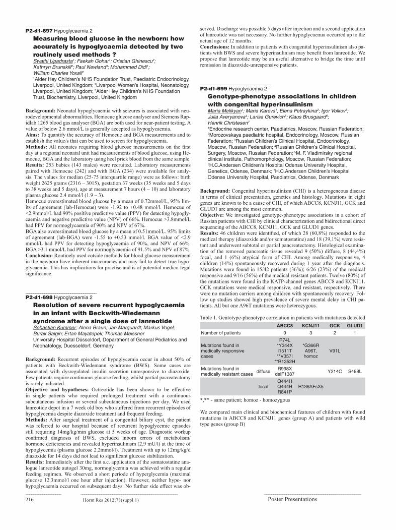
216_______________________________________________________________________________________________________________________
Horm Res 2012;78(suppl 1)
______________________________________P2-d1-697 Hypoglycaemia 2
Measuring blood glucose in the newborn: how accurately is hypoglycaemia detected by two routinely used methods ?Swathi Upadrasta1; Faekah Gohar1; Cristian Ghinescu2; Kathryn Brunskill2; Paul Newland3; Mohammed Didi1; William Charles Yoxall21Alder Hey Children’s NHS Foundation Trust, Paediatric Endocrinology, Liverpool, United Kingdom; 2Liverpool Women’s Hospital, Neonatology, Liverpool, United Kingdom; 3Alder Hey Children’s NHS Foundation Trust, Biochemistry, Liverpool, United Kingdom
Background: Neonatal hypoglycaemia with seizures is associated with neu-rodevelopmental abnormalities. Hemocue glucose analyser and Siemens Rap-idlab 1265 blood gas analyser (BGA) are both used for near-patient testing. A value of below 2.6 mmol/L is generally accepted as hypoglycaemia. Aims: To quantify the accuracy of Hemocue and BGA measurements and to establish the value/s that can be used to screen for hypoglycaemia. Methods: All neonates requiring blood glucose measurements on the first day at a regional neonatal unit had measurements of blood glucose, using He-mocue, BGA and the laboratory using heel prick blood from the same sample. Results: 253 babies (143 males) were recruited. Laboratory measurements paired with Hemocue (242) and with BGA (234) were available for analy-sis. The values for median (25-75 interquartile range) were as follows: birth weight 2625 grams (2316 – 3015), gestation 37 weeks (35 weeks and 5 days to 38 weeks and 5 days), age at measurement 7 hours (4 – 10) and laboratory plasma glucose 2.4 mmol/l (1.9 – 3). Hemocue overestimated blood glucose by a mean of 0.72mmol/L, 95% lim-its of agreement (lab-Hemocue) were -1.92 to +0.48 mmol/l. Hemocue of <2.9mmol/L had 90% positive predictive value (PPV) for detecting hypogly-caemia and negative predictive value (NPV) of 66%. Hemocue >3.8mmol/L had PPV for normoglycaemia of 90% and NPV of 67%. BGA also overestimated blood glucose by a mean of 0.51mmol/L. 95% limits of agreement (lab-BGA) were -1.55 to +0.53 mmol/l. BGA value of <2.9 mmol/L had PPV for detecting hypoglycaemia of 90%, and NPV of 66%. BGA >3.1 mmol/L had PPV for normoglycaemia of 91.5% and NPV of 87%. Conclusion: Routinely used cotside methods for blood glucose measurement in the newborn have inherent inaccuracies and may fail to detect true hypo-glycaemia. This has implications for practise and is of potential medico-legal significance.
______________________________________P2-d1-698 Hypoglycaemia 2
Resolution of severe recurrent hypoglycaemia in an infant with Beckwith-Wiedemann syndrome after a single dose of lanreotideSebastian Kummer; Alena Braun; Jan Marquardt; Markus Vogel; Burak Salgin; Ertan Mayatepek; Thomas MeissnerUniversity Hospital Düsseldorf, Department of General Pediatrics and Neonatology, Duesseldorf, Germany
Background: Recurrent episodes of hypoglycemia occur in about 50% of patients with Beckwith-Wiedemann syndrome (BWS). Some cases are associated with dysregulated insulin secretion unresponsive to diazoxide. Few patients require continuous glucose feeding, whilst partial pacreatectomy is rarely indicated. Objective and hypotheses: Octreotide has been shown to be effective in single patients who required prolonged treatment with a continuous subcutaneous infusion or several subcutaneous injections per day. We used lanreotide depot in a 7 week old boy who suffered from recurrent episodes of hypoglycemia despite diazoxide treatment and frequent feeding. Methods: After surgical treatment of a congenital biliary cyst, the patient was referred to our hospital because of recurrent hypoglycemic episodes still requiring 14mg/kg/min glucose at 5 weeks of age. Diagnostic workup confirmed diagnosis of BWS, excluded inborn errors of metabolism/hormone deficiencies and revealed hyperinsulinism (2,9 mU/l) at the time of hypoglycemia (plasma glucose 2.2mmol/l). Treatment with up to 12mg/kg/d diazoxide for 14 days did not lead to significant glucose stabilization. Results: Immediately after the first s.c. application of the somatostatine ana-logue lanreotide autogel 30mg, normoglycemia was achieved with a regular feeding regimen. We observed a short periode of hyperglycemia (maximal glucose 12.3mmol/l one hour after injection). However, neither hypo- nor hypoglycaemia occurred on subsequent days. No further side effect was ob-
served. Discharge was possible 5 days after injection and a second application of lanreotide was not necessary. No further hypoglycaemia occurred up to the actual age of 12 months. Conclusions: In addition to patients with congenital hyperinsulinism also pa-tients with BWS and severe hyperinsulinism may benefit from lanreotide. We propose that lanreotide may be an useful alternative to bridge the time until remission in diazoxide-unresponsive patients.
______________________________________P2-d1-699 Hypoglycaemia 2
Genotype-phenotype associations in children with congenital hyperinsulinismMaria Melikyan1; Maria Kareva1; Elena Petraykina2; Igor Volkov3; Julia Averyanova4; Larisa Gurevich5; Klaus Brusgaard6; Henrik Christesen7
1Endocrine research center, Paediatrics, Moscow, Russian Federation; 2Morozovskaya paediatric hospital, Endocrinology, Moscow, Russian Federation; 3Russian Children’s Clinical Hospital, Endocrinology, Moscow, Russian Federation; 4Russian Children’s Clinical Hospital, Surgery, Moscow, Russian Federation; 5M. F. Vladimirsky regional clinical institute, Pathomorphology, Moscow, Russian Federation; 6H.C.Andersen Children’s Hospital Odense University Hospital, Genetics, Odense, Denmark; 7H.C.Andersen Children’s Hospital Odense University Hospital, Paediatrics, Odense, Denmark
Background: Congenital hyperinsulinism (CHI) is a heterogeneous disease in terms of clinical presentation, genetics and histology. Mutations in eight genes are known to be a cause of CHI, of which ABCC8, KCNJ11, GCK and GLUD1 are among the most common. Objective: We investigated genotype-phenotype associations in a cohort of Russian patients with CHI by clinical characterization and bidirectional direct sequencing of the ABCC8, KCNJ11, GCK and GLUD1 genes. Results: 46 children were identified, of which 28 (60,8%) responded to the medical therapy (diazoxide and/or somatostatine) and 18 (39,1%) were resis-tant and underwent subtotal or partial pancreatectomy. Histological examina-tion of the removed pancreatic tissue revealed 9 (50%) diffuse, 8 (44,4%) focal, and 1 (6%) atypical form of CHI. Among medically responsive, 4 children (14%) spontaneously recovered during 1 year after the diagnosis. Mutations were found in 15/42 patients (36%); 6/26 (23%) of the medical responsive and 9/16 (56%) of the medical resistant patients. Twelve (80%) of the mutations were found in the KATP-channel genes ABCC8 and KCNJ11. GCK mutations were medical responsive, and resistant, respectively. There were no mutation carriers among children with spontaneously recovery. Fol-low up studies showed high prevalence of severe mental delay in CHI pa-tients. All but one A96T mutations were heterozygous.
Table 1. Gentotype-phenotype correlation in patients with mutations detectedABCC8 KCNJ11 GCK GLUD1
Number of patients 9 3 2 1
Mutations found in medically responsive cases
R74L *Y344X I1511T **V357I
**R1352H
*G366R A96T, homoz
V91L
Mutations found in medically resistant cases diffuse R998X
delF1387 Y214C S498L
focalQ444H Q444H R841P
R136AFsX5
*,** - same patient; homoz - homozygous
We compared main clinical and biochemical features of children with found mutations in ABCC8 and KCNJ11 genes (group A) and patients with wild type genes (group B)
Poster Presentations

21751st Annual Meeting of the ESPE __________________________________________________ _____________________________________________________________________
Horm Res 2012;78(suppl 1)
Table 2. Group A (n=11) Group B (n=28) p
Age at hypoglycemia manifestation (months) Median [25%-75%] 0.03 [0.03-0.06] 3.5 [0.5-6] 0.000098
Insulin during the hypoglycemia (U/l) Median [25%-75%] 28.63 [12.6-38.8] 14.82 [6.8-31.7] 0.244
Birth weight (g) mean (±SD) 3842.5 ± 811.8 3514 ± 440.8 0.21
Number of severe cases (n, %) 8 (72.7%) 7 (25.9%) 0.005
Number of medically resistant cases (n, %) 7 (63.4%) 7 (25.9%) 0.02
Number of cases with severe mental delay during follow up (n, %) 7/7 (100%) 5/20 (25%) 0.0006
Cases of spontaneous recovery (n) 0 4 0.18
Conclusion: In conclusion, a genetic cause was detected in 23%, and 56%, of children with mild, and severe CHI, respectively, in Russia. Mutations in ABCC8 and KCNJ11 were found to be the most common cause and associ-ated with severe course of the disease and poor neurologic outcome.
______________________________________P2-d1-700 Hypoglycaemia 2
Hypoglycaemia in childrenMaria Melikyan1; Maria Kareva1; Elena Petraykina2; Julia Averyanova3; Henrik Christesen4
1Endocrine Research Center, Paediatrics, Moscow, Russian Federation; 2Morozovskaya Paediatric Hospital, Endocrinology, Moscow, Russian Federation; 3Russian Children’s Clinical Hospital, Surgery, Moscow, Russian Federation; 4H.C.Andersen Children’s Hospital Odense University Hospital, Paediatrics, Odense, Denmark
Background: In hypoglycemia, verification of specific diagnosis is important for the treatment and future prognosis. Objective and hypotheses: We aimed to investigate different causes of per-sistent/recurrent hypoglycemia in children and evaluate clinical characteris-tics and neurologic features during follow up. Methods: We included 91 patients aged 1 day-16 years diagnosed the last 4 years. Standard evaluation included fasting test, insulin, C-peptide, cortisol, GH, urine ketone bodies during hypoglycemia (<2,2 mmol/l), IGF-I, ACTH, TSH, fT4, lipid profile, lactate, Masspectrometry of aminoacids and carnitins abdominal US/CT . Glucagon test, GH-stimulation test, OGTT, brain MRI were done when indicated. Severe course was classified as constant iv glucose demand. Results: Hyperinsulinemic hypoglycemia (insulin level > 2 mU/l during hy-poglycemia) were found in 56 patients, congenital hyperinsulinism; n=46, pancreatic insulin-producing tumors; n=10. Among 35 children with low in-sulin levels, 31 patients had ketotic hypoglycemia, 4 had low ketone bodies. Except in pancreatic insulinomas, all patients had hypoglycemia onset during the first 3 years of age.
Table 1
Diagnosis Number of cases (%)
Age at manifestation of hypoglycemia
(mean, (range))
Number (%) of severe
cases
Neurologic outcome
(mean follow up period 2
years)
Neurologic outcome
(mean follow up period 2
years)
Mental delay (n, (%))
Autonomic epilepsy (n,
(%))
CHI 46 (50.5%) 1 (0,03-30) months 29/46 (63%) 20/33 (61%) 8/33 (24%)
Insulinoma 10 (11%) 10 years (8-16) 4/10 (40%) 1/9 (11%) 1/9 (11%)
Hypopituitarism 9 (10%) 31 (9-35) months 2/9 (22,2%) none none
Glycogen storage disease 4 (4,5%) 1,25 (0,5-3)
months 4/4 (100%) 1/3 1/3
Ideopathic ketotic hypoglycemia
17 (18,5%) 18 (1-36) months none none none
Defects of fatty acids beta-oxidation
1 (1,1%) 0,5 months 1 1 none
POMC deficiency 1 (1,1%) 12 months none 1 none
Cases with unverified etiology
3 (3,3%) 24 (8-49) months none none none
Conclusions: CHI was the most common cause of persistent severe hypo-glycemia and had the worst neurologic outcome. In 3.3% of cases with non-ketotic hypoglycemia, specific diagnosis was not verified.
______________________________________P2-d1-701 Hypoglycaemia 2
Hyperinsulinemic hypoglycemia: experience with 18 casesSebahat Yilmaz Agladioglu; Senay Savas Erdeve; Semra Cetinkaya; Veysel Nijat Bas; Havva Nur Peltek Kendirci; Asan Onder; Zehra AycanDr. Sami Ulus Obstetrics and Gynecology, Pediatric Health and Disease Training and Research Hospital, Clinics of Pediatric Endocrinology, Ankara, Turkey
Background: Patients with hyperinsulinemic hypoglycemia (HIH) present neonatally, infancy or childhood period and show transient, prolonged and persistent characteristics.Objective and hypotheses: To analyzed the clinical and biochemical char-acteristics and treatment and neurodevelopmental outcomes in HIH patients.Methods: We evaluated 18 children with HIH during 14 year period.Results: Patients presented neonatally (11), during infancy (6) and childhood (1).The age at time of presentation ranged from the first day of life to 7 years. Consanguity was reported in 11 cases (61%). None of the mothers had ges-tational diabetes. Mean birth weight and gestational age were 3335 gr, 38 weeks, respectively. Hypoglycemic seizure was the most common present-ing sympyoms (13 patients). Poor feeding (4) and asymptomatic hypogly-cemia (1) were seen at diagnosis. Insulin and C-peptide levels at the time of hypoglycemia ranged from 3.9-88 mIU/ml, 1.3-12 ng/ml, respectively. The ammonia level was found to be elevated in two patients, one of whom also had high alanin aminotransferase level and was diagnosed Wilson disease to-gether with HIH. All patients received intravenous glucose infusions (9.2 mg/kg/min) and were started diazoxide at diagnosis. Diazoxide treatment was ceased in nine patients (transient HIH) and 7 of them had diagnosed neonatal period. Medical treatment was failed to maintain normoglycemia in two pa-tients. Diazoxide unresponsive one patient underwent near total pancreatecto-my and hypoglycemic episodes continued after surgery and this episodes was controlled by diazoxide, octreotide and hydroclorotiazide. The other patient refused surgery and continued medical treatment and severe motor mental re-tardation was developed at the follow-up. Visual evoked response (2 patients), cranial magnetic resonance imaging (3 patients), electroencephalogram (2 pa-tients) were abnormal in all group. Conclusions: The natural history of HIH is not well understood. The long term careful monitoring is needed to avoid complications.
______________________________________P2-d1-702 Hypoglycaemia 2
Hypoglycaemia as a complication of the metabolic disease aromatic L-amino acid decarboxylase deficiencyFrancois EyskensUniversity Hospital/Queen Paola Chidlren’s Hospital, Paediatrics, Antwerp, Belgium
Background: Aromatic L-amino acid decarboxylase (AADC) is an essential enzyme in the biosynthesis of the monoamine neurotransmitters serotonin and dopamine. AADC-deficiency is a rare autosomal recessive inborn error of metabolism characterized by severe developmental delay, prominent motor abnormalities, oculogyric crises and autonomic features. Prognosis is poor. Objective and hypotheses: We describe a five year old boy with AADC-de-ficiency with low neurotransmitters level in CSF (low HVA and 5-HIAA and elevated 3-O-Methyldopa) confrmed by deficient AADC plasma enzymatic activity (0.7 mU/L) and mutation analysis (compound heterozygote A91V/A275T) . He showed a severe neurologic clinical picture and no improvement was found under several dopamine agonists (DA-agonists), high dosis of vita-min B6 and an atropine agonist (see table 2). Severe, unpredictable, episodes of hypglycaemia were documented when he was switched from bromocryp-tine to pramipexol, a more potent dopamine agonist, in order to try to improve his motoric disabilities. Episodes of hypoglycaemia are documented in other patients with this metabolic disease although they were mostly misdiagnosed as having epilepsy. The pathogenesis of hypoglycaemia in these patients is unknown: they are thought to reflect the relative lack of catecholamines as anti-inulinergic hormones. I hypothezise that the potent dopamine agonists in

218_______________________________________________________________________________________________________________________
Horm Res 2012;78(suppl 1)
these patients can give rise to hypoglycaemia based on inhibition of growth hormone secretion through activation of dopanine D2 receptors and/or by the autonomic dysfunction in these patients with virtually no sympathetic activ-ity left. Methods: During episodes of hypoglycaemia, serum growth hormone, serum insuline and serum cortisol and urinary free cortisol and catecholamines were measured. Results: No overt hormonal abnormalities (growth hormone, insuline, cor-tisol) were found. The episodes of hypoglycaemia disappeared, and other severe side effects, e.g. arterial hypotension improved when the patient was switched back to bromocryptine.
______________________________________P2-d1-703 Perinatal and Neonatal Endocrinology 2
Correlation of early morning plasma cortisol and salivary cortisol in extremely premature infantsSze May Ng1; Mark Turner1; Josephine Drury1; Mohammed Didi2; Suresh Victor3; Paul Newland4; A Michael Weindling5
1University of Liverpool, Department of Women’s and Children’s Health, Institute of Translational Medicine, Liverpool, United Kingdom; 2Alder Hey Children’s Foundation Trust, Department of Endocrinology, Liverpool, United Kingdom; 3University of Manchester, Manchester Academic Health Sciences Centre, Manchester, United Kingdom; 4Alder Hey Children’s Foundation Trust, Department of Biochemistry, Liverpool, United Kingdom; 5University of Manchester, Department of Women’s and Children’s Health, Institute of Translational Medicine, Liverpool, United Kingdom
Background: Cortisol is important for survival during illness and cortisol concentrations are expected to increase during significant stress. Extreme pre-term infants may develop adrenal insufficiency in the early neonatal period. Cortisol is 90% bound to cortisol binding globulins (CBG) in the circulation, therefore measurements of plasma cortisol can be compromised by condi-tions that alter CBG levels. Measurement of free cortisol is the best indicator of adrenal glucocorticoid secretion and can be determined in the saliva. Few studies have been reported on salivary cortisol determination in neonates and particularly, premature infants. Objective and hypotheses: The aim of the study was to determine the cor-relation of early morning plasma cortisol and salivary cortisol in extremely premature infants. Methods: We prospectively obtained early morning plasma and salivary cor-tisol sampling at Day 1 of life, Day 14, Day 28 and at 36 weeks corrected ges-tational age (CGA). Saliva was obtained using 4 standard universal swabs by placing one swab at a time in the infant’s mouth for 1-2 minutes. No salivary stimulants were used. Salivary cortisol was measured by competitive ELISA using a commercially available kit SLV-2930 (DRG, Germany) according to the manufacturers instructions. Plasma cortisol was measured using DPC Im-mulite2000 using a solid phase 2 site chemiluminescent immunometric assay Results: There were 65 infants (36 males) included in the study. Mean ges-tation was 25.3±1.3 weeks. Mean plasma cortisol levels were 400nmol/L ± 42.8 SEM, and mean salivary cortisol levels were 127.5 nmol/L ± 66.5 SEM. Plasma cortisol was positively correlated with salivary cortisol levels (r=0.44, p=0.001). Conclusions: The study showed a good correlation between salivary and plasma cortisol concentrations obtained early in the morning in extremely premature infants.
______________________________________P2-d1-704 Perinatal and Neonatal Endocrinology 2
Ultrasound measurements of thyroid gland volume in extreme preterm infants at 36 weeks corrected gestational age in a UK populationSze May Ng; Mark Turner; A Michael WeindlingUniversity of Liverpool, Department of Women’s and Children’s Health, Institute of Translational Medicine, Liverpool, United Kingdom
Background: Neonatal thyroid ultrasonography has an important role in as-sessing thyroid anatomy in newborns with thyroid dysfunction. Hypothyroxi-naemia of prematurity is commonly reported in premature neonates. Objective and hypotheses: We aimed to determine the mean thyroid vol-ume in infants with extreme prematurity at 36 weeks corrected gestational
age (CGA) in a UK population compared to normative data reported in term infants in the literature. Methods: We prospectively examined babies born below 28 weeks’ gestation. The thyroid gland was measured using a linear array transducer by a single observer. Thyroid volume of each lobe was calculated by the formula depth x length x width x π/6. The means and standard deviations were calculated . Results: There were 55 babies (28 males) who had ultrasound assessment of the thyroid gland at 36 weeks’ CGA. Mean gestational age was 26.2 ± 1.0 weeks and mean birth weight was 893.8 ± 178.1 grams. Mean thyroid volume was 0.57mls ± 0.17. Conclusions: In this study, premature infants born below 28 weeks’ gesta-tion had their thyroid volume measurement assessed at 36 weeks’ CGA with a mean volume of 0.57 mls compared to a Scottish data of term infants re-porting a mean thyroid volume of 1.6mls. In a Turkish population, the mean thyroid volume in preterm infants of gestational age 25-28 weeks was 0.4 mls and in term infants, their mean volume was 0.72 ml. Mean thyroid volumes in term infants from a Belgium population was 0.84 ml and from a German population was 1.1ml. Thyroid volume can vary from country to country due to iodine intake and may be dependent of gestational age at birth.
______________________________________P2-d1-705 Perinatal and Neonatal Endocrinology 2
Infant girls have a postnatal activation of ovarian steroidogenesis associated with biological estrogen effectsTanja Kuiri-Hänninen1; Ulla Sankilampi1; Ursula Turpeinen2; Mikko Haanpää2; Esa Hämäläinen2; Leo Dunkel31Kuopio University Hospital and University of Eastern Finland, Department of Pediatrics, Kuopio, Finland; 2Helsinki University Central Hospital, Department of Clinical Chemistry, Helsinki, Finland; 3Queen Mary University of London, William Harvey Research Institute, London, United Kingdom
Background: An increase in testosterone levels and testicular and penile growth after the transient postnatal gonadotropin surge is well described in boys. In girls, analogous activation of ovarian steroidogenesis has not been substantiated. The timing and magnitude of the postnatal pituitary-gonadal activation differ between full term and premature infant boys. Objective and hypotheses: To determine longitudinal changes in E2 levels during the first six months of life and biological estrogen effects by measuring simultaneous changes in E2 sensitive tissue, the mammary gland. Methods: Urinary E2 levels were measured monthly from one week (D7) to six months of age (M1-M6) by LC-MS/MS and compared to manually measured mammary gland diameter (MGD) in 125 infants in three gestational age (GA) cohorts: full term (FT, GA>37 weeks, n=58, 29 boys), moderately premature (MPT, GA 32-36+6 weeks, n=42, boys 19) and very premature (VPT, GA<32 weeks, n= 25, 14 boys).
Urinary E2 levels in full term infants during the first six months of life (presented as median and quartiles, values below the detection limit of the assay are set to zero).
Results: In girls, median E2 levels were lowest at D7, increased significantly by M2 in FT, by M1 in MPT and by M4 in VPT girls and then remained ele-vated until M6. Similar increase was not observed in boys and E2 levels were significantly higher in girls from M2 in FT, from M1 in MPT and from M3 in VPT infants (P<0.01-0.001). MGD was largest in FT infants at D7 (median 1.1 cm (range 0-1.9) in girls and 1.0 cm, (0-1.5) in boys) probably reflecting the effect of high estrogen levels during the last trimester of pregnancy. In premature cohorts, MGD was small (<0.5 cm) in both sexes until M2, when
Poster Presentations
TimepointD7 M1 M2 M3 M4 M5 M6
150
100
50
0
Girls
Boys
Uri
nar
y E
2 (p
mo
l/mm
ol C
r)

21951st Annual Meeting of the ESPE __________________________________________________ _____________________________________________________________________
Horm Res 2012;78(suppl 1)
it started to increase in girls parallel to rising E2 levels. By M6, MGD had increased to 1.2 cm (0.6-1.8) in MPT and to 0.9 cm (0.7-1.6) in VPT girls and was larger than in FT girls (0.7 cm, 0-1.4, P<0.01). MGD was significantly larger in girls than in boys from M4 onwards in FT and from M2 onwards in MPT and VPT cohorts. Conclusions: E2 levels were higher in girls than in boys and temporally as-sociated to larger MGD suggesting that ovarian steroidogenesis is active and has biological effects during the first months of life.
______________________________________P2-d1-706 Perinatal and Neonatal Endocrinology 2
Placental expression of peroxisome proliferator-activated receptor γ (PPARγ): relation to placental and fetal growthMarta Díaz1; Judit Bassols2; Maria Dolores Gómez-Roig3; Abel López- Bermejo2; Francis de Zegher4; Lourdes Ibáñez5
1Sant Joan de Déu Hospital, University of Barcelona, Endocrinology, Esplugues, Barcelona, Spain; 2Josep Trueta Hospital, Pediatrics, Girona, Spain; 3Sant Joan de Déu Hospital, University of Barcelona, Obstetrics and Gynecology, Esplugues, Barcelona, Spain; 4University of Leuven, Department of Woman and Child, Leuven, Belgium; 5Sant Joan de Deú Hospital, University of Barcelona, Endocrinology, Esplugues, Barcelona, Spain
Background: The nuclear receptor peroxisome proliferator activated recep-tor γ (PPARγ) contributes to placental development and thus to the materno-fetal transfer of oxygen and nutrients that allow for prenatal growth. Objective and hypotheses: To stablish whether the placental PPARγ expres-sion relates to placental and fetal growth. Population and methods: Placentas (N=116) were collected at term delivery of singleton infants, who were born either small-, appropriate- or large-for-gestational-age [32 SGA, 55 AGA or 29 LGA]. Placentas and newborns were weighed at birth. Real-time PCR was used to assess placental expression of PPARγ as compared to the housekeeping gene GAPDH. Results: PPARγ expression in placentas from AGA and LGA infants were nearly 2-fold higher than in placentas from SGA infants (P=0.01). Placental PPARγ expression associated positively to placental and/or fetal weight at birth, particularly within the SGA subpopulation (P=0.001). Conclusion: PPARγ expression was found to be low in placentas of SGA fetuses and to associate positively to feto-placental growth. It remains to be studied whether feto-placental growth, if reduced, can be safely augmented by upregulating placental PPARγ expression.
______________________________________P2-d1-707 Perinatal and Neonatal Endocrinology 2
Nonalcoholic fatty liver disease and visceral fat accumulation in small for gestational age childrenMaria Felicia Faienza1; Angelo Acquafredda1; Mariangela D’Aniello1; Mariantonietta Monteduro2; Anna Lucia Stefania Di Giovinazzo1; Luciano Cavallo1
1University of Bari, Interdisciplinary Department of Medicine, Bari, Italy; 2University of Bari, Diagnostic imaging, Bari, Italy
Background: Low birth weight followed by rapid postnatal weight gain is associated with long-term risks for central obesity and insulin resistance (IR). A close association between obesity, IR and nonalcoholic fatty liver disease (NAFLD) has been demonstrated. Objective and hypotheses: Aim of this study was to evaluate the effect of low birth weight and rapid catch up growth on NAFDL and visceral fat ac-cumulation, assessed by ultrasound measurements. Methods: We enrolled 45 children, 23 SGA (14 F, mean age 7.35 ± 1.97 years), who presented a rapid catch-up growth within the first 6-12 months, and 22 appropriate for gestational age (AGA) (7 F, mean age 8.85 ± 3.36 years). The definition of ultrasonic NAFDL was based on previously reported diagnostic criteria and classified as grade 1 mild, 2 moderate, 3 severe. Vis-ceral fat was evaluated as the distance from the internal surface of the rectus abdominis muscle to the anterior wall of the aorta. Bioimpedance analysis (BIA) was also performed using a bioelectrical impedance analyzer (model BIA-101/S, RJL system, Akern), for the evaluation of body composition. Results: Moderate NAFDL was present in 4/23 (17.39 %) SGA and absent in AGA children; an association between NAFDL and IR (HOMA >2.5) was
found in 3/4 SGA having ultrasonic signs of liver steatosis. No significant correlation was found between NAFDL and visceral fat accumulation. A posi-tive correlation between visceral fat accumulation and waist circumference (P <0.0001) was found in SGA children. The percentage of body fat was higher in SGA compared to AGA children. Conclusions: Our results demonstrated that a rapid catch-up growth plays an important role in determining the NAFDL and the excess of fat mass in children born SGA. Moreover, the NAFDL in SGA children seems to be as-sociated with IR and not with the visceral fat accumulation.
______________________________________P2-d1-708 Perinatal and Neonatal Endocrinology 2
A novel homozygous TMEM70 mutation results in congenital cataract and neonatal mitochondrial encephalo-cardiomyopathyZeynep Atay1; Abdullah Bereket1; Serap Turan1; Belma Haliloglu1; Asli Memisoglu2; Stavit A Shalev3; Morad Khayat3; Ronen Spiegel41Marmara University, School of Medicine, Pediatric Endocrinology, Istanbul, Turkey; 2Marmara University, School of Medicine, Neonatology, Istanbul, Turkey; 3Ha’Emek Medical Center, Rappaport School of Medicine, Genetic Institute, Haifa, Israel; 4Ha’Emek Medical Center, Rappaport School of Medicine, Pediatric Deparment and Genetic Institute, Haifa, Israel
Background: Mutations in the TMEM70 gene are the most common cause of nuclear encoded ATP synthase deficiency resulting in a syndrome character-ized by neonatal lactic acidosis, cardiomyopathy, and encephalomyopathy. Objective: To present the first Turkish patient with TMEM70 mutation who presented with hypercalcemia and bilateral congenital cataract in addition to the typical previously reported features. Patient and results: The patient is the second child of parents who are first degree cousins, born at term with a birth weight of 2230gr. He had hypertro-phic cardiomyopathy, bilateral cataract, inguinal hernia, cryptoorchidism and hypospadias.He also had clear facial dysmorphism including retromicrogna-thia, high arched palate, long philtrum, flat and thin upper lip, arched eye-brows, prominent nasal bridge and upturned nares, large posteriorly rotated low set ears, flat occiput and upper gingival hypertrophy. The serum Ca levels 15 mg/dl with supressed PTH and resolved with IV fluid administration and prednisolone in 3 days. Metabolic screening revealed elevated serum lactate (6.9 mmol/L, normal range 0.9-2.2 mmol/L), and elevated serum ammonia (163,4 µmol/l normal range <86 µmol/l ). Serum amino acid analysis revealed significantly elevated alanine and urinary organic acid analysis showed in-creased excretion of fumaric acid and methylglutaric acid. TMEM70 genetic analysis revealed the causative homozygous c.535C>T novel mutation which is predicted to result in substitution of a highly evolutionary conserved tyro-sine residue into histidine (p.Y179H). Both parents were heterozygous for the mutation. Conclusion: This is the first Turkish case of TMEM70 gene dysfunction caused by a novel c.535C>T mutation. In addition to typical features of the disease he had bilateral congenital cataract and hypercalcemia which further extends the clinical spectrum of TMEM70 gene defects. When suspected, TMEM70 genetic analysis should be performed before the traditional assess-ment of respiratory chain complex activities in muscle.
______________________________________P2-d1-709 Perinatal and Neonatal Endocrinology 2
Development of postnatal head circumference and its impact on coordinative skills in very premature infants at 9.5 years of ageSonja Stutte1; Bettina Gohlke1; Joachim Wölfle1; Peter Bartmann2; Annika Peiler1
1Universitäts-Kinderklinik Bonn, Pädiatrische Endokrinologie, Bonn, Germany; 2Universitäts-Kinderklinik Bonn, Neonatologie, Bonn, Germany
Background: It’s a well-known fact that the development of head circumfer-ence (HC) can be considered as a measure of brain growth in children. Brain volume however is decisive for cognitive and motor functions. Can these in-sights be applied to extreme preterm infants as well? Objective and hypotheses: We analyzed the dependence of coordination skills on postnatal growth of head circumference in extremely low birth weight (ELBW) infants at a mean age of 9.5 years.

220_______________________________________________________________________________________________________________________
Horm Res 2012;78(suppl 1)
Methods: A total of 39 children (17 male; mean age: 9.5 years; range: 7.9-11.9 years) of normal development were recruited. All were born with a birth weight < 1000g (BW – SDS: - 0.75 +/- 0.2; birth length (BL) – SDS: 0.39 +/- 0.3). All but eight (six male) were prepubertal. Clinical data were gathered retrospectively from patient files. Head circumference was measured at birth, at discharge and during regular annually visits. At the last visit at a mean age of 9.5 years, peak jump power (PJP) was quantified using Leonardo jumping platform. Results: 1) Main factors influencing postnatal growth of head circumference: HC was significantly associated with beginning of full enteral feeding (r=-0.38; p=0.045); duration of respiratory support (r=-0.58; p=0.001), and dura-tion of catecholamine dependency (r=-0.39; p=0.032). 2) Head circumference from point of discharge onwards correlates highly significantly with PJP later on.
Pearson HC - SDS birth
HC - SDS discharge
HC - SDS 0.5 years
HC - SDS 2 years
HC - SDS 4 years
HC - SDS 9.5 years
PJP - SDS for age NS r = 0.49 p =
0.005r = 0.49 p=
0.003r= 0.37 P =
0.038r = 0.38 p =
0.031r = 0.38 p =
0.019
Conclusions: Postnatal and not fetal growth of head circumference was as-sociated with better coordinative skills at the age of 9.5 years in formerly ELBW infants. Early postnatal growth of HC was dependent on clinical con-dition of the infants. Beside duration of respiratory support and duration of catecholamine dependency it was the start of full enteral feeding, which was associated with better growth of HC.
______________________________________P2-d1-710 Perinatal and Neonatal Endocrinology 2
Neonatal salt loss: not only 21-hydroxilase deficiency. Report of six cases with aldosterone deficiency or resistance Diego Rinaldini1; Felix Riepe2; Florance Roucher3; Maria Dracopoulou4; Angelica Marsigli1; Piero Pirazzoli1; Antonio Balsamo1
1Azienda policlinico S.Orsola-Malpighi; University of Bologna, Gynecology, Obstetrics & Pediatrics Science, Bologna, Italy; 2University of Kiel, Pediatrics, Kiel, Germany; 3University of Lyon, Laboratoire d’Endocrinologie Moléculaire et Maladies rares, Lyon, France; 4Athens University Medical School, First Department of Pediatrics, Athens, Greece
Background: Aldosterone Sinthase Deficiency (ASD) and Pseudohypoaldo-steronism Type 1 (PAH1) are rare causes of salt wasting in infancy. Objective and hypotheses: characterization of 6 cases with salt loss not re-lated to 21OHD Methods: 6 patients analysed by means of CYP11B2, SCNN1A, B, C and NR3C2 genes. Results: Patient 1 and 2 were girls presenting at 28 and 16 days of life, re-spectively, with vomiting, failure to thrive and salt loss. Normal female geni-talia and cortisol production, low aldosterone levels and high PRA induced to suspect ASD. Fludrocortisone and NaCl administration restored the elec-trolyte equilibrium. The CYP11B2 gene analysis confirmed a novel homozy-gous mutation (c.395+1G>T) in the first patient, and the mutations p.R174-R175del, and novel mutation (not yet described)+ p.V386A in the second (performed in Greece). Patient 3 was a boy who presented at 19 days of life with poor feeding and salt loss. In this case Aldosterone and renin levels were judged high. Although PAH1 was suspected at the beginning, treatment with FC and NaCl administration ameliorated the clinical outcome. Molecular analysis of CYP11B2 gene identified two new mutations (p.R141X/p.E198G) in compound heterozygosis. Patient 4 and 5 were boys presenting at 56 and 21 days of life, respectively, with vomiting, failure to thrive, hyperkalemia, hyponatremia and elevate aldosterone and PRA levels suggesting PAH1. With NaCl supplementation they rapidly reached electrolyte balance and weight gain. NR3C2 gene analysis revealed two novel mutations Y134X and L722X respectively. Patient 6 was a girl showing a clinical and laboratory pattern like that of the patient 4 and 5. The severer electrolyte disequilibrium and the higher Aldosterone levels than the previous two patients induced to suspect a systemic PAH1. Therapy with Parenteral saline was successful. Molecular analysis of the SCNN1A, B, C and NR3C2 genes didn’t identify mutations Conclusions: After 21-OHD, ASD and PAH 1 have to be taken into consider-ation as a possible alternative causes of neonatal endocrine salt loss.
______________________________________P2-d1-711 Perinatal and Neonatal Endocrinology 2
Normal values of anti mullerian hormone in boys during the first 4 years of life: evaluation of a new immunoassay (AMH GenII)Ingrid Plotton1; Claire-Lise Gay2; Jean Marc Labaune3; Yves Morel11Hospices Civils de Lyon, Laboratoire d’hormonologie et Enndocrinologie moléculaire, Bron, France; 2Hospices Civils de Lyon, Endocrinologie Pediadry, Bron, France; 3Hospices Civils de Lyon, Neonatology, Lyon, France
Background: AMH is a good marker of Sertoli cell function and is very useful for evaluation patient 46,XY DSD. In 2009, we have reported the normal val-ues of AMH using immunoassay AMH/MIS Elisa® (Immunotech-Coulter) in a cohort of 110 male neonates without abnormalities of external genitalia and detectable diseases. AMH values were determined in each neonate three times around 15thday, 3th and 9thmonths of life and were expressed in pmol/L as following: mean±1SD (value range): day13-20, 882±335 (286-2116); 2,8-5,1 months, 1816±577 (704-3250); 8,5-9,8 months, 1736±616 (639-4364). Objective and hypotheses: The aim of this study was to continue this longi-tudinal study with an evaluation at 4 years of life and to compare our results with a new immunoassay AMH Gen II® Beckman Coulter. Methods: The pre-analytical conditions regarding the collection of blood sample (serum, EDTA plasma), the storage (+4 °C or room temperature ) and the time of the freezing after the collection (24h, 48h ,72h, 96h) have been evaluated using the two AMH kits. AMH values were determined in our co-hort by the new AMH Gen II® Beckman Coulter kit. Results: Whatever the conditions of blood collect, AMH value was the same with AMH/MIS Elisa® . The AMH values with the both AMH kits were iden-tical if the blood was collected on EDTA, but decrease from about 40% with the AMH Gen II in the other conditions of collection. The measure plasma EDTA in the cohort of 110 obtained with AMH Gen II show the result in pmol/L [mean+/-SD (min-max) :13-20days: 1256+/-714.84 (413-2493); 2.8-5.1months : 2443.11 (811-5320), 8.5-9.8:months 2483.60+/-152.49 (916-6676) ; 3.9-4.1 years: 1459.93+/-613.93 (191-2826). Conclusions: We must be careful at the preanalytic step for the measure of AMH with the kit AMH Gen II® . The normal values obtained with the immunoassay AMH Gen II® show normal value similar to the immunoas-say AMH/MIS Elisa® (Immunotech-Coulter) provided that collect of blood sample with EDTA .
______________________________________P2-d1-712 Perinatal and Neonatal Endocrinology 2
IGF-I levels are related to growth of preterm infants between term age and six months post-termMonique van de Lagemaat; Joost Rotteveel; Harrie Lafeber; Mirjam van WeissenbruchVU University Medical Center, Pediatrics, Amsterdam, Netherlands
Background: IGF-I regulates fetal growth during the third trimester as well as postnatal growth in term infants and its production is regulated by insulin and protein intake. Objective and hypotheses: To study IGF-I in relation to growth and protein intake between term age and six months corrected age (CA). We hypothesized that IGF-I levels are related to protein intake and growth between term age and six months CA in preterm infants. Methods: In 139 preterm infants (51% males, gestational age 30.3 ± 1.5 weeks, birth weight 1341 ± 288 gram) IGF-I levels were measured at term and at three and six months CA. Protein intake (g/kg/d) between term age and six months CA was calculated. At six months CA, infants were categorized as non-growth-restricted (weight and length ≥-2 SDS) or growth-restricted (weight and/or length <-2 SDS). Results: Between term age and six months CA, change in IGF-I levels was associated with change in weight, length, and head circumference (weight: β=0.17, 95%CI 0.12-0.22; length: β=0.51, 95%CI 0.33-69; and head cir-cumference: β=0.23, 95%CI 0.15-0.31; all p<0.001; Table 1). IGF-I levels were associated with insulin levels at term and at three months CA (term: β=0.13, 95%CI 0.07-0.18, p<0.001; three months: β=0.20, 95%CI 0.09-0.13, p=0.001), but not at six months CA. Protein intake was not associated with IGF-I levels.
Poster Presentations

22151st Annual Meeting of the ESPE __________________________________________________ _____________________________________________________________________
Horm Res 2012;78(suppl 1)
Term six months CA
N mean ± sd N mean ± sd
Weight (g) 139 3154 ± 502 139 7388 ± 1049
Length (cm) 139 48.5 ± 2.3 139 66.6 ± 2.8
Head circumference (cm) 139 35.8 ± 1.4 139 43.9 ± 1.5
IGF-I (nmol/l) 130 6.4 ± 1.9 127 8.3 ± 3.4
Conclusions: In preterms, IGF-I plays an important role in growth regula-tion, independent of protein intake. The lack of association between IGF-I and insulin after three months CA suggests that growth regulation switches from nutrition/IGF-I dependent towards GH/IGF-I dependent growth.
______________________________________P2-d1-713 Perinatal and Neonatal Endocrinology 2
Small for gestational age (SGA) preterm infants already have lower bone mineralization during early infancyMonique van de Lagemaat; Joost Rotteveel; Mirjam van Weissenbruch; Harrie LafeberVU University Medical Center, Pediatrics, Amsterdam, Netherlands
Background: Adult bone mass in preterm subjects is deteriorated by low birth weight and decreased bone accretion. Whether low birth weight is al-ready associated with decreased bone mass during early infancy is unknown. Objective and hypotheses: To study the effect of birth weight on bone ac-cretion between term and six months corrected age (CA) in preterm infants. We hypothesized that low birth weight was related to decreased bone mass during early infancy. Methods: In 139 preterm infants (51% male, gestational age (GA) 30.3 ± 1.5 weeks, birth weight 1341 ± 288 gram) whole body bone mineral con-tent (BMC), measured by dual-energy x-ray absorptiometry (Hologic QDR4500A), was determined at term and six months CA. At birth, infants were SGA (n=33, weight and/or length <-2 SDS) or appropriate for gesta-tional age (AGA, n=98, weight and length ≥-2 SDS). Results: At term and six months CA, BMC adjusted for gender and GA was significantly lower in SGA than AGA infants (Table 1). At six months CA, BMC remained lower in SGA infants after adjustment for actual weight and length. Gain in BMC adjusted for gender and GA was lower in SGA than AGA infants and remained lower after adjustment for actual weight and length (Table 1).
AGA SGAadjusted for gender and
GA
adjusted for gender, GA,
actual weight and length
mean ± sd
mean ± sd β 95%CI p β 95%CI p
BMC (g) term age
48.6 ± 10.1
38.1 ± 9.5 -0.26 -0.37;-
0.16 <0.001 n.s.
BMC (g) 6m
145.4 ± 22.9
130.1 ± 25.7 -0.16 -0.25;-
0.08 <0.001 -0.06 -0.12;-0.01 0.01
ΔBMC (g) term-6m
98.2 ± 20.7
91.7 ± 22.8 -0.12 -0.24;-
0.003 0.04 -0.11 -0.18;-0.03 0.01
Table 1. Bone mineral content in AGA and SGA infants between term and six months CA. Multivariate regression analyses with natural log transformed parameters and covariates for between-group differences; n.s.: not significant Conclusions: The effect of low birth weight on bone mass is already pres-ent during early infancy. Tracking from infancy to adulthood may explain decreased adult bone mass in SGA preterm subjects.
______________________________________P2-d1-714 Perinatal and Neonatal Endocrinology 2
Penile and clitoral sizes of normal full term newborns in KoreaSo-Eun Park1; Sung-Yeon Ahn2
1College of Medicine, CHA University, Pediatrics, Seoul, Republic of Korea; 2Kangwon National University Hospital, Pediatrics, Chuncheon, Republic of Korea
Background: Abnormalities of genital sizes could be early signs of underly-ing endocrine diseases and previous reports show ethnic differences in normal length. Objective and hypotheses: We studied to establish reference range of genital size in normal full term in Korea and compare that with other ethnicities. We present the largest study of penile and clitoral length measurements from Korea. Methods: Normal full term males (n=440) and females (n=390) with appro-priate birth weight for gestational age were included. Newborns with am-biguous genitalia, hypospadia, marked chordee, major congenital anomalies, or clinical evidence of endocrine disease were excluded. Penile length was measured with a rigid ruler as the distance from the pubic bone to the tip of glans or stretched flaccid length. Clitoral length and width were measured with a tumorimeter in after the labia major were spread in frog-leg position. Measurements were taken 3 times by a single examiner during the first 3 days of life. The study was carried out in Gangnam medical center, CHA Univer-sity over October 2011 to December 2011. Results: The mean penile length was 2.7cm(standard deviation(SD) 0.3). The mean clitoral width and length were 6.1mm(SD 1.4) and 3.4mm(SD 0.9) re-spectively. All the penile length, clitoral width and length were sinificantly shorter than many previous reports in other ethnicities. (p< .0001) Conclusions: This study establishes range for genital size of normal full term newborns in Korea which would help clinicians to diagnose early and treat an underlying diseases promptly.
______________________________________P2-d1-715 Perinatal and Neonatal Endocrinology 2
Maternal dietary intervention prevents postnatal weight gain and impaired glucose tolerance in the offspring of obese miceRuth Kuschewski; Inga Bae-Gartz; Jörg Dötsch; Eva RotherUniversity Hospital of Cologne, Department of Pediatrics, Cologne, Germany
Background: The global prevalence of pediatric overweight and obesity has increased substantially over the past decades, and the majority of obese chil-dren will remain obese through adulthood. One major risk factor for child-hood overweight is maternal obesity. Objective and hypotheses: However, the molecular mechanisms responsible for this phenomenon are so far ill-defined. More importantly, effective strate-gies for preventing this maternally transduced predisposition for childhood obesity are missing. This study aimed to investigate, whether maternal dietary intervention during pregnancy and lactation in obese dams is able to counter-act the predisposition for obesity in their offspring. Methods: Female C57BL/6N mice were rendered obese and glucose intol-erant by high fat diet feeding from 3 to 14 weeks of age. After mating, the intervention group was changed to a standard chow, rich in carbohydrates and low in fat, for the duration of pregnancy and lactation, while the control group stayed on HFD. Results: Strikingly, offspring of the intervention group showed a higher birth weight, but exhibited massively decreased postnatal weight gain, resulting in a 22.5% reduced body weight at postnatal day (P) 21 (p<0.001). Additionally, epigonadal fat pad weight was even reduced to 19.6% (p<0.001). Moreover, offspring of the intervention group remained significantly lighter than off-spring of controls until adulthood. Furthermore, blood glucose levels at P21 were significantly lower in offspring of the intervention group (p<0.001) and glucose tolerance tests at P56 revealed ameliorated glucose tolerance as a consequence of maternal dietary intervention during pregnancy and lactation. Conclusions: Thus, we conclude that improving maternal perinatal nutrition has impact on postnatal body weight gain and glucose tolerance in the off-spring. Further experiments will shed light on the underlying mechanisms mediating this effect and will thereby contribute to the development of effec-tive strategies for the prevention of obesity and its associated co-morbidities.

222_______________________________________________________________________________________________________________________
Horm Res 2012;78(suppl 1)
______________________________________P2-d1-716 Perinatal and Neonatal Endocrinology 2
Maternal and cord blood levels of leptin, adiponectin, resistin, ghrelin, IGF-1, and insulin: relationship with fetal growthAysenur Ökten1; Emel Gül Acikgöz1; Tugba Esen2; Gülay Karagüzel11Karadeniz Technical University, School of Medicine, Pediatric Endocrinology, Trabzon, Turkey; 2Karadeniz Technical University, School of Medicine, Biochemistry, Trabzon, Turkey
Background: Maternal bioactive substances, such as hormones and neuro-peptides, are thought to be important for fetal development. However, all of the factors that effect fetal growth and birth weight still unknown. Objectives: We investigated the maternal and cord blood levels of ghrelin, some adipokines and growth factors and their effects on fetal growth.Population and methods: Maternal serum and cord blood levels of ghrelin, leptin, adiponectin, resistin, insulin, IGF-1, and insulin were measured 100 healthy mothers and their offspring at delivery. Maternal body mass index (BMI) and weight were obtained before pregnancy, at first and 2nd trimesters, and at delivery. Neonatal anthropometric characteristics (birth weight [BW] and lenght [BL], ponderal index, head and abdominal circumferences) and placental weight were recorded. Results: There was a significant positive correlation between all the neona-tal antropometrics and mother’s weight that measured before and during the pregnancy (p<0,05). Plasental weights were correlated positively with BW and BL, head and abdominal circumferences (p<0.005). The maternal serum levels of leptin, IGF-1, glucose and insulin were higher than those cord blood (p<0,001), whereas maternal adiponectin levels were lower (p<0,01). Mater-nal and cord blood levels of resistin and ghrelin were not found different (p>0,05). There was a positive correlation between BW and maternal and cord blood levels of leptin and IGF-1 (p<0,005). The same relation was found for BL (p<0,005). Conclusions: Our data showed that fetal growth was related to maternal weight not only during pregnancy but also before pregnancy. We found that leptin was associated with fetal growth, however other adipokines and ghrelin were not related to fetal growth.
______________________________________P2-d1-717 Pituitary 2
Utility of the timed-12h urine collections during gonadotropin releasing hormone analogue stimulation testing in monitoring therapy of central precocious puberty in girlsZhuangjian Xu; Yaping Ma; Qing Wang; Shuyan Xie; Tingting ZhangTheFourthAffiliatedHospitalofSoochowUniversity,Pediatrics,Wuxi,China
Background: GnRH stimulation testing is frequently uncomfortable for chil-dren. Moreover, GnRH is commercially limited. Objective: To determine whether timed-12h urinary gonadotropin (UGn) during GnRH analogue (GnRHa) stimulation testing can be used for monitor-ing GnRHa therapy. Methods: In 19 girls with central precocious puberty (CPP) who were treated with diphereline (3.75 mg, i.m., q4w), GnRHa stimulation testing were per-formed before the initiation of treatment as well as 3 months after the begin-ning of the therapy. Following injecting triptorelin (08:30 am, Decapeptyl, 0.1 mg, s.c.), consecutive double timed-12h urine samples were respectively collected. Gonadotropin were assayed by immunochemiluminometric assays (ICMA). Results: One in 19 cases had poor clinical efficacy. In the other 18 cases who had good efficacy, the correlation coefficient between the serum peak LH (PLH) and the content (concentration × volume) of the first urinary LH (FULH) was 0.754, and for PLH and the second urinary LH (SULH) con-tent 0.697; for serum peak FSH (PFSH) and the first urinary FSH (FUFSH) content 0.784, and for PFSH and the second urinary FSH (SUFSH) content 0.831. And in these 18 girls, the means of PLH, PFSH and UGn content be-fore GnRHa therapy were all more than ones after the therapy (P < 0.001). Before therapy, the range of PLH was 5.20 — 62.31 IU/l, PFSH 9.24 — 35.18 IU/l; FULH content 0.543 — 9.737 IU, FUFSH content 9.435 — 56.925 IU; SULH content 0.185 — 15.131 IU, SUFSH content 8.779 — 57.722 IU. After therapy, the range of PLH was 0.44 — 1.67 IU/l, PFSH 0.72 — 5.70 IU/l; FULH content 0.043 — 0.821 IU, FUFSH content 0.673 — 6.725 IU; SULH content 0.017 — 0.540 IU, SUFSH content 0.417 — 3.215 IU.
Conclusions: It is concluded that double timed-12h urinary gonadotropin content assayed by ICMA during GnRHa stimulation testing provides an ef-fective, noninvasive methods for monitoring therapy in girls with CPP.
______________________________________P2-d1-718 Pituitary 2
Different reasons for hyponatraemia in a child with panhypopituitarismSharon LimBroomfieldHospital,PaediatricDepartment,Chelsmford,UnitedKingdom
Background: Growth hormone (GH) influences salt and water handling in the renal tubules. Tubular response to DDAVP is influenced by cortisol. Hypotha-lamic osmoreceptors may have reduced sensitivity from chronic Valproic acid administration, resulting in antidiuresis. In a child with panhypopituitarism, epilepsy and recurrent chest infections, medical management is a challenge. Methods: A 10-year-old girl with panhypopituitarism and cerebral palsy sec-ondary to Group B streptococcal meningitis as a neonate had full hormonal replacement therapy since infancy. Sodium levels were always stable in the high normal range. She became epileptic aged 7 and was started on Sodium Valproate. Reccurent chest infections occured age 8 and she became colo-nised with Pseudomonas and required occasional home oxygen. In the same year, she stopped feeding orally and became totally gastrostomy fed on a fixed volume of 1000ml a day. Hydrocortisone was changed to prednisolone at aged 7 to facilitate the effects of DDAVP through the day. GH was stopped aged 9 when it appeared that she was not growing very well. She had been quite ill that year with her chest. Her weight escalated despite a calorie reduction in feeds. Prior to stopping GH, her plasma sodium was 138 mmol/L. This fell to 129 mmol/L despite sodium supplements of 3mmol/kg/day. Urine output was < 3 ml/kg/hr despite dose reduction of DDAVP. Urinary sodium peaked at 133 mmol/L. She was admitted for input output assessment and blood profile whilst hydrocortisone was re-introduced 6 hourly and DDAVP and sodium supplements stopped. Results: Hyponatraemia and natriuresis persisted till GH was reintroduced at 0.026mg/kg/day. Urinary sodium fell to <10 mmol/L in the first week of treatment. Urine output was < 3ml/kg/hr on no DDAVP. Conclusions: There are a number of reasons for this complex child to have hy-ponatraemia, the most influential one being GH deficiency. This, coupled with the antidiuretic effect of valproic acid must not be underestimated. 6-hourly dosing of hydorcortisone ensures residual DDAVP works effectively.
______________________________________P2-d1-719 Pituitary 2
Genetic screening in an Argentinean population with congenital combined pituitary hormone deficiencyDébora Braslavsky1; Alexandru Savenau2; Ana Keselman1; Anne Barlier2; Rachel Reynaud3; Melanie Philippon4; Frederic Castinetti4; Thierry Brue4; Ignacio Bergadá1
1Hospital de Niños Ricardo Gutiérrez, Centro de Investigaciones Endocrinológicas (CEDIE), División de Endocrinología, Buenos Aires, Argentina; 2Aix-Marseille Université, Centre National Recherche Scientifique,FacultéMédecine,APHM,HôpitalTimone,HôpitalConception, CRN2M, Unité Mixte de Recherche 7286, Department of Endocrinology, Paediatric Endocrinology Unit, Laboratory Biochemistry Molecu, Marseille, France; 3Aix-Marseille Université, Centre National delaRechercheScientifique,FacultédeMédecine,APHM,Hôpitalde la Timone, CRN2M, Unité Mixte de Recherche 7286, Department of Paediatrics, Paediatric Endocrinology Unit, Marseille, France; 4Aix-MarseilleUniversité,CentreNationalRechercheScientifique,FacultéMédecine, APHM, Hôpital Timone, CRN2M, Unité Mixte de Recherche 7286, Department of Endocrinology, Marseille, France
Background: Mutation frequency of genes involved in Congenital multiple pituitary hormone deficiency (CMPHD) varies substantially between popula-tions. Objective and hypotheses: Genetic screening in CMPHD from Argentina. Methods: 25 children (8 girls) with sporadic CMPHD diagnosed before 2 years of age. Genetic studies of the transcription factors known to be involved in pituitary ontogenesis were guided by respective phenotypes: hormone de-ficiencies profile and neuroradiological findings characteristics of the pitu-
Poster Presentations

22351st Annual Meeting of the ESPE __________________________________________________ _____________________________________________________________________
Horm Res 2012;78(suppl 1)
itary anatomy seen on the Magnetic Resonance imaging. Studied genes were PROP1, HESX1, SOX3, PITX2, OTX2, LHX4 and LHX3. Results: Mutations were found in two (8% of the patients). A heterozy-gous LHX4 mutation (p. Lys242del) was found in a boy with corticotroph, somatotroph, thyrotroph and lactotroph deficiencies, pituitary hypoplasia, and no extrapituitary abnormalities. A heterozygous LHX3 allele variation (p.Arg310Pro) was present in a boy with nystagmus, microcephaly, normal neck rotation; gonadotroph, corticotroph, somatotroph, thyrotroph and lac-totroph deficiencies and pituitary hypoplasia, ectopic neurohypophysis and disrupted stalk. We did not find any mutation or deletion in the other genes tested. The LHX4 mutation affected a biologically critical and highly con-served residue, in favor of the pathogenic role of the variant in the observed phenotype. In contrast, functional studies of the LHX3 mutated protein were suggestive of a rare polymorphism, thus unlikely to account for the pheno-type. Conclusions: A genetic screening strategy, based on endocrine and neu-roradiological phenotype allowed the identification of gene defects in the CMPHD patients helping the management of the patients and contributing to genetic counseling. In the literature, the frequency of mutations in the LHX3 and LHX4 genes in CMPHD is low. Clinical relevance of such mutations should take into account functional studies and cosegregation pattern. The etiology of most of the cases of CMPHD remains to be established.
______________________________________P2-d1-720 Pituitary 2
Anterior pituitary hormone deficiency in GHD children with pituitary stalk interruption syndromeHongshan Chen1; Minlian Du1; Boning Luo2
1TheFirstAffiliatedHospitalofSunYat-senUniversityofMedicalSciences, Paediatric Department, Guangzhou, China; 2The First AffiliatedHospitalofSunYat-senUniversityofMedicalSciences,Radiology Department, Guangzhou, China
Objective: To investigate the anterior pituitary hormone secretion in growth hormone deficiency children with pituitary stalk interruption syndrome (PSIS). Method: By reviewing the data of GHD children with PSIS in our hospital for recent 10 years, we analyze the incidence of the other anterior pituitary hormone deficiency in those GHD children with PSIS, both before and after rhGH treatment. Results:There were 69 growth hormone deficiency children with PSIS (53 boys, 16 girls) out of 348 growth hormone deficiency patients who were car-ried out MRI scans of the hypothalamo- hypophyseal tract. Of the 69 chil-dren with PSIS, 66 were complete growth hormone deficiency , and 63 were multiple pituitary hormone deficiency. MRI scans showed that 30 out of 69 PSIS children were with thin stalk and 39 with missing stalk. No statistically significant difference was found in the ratio of CGHD between the thin stalk group and missing stalk group (93.33% vs 97.44%, P>0.05) , but the ratio of multiple pituitary hormone deficiency in the thin stalk group were lower than that of the missing stalk group (83.33% vs 97.44%, P<0.05). Conclusion: The result indicated that the majority children with PSIS were MPHD. MRI scans of the hypothalamo- hypophyseal tract should be carried out for all GHD patients. Carefully monitoring the other anterior pituitary hormone deficiencies is clinically important for GHD children with PSIS in order to early diagnose and adequately manage the possible deficiency hap-pened later during rhGH treatment.
______________________________________P2-d1-721 Pituitary 2
Nocturnal urinary gonadotropin in evaluation of precocious puberty in girlsZhuangjian Xu; Yaping Ma; Tingting Zhang; Qing WangtheFourthAffiliatedHospitalofSoochowUniversity,Pediatrics,Wuxi,China
Background: GnRH test is a gold standard for confirming the diagnosis of central precocious puberty (CPP), an accurate but at times not always com-fortable method. Objective and hypotheses: To assess the diagnostic value of the nocturnal urinary gonadotropin for precocious puberty in girls. Methods: Sixty-three girls with precocious puberty, in which 42 girls with
CPP, other 21 girls with non-CPP, were hospitalized for gonadotropin releas-ing hormone analogue stimulating test. Timed 12-hour nocturnal spontaneous urine samples were collected before the test. Serum samples, which were ob-tained at midnight before the test and 0 min during the test, were respectively regarded as the samples of spontaneous nocturnal and diurnal gonadotropin. Luteinizing hormone (LH) and follicle-stimulating hormone (FSH) in serum and urine were assayed by immunochemiluminometric assay. Results: The correlation coefficient between the urinary LH quantity (concentration×volume) and serum peak LH (PLH) was 0.597 (P<0.001), for diurnal spontaneous serum LH and PLH 0.514 (P<0.001), for nocturnal spon-taneous serum LH and PLH 0.423 (P<0.01). The areas under the receiver op-erator characteristic curves of urinary LH quantity, diurnal spontaneous serum LH, nocturnal spontaneous serum LH for the diagnoses of CPP were 0.825, 0.680 and 0.654, respectively. The sensitivity and specificity for the diagnoses of CPP were respectively 71.4% and 90.5% when urinary LH quantity was no less than 0.113IU. Conclusions: Noninvasive determination of timed 12-hour nocturnal spon-taneous urinary gonadotropin by immunochemiluminometric assay can be as a screening tool for CPP in girls, the value of nocturnal urinary LH quantity may be higher than that of spontaneous LH.
______________________________________P2-d1-722 Pituitary 2
Factors associated with social outcomes in patients with hypothalamic pituitary conditions and growth hormone deficiencyHelena Gleeson1; Peter Jonsson2; Peter Clayton3; Marta Korbonits4; Maria Koltowska-Haggstrom5; Andy Toogood6; Ann-Charlotte Akerblad5
1LeicesterRoyalInfirmary,DepartmentofEndocrinology,Leicester,United Kingdom; 2KIMS,PfizerEndocrineCare,Sollentuna,Sweden;3Royal Manchester Children’s Hospital, Manchester Academic Health Sciences Centre, Manchester, United Kingdom; 4Barts and the London School of Medicine, Department of Endocrinology, London, United Kingdom; 5PfizerEndocrineCare,KIMS,Sollentuna,Sweden;6Queen Elizabeth Hospital Birmingham, Department of Endocrinology, Birmingham, United Kingdom
Background: Physical but not social outcomes in patients with growth hor-mone deficiency (GHD) have been studied. Objective: To describe the social outcomes of growing up with GHD. Methods: KIMS patients (n=5675) who completed a Patient Life Situation Form (KIMSPLSF) were divided by age of hypothalamic pituitary condition onset (childhood onset (CO) <16yrs, late adolescence/young adulthood onset (YAO) 16-<25yrs, adult onset (AO) >25yrs). Data from individual patients were analysed in 3 age bands (25-35, 35-45, 45-55), using data closest to the middle of each age band. A patient can be represented in more than one age band but only once in each age band. Chi2 test was used for pairwise propor-tions and forward stepwise regression to find factors. Results: CO and YAO were less likely to live independently, get married and have children compared with AO patients (figure).
Figure: % of patients aged 25-35 achieving social milestones (CO vs YAO vs AO p<0.05 except ns)
Factors associated with independent living and marriage in age bands 25-45 were male gender, older age at condition onset, fewer pituitary hormone defi-cits, lower BMI and taller height. The only difference in the age band 45-55 was that women were more likely to be married than men. Longer duration of GH therapy was significant only for independent living in age bands 25-45. Having a non acquired aetiology (GHD not secondary to tumour or radio-therapy) was a factor for being married in age bands 25-35 and 45-55 and
Independent living Married Children
ns ns
100
90
80
70
60
50
40
30
20
10
0
CO
YAO
AO
%

224_______________________________________________________________________________________________________________________
Horm Res 2012;78(suppl 1)
for living with parents in oldest age band. Previous surgery was a factor for independent living and being married in age band 25-35 and irradiation for living with parents in age band 35-45. Factors associated with having children were older age at condition onset, fewer pituitary hormone deficits and longer duration of GH-treatment. Conclusions: Patients affected by a hypothalamic pituitary condition at a younger age with more severe hypopituitarism are less likely to achieve so-cial milestones.
______________________________________P2-d1-723 Pituitary 2
Aetiologies and clinical characteristics of childhood central diabetes insipidusGonul Catli1; Ayhan Abaci1; Korcan Demir1; Emel Ulusoy2; Ayca Altincik1; Atilla Buyukgebiz3; Ece Bober1
1Dokuz Eylul University, Pediatric Endocrinology, Izmir, Turkey; 2Dokuz Eylul University, Pediatrics, Izmir, Turkey; 3Bilim University, Pediatric Endocrinology, Istanbul, Turkey
Background: Central diabetes insipidus is characterized with deficiency in vasopressin secretion and extremely large volumes of dilute urine that results in polyuria and polydipsia. Common causes of childhood diabetes insipidus are central nervous system (CNS) neoplasms, neurosurgical intervention, Langerhans’-cell histiocytosis, CNS malformations, CNS infections and trau-ma. Approximately 50% of cases of childhood DI are idiopathic. Objective and hypotheses: To evaluate the clinical, anthropometric, hor-monal and radiological characteristics of children with central diabetes in-sipidus. Methods: Case records of 34 children (22 males) with documented central diabetes insipidus were reviewed. The mean age at diagnosis was 76.5±67.5 months (range 1-192 months). All the patients underwent anterior pituitary function assessment and magnetic resonance imaging of pituitary at diagno-sis. The median duration of follow-up was 94.4±54.1 months. Results: The etiology of central diabetes insipidus was organic in 22 patients (64.7% ), trauma in 2 patients (5.9%) and idiopathic in 10 patients (29.4%). Organic causes consisted of craniopharyngioma in 7 patients, Langerhans’-cell histiocytosis in 4 patients, germinoma in 4 patients, holoprosencephaly in 3 patients, astrocytoma in 1 patient, cavernous hemangioma in 1 patient, Rathke’s cleft kist in 1 patient and autoimmune polyendocrinopathy in 1 pa-tient. Anterior pituitary hormone deficiencies were documented in 18 patients (%53). Organic central diabetes insipidus group had a greater proportion an-terior pituitary hormone deficiency when we compared with the idiopathic group (respectively, 66% & 10%, p=0.007). The final height of patients with organic etiology were significantly lower than the idiopathic group (155 cm and 178 cm respectively, p=0.021). Conclusions: Central nervous system tumors are the most frequent cause of organic etiology. Findings of this study suggest that accompanying anterior pituitary hormone deficiencies and short stature may be considered as indica-tors of organic etiology.
______________________________________P2-d1-724 Pituitary 2
Endocrinological effects in survivors of central nervous system tumours (CNST) after a 5 year follow-upMaria Guemes1; Laura Fuente1; Francisco J Caballero1; Gabriel Martos-Moreno1; Maria T Munoz-Calvo1; Jesus Argente2
1Hospital Infantil Universitario Niño Jesús, Endocrinology Department, Madrid, Spain; 2Universidad Autónoma de Madrid, Instituto de Investigación La Princesa. CIBERobm. ISCIII., Madrid, Spain
Background: Primary brain tumors are the most frequent type of solid neo-plasms in children and account for approximately 20% of all childhood ma-lignancies. Objective: Our aim was to evaluate the rates of endocrine abnormalities in children with brain neoplasms according to the treatment received. Patients and methods: A retrospective study of 38 children treated for CNST and followed for 5 years was performed. Mean age at diagnosis was 5.34 ± 3.07 years (36.8% females and 63.2% males). Results: The distribution of tumors was as follows: medulloblastoma 26.3%, craniopharyngioma 21.1%, astrocytoma 18.4%, germinoma 10.5%, ependy-moma 10.5%, glioma 2.6% and other 10.5%. In the whole cohort, 76.3% pa-
tients presented at least one hormone deficiency. The most prevalent was TSH deficiency, present in 68.4% of the patients, followed by GH (63.7%), ACTH (31.6%) or LH/FSH (21.1%) deficiency. Precocious puberty was found in 21.1% of the children. After 5 years of follow-up, 28.9% showed a body mass index above ±2 SDS. The mean time interval between treatment and the pre-sentation of hormone deficiency is shown in the table. Final height was avail-able for 20 patients (mean -1.2 SDS), with a mean difference of -0.53 SDS regarding their target height.
Hormone deficit
Years since tumor surgery (mean ± SDS)
Years since radiotherapy (mean ± SDS)
Years since chemotherapy (mean ± SDS)
TSH 2.9 ± 3.4 3.4 ± 3.5 3.8 ± 3.7
GH 3.9 ± 2.8 4.1 ± 2.1 4.4 ± 2.6
ACTH 2.0 ± 3.9 3.6 ± 4.4 5.2 ± 5.5
LH/FSH 7.3 ± 3.7 8.1 ± 3.1 8.7 ± 3.6
Conclusion: Children undergoing treatment for central nervous system tumors are at risk of hormonal deficiencies, as well as obesity, which should be assessed throughout follow-up.
______________________________________P2-d1-725 Pituitary 2
Hyperprolactinaemia in the paediatric age group. Clinical description and management in 12 patientsLaura Losada; Miquel Gusinye; Diego Yeste; Mariam Albisu; Lina Velazquez; Antonio CarrascosaHospital Vall D´Hebron, Endocrinology Paediatric, Barcelona, Spain
Background: Hyperprolactinaemia is an uncommon diagnosis in childhood and adolescence. Objective and hypotheses: A single-centre experience in the diagnosis and treatment of this entity is presented. Methods: Patients: 12 patients ( 2 boys, 10 girls; age range 10-18 years) di-agnosed with hyperprolactinaemia and a follow-up of 6 months to 22 years. Results: Mean prolactin values in girls were: 146.12 ng/ml (59-343 ng / ml). Clinical findings were: galactorrhoea (n:3), oligomenorrhoea (n:3), primary (n:2) or secondary (n:2) amenorrhoea. Mean prolactin values in boys were: 463 ng/ml (380-546ng/ml) and manifest delayed puberty (n:1) and puberty and growth arrest (n:1). Thyroid function was within normal range. Oph-talmological and neurological impairments were not present at diagnosis or during follow-up. MRI confirmed the presence of a tumour in 6 of 12 pa-tients, macroadenoma: in 1 boy and 2 girls and microadenoma in 3 girls. Drug therapy was indicated in 9 patients (7 girls, 2 boys): bromocriptine (n:3) at doses from 2.5 to 15 mg / day and cabergoline (n:6) at doses of 1 to 2 mg / week. 3 patients did not require drug therapy: 2 received risperidone indi-cated by a psychiatrist, and 1 was 1 microprolactinoma asymptomatic at the time of diagnosis. With a dose reduction or even discontinued at resolution of symptoms or after 2 years, with reduction prolactin and disappearance and / or tumor shrinkage. Prolactin levels after the onset of treatment at 1 to 1.5 months were within normal limits; symptoms were resolved between 3 and 6 months in all groups. Conclusions: Prolactinomas are rare in children and adolescents, with a high-er prevalence in girls. In our experience, pubertal development abnormalities are primarily associated with boys. Whereas in girls, symptoms are associated with menstrual cycle alterations. Prolactin values > 250 ng / ml are associated with macroprolactinomas.
______________________________________P2-d1-726 Pituitary 2
Pituitary duplication and precocious puberty in a boySarah Castets1; Mirjam Dirlewanger1; Tristan Zand2; Valérie Schwitzgebel11Geneva University Hospital, Pediatric Endocrinology and Diabetology, Geneva, Switzerland; 2Geneva University Hospital, Pediatric Radiology, Geneva, Switzerland
Background: Pituitary duplication is a rare malformation. Most of the cases are associated with severe congenital craniofacial, neural and arterial midline anomalies, but in some cases pituitary duplication is isolated. Its association with precocious puberty is well established in girls.
Poster Presentations

22551st Annual Meeting of the ESPE __________________________________________________ _____________________________________________________________________
Horm Res 2012;78(suppl 1)
Objective and hypotheses: To describe the first case of pituitary duplication with precocious puberty in a boy. Methods: We used a combined analysis of MRI and CT to visualize the hy-pothalamic and pituitary area and to reconstruct 3D images. Results: This communication describes precocious puberty in a boy without evident dysmorphias, for whom MRI shows two separate glands, two respec-tive stalks, ectopic neurohypophysis, tubomamillary fusion, fenestration and duplication of the basilar artery and vertebral anomalies from C1 to C7. The patient was treated successfully with an LHRH agonist. A majority of patients described in the literature have an abnormal hypothalamic area with thicken-ing of the hypothalamus, sometimes described as a pseudo-hamartoma, and sometimes as a tubomamillary fusion, as is the case here. The embryological mechanisms of pituitary duplication are not well understood. The pituitary gland develops at an early stage (25-26 days), from an adhesion of neurec-todermal and stomodeal ectoderm, closely related to the rostral end of the notochord and the prechordal plate. Various hypotheses have been proposed to explain the malformation, including duplication of the prechordal plate and rostral end of the notochord, or primary disruption in the area of the neuroec-todermal adhesion. Conclusions: Even if more and more genes involved in pituitary development are identified, with advancements in the understanding of the physiopatholog-ical basis of pituitary hormone disease, all known genes so far are involved in pituitary hormone deficiency and pituitary hypoplasia, and to date no gene has been associated with pituitary duplication and precocious puberty.
______________________________________P2-d1-727 Pituitary 2
A case of Shapiro’s syndrome treated successfully with a combination of clonidine and propranolol: a 1-year follow upSelim Kurtoglu; Leyla Akin; Mustafa Kendirci; Nihal Hatipoglu; Deniz ÖktemirErciyes University Faculty of Medicine, Pediatric Endocrinology, Kayseri, Turkey
Background: Shapiro syndrome is a disorder characterized by episodes of spontaneous periodic hypothermia and hyperhidrosis. The agenesis of the cor-pus callosum is usually but not always present. Some patients exhibit polyuria and polydipsia during the attacks. There are only about 50 cases reported in the literature. There is no definite data about the treatment of the syndrome due to its rarity. Anticonvulsants, clonidine, cyproheptadine, glycopyrrolate, bromocriptine, chlorpromazine, or sympathectomy has been used, with vary-ing responses. Case: A 2-year-old-girl was admitted to the hospital due to complaints of profuse sweating and cooling attacks associated with polyuria and polydipsia for two months. Body temperature was 33-34 Cº during these episodes which occurred almost daily and lasted for about 2 hours. She did not respond to heating with warm clothes. Laboratory findings including urinanalysis, total blood count, serum glucose and electrolytes, renal and liver function tests, acute phase reactants, thyroid function tests, and serum cortisol levels were all normal excluding any infectious or endocrinological disease, particularly diabetes insipidus. Cranial magnetic resonance imaging was normal. The pa-tient was diagnosed with Shapiro’s syndrome. She had no response to carba-mazepine (20 mg/kg) treatment. Clonidin (100 mcg/1.73 m2/d) was used with partial reduction in frequencies of episodes. After addition of propronalol (0.5 mg/kg) to the treatment, the intervals of the episodes further prolonged and became two to three months. No adverse effect occurred during a one year follow up. Conclusions: Although rare, Shapiro’s syndrome should be considered in the differential diagnosis of patients with polyuria and polydipsia. We describe our experience with a case of Shapiro’s syndrome successfully treated with a combination of clonidine and propranolol.
______________________________________P2-d1-728 Pituitary 2
Prolactinomas in children and adolescents: a single centre experienceVrinda Saraff; Wolfgang Hogler; Nick ShawBirmingham Children’s Hospital NHS Foundation Trust, Endocrinology, Birmingham, United Kingdom
Background: Prolactinomas are uncommon pituitary tumors in the paediatric population, however they account for 50% of the pituitary adenomas. They are more common among adolescent girls. Objective and hypotheses: To establish the mode of clinical presentation, MRI findings and response to treatment of the children who presented with a diagnosis of prolactinoma to a tertiary paediatric centre over a period of 9 years between 2003 to 2011. Methods: Retrospective case note review was performed for the 12 paediatric patients who were identified and treated for a prolactinoma between 2003 to 2011. Results: The mean age at presentation was 13.8 ± 1.5 years (11-16yrs) with 2 boys and 10 girls. 11 patients (91%) were diagnosed to have a macropro-lactinoma. The most common clinical presentation was headache in 58% of the patients, with galactorrhoea and secondary amenorrhoea in 50%. Visual disturbances and lethargy were less common presenting symptoms (16%). Hypothyroidism was the most common comorbidity seen in 50% of the pa-tients, whilst growth hormone deficiency and diabetes insipidus occurred as a sequel to surgical intervention in 1 patient. All the patients were initially com-menced on Cabergoline (average dose of 500 micrograms/week) with prolac-tin levels halved by three months of treatment in all 12 patients. Shrinkage of the prolactinoma on MRI scan at 6 months post drug treatment was seen in 75% cases. Improvement in symptoms was noted by 50% of the patients by 3 months. Two patients who failed medical treatment with persistently raised prolactin levels and lack of shrinkage of the adenoma underwent surgery with one patient requiring additional radiotherapy. Conclusions: The majority of patients showed a good response to medical treatment within six months. Absence of significant response to medical treat-ment biochemically and on pituitary imaging by 12 months was more likely to require surgical intervention.
______________________________________P2-d1-729 Pituitary 2
Low-dose mitotane in pediatric Cushing’s diseaseEmmanuelle Motte; Agnès Linglart; Pierre BougneresBicêtre Hospital, Pediatric Endocrinology, Le Kremlin Bicetre, France
Background: The main clinical feature of pediatric Cushing’s disease is growth retardation. The conventional treatment is the transphenoidal adeno-mectomy yet with an important 50% failure rate, It is therefore necessary to control the cortisol secretion through a medical treatment. Mitotane is an adrenolytic drug used in metastatic adrenocortical carcinomas. Objective and hypotheses: The aim of this study is to describe its efficacy and safety in pediatric Cushing’s disease as an alternative to the transsphe-noidal surgery. Methods: This is a retrospective study reporting 26 pediatric Cushing’s dis-ease patients, 7 out of 26 were treated with mitotane. I compared growth ve-

226_______________________________________________________________________________________________________________________
Horm Res 2012;78(suppl 1)
locity, height, body mass index and puberty in « cured after surgery » and « mitotane » groups. Results: 1) 69% of the patients were cured after surgery and 22% relapsed. Surgeon experience and visualization of the adenoma on MRI were predictive factors of successful adenomectomy. Postoperative corticotrope deficiency was associated with successful surgery (p<0,001). 2) after 3 months of treat-ment, the cortisol secretion was controlled. In our hands, mitotane was safe, except for transient adrenal insufficiency in some patients. We found no cor-relation between urinary cortisol and mitotane blood levels, which should not be used to monitor the treatment. 3) Low-dose mitotane treatment restores the growth velocity, pubertal development and improves the body mass index z-score. Moreover,in one patient, we observed the progressibve growth of the pituitary adenoma on MRI over the 40 months of treatment, which allowed a successful surgery. Conclusions: Low-dose mitotane may be used in pediatric Cushing’s disease, after surgery failure or in patients without adenoma identified on MRI. There is a need for a multicenter clinical trial to demonstrate the efficacy and safety of mitotane after these pilot observations;
______________________________________P2-d2-730 Programming/Epigenetics 2
Birthweight is directly related to body mass index and inversely related to blood pressure levels in school childrenJeanne Teixeira Bessa Fuly1; Nayara Paula Bermudes Giovaninni1; Jessica Dutra Sampaio2; Leonardo Iezzi de Moraes2; Daniele Gasparini Marcato2; Eduardo Roberty Badiani Alves2; Everlayny Fiorot Costalonga1
1Vila Velha University, Post Graduation Program in Pharmaceutical Sciences, Vila Velha-ES, Brazil; 2Vila Velha University, Graduation in Medicine, Vila Velha-ES, Brazil
Background: Since the so-called Barker Hypothesis, which pointed to the intrauterine life environment an important determinant of metabolic condi-tions, much has been published about the association between birthweight, hypertension and obesity. However, few studies have explored this associa-tion in children, especially in developing countries. Objectives and hypotheses: To evaluate the relation among birthweight, childhood body mass index (BMI) and blood pressure (BP) levels in school-children from a medium-sized city in southeast, Brazil. Methods: In a sample of 175 children aged 6-13 years, weight, height and BP levels were measured three times. BMI and BP levels were converted to standard deviation scores (SDS) adjusted to sex and age (BMI) or sex, age and height (BP levels). Birthweight was accessed through parents’ interviews and hospital registries (child’s card). Pearson’s test, One Way ANOVA and linear regressions were performed on statistics. Results: It was observed a positive and linear correlation between the present BMI SDS and the birthweight SDS (p<0.001). BMI SDS got average -0.5, +0.2 and +0.7 in children classified as small, adequate or large for gestational age, respectively (p=0.009).Blood pressure levels SDS were positively in-fluenced by the child’s BMI (p=0.003 for systolic and p=0.005 for diastolic BP). By the other side, there was a negative and linear association between birthweight and systolic BP levels SDS (p=0.03). Hence, the most important predictor of BP levels was the weight gain from birth to school age, which ex-plained 5% (p=0.019) of systolic and 4% (p<0.001) of diastolic BP variation. Conclusions: In this sample, the combination of low birthweight and high BMI in school age is an important predictor of increased blood pressure levels in children.
______________________________________P2-d2-731 Programming/Epigenetics 2
Health profile of young adults born preterm: negative effects of rapid weight gain in early life Gerthe Kerkhof; Ruben Willemsen; Ralph Leunissen; Petra Breukhoven; Anita Hokken-KoelegaErasmusMC Sophia Children’s Hospital, Pediatrics, Subdivision of Endocrinology, Rotterdam, Netherlands
Background: Early postnatal weight gain is associated with determinants of cardiovascular disease (CVD) and type 2 diabetes (DM2) in adults born term. However, this association remains to be elucidated in adults born preterm.
Objective: To investigate the association of weight gain during different peri-ods, and weight trajectories in early life after preterm birth with determinants of CVD and DM2 in early adulthood. Methods: Observational study using longitudinal data collected in the PREMS study in 162 healthy participants born preterm (gestational age <36 weeks), aged 18 to 24 years. Associations between early and first-year growth, tempo of weight gain, and determinants of CVD and DM2 in young adults born preterm were determined. Results: Weight gain, adjusted for length, in the period from birth up to term age and in the first 3 months after term age, was positively associated with body fat percentage and waist circumference at 21 years. Weight gain in the first 3 months after term age was in addition positively associated with total cholesterol and LDL-c levels in early adulthood. Subjects with the highest gain in weight from birth to term age (highest quartile) had higher body fat percentage, waist circumference, acute insulin response, and disposition in-dex in early adulthood than the subgroups with moderate and low gain in weight. Rapid catch-up in weight during the first 3 months after term age, re-sulted in a higher fat percentage, waist circumference and serum triglycerides level than slower catch-up in weight. (Figure 1) Conclusions: Accelerated neonatal weight gain compared to gain in length after preterm birth (immediately after birth and during the first three months after term age) is associated with risk factors for cardiovascular disease in early adulthood, and should therefore be avoided.
Figure 1. Weight SDS, and weight SDS relative to height SDS in the first year after term age and in early adulthood, in subjects born preterm with slow and rapid catch-up growth
______________________________________P2-d2-732 Programming/Epigenetics 2
Children conceived by ovarian stimulation have shorter statureTim Savage1; John Peek2; Mark Green1; Fran Mouat3; Harriet Miles1; Paul Hofman1; Wayne Cutfield1
1University of Auckland, Liggins Institute, Auckland, New Zealand; 2FertilityAssociates,ScientificAdvisoryDepartment,Auckland,NewZealand; 3Starship Children’s Hospital, Endocrinology & Diabetes, Auckland, New Zealand
Background: Approximately 5% of children in the developed world are con-ceived with the help of fertility drugs for ovarian stimulation alone (OSA).This is in addition to the 2 to 4% of children conceived by IVF. Ovarian stimulation is also part of the IVF process,and studies have shown that IVF children are taller than naturally conceived children. Objective and hypothesis: We aimed to determine if children conceived with OSA would differ from naturally conceived children of fertile (time to con-ception<12 months) and subfertile (time to conception>12 months) parents. We hypothesised that OSA children would be taller than naturally conceived children. Methods: Children aged 3–10 years(term, singletons)were recruited and al-located into 3 groups:i) conception following OSA, & naturally conceived children of ii)subfertile and iii) fertile parents.All children had height, weight,
Poster Presentations

22751st Annual Meeting of the ESPE __________________________________________________ _____________________________________________________________________
Horm Res 2012;78(suppl 1)
body composition by DEXA scan and fasting serum IGF-1, IGF-2, IGFBP3, lipids, glucose and insulin levels recorded. Children’s heights & BMI were expressed as SDS and corrected for genetic potential. Results: 352 children (mean age 7.25±0.2 years) were studied:84 OSA subjects, 54 subfertile and 214 fertile controls. Naturally conceived chil-dren of subfertile and fertile parents did not differ in measured outcomes. OSA children were shorter than subfertile (SDS score -0.08 ±0.09 vs 0.32 ±0.07;P=0.001) and fertile (SDS score -0.08 ±0.09 vs 0.45 ±0.10;P=0.004) control children. OSA children had lower corrected BMISDS than subfertile (SDS score -0.90 ±0.15 vs -0.37 ±0.17;P=0.05) and fertile (-0.90 ±0.15 vs -0.34±0.10;P=0.008) controls. The only detected difference in serum metabo-lites between groups was that fasting glucose was lower in OSA children than in fertile controls (4.62 ±0.07 vs 4.81 ±0.04;P=0.006). Conclusions: Children conceived by ovarian stimulation are, on average, 2 to 3 centimetres shorter than naturally conceived children. We speculate that this height difference may be due to ovarian stimulation leading to changes in peri-conceptual imprinting and consequent altered fetal programming.
______________________________________P2-d2-733 Programming/Epigenetics 2
First born children have reduced insulin sensitivity and taller statureTim Savage; Ahila Ayyavoo; Jose Derraik; Fran Mouat; Harriet Miles; Paul Hofman; Wayne CutfieldUniversity of Auckland, Liggins Institute, Auckland, New Zealand
Background: There is a strong trend towards couples having fewer children, leading to an increase in the population of first born compared to later born children. Objective and hypotheses: We aimed to determine if birth order influences childhood physical or metabolic parameters. We hypothesised that first born children would have a different auxological and metabolic profile to later born children. Methods: Prepubertal children aged 3–10 years, (born at full term and not SGA) were recruited and grouped by birth order. All children had height, weight, body composition by DEXA scan and fasting serum IGF-1, IGF-2, IGFBP3, lipids, leptin, adiponectin, glucose and insulin levels recorded. Chil-dren’s heights and BMI were expressed as an SDS, and corrected for mean parental height and BMI SDS. A subgroup of children underwent 24 hr ambu-latory BP monitoring and a frequently sampled IVGTT to determine insulin sensitivity with the minimal model. Results are expressed as mean ±SEM. Results: 394 children (mean age 7.4±0.9 years) were studied: 188 first, 146 second, and 60 third borns. First borns were taller than second (HtS-DS-MPHtSDS 0.41±0.06 vs 0.18±0.07, p=0.002) and third borns (HtSDS-MPHtSDS 0.41±0.06 vs -0.038±0.16, p<0.001), when adjusted for their ge-netic potential. Second borns were also taller than third borns (p=0.011). First borns (n=32) were less insulin sensitive than second and third borns together (n=45) (Si 10.7±0.82 vs 13.10±0.94, p=0.033). Across all birth order groups, there were no differences in BMI, body composition, ambulatory BP or other measured metabolites. Conclusions: Birth order has a graded effect on height, with incremental height reduction from first to third birth order. The additional novel finding of reduced insulin sensitivity in first born children may be a risk factor for later development of type 2 diabetes in this growing population.
______________________________________P2-d2-734 Programming/Epigenetics 2
Long-Term clinical follow-up and molecular diagnosis in 9 cases of cockayne syndromeMei-Chyn Chao1; Hui-Pin Hsiao2; Cheng-Yu Liao3; Li-Min Chen1; Cheng-Chu Wang4; Ya-Huei Shiau4
1Kaohsiung Medical University Hospital, Division of Genetics, Endocrinology and Metabolism, Department of Pediatrics, Kaohsiung, Taiwan; 2Kaohsiung Municipal Hsiao-Kang Hospital, Department of Pediatrics, Kaohsiung, Taiwan; 3Kaohsiung Municipal Ta-Tung Hospital, Department of Pediatrics, Kaohsiung, Taiwan; 4Kaohsiung Medical University Hospital, Genetic Counseling Center, Kaohsiung, Taiwan
Background: Cockayne syndrome (CS) is a rare autosomal recessive genetic disorder characterized by developmental and progressive defects. Premature aging, cachectic dwarfism and neurological abnormalities are hallmark symp-toms of CS. There are 2 types of mutations within two genes form the genetic
basis: CSA (ERCC8) and CSB (ERCC6), comprising ~20 and ~80% of pa-tients, respectively. Objective: Growth and development of CS usually proceed at a normal rate in early infancy and growth failure is usually the first sign of CS until 2 to 4 years of age. Early intervention of growth failure, endocrine and molecular diagnosis are necessary for CS patients. Methods: We report on 9 cases of CS (7 males and 2 females). The clinical features and endocrine examination were follow-up for more than 15 years. Molecular diagnosis of Cockayne syndrome for CSA (ERCC8) and CSB (ERCC6) were done in 5 cases. Results: Clinical features included profound postnatal growth failure and mental deficiency with loss of adipose tissue. Other abnormalities showed microcephaly, unsteady gait and incoordination. All of the cases had profound sensorineural hearing loss. Photosensitive dermatitis was no prominent fea-ture. The endocrine and biochemical tests included growth hormone, thyrox-in, parathyroxin, liver function, renal function, lipoprotein profile and blood glucose and electrolytes were all within normal limit. Two cases had muta-tion of ERCC6 with c.1196G>A (p.Gly399Asp), 1 case had heterogeneous ERCC6 gene mutation, 1 case showed complete internal deletion at one exon of ERCC8, and no gene mutation was found in 1 case. Conclusions: The pathology of CS underlines the complexity of this disorders with a relatively more homogeneous phenotype. CS affects of growth, accel-erates aging and induces degeneration of nervous system. Genetic counseling and molecular diagnosis are recommended for this intractable disorders.
______________________________________P2-d2-735 Puberty and Neuroendocrinology 2
Efficacy of the single luteinizing hormone level after GnRH agonist administration for the therapeutic monitoring of girls with central precocious pubertyYoo-Mi Kim; Young-Eun Seo; Chang-Woo Jung; Beom Hee Lee; Jin-Ho Choi; Han-Wook YooAsan Medical Center, Pediatrics, Seoul, Republic of Korea
Background: The effectiveness of gonadotropin releasing hormone (GnRH) agonist therapy in central precocious puberty (CPP) depends on the suppres-sion of leuteinizing hormone (LH) secretion. The intravenous GnRH stimu-lation test is the gold standard for evaluating LH supression, however, it is difficult for pediatric patients because of the multiple blood sampling. Objective and hypotheses: The purpose of this study was to determine the utility of single luteinizing hormone level after GnRH agonist injection for the therapeutic monitoring of CPP. Methods: One hundred and forty-eight females with CPP who have been treated with GnRH agonist were included. During the therapy, it was as-sessed whether their pubertal development was suppressed by assessing their height standard deviation score (SDS), bone age, and pubertal stage. Every six months their serum LH and follicular stimulating hormone (FSH) were measured using immunoradiometric assay. Their estradiol was also assayed using radioimmunoassay two hours following GnRH agonist administration. Results: The mean of the onset age, bone age, and chronological age were 7.2±0.93, 10.1±1.1, and 8.2±0.94 years, respectively. There were eight pa-tients with pathologic intracranial lesions including harmatoma, hydrocepha-lus, meningitis, astrocytoma, and Rathke’s cleft cyst. The basal LH and peak LH levels were 1.56±1.1 and 18.2±12.1 IU/L, respectively. In 39 females with central precocious puberty, the pubertal development was not sufficiently sup-pressed even after one year of therapy. The receiver operating characteristic (ROC) curve shows that the cutoff value of LH for pubertal suppression is less than 2.5 IU/L. The area under the curve (AUC) is 0.674. Conclusions: The cut-off value of 2.5 IU/L of the two-hour LH following subcutaneous GnRH agonist injection, is adequate for therapeutic monitoring of females with central precocious puberty because of its convenience and cost effectiveness.

228_______________________________________________________________________________________________________________________
Horm Res 2012;78(suppl 1)
______________________________________P2-d2-736 Puberty and Neuroendocrinology 2
Influence of obesity on sexual maturation in girls with idiopathic precocious pubertyHae Sang Lee; Hong Kyu Park; Jung Hee Ko; You Jin Kim; Jin Soon HwangAjou University School of Medicine, Ajou University Hospital, Pediatrics, Suwon, Republic of Korea
Objective: Girls with precocious puberty have high LH levels and advanced bone age more than 1 year above chronological age. Obese children enter pu-berty at earlier ages than nonobese children. The mechanisms whereby obese children grow faster since early childhood are not well defined. We analyzed the effects of obesity on luteinizing hormone secretion to gonadotropin-re-leasing hormone (GnRH) tests in girls with precocious puberty. Methods: Clinical data was collected retrospectively by chart review from the Pediatric Endocrine Unit at Ajou University Hospital. A total of 673 subjects with idiopathic precocious puberty who completed a gonadotropin-releasing hormone stimulation testing between 2008 and 2010 were included in the study. Results: In Tanner 2 girls, peak stimulated LH levels on GnRH test were 8.5 ± 7.2, 7.1 ± 5.5, and 6.0 ± 4.9 mIU/mL among normal weight, overweight, and obese subjects, respectively (P<0.001 for all comparisons). In Tanner 3 girls, peak stimulated LH levels were 13.4 ± 11.9, 8.8 ± 7.4, and 7.6 ± 7.2 mIU/mL, respectively (P=0.019 for all comparisons). However, in Tanner 4 and 5 girls, peak stimulated LH levels were not significantly different among normal, overweight, and obese children. On multivariate analysis, BMI was significantly and negatively associated with peak LH (β=-0.532, P<0.001). Conclusion: In girls with precocious puberty, obesity affects peak stimulated LH levels. In addition, obesity may directly modulate skeletal growth and sexual maturation in early puberty.
______________________________________P2-d2-737 Puberty and Neuroendocrinology 2
Changes in bone mineral density and body composition in girls with central precocious pubertyHong Kyu Park; Jung Hee Ko; You Jin Kim; Hae Sang Lee; Jin Soon HwangAjou University Hospital, Department of Pediatrics, Suwon, Republic of Korea
Background: Puberty is a period characterized by growth spurt and rapid change in body composition. The effect of GnRH agonist therapy for central precocious puberty on bone mineral density is unclear. Objective and hypotheses: We demonstrated changes of bone mineral den-sity and body composition in subjects with central precocious puberty who were treated with GnRH agonist for more than three years. Methods: One hundred ninety-five Korean girls with central precocious pu-berty were treated with GnRH agonist and, among these subjects, 39 patients were treated for more than three years. The changes in bone mineral density and body compositions were tested with analysis of variance with repeated measures to identify statistical significance over the treatment period. Results: The crude values for the bone mineral density at the lumbar spine, at the femur neck, of total body and of total body less head were increased. The parameters for chronological age tended to decrease near the mean for the treatment period, however, they increased significantly for bone age exclud-ing bone mineral apparent density. An increment of the BMI was not signifi-cant for their chronological ages. Conclusions: Three-year treatment with GnRH agonist in central precocious puberty patients demonstrated a positive effect on bone maturation. Early treatment and administration of GnRHa in subjects who have more advanced bone age can improve their bone mineral density outcome.
______________________________________P2-d2-738 Puberty and Neuroendocrinology 2
Near adult height in girls with central precocious puberty and early puberty after GnRH agonist treatmentHae Sang Lee; Hong Kyu Park; Jung Hee Ko; You Jin Kim; Jin Soon HwangAjou University Hospital, Department of Pediatrics, Suwon, Republic of Korea
Background: Gonadotropin-releasing hormone (GnRH) agonist has been used for the treatment of girls with central precocious puberty (CPP) and early puberty (EP) for more than two decades. Effect on final height outcome is uncertain yet. Objective and hypotheses: The objective of this investigation was to assess final height outcome after discontinuation of GnRH agonist in girls with CPP and EP. Methods: A prospective analysis of 39 patients (6 with CPP and 39 with EP) was assessed to evaluate the correlation between predicted adult height and near final height. Results: The mean age of the subjects at the start of treatment was 9.1 ± 0.8 years old (8.4 ± 0.3 in CPP, 9.3 ± 0.8 in EP), and bone age was 10.3 ± 0.8 years old (10.1 ± 0.7 in CPP, 10.7 ± 0.8 in EP). Bone age was advanced by 1.5 ± 0.5 years. Standard deviation score (SDS) of height was 0.63 ± 0.66 before the treatment. Predicted adult height (PAH) was 153.0 ± 5.7 cm, and it was 1.60 ± 1.20 standard deviation below the mean of young adult women. The mean duration of treatment was 2.1 ± 0.6 years. PAH at the end of treatment was increased to 157.7 ± 5.6 cm (-0.63 ± 1.14 SDS). The mean of height gain from the discontinuation of GnRH agonist was 13.9 cm (7.72 - 25.3 cm) and menarche occurred 17 months (3 ± 36 months) after discontinuation of treat-ment. The final height (FH) was 159.3 ± 4.6 cm (-0.3 ± 0.9 SDS) and was 2.2 ± 3.7 cm taller than mid-parental height. FH was 5.9 ± 3.3 cm higher than the initial PAH , and 1.2 ± 2.7 cm higher than the PAH at the discontinuation. The Pearson correlation of PAH and FH was 0.809 (p < 0.001). Five of 39 patients (12.8%) were received combined treatment with human recombinant growth hormone. The mean duration was 12 months. No statistical difference was observed between the PAH, FH, or duration to menarche, and growth hormone administration. Conclusions: GnRH agonist administration in girls with CPP and EP im-proved final height outcome the predicted adult height based on the Bayley-Pinneau method.
______________________________________P2-d2-739 Puberty and Neuroendocrinology 2
The effects of GnRHa therapy on cognition, emotional reactivity, and heart in girls treated for central precocious pubertySlawomir Wojniusz1; Nina Callens2; Stefan Suetterlin3; Stein Andersson4; Jean De Schepper5; Kathleen De Waele2; Sara Van Aken2; Margarita Craen2; Martine Cools2; Ira Hebold Haraldsen4
1Oslo University Hospital and Oslo University College, Department of Neuropsychiatry and Psychosomatic Medicine, Oslo, Norway; 2Ghent University Hospital, Department of Pediatric Endocrinology, Ghent, Belgium; 3University of Luxemburg and University of Leuven, INSIDE Research Centre, Luxembourg, Luxembourg; 4Oslo University Hospital, Department of Neuropsychiatry and Psychosomatic Medicine, Oslo, Norway; 5University Hospital Brussels, Department of Pediatric Endocrinology, Ghent, Belgium
Background: Gonadotropin-releasing hormone analogs (GnRHa) have been used with success in the clinical management of central precocious puberty (CPP) and somatic side-effects are mild. However, cognitive and psychode-velopmental treatment effects in children have not been investigated. In con-trast, in adults GnRHa therapy has been associated with modulation of cogni-tive functions, depressive symptoms, and in men with prostate cancer with greater risk of coronary heart disease and myocardial infarction. Objective and hypotheses: Exploration of GnRHa effects on neurocognitive function, emotional reactivity, and heart rate variability (HRV) in children treated for idiopathic CPP. Methods: 15 girls with idiopathic CPP and 15 age-matched controls (mean age: 9.7 in both groups) were assessed by physical examination (Tanner stage); serum analysis (gonadotropins, sex steroids); X-ray for determination of bone
Poster Presentations

22951st Annual Meeting of the ESPE __________________________________________________ _____________________________________________________________________
Horm Res 2012;78(suppl 1)
age; neuropsychological testing (IQ, verbal and visual memory, visuo-spatial ability, cognitive executive functions, processing speed); emotional reactivity (computerized emotional distractibility task), behavioral measures (parents questionnaires); and HRV (inter-beat intervals recorded by Polar©). Results: CPP patients displayed significantly increased emotional reactivity (p < 0.05), and higher HRV (p < 0.01) compared to healthy controls. Conclusions: CPP girls treated with GnRHa did not differ in basic cognitive functioning from matched healthy controls. However, they showed greater emotional reactivity than healthy children. The significant differences regard-ing HRV might indicate a direct effect of GnRHa treatment on heart action through its cardiac receptors. Longitudinal studies comparing pre- and post-treatment stages are needed to establish precise effects of GnRHa on possible emotional problems and heart function.
______________________________________P2-d2-740 Puberty and Neuroendocrinology 2
Three Chinese cases of familial male-limited precocious puberty (FMPP) caused by a heterozygous mutation (M398T) in luteinizing hormone/choriogonadotropin receptor gene (LHCGR)Zhe Su1; Si-nian Pan2; Hua-mei Ma1; Min-lian Du1
1TheFirstAffiliatedHospitalofSunYat-SenUniversity,DepartmentofPediatrics, Guangzhou, China; 2TheThirdAffiliatedHospitalofSunYat-Sen University, Department of Pediatrics, Guangzhou, China
Background: Familial male-limited precocious puberty (FMPP) is an autoso-mal dominant form of gonadotropin-independent precocious puberty caused by heterozygous constitutively activating mutations of the luteinizing hor-mone/choriogonadotropin receptor gene (LHCGR). There were only a few cases reported from China. Objective and hypotheses: To present three cases of FMPP from China with the same heterozygous mutation (M398T) in LHCGR gene. Methods: The patient 1 and patient 2 were both 5-year-old boys who started to develop penile enlargement and accelerated height velocity at the age of 4 years. They both had elevated serum testosterone levels, but exhibited a pre-pubertal response of gonadotropins to GnRH. The patient 3 was the father of patient 1. He reported to have experienced precocious puberty and his height remained unchanged after the age of 11 years. His measured height was 160 cm. Father and one paternal uncle of patient 3 and a paternal cousin of patient 1 were reported to have similar history. Their reported adult heights ranged from 158 cm to 164.5cm. There is no history of short stature and precocious puberty in the family of patient 2. The LHCGR gene was analyzed by direct DNA sequencing of amplified PCR products from the patient 1 and his par-ents, patient 2 and his father. Results: All the three patients were found to carry a heterozygous c.1193T>C (M398T) mutation, which was not found from the mother of patient 1 and father of patient 2. The three patients carried the same mutation in LHCGR with a previous report from north China. Conclusions: Being different from the reported most common mutation (A578G) in LHCGR gene found in FMPP patients, so far all the four reported Chinese FMPP patients who had undergone genetic analyses had the same heterozygous mutation (M398T) in LHCGR gene.
______________________________________P2-d2-741 Puberty and Neuroendocrinology 2
McCune-Albright syndrome: experience in a pediatric populationCaroline de Gouveia Buff Passone; Rachel Lana Obata Giroto; Hamilton Cabral de Menezes-Filho; Camila Mascaretti Dias; Luciana Felipe Férrer; Lygia Spassapan Oliveira; Karina Rinaldo; Hilton Kuperman; Louise Cominato; Vaê Dichtchekenian; Durval DamianiInstituto da Criança - Sāo Paulo University Medical School, Pediatric Endocrinology Unit, São Paulo, Brazil
Background: McCune-Albright syndrome (MAS) is characterized by fibrous dysplasia (FD), skin hyperpigmentation (café-au-lait spots) and autonomous hyperfunction of endocrine organs, frequently seen in females as gonadotro-pin-independent precocious puberty(GIPP). In MAS the mosaicism of acti-vating somatic mutations of the alpha-subunit of Gs results in a variable pat-tern of clinical presentation.
Objective: To describe the clinical manifestations and treatment of patients with MAS. Methods: Ten female patients with MAS characterized by café-au-lait spots and GIPP, followed for a mean of 6.6years (1-15yrs). Results: All patients were diagnosed before 3 years of age with GIPP, el-evated estradiol levels (mean: 40,3pg/mL; normal:<24pg/mL), prepubertal gonadotropin levels and large ovarian follicles at ultrasound. Tamoxifen was used in 9 cases to improve the final height(FH) through the interrup-tion of bone age(BA) acceleration. The patients treated with tamoxifen evolved without vaginal bleeding and with stabilization of BA maturation. There was a significant difference between the FH predicted at the begin-ning of treatment (144,7cm±9,32;Zs:-2,88±1,56) and at the last evaluation (156,86cm±9,93;Zs:-0,86±1,62)(p<0,01). Oophorectomy was required in one patient, never treated with tamoxifen, due to a large ovarian cyst. One patient had adenomatous goiter and two hyperthyroidism, both requiring total thyre-oidectomy after failure of antithyreoidal drugs. FD was treated with pamidro-nate, vitamin D and calcium in four cases. FD resulted in walking disability in two patients and amaurosis in one of them. The treatment with tamoxifen or the FD did not interfere significantly with bone mineral accretion as the Zs of BMD was higher than -2.0 in all patients. Liver disfunction was noted in 4 patients (two with colestasis and two with increased liver enzymes). Conclusions: The patients with MAS display a broad clinical spectrum of MAS, which may complicate the diagnosis and follow-up. In our patients tamoxifen improved the FH prediction and did not affect significantly the bone mass.
______________________________________P2-d2-742 Puberty and Neuroendocrinology 2
Is “intermediate thelarche” a new entity following a different clinical course than premature thelarche and central precocious puberty?Carolina Di Blasi1; Christian Roth1; Joyce Yi-Frazier2; Angela Badaru1
1Seattle Children’s Hospital, University of Washington, Division of Endocrinology and Diabetes, Seattle, United States; 2Seattle Children’s Research Institute, University of Washington, Pediatrics, Seattle, United States
Background: The spectrum of precocious puberty (PP) is broad. Interme-diate forms between benign premature thelarche (PT) and central preco-cious puberty (CPP) are common. We describe an entity of “Intermediate Thelarche”(IT) as breast development in girls <8yr with features of CPP in-cluding pubic hair, and/or bone age advancement (adBA) but lack pubertal LH secretion (basal ≥0.3 ultrasensitive LH by ICMA or peak LH ≥5mIU/mL aq leuprolide). Objective: 1) Describe diagnosis in a large cohort of girls <8yr referred for evaluation of PP; 2) Compare baseline clinical and biochemical characteris-tics in patients with idiopathic CPP (iCPP), PT and IT and 3) Describe the natural history of progression of PT and IT. Methods: Retrospective chart review of 269 girls referred for PP 2006-2011, 42 were excluded due to incomplete data. 64 girls who met criteria for iCPP, PT or IT were analyzed for baseline and followup characteristics. iCPP was defined as B≥2, PH≥2 and/or adBA (≥2SD) with pubertal LH and normal cranial MRI; PT as B≥2, no PH, no adBA and prepubertal LH (baseline<0.1, peak<5); IT as B≥2, PH≥2 and/or adBA (≥2SD) and prepubertal LH (base-line<0.3, peak<5). Results: Of the 64, 8 (12.5%) had iCPP, 17 (26.5%) PT and 39 (61%) IT. Of the remaining 163, 98 had precocious adrenarche, 30 prepubertal, 26 with brain pathology, 9 peripheral PP. At presentation, IT girls were similar in age to PT but younger than iCPP (IT:6.62 yr [0.5-7.91] vs CPP:7.54 yr [6.33-7.91] p<0.05). Height and BMI z-scores were not different among groups, neither was degree of adBA in IT and iCPP. Basal LH was higher in iCPP (Mean±SD 2.5 ±4.47), than in IT (0.04±0.05) p<0.01, but not different in IT vs PT (0.05±0.07).Over the course of ≥6 mos IT girls showed adBA in 30% and pubertal LH in 23% of patients. Groups were compared by one way ANOVA and post hoc multiple comparison test. Conclusion: IT is a common clinical entity that does not fit criteria for PT or iCPP. Despite prepubertal LH levels IT girls may be at risk for pubertal progression.

230_______________________________________________________________________________________________________________________
Horm Res 2012;78(suppl 1)
______________________________________P2-d2-743 Puberty and Neuroendocrinology 2
Melatonin deficiency and disrupted circadian rhythms in pediatric survivors of craniopharyngiomaElena Ilyina; Natalia Strebkova; Valentina PeterkovaEndocrinology Research Centre, Institute of Pediatric Endocrinology, Moskow, Russian Federation
Background: Craniopharyngioma (CP) are rare embryogenic malformations of the sellar area with low-grade histological malignancy. Patients frequently suffer from endocrine deficiencies, sleep disturbances and severe obesity due to pituitary and hypothalamic lesions. Objective and hypotheses: We hypothesized that disrupted sleep patterns in patients with CP result from dysfunction of the hypothalamic circadian pace-maker located in the suprachiasmatic nucleus. Daily variations in levels of the pineal hormone melatonin serve as a marker of the function of this system. Methods: Salivary melatonin was evaluated in 37 childhood CP patients and 34 controls. Body weight was evaluated by calculating the body mass index (BMI) [BMI=weight (kg)/height2 (m2)]. Expression of the BMI as a standard deviation score. Saliva was collected at 3 time points: morning, midday, night. Salivary melatonin concentrations were measured by a commercially avail-able direct saliva melatonin RIA (Buhlmann Laboratories AG, Switzerland). The Epworth Sleepiness Scale (ESS) was used to assess subjective daytime sleepiness. Results: CP patients with severe obesity had higher scores on ESS (p<0,01) in comparison with less obese or normal weight CP patients, indicating in-creased daytime sleepiness. Average nocturnal melatonin level in CP pa-tients was markedly decreased (14,8±8,4 pg/mL) as compared to controls (40,7±19,1 pg/mL), p<0,001. Morning melatonin level was decreased in only severely obese CP patients (3,6±1,04 pg/mL) as compared to controls in the same group (12,0±2,5 pg/mL), p<0,01. Morning and nighttime melatonin lev-els in CP patients were related to BMI (R=-0,8, p<0,001 and R=-0,9, p<0,001) and patient’s ESS score (R=-0,6, p<0,001 and R=-0,4, p<0,01). Conclusions: We assume that hypothalamic lesions might be responsible for both obesity and increased daytime sleepiness. Our findings suggest that increased daytime sleepiness in patients with childhood CP was related to decreased nocturnal melatonin level and the degree of obesity.
______________________________________P2-d2-744 Puberty and Neuroendocrinology 2
Vitamin A and iron as an adjuvant therapy in addition to GnRH agonist in precocious pubertyZohre Karamizadeh; Hamid MohammadiShiraz university of medical sciences, Pediatrics, Shiraz, Islamic Republic of Iran
Background: To examine the effect of vitamin A and Iron supplementation on growth outcome of central precocious puberty (CPP) patients who receive GnRH agonist. Methods: 36 female CPP patients were randomized in control (17 cases) and trial (19 cases) groups. Both groups received GnRH agonist and the trial group received iron (10m/day) and vitamin A 6000 U/ week as well. The pa-tients were revisited every 3 months, their weights, height, BMI were mea-sured, and their bone age was determined in the beginning and end of the study. Statistical analysis was performed between groups and in each group. Results: The mean age of the patients was 106.7±10.57 vs. 102.7 ± 13.7 months in trial and control groups. No statistical difference was observed in the base-line age, weight, height, BMI, bone age and predicted adult height (PAH). Height Z score (1.22±0.9 vs. 0.39±0.7, p value<0.01), and height ve-locity Z score (1.4±2.1 vs -0.36±1.9, p value<0.01) were significantly higher in the trial as compared to the control group at the end of the study. PAH-SDS had no significant changes in each group and between the two groups (trial: -0.29±0.9 vs. control: -0.95±1.1, p value >0.05). Height Velocity with age had a negative linear correlation and height Z score was positively related to the initial height and weight (p value <0.05) in both groups. Conclusions: In CPP, adjuvant therapy with Vitamin A and iron in combina-tion with GnRH agonist could be considered to improve height velocity.
______________________________________P2-d2-745 Puberty and Neuroendocrinology 2
Isolated giant café-au-lait spot: an unusual presentation of McCune-Albright syndromeFrançoise Paris1; Sylvie Soskin2; Dan Lipsker3; Hélène Dollfus4; Nadège Servant5; Charles Sultan6
1CHU Arnaud de Villeneuve, Lapeyronie Montpellier, Endocrinologie Pédiatrique, Hormonologie, Montpellier, France; 2CHU Hautepierre, Strasbourg, Endocrinologie Pédiatrique, Strasbourg, France; 3Hôpital Civil, Strasbourg, Dermatologie, Strasbourg, France; 4CHU Hautepierre, Génétique Médicale, Strasbourg, France; 5CHU Lapeyronie, Hormonologie, Montpellier, France; 6Hôpital Arnaud de Villeneuve, Lapeyronie Montpellier, Endocrinologie Pédiatrique, Hormonologie, Montpellier, France
Background: McCune-Albright syndrome (MAS) is classically revealed by precocious puberty, most often associated with bone fibrous dysplasia and café-au-lait skin lesions. Objective and hypotheses: We report here a very unusual case of MAS re-vealed by an isolated café-au-lait spot. Methods: This 5-year-old girl was first referred to dermatologists for a café-au-lait spot located at the upper back. This child was born at term with a birth length of 51 cm and a birth weight of 3040 g. Clinical examination found an irregular spot situated to the left of the midline and extending from D4,D5 to the posterior face of the left shoulder, which challenged dermatolo-gists. Although the skin lesion seemed isolated, MAS was suspected. She was thus referred to our Pediatric Endocrinology Unit. Pubertal development was S1P1, height was 117 cm (+2.8 SD), and weight was 20.3 kg (+1.2 SD). The growth curve showed acceleration over the last year. Bone age was 5 years and 6 months. Basal LH and FSH were both <0.5 mU/ml (N<1.5, respec-tively). Plasma E2 level was 20.8 pmol/l (N<35). IGF1 level was 271 ng/ml (N< 250 ). Results: The size of the café-au-lait spot, especially in association with slight growth acceleration, led us to perform Gsα gene analysis. A R201H substi-tution was found in DNA extracted from blood, establishing the diagnosis of MAS. Basal TSH, T4L and PRL levels were normal. Bone radiographs revealed no bone lesions. Conclusions: The minor clinical presentation of MAS in this girl is very un-usual. These data confirm the existence of dissociated forms of MAS and demonstrate the usefulness of Gsα gene analysis in the management of these children who may indeed benefit, even in these very minor forms, of screening for other autonomous endocrine hyperfunctions, as well as for bone lesions.
______________________________________P2-d2-746 Puberty and Neuroendocrinology 2
Ovarian and uterine ultrasonography and relation to puberty in healthy girls between 6 to 16 years in Turkish population: a cross-sectional studyErsen Atilla1; Hasan Onal2; Duzgun Yildirim3; Erdal Adal21Kasimpasa Military Hospital, Department of Pediatrics, Istanbul, Turkey; 2Ministry of Health Bakirkoy Maternity and Children Education Hospital, Department of Pediatric Endocrinology, Istanbul, Turkey; 3Kasimpasa Military Hospital, Department of Radiology, Istanbul, Turkey
Background: Awareness of female reproductive system changes around pu-berty has acquired great importance, especially with the global trend of preco-cious puberty. Objective and hypotheses: Pelvic ultrasonography is mostly emphasized as a safe and non-invasive method; more up-to-date information is needed, since the quality of ultrasound has improved everyday. We investigated uterine and ovarian ultrasonography in prepubertal-pubertal girls and established reliable cut-off limits for Turkish population. Methods: The study was performed on 90 healthy girls (mean age ± SD, 10.48 ±2.68 years) with bone age and hor-monal evaluation and pelvic ultrasounds. Total uterine length (TUL), antero-posterior diameters of corpus (COAP), anteroposterior diameters of cervix (CEAP), fundus/cervix ratio (F/C), uterine volume (UV), ovarian volume (OV) and morphology were obtained. Results: The data was stratified according to various ages and pubertal stages. Age-related increases of pelvic organs were noted after 10-11 years. Signifi-cant correlation was detectable between age and OV, TUL, UV in pubertal girls, but age only correlated with OV in prepubertal girls. Ovarian and uter-ine sizes in post-menarche cases were significantly greater than the pubertal
Poster Presentations

23151st Annual Meeting of the ESPE __________________________________________________ _____________________________________________________________________
Horm Res 2012;78(suppl 1)
cases without menarche. A cut-off of 4 cm for TUL, 2,57 cm3 for UV and 1,58 cm3 for OV were the best discrimination values of pubertal status. Conclusion: Assessment of uterine volume is valuable in pubertal stages and menarche should be taken into consideration in evaluating pelvic organs. The data herein may be useful in screening cases around puberty when continuous changes take place.
Age (year) Number TUL (cm) COAP (cm)
CEAP (cm) F/C ratio UV
(cm3)OV
(cm3)
6.33-7.0 12 2,85 ± 0,6
0,72 ± 0,2
0,60 ± 0,1
1,2 ± 0,2
1,28 ± 0,93
0,86 ± 0,70
7.01-8.0 6 3,10 ± 0,9
0,74 ± 0,2
0,65 ±0,1
1,1 ± 0,1
1,51 ± 1,35
0,78 ± 0,78
8.01-9.0 10 2,79 ± 0,6
0,69 ±0,1
0,56 ± 0,6
1,2 ± 0,2
1,08 ± 0,54
0,74 ± 0,40
9.01-10.0 12 3,06 ± 0,6
0,80 ± 0,2
0,61 ± 0,1
1,2 ± 0,1
1,57 ± 0,78
1,05 ± 0,65
10.01-11.0 11 4,43 ± 0,8
1,18 ±0,3
1,00 ± 0,3
1,2 ± 0,2
5,49 ± 4,09
2,92 ± 1,70
11.01-12.0 11 4,71 ± 1,1
1,47 ± 0,6
1,00 ± 0,3
1,4 ± 0,2
9,12 ± 8,57
3,04 ± 2,01
12.01-13.0 10 5,37 ± 1,0
1,73 ± 0,5
1,26 ± 0,2
1,3 ± 0,4
14,45 ± 9,35
5,14 ± 2,43
13.01-14.0 8 5,37 ± 0,9
2,44 ± 0,9
1,61 ± 0,5
1,5 ± 0,2
25,47 ± 17,95
4,57 ± 2,61
14.01-15.0 5 6,23 ± 1,0
2,49 ± 0,3
1,44 ± 0,3
1,8 ± 0,5
26,96 ± 4,84
6,82 ± 4,76
15.01-15.6 5 5,64 ± 1,0
2,12 ± 0,4
1,48 ± 0,3
1,4 ± 0,3
20,02 ± 8,98
8,97 ± 3,79
Tanner stage Number Age
(year)TUL (cm)
COAP (cm)
CEAP (cm)
F/C ratio
UV (cm3)
OV (cm3)
1 41 8,15 ± 1,50
3,00 ± 0,68
0,74 ± 0,21
0,62 ± 0,15
1,21 ± 0,23
1,43 ± 0,95
1,01 ± 0,88
2 15 10,93 ± 1,19
4,11 ± 0,90
1,17 ± 0,35
0,87 ± 0,28
1,36 ± 0,26
5,05 ± 3,71
2,60 ± 1,54
3 11 12,09 ± 1,59
5,22 ± 0,9
1,79 ± 0,8
1,31 ± 0,4
1,36 ± 0,28
13,68 ± 12,73
4,01 ± 3,28
4 17 13,44 ± 1,12
5,58 ± 1,0
2,13 ± 0,6
1,41 ± 0,4
1,55 ± 0,46
20,54 ± 12,31
5,86 ± 3,29
5 6 13,94 ± 1,53
6,28 ± 0,52
2,45 ± 0,3
1,55 ± 0,2
1,59 ± 0,25
27,33 ± 5,88
7,24 ± 3,53
______________________________________P2-d2-747 Puberty and Neuroendocrinology 2
Clinical and hormonal determinants of dysfunctional uterine bleeding in precocious puberty girlsAlexandra Dynnik1; Oksana Khyzhnyak2; Victoria Dynnik3
1Kharkiv National Medical University, Department of Obstetrics and Gynecology, Kharkiv, Ukraine; 2Institute for Endocrine Pathology Problems, Departments of Age-Releated Endocrinology, Kharkiv, Ukraine; 3Institute for Children and Adolescent Health Problems, Department of Pediatric Gynecology, Kharkiv, Ukraine
Background: It was previously shown that course of dysfunctional uterine bleeding depends on time of puberty onset.Aims: To highlight clinical and hormonal pattern of dysfunctional uterine bleeding appropriate to precocious puberty girls. Methods: 120 girls (12-17 years) with dysfunctional uterine bleeding (DUB) were under investigation. Physical examination included height, weight, waist circumference and blood pressure measurements, pubertal development (Tanner stage) estimation. Blood samples for insulin (IRI), gonadotropins (FSH, LH and prolactin (PRL), testosteron (T) and estradiol (E2) measure-ments were taken in fasting state. Dicriminant analysis was performed to ob-taine set of variables pertaining to course of uterine bleeding in girls with normal and precocious puberty. Data are given as standardized coefficients of discriminant function (CDF). Results: It was found out that in normal puberty girls uterine bleeding courses on the background of increased level of gonadotropins: FSH (CDF = 0.17); LH (CDF =0.08); PRL (CDF =0.23). In normal puberty girls DUB distiguish-es by late menstrual history onset (CDF=0.67). In precocious puberty girls
DUB accompanes by increased level of T (CDF=0.14) and E2 (CDF=0.11), and IRI (CDF=0.32). Excessive menstrual bleeding since early menarche on-set (CDF=0.57), excess of body mass (CDF=0.51) and higher body mass at birth (CDF=0.18) were notable in these patients. Conclusions: In was found out that depend on time of patient’s puberty on-set DUBs have different history, clinical manifestation and hormonal back-ground. In normal puberty girls increased level of gonadotropins indicates on central regulation disorders of menstrual cycle. In precocious puberty girls metabolic dysfunction affects sex steroids production and creates background for DUBs.
______________________________________P2-d2-748 Puberty and Neuroendocrinology 2
Influence of residual growth and menarche on adult height and adiposity in girls with idiopathic central precocious pubertyJean De Schepper1; Margareta Craen1; Charlotte Vandenhove1; Kathleen De Waele1; Sara Van Aken1; Dirk Voet2; Valerie Meersschaut2; Tom Fiers3; Martine Cools1
1Universitair Ziekenhuis Gent, Pediatrics, Gent, Belgium; 2Universitair Ziekenhuis Gent, Department of Medical Imaging, Gent, Belgium; 3Universitair Ziekenhuis Gent, Laboratory of Hormonology, Gent, Belgium
Background: Depot gonadotropin –releasing hormone analogs (GnRHa) are the recommended therapy for idiopathic central precocious puberty (ICPP). Controversy exists on when to stop treatment and on the potential risk of obesity during and after therapy. Objective: We assessed in a retrospective unicenter study adult height (AH) and body mass index (ABMI), as well as the timing of menarche and residual height and BMI gain after stopping GnRHa therapy in girls with ICPP. Popu-lation and methods :Forty (8 adopted) girls presenting a progressive form of ICPP were treated for a mean (SD) duration of 4.1 (1.6) years with Decapep-tyl SR ( 3.75 mg/28 days). Results: Mean(SD)AH of the non-adopted girls was 164 (7.6) cm, 87 % end-ed within their target ( mid parental) height range. For the whole group, AH was 1.3 (6.0) cm above the Bayley and Pinneau predicted AH at start. ABMI was 21 (3.1) kg/m2 or 0.44 (0.84) SDS), which differed by 1.4 (1.6) kg/m2 or -0.87(0.58)SDS from the measurements at stop of therapy. Menstruation occured after 0.5- 3 years; 50 % had menarche within 12 months following GNRHa discontinuation. Height gain after stopping therapy was 9.3 (3.3) cm; 50 % had a height gain of minimum 8.8 cm. Residual growth ( r = -.38; p< 0.05), but not the timing of menarche correlated with bone age at stop of therapy. The timing of menarche was unrelated to the BMI at stop and the BMI increase later on. Conclusions: Our data confirm that GnRHa therapy can preserve the genetic adult height potential in most girls with ICPP and does not have a prolonged negative effect on adult adiposity. The great variability of individual respons-es in residual growth and timing of menarche and the absence of a correlation between timing of menarche and adult adiposity, justify an individualized de-cision on therapy discontinuation.
______________________________________P2-d2-749 Puberty and Neuroendocrinology 2
High body fat percentage at early school age is associated with earlier puberty in girlsChristine Wohlfahrt-Veje; Annette Mouritsen; Malene Boas; Casper Hagen; Jeannette Tinggaard; Mikkel Grunnet Mieritz; Katharina MainRigshospitalet, University of Copenhagen, Department of Growth and Reproduction 5064, Copenhagen, Denmark
Background: The obesity epidemic may play a role in the trend of earlier breast development among girls. As BMI increases with height and puberty, skinfold thickness appears to be a better marker of body fatness. Objective and hypotheses: We aimed to investigate the impact of childhood body fat % on timing of puberty. Methods: A prospective cohort 525 Danish girls in, was examined (pubertal stages and skinfolds) at age 0, 3, 18, 36 months, at early school age (median age: 7 years, range: 4-9) and at mid school age (median age: 11 years, range: 6-14). Fat % was calculated from skinfolds. Influence of fat % (Z-scores) on probability of breast development (B2+) and pubic hair (PH2+) for age was

232_______________________________________________________________________________________________________________________
Horm Res 2012;78(suppl 1)
analyzed with logistic regression adjusting for maternal menarche, smoking in pregnancy and socio economic status. Results: Fat % (both the current fat % and the fat % 4 years earlier) was positively correlated to earlier puberty (B2+ and PH2+). Girls with fat % above mean at early school age developed breasts 7 months earlier and pubic hair 4 months earlier than girls with fat% under the mean (figure 1). Fat % increase (ΔZ-score) from 3 months to 7 years was also positively associated with early PH2+. Conclusions: A higher fat % at early school age was associated with earlier pubertal onset indicating a correlation between the increased prevalence of overweight and the trend of earlier puberty among girls. Secondarily it reflects that timing of puberty may be determined years before the actual first signs.
______________________________________P2-d2-750 Puberty and Neuroendocrinology 2
Effectiveness of leuprolide acetate 3-month depot (11.25 mg) in the treatment of patients with central precocious puberty: experience of 3 yearsVinicius Nahime Brito; Priscilla Cukier; Lorena Oliveira Lima; Leticia F Gontijo Silveira; Milena Teles; Ivo J Prado Arnhold; Berenice Bilharinho Mendonca; Ana Claudia LatronicoUniversity of Sao Paulo, Unidade de Endocrinologia do Desenvolvimento e Laboratório de Hormônios e Genética Molecular LIM42, Sao Paulo, Brazil
Background: Depot GnRH agonists are the first-line therapy of central pre-cocious puberty (CPP). One-month depot preparations have been shown to be effective and safe. In the last years, 3-month depots have become available allowing prolonged injection interval. Aim: To evaluate the clinical and hormonal response of patients with CPP treated with leuprolide acetate 3-month depot (LA 11.25 mg) given every 3 months in comparison with the 1-month 3.75 mg formulation. Patients and Methods: 48 children (45 girls) with clinical and hormonal diagnosis of pro-gressive CPP were included. Two groups of patients were evaluated: group 1 - children receiving leuprolide acetate (LA) 3.75 mg monthly with ade-quate control (n=42); group 2 - all children from group 1 who migrated to 3-monthly LA 11.25 mg and 6 girls who started therapy with 3-monthly depot leuprolide acetate (n=48). Results: In girls, the mean chronological age at onset of puberty was 5.6 ± 1.8 yr and at start of therapy was 7.3 ± 1.5 yr. The mean bone age at start of therapy was 8.8 ± 2 yr. The mean duration of therapy was 2.5 ± 1.4 yr. In all but one patient, who migrated from 1- to 3-monthly therapy, mean growth velocity was similar during both regimens, breast development and testicular enlargement remained unchanged and slower bone age advance-ment was observed. Basal LH levels were suppressed and mean LH levels 2 h after both LA 3.75mg and 11.25 mg injection indicated adequate hormonal suppression. Basal E2 and testosterone levels were suppressed. In only one girl (BMI>97.5th percentile) LH and E2 levels were not suppressed during 3-month LA therapy, and 1-month LA therapy was restarted in this patient resulting in satisfactory suppression. All 3 boys achieved adequate pubertal suppression. Two patients (4.2%) had transient mild local reaction with LA 11.25 mg. Conclusions: Clinical and hormonal parameters of children with CPP during therapy with either 1- or 3-monthly LA therapy were similar, confirming that 3-monthly depot preparations are effective and safe therapy for CPP in most of patients.
______________________________________P2-d2-751 Puberty and Neuroendocrinology 2
Persistent high frequency of PCOS, hirsutism, menstrual disorders and hyperandrogenism in T1DM adolescentsKanetee Busiah1; Ana Colmenares2; Dinane Samara-Boustani2; Caroline Elie3; Nadia Tubiana-Rufi4; Claire Levy-Marchal4; Christine Delcroix4; Paul Jacquin4; Delphine Martin2; Lila Benadjaoud5; Evelyne Jacqz Aigrain5; Kathleen Laborde6; Elisabeth Thibaud2; Jean-Jacques Robert2; Michel Polak7
1INSERM U845, Centre de Recherche Croissance et Signalisation, Université Paris Descartes, Sorbonne Paris Cité, Paris, France; 2Department of Paediatric Endocrinology and diabetology, Necker Enfants-Malades Hospital, APHP, Paris, France; 3Department of Biostatistics, Necker Enfants-Malades Hospital, APHP, Paris, France; 4Department of Paediatric Endocrinology and diabetology, Robert Debré Hospital, APHP, Paris, France; 5Clinical Investigation Center, Robert Debré Hospital, APHP, Paris, France; 6Functional Testing Unit, Necker Enfants-Malades Hospital, APHP, Paris, France; 7Department of Paediatric Endocrinology and diabetology, Necker Enfants-Malades Hospital, APHP, INSERM U845, Centre de Recherche Croissance et Signalisation, Université Paris Descartes, Sorbonne Paris Cité, Paris, France
Background: Polycystic ovarian syndrome (PCOS), hirsutism and menstrual disorders are frequently described in type 1 diabetes mellitus (T1DM) adoles-cent. Little is known about their prospective evolution. Objective and hypotheses: To evaluate at 1 year of follow up the clinical characteristic, hormonal profile and the prevalence of PCOS (according to Rotterdam criteria), hirsutism (H) and menstrual disorders (MD) in adoles-cent girls with T1DM. Methods: 78 adolescents with T1DM (12 to 17 years old, Tanner IV-V) were included at first examination (V1). Data from 54 adolescents were analysed at second examination (V2). Results: A similar high prevalence of PCOS (50% and 37%, p=0.5), Hir-sutism (20.5% and 32.6% p=0.12) and Menstrual Disorders (44% and 26% p=0.1) were found at V1 and V2 respectively.
Clinical and androgenic characteristics (median [min-max])
V1 V2
PCOS + PCOS - PCOS + PCOS -
N (>2 y of menstruations) 17 17 13 22
Delta 4 A (ng/ml) 3.4 [1.6-5.7] 1.9 [1.1-3.2]** 2.8 [1.4-4.4] 2.1 [0.9-4]*
H+ H- H+ H-
N 16 62 16 37
Age (y) 15.5 [13-17.4] 14 [11.9-7.5]** 16.4 [14.3-19] 15.4 [12.8-17.9]*
Waist circumference (cm)
77 [69-90] 70 [61-85]** 73 [68-94] 72 [65-72]
Delta 4 A (ng/ml) 2.6 [1.1-6.7] 1.9 [0.7-3.8]* 2.48 [1.3-4.4] 2.03 [0.86-3.98]*
DHEAS (ng/ml) 1978 [622-5196] 1004 [208-2729]** 2322 [705-3870] 1429 [406-3683]*§
M+ M- M+ M-
N (>2 y of menstruations) 15 19 9 26
Age at menarchy (y) 13 [11-14] 12 [10-14.5]* 13.8 [13-15.1] 12.3 [9-14.5]*§
Delta 4 A (ng/ml) 2.9 [1.6-5.7] 1.9 [1.1-3.6]** 3.1 [1.78-4.4] 2.22 [0.86-3.98]*
Delta 4 A: Delta 4 androstenedione. *<0.05; *§<0.01; **<0.001
HbA1c and insulin dose were not different in patients with or without PCOS, Hirsutism or Menstrual Disorders at V1 and V2. In 9 girls with Hirsutism seen at both V1 and V2, median Ferriman-Gallwey score decreased from 15 [9-22] to 10 [8-22]; p=0.04. We found no modification in clinical and androgenic profile of girls with PCOS (n=8) and Menstrual Disorders (n=6) seen at both V1 and V2.
Poster Presentations

23351st Annual Meeting of the ESPE __________________________________________________ _____________________________________________________________________
Horm Res 2012;78(suppl 1)
Conclusions: Reproducibility of the clinical and biological differences in time suggests that they are strong features that should be investigates in all T1DM adolescents. Large scale studies are needed to assess the role of diabe-tes on hormonal disorders and allow for predictive diagnostic.
______________________________________P2-d2-752 Puberty and Neuroendocrinology 2
Predictive markers of complete recovery in anorexia nervosaSilvia Della Casa; Caterina Policola; Barbara Altieri; Enrica Salmone; Maria Chiara Fabiano; Chiara Cefalo; Annapia Lassandro; Anna Capozzi; Teresa Schiro’Catholic University, Endocrinology, Rome, Italy
Background: Anorexia Nervosa (AN) is an eating disorder affecting most adolescent characterized by a decreased caloric intake and low weight that lead to metabolic and hormonal complications. Weight gain and improvement of hormones do not necessarily involve the normalization of gonadal func-tion. Objective and hypotheses: Aim of this study is to evaluate the link between hormonal changes and resumption of menses. Methods: We enrolled 30 patients, aged 15-25 (mean 20,03±30,6), with ac-tive or past diagnosis AN according to DSM IV. Anorexic patients were di-vided into 3 groups: AA (10 acute, with amenorrhea), RA (10 recovered with amenorrhea), RM (10 recovered, eumenorrheic). Mean BMI was 17,97±3,38. We evaluated hormonal parameters (serum cortisol, sexual steroids, thyroid hormones, IGF-1, PTH) and markers of bone turnover (vit. D, osteocalcin, beta-cross-laps). Total body BMD was determined using Dual Energy x-ray absorptiometry (DEXA) and was expressed as T-score. Results: In the AA group a positive correlation was found between IGF-1 and BMI (r=0,06), fat mass (r=0,06) and osteocalcin (r=0,11); in the RA group a positive correlation was found between IGF-1 and osteocalcin (r=0,01), BMI (r=0,01) and area under curve of LH during GnRH test (AUC LH) (r=0,04). In the RM group a positive correlation was observed between estradiol and fat mass/lean mass ratio (r=0,04, ), IGF1 (r=0,03) and osteocalcin (r=0,044); osteocalcin was also positively correlated with BMI (r=0,06), fat mass/lean mass ratio (r=0,025), IGF-1 (r=0,04 ) and T-score (r=0,07). Conclusions: Our data show that the weight gain alone, measured as BMI, does not mean normalization of all compromised parameters and does not represent recovery from AN. Markers of bone metabolism, positively cor-related with restoring normal nutritional conditions, as IGF1, which is cor-related with recovery of gonadal function, may be more representative. This evidence may suggest that BMI plus osteocalcin may be a predictive marker of complete healing in recovering of AN.
______________________________________P2-d2-753 Puberty and Neuroendocrinology 2
Assesment of gonadotropin suppresion in patients treated with GnRH analogue for central precocious pubertyNesibe Andiran; Ayse Derya Bulus; Elif Yagli ColakogluKeçiören Training and Education Hospital, Pediatric Endocrinology, Ankara, Turkey
Background: GnRH stimulation test is the gold standard for the diagnosis and assesment of gonadotropin suppression during the therapy in patients with CPP. However the test is costly and uncomfortable for the patients. Therefore, LH determination in a single sample at 90 min after GnRHa enjec-tion is suggested to control the therapeutic efficiency. In this study, we aimed to compare the single LH cut-off >2.5 mIU/ml at 90 minute after GnRHa enjection with the classical GnRH stimulation test for the evalution of go-nadotrophin suppresion. Patients and methods: Study group consisted of 31 girls and 2 boys with CPP who were treated with intramuscular/sc Leuprolide acetate(LA) every 28 days. LH level at 90 min after 3rd dose of LA was measured. If LH level >2.5 mIU/ml, was also performed classical iv GnRH stimulation test. Results: At the time of diagnosis,mean chronologic , height and bone ages were 8.9 ±0,6(7,3-10), 9,8±1,1(7,5-12),11,1±0,89(9-12) years, respectively. Nineteen patients(57.6 %) had tanner stage 2,12 (36.4%) had stage 3 and 2(6.1 %) had stage 4 pubertal development at the time of the diagnosis. Two (6.1%) patients were presented with menarche before the age of 10 years. The mean postinjection LH level was 3.9±1,91(2.5-10.9) mIU/ml and mean of
peak LH levels of classical GnRH stimulation tests was 1.92±0.96 (0.9-4.80) mIU/ml. 28(%85) patients had suppressed LH level(<2 mIU/ml) at classical GnRH test although their LH levels at 90 minute after GnRHa enjection were >2.5. Only 5 patients(%15) did not achieved gonadotrophin suppression with respect to both tests. Discussion: For evaluation of therapeutic efficiency of CPP clinical findins are very important. Additionally we need laboratuary confirmation. A single LH level>2.5 mIU/ml at 90 min after GnRHa enjection seems not a reliable marker alone. In these patients classical GnRH stimulation test should be per-formed for confirmation.
______________________________________P2-d2-754 Puberty and Neuroendocrinology 2
Aromatase expression in patients with pubertal gynecomastiaAlessandra Vottero1; Lucia Ghizzoni2; Isabella Viani1; Roberta Minari1; Sergio Bernasconi11University of Parma, Department of Pediatrics, Parma, Italy; 2University of Turin, Department of Internal Medicine, Turin, Italy
Background: Gynecomastia represents a benign proliferation of the breast glandular tissue and can be detected in up to 60% of boys during puberty. This clinical condition is thought to result from an imbalance between the estrogen stimulatory effects and the androgen inhibitory effects at the breast tissue level. Objective and hypotheses: Previous studies on patients with prepubertal gynecomastia have demonstrated that aromatase plays a role in the develop-ment of this condition. The aim of this study was to try to evaluate whether aromatase expression and activity are involved in the pathogenesis of pubertal gynecomastia.Methods: 10 patients with pubertal gynecomastia were studied. DNA and proteins were extracted from peripheral blood and glandular tissue. Aroma-tase expression was analyzed by Western Blotting. Sequencing of the aroma-tase gene was performed using the ABI 310 genetic analyzer. The promoter region was studied following the 5’ Race protocol. Results: Patients with pubertal gynecomastia presented a higher peripheral blood aromatase expression in comparison with normal controls (242380 ± 40387 vs 18796 ± 785, mean ± SD; P<0.001). Sequencing of the aromatase gene did not reveal any mutation of the coding region, although a polymor-phism in exon 3 was detected. Amplification of the promoter region both in peripheral blood and glandular tissue showed the presence of I.3 and PII pro-moters and both of them presented a normal sequence. Conclusions: Patients with pubertal gynecomastia presented increased ex-pression of aromatase in peripheral blood but this finding was not accompa-nied by alterations of the aromatase gene and promoter region sequences. The cause of the increased aromatase expression in peripheral blood is not clear, but it can be hypothesized, based on previous studies, that it is the result of abnormalities in the hormonal melieu.
______________________________________P2-d2-755 Puberty and Neuroendocrinology 2
A prospective study of pubertal growth in children with inflammatory bowel diseaseA Mason1; J Bishop2; P Mcgrogan2; RK Russell2; J McNeilly3; SF Ahmed1
1RHSC Glasgow, Department of Bone and Endocrine Research, Glasgow, United Kingdom; 2RHSC Glasgow, Department of Paediatric Gastroenterology, Glasgow, United Kingdom; 3RHSC Glasgow, Department of Clinical Biochemistry, Glasgow, United Kingdom
Background: Puberty is understood to be commonly affected in Crohn’s Dis-ease (CD) and ulcerative colitis (UC). Objective: Determine the impact of CD and UC on pubertal status. Methods: Prospective study of 63 children:CD-M(23); CD-F(22); UC-M(12) and UC-F(6) with a median (range) age at diagnosis and age at baseline (T0) of 10.9; 10.9; 11.8; and 11.9yrs, and 13.4; 13.9; 13.4; and 13.2yrs, respective-ly. Height at Diagnosis (HtAD) and height at T0(Ht0) and 12(Ht12) months was converted to SDS; and height velocity, 0-12months, was converted to SDS adjusted for bone age (HVba). Age at Tanner 1, Tanner 2/3 and Tanner 4/5 was compared to the normal population at 0 and 12months. Results were expressed as median (range). Urinary gonadatrophins at baseline, Luteinising Hormone (LH) and Follicle Stimulating Hormone (FSH) corrected for creati-nine (Cr), were compared with those of healthy children. C-reactive protein

234_______________________________________________________________________________________________________________________
Horm Res 2012;78(suppl 1)
(CRP), erythrocyte sedimentation rate (ESR), and a disease activity index (PCDAI/PUCAI) were compared with HV and HVSDS. Results:
Baseline Baseline 12months
Group Ht0SDS T1(yrs) T2/3(yrs) T4/5(yrs Ht12SDS T1(yrs) T2/3(yrs) T4/5(yrs)
CDM-0.15
(-2.6;1.5)11.2
(10.4;11.4)12.9
(10.9,16.6)15.8
(13.9,16.4)-0.13
(-2.5;1.6)12.4
12.4(10.4;14.3)
16.5(13.5;17.6)
CDF-0.32
(-2.5;2.1)11.2
(10;11.7)13
(11.2;14.6)15.7
(14.1;16.5)0.55
(-3;2.1)12.6
12.7(12.1;15.6)
16.1(14.8;17.5)
UCM0.27
(-1.7;2.7)10.9
(10;11)13.4
(12.1;14.5)16.2
(15.8;16.5)0.27
(-1.6;2.2)11.3
(10.8;11.8)13.9
(12.1;14.9)14.6
(13.4;15.8)
UCF0.18
(-1.8;1.1)-
13.2(11.2;14.7)
13.20.39
(-1.3;0.6)-
14.2(12.1;15.2)
13.9(13;14.7)
The median HVSDS was 0.1 (-2.3;3.6), -0.4 (-4;4.7), 0.3 (-4.6;2.3) and 2.5 (-2;8.2) respectively in CD-M, CD-F, UC-M and UC-F. A statistically significant negative impact on parameter, Ht12SDS (p=0.05) was seen in CD-M. Individually, 7/23 CD-M cases had one or more param-eter affected: 6 had HtADSDS <-2, 3 had Ht0SDS and Ht12SDS <-2, and 1 had HVSDS <-2. 3/22 CD-F cases had one or more parameter affected:1 had HtADSDS <-2, 3 had Ht0SDS and Ht12SDS <-2, and 1 had HVSDS <-2. In each of UC-F and UC-M 1 subject had HVSDS <-2. In post-pubertal CDM, urinary LH:Cr and FSH:Cr were significantly lower than the healthy popula-tion (p=0.01 and p=0.0001). Median ESR showed a significant association with HV in the whole group (r,-0.355; p=0.01) and median PCDAI showed a significant association with HVSDS in CD-M and CD-F(r,-0.404; p=0.015). Conclusion: Disorders of the pubertal growth spurt are more likely to occur in CD and may be related to disease activity. There is a need for detailed bio-chemical evaluation in these patients.
______________________________________P2-d2-756 Puberty and Neuroendocrinology 2
Estradiol levels in young girls with premature thelarche; a west Swedish studyMartin Österbrand1; Kerstin Allvin2; Hans Fors1; Ensio Norjavaara2
1NÄL, NU-sjukvården, Pediatric Clinic, Trollhättan, Sweden; 2Sahlgrenska Academy, Gothenburg University Institute clinical science, Department of Pediatrics, Growth Research Center, Gothenburg, Sweden
Background: Premature thelarche is defined as breast development without any other signs of puberty in a young girl. Premature thelarche is usually a benign condition but can create clinical problems if still seen after the age of 2 years or, if there is a progress. We need to diagnostic tools separate benign premature thelarche from pubertal precocity and other pathological condi-tions in young girls. This is 2nd rapport from the ongoing study to ESPE. Objective and hypotheses: Study estradiol levels in girls with breast devel-opment younger than 5 years in relation to clinical outcome. Methods: Since 2002 a study of girls with premature thelarche in the western part of Sweden is ongoing. All serum samples of for estradiol determinations in girls aged 10 months – 4 year due to thelarche that were sent to the labora-tory at Gothenburg Pediatric Growth Research Center Laboratory, also with the analyses result, a questions was sent to the referral about the outcome of thelarche. The estradiol where analysed with a high sensitive extraction RIA method that measures estradiol down to 4 pmol/L. Prepubertal range in girls older than 5 years is up to 10 pmol/L and for girls in early puberty range 11-42 pmol/L. Results: 150 girls qualified for the study and 98 of these girls parents accepted to participate in the study. Three girls had high estradiol levels 74, 90 and 114 pmol/L and were diagnosed with pubertal precocity, hamartoma and McCune Albright syndrome. The other the girls considered to have benign premature thelarche: 3girls had an estradiol of 30-31 pmol/L and 7 girls had estradiol of 25–30 pmol/L. The remaining 85 girls had estradiol under 25 pmol/L. We received 67 growth charts and 14 of the girls showed a growth-acceleration (½ SD increase during 6 months). No association with the estradiol levels. Conclusions: Girls aged up to 4 year with breast development without any other signs of puberty and estradiol values of 30 pmol/L or less determined with highly sensitive extraction RIA is considered to have benign premature thelache.
______________________________________P2-d2-757 Puberty and Neuroendocrinology 2
Mutational analysis of KISS1 and KISS1R genes in idiopathic central precocious pubertyZoran S. Gucev1; Aleksandra Janchevska1; Jana Jovanovska1; Luciana R. Montenegro2; Daiane Beneduzzi2; Letícia F. Gontijo Silveira2; Marina Krstevska-Konstantinova1
1Medical Faculty Skopje, Endocrinology and Genetics, Skopje, Macedonia, Fyrom; 2Laboratorio de Hormonios e Biologia Molecular – LIM/42, HC/FMUSP, Disciplina de Endocrinologia e Metabologia, Sao Paulo, Brazil
Background: The genetic background of idiopathic central precocious pu-berty (ICPP) is not well understood. The genetic activation of pubertal onset is tought to arise from the effect of multiple genes. Kisspeptin and its receptor are important stimulators of GnRH secretion and puberty onset. Mutations in KISS1 and KISSR genes have been reported in rare cases of ICPP. Patients and methods: ICPP was defined by pubertal onset before 8 yrs of age, and a pubertal LH response to GnRH testing. Twenty eight girls with ICPP were included in the study (age at diagnosis 5.72+/2.59; bone age 6.12+/-2.81). Height SD at the start of treatment was 0.90+/-1.48 for chrono-logical age. LHRH test was performed and was pubertal in all the subjects (LH20.35+/-32.37 mIU/ml; FSH 23.32+-15.72 mIU/ml). Genomic DNA was extracted from peripheral leukocytes and the entire coding and boundary re-gions of KISS1 and KISS1R were amplified and sequenced. Results: No rare variants were detected in KISS1 or KISS1R in the 28 sub-jects with ICPP. Conclusions: Our results confirm that mutations in KISS1 and KISS1R are not a common cause for ICPP.
______________________________________P2-d2-758 Puberty and Neuroendocrinology 2
Letrozole treatment prevents development of gynecomastia and leads to normal growth in a boy with aromatase excess syndromeHeike Vollbach; Martin WabitschUniversity Hospital Ulm, Paediatric Endocrinology, Ulm, Germany
Objective: To describe a family with aromatase excess syndrome (AES) and the effect of letrozole in affected growing boys. Case report: At the age of 8 years the index patient presented pseudopubertas praecox with gynecomastia, accelerated growth velocity and a bone age (BA) of 13 ½ years (+5.9 SDS). He underwent mastectomy twice. In late puberty increased estradiol levels (54 ng/l) became apparent. Serum testosterone was below normal range (1.4 µg/l). Presuming an AES, at the age of 18 years a therapy with letrozole 2.5 mg daily was initiated. Hereby estradiol levels decreased (6.4 ng/l) and testosterone levels raised (8.64 µg/l). At the age of 3 years a younger brother also showed an accelerated growth velocity (7.75 cm/year) and BA (5 ½ years). Suspecting an AES, he was started on a therapy with letrozole 2.5 mg daily at the age of six years, which is continued to pres-ent (0.3 mg daily). Several other male and female family members showed early puberty and relatively short final height (around 150 cm) over three generations. Mutation analysis is pending. Results: Treatment with letrozol of the younger brother resulted in a decline of growth velocity (7.93 cm before/ 2.86 cm under therapy) and a dramati-cal attenuation of BA acceleration (2002: chronological age (CA) 7years/ 7 month, BA 12 years/ 3 month; 2006: CA 11 years/ 6 month, BA 12 years/ 7 month). At the age of 16 8/12 years the patient actually reached a height of 177.2 cm. He did not developed gynecomastia. Conclusion: AES is an autosomal dominant disorder. In males it is charac-terized by elevated systemic estrogen levels, short final stature, prepubertal gynecomastia and testicular failure. As shown here, therapy with letrozol can normalize growth, attenuates bone age acceleration with a probable increase of final height and prohibits the development of gynecomastia.
Poster Presentations

23551st Annual Meeting of the ESPE __________________________________________________ _____________________________________________________________________
Horm Res 2012;78(suppl 1)
______________________________________P2-d2-759 Puberty and Neuroendocrinology 2
Precise description of pubertal development and clinical practices in women with early premature ovarian failureImane Benabbad1; Anne Bachelot2; Caroline Elie3; Maud Bidet4; Jérome Dulon2; Thibaud Elisabeth4; Polak Michel5; Touraine Philippe2
1Université Paris V, Hôpital Necker-Enfants Malades, Pediatric Endocrinology and Diabetes Unit, Paris, France; 2APHP, Hôpital La Pitié Salpétrière, Endocrinology and Reproductive Medicine Unit, Paris, France; 3APHP, Hôpital Necker-Enfants Malades, Biostatistics department, Paris, France; 4APHP, Hôpital Necker-Enfants Malades, Pediatric Endocrinology and Diabetes Unit, Paris, France; 5Université Paris V, APHP, Hôpital Necker-Enfants Malades, Pediatric Endocrinology and Diabetes Unit, Paris, France
Background: Early Premature ovarian failure (ePOF) defined by cessation of ovarian function before age 18 is a rare condition. The link between puberty progression and POF hasn’t been explored in details. Aim of study: To de-scribe pubertal development and clinical practices of women with ePOF in a referral center within the first two years follow-up. Method: A mixed retrospective and prospective study was performed. A total of 358 consecutive POF patients were followed from 1997 to 2010. From this cohort 108 patients with ePOF (young girls, adolescents < 18 years of age at diagnosis of POF) with karoytype excluding Turner syndrome, and no iatrogenic cause are the focus of our study. Their clinical (age, pubertal evolution, circumstance diagnosis), hormonal (FSH, LH, E2) and morpho-logical features (pelvic ultrasonography, laparoscopy) were analyzed. We also documented their management’s characteristics (type of/age of hormonal treatment). Results: Primary amenorrhea (PA) was more often associated with Tan-ner breast’s stage 1-2-3 (73.3%) than breast’s stage 4-5 (26.3%). Secondary amenorrhea (SA) was always associated to stage 4-5 (100%), (p<0.001). PA was more often associated with partial and delayed pubertal development than normal and conversely for SA (p<0.001).SA’s mean age was 16.22 [15-18] years with menarchy at 13.56 [10.5-17] years. The diagnoses are made on high FSH and mean level was 94.76 UI/l and estradiol 17ng/ml, LH level was higher with normal puberty than delayed(p=0.002). Both estradiol and LH level were higher for 4-5 breast stages (p=0.02) than 1-2-3 stages. Mean age at start of hormonal treatment was 17.55 [15-28] years, progestogen’s onset was one year later. Combined treatment started at 18 years old. During these two years 1/5 of patients were lost for follow-up. Conclusion: The fact to complete puberty doesn’t exclude PA. However in SA a normal puberty is most often found then in PA. Suboptimal care of these ePOF is documented therefore widespread guidelines to improve care of pa-tients with early POF are needed.
______________________________________P2-d2-760 Puberty and Neuroendocrinology 2
Central precocious puberty and cerebral magnetic resonance: when do we perform?Stefania Pedicelli1; Giuseppe Scirè2; Annalisa Deodati1; Gian Luigi Spadoni2; Emanuela Peschiaroli1; Paola Cambiaso2; Marco Cappa2; Stefano Cianfarani11Bambino Gesù Children’s Hospital - University “Tor Vergata”, Molecular Endocrinology Unit, Rome, Italy; 2Bambino Gesù Children’s Hospital - University “Tor Vergata”, Endocrinology Unit, Rome, Italy
Background: The “Consensus Statement on the Use of Gonadotropin-Re-leasing Hormone Analogs in Children”, published in 2009, established that cerebral Magnetic Resonance Imaging (MRI) in Central Precocious Puberty (CPP) should be performed in boys, whatever is the age of onset of CPP, and in girls under 6 years. Its role in girls between 6 and 8 years is still contro-versial. Objective and hypotheses: We analyzed the results of the MRI performed in children with diagnosis of CPP to better understand the frequency of organic pathology, considering the age and the sex. Population and methods: We performed a retrospective study in 84 children (70 girls and 14 boys, with mean age of 6.04 years), followed in two Pae-diatric Endocrinological Centres for CPP, diagnosed by LHRH test. All the children performed cerebral MRI. We excluded children with known neurofi-bromatosis and cerebral, genetic or other endocrinological disorders. Results: The MRI resulted normal in 70.2% of cases (59/84), pathological (hamartoma or microadenoma) in 11.9% (10/84) and it showed abnormali-
ties not related with CPP in the remaining 17.9% (15/84). In all the adopted children (5 girls and 3 boys) the MRI was normal. In the group with signs of puberty under 2 years (6 girls) the MRI was normal in 3 girls and pathological in the other 3. The group with onset of puberty between 2 and 6 years included 2 boys and 15 girls: the MRI was pathological in 1 girl and it showed abnor-malities not related to CPP in 1 boy. The MRI was normal in the other boy and in the other 14 girls. In the last group (onset of puberty after 6 years) the MRI, performed in 53 children (44 girls and 9 boys), was normal in 55.6% of boys and in 63.6% of girls, pathological in 44.5% of boys and 4.6% of girls and showed other abnormalities in 31.8% of girls. Conclusions: Given the incidence in our data of organic disease or malforma-tions, the use of cerebral MRI in girls with CPP between 6 and 8 years of age should be considered.
______________________________________P2-d2-761 Puberty and Neuroendocrinology 2
Curcumin helps in treatment of neurodegenerative disordersAnand Prakash Singh; Saima AijazJawaharlal Nehru University, Special Centre for Molecular Medicine, New Delhi, India
Background: Curcumin is a polyphenol extracted from the rhizome of Cur-cuma longa and well known as a multi-functional drug with antioxidative, anti-cancerous and anti-inflammatory activities. Curcumin’s antiaging and neuroprotective potential is widely reported. Objective: To study the effect of curcumin in neurodegenerative disorders Observations: In the present study, effect of curcumin treatment dose 30 mg kg(-1) day(-1) was investigated against aluminium neurotoxicity in young and old animals. Direct and indirect intakes of aluminium have been reported to be involved in the etiology of several neurodegenerative disorders like Al-zheimer’s and Parkinson’s diseases. Long term Al was administered through drinking water at a dose of 50 mg/kg/day for 6 months in both young (4 months) and old (18 months) male Wistar rats. Results: Result obtained demonstrates that curcumin treatment attenuates the Al-induced alterations at biochemical, behavioral and ultrastructural levels which was well reflected in the electrophysiological recordings. Our results indicate that curcumin’s ability to bind redox active metals and cross the blood-brain barrier could be playing crucial role in preventing against Al-induced neurotoxicity.
______________________________________P2-d3-762 Sex Differentiation 2
48 XXYY syndrome in a newborn with ambiguous genitalia Maria Francesca Messina; Carmelo Mami’; Mariella Valenzise; Barbara Russo; Filippo De LucaUniversity of Messina, Department of Pediatrics, Messina, Italy
Background: Ambiguous genitalia are a complex medical problem that re-quires a multidisciplinary approach and may represent in the newborn period a medical emergency. The first problem is the appropriate sex of rearing, the second one is to prevent the associated metabolic imbalances. Sex chromo-some aneuploidies are the most frequently occurring chromosome abnormali-ties and involve the addition/ deletion of an X or Y chromosome to a normal karyotype. The addition of more than one extra sex is extremely rare and in particular the 48 XXYY constitution is 1/50000 among newborns. Case report: We present the case of a newborn with ambiguous genitalia and a 48 XXYY pconstitution. He was born at term by cesarian section with a birth weight of 2480 gr and a birth length of 47.0 cm. On physical examina-tion he showed mild dysmorphic features and ambiguous genitalia with penis length 2.5 cm, bifid scrotum, scrotal hypospadias and both testes palpable in the scrotum (Fig. 1). Hormonal results were normal but karyotype analy-sis evidenced the presence of 48 XXYY aneuploidy. No renal malformations were detected. An X-ray of the left wrist showed radio-ulnar synostosis. He was discharged by the postnatal ward after rearing normal male sex and surgi-cal follow-up was suggested in order to plan the timing of corrective genital surgery. Conclusions: The peculiarity of our case report is represented by the fact that a patient with 48 XXYY polysomy has been diagnosed in the newborn period, due to genital ambiguity. Most of patients with KS and rarer aneuploidies are diagnosed mainly during prenatal period by prenatal cytogenetic examination or after adolescence. The rate of diagnosis in prepubertal age is extremely low

236_______________________________________________________________________________________________________________________
Horm Res 2012;78(suppl 1)
and the common elements that trigger the diagnosis at this stage are represent-ed by neurodevelopmental and psychological features. Ambiguous genitalia is an exceptional clinical presentation of 48 XXYY syndrome that has never been reported in the literature.
______________________________________P2-d3-763 Sex Differentiation 2
A partial androgen receptor duplication can mimic postzygotic mosaicismRalf Werner1; Helmut Heilbronner2; Olaf Hiort1
1University of Luebeck, Dept. Paediatric Endocrinology and Diabetes., Luebeck, Germany; 2Inst. Clinical Genetics, Human Genetics, Stuttgart, Germany
Background: Partial androgen insensitivity syndrome (PAIS) can have a broad phenotypic variability, depending also on the site and type of the un-derlying mutation in the X-chromosomal androgen receptor (AR) gene. Post-zygotic somatic mosaicism has been postulated to influence phenotype and its detection is of high importance to genetic counselling. Objective: To investigate a family with several affected members with PAIS and define the basis of a seemingly recurrent somatic mutation in the AR-gene. Methods: We sequenced the entire coding region of the AR-gene in the pro-positus, the healthy sister and the niece, who also had PAIS. In the niece a CGH array analysis was performed. Results: In the index patient with 46,XY PAIS, initially both the wild-type allele and a c.2567G>A (p.R856H) mutation in exon 7 of the AR-gene was detected. The healthy sister also bore this mutation in a heterozygous fashion and later in her daughter with 46,XY karyotype both the wild type and the mu-tated allele was detected. CGH-array analysis in the niece of the index patient revealed a 158 kbp partial duplication of the AR gene. The first breakpoint of the duplication is between exon 2 and 3, the second breakpoint is 120kb distal of the AR gene. Conclusions: Partial duplication of the AR gene can mimic postzygotic mo-saicism. This is of high importance for genetic counselling in affected fami-lies who should always be screened for carrier status.
______________________________________P2-d3-764 Sex Differentiation 2
Study on the steroidogenic factor I (SF-1) and 46, XY cryptorchidism including hypospadiasYueshuang CunThe Children’s Hospital of Chongqing Medical University of China, Endocrinology, ChongQing, China
Background: Steroidogenic factor I (SF-1; Ad4BP/NR5A1) is a nuclear re-ceptor. SF-1 regulates many genes involved in adrenal and gonadal develop-ment, steroidogenesis synthesis and reproductive. We found a heterozygous delection(c.11835del.C) in the exon 4 of SF-1 gene from sixty 46, XY crypt-orchidism including hypospadias patients without adrenal insufficiency. Objective and hypotheses: To research the mutation of steroidogenic fac-tor-1 (SF-1) gene in 46,XY cryptorchidism including hypospadias and ex-plore the significance of SF-1 gene in human gonadal differentiation and development. Methods: In the Children’s Hospital of Chongqing Medical University of China, sixty 46,XY cryptorchidism including hypospadias subjects without adrenal insufficiency and forty healthy male children with normal genital development for control were collected. Genomic DNA was extracted from fresh peripheral whole blood. The exons 2-7 and exon-intron boundaries of SF-1 gene were amplified by polymerase chain reaction(PCR). The PCR products were directly sequenced to identify gene mutation. Results: we detected a heterozygous deletion in the enox 4 (c.11835del.C) of SF-1 gene from sixty 46, XY cryptorchidism including hypospadias pa-tients without adrenal insufficiency. The patient was diagnosed with female genitalia at birth and perineal hypospadias with bilateral cryptorchidism.This mutation replaced proline(P/Pro) at position 125 by a arginine(R/Arg), caused a frameshift, and prematurely terminated translation at position 295(p.P125RfsX170). One single nucleotide polymorphism(SNP) rs1110061 (G / C) in the exon 4 of SF-1 gene was detected. No significant statistical differences were found of the allele of G / C and genotype (GG/GC/ CC ) frequencies between the 46, XY cryptorchidism in-cluding hypospadias patients and controls.
Conclusions: SF-1 gene plays an important role in human gonadal differen-tiation and development. The detection of SF-1 gene is important for the early diagnois of the 46,XY. external genitalia ambiguous or female genital with testicular dysgenesis.:
______________________________________P2-d3-765 Sex Differentiation 2
Is 46 XY gonadal dysgenesis dependent on microdeletion of the short arm of a chromosome 8? Report of one case and review of the literature Sophie Monnot1; Patricia Ferrari2; Jean Christophe Mas3; Etienne Bérard3; Fabienne Giuliano4; Houda Karmous Benailly4; Jean Claude Lambert4; Céline Jouanelle3; Marie Hoflack3; Frédérique Gastaud3; Yves Morel5; Kathy Wagner Mahler3; Jean Yves Kurzenne3
1CHU Nice, Service de génétique, Nice, France; 2CHU Nice, Laboratoire de biochimie, Nice, France; 3CHU Nice, Pédiatrie, Nice, France; 4CHU Nice, Génétique, Nice, France; 5CHU Lyon, Laboratoire de recherche, Lyon, France
Objective: Chromosome 8 deletions, especially of the short arm, are associ-ated with varying phenotypic expression: cardiac abnormalities, mental and behavioural problems with variable morphotype. Associated genitourinary abnormalities are mentioned in 25% of the cases and two cases of XY DSD have been reported until now. Describing a third case, we tried to establish a relationship with the above mentioned deletion. Design: Case report, literature review. Methods end results: Our case concerns a boy born with insufficient mas-culinisation, 46, XY DSD and few other associated signs. Imaging, pathol-ogy and hormonal exploration suggested gonadal dysgenesis. The child’s further development made genetic studies necessary (mental developmental delay, microcephaly, dysmophic facies, but no cardiac abnormality). Three standard karyotypes were considered normal but a high resolution analysis showed an interstitial deletion on chromosome 8’s short arm: 46, XY,del(8)(p23.1p23.1). We compared the phenotype and genotype of our case with the 61 previously reported, especially with cases reported after 1990 (molecular analysis). 81% of them had mental problems, 35% dysmorphic disorder, 50% cardiac anomalies, 25% genitourinary anomalies. The phenotype of our pa-tient with 8p microdeletion can be explained, for the genital abnormalities, by the haploinsufficiency of the genes, such as GATA4 and SOX7, included in the deleted area, whereas the small distal size of the deletion may explain the absence of cardiopathy. Conclusion: Compared with the data from the literature, this third case of severe XY DSD raises the question of the role played by 8p deletion in go-nadal dysgenesis. If genitourinary abnormalities are common, severe sexual differentiation disorders are rare. Further genetic studies are necessary.
Poster Presentations

23751st Annual Meeting of the ESPE __________________________________________________ _____________________________________________________________________
Horm Res 2012;78(suppl 1)
______________________________________P2-d3-766 Sex Differentiation 2
Bilateral inguinal hernia in neonatal “girls” rarely reveals incomplete androgen insensitivity syndromeNicolas Kalfa1; Pascal Philibert2; Françoise Audran2; Francoise Paris2; Nadege Servant3; Rene Bnoit Galifer4; Charles Sultan5
1CHU Montpellier, Pediatric Urology, Montpellier, France; 2Hôpital Lapeyronie, CHU de Montpellier, Service d’Hormonologie, Montpellier, France; 3CHU Montpellier, Service d’Hormonologie, Montpellier, France; 4CHU Montpellier, Service de Chirurgie et Urologie Pédiatrique, Montpellier, France; 5CHU Montpellier, Unité d’Endocrinologie et Gynécologie Pédiatriques, Montpellier, France
Background: The incidence of CAIS revealed by inguinal hernia in little “girls” is variable due to the clinical heterogeneity of the series. Objective and hypotheses: The aim of this study is to estimate the percentage of CAIS in children with female phenotype who underwent bilateral inguinal herniotomy. Methods: This is a retrospective study based on a population of 129 “girls” treated for bilateral hernia repair. The gonads are assessed either by US or by intra operative direct examination. In case of CAIS suspicion, gonadic tissue was sampled, karyotyping and hormonal analysis were performed. Diagnosis of CAIS was confirmed by direct AR gene sequencing (exons 1-8) Results: We identified 2 cases of CAIS (mutations pS204N+delR615 and del F584). The percentage of CAIS depends on the studied population. On the entire series (including simple permeability of the peritoneo-vaginal channel, n=129), the rate of CAIS is low, 1.6%. In case of clinical bilateral hernia whatever its content, the rate of CAIS climbs at 6.9%. For bilateral gonadal hernia (n=7), the rate of CAIS is 28.6%. Conclusions: The incidence of CAIS among “girls” undergoing bilateral hernia repair is low and depends on the population involved. The simple permeability of the contra lateral channel is not a risk factor for CAIS. Systematic research of CAIS may be justified in selected patients only, especially those with gonadal content on both sides.
______________________________________P2-d3-767 Sex Differentiation 2
Incidence of ambiguous genitalia in 14177 newborns in TurkeyBanu Kucukemre Aydin1; Nurcin Saka1; Firdevs Bas1; Tulay Guran2; Evrim Kiray Bas3; Asuman Coban4
1Istanbul University, Istanbul Faculty of Medicine, Department of Pediatrics, Pediatric Endocrinology Unit, Istanbul, Turkey; 2Zeynep Kamil Training and Research Hospital, Pediatric Endocrinology Unit, Istanbul, Turkey; 3Sisli Etfal Training and Research Hospital, Neonatology Unit, Istanbul, Turkey; 4Istanbul University, Istanbul Faculty of Medicine, Department of Pediatrics, Neonatology Unit, Istanbul, Turkey
Background: Disorders of sex development (DSD) is a clinically heterog-enous condition. While some of the cases present with ambiguous genitalia during the newborn period, anothers are diagnosed later in life. In only limited
number of studies, incidence of DSD were found about 1/5000 birth. Objective and hypotheses: To determine the incidence and etiology of DSD in newborns and to promote early diagnosis and genetic councelling by in-creasing the awareness on this topic. Methods: All newborns born in 3 hospitals between October 2009 and March 2011 were included in this study. A questionnaire including data on family and pregnancy history, systemic examination, and results of laboratory ex-aminations was filled. Results: 14177 newborns were evaluted during the study period and 19 of them (0.13%) had ambiguous genitalia. The average age of the mothers of newborns with ambiguous genitalia was 31.3±6.3 years. Ten mothers had additional medical conditions such as preeclampsia (n:7), depression (n:2), insulin resistance and gestational diabetes (n:2), and 5 mothers had a his-tory of drug use during pregnancy (Methyldopa in 2 mothers and paroksetine, glucophage, and progesterone in others). Three parents had consanguineous marriage, 3 families had other members with a similar condition. The ratio of prematurity and average gestational age were 44.4% and 35.6±3.1 weeks in DSD group and 19.6% and 38.2±2.5 weeks in the newborns without DSD. Seventeen cases were 46,XY DSD (3 of them multiple congenital anomalies), 1 had 46,XX congenital adrenal hyperplasia and 1 had 45,X/46,XY mixed gonadal dysgenesis. Conclusions: DSD incidence in this study was 1.3 in 1000 births, which is higher compared to previous studies in literature. This outcome may be result of mother’s medical conditions at least in some cases. Pediatricians and gen-eral practitioners could be informed about DSD to provide early diagnosis and genetic councelling.
______________________________________P2-d3-768 Sex Differentiation 2
Novel mutation of the aromatase gene (CYP19) causes disorder of sexual differentiation (DSD) in two sisters Nicolin Datz1; Barbara Ludwikowski2; Ricardo Gonzalez2; Annette Richter-Unruh3; Sabine Heger4
1Auf der Bult, Department of Paediatric Endocrinology, Hannover, Germany; 2Auf der Bult, Department of Surgery and Urology, Hannover, Germany; 3Endokrinologikum Ruhr, Endocrinology, Bochum, Germany; 4Kinderkrankenhaus auf der Bult, Diabetology and Endocrinology, Hannover, Germany
Background: Aromatase deficiency is characterized by loss of placental con-version of androgenic precursors to oestrogens in both the mother and the foetus, resulting in reversible maternal virilisation during pregnancy and am-biguous genitalia in female newborns. Objective and hypotheses: Aromatase deficiency is an extremely rare cause of 46, XX Disorder of Sex Differentiation (DSD). An early diagnosis is es-sential for sufficient management of affected patients. Methods: We report on two sisters 1.8 and 4.5 years of age from Palestine, who were presented at our clinic for operative correction of genital virilisa-tion (Prader IV) present since birth. The parents were I° cousins. During both pregnancies the mother developed signs of virilisation with a deeper voice and acne. Results: The karyotype of the children was 46; XX. Adrenal insufficiency due to adrenal hyperplasia was ruled out by corticotropin (ACTH) stimula-tion test. No increase of androgens was present after HCG-stimulation test (human chorion gonadotropin) indicating the absence of testicular tissue. In hMG-test (human menopausal gonadotropin) no increase of oestradiol was detected, suggestive for ovarian insufficiency or absence of ovarian tissue. Basal LH and FSH serum levels were elevated. Ultrasound investigation and subsequent intra-operative exploration revealed the presence of an uterus and ovaries. The combination of high LH-, FSH-, low oestradiol levels and the history of the mother´s virilisation during pregnancy was suspicious for aro-matase deficiency. Molecular analysis of the CYP19 A1-gene revealed the presence of a novel homozygous mutation in Exon 4, Codon 141(TGG>TAG) resulting in a stop codon in both girls. The parents were heterozygote for the same mutation. Both girls underwent a feminising genitoplasty. Conclusions: Placental aromatase deficiency is a rare cause of DSD in 46; XX individuals and should be considered by primary care takers.

238_______________________________________________________________________________________________________________________
Horm Res 2012;78(suppl 1)
______________________________________P2-d3-769 Sex Differentiation 2
Bone mineral density (BMD) in women with complete androgen insensitivity syndromeEleonora Dati; Silvia Ghione; Giampiero Baroncelli; Silvano BertelloniClinica Pediatrica I, Dep. Materno Infantile AOUP Pisa, Pisa, Italy
Background: Bone health represent a clinical concern in women with com-plete androgen insensitivity syndrome (cAIS). Reduced BMD has been re-ported, but methodological issues may give some inferences on data interpre-tation (methods of BMD assessment or AIS molecular diagnosis). Another problem can be the use of female (phenotypic sex) or male (genetic/gonadal sex) references values to evaluated BMD status. Objective and hypotheses: To evaluate BMD in women with molecular di-agnosis of complete AIS. Methods: 28 women (age 15-47 years) with cAIS (diagnosis based on phe-notype, 46,XY karyotype, hormonal pattern and androgen receptor gene mu-tation); 7 patients were not gonadectomised at BMD assessment, while the others were. The women with removed gonads were on hormone replacement therapy (HRT). BMD (lumbar spine (L2-L4), femoral neck (FN), total body (TB)) were assessed by DXA. The values are expressed as T-score in com-parison with normative values for females. Results: Mean BMD values (SDS) in the whole group are: L2-L4 -1.57 ± 1.17 (p<0.001 vs 0); FN -0.62 ± 1.29 (p<0.002 vs 0); TB 0.68 ± 1.37 (p=NS vs 0). BMD was significantly reduced in women with removed gonads in comparison with women with intact gonads (FN, SDS: gonadectomized -0.98 ± 1.04 vs not goadectomized 0.63 ± 1.38 (p=0.003); L2-L4: gonadectomized -1.86 ± 0.90 vs not goadectomized -0.55 ± 1.51 (p=0.0095); TB: gonadec-tomized -0.75 ± 1.07 vs not goadectomized 1.35 ± 1.65 (p=0.0006)). Good compliance with HRT was associated with better BMD values. Conclusions: Gonadal removed is associated with poorer bone health in women with cAIS. HRT may improve BMD status in cAIS women who had gonads removed. Testicular protein hormones (f.e. INSL3) may have a role on bone health. Larger series of homogeneous patients will be investigated to obtain conclusive data and clear indications for the optimal management of people with cAIS.
______________________________________P2-d3-770 Sex Differentiation 2
Cytogenetics analysis in 46,XX (SRY positive) infant with ambiguous genitaliaShinichi Nakashima; Akira Ohishi; Ryota Kawakami; Shinichiro Sano; Eiichiro Satake; Toshiki Nakanishi; Tsutomu OgataHamamatsu University School of Medicine, Pediatrics, Hamamatsu, Japan
Background: The XX male syndrome is a rare condition characterized by a spectrum of clinical presentations, ranging from ambiguous to normal male genitalia. Although most of the SRY-positive patients have normal external genitalia, about 20% of XX male with SRY-positive present with ambigu-ous genitalia. The phenotypic variability may be explained by the differential inactivation of the X chromosome carrying SRY or disruption of normal SRY expression by position effect of translocation. Objective and hypotheses: The aim of this study was to analyze the case of a boy with XX male with ambiguous genitalia, emphasizing its hormonal, molecular and cytogenetic aspects. Methods: A one day old infant was referred with hypospadias and bifid scro-tum. We performed serum hormonal levels, karyotype, fluorescence in situ hybridization (FISH) and array comparative genomic hybridization (CGH). Results: Chromosome analysis showed a 46,XX karyotype and transloca-tion of SRY from chromosome Y to chromosome X was identified by FISH. Testosterone level was 480 ng/dL by electrochemiluminescent immunoassay (ECLIA) method and 0.796 ng/mL by LC-MS/MS. LH and FSH levels were undetectable. ACTH was 35.1 pg/mL and 17α hydroxyprogesterone (17-OHP) was 2 ng/mL. By using array CGH, we found translocations between 3 Mb region of Y chromosome including SRY and 6 Mb region of short arm terminal of the X chromosome. Furthermore, X chromosome inactivation (XCI) ratio was 79:21. Conclusions: We suppose that the reasons for the ambiguity may be the small size of the translocated Y chromosome region and the effect of random inac-tivation of X chromosome including SRY.
______________________________________P2-d2-771 Thyroid 2
A rare case of encephalopathy in a teenage boyPiero Balice; Sara QueiroloCivico Hospital, Pediatric endocrinology & diabetes unit, Pediatric Department,, Lugano, Switzerland
Background: We describe the case of an adolescent male with a rare cause of encephalopathy that required several hospitalizations before receiving the right diagnosis and consequently the adequate therapy. Methods: He presented many episodes of generalized tonic-clonic seizures followed by aggressiveness and extreme psychomotor excitement. In one case he was complaining of an intense sensation of cold and showed hypotermia (AT 34.1°C). Biochemical and radiological exams (X-ray, cerebral CT and MRI) were all normal but cerebrospinal fluid revealed a hyperproteinorrachia with increased albumin and IgG. The blood culture and the search for com-mon causes of infective encephalitis by serological and PCR based-analysis of blood and cerebrospinal fluid were negative. EEG showed an asymmetry in basal activity with slow waves referred at the posterior part of the right cerebral hemisphere. The endocrine assessment revealed the presence of a hypothyroidism: TSH 21.390 mIU/L (NV 0.4-4.0), FT4 7.1 pmol/l (NV 7.5-21.1), FT3 4.7 pmol/l (NV 3.8-6.0) with autoimmunity (thyroperoxidase an-tibodies 10640 IU/ml (NV <60.0), thyroglobulin antibodies 55.5 IU/ml (NV <33). Furthermore the research of thyroid antibodies in CSF was positive (thyroperoxidase antibodies 80.10 IU/ml (NV negative). The other markers of autoimmunity were negative. Results: This findings allowed us to diagnose an Hashimoto’s encephalopa-thy. Therefore a therapy with high-dose of steroids and levothyroxine was started. The patient showed a progressive improvement and he had no more seizures. At the last visit he had normal thyroid function test and a decrease of autoimmunity but he presented an impairment of cognitive functions with involvement of short term memory and attention. Therefore he is actually supported both psychologically and neurologically. Conclusions: We underline that a high degree of suspicion is necessary for a prompt diagnosis of this rare disease in pediatric patients with neuropsyco-logical symptoms not well explained by other disorders.
______________________________________P2-d2-772 Thyroid 2
Hashimoto thyroiditis in a 9 months old girl?Felix Reschke1; Kai-Nils Pargac2; Angela Huebner1
1University Children`s Hospital Dresden, Division of Paediatric Endocrinology and Diabetology, Dresden, Germany; 2Elblandklinikum Meissen, Paediatrics, Meissen, Germany
Background: In contrast to congenital hypothyroidism, hypothyroidism starting in infancy is a rare condition. Immediate diagnosis and treatment is required to ensure normal statomotor and mental development. A transplacen-tal passage of thyroid peroxidase (TPO) antibodies from mothers suffering from Hashimoto thyroiditis can cause transient hypothyroidism in the new-born. Here we present a case of a 9 month old infant with severe hypothyroid-ism due to highly elevated TPO antibodies but absent TPO antibodies in the mother. Case report: A girl presented with apathia, feeding problems and weight stagnation at an age of 9 months. Laboratory tests revealed an elevated thyroid stimulating hormone (TSH) level (> 200 mU/l), an extremely low free-thyroxine (fT4) level and slightly elevated TPO-antibodies 140 U/ml. The infant`s development was normal. Ultrasound showed an orthotop nor-moplastic thyroid gland. Retrospectively, newborn screening was normal. A treatment with levothyroxine (LT4) 75 µg/d was started immediately. In the course we observed a successive rise of TPO-antibodies up to at last 1100 U/ml, while TSH and fT4 amounts normalized on L-thyroxine substitution. The girl shows a normal development, hearing tests, ultrasound of the heart, kid-neys and thyroid remained unremarkable. The child’s mother was diagnosed to suffer from an antiphospholipid syndrome, a chronic IgA nephropathty and hypothroidism for which she was treated with ASS until the 34th week of pregnancy and Nadriparine as well as LT4 during the full time of pregnancy. Maternal thyroid autoantibodies were always negative. Before this pregnancy she had one missed abortion. Conclusion: Infantile autochthoneous TPO antibody expression is a very rare condition. From the clinical and laboratory findings we conclude that the girl shows an unusual early presentation of Hashimoto thyroiditis with consecu-tive severe hypothyroidism. Monogenic polyglandular syndromes (e.g. APS type 1, IPEX) where discussed but not considered at the moment due to the current lack of associated symptoms.
Poster Presentations

23951st Annual Meeting of the ESPE __________________________________________________ _____________________________________________________________________
Horm Res 2012;78(suppl 1)
______________________________________P2-d2-773 Thyroid 2
Presentation of paediatric Graves’ disease in Chilean children do not differ with respect to their pubertal statusFrancisca Grob1; Mario Zanolli1; Andrea Araya1; Diego Carrillo2; Andreina Cattani1; Hernan García1; Claudia Godoy1; Pilar Orellana3; Alejandro Martinez- Aguayo3
1PontificiaUniversidadCatolicadeChile,PediatricEndocrinology,Santiago, Chile; 2PontificiaUniversidadCatolicadeChile,SchoolofMedicine, Santiago, Chile; 3PontificiaUniversidadCatolicadeChile,Nuclear Medicine Unit, Radiology Department, Santiago, Chile
Introduction: Graves’ disease (GD) is rare in children, being diagnosed more frequently in adolescents. It has been suggested that the clinical presentation in prepubertal children would be more severe than the pubertal with regard to the presenting symptomatology. Objective: Our objective was to determine whether differences exist in the presentation of Graves’ disease between these two age groups. Method: The diagnosis of GD in 27 prepubertal children (Median, Q1- Q3) (7; 5.7- 9.3) was compared with that in 31 pubertal patients (12.7; 10.9- 14). The medical records of the patients (46 girls) diagnosed between years 1992 and 2011 at Pontificia Universidad Católica de Chile were retrospectively investigated. Results: At diagnosis, 46.5% of patients were prepubertal with ages between 2.9 and 17.1 years (10.35 years; 6.92- 13 years; P<0.0001), female to male ratio 4/1. Height-SDS (Median, Q1- Q3) [(0.67; -0.52- 1.86)/vs (0.32; -0.12- 0.87); P=NS] and BMI-SDS [(0.18; -0.97- 0.75)/vs (0.18; -0.82- 0.62; P=NS] did not differ between prepubertal and pubertal. The most common clini-cal presentations between both groups were diffuse goiter (96.3/vs 96.6%); P=NS), hyperactivity (63/vs 41.9%; P=NS), frequent bowel movements (48.1/vs 51.6%; P=NS), sleep disturbances (44.4/vs 54.6%; P=NS) and pal-pitations (37/vs 48.4%; P=NS). The most frequent ocular manifestation were exophthalmus (48.1/vs 58.1%; P=NS) and eyelid retraction (14.8/vs 16.1; P=NS). Values of TSH (uUI/mL) [(0.01; 0.01-0.03)/vs (0.01; 0.01-0.02); P=NS], free T4 (ng/dL) [(2.44; 1.61-5.43)/vs (3.42; 2.18-6.35); P=NS] and T3 (ng/dL) [(401.9; 258.5-644.8)/vs (410; 223-572); P=NS] did not differ between groups. Conclusions: We did not find any differences at presentation of GD among prepubertal and pubertal patients. Neuropsychiatric symptoms such as hy-peractivity and sleep disturbances, together with exophthalmus are common features in children with GD.
______________________________________P2-d2-774 Thyroid 2
Impacts of triiodothyronine and α-tocopherol treatment in a patient with SBP2 mutationsTakashi Hamajima1; Masako Izawa1; Hidenori Tada1; Yuichi Mushimoto2; Hironori Kobayashi2; Kazumichi Onigata2
1Aichi children’s health and medical center, Pediatric endocrinology and metabolism, Obu, Japan; 2Shimane university faculty of medicine, Pediatrics, Shimane, Japan
Background: Selenocysteine insertion sequence binding protein 2 (SBP2) plays a crucial role in selenoprotein synthesis. Selenoproteins, such as iodo-thyronine deiodinases and glutathione peroxidases, have physiological func-tions in thyroid hormone metabolism and antioxidant defense, respectively. The phenotypes of SBP2 deficiency are considered to be mediated by tissue-specific selenoprotein deficiencies and impaired antioxidant defense. Objective: To investigate the effects of triiodothyronine (T3) and α-tocopherol (E) treatment for a patient with SBP2 mutations. Methods: An 11-year-old Japanese boy with compound heterozygous SBP2 mutations (p.M515fsX563/Q79X) was treated with T3 (5 µg/day) and E (100 mg/day). The patient showed short stature, delayed bone maturation, easy fatigability, and characteristic thyroid hormone abnormalities, and had been receiving GH (0.175 mg/kg/week) since 4 years old. We monitored patient growth and bone age, along with various laboratory values, including levels of oxidative stress markers. Results: Six-month T3 treatments slightly improved height score from -1.8 SD to -1.6 SD. Although GH monotherapy was unable to narrow the gap between chronological age and bone age, combined therapy T3 with GH ad-vanced bone age by 22 months during an 8-month period that included 6 months of T3 therapy. Abnormal results for thyroid function tests were almost normalized after commencement of T3 therapy. Easy fatigability of the pa-tient was not ameliorated after initiating treatment. Cardiovascular hyperac-
tivities, such as tachycardia, were not recognized during treatment. In terms of lipoperoxide, total hydroxyoctadecadienoic acid levels did not decrease, but 7-β-hydroxycholesterol levels decreased after administration of E. Conclusions: T3 replacement therapy appears safe and effective for normal-izing thyroid function test results and advancing longitudinal bone growth and maturation, although positive effects on easy fatigability are lacking. E may decrease oxidative stress to some extent in SBP2 deficiency disorder.
______________________________________P2-d2-775 Thyroid 2
Evaluation of CD4+CD161+CD196+ and CD4+IL-17+ Th17 cells in the peripheral blood of young patients with graves’ disease and hashimoto’s thyroiditisArtur Bossowski1; Marcin Moniuszko2; Milena Dabrowska3; Marta Jeznach2; Beata Sawicka1; Anna Bossowska4; Malgorzata Rusak3; Anna Bodzenta-Lukaszyk2
1Medical University in Bialystok., Dep. of Pediatrics, Endocrinology, Diabetology with Cardiology Division, Bialystok, Poland; 2Medical University in Bialystok., Department of Allergology and Internal Medicine., Bialystok, Poland; 3Medical University in Bialystok., Department of Hematology Diagnostic, Bialystok, Poland; 4Hospital of MSW and A, Dep. of Cardiology, Bialystok, Poland
Background: Up till now, altered balance of Th1 and Th2 immune cells has been postulated to play an important role in the pathogenesis of autoimmune thyroid diseases (AITD). However, recent studies on thyroid diseases suggest a new role for Th17 (T helper 17) cells that have been classified as a new lineage, distinct from Th1, Th2 and Treg cells. Despite wide interest, role of Th17 cells in the pathogenesis of inflammatory and autoimmune diseases is still debated. Th17 cells are involved in immune responses against extracel-lular pathogens and have the ability to secrete cytokines: IL-17, IL-17F, IL-22 and IL-21. Th17 cells can be characetrized by several surface markers, i.e. CCR6 (CD196), IL-23R, IL-12Rbeta2 and CD161. Hypothesis: The aim of the study was to estimate the frequencies of circu-lating CD4+CD161+CD196+ and CD4+IL-17+ Th17 cells in patients with Graves’ disease (GD), Hashimoto’s thyroiditis (HT) and in healthy controls. Method: Polychromatic flow cytometry and several fluorochrome-conjugat-ed monoclonal antibodies were applied to delineate Th17 cells with either CD4+CD161+CD196+ or CD4+IL-17+ phenotype. Results: In untreated patients with AITD we observed a tendency to increased frequencies of CD4+CD161+ CD196+ and CD4+IL-17+ Th17 lymphocytes in comparison to the healthy controls. In cases with HT, positive correlation between percentage of CD4+CD161+CD196+ T cells and serum level of anti-TPO antibodies was detected. Conclusions: We conclude that the increase percentage of Th17 cells in chil-dren with Graves’ disease and Hashimoto’s thyroiditis can suggest their role in initiation and development of immune processes in AITD.

240_______________________________________________________________________________________________________________________
Horm Res 2012;78(suppl 1)
______________________________________P2-d2-776 Thyroid 2
Analysis of selected FOXP3 gene polymorphisms in children and adolescents with autoimmune thyroid diseasesArtur Bossowski1; Natalia Wawrusiewicz-Kurylonek2; Adam Kretowski2; Anna Kucharska3; Malgorzata Mysliwiec4; Ewa Barg5; Beata Wikiera5; Maciej Hilczer6; Mieczyslaw Szalecki7; Maria Gorska2; Anna Bossowska8
1Medical University in Bialystok, Department of Pediatrics, Endocrinology, Diabetology with Cardiology Division, Bialystok, Poland; 2Medical University in Bialystok, Department of Endocrinology and Diabetes with Internal Medicine, Bialystok, Poland; 3Medical University in Warsaw, Department of Pediatrics and Endocrinology, Warsaw, Poland; 4Medical University in Gdansk, Department of Pediatrics, Hematology, Oncology and Endocrinology, Gdansk, Poland; 5Medical University in Wroclaw, Department of Endocrinology and Diabetology for Children and Adolescents, Wroclaw, Poland; 6Medical University in Lodz, Department of Endocrinology and Metabolic Diseases, Lodz, Poland; 7Children’s Memorial Health Institute, Clinic of Endocrinology and Diabetology, Warsaw, Poland; 8Hospital MWS and A, Department of Cardiology, Bialystok, Poland
Introduction: FOXP3 is a critical determinant of T regulatory cells (Tregs) development and function. Treg cells play a crucial role in modulating po-tentially self-reactive clones, and dysfunction of this cell type contributes to autoimmune disease such as Graves’ disease (GD) and Hashimoto’s thyroid-itis (HT). The aim of our study was to estimate the association of three poly-morphism of FOXP3 gene with the predisposition to GD and HT in Polish population. Description of methods: The study was performed in the group consisting of 98 patients with GD (mean age, 17.3±6), 39 patients with HT (mean age, 18±4.5) sequentially recruited from the endocrinology outpatient clinic and 158 healthy volunteers (mean age, 16.3±3). DNA was extracted from the pe-ripheral blood leukocytes using a classical salting out method. The three SNPs rs3761549 (-2383C/T), rs3761548 (-3279G/T) and rs3761547 (-3499T/C) in the FOXP3 gene were genotyped by TaqMan SNP genotyping assay using the real-time PCR method. The levels of thyroid hormones, TSH and anti-thyroid autoantibody were determined using chemiluminescence method. Results and conclusion: In our study the frequencies of -3279TT (rs3761548) genotype was less frequent in female patients with HT in comparison to healthy female (1% vs. 18%, p=0.033). There were no differences in the dis-tribution of other analyzed polymorphisms of FOXP3 gene between the stud-ied group. This result may suggests that -3279G/T polymorphism in FOXP3 gene could have a protective role in predisposition to HT.
______________________________________P2-d2-777 Thyroid 2
Iodine supply and prevalence of thyroid autoimmunity and autoimmune thyroiditis in children and adolescents between 1 and 16 years oldEmilio García-García1; Icíar García-Escobar2; Patricia Oliva3; José-Luis Gómez-Llorente3; Jerónimo Momblan3; María-Angeles Vázquez4; Antonio Bonillo4
1Virgen del Rocio Hospital, Pediatric Endocrinology, Sevilla, Spain; 2Punta de Europa Hospital, Pediatric Endocrinology, Algeciras, Spain; 3Torrecárdenas Hospital, Pediatric Endocrinology, Almería, Spain; 4Torrecárdenas Hospital, Pediatrics, Almería, Spain
Background: Thyroid autoimmunity is related to iodine supply. Iodine nutri-tion has improved in our country, but the prevalence of autoimmune thyroid-itis in our children is unknown. Objective and hypotheses: To determine the status of iodine nutrition in children and adolescents in our city. To calculate local prevalence of thyroid autoimmunity and autoimmune thyroiditis in pediatric ages and to research into associated factors. Methods: Cross-sectional epidemiological study. By a multistage probability sampling 1387 children and adolescents aged between 1 and 16 were selected. Physical examination was carried out including neck palpation. Parents were asked about eating habits as well as about social and demographic aspects. Urinary iodine, free thyroxine, TSH, antiperoxidase and antityroglobulin antibodies were measured. Thyroid autoimmunity was diagnosed when any
antibody was positive and autoimmune thyroiditis when autoimmunity was associated with impaired thyroid function or goitre. Results are shown using percentages (and its 95% confidence interval). To study associated variables with thyroid autoimmunity and with urinary excretion of iodine we used mul-tiple logistic regression, quantifying the relation with odds ratio (OR), and multiple lineal regression, respectively. Results: Median urinary iodine concentration was 199.5 mcg/L. The preva-lence of thyroid autoimmunity and autoimmune thyroiditis were 3.7% (2.4-5.0) and 1.4% (0.4-2.4). Thyroid autoimmunity is associated with female sex (OR 2.78; p<0.001) and age (OR 1.30; p<0.001). Iodine status is associated with milk and dairy products (p<0.001) and vegetable intake (p=0.021), but not with use of iodated salt at home (p=0.1). Conclusions: The iodine supply in children and adolescents in our city is op-timal. Milk and dairy products are the most important iodine sources. Thyroid autoimmunity and autoimmune thyroiditis are prevalent in pediatric ages in our city mainly in females and older subjects.
______________________________________P2-d2-778 Thyroid 2
Effects of selenium in early stage of autoimmune thyroiditis in childhood: an open-label pilot studyHasan Onal1; Gonca Keskindemirci2; Erdal Adal1; Atilla Ersen3; Orhan Korkmaz4
1Ministry of Health Bakirkoy Maternity and Children Education Hospital, Department of Pediatric Endocrinology, Istanbul, Turkey; 2Ministry of Health Bakirkoy Maternity and Children Education Hospital, Department of Pediatrics, Istanbul, Turkey; 3Kasimpasa Military Hospital, Department of Pediatrics, Istanbul, Turkey; 4Ministry of Health Bakirkoy Maternity and Children Education Hospital, Department of Radiology, Istanbul, Turkey
Background: Evidence mostly declared in adults suggests that selenium (Se) could be useful as an adjunctive therapy in autoimmune thyroiditis. Objective and hypotheses: To evaluate the role of Se supplementation in childhood autoimmune thyroiditis regarding its effect on thyroid-stimulating hormone (TSH), free T4 (fT4), thyroid peroxidase antibody (TPOAb) and thyroglobulin antibody (TgAb) and thyroid volume. Methods: We performed an open-label study in newly diagnosed 23 outpa-tient euthyroid children (mean age: 12.3 ± 2.4 years) with Hashimoto’s thy-roiditis (HT). They received only 50 µg L-selenomethionine per day, orally for 3 months. The baseline urinary iodine level, serum Se, TSH, fT4, TPOAb, Tg Ab concentrations and thyroid morphology by ultrasound were detected. We reanalyzed our group at the third month about TPOAb and Tg Ab changes, and compared with 30 healthy individuals (mean age: 12.10 ± 2.1 years) at the sixth month about thyroid volume and echogenicity changes. Results: Serum TPOAb, TgAb titers and thyroid echogenicity were un-changed according to the baseline with Se supplementation (Table 1). In the sixth month, prominent decrease in thyroid volume was noteworthy in our subjects with the ratio of 34.8% of whom had thyroid volume regression rate of ≥ 30% (Table 2); thyroid volumes were similar to healthy children. Conclusions: In terms of TPOAb and TgAb, Se may not be effective in eu-thyroid period of HT but, Se seems to lead regression of thyroid volume in children with HT. Besides, our data showed that the role of Se in childhood autoimmune thyroiditis should be further investigated.
Study group Baseline 3.month p
Serum selenium (µg/L) 88.52 ± 17.5 70.10 ± 17.5 0.77
TSH (mU/L) 4.12 ± 2.94 3.61 ± 2.01 0.61
fT4 (ng/dl) 1.31 ± 0.23 1.18 ± 0.15 0.92
TPO Ab (IU/ml) 406 320 0.33
TG Ab (IU/ml) 148 123 0.68
Table 1. The laboratory parameters before and after Se supplementation
Study group Baseline 6. month p
Mean thyroid volume regression
(%)
The ratio of patients with volume regression
≥ 30%
Thyroid volume (cm3)
10.49 ± 6.28 4.77 ± 4.1 0.007 45.47 % %34.8 (8/23)
Table 2. Thyroid volume changes in study group with Se supplementation
Poster Presentations

24151st Annual Meeting of the ESPE __________________________________________________ _____________________________________________________________________
Horm Res 2012;78(suppl 1)
______________________________________P2-d2-779 Thyroid 2
The long-term effects of T4 plus T3 treatment in children with congenital hypothyroidism with inappropriately elevated TSHBelma Haliloglu1; Teoman Akcay2; Tulay Guran1; Zeynep Atay1; Abdullah Bereket1; Serap Turan1
1Marmara University, School of Medicine, Pediatric Endocrinology, Istanbul, Turkey; 2Sisli Etfal Education and Research Hospital, Pediatric Endocrinology, Istanbul, Turkey
Background: Inappropriately elevated TSH levels have been reported in 20-50% of L-T4 (levothyroxine) treated cases with congenital hypothyroidism (CH), despite clinical euthyroidism and normal thyroid hormone concentra-tions. Objective and hypotheses: We have previously demonstrated that, combined T4 plus T3 treatment achieved eutyhrotropinemia without causing clinical hyperthyroidism in children with CH who have inappropriately elevated TSH on L-thyroxine treatment (1). Our initial report included 10 patients who com-pleted 1 year on combination treatment. Here, we present long-term evalua-tion of this treatment of 8 patients who completed 5 years on combination treatment. Methods: Eight cases (5M, 3F) with CH (6 dysgenesis, 2 goitrous) whose TSH levels were persistently elevated despite high-normal fT4 levels and can only be normalized with supraphysiological fT4 levels were included in the study. After five years follow up, we investigated anthropometric parameters, thyroid hormone levels and changes in the bone age. Results: TSH levels normalized at the end of the first year of combination treatment and remained within normal ranges during consecutive years. Mean TSH and fT4 levels were significantly lower (p<0.005) during combination treatment compared to those during the last 1 year of LT4-only regimen (Ta-ble-1). The mean change in bone age in patients was 1.0±0.3/year for last 2 years under L-T4 treatment only and 1.15±0.3/year for 5 years with T4 plus T3 treatment (p=0.3). Height SDS did not show a significant change but BMI SDS decreased at the end of the first year of combination treatment and re-mained so, for the following 5 years. Conclusion: Combined T4 plus T3 therapy is successful in suppression of resistant TSH and has positive metabolic effects continued in the long term with no demonstrable adverse outcome.
T4-only treatment-the mean of last 1
year
T4 plus T3-the
mean of 1st year
T4 plus T3-the
mean of 2nd year
T4 plus T3-the
mean of 3rd year
T4 plus T3-the
mean of 4th year
T4 plus T3-the
mean of 5th year
p value (T4-only vs. 5th
year of T4 plus T3)
TSH (0.28-4.3 IU/ml)
19.95 ± 7.33
4.90 ± 4.03
6.66 ± 7.05
7.89 ± 6.37
2.75 ± 3.22
3.35 ± 3.21 <0.001
f-T4 (0.93-1.7 ng/dl)
1.53 ± 0.21
1.05 ± 0.13
1.03 ± 0.16
1.00 ± 0.05
1.00 ± 0.28
0.96 ± 0.11 0.003
f-T3 (2.6-4.4 pg/ml)
3.89 ± 0.45
4.09 ± 0.41
3.84 ± 0.47
3.60 ± 0.48
3.86 ± 0.29
3.85 ± 0.81 0.79
Equivalent T4 dose
3.36 ± 0.94
4.31 ± 0.96
3.98 ± 0.80
4.17 ± 1.21
4.07 ± 0.85
3.64 ± 0.82 0.068
Height SDS
-0.67 ± 0.77
-0.55 ± 0.85
-0.50 ± 1.08
-0.56 ± 1.05
-0.50 ± 1.03
-0.61 ± 0.78 0.68
BMI SDS 0.60 ± 0.43
-0.00 ± 0.73
0.00 ± 0.71
0.13 ± 0.62
-0.06 ± 0.74
-0.34 ± 0.51 0.005
1. Akcay T, Turan S, Guran T, Unluguzel G, Haklar G, Bereket A. T4 plus T3 treatment in children with hypothyroidism and inappropriately elevated TSH despite euthyroidism on T4 treatment. Horm Res Peadiatr 2010;73(2):108-14.
______________________________________P2-d2-780 Thyroid 2
Assessment of IL-17 and IL-23 levels in dynamics of autoimmune thyroid diseases in pediatric patientsBeata Sawicka1; Artur Bossowski1; Hanna Borysewicz-Sanczyk1; Edyta Pietrewicz1; Anna Bossowska2
1Medical University in Bialystok, Department of Pediatrics, Endocrinology, Diabetology with Cardiology Division, Bialystok, Poland; 2Internal Affairs and Administration Ministry Hospital in Bia³ystok, Division of Cardiology, Bialystok, Poland
Background: Autoimmune thyroid diseases(AITD)account for 30% of all organspecyfic autoimmune disorders.Genetic background,environmental and endogenous factors play an important role in determining the activation of immune cells or the efficacy of the immunoregulatory pathways. Objective and hypotheses: In recent years the world literature also under-lined the correlation between inflammatory response against the thyroid gland and raised cytokines secretion.The aim of the study was to estimate the con-centration of IL-17 and IL-23 in serum in patients with Graves’ disease(GD), Hashimoto’s thyroiditis (HT)and in healthy control subjects. Method: The research was performed on the group of 22 patients with GD(average age 11.5±5.7years old),37 HT patients (13.8±2.6years old)and 21 healthy children(14.3±2.2years old). Laboratory analysis included the as-sessment of cytokines serum concentration, levels of anti-thyroid antibodies and basic hormone values. Results: In untreated patients with autoimmune thyroid diseases we ob-served a significant elevation of IL-23 concentration in comparison to control group (GD:156.29±118.22 vs 69.04±38.23,p=0.004,HT:135.04±140.19 vs 69.04±38.23, p=0.046). The thyrostatics treatment in Graves’ disease led to decrease of cytokines levels in a period of 6-12 months. However, during 6-24 months of L-thyroxine therapy in Hashimoto’s thyroiditis there wasn’t any reduction of IL-23 concentration compared with healthy children.The analysis of IL-17 showed increased levels of this cytokine in cases with HT in comparison to the controls,(17.17±10.49vs11.38±2.99,p=0.021), which nor-malized in a process of treatment. The examined relationships between cyto-kines concentrations, anti-thyroid antibodies and hormone values in the cases of untreated GD revealed positive correlation between IL-23 and anti-TPO (r=0.368, p=0.038) as well as between IL23 and TRAK(r=0.478, p=0.014). Conclusions: Both IL-17 and IL-23 cytokines inducing inflammatory pro-cesses play an important role in developing of autoimmune thyroid diseases in children.
______________________________________P2-d2-781 Thyroid 2
The role of PAX-8, TTF-1 and TTF-2 gene polymorphism in children with congenital hypothyroidism due to thyroid dysgenesisMehmet Keskin1; Ali Yalcin2; Serdar Oztuzcu3; Yilmaz Kor1; Yavuz Coskun2
1Gaziantep University, Faculty of Medicine, Pediatric Endocrinology and Metabolism, Gaziantep, Turkey; 2Gaziantep University, Faculty of Medicine, Pediatrics, Gaziantep, Turkey; 3Gaziantep University, Faculty of Medicine, Medical Biology, Gaziantep, Turkey
Background: We aimed to investigate whether there is any relation between congenital hypothyroidism due to thyroid dysgenesis and PAX-8, TTF-1, TTF-2 gene polymorphism in children. Objective and hypotheses: Investigations of functions of polymorphisms in these genes can provide valuable information for further studies about diagno-sis, treatment, pathogenesis of congenital hypothyroidism and can give new ideas about preventing of this disease. Methods: 200 children were taken to this study. 100 of 200 children were suffering from congenital hypothyroidism and the remainder was healthy. Age of diagnosis, gender, TSH level, free T4 level, thyroglobulin level and ultrasonographic findings of all patients were recorded. TTF1 (rs3739914), TTF1 (2054238), TTF2 (rs998532), and PAX8 (rs80049915) gene regions were proliferated by PCR method. Sequences which have single nucleotide polymorphism (SNP) have determined for each gene region after lining up procedure for DNA. Results: No polymorphism was found on TTF1 (rs3739914) gene region in neither congenital hypothyroidism group nor healthy group. Polymorphism was found on gene region of TTF1 (2054238 C/T) in 76 children with con-genital hypothyroidism group and also CT heterozygote mutation was in 24

242_______________________________________________________________________________________________________________________
Horm Res 2012;78(suppl 1)
patients of the same group. No polymorphism was found in 80 children of healthy group but CT heterozygote mutation was detected in 20 ones of the same groups. It was found that gene polymorphism TTF1 (2054238 C/T) are not a risk factor for this disease. No polymorphism was detected on TTF-2 (rs998532) gene region in 94 children of congenital hypothyroidism group; GA heterozygote mutation in 4 children and GG homozygote mutation in 2 children were detected in the same group. Conclusions: No polymorphism on the mostly seen regions of PAX-8, TTF-1, and TTF-2 genes which shows SNP was found in children with congenital hypothyroidism due to thyroid dysgenesis.
______________________________________P2-d2-782 Thyroid 2
Bone status assessment in children with untreated idiopathic subclinical hypothyroidismRaffaella Di Mase1; Manuela Cerbone1; Andrea Esposito1; Flavia Barbieri1; Martina Rezzuto1; Carla Ungaro1; Francesco Porcaro2; Ciro Mainolfi2; Mariacarolina Salerno1
1University of Naples, Department of Pediatrics, Naples, Italy; 2University of Naples, Department of Radiology, Naples, Italy
Background: TSH is a negative regulator of skeletal remodeling by reducing formation and increasing resorption of bone. Therefore, even mild elevations of TSH may affect bone health. Dual-energy X-ray densitometry (DXA) is the most widespread diagnostic tool to assess bone status. Objective and hypotheses: To evaluate whether untreated idiopathic sub-clinical hypothyroidism (SH) may affect bone health in childhood and to compare two different diagnostic tools such as DXA and quantitative ultra-sound (QUS). Methods: Twenty-five children and adolescents (11 males) aged 9.8 ± 3.5 years (range 4.2-18.7) with untreated idiopathic SH were enrolled in the study. SH was diagnosed on the basis of normal FT4 levels with TSH concen-trations between 4.2 and 10 mcU/ml. Children have been followed for 3.3 ± 0.3 years from the time of SH diagnosis. Twenty-five healthy children, age- and sex-matched, were enrolled as controls. Patients and controls underwent DXA to evaluate lumbar spine bone mineral density (LS-BMD) and QUS to assess bone quality, measured as amplitude-dependent speed of sound (Ad-SoS) and bone transmission time (BTT). Results: Mean LS-BMD z-score was -0.4 ± 1.4 in patients and -0.2 ± 1.2 in controls. Mean Ad-SoS z-score was 0.01 ± 1.0 in patients and 0.1 ± 1.2 in con-trols and mean BTT z-score was -0.03 ± 0.8 and 0.04 ± 1.1 respectively. All values were within the normal range, both in patients and in controls. There were no statistically significant differences between the two groups. Conclusions: Bone health, evaluated by DXA and QUS, is not impaired in our children, despite long-term duration of idiopathic SH. Data about bone status provided by QUS are comparable to those provided by DXA. There-fore, QUS may represent a good, cheaper and safe screening test for bone evaluation in children with SH.
______________________________________P2-d2-783 Thyroid 2
A family with pendrin gene mutationsMilva Bal; Sara Monti; Angela Rizzello; Martina Zanotti; Emanuela Zazzetta; Claudia Balsamo; Cassio AlessandraUniversity of Bologna, Pediatrics Department, Bologna, Italy
Background: Pendrin is a multifunctional anion transporter that exchanges chloride and iodide in the thyroid, as well as chloride and bicarbonate in the inner ear, kidney and airways. Loss or reduction in the function of pendrin results in both syndromic (Pendred syndrome) and non syndromic (non syn-dromic Enlarged Vestibular Aqueduct). Pendred Syndrome (OMIM#274600) is an autosomal recessive disease characterized by the association of senso-rineural hearing loss and a partial iodide organification defect, clinically re-vealed by a positive perchlorate discharge test, with or without goiter and hypothyroidism. Deafness is due to malformations of the inner ear. SLC26A4 gene encodes pendrin. Objective: We report a family with mutations in the SLC26A4 gene and dif-ferent phenotype. Case report: Our patient was followed-up from six years old because she presented deafness, goiter and hypothyroidism treated with L-tiroxine. The perchlorate discharge test was positive. The renal and respiratory function remains normal. At the computed tomography there was EVA bilaterally and MRI showed endolimphatic duct and sac enlarged. The SLC26A4 gene analy-
sis showed two mutations (1197delT, FS400 stop 431 and 1614 + 1G>A). Her partner is deaf without goiter or hypothyroidism. A perchlorate discharge test will be performed. Computed tomography showed EVA. He presented two mutations in the SLC26A4 gene (L597S and Q706X). The second one is not reported in literature yet. After genetic counselling they decided to procreate and five moths ago was born a deaf male. He presented a normal neonatal screening for TSH, not goiter and normal thyroid function in all examinations. The SLC26A4 gene analysis is ongoing. Conclusion: The phenotypic variability has been reported for SLC26A4 related-diseases and in some cases other genes can be postulated (FOXI1, KCNJ10) to explain the different intrafamiliar expression. Some authors sug-gest to review the classification in syndromic and not syndromic EVA.
______________________________________P2-d2-784 Thyroid 2
Thyrotoxicosis in prepubertal compared with pubertal childrenKatarina Kocic1; Silvija Sajic2; Maja Jesic2; Vladislav Bojic2; Vera Zdravkovic2
1University of Belgrade, Medical School, Belgrade, Serbia; 2University Children Hospital, Endocrinology, Belgrade, Serbia
Background: Graves disease (GD) is the main cause of thyrotoxicosis in childhood. It is rare at this age and diagnosis and management of GD is more complicated in children than in adults. Objective and hypotheses: To determine the incidence, clinical presentaion and remission rate of thyrotoxicois in childhood and to compare the clinical course between pre and pubertal group of patients. Methods: We conducted a chart review of patients with GD admitted to our institution between 1996-2011. We collected data on their symptoms, physi-cal examination, thyroid hormones levels at diagnosis and treatment. For sep-arate analysis, we divided them into two groups according to pubertal stage at diagnosis. Results: The study population consisted of 56 patients aged 11,8± 4,0 years, M/F ratio was 13/43. Family history was positive in 62% of patients and 22% of patients were prepubertal. The average symptom duration was 10.5 months. The remission was achieved in 18 % of patients after 1.5-5 years with antithyroid drugs, 7% had thyroidectomy and 3% of patients received radioiodine treatment. Prepubertal children were taller (1.8±0.6 vs 0.9±0.8 SDS, p=0.05) and had lower BMI SDS (-1.1 ± 0.9 vs -0.2± 0.9 SDS, p=0.07) compared to those who were in puberty. Mean FT3 values were higher in pu-bertal group (23.2±12.0 vs 16±12 pmol/L , p<0.05). Majority of prepubertal patients (67%) presented in last 5 years. Conclusions: We noticed the increased incidence of GD, especially at young-er age. There is a delay in diagnosis, ATD therapy is associated with advere effects and remission rate is very low in this group of patients.
______________________________________P2-d2-785 Thyroid 2
Autoimmune diseases induced by antithyroid drugs in childhood: is it time for close autoimmune follow-up?Claudio Giacomozzi; Eva Goldoni; Antonella Migliaccio; Massimo Palumbo; Claudia ArnaldiBelcolle Hospital, Center of Pediatric Endocrinology, U.O.C. Pediatria, Viterbo, Italy
Background: Rheumatic manifestations are a rare side effect of anti-thyroid drugs (ATD) in hyperthyroidism, especially propythiouracil (PTU). Few cas-es have been reported in adults, but more rarely in childhood. Objective and hypotheses: We report a case of PTU induced lupus in a young girl and we review the literature focusing on childhood. Case presentation: A 167/12 year old girl was referred to our department with a 2-month history of arthralgias and conjunctivitis. She had a history of Graves disease, treated with PTU and Methimazole (MM) alternatively. She had a palpable non tender goiter, migrant polyarthralgia, especially to right jointer join, and bilateral purulent conjunctivitis. Results: The erythrocyte sedimentation rate and serum C-reactive protein level were both normal. ANA, p-ANCA and c-ANCA antibody were posi-tives. An accurate eye examination showed giant papillary conjunctivitis and keratitis punctata. Stopping PTU induced complete resolution of eyes symp-toms and arthralgias. Two months later all antibody were normalized, but hy-
Poster Presentations

24351st Annual Meeting of the ESPE __________________________________________________ _____________________________________________________________________
Horm Res 2012;78(suppl 1)
perthyroidism reappeared. Therapy with MM was resumed with recurrence of arthalgias, therefore the patient was treated by total thyrodectomy. Conclusions: Literature shows a lack o data available on ATD induced auto-immune disease in childhood. Despite some authors suggest a prompt therapy discontinuation in case arthralgias appears, in young patients they are still considered as minor side effects self-limiting in the early months of therapy. No data are available about autoimmune profile in young patients who experi-ence this symptoms, and about risk of developing ATD induced autoimmune diseases. On the contrary in adults with Graves’ disease is known that ANCA positivity increases receiving ATD and remains positive in 30% of patients who had discontinued therapy. We suggest to perform autoimmunity assess-ment before and during ATD therapy in childhood, and a long follow-up to evaluate the risk rate of developing autoimmune diseases later.
______________________________________P2-d2-786 Thyroid 2
R320H mutation in the thyroid hormone peceptor beta (TRβ) associated with autoimmune thyroid disease in a Turkish family Mesut Parlak1; Erdem Durmaz1; Riza Taner Baran1; Esra Manguoglu2; Iffet Bircan1; Sema Akcurin1
1Akdeniz University, Pediatric Endocrinology, Antalya, Turkey; 2Akdeniz University, Medical Biology And Genetics, Antalya, Turkey
Background: Resistance to thyroid hormone (RTH) is rare and dominantly inherited syndrome of reduced responsiveness of target tissues to thyroid hor-mone (TH). It is characterised by raised circulating free thyroxine (fT4) and serum thyrotropin (TSH) levels. The TRβ gene mutations is associated with RTH. Hashimoto’s thyroiditis is the most common autoimmun thyroid disease (AITD). The combination of RTH with AITD is reported that rare, approxi-mately 1 in 1.3 million. However, RTH can be easily overlooked in the AITD. Objective and hypotheses: We present R320H mutation of the TRβ gene in a Turkish family with RTH and AITD. Methods: The patient was admitted to the pediatric endocrine department for evaluation of hyperthyroidism and palpabl goiter at the 13 years of age. He showed eleveted level of sT3, sT4 and TSH. Anti-Tg and anti-TPO antibodies were slightly positive, suggesting AITD. His brother, aged 20 years, showed elevated levels of TSH and sT4 who had positive anti-Tg and anti-TPO an-tibody with palpabl goiter. Their mother had non palpable goiter, euthyroid-ism with AITD. Heterozygous R320H mutation was identified all cases in the genetic analysis. Results: To date, more than 150 TRβ gene mutations have been identified. Molecular analyse, showed that R320H mutation was known disease-causing mutation in the TRβ gene, but is the first published in the Turkish population. In addition RTH and coincidental AITD has not been reported with R320H mutation of the TRβ gene. Moreover, the same mutation can result in consid-erable heterogeneity in the clinical manifestations. The reason is unknown but the same laboratory findings of both disease may be the precence of thyroid autoantibodies with RTH often leads to misdiagnosis and treatment. Conclusions: As the case of our patients shows, hight serum TH and TSH levels with diagnosis of AITD, should consider the question of RTH and must be corrected with molecular analyse.
______________________________________P2-d2-787 Thyroid 2
A boy with thyroid hormone resistance: from diagnosis to final heightAdriana Mangue Esquiaveto-Aun1; Maricilda Palandi De-Mello2; Maria Tereza Matias Baptista1; Gil Guerra-Junior1; Georgette Beatriz De-Paula1; Sofia Helena Valente Lemos-Marini11Faculty of Medical Science - University of Campinas (Unicamp), Endocrinology Unit - Pediatric Department/CIPED, Campinas, Brazil; 2University of Campinas (Unicamp), Laboratory of Human Molecular Genetics - CBMEG, Campinas, Brazil
Background: Thyroid Hormone Resistance is a rare inherited disease, caused by mutations in thyroid hormone receptor β gene (THRβ). The variable mag-nitude of hormone resistance in the different tissues is responsible for the di-versity of phenotypes. Therefore, clinical and laboratory evidence of thyroid hormone (TH) excess and deficiency may coexist. Objective: We report a six-year-old boy presenting goiter, high levels of THs with the initial diagnosis of hyperthyroidism. Aggressive and hyperactive be-
havior and learning difficulty have been reported by his parents. Methods: Clinical findings included hyperkinetic behavior, goiter, normal resting pulse and low weight-for-height (Zsc= -1.85). THs serum levels were always increased with normal TSH secretion (basal and after TRH) and nor-mal levels of thyroxin-binding globulin. At the age of 10 years, T3 therapy was started in order to ameliorate the attention-deficit hyperactive disorder, despite clinical euthyroidism. Results: After 6 months of treatment his parents reported behavior improve-ment: he was doing better in school and his hyperactivity was reduced. During the treatment, he presented bone age acceleration and concluded his puberty in 19 months, reaching a final height of 163.9 cm (Zsc= -1.7) - target-height of 170.0 cm (Zsc=0.9). THRβ gene was amplified and sequenced. The molecular analyses identified a leucine to valine substitution in codon 454 (L454V). Conclusion: The clinical presentation of Thyroid Hormone Resistance ranges from asymptomatic and euthyroid patients to variable signs of thyrotoxico-sis and hypothyroidism. The same mutation may exhibit different clinical manifestations. We describe a boy with signs of hypothyroidism (learning disability and initial delayed bone age) presented along with hyperactivity (suggesting excess of TH). The most likely explanations for these findings are the dominant negative effect of a mutant THRβ (mTHRβ) interfering with a normal THRβ and the impaired association of mTHRβ with cofactors (co-activators and corepressors) that modulate receptor-dependent action of TH.
______________________________________P2-d2-788 Thyroid 2
Normal values of thyroid volume, TSH, T3 and T4 in school children aged 5-18yrs living in a long-standing iodine replete area; Relation to BMIIrene Kaloumenou1; Antonis Voutetakis1; Maria Alevizaki2; Leonidas Duntas2; Dimitrios Chiotis3; Christos Ladopoulos4; George Mastorakos2; Catherine Dacou-Voutetakis1
1University of Athens, Pediatrics, Athens, Greece; 2University of Athens, Endocrinology, Athens, Greece; 3Agia Sophia Children’s Hospital, Pediatrics, Athens, Greece; 4University of Athens, Radiology, Athens, Greece
Background: The definition of normal values of thyroid function indices in the pediatric population has created controversies and therapeutic dilemmas, especially with regard to TSH, and its relation to BMI. Objective and hypotheses: To outline the mean as well as the 5th and 95th percentile values of thyroid hormones, TSH and thyroid volume, at different ages, in a cohort of school children, after excluding all subjects with any ob-jective evidence of thyroid abnormality, and to also determine whether or not in this “normal” sample, TSH is related to BMI. Population and/or methods: Out of 440, apparently healthy school children, aged 5-18yrs, 69 were excluded on the basis of any abnormal sonographic findings (nodules, hypoechogenicity) or presence of antithyroid antibodies. In the remaining 371, the mean as well as the 5th and 95th percentile values for TSH, thyroid hormones and thyroid volume (by sonography) were calculated. The relation of TSH to BMI SDS was also determined. Results: Table 1. Results in the entire group (males and females)
Parameter mean (±SD) 5th percentile 95th percentile
TSH mcU/ml 2.3 (0.96) 0.9 4
T3 ng/dl 1.67 (0.33) 1.1 2.18
T4 mcg/dl 8.7 (4.5) 6.5 10.74
Thyroid volume (ml) 4.82 (2.63) 2.16 11.11
No relation of TSH to BMI SDS was detected in this sample (in either males or females) in which the mean±SD values of BMI SDS were in males 0.17± 1.17 and in females 0.08± 1.0, whereas there was relation of BMI to thyroid volume (P 0.012), Pearson correlation. Conclusions: We consider the range of values in this, specifically selected cohort, as representing reference values for thyroid indices at this develop-mental stage. Moreover, the lack of correlation between TSH and BMI indi-cates that although, reportedly, TSH values may be higher in obese children, in children within the normal BMI range, TSH is not related to BMI.

244_______________________________________________________________________________________________________________________
Horm Res 2012;78(suppl 1)
______________________________________P2-d2-789 Thyroid 2
Do children with Hashimoto’s thyroiditis need a life-long treatment with l-thyroxine?Monica Benassai1; Silvia Longhi1; Roberto Gastaldi2; Sandro Loche3; Chiara Guzzetti3; Marco Cappa4; Giuseppe Scirè4; Giulia Tronconi5; Arianna Passoni5; Alessandra Cassio6; Graziano Cesaretti7; Carolina Salerno8; Andrea Corrias9; Giorgio Radetti11Regional Hospital of Bolzano, Department of Pediatrics, Bolzano, Italy; 2Institute G. Gaslini, University of Genova, Department of Paediatrics, Genova, Italy; 3Microcitemico Hospital, ASL Cagliari, Paediatric Endocrinology Unit, Cagliari, Italy; 4Bambin Gesù Children’s Hospital, IRCCS Roma, Unit of Endocrinology and Diabetes, Roma, Italy; 5Institute San Raffaele, Milano, Department of Pediatrics, Milano, Italy; 6S.Orsola Malpighi Hospital, Bologna, Department of Pediatrics, Bologna, Italy; 7Regional Hospital of Pisa, Department of Pediatrics, Pisa, Italy; 8University of Naples Federico II, Napoli, Department of Pediatrics, Napoli, Italy; 9Regina Margherita Children Hospital, Torino, Department of Paediatric Endocrinology, Torino, Italy
Background: Recent studies on the natural history of Hashimoto’s thyroiditis (HT) showed that in many patients thyroid function recovers over the years. Moreover the criteria to start a treatment with l-thyroxine has changed (TSH > 10 TAGfontoffTAGsymfontmTAGfontoffTAGstdfontU/ml). Objective and hypotheses: To verify after discontinuation of treatment, how many patients needed to resume it, based on the TSH and fT4 levels. Population and methods: We evaluated 51 children and adolescents(9 m e 42 f) affected with HT and in treatment with l-thyroxine for at least 1 year, who were examined in 5 italian Centres of Paediatric Endocrinology. TSH and fT4 serum level were assayed at baseline and after 2, 6 and 12 months. Treatment with l-thyroxine was restarted when TSH raised above 20 µU/ml and/or fT4 was below the normal range, while the patients were followed-up when TSH levels were between 10 and 20 µU/ml and fT4 was inn the normal range. Preliminary results: at baseline TSH was in the normal range in all 51 pa-tients; treatment was restarted after 2 months in 15/51 (29.4%) patients, after 6 months in 1/30 (3.3%) and after 12 months in 1/13 (7.7%) (see figure). Conclusion: from our preliminary data it seems that not all children affected with Hashimoto’s thyroiditis need a life-long therapy.
______________________________________P2-d2-790 Thyroid 2
No evidence for central hypothyroidism at birth in Prader-Willi syndrome (PWS)Stephanie Michaud1; Mohamad Sharkia2; Marie-Therese Berthier3; Yves Giguere3; Dan Metzger2; Laura Stewart2; Cheri L. Deal1; Johnny Deladoey1; Jean-Pierre Chanoine2; Guy Van Vliet1
1Saint-Justine Hospital and University of Montreal, Pediatrics, Montreal, Canada; 2British Columbia Children’s Hospital and University of British Columbia, Endocrinology and Diabetes Unit, Vancouver, Canada; 3Quebec University Health Center and Laval University, Neonatal Hypothyroidism Screening Program; Medical Biology Department, Quebec City, Canada
Background: Central endocrine dysfunction is a hallmark of PWS. How-ever, it may evolve over time, as shown by the contrast between the normal mini-puberty of infancy (reference 1) and the hypogonadotropic hypogonad-ism commonly seen at later ages. Central hypothyroidism has recently been reported to occur in infants with PWS (reference 2), but whether it is present at birth is unknown. Research question: Is thyroid function normal at birth in PWS? Methods: We retrieved the newborn screening blood spot to measure total T4 in 21 patients with genetically-confirmed PWS born between 2002 and 2011. The results of blood spot TSH, which is the primary screening test for congenital hypothyroidism in our jurisdictions, were also analyzed. The re-sults of each patient were compared to those of 4 healthy neonates born on the same day and matched for sex, birth weight (±200 g) and age at screening (±2 h). To correct for T4 decay in stored blood spots over time, T4 results are expressed as the ratio of the value measured in the neonate with PWS to that of the 4 matched controls. Results: The median ratio of T4 of the neonates with PWS to that of the matched controls was 1.09 (range: 0.17 to 2.03, p= 0.20, t-test). Likewise, median TSH (mU/L whole blood) was similar in neonates with PWS and controls: 2 (range 0 to 5) vs 3 (range 0 to 12), p > 0.20, Mann-Whitney test. Conclusions: We found no evidence for central hypothyroidism at birth in PWS. Further study is needed to determine how frequently it occurs at later ages, when screening should start and whether treatment affects the PWS phe-notype.
______________________________________P2-d2-791 Thyroid 2
A patient with Allan-Herndon-Dudley syndrome due to a complete deletion of exon 1 of the MCT8 (SLC16A2) geneMaria de la Luz Ruiz-Reyes1; Evelin Perez Trejo1; Miguel Angel Alcantara Ortigoza2; Benilde Garcia de Teresa3; Bernardo Perez Enriquez4; Ariadna González del Angel2; Raul Calzada-León1
1Instituto Nacional de Pediatria, Endocrinology, Mexico City, Mexico; 2Instituto Nacional de Pediatria, Subdireccion de Investigación Medica, Mexico City, Mexico; 3Instituto Nacional de Pediatria, Laboratorio Biologia Molecular, Mexico City, Mexico; 4Instituto Nacional de Nutricion Salvador Zubiran, Endocrinology, Mexico City, Mexico
Background: In 1944, Allan, Herndon and Dudley described a severe form of X-linked mental retardation with a particular thyroid hormone profile that was later known as AHD syndrome. Years later, mutations in MCT8 (SLC16A2), a gene that codes for a thyroid hormone transporter, were found to cause this syndrome. Since then, affected males from 47 families carrying an hemizy-gous mutation in MCT8 have been described. Objective and hypotheses: 30 month old patient, second son of a non-consanguineous couple, delivered by C section at 39 weeks. The patient’s neonatal screening for hypothyroidism was reported normal. At 3 months of age, he was somnolence and difficult feeding, which required a gastrostomy. At 2 years old he was central hypotonia, not cephalic support, wide anterior fontanel, depressed nasal bridge, telecantus, bulbous nose, prominent upper lip and is unable to speak. Thyroid tests show a particular profile: high T3, T4 in the lower limit of normal range, and normal TSH, sugest AHD syndrome. Methods: The molecular study of MCT8 was performed by amplifying each of its six exons, following the method described by Schwartz 2005. Results: The genomic DNA sample of this patient only allowed the amplifi-cation of five exons of MCT8. Despite many attempts, the expected 773 pb product of exon 1 was never obtained. The molecular study of MCT8 was the same in the mother.
Poster Presentations

24551st Annual Meeting of the ESPE __________________________________________________ _____________________________________________________________________
Horm Res 2012;78(suppl 1)
Conclusions: The results are consistent with a deletion that completely spans exon 1 of MCT8. To our knowledge, this would be the 48th family in which a MCT8 mutation has been identified. Of all mutations, 52% are point muta-tions, 31% small in/dels and the remaining 17% large deletions. The complete deletion of exon 1 has been found in 4 different families, the rest are private mutations. This recurrence of the complete exon 1 deletion could be indica-tive of a genomic region prone to deletions. The loss of exon 1 predicts a complete inactivation of the MCT8 transporter and is associated with a severe phenotype where independent walk or speech are never attained. Our patient presents this severe phenotype.
______________________________________P2-d2-792 Thyroid 2
Approach to the etiologic diagnosis of dyshormonogenesisSilvia Grau; Albisu Maria Angeles; Ariadna Campos; Maria Clemente; Diego Yeste; Antonio CarrascosaHospital Vall d’Hebron, Pediatric Endocrinology, Barcelona, Spain
Background: Defects in thyroid hormone biosynthesis, which accounts for 15-20 % of the primary Congenital Hypothyroidism (CH) cases, has been linked to mutations in the sodium iodide symporter (NIS), thyroid peroxidase (TPO), dual oxidase 2 (DUOX2), DUOX maturation factor 1 (DUOXA2), Iodotyrosine deionidase 1 (DEHAL1) and thyroglobulin (TG) genes. Objective and hypotheses: Approach to the etiology of hormonogenesis de-fect in patients with congenital hypothyroidism and thyroid orthotopic. Methods: 50 infants with CH and thyroid gland in place were included in the study. Children older than 2 years old, and still undergoing the L-thyroxine (LT4) treatment underwent a diagnostic algorithm. After LT4 was discontin-ued for three weeks, thyroid function tests (TFT) were obtained. A scintigra-phy with I123 and perchlorate discharge test (PDT) was performed. Results: 43 patients (23 men and 20 women) completed the trial. Among these children, eight had transient CH. Among patients with permanent CH, nine had total iodine organification defect with positive PDT. Six showed par-tial organification defect (Duox, Tpo partial). Six showed TG deficit. One had a mutation in the NIS (normal ultrasound and absence iodine uptake in thyroid), three had other dyshormonogenesis with negative PDT and ten displayed hypertirotropinemia when reevaluated. Table: Values and limits of TFT and perchlorate discharge, by possible etiologies.
TSH (n) (mUI/mL)
TSH (r) (mUI/mL)
T4L (r) (ng/dL)
TG (ng/mL)
Perchlorate discharge
TPO (n: 9)
438 (63 - 685)
136 (42,6 - 295)
0,29 (0,2 - 0,5)
964 (300 - 1500)
83,11% (65 - 100)
TG (n: 6)
370 (75 - 605)
138 (62 - 187)
0,41 (0,1 - 0,6)
2 (0,3 - 9)
13% (0 - 36,5)
DUOX / TPO partial (n: 6)
135,9 (13 - 268)
11,49 (3,84 - 27,2)
1,13 (0,89 - 1,6)
119,27 (11,6 - 578)
21,4% (10,2 - 41,5)
Transient (n: 8)
130,5 (48 - 223)
3,13 (2,1 - 4,2)
1,3 (1,2 - 1,59)
120 (13,4 - 300)
4,4% (0 - 8)
Hypertyrotropi-nemia (n: 11)
158 (18,1 - 586)
17,05 (6,2 - 50,5)
1,09 (0,7 - 1,59)
272,36 (42-1500)
1% (0-9)
NIS (n: 1) > 100 376 < 0,1 50 not catchment
Conclusions: The diagnostic reassessment by analytical determination, scin-tigraphy and perchlorate discharge test guides the etiological diagnosis of pa-tients with dyshormonogenesis, prior to genetic testing.
______________________________________P2-d1-793 Turner Syndrome 2
Uterine development in patients with Turner syndrome: relation to hormone replacement therapy and karyotypeHeba Elsedfy1; Rasha Hamza1; Mohamed Farghaly1; Mohamed Ghazy2
1Ain Shams University, Pediatrics, Cairo, Egypt; 2Ain Shams University, Radiodiagnosis, Cairo, Egypt
Background: It is not clear from previous studies whether impaired uterine development in Turner syndrome (TS) patients is due to delayed estrogen replacement, too low dosage, or is related to karyotype. Objective and hypotheses: To assess uterine development in TS patients and its relation to dose and type of estrogen therapy; and karyotype.
Methods: Pelvic ultrasound was used to assess uterine size and shape, and ovarian volume in 40 TS patients. Information on hormone replacement therapy (HRT) was collected from patients’ notes Results: Among 40 studied subjects, 57.5% started estrogen therapy and 30% were taking progestins. Sixty five% had immature uterus, 17.5% had fully mature uterus and 17.5% had transitional uterus. Uterine volume was associated with age (p<0.001), height (p=0.002), weight (p=0.001), years of estrogen use (p<0.001), estrogen dose (p=0.016), current estrogen use (p=0.001) and Tanner breast stage (p<0.001). Uterine volume was not affected by type of estrogen used (p=0.40) and Karyotype (p=0.40). Conclusions: Patients with TS treated with estrogen (adequate dose and duration) may attain a normal, mature uterine size and configuration even at a late start of HRT and regardless of karyotype. Table 1:Immature versus mature uterus in TS patients (n=40). Fig. 1:Correlation between uterine volume and duration of estrogen therapy Fig.2: Correlation between uterine volume and dose of estrogen therapy.
Immature uterus (n=26) Mature uterus (n=7) t/x2 p
Age (years) 16.1 ± 3.7 (9.7 – 24.3) 23.9 ± 1.5 (21.8 – 26.3) 6.18 0.040*
Karyotype, 45X, n (%) 11(43.2) 2(28.6) 12.15 0.021*
Karyotype, 46X,i(Xq), n (%) 5(19.2) 1(14.2) 3.65 0.040*
Currently taking HRT, n (%) 10(38.5) 7(100) 16.34 0.002**
Uterine length (cm) 3.6 ± 0.6 (2.5 – 4.9) 7.0 ± 0.4 (6.5 – 7.6) 13.55 0.01*
Uterine width (cm) 1.4 ± 0.7 (0.7 – 3.0) 3.4 ± 0.3 (2.9 – 3.8) 11.12 0.030*
Uterine thickness (cm) 1± 0.4 (0.5 – 1.7) 2.3 ± 0.3 (1.8 – 2.6) 10.38 0.031*
Uterine volume (ml) 3.4 ± 3.1 (0.6 – 10.3) 27.9 ± 4.4 (20.2 – 33.2) 17.21 0.001**
FCR 1.1 ± 0.1 (0.9 – 1.5) 1.4 ± 0.08 (1.3 – 1.5) 4.99 0.041*

246_______________________________________________________________________________________________________________________
Horm Res 2012;78(suppl 1)
______________________________________P2-d1-794 Turner Syndrome 2
Combined treatment of recombinant human growth hormone and stanazolol improve growth velocity and final adult height in girls with Turner syndromeHong-shan Chen; Hui Xiong; Min-lian Du; Yan-hong Li; Zhe Su; Hua-mei Ma; Qiu-li ChenTheFirstAffiliatedHospitalofSunYat-senUniversity,Pediatrics,Guang zhou, China
Objective: To evaluate the effect of combined treatment with recombi-nant human growth hormone (rhGH) and low dosage stanazolol on growth velocity(GV) and final adult height (FAH) in chinese girls with Turner syn-drome (TS). Methods: Forty-three chinese girls with TS, aged (12.6 +/- 1.9) yr, were treated with daily subcutaneous injections of rhGH (1.0-1.1 IU/kg.w) and oral stanozolol (0.02-0.04 mg/kg.d) for 0.5-5 years. GV, height standard deviation score (SDS) by reference of healthy Chinese girls (HtSDSNor) and height SDS by reference of untreated Chinese TS girls (HtSDSTS) were evaluated regularly. Of the forty-three girls studied, thirteen had discontinued the treat-ment after a mean duration of (2.9 +/- 1.2) years when GV was less than 2 cm/yr or when patients were satisfied with the achieved height. FAH or near-final height, which defined as the most recent available height after discontinuation of treatment, and the height gain for the thirteen girls were evaluated. Results: GV increased from <4 cm/yr to (8.8 +/- 2.3) cm/yr in the first sixth month, and GV for the 1st year, 2nd year, 3rd year, 4th year and 5th year was (8.6 +/- 2.0) cm/yr, (6.4 +/- 1.6) cm/yr , (5.5 +/- 1.5) cm/yr, (4.7 +/- 2.1) cm/yr, and (4.4 +/- 0.1) cm/yr respectively. HtSDSNor also increased from (-4.2 +/- 1.0) to (-3.4 +/- 1.0) in the first year, and (-2.8 +/- 1.0), (-2.4 +/- 0.8), (-2.5 +/- 0.5), (-2.3 +/- 0.3) respectively in the subsequent year. The change of HtSDSTS was similar to HtSDSNor. The 13 girls predicted adult height(PAH) was (142.8 +/- 4.2) cm before treatment. FAH was (151.7 +/- 4.1) cm, which was significantly higher than PAH (P < 0.001), the mean height gain was (8.9 +/- 2.8) cm (5.1-12cm). FAHSDSNor increased to (-1.6 +/- 0.8) from (-3.8 +/- 0.8). Conclusions: For chinese girls with TS above approximately 9 yr of age, combined therapy with rhGH and the low dosage stanazolol significantly in-crease growth velocity and improve final adult height.
______________________________________P2-d1-795 Turner Syndrome 2
Selected factors in the development of atherosclerosis in patients with Turner syndromeBeata Wikiera1; Irena Wikiera-Magott2; Magdalena Hurkacz3; Anna Noczynska1
1University of Medicine, Department of Endocrinology and Diabetology for Children and Adolescents, Wroclaw, Poland; 2University of Medicine, Department of Pediatric Nephrology, Wroclaw, Poland; 3University of Medicine, Department of Clinical Pharmacology, Wroclaw, Poland
Background: Coronary heart disease (CHD) is the most common cause of death in Turner syndrome (TS). Generally known risk factors for atheroscle-rosis such as obesity, diabetes, hypertension and dyslipidaemia although ap-pear in adults with TS are rarely observed in children. The increased synthesis of factors predisposing to the development of atherosclerosis such as homo-cysteine (HC), endothelin1 (Et) and ADMA can be one of TS features. Objective and hypotheses: The estimation of the selected parameters of de-velopment of atherosclerosis in patients with TS. Methods: 44 patients with TS aged 12.5±4.5 years were analyzed. Anthro-pometric data were as follows: mean height 135.2±22.1 cm, height SDS -1,8±0,9. weight 38.4±15.3 kg, BMI 19.7±3.57 kg/m2, BMI SDS 0.3±1.1. 15 patients matched with age and weight constituted control group. The analy-sis of serum biochemical parameters (CRP, cholesterol, triacylglycerol [TG]) was performed immediately; plasma for estimation of HC, Et and ADMA was frozen, and subsequently analyzed with immunoenzymatic methods. Results: Mean concentration of HC in TS (7.9±2.5 µmol/l) was significantly higher then in control group (6.3±2.4 µmol/l), p = 0.03. Mean concentra-tion of Et in TS (24.7±45.4 pg/ml) was also significantly higher than in con-trol group (3.6±0.4 pg/ml), p = 0.00035. There was no similar difference in ADMA level. The concentration of HC correlated with age (r = 0.53), height
(r = 0.46) and weight (r = 0.46) in patients with TS. Concentrations of Et i ADMA did not correlated with anthropometric parameters, but only with TG level (r = 0.41, r = 0.40). A significant correlation between Et and ADMA (r = 0.40) was found. Conclusions: Levels of HC and Et are higher in young patients with TS de-spite of unchanged body mass and normal concentration of lipids . The es-timation of them in youth is therefore important in the prevention of CHD.
______________________________________P2-d1-796 Turner Syndrome 2
Results of molecular genetic studies on latent mosaicism and parental origin of X chromosome in girls with Turner syndromeNilufar Ibragimova1; Saidgonikhoja Ismailov1; R Mukhamedov1; M Mirkhaydarova2
1Republican specialized research and practical medical center of endocrinology, laboratory of endocrine deseases epidemiology and organization of endocrinological service, Tashkent, Uzbekistan; 2Republican specialized research and practical medical center of endocrinology, endocrinology, Tashkent, Uzbekistan
Background: The determination of latent mosaicism and parental origin of X chromosome in girls with Turner syndrome is very important from the view-point of setting correlations between a phenotype and karyotype. Objective and hypotheses: Identification of latent mosaicism and determina-tion of a parental origin of an X chromosome in TS patients in Uzbek popula-tion. Methods: Molecular genetic studies are carried out in 35 patients with TS (26 with monosomy, 9 with mosaicism) at the age of 7 to 16 and their parents with a set of DIATOMTM DNA prep 200 reagents. DNA amplification was performed in Applied Biosystems thermocyclers. PCR products were sub-jected to electrophoresis on 12% acrylamide bis-acrylamide gel (29:1) with subsequent DNA staining with ethidium bromide and visualization by a Bio-DocAnalyze (Biometra) system. Results: Three X-linked markers (DMD 49, AR and DX1283E) were stud-ied on the basis of their high level of heterozygosis (varying from 88.6 to 93.3%), a number of alleles (11 to 19) and localization both on a short and long X chromosome arm. The results obtained confirm that the use of a set of these primers (DMD 49, AR and DX1283E) will allow enhancing a probabil-ity of obtaining an informative marker and detection of latent X-mosaicism. Monozygosity on all 3 markers indicates the presence of only one X chromo-some that in female patients will correspond to true monosomy (X0). Het-erozygosis of one marker suggests on the presence of an additional second X chromosome or a part of an X chromosome which is observed both in 46XX karyotype (healthy) and in mosaic variants of chromosomal anomalies (45X0-46XX, 45X0-46XY). Conclusions: A comparative analysis of polymorphic markers in TS patients and their parents enable us to establish the origin of an X chromosome and determine in gametogenesis of which parents meiotic impairment occurred. Identification of mosaicism in Turner syndrome is very important from the viewpoint of setting correlations between a phenotype and karyotype.
______________________________________P2-d1-797 Turner Syndrome 2
Time distant effects of growth hormone treatment in patients with Turner syndrome: the influence on anthropometric parameters and metabolic statusHanna Magnuszewska1; Maria Szymanska2; Piotr Wisniewski2; Dorota Birkholz-Walerzak1; Maria Korpal-Szczyrska1; Krzysztof Sworczak2
1Medical University of Gdañsk, Department of Paediatrics, Hematology, Oncology and Endocrinology, Gdañsk, Poland; 2Medical University of Gdañsk, Department of Endocrinology and Internal Medicine, Gdañsk, Poland
Background: Beneficial effect of growth hormone (GH) therapy on height in patients with Turner syndrome (TS) is confirmed. Data suggest also other advantages: reduction of overweight, decrease in body fat mass (BFM) and serum lipids level. Influence on carbohydrate metabolism, especially after discontinuation of GH therapy is still discussed, the more that patients with TS are at increased risk of metabolic disturbances.
Poster Presentations

24751st Annual Meeting of the ESPE __________________________________________________ _____________________________________________________________________
Horm Res 2012;78(suppl 1)
Objective and hypotheses: Evaluation of time distant results of GH treat-ment in TS, including height, weight, body composition and metabolic status. Methods: 49 girls with TS: 33 after GH treatment (45,X: 39%, mosaic: 36%, abnormal X: 24 %, age 20.8±3.7 yrs) and 16 girls never treated GH (45,X: 31%, mosaic: 56%, abnormal X: 13%, age 21.3±5.9 yrs). Age on start of GH therapy in treated group was 11.8±2.7 yrs, treatment duration 5.2±3.4 yrs. Interval between GH discontinuation and study 4.3±3.1 yrs. All included pa-tients received estrogen replacement therapy or were after spontaneous pu-berty. Height, weight, BMI, waist to hip ratio (WHR), BFM were measured. Serum lipids, thyroid hormones, oral glucose tolerance test (OGTT) were performed. Indices of insulin sensitivity and β-cell function were calculated (HOMA-IR, Matsuda index (MI) and HOMA-β). Results: TS patients previously treated GH were higher (154.6±6.2 vs 148.1±5.7cm, p=0.001), the difference was also significant when compared with parental height (hSDS - mp hSDS: -1.3±0.9 vs -2.2±0.9, p=0.01). TS treated with GH had lower BFM (27.3±5.9 vs 31.7±6.25%, p=0.04). BMI, WHR, serum lipids doesn’t differ significantly between groups. Indices of insulin sensitivity and β-cell function derived from fasting state were similar, but considering MI derived from OGTT, the percentage of insulin resistant patients in GH treated group was lower (33.3% vs 62.5%, p=0.05). Conclusions: GH therapy in TS significantly improves height, decreases BFM and insulin resistance. Benefits maintain after years of discontinuation of treatment.
______________________________________P2-d1-798 Turner Syndrome 2
Medical follow-up of young women with Turner syndrome in the transition phase is still inadequateAneta Gawlik; Agnieszka Zachurzok-Buczynska; Barbara Kaczor; Halla Kaminska; Ewa Malecka-TenderaMedical University of Silesia, Department of Paediatrics, Paediatric Endocrinology and Diabetes, Katowice, Poland
Background: Smooth transition from pediatric to adult health care is a criti-cal point for patients with chronic disorders. Objective and hypotheses: The aim of the study was to evaluate the quality of medical follow-up of the young women with Turner syndrome (TS) a few years after the guidelines introduced by the TS Study Group. Methods: A questionnaire study was performed in 59 TS adults selected from a database of 117 patients. Twenty-two of them, aged 23.0±2.8 yr, consented to participate. Results: 19 (86.4%) patients were followed up by general practitioners (GPs) who were not aware of the TS diagnosis in 14 (63.6%) cases. Eight (36.4%) were seen regularly by the relevant specialists, 4 (18.2%) were followed up by both endocrinologist and gynecologist. Adequate medical assessment varied from 5% (celiac serology) to 74% (gynecology assessment) and 82% (ear-nose-throat assessment) of participants. None of the patients had undergone all of the recommended investigations. Height deficiency, BMI, age of TS diagnosis and level of education did not correlate with the number of assess-ments performed (p =0.687; p=0.810; p=0.641, p=0.568, respectively). Eigh-teen (81.8%) responders had regular menses while on HRT: thirteen (59.1 %) received estrogen and progestagens delivered by transdermal routes and five (22.7%) were treated with oral contraceptive pills. Conclusions: It is concluded that three years after the guidelines introduction medical follow-up in the transition phase is still inadequate. Improvement in transitional health care is warranted through patients’ better education, refer-ring to physicians caring for adults with TS and better cooperation with GPs with wider popularization of the TS recommendations among them.
______________________________________P2-d1-799 Turner Syndrome 2
The utility of ultrasound examination in diagnosis of autoimmune thyroid disorders in girls with Turner syndromeAneta Gawlik1; Aleksandra Januszek-Trzciakowska1; Tomasz Gawlik2; Agnieszka Zachurzok-Buczynska1; Ewa Malecka-Tendera1
1Medical University of Silesia, Department of Paediatrics, Paediatric Endocrinology and Diabetes, Katowice, Poland; 2Maria Sklodowska-Curie Memorial Cancer Center and Institute of Oncology, Gliwice Branch, Department of Nuclear Medicine and Endocrine Oncology, Gliwice, Poland
Background: Turner’s syndrome patients (TS) are known to have an in-creased risk for thyroid disorders, especially Hashimoto’s thyroiditis (HT). Objective and hypotheses: The aim of the study was to analyze the utility of ultrasound examination (US) in diagnosis of autoimmune thyroid disorders in TS girls. Methods: Eighty six TS patients with a median age of 10.6 years (mean 10.0±4.0; range from birth to 17.4) were followed for 4.6±3.0 years. Out of them, 31 (36%) had positive thyroid autoantibodies (TAb) and 27 (31.4%) had subclinical hypothyroidism. HT was diagnosed in 15 girls. US examina-tion was performed at least once during follow-up period and diffuse low echogenicity was considered as an indicator of thyroid autoimmune disorder. Results: US showed thyroid hypoechogenic pattern in 32.5% of all subjects. Typical decreased thyroid echogenicity was present in 66% of TS patients with autoimmune thyroiditis (HT) and in 14 % of TS girls without thyroid disturbances (p = 0.008). In 4 (26.6%) TS girls with HT and in 2 (12.5%) with only positive TAb, characteristic hypoechogenicity was ahead of biochemical markers of autoimmune thyroid disorders. Positive and negative predictive value, sensitivity and specificity for low echogenicity as an indicator of HT were 45.4%, 91.4%, 66.7% and 81.5%, respectively. Conclusions: It is concluded that thyroid ultrasound examination could be a valuable tool for the diagnosis and follow-up of thyroid disorder.
______________________________________P2-d1-800 Turner Syndrome 2
Turner syndrome and madelung deformity: influence of GH treatment and karyotypeAlessia Sallemi; Alessandra Nicolosi; Valeria Panebianco; Stefano Passanisi; Maria Andaloro; Manuela Caruso NicolettiUniversity of Catania, Department of Medical and Pediatric Sciences, Catania, Italy
Background: Madelung deformity (Md) is a marker of the bone dysplasia present in Turner Syndrome (TS). We previously reported that Md, not clini-cally evident, detected by measure of Ulnar Tilt (UT), Lunate Subsidence (LS), Triangulation index (T) in hand and wrist X-Ray is frequent in TS pa-tients (Horm Res Pediatr, 76,S2,2011, P2-d2-828). Response to GH treatment is variable and influenced by several factors. Objective and hypotheses: To assess the relationship between the response to GH treatment in TS patients, Madelung deformity (detected measuring UT, LS and T) and karyotype. Methods: We analyzed in 21 TS patients: Karyotypes, height SDS and age at start of GH treatment, GH dose, length of treatment, parents height and final stature SDS. We evaluated the presence of Md by measuring UT, LS and T (McCarroll HR Jr et al., J Hand Surg Am, 2005). Results: Mean final height SDS was -2.1±1; mean height SDS gain was 0.44±1. No correlation was found between these parameters and Md mea-sures. Patients with 45,X0/46,XisoXq karyotype showed the worst final height and the worst UT and LS values, compared to patients with others karyotype (45,X0, mosaicism, other X chromosome anomalies). Conclusions: Our data suggest that response to GH treatment is not sig-nificantly affected by the presence of minor hand and wrist bone dysplasia; however we found that patients with 45,X0/46,XisoXq karyotype had both worst final height and UT and LS values; therefore we cannot exclude that this karyotype is more frequently associated with bone dyplasia and that this negatively influence response to GH treatment.

248_______________________________________________________________________________________________________________________
Horm Res 2012;78(suppl 1)
______________________________________P2-d1-801 Turner Syndrome 2
Pelvic magnetic resonance imaging in Turner syndromeMaria Cristina Maggio1; Anita De Pietro2; Francesca Serraino1; Paolo Porcelli3; Tommaso Angileri2; Giovanni Corsello1
1University of Palermo, Materno-Infantile, of Andrology and Urology, Palermo, Italy; 2Villa S. Teresa Diagnostica per Immagini e Radioterapia, Diagnostic Operative Unit, Bagheria, Italy; 3Azienda OspedaliRiunitiVillaSofia-Cervello,OperativeUnitofEndocrinology,Palermo, Italy
Background: Adolescents with Turner Syndrome (TS) live a difficulty re-lated to the prospective to have spontaneous pubertal development and menarche as well as to their future fertility. These questions have relevant psychological-therapeutic implications on clinical and endocrine follow-up and represent critical points in the TS management. Some patients have spon-taneous menarche and do not need estroprogestinic replacement in the first years of adolescence. This evolution is not always predictable on the basis of hormonal pattern and echographic imaging, while it is described in patients with mosaicism. Methods: We studied 17 patients with TS, age: 9-16 years, caryotype 45,X in 9 patients, mosaic in 8. We did not included TS with Y chromosome, because they underwent to gonadectomy to prevent neoplasm risk. We studied caryo-type, endocrine assess (FSH, LH; Prolactin, TSH, fT3, fT4, IGF-1), pelvic imaging to determine uterine and ovarian size and to compare uterine and ovarian size evaluated by transabdominal ultrasound (US) and by Magnetic Resonance imaging (MRI). We correlated the data described by imaging and caryotype, FSH, LH, estradiol levels. Results: MRI revealed higher definition of uterine, ovarian volume and mor-phology, follicular volume, endometrial thickness, uterine body/neck ratio. MRI measured a bigger ovarian volume compared to US; ovarian follicles were detected by MRI not by US. The presence of a bigger ovarian volume and ovarian follicles was not related to caryotype (mosaicism vs. 45,X); two patients with a spontaneous pubertal development and menstrual cycles were 45,X. 8 patients with mosaicism did not show a spontaneous pubertal devel-opment and needed estroprogestinic replacement treatment. The presence of ovarian follicles was relieved by MRI in patients with spontaneous menarche and the persistence of menstrual cycles was related to a significant ovarian volume. Conclusions: We stress the utility of MRI in the pelvic study in the follow-up of TS to help in the decision of cryopreservation.
______________________________________P2-d1-802 Turner Syndrome 2
Case of heterokaryotic monozygotic twins discordant for Turner syndrome diagnosed with amniocentesisClaudia Piona; Elena Monti; Grazia Morandi; Giulia Rodella; Evelina Maines; Giuseppina De Luca; Rossella Gaudino; Franco AntoniazziUniversity of Verona, department of Life and Reproduction, Verona, Italy
Introduction: Different karyotype in monozygotic twins is called hetero-karyotic monozygosity. We describe a case of monochorial diamniotic twins both of whom have 45,X/46,XX mosaicism resulted from karyotype analysis on postnatal blood samples but differ in prenatal diagnosis of complete mono-somy 45 X0 in twin B and a normal karyotype 46,XX in twin A resulted from amniocentesis. Case report: After a pregnancy characterized by maternal preeclampsia and amniocentesis demonstrating a complete monosomy 45X0 in twin B and a normal karyotype in twin A, the female twins were born at 33 weeks of gesta-tion by caesarean section. Twin A had Apgar score of 7 at 1 min and 9 at 5 min, birth weight was 1540 g (10 th percentile), she had mild neonatal TS phenotype. Twin B had Apgar score of 7 at 1 min and 9 at 5 min, weight was 1710 g (10-50th percentile), length 42 cm (10-50th percentile) and she had neonatal TS phenotype. Echocardiography showed patent ductus arteriosus, patent foramen ovale and bicuspid aortic valve. Karyotype analysis on post-natal blood samples showed 45,X/46,XX mosaicism (25 analysed cells) in both twins: 45,X (8)/46,XX (17) in Twin A and 45,X (10)/46,XX (19) in Twin B. FISH with chromosome X-specific centromeric probes demonstrated the presence of specific signal in 32,5% nucleus in Twin A and 35% in Twin B. A total of 800 nucleus were analyzed.
Conclusion: Karyotype analysis on peripheral lymphocytes in monozygotic and monochorial twins may be different from prenatal karyotype . This diver-gence is presumably due to anastomoses between the placentae resulting in a mixture of the two cell populations in the hematopoietic tissue. Therefore a regular clinic follow up and chromosomal analyses on skin fibroblasts and buccal smears could better clarify the phenotypical difference between twins, discrepance results of two karyotype and the therapeutic approch.
Poster Presentations
Copyright © 2022 FDOKUMEN