Brainstem circuitry of tracheal–bronchial cough: c- fos study in anesthetized cats
12
Available online at www.sciencedirect.com Respiratory Physiology & Neurobiology 160 (2008) 289–300 Brainstem circuitry of tracheal–bronchial cough: c-fos study in anesthetized cats Jan Jakus a,∗ , Ivan Poliacek a,d , Erika Halasova b , Peter Murin a , Juliana Knocikova a , Zoltan Tomori c , Donald C. Bolser d a Department of Medical Biophysics, Comenius University Bratislava, Jessenius Faculty of Medicine, Mal´ a Hora 4, 03754 Martin, Slovakia b Department of Medical Biology, Comenius University Bratislava, Jessenius Faculty of Medicine, Mal´ a Hora 4, 03754 Martin, Slovakia c Department of Physiology, Faculty of Medicine, ˇ Saf´ arik University, Trieda SNP 1, 040 11 Koˇ sice, Slovak Republic d Department of Physiological Sciences, College of Veterinary Medicine, University of Florida, PO Box 100144, 1600 SW Archer Road, Gainesville, FL 32610-0144, USA Accepted 23 October 2007 Abstract The c-fos gene expression method was used to localize brainstem neurons functionally related to the tracheal–bronchial cough on 13 sponta- neously breathing, pentobarbitone anesthetized cats. The level of Fos-like immunoreactivity (FLI) in 6 animals with repetitive coughs (170 ± 12) induced by mechanical stimulation of the tracheobronchial mucosa was compared to FLI in 7 control non-stimulated cats. Thirty-four nuclei were compared for the number of labeled cells. Enhanced cough FLI was found bilaterally at following brainstem structures, as compared to controls: In the medulla, FLI was increased in the medial, interstitial and ventrolateral subnuclei of the solitary tract (p < 0.02), in the retroambigual nucleus of the caudal medulla (p < 0.05), in the ambigual, paraambigual and retrofacial nuclei of the rostral medulla along with the lateral reticular nuclei, the ventrolateral reticular tegmental field (p < 0.05), and the raphe nuclei (p < 0.05). In pons, increased FLI was detected in the lateral parabrachial and K¨ olliker–Fuse nuclei (p < 0.01), in the posteroventral cochlear nuclei (p < 0.01), and the raphe midline (p < 0.05). Within the mesencephalon cough- related FLI was enhanced at the rostral midline area (p < 0.05), but a decrease was found at its caudal part in the periaqueductal gray (p < 0.02). Results of this study suggest a large medullary – pontine – mesencephalic neuronal circuit involved in the control of the tracheal–bronchial cough in cats. © 2007 Elsevier B.V. All rights reserved. Keywords: c-fos; Brainstem; Tracheal–bronchial cough; Cat 1. Introduction Cough is a three-phase expulsive motor act characterized by an inspiratory effort (inspiratory phase), followed by forced expiratory effort against a closed glottis (compressive phase) and then by opening of the glottis allowing rapid airflow (expul- sive phase). This behavior is a non-ventilatory airway reflex that protects the lungs against aspiration of foreign particles and nox- ious challenges both in animals and humans (Korpas and Tomori, 1979; Widdicombe and Fontana, 2006; Pecova and Tatar, 2007). A hypothesis regarding the neurogenesis of tracheal–bronchial cough holds that respiratory pattern generator undergoes recon- figuration to produce this behavior (Shannon et al., 2000; Baekey et al., 2001). However, we have recently proposed a hypothesis ∗ Corresponding author. Tel.: +421 43 4221422; fax: +421 43 423 82 99. E-mail address: [email protected] (J. Jakus). that cough is produced by a larger holarchical (multilevel and multibehavioral) network (Bolser et al., 2006) that includes the respiratory central pattern generator, as well as one or more other control elements (termed holons; Koestler, 1967). The locations and fundamental behavioral aspects of specific neuronal popula- tions that contribute to the respiratory central pattern generator were described. The generation of the respiratory rhythm might include some neurons with pacemaker characteristics, as well as probably two separate but coordinated rhythm generators. These generators, which produce the rhythm by interactions (primar- ily by reciprocal inhibition) of several neuronal populations, are located in the pre-B¨ otzinger complex area of the rostral ventral respiratory group and in the retrotrapezoid-parafacial respira- tory group of neurons (Feldman et al., 2003; Feldman and Del Negro, 2006). On the other hand, the identity and locations of neurons that contribute to control elements in the cough net- work are unknown (Jakus et al., 2004a; Bolser et al., 2006; Jakus, 2007). Gestreau et al. (1997) utilized the c-fos method 1569-9048/$ – see front matter © 2007 Elsevier B.V. All rights reserved. doi:10.1016/j.resp.2007.10.014
Transcript of Brainstem circuitry of tracheal–bronchial cough: c- fos study in anesthetized cats

A
nicttvKro©
K
1
beaspi1Acfie
1d
Available online at www.sciencedirect.com
Respiratory Physiology & Neurobiology 160 (2008) 289–300
Brainstem circuitry of tracheal–bronchial cough:c-fos study in anesthetized cats
Jan Jakus a,∗, Ivan Poliacek a,d, Erika Halasova b, Peter Murin a,Juliana Knocikova a, Zoltan Tomori c, Donald C. Bolser d
a Department of Medical Biophysics, Comenius University Bratislava, Jessenius Faculty of Medicine, Mala Hora 4, 03754 Martin, Slovakiab Department of Medical Biology, Comenius University Bratislava, Jessenius Faculty of Medicine, Mala Hora 4, 03754 Martin, Slovakia
c Department of Physiology, Faculty of Medicine, Safarik University, Trieda SNP 1, 040 11 Kosice, Slovak Republicd Department of Physiological Sciences, College of Veterinary Medicine, University of Florida, PO Box 100144,
1600 SW Archer Road, Gainesville, FL 32610-0144, USA
Accepted 23 October 2007
bstract
The c-fos gene expression method was used to localize brainstem neurons functionally related to the tracheal–bronchial cough on 13 sponta-eously breathing, pentobarbitone anesthetized cats. The level of Fos-like immunoreactivity (FLI) in 6 animals with repetitive coughs (170 ± 12)nduced by mechanical stimulation of the tracheobronchial mucosa was compared to FLI in 7 control non-stimulated cats. Thirty-four nuclei wereompared for the number of labeled cells. Enhanced cough FLI was found bilaterally at following brainstem structures, as compared to controls: Inhe medulla, FLI was increased in the medial, interstitial and ventrolateral subnuclei of the solitary tract (p < 0.02), in the retroambigual nucleus ofhe caudal medulla (p < 0.05), in the ambigual, paraambigual and retrofacial nuclei of the rostral medulla along with the lateral reticular nuclei, the
entrolateral reticular tegmental field (p < 0.05), and the raphe nuclei (p < 0.05). In pons, increased FLI was detected in the lateral parabrachial andolliker–Fuse nuclei (p < 0.01), in the posteroventral cochlear nuclei (p < 0.01), and the raphe midline (p < 0.05). Within the mesencephalon cough-elated FLI was enhanced at the rostral midline area (p < 0.05), but a decrease was found at its caudal part in the periaqueductal gray (p < 0.02). Resultsf this study suggest a large medullary – pontine – mesencephalic neuronal circuit involved in the control of the tracheal–bronchial cough in cats.
2007 Elsevier B.V. All rights reserved.
tmrcatwipgi
eywords: c-fos; Brainstem; Tracheal–bronchial cough; Cat
. Introduction
Cough is a three-phase expulsive motor act characterizedy an inspiratory effort (inspiratory phase), followed by forcedxpiratory effort against a closed glottis (compressive phase)nd then by opening of the glottis allowing rapid airflow (expul-ive phase). This behavior is a non-ventilatory airway reflex thatrotects the lungs against aspiration of foreign particles and nox-ous challenges both in animals and humans (Korpas and Tomori,979; Widdicombe and Fontana, 2006; Pecova and Tatar, 2007).
hypothesis regarding the neurogenesis of tracheal–bronchial
ough holds that respiratory pattern generator undergoes recon-guration to produce this behavior (Shannon et al., 2000; Baekeyt al., 2001). However, we have recently proposed a hypothesis∗ Corresponding author. Tel.: +421 43 4221422; fax: +421 43 423 82 99.E-mail address: [email protected] (J. Jakus).
lrtNnwJ
569-9048/$ – see front matter © 2007 Elsevier B.V. All rights reserved.oi:10.1016/j.resp.2007.10.014
hat cough is produced by a larger holarchical (multilevel andultibehavioral) network (Bolser et al., 2006) that includes the
espiratory central pattern generator, as well as one or more otherontrol elements (termed holons; Koestler, 1967). The locationsnd fundamental behavioral aspects of specific neuronal popula-ions that contribute to the respiratory central pattern generatorere described. The generation of the respiratory rhythm might
nclude some neurons with pacemaker characteristics, as well asrobably two separate but coordinated rhythm generators. Theseenerators, which produce the rhythm by interactions (primar-ly by reciprocal inhibition) of several neuronal populations, areocated in the pre-Botzinger complex area of the rostral ventralespiratory group and in the retrotrapezoid-parafacial respira-ory group of neurons (Feldman et al., 2003; Feldman and Del
egro, 2006). On the other hand, the identity and locations ofeurons that contribute to control elements in the cough net-ork are unknown (Jakus et al., 2004a; Bolser et al., 2006;akus, 2007). Gestreau et al. (1997) utilized the c-fos method

2 gy & Neurobiology 160 (2008) 289–300
tcnsFWrttntt
2
2
ffawdlTtfopooalis
sptptaCtooevCssTm(oBt
Fum
ca
u(caliw
etfip((tt(m
2p
stbsfafa7wi
90 J. Jakus et al. / Respiratory Physiolo
o examine the brainstem locations of neurons activated duringough induced by electrical stimulation of the superior laryngealerve. However, such stimulation also elicits expiration reflex,wallowing, and augmented breaths, which may have inducedos in neurons that did not participate in the cough network.e hypothesized that in addition to the well-defined medullary
espiratory areas, the c-fos method would reveal the contribu-ion of other important brainstem regions in the neurogenesis ofracheal–bronchial cough. Furthermore, we speculated that theeuronal network responsible for the production and control ofhe cough would be refined in our study by utilizing of a stimulushat only elicits coughing.
. Material and methods
.1. General experimental procedures
Animal care in this study was in agreement with NIH Guideor the Use and Care of Laboratory Animals, the Animal Wel-are Guidelines of the University of Florida, and the ethical rulesnd law regulations of the Slovak Government. Experimentsere performed on 13 adult cats of either sex. The animals wereivided into two groups. Six cats (2.90 ± 0.23 kg) were stimu-ated in order to induce repetitive tracheal–bronchial coughing.he other 7 animals (3.17 ± 0.32 kg) were non-stimulated and
hey represent a control group. Cats were anesthetized in animalacility with pentobarbital (Vetbutal, Polfa) by an initial dosef 35–40 mg/kg i.p. and then transported to the laboratory. Theroper level of anesthesia was maintained by supplemental dosesf pentobarbital given intravenously (1–3 mg/kg/h) dependingn an appearance of the palpebral reflex, eye blink reflex andjaw tone. Skin and muscle infiltrations were performed by
ocal anesthetics (Mesocaine, Zentiva) at the sites of surgicalnterventions to minimize induction of c-fos being related to atimulation of nociceptors (Lykkegaard et al., 2005).
The surgical preparation, processing of the signals, brain-tem tissue perfusion, fixation and immunohistochemical c-fosrocessing, completed by data analysis and statistical evalua-ion were the same for all cats and have been described in detailreviously (Jakus et al., 2004b). Briefly, a cannula was placedo the trachea allowing the animal to breathe spontaneouslynd for mechanical stimulation of tracheobronchial mucosa.atheters were inserted into the right femoral vein and artery
o inject supplemental doses of anesthetic and for monitoringf the arterial blood pressure (BP), respectively. In both groupsf animals arterial BP, airflow, tidal volume (Fig. 1), and thend-tidal CO2 concentration were monitored. Then the meanalues of respiratory rate, systolic and diastolic BP, and end-tidalO2 concentration were taken in the pre-stimulation control,
timulation and survival time periods (analogously for non-timulated cats in early – control and late – survival periods).hese values were not different during the course of experi-ent and between the control and coughing groups of animals
ANOVA). Immunohistological tissue processing was restrictednly to animals that had maintained systolic/diastolic values ofP within the range 120–195/70–130 mmHg (16–26/9–17 kPa),
he respiratory rate 13–25 breaths/min, and the end-tidal CO2
p3gp
ig. 1. An original record of blood pressure (BP), airflow (flow; expirationp) and tidal volume (VT) during quiet breathing and coughing induced byechanical stimulation (stim) of the tracheobronchial mucosa.
oncentration between 4 and 6%. Rectal temperature was keptt 36.5–38.5 ◦C using a heating lamp and a pad.
Repetitive coughs (Fig. 1) were evoked by mechanical stim-lation (5–20 s) of trachea and large bronchi with a nylon fiberdiameter 0.2–0.35 mm). Several “non-stimulating” respiratoryycles and some longer (up to 3 min) resting periods werellowed to occur between the stimuli. In the series of stimu-ated cats 170 ± 12 (140–217) tracheal–bronchial coughs werenduced during the 30–40 min period of stimulation (control catsere only monitored).Our preliminary experiments did not reveal prominent differ-
nces in basal Fos-like immunoreactivity (FLI) between 3 or 6 hime periods from the surgical preparation to the perfusion. Thus,ollowing the preparatory period (approximately 1 h from thenduction of anesthesia to the completion of surgery), the coughrotocol was initiated 30 min later. After the stimulation period30–40 min), both groups of cats had the same 2 h survival timeDragunow and Robertson, 1988; Orendacova et al., 2000), sohe perfusion of all animals occurred 3 h after the completion ofhe surgical preparation. Animals were then deeply anesthetizedby an additional dose of 10 mg/kg of pentobarbital i.v.) and aedial thoracothomy was performed.
.2. Perfusion, fixation, and immunocytochemical tissuerocessing
Cats were perfused transcardially with a 2500 ml bolus ofaline containing heparin (1000 U/100 ml), followed by a solu-ion of 1500 ml of 4% paraformaldehyde in 0.1 M phosphateuffer (PBS) at pH 7.4. Immediately after perfusion, the brain-tem was removed and post-fixed in the same fixative solutionor 6 h. Tissue was then processed in solutions containing 10, 20nd 30% sucrose at 4 ◦C during a period of 48–72 h. Transverserozen tissue sections (40 �m) were prepared using a cryostatnd collected in PBS (0.9% NaCl in 0.1 M phosphate buffer, pH.4). In order to block peroxidase activity, free-floating sectionsere incubated (30 min at room temperature) in PBS contain-
ng 0.3% H2O2. The slices were then rinsed twice in PBS. To
revent non-specific binding, the sections were incubated forh in 0.1 M PBS containing 1% bovine albumin, 3% normaloat serum and 0.2% Triton X-100. An incubation in polyclonalrimary Fos rabbit antibody (c-fos Ab-5 PC 38, Calbiochem,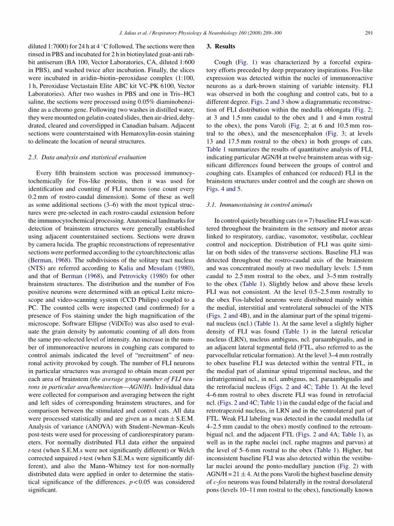
gy &
drbiw1Lsdtdst
2
ti0attdubs((abpsPpmutbcrierwacwApetcfdts
3
tenwdtatt1TincbF
3
tlcldactFtt(ndnapttit4nrF4bwti
J. Jakus et al. / Respiratory Physiolo
iluted 1:7000) for 24 h at 4 ◦C followed. The sections were theninsed in PBS and incubated for 2 h in biotinylated goat-anti rab-it antiserum (BA 100, Vector Laboratories, CA, diluted 1:600n PBS), and washed twice after incubation. Finally, the slicesere incubated in avidin–biotin–peroxidase complex (1:100,h, Peroxidase Vectastain Elite ABC kit VC-PK 6100, Vectoraboratories). After two washes in PBS and one in Tris–HClaline, the sections were processed using 0.05% diaminobenzi-ine as a chromo gene. Following two washes in distilled water,hey were mounted on gelatin-coated slides, then air-dried, dehy-rated, cleared and coverslipped in Canadian balsam. Adjacentections were counterstained with Hematoxylin-eosin stainingo delineate the location of neural structures.
.3. Data analysis and statistical evaluation
Every fifth brainstem section was processed immunocy-ochemically for Fos-like proteins, then it was used fordentification and counting of FLI neurons (one count every.2 mm of rostro-caudal dimension). Some of these as wells some additional sections (3–6) with the most typical struc-ures were pre-selected in each rostro-caudal extension beforehe immunocytochemical processing. Anatomical landmarks foretection of brainstem structures were generally establishedsing adjacent counterstained sections. Sections were drawny camera lucida. The graphic reconstructions of representativeections were performed according to the cytoarchitectonic atlasBerman, 1968). The subdivisions of the solitary tract nucleusNTS) are referred according to Kalia and Mesulam (1980),nd that of Berman (1968), and Petrovicky (1980) for otherrainstem structures. The distribution and the number of Fosositive neurons were determined with an optical Leitz micro-cope and video-scanning system (CCD Philips) coupled to aC. The counted cells were inspected (and confirmed) for aresence of Fos staining under the high magnification of theicroscope. Software Ellipse (ViDiTo) was also used to eval-
ate the grain density by automatic counting of all dots fromhe same pre-selected level of intensity. An increase in the num-er of immunoreactive neurons in coughing cats compared toontrol animals indicated the level of “recruitment” of neu-onal activity provoked by cough. The number of FLI neuronsn particular structures was averaged to obtain mean count perach area of brainstem (the average group number of FLI neu-ons in particular area/hemisection—AGN/H). Individual dataere collected for comparison and averaging between the right
nd left sides of corresponding brainstem structures, and foromparison between the stimulated and control cats. All dataere processed statistically and are given as a mean ± S.E.M.nalysis of variance (ANOVA) with Student–Newman–Keulsost-tests were used for processing of cardiorespiratory param-ters. For normally distributed FLI data either the unpaired-test (when S.E.M.s were not significantly different) or Welchorrected unpaired t-test (when S.E.M.s were significantly dif-
erent), and also the Mann–Whitney test for non-normallyistributed data were applied in order to determine the statis-ical significance of the differences. p < 0.05 was consideredignificant.lAop
Neurobiology 160 (2008) 289–300 291
. Results
Cough (Fig. 1) was characterized by a forceful expira-ory efforts preceded by deep preparatory inspirations. Fos-likexpression was detected within the nuclei of immunoreactiveeurons as a dark-brown staining of variable intensity. FLIas observed in both the coughing and control cats, but to aifferent degree. Figs. 2 and 3 show a diagrammatic reconstruc-ion of FLI distribution within the medulla oblongata (Fig. 2;t 3 and 1.5 mm caudal to the obex and 1 and 4 mm rostralo the obex), the pons Varoli (Fig. 2; at 6 and 10.5 mm ros-ral to the obex), and the mesencephalon (Fig. 3; at levels3 and 17.5 mm rostral to the obex) in both groups of cats.able 1 summarizes the results of quantitative analysis of FLI,
ndicating particular AGN/H at twelve brainstem areas with sig-ificant differences found between the groups of control andoughing cats. Examples of enhanced (or reduced) FLI in therainstem structures under control and the cough are shown onigs. 4 and 5.
.1. Immunostaining in control animals
In control quietly breathing cats (n = 7) baseline FLI was scat-ered throughout the brainstem in the sensory and motor areasinked to respiratory, cardiac, vasomotor, vestibular, cochlearontrol and nociception. Distribution of FLI was quite simi-ar on both sides of the transverse sections. Baseline FLI wasetected throughout the rostro-caudal axis of the brainstemnd was concentrated mostly at two medullary levels: 1.5 mmaudal to 2.5 mm rostral to the obex, and 3–5 mm rostrallyo the obex (Table 1). Slightly below and above these levelsLI was not consistent. At the level 0.5–2.5 mm rostrally to
he obex Fos-labeled neurons were distributed mainly withinhe medial, interstitial and ventrolateral subnuclei of the NTSFigs. 2 and 4B), and in the alaminar part of the spinal trigemi-al nucleus (ncl.) (Table 1). At the same level a slightly higherensity of FLI was found (Table 1) in the lateral reticularucleus (LRN), nucleus ambiguus, ncl. paraambigualis, and inn adjacent lateral tegmental field (FTL, also referred to as thearvocellular reticular formation). At the level 3–4 mm rostrallyo obex baseline FLI was detected within the ventral FTL, inhe medial part of alaminar spinal trigeminal nucleus, and thenfratrigeminal ncl., in ncl. ambiguus, ncl. paraambigualis andhe retrofacial nucleus (Figs. 2 and 4C; Table 1). At the level–6 mm rostral to obex discrete FLI was found in retrofacialcl. (Figs. 2 and 4C; Table 1) in the caudal edge of the facial andetrotrapezoid nucleus, in LRN and in the ventrolateral part ofTL. Weak FLI labeling was detected in the caudal medulla (at–2.5 mm caudal to the obex) mostly confined to the retroam-igual ncl. and the adjacent FTL (Figs. 2 and 4A; Table 1), asell as in the raphe nuclei (ncl. raphe magnus and parvus) at
he level of 5–6 mm rostral to the obex (Table 1). Higher, butnconsistent baseline FLI was also detected within the vestibu-
ar nuclei around the ponto-medullary junction (Fig. 2) withGN/H = 21 ± 4. At the pons Varoli the highest baseline densityf c-fos neurons was found bilaterally in the rostral dorsolateralons (levels 10–11 mm rostral to the obex), functionally known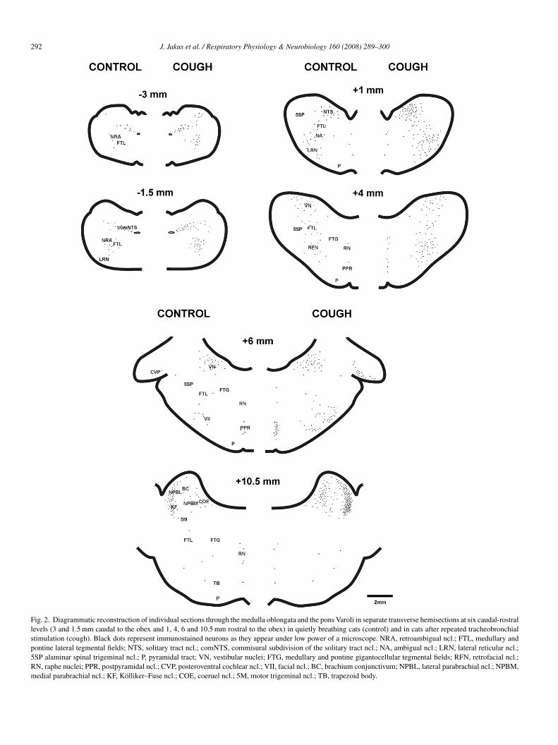
292 J. Jakus et al. / Respiratory Physiology & Neurobiology 160 (2008) 289–300
Fig. 2. Diagrammatic reconstruction of individual sections through the medulla oblongata and the pons Varoli in separate transverse hemisections at six caudal-rostrallevels (3 and 1.5 mm caudal to the obex and 1, 4, 6 and 10.5 mm rostral to the obex) in quietly breathing cats (control) and in cats after repeated tracheobronchialstimulation (cough). Black dots represent immunostained neurons as they appear under low power of a microscope. NRA, retroambigual ncl.; FTL, medullary andpontine lateral tegmental fields; NTS, solitary tract ncl.; comNTS, commisural subdivision of the solitary tract ncl.; NA, ambigual ncl.; LRN, lateral reticular ncl.;5SP alaminar spinal trigeminal ncl.; P, pyramidal tract; VN, vestibular nuclei; FTG, medullary and pontine gigantocellular tegmental fields; RFN, retrofacial ncl.;RN, raphe nuclei; PPR, postpyramidal ncl.; CVP, posteroventral cochlear ncl.; VII, facial ncl.; BC, brachium conjunctivum; NPBL, lateral parabrachial ncl.; NPBM,medial parabrachial ncl.; KF, Kolliker–Fuse ncl.; COE, coeruel ncl.; 5M, motor trigeminal ncl.; TB, trapezoid body.
J. Jakus et al. / Respiratory Physiology & Neurobiology 160 (2008) 289–300 293
Fig. 3. Diagrammatic reconstruction of transverse mesencephalic hemisections at the level of inferior colliculi (13 mm rostral to the obex) and superior colliculi(17.5 mm rostral to the obex) with baseline FLI found in quietly breathing cats (control) and in coughing cats (cough). ICC, central nucleus of the inferior colliculus;P gitudc ulomon
tdien7AtwmtgtT(
niwptngiat
AG, periaqueductal gray; AQ, aqueduct; COE, coeruel ncl.; MLB, medial lonentral ncl.; FTP, paralemniscal tegmental field; SCI, superior colliculus; 3, occl.; RRN, retrorubral ncl.
o as “pontine respiratory group”. Higher baseline FLI wasetected at the level 10.5–11 mm in the brachium conjunctivum,n the coeruel nucleus, in the medial (NPBM), and the lat-ral (NPBL) parabrachial nuclei, and within the Kolliker–Fuseucleus (Figs. 2 and 5A; Table 1). From this total value almost0% was detected in NPBL and the Kolliker–Fuse ncl. (Table 1).faint baseline FLI was detected at the lower (6–7 mm rostral
o the obex), and the middle (7–9 mm rostral to the obex) pons,ithin the posteroventral cochlear nucleus and also in the rapheidline (Figs. 2 and 4D; Table 1). Within the mesencephalon
he highest baseline FLI was found around the periaqeductal
ray (PAG) of the lower mesencephalon (at 12.5–14 mm rostralo the obex) mainly laterally to the aqueduct (Figs. 3 and 5C;able 1), and in the midline area of the rostral mesencephalon17–18 mm), including the oculomotor ncl., medial longitudi-3
b
inal bundle; FTC, central tegmental field of the mesencephalon; CS, superiortor ncl.; TX, tegmental decussation; LC, central linear ncl.; IF, interfascicular
al bundle, tegmental decussation, the central linear ncl., thenterfascicular ncl. (Figs. 3 and 5B; Table 1). No baseline FLIas observed in the following brainstem structures: the areaostrema, medullary hypoglossal nucleus, the spinal trigeminalract and nucleus, the subretrofacial nucleus, the accessory oliveuclei—medial, dorsal, inferior, in the medullary and pontineigantocellular tegmental fields (FTG), the preolivary nucleus,n the trapezoid body, in the external cuneate nucleus, in the par-lemniscal nuclei (tegmental field) of the mesencephalon and inhe superior colliculi.
.2. Immunostaining in coughing animals
In coughing cats (n = 6) dense FLI staining was detectedilaterally throughout the brainstem. At the medulla oblongata
294 J. Jakus et al. / Respiratory Physiology & Neurobiology 160 (2008) 289–300
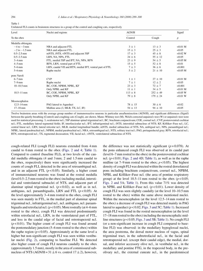
Table 1Unilateral FLI counts in brainstem structures in a group of the control and coughing cats, respectively
Level Nuclei and regions AGN/H Signif
To the obex Control Cough p
Medulla oblongata−4 to −3 mm NRA and adjacent FTL 3 ± 1 13 ± 3 <0.01 M−2 to −1.5 mm NRA and adjacent FTL 15 ± 3 25 ± 3 <0.050.5–2.5 mm mNTS, iNTS, vlNTS and adjacent 5SP 17 ± 3 45 ± 8 <0.02 W0.5–2.5 mm LRN, NA, NPA, FTL 24 ± 6 55 ± 12 <0.053–4 mm FTL, medial 5SP and IFT, NA, NPA, RFN 23 ± 9 54 ± 5 <0.05 M4–5 mm RFN, LRN, ventral part of FTL 15 ± 5 52 ± 8 <0.015–6 mm LRN, caudal VII and RTN, medial IFT, ventral part of FTL 7 ± 3 36 ± 6 <0.0015–6 mm Raphe nuclei 5 ± 2 21 ± 10 <0.05 M
pons Varoli6–7 mm CVP 1 ± 1 27 ± 10 <0.01 M7–9 mm Raphe nuclei 7 ± 1 12 ± 2 <0.0510–10.5 mm BC, COE, NPBM, NPBL, KF 23 ± 3 72 ± 7 <0.001
Only NPBL and KF 11 ± 1 34 ± 5 <0.01 M10.5–11 mm BC, COE, NPBM, NPBL, KF 113 ± 8 252 ± 49 <0.05 W
Only NPBL and KF 79 ± 8 179 ± 24 <0.01 W
Mesencephalon12.5–14 mm PAG lateral to Aqueduct 78 ± 15 30 ± 6 <0.0217–18 mm Midline area (3, MLB, TX, LC, IF) 54 ± 13 94 ± 10 <0.05
Twelve brainstem areas with the average group number of immunoreactive neurons in particular area/hemisection (AGN/H), and significant differences (Signif)between the quietly breathing (Control) and coughing cats (Cough), are shown. Mann–Whitney test (M), Welch corrected unpaired t-test (W) or unpaired t-test wereused for statistical processing. 3, oculomotor ncl.; 5SP alaminar spinal trigeminal ncl.; BC, brachium conjunctivum; COE, coeruel ncl.; CVP, posteroventral cochlearncl.; FTL, medullary lateral tegmental fields; IF, interfascicular ncl.; IFT, infratrigeminal ncl.; iNTS, interstitial subnucleus of NTS; KF, Kolliker–Fuse ncl.; LC,central linear ncl.; LRN, lateral reticular ncl.; MLB, medial longitudinal bundle; mNTS, medial subnucleus of NTS, NA, ambigual ncl.; NPA, paraambigual ncl.;N biguaR ntrol
ccCdtcao(taatwtbrwa(tthla(n
tt(nmdpNgFicrWtlc1l(ltt
PBL, lateral parabrachial ncl.; NPBM, medial parabrachial ncl.; NRA, retroamTN, retrotrapezoid ncl.; TX, tegmental decussation; VII, facial ncl.; vlNTS, ve
ough-related FLI (cough FLI) neurons extended from 4 mmaudal to 6 mm rostral to the obex (Figs. 2 and 4; Table 1).ompared to control (baseline FLI), at two levels of the cau-al medulla oblongata (4 and 3 mm; 2 and 1.5 mm caudal tohe obex, respectively) there were significantly increased theounts of cough FLI, detected in the area of retroambigual ncl.nd in an adjacent FTL (p < 0.05). Similarly, a higher countf immunostained neurons was found at the rostral medullalevel 0.5–2.5 mm rostral to the obex) including medial, intersti-ial and ventrolateral subnuclei of NTS, and adjacent part oflaminar spinal trigeminal ncl. (p < 0.02), as well as in ncl.mbiguus, ncl. paraambigualis, LRN and FTL (p < 0.05). Athe level approximately 3–4 mm rostral to the obex, cough FLIas seen mainly in FTL, in the medial part of alaminar spinal
rigeminal ncl., infratrigeminal ncl., ncl. ambiguus, ncl. paraam-igualis and retrofacial ncl. (p < 0.05). More rostrally (at 4–6 mmostral to the obex), cough FLI was detected predominantlyithin retrofacial ncl., LRN, in the ventrolateral part of FTL,
nd less in the caudal edge of facial and retrotrapezoid ncl.p < 0.01). The higher count of cough FLI was found aroundhe pontomedulary junction (5–6 mm rostral to the obex) withinhe raphe region (p < 0.05). Approximately at the same level aigher but non-significant cough FLI was seen within vestibu-
ar nuclei (Fig. 2), comparing to baseline FLI. We detectedlso higher count of cough FLI neurons caudally to the obexapproximately 1.5 mm), mostly in the area of commissural sub-ucleus of NTS (AGN/H = 31 ± 6 vs. control 17 ± 2), however,rsmo
l ncl.; NTS, solitary tract ncl.; PAG, periaqueductal gray; RFN, retrofacial ncl.;ateral subnucleus of NTS.
he difference was not statistically significant (p = 0.078). Athe pons enhanced cough FLI was observed at its caudal partlevel 6–7 mm rostral to the obex) in the posteroventral cochlearcl. (p < 0.01; Figs. 2 and 4D; Table 1), as well as in the rapheidline (at 7–9 mm rostral to the obex; p < 0.05). The highest
ensity of cough FLI was detected within the rostral dorsolateralons including brachium conjunctivum, coeruel ncl., NPBM,PBL and Kolliker–Fuse ncl. (the area of pontine respiratoryroup) at the level 10.5–11 mm rostral to the obex (p < 0.05;igs. 2 and 5A; Table 1). From this value 71% was detected
n NPBL and Kolliker–Fuse ncl. (p < 0.01). Lower density ofough FLI was seen slightly caudally (at the level 10–10.5 mmostral to the obex) within the same above mentioned nuclei.
ithin the mesencephalon (at the level 12.5–14 mm rostral tohe obex) a decrease of cough FLI was detected mainly in PAGateral to aqueduct (p < 0.02; Figs. 3 and 5C; Table 1). Increasedough FLI was found in the rostral mesencephalon (at the level7–18 mm rostral to the obex) including the mesencephalic mid-ine structures (p < 0.05; Figs. 3 and 5B; Table 1). No cough FLIor a non-significant increase in cough FLI compared to base-ine FLI) was observed: in the medullary hypoglossal nuclei,he area postrema, the dorsal motor nucleus of vagus, spinalrigeminal tract, in the subretrofacial ncl., in the facial and
etrotrapezoid ncl. (except their caudal edge), the medial, dor-al, and inferior accessory olive ncl., in vestibular ncl., in theedullary and pontine FTG, in the trapezoid body, in the pre-livary ncl., the external cuneate ncl., in the paralemniscal

J. Jakus et al. / Respiratory Physiology & Neurobiology 160 (2008) 289–300 295
Fig. 4. Examples of cough-related FLI on right panels (cough) compared to baseline FLI in quietly breathing cats shown on the left panels (control), taken from thetransversal sections at the medullary levels: (A) 3 mm caudal to the obex, (B) 0.5 mm rostral to the obex, (C) 3.5 mm rostral to the obex, and the pontine level: (D)6 icate dm sionsv
nc
3
t
et1
.6 mm rostral to the obex. For explanation see the text (Section 3). Arrows indedullary lateral tegmental field; mNTS, iNTS, medial and interstitial subdivi
agus; RFN, retrofacial ncl.; CVP, posteroventral cochlear ncl.
cl. of the mesencephalon and in both inferior and superiorolliculi.
.3. Alterations of BP in coughing animals
In stimulated animals, we detected two distinct pat-erns of BP changes caused by coughing (Fig. 1). Cough
sdp8
orsal and lateral directions. Scale bar 0.2 mm. NRA, retroambigual ncl.; FTL,of the solitary tract ncl.; S, solitary tract; DMNV, dorsal motor nucleus of the
xpulsions frequently caused elevated BP of one sys-ole (average increase in systolic pressure: 13 ± 3 mmHg,.7 ± 0.4 kPa; p < 0.01; Fig. 1). During the later part of
timulation and several seconds after the coughing, BPecreased (mean BP decrease: 16 ± 3 mmHg, 2.1 ± 0.4 kPa;< 0.001; Fig. 1). The maximum BP depressions lasted± 1 s.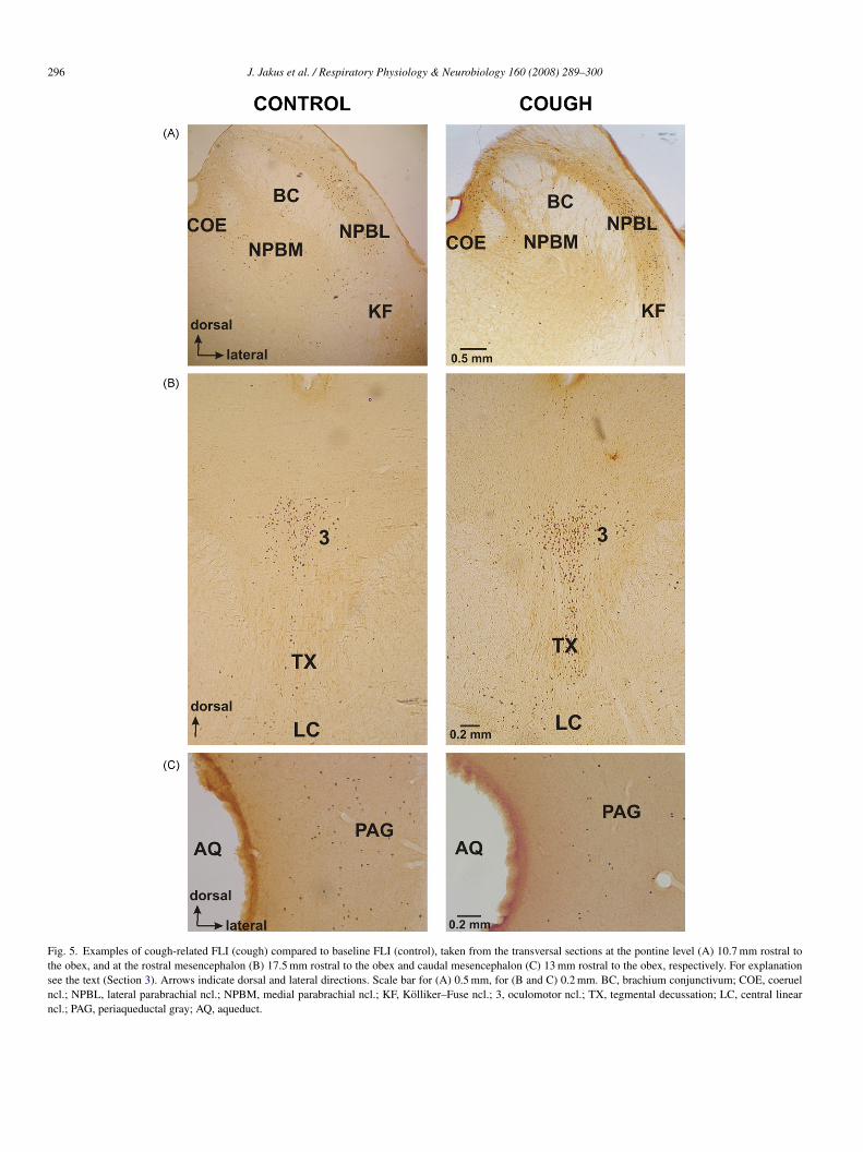
296 J. Jakus et al. / Respiratory Physiology & Neurobiology 160 (2008) 289–300
Fig. 5. Examples of cough-related FLI (cough) compared to baseline FLI (control), taken from the transversal sections at the pontine level (A) 10.7 mm rostral tothe obex, and at the rostral mesencephalon (B) 17.5 mm rostral to the obex and caudal mesencephalon (C) 13 mm rostral to the obex, respectively. For explanationsee the text (Section 3). Arrows indicate dorsal and lateral directions. Scale bar for (A) 0.5 mm, for (B and C) 0.2 mm. BC, brachium conjunctivum; COE, coeruelncl.; NPBL, lateral parabrachial ncl.; NPBM, medial parabrachial ncl.; KF, Kolliker–Fuse ncl.; 3, oculomotor ncl.; TX, tegmental decussation; LC, central linearncl.; PAG, periaqueductal gray; AQ, aqueduct.
gy &
4
sbmiarcfi
nn(cmetmi(fab(ndaJrwftetdplwetra(ceatitotsldt
eactaoo
cat1asetf(ib2cn
tcpruas“cvFp
4c
lomliftdo1cN
J. Jakus et al. / Respiratory Physiolo
. Discussion
The present study is the first attempt to identify brain-tem circuitry of the tracheobronchial cough in spontaneouslyreathing cats by Fos-like immunoreactive technique. Frequentechanical stimulation of the lower airways resulted in repet-
tive tracheal–bronchial coughs comprising stronger expiratorynd weaker inspiratory efforts, differing from the stronger inspi-atory and weaker expiratory efforts typical for the laryngealough (Korpas and Tomori, 1979), or from motor pattern duringctive laryngeal cough (Gestreau et al., 1997).
Although Fos expression is known as a reliable marker ofeuronal activation (Dragunow and Robertson, 1988), not alleurons activated during the given motor reflex express FosGestreau et al., 1997; Jakus et al., 2004b). This was alsoonfirmed in this study because e.g. the hypoglossal, or otherotoneurons involved in tracheal–bronchial coughing, did not
xhibit marked FLI. Another limitation of the method is that sub-hreshold stimuli do not induce detectable levels of Fos, however,
anipulations such as surgery and stress can cause Fos label-ng. The period of time required for FLI related to non-specifice.g. nociceptive) stimulation to decline to minimal levels dif-ers depending on the type, intensity and duration of the stimuli,nd also on anesthetic conditions (Bullitt et al., 1992). Pentobar-ital anesthesia is known as a suppressant of c-fos expressionMorgan et al., 1987) and also as a depressant of the respiratoryeuronal network. However, this anesthetic has been utilized forecades in the cat to study cough and this species will cough reli-bly under pentobarbital anesthesia (Korpas and Tomori, 1979;akus et al., 2004a). In pentobarbital anesthetized cats, markedeductions of FLI induced by dynamic and repetitive sneezingere observed after a 3-h period from the stimulation to the per-
usion (Wallois et al., 1995). We chose a 30 min period betweenhe surgical preparation and tracheal stimulation (3 h from thend of preparation to the perfusion) because prolonged of anes-hesia can reduce cough excitability and we also did not detectifferences in baseline FLI in our control animals with 3 or 6 host-surgical periods. In control cats we found low levels of base-ine FLI in most brainstem areas and these findings correspondell with the results of e.g. Wallois et al. (1995) and Gestreau
t al. (1997). Limited manipulation of the animal and surgery,he use of local anesthetic in the wound margins leading to aeduction in nociception-related FLI (Lykkegaard et al., 2005),nd suppression of c-fos expression by pentobarbital anesthesiaMorgan et al., 1987) all could account for our observations. Inontrast with some studies (Miller and Ruggiero, 1994; Gestreaut al., 1997) that reported robust FLI in the vestibular nuclei andlaminar spinal trigeminal ncl. (probably as a result of stimula-ion of nociceptors due to more extensive surgical preparationsn decerebrated animals), we found only a weak baseline FLI inhose structures. In addition to the caudal trigeminal complex,ther pain-related brainstem areas such the medullary and pon-ine raphe, caudal pontine reticular formation, olivary nuclei,
uperior colliculi (Bullitt, 1990; Liu et al., 1998) expressed veryittle FLI in our animals. Similarly, a faint baseline FLI wasetected in the retrotrapezoid ncl., which is known to be relatedo breathing under hypercapnia (Teppema et al., 1994). How-otsi
Neurobiology 160 (2008) 289–300 297
ver, we revealed a large baseline FLI at the rostrolateral pons,nd also at the caudal mesencephalon, probably indicating theiromplex integrative role in a control of many physiological func-ions e.g. somatosensory, alimentary, cardiovascular, respiratory,rousal, behavioral reactions, vocalization, etc. Taken together,ur control animals expressed FLI levels that were in line withther studies in the cat that employed longer post-surgical times.
As we stated before, we successfully maintained stableardiorespiratory parameters in our cats. However, transientlterations of BP were associated with coughing in our study andhese changes have been observed before (Korpas and Tomori,979). The compensatory mechanisms of autonomic controlctivated by sustained hypotension or hypertension can induceubstantial expression of c-fos (McAllen et al., 1992; Dampneyt al., 2003). However, the magnitude and in particular the dura-ion of the BP increases or decreases in our experiments werear smaller than those required to induce the c-fos by othersDampney et al., 2003). Our cats showed scarce Fos labelingn the area postrema, which expresses high levels of FLI inoth hypertensive and hypotensive animals (Dampney et al.,003). Also dense clustering of labeled cells in the subretrofa-ial nucleus related to hypotension (McAllen et al., 1992) wasot present in our brainstems.
We evoked cough by mechanical stimulation restricted tohe regions of trachea and large bronchi. Other approaches (e.g.apsaicin challenge) are less efficient in production of multi-le cough trials, less selective (stimulating also intrapulmonaryeceptors), and it is difficult to control the intensity of such stim-li. Coughing is accompanied by additional involvements, suchs pleural pressure changes, glottal movement, bronchocon-triction, hyperventilation, etc. (Korpas and Tomori, 1979).Over-stimulation” (too many coughs in the trial due to e.g. highoncentration of tussive agent) could create excessive cardio-ascular and respiratory effects with an increase of non-specificLI. Conversely, weak coughing e.g. when cough is induced onaralysed animals might make cough FLI undetectable.
.1. Involvement of the medulla oblongata structures inoughing
Tracheal–bronchial stimulation in our experiments caused aarge increase of cough FLI in the caudal and rostral medullablongata, through the rostral, dorsolateral pons, up to theidline area of the rostral mesencephalon. There is relatively
ittle information on the properties of the “cough” second-ordernterneurons in the dorsal medulla. The afferent informationrom the tracheal–bronchial “cough” receptors is conveyed byhe vagus nerves terminating at different subnuclei of the cau-al two thirds of NTS, where they make synaptic contactsn the second-order cough interneurons (Kalia and Mesulam,980; Kubin and Davies, 1988; Jordan, 2001). We found denseough FLI mostly in the medial, interstitial, and ventrolateralTS, consistent with the fact that the second-order neurons
f the tracheal–bronchial cough reflex arc are located also inhese areas (Kalia and Mesulam, 1980). In addition, pulmonarytretch receptors, which are vigorously activated during cough-ng, project to the NTS rostral to the obex as well (Davies et
2 gy &
asiDnRasAiftbnaitpaaeFaEeepwr1crebb1inavriaercinmloodeaec
sJsie
4i
tpapohpeciicbwmtiste
antsrbawmatmtal(
tdos
98 J. Jakus et al. / Respiratory Physiolo
l., 1987; Kubin et al., 2006). However, the primary site of theecond-order neurons in the afferent pathway of the cough reflexs the commissural subnucleus in the caudal NTS (Kubin andavies, 1988; Kubin et al., 2006; Mutolo et al., 2007). We didot detect significant increases of the cough FLI in this region.elatively high basal staining just below the obex (observedlso e.g. by Wallois et al. (1995) and Gestreau et al. (1997)) pre-umably interfered with the level of recruitment of cough FLI.
bilateral distribution of an intense cough FLI supports thedea that during tracheal–bronchial stimulation neural activityrom the second-order interneurons may transmit informationo the ventral and lateral parts of medulla, spreading it fartheroth in ascending and descending manner through the rapheuclei to the ventrolateral FTL, the LRN, the regions of ncl.mbiguus and ncl. paraambigualis, the retrofacial ncl., all local-zed in the rostral ventrolateral medulla. It is known that many ofhe aforementioned structures located in the rostral ventral res-iratory group (e.g. those around the facial or retrofacial nucleire closely related with the respiratory central pattern generator,nd are also involved in production of the cough (Bongiannit al., 1998; Shannon et al., 2000). Furthermore, our coughLI overlapped the region of ventral respiratory group neuronsffecting both its caudal part, consisting mainly of the pre-motor-AUG neurons with spinal projections (supplying the spinalxpiratory motoneurons during breathing, coughing, vomiting,tc.; Iscoe, 1998; Bongianni et al., 2005) as well as its rostralart, containing predominantly the pre-motor I-AUG neurons,hich drive the phrenic and the external intercostal motoneu-
ons, having also collaterals to the retroambigual area (Feldman,986; Lipsky et al., 1991). In particular, in tracheal–bronchialoughing inspiratory and expiratory neurons of ventral respi-atory group fire during either the inspiratory, compressive orxpiratory phases with a higher frequency than during quietreathing, and even new expiratory units (silent during quietreathing) were found to be regularly recruited (Jakus et al.,985; Shannon et al., 1998). Hence, our dense cough Fos label-ng detected in ncl. retroambigualis, FTL, ncl. paraambigualis,cl. ambiguus and retrofacial ncl. may reflect the activities of allbove mentioned neurons in tracheal–bronchial coughing. Acti-ation of these neurons subserves various components of cougheflex (preparatory inspiration, inspiratory pharyngeal dilation,nspiratory glottal opening, closure during compressive phasend dilation to allow expulsion, bronchoconstriction supportingxpulsion, etc). Whether laryngeal and pharyngeal motoneu-ons form part of our labeled neurons in ventrolateral medullaould not be confirmed. In our coughing cats a large Fos label-ng was detected in LRN, and solely at the medullary FTL, butot in the FTG, whereas both the FTL and FTG along with theedial accessory olive ncl. were clearly labeled for “fictive”
aryngeal coughing (Gestreau et al., 1997). The involvement ofnly LRN and FTL may be specific for the brainstem circuitryf tracheal–bronchial cough, and they may be necessary for pro-uction or modulation of the cough motor pattern (see also Jakus
t al., 2000, 2004b). This part of the reticular formation is notssociated with the respiratory central pattern generator. How-ver, activation of medullary and pontine reticular neurons wasoupled with the contraction of respiratory-related muscles, aspnTf
Neurobiology 160 (2008) 289–300
een e.g. in the gasp-like aspiration reflex (Fung et al., 1994;akus et al., 2004b), in vomiting (Miller and Ruggiero, 1994),neezing (Wallois et al., 1995), or in “fictive” laryngeal cough-ng (Gestreau et al., 1997), and the expiration reflex (Poliacekt al., 2007).
.2. Involvement of pontine and mesencephalic structuresn coughing
In the pons, the large cough FLI was detected primarily withinhe NPBL and the Kolliker–Fuse ncl., referred to as parts of theontine respiratory group (respiratory neurons located withinnd around the NPBM, NPBL and Kolliker–Fuse ncl.), whichromotes inspiratory termination (Bianchi et al., 1995). More-ver, neurons at NPBL, Kolliker–Fuse ncl. (also in NPBM) alsoave firing patterns consistent with their involvement in coughroduction (Baekey et al., 1999; Shannon et al., 2004a). How-ver, in contrast to involvement of NPBM in fictive laryngealoughing (Gestreau et al., 1997), our finding of the cough FLIn that nucleus indicates that NPBM probably does not have anmportant role in the brainstem circuitry for tracheal–bronchialoughing. Also, we found a higher density of cough Fos neuronsilaterally in the posteroventral cochlear nucleus. This findingas unexpected. However, because this nucleus processes infor-ation coming from the cochlea to the caudal pons, it is unlikely
hat it is directly involved in the production of coughing. Rathert may play a role of a cough modulator, similarly as has beenuggested for the cerebellar interposite nucleus (Xu et al., 1997),he raphe nuclei (Jakus et al., 1998) or the medullary FTL (Jakust al., 2000).
In the mesencephalon enhanced cough FLI was seen bilater-lly at its rostral midline structures, and surprisingly a reducedumber of cough Fos neurons was detected at the caudal levels inhe PAG. As it was stressed above, the function of the midbraintructures at those levels is very complex. However, as reportedecently (Baekey et al., 2004; Shannon et al., 2004b) the mid-rain and diencephalon structures are not crucially involved incontrol of the cough or expiration reflex in cats. Nevertheless,e reported a dense Fos labeling at the caudal and the mid-esencephalon in our cats with the aspiration reflex (Jakus et
l., 2004b). Thus, it is possible that the structures located aroundhe central tegmental field of the caudal and mid-mesencephalon
ay participate in a modulation of behaviors with strong inspira-ory efforts (e.g. the aspiration reflex or gasping), whereas thoseround the midline of the rostral mesencephalon may modu-ate the expiratory expulsions in the cough or expiration reflexPoliacek et al., 2007).
The motor pattern of coughing can differ in various situa-ions (different stimulation, pathological conditions, etc.). Theseifferences in motor pattern could be due to plasticity at vari-us level of the reflex pathways and network (receptors, NTSecond-order neurons, downstream central control elements,
re-motor neurons) resulting in altered excitability of individualeuronal populations (Bolser et al., 2006; Bonham et al., 2006).hese issues could contribute to the differences of cough FLIound in our study and the study of Gestreau et al. (1997).

gy &
elmot1eaNFtIlp(si
A
Fpn
R
B
B
B
B
B
B
B
B
B
B
B
D
D
D
F
F
F
F
G
IJ
J
J
J
J
J
J
K
K
K
K
K
L
L
L
J. Jakus et al. / Respiratory Physiolo
In conclusion, stimulation of the tracheal–bronchial mucosavokes tracheobronchial cough that induces a selective Fosabeling in several brainstem nuclei, with either control and/or
odulatory functions, being involved in the brainstem circuitryf the tracheal–bronchial cough. Selective cough FLI, similaro that being found in fictive laryngeal cough (Gestreau et al.,997), was detected in the medial, interstitial and ventrolat-ral subdivisions of the NTS, in the retroambigual, ambigualnd paraambigual area, in the region of retrofacial ncl., LRN,PBL and Kolliker–Fuse ncl. Nevertheless, differences in theos labeling for tracheal–bronchial cough in our study and fic-
ive laryngeal cough (Gestreau et al., 1997) were clearly seen.n the present study, cough FLI neurons were found within theateral medullary FTL, medullary and pontine raphe, and in theosteroventral cochlear ncl. Unlike the study of Gestreau et al.1997) we did not find an involvement of FTG, the medial acces-ory olive ncl., and NPBM. In addition, we also found cough FLIn the midbrain.
cknowledgements
We gratefully acknowledge the technical assistance of Evarolova, Peter Machac and Roman Kubizna.This study was sup-orted by grant no. 1/2274/05 (VEGA) to J. Jakus, and grant NIHo. R01 07125 to D.C. Bolser.
eferences
aekey, D.M., Morris, K.F., Nuding, S.C., Lindsey, B.G., Shannon, R., 1999.Concurrent changes in pontine respiratory group neuron activities duringfictive coughing. Fed. Am. Soc. Exp. Physiol. 13, 824.
aekey, D.M., Morris, K.F., Gestreau, C., Li, Z., Lindsey, B.G., Shannon, R.,2001. Medulary respiratory neurones and control of laryngeal motoneuronesduring fictive eupnoea and cough in the cat. J. Physiol. Lond. 534, 565–581.
aekey, D.M., Morris, K.F., Nuding, S.C., Segers, L.S., Lindsey, B.G., Shannon,R., 2004. Ventrolateral medullary respiratory network participation in theexpiration reflex in the cat. J. Appl. Physiol. 96, 2057–2072.
erman, A.L., 1968. The Brainstem of the Cat A Cytoarchitectonic Atlas withStereotaxic Coordinates. The University of Wisconsin Press, Madison.
ianchi, A.L., Denavit-Saubie, M., Champagnat, J., 1995. Central control ofbreathing in mammals: neuronal circuitry, membrane properties, and neuro-transmitters. Physiol. Rev. 75, 1–45.
olser, D.C., Poliacek, I., Jakus, J., Fuller, D.D., Davenport, P.W., 2006. Neu-rogenesis of cough, other airway defensive behaviors and breathing: aholarchical system? Respir. Physiol. Neurobiol. 152, 255–265.
ongianni, F., Mutolo, D., Fontana, G.A., Pantaleo, T., 1998. Discharge patternsof Botzinger complex neurons during cough in the cat. Am. J. Physiol. 274,R1015–R1024.
ongianni, F., Mutolo, D., Nardone, F., Pantaleo, T., 2005. Ionotropic glutamatereceptors mediate excitatory drive to caudal medullary expiratory neuronsin the rabbit. Brain Res. 2 (1056), 145–157.
onham, A.C., Chen, C.Y., Sekizawa, S.I., Joad, J.P., 2006. Plasticity in thenucleus tractus solitarius and its influence on lung and airway reflexes. J.Appl. Physiol. 101, 322–327.
ullitt, E., 1990. Expression of c-fos-like protein as a marker for neuronalactivity following noxious stimulation in the rat. J. Comp. Neurol. 296 (4),517–530.
ullitt, E., Lee, C.L., Light, A.R., Willcockson, H., 1992. The effect of stimulusduration on noxious-stimulus induced c-fos expression in the rodent spinalcord. Brain Res. 580, 172–179.
ampney, R.A.L., Polson, J.W., Potts, P.D., Hirooka, Y., Horiuchi, J., 2003.Functional organization of brain pathways subserving the baroreceptor
M
Neurobiology 160 (2008) 289–300 299
reflex: studies in conscious animals using immediate early gene expression.Cell. Mol. Neurobiol. 23, 597–616.
avies, R.O., Kubin, L., Pack, A.L., 1987. Pulmonary stretch receptor relay neu-rones of the cat: location and contralateral medullary projections. J. Physiol.383, 571–585.
ragunow, M., Robertson, H.A., 1988. Localization and induction of c-fosproteine-like immuno-reactive material in the nuclei of adult mammalianneurons. Brain Res. 440, 252–260.
eldman, J.L., 1986. Neurophysiology of breathing in mammals. In: Bloom, F.E.(Ed.), Handbook of Physiology, Section 1: The Nervous System, IntrinsicRegulatory Systems of the Brain, vol. IV. American Physiological Society,Bethesda, Maryland, pp. 463–524.
eldman, J.L., Mitchell, G.S., Nattie, E.E., 2003. Breathing: rhythmicity, plas-ticity chemosensitivity. Annu. Rev. Neurosci. 26, 239–266.
eldman, J.L., Del Negro, C.A., 2006. Looking for inspiration: new perspectiveson respiratory rhythm. Nat. Rev. Neurosci. 7 (3), 232–242.
ung, M.L., StJohn, W.M., Tomori, Z., 1994. Reflex recruitment of medullarygasping mechanisms in eupnoea by pharyngeal stimulation and hypoxia.Respir. Physiol. 100, 195–202.
estreau, C., Bianchi, A.L., Grelot, L., 1997. Differential brainstem Fos-likeimmunoreactivity after laryngeal-induced coughing and its reduction bycodeine. J. Neurosci. 17, 9340–9352.
scoe, S., 1998. Control of abdominal muscles. Prog. Neurobiol. 56 (4), 433–506.akus, J., Tomori, Z., Stransky, A., 1985. Activity of bulbar respiratory neu-
rones during cough and other respiratory tract reflexes in cats. Physiol.Bohemoslov. 34, 127–136.
akus, J., Stransky, A., Poliacek, I., Barani, H., Boselova, L., 1998. Effects ofmedullary midline lesions on cough and other airway reflexes in anesthetizedcats. Physiol. Res. 47, 203–213.
akus, J., Stransky, A., Poliacek, I., Barani, H., Boselova, L., 2000. Kainic acidlesions to the lateral tegmental field of medulla: effects on cough, expirationand aspiration reflexes in anesthetized cats. Physiol. Res. 49, 387–398.
akus, J., Tomori, Z., Stransky, A., 2004a. Neuronal Determinants of BreathingCoughing and Related Motor Behaviours: Basics of Nervous Control andReflex Mechanisms. Wist, Martin.
akus, J., Halasova, E., Poliacek, I., Tomori, Z., Stransky, A., 2004b. Brain-stem areas involved in the aspiration reflex: c-fos study in anesthetized cats.Physiol. Res. 53, 703–717.
akus, J., 2007. Central control of the cough reflex. In: Korpas, J., Paintal, A.S.,Anand, A. (Eds.), Cough from Lab to Clinic. Ane Books India, New Delhi.
ordan, D., 2001. Central nervous pathways and control of the airways. Respir.Physiol. 125, 67–81.
alia, M., Mesulam, M.M., 1980. Brain stem projections of sensory and motorcomponents of the vagus complex in cat. II. Laryngeal, tracheobronchial,pulmonary, cardiac, and gastrointestinal branches. J. Comp. Neurol. 193,467–508.
oestler, A., 1967. The Ghost in the Machine. The Maxmillan Company, NewYork.
orpas, J., Tomori, Z., 1979. Cough and Other Respiratory Reflexes. S. Karger,Basel, New York.
ubin, L., Davies, R.O., 1988. Sites of termination and relay of pulmonaryrapidly adapting receptors as studied by spike-triggered averaging. BrainRes. 443 (1–2), 215–221.
ubin, L., Alheid, G.F., Zuperku, E.J., McCrimmon, D.R., 2006. Central path-ways of pulmonary and lower airway vagal afferents. J. Appl. Physiol. 101,618–627.
ipsky, J., Ezure, K., Wong-She, R.B., 1991. Identification of neurons receivinginput from pulmonary rapidly adapting receptors in the cat. J. Physiol. 443,55–77.
iu, R.J., Qiang, M., Qiao, J.T., 1998. Nociceptive c-fos expression insupraspinal areas in avoidance of descending suppression at the spinal relaystation. Neuroscience 85 (4), 1073–1087.
ykkegaard, K., Lauritzen, B., Tessem, L., Weikop, P., Svendsen, O., 2005.
Local anaesthetics attenuates spinal nociception and HPA-axis activationduring experimental laparotomy in pigs. Res. Vet. Sci. 79, 245–251.cAllen, R.M., Badoer, E., Shafton, A.D., Oldfield, B.J., McKinley, M.J., 1992.Hemorrhage induces c-fos immunoreactivity in spinally projecting neuronsof cat subretrofacial nucleus. Brain Res. 575, 329–332.

3 gy &
M
M
M
O
P
P
P
S
S
S
S
T
W
00 J. Jakus et al. / Respiratory Physiolo
iller, A.D., Ruggiero, D., 1994. Emetic reflex arc revealed by expression ofthe immediate-early gene c-Fos in the cat. J. Neurosci. 14, 871–888.
organ, J.I., Cohen, D.R., Hempstead, J.L., Curran, T., 1987. Mapping patternsof c-fos expression in the central nervous system after seizure. Science 237(4811), 192–197.
utolo, D., Bongianni, F., Fontana, G.A., Pantaleo, T., 2007. The role of exci-tatory amino acids and substance P in the mediation of the cough reflexwithin the nucleus tractus solitarii of the rabbit. Brain Res.Bull. 74 (4), 284–293.
rendacova, J., Marsala, M., Sulla, I., Kafka, J., Jalc, P., Cizova, D., Taira,Y., Marsala, J., 2000. Incipient cauda equina syndrome as a model ofsomatovisceral pain in dogs: spinal cord structures involved as revealed bythe expression of c-fos and NADPH diaphorase activity. Neuroscience 95,543–557.
ecova, R., Tatar, M., 2007. Testing cough sensitivity in the clinic. In: Korpas,J., Paintal, A.S., Anand, A. (Eds.), Cough from Lab to Clinic. Ane BooksIndia, New Delhi, pp. 237–252.
etrovicky, P., 1980. Reticular formation and its raphe system I. Cytoarchitec-
tonics with comparative aspects. Monographia XLIX. Acta Univ. CarolinaeMed., Prague.oliacek, I., Halasova, E., Jakus, J., Murin, P., Barani, H., Stransky, A., Bolser,D.C., 2007. Brainstem regions involved in the expiration reflex. A c-fos studyin anesthetized cats. Brain Res. 1184, 168–177.
W
X
Neurobiology 160 (2008) 289–300
hannon, R., Baekey, D.M., Morris, K.F., Lindsey, B.G., 1998. Ventrolateralmedullary respiratory networks and a model of cough mottor pattern gener-ation. J. Appl. Physiol. 84, 2020–2035.
hannon, R., Baekey, D.M., Morris, K.F., Li, Z., Lindsey, B.G., 2000.Functional connectivity among ventrolateral medullary respiratory neu-rons and responses during ficitve cough in cat. J. Physiol. 525, 207–224.
hannon, R., Baekey, D.M., Morris, K.F., Nuding, S.C., Segers, L.S., Lindsey,B.G., 2004a. Pontine respiratory group neuron discharge is altered duringfictive cough in the decerebrate cat. Respir. Physiol. Neurobiol. 142, 43–54.
hannon, R., Baekey, D.M., Morris, K.F., Nuding, S.C., Segers, L.S., Lindsey,B.G., 2004b. Production of reflex cough by brainstem respiratory networks.Pulm. Pharmacol. Ther. 17, 369–376.
eppema, L.J., Berkenbosh, A., Veening, J.G., Olivier, C.N., 1994. Hypercapniainduces c-fos expression in neurons of retrotrapezoid nucleus in cats. BrainRes. 635, 353–356.
allois, F., Gros, F., Masmoudi, K., Larnicol, N., 1995. C-Fos-like immunore-activity in the cat brainstem evoked by sneeze-inducing air puff stimulation
of the nasal mucosa. Brain Res. 687, 143–154.iddicombe, J., Fontana, G., 2006. Cough: what’s in a name? Eur. Respir. J. 28(1), 10–15.
u, F.D., Frazier, D.T., Zhang, Z., Baekey, D.M., Shannon, R., 1997. Cerebellarmodulation of cough motor pattern in cats. J. Appl. Physiol. 83, 391–397.