Ultrastructure and fluorescence histochemistry of endocrine (APUD-type) cells in tracheal mucosa of...
13
Cell Tiss. Res. 158, 4 2 5 ~ 3 7 (1975) by Springer-Verlag 1975 Uhrastructure and Fluorescence Histochemistry of Endocrine (APUD-Type) Cells in Tracheal Mucosa of Human and Various Animal Species* E. Cutz, W. Chan, V. Wong, and P. E. Cohen Department of Pathology and Research Institute, The Hospital for Sick Children, and University of Toronto, Toronto, Ontario, Canada Received December 13, 1974 Summary. This study describes distinctive cells with ultrastructural and histochemical features of APUD-type endocrine cells within the tracheal epithelium of human fetuses, newborns and children as well as different animal species. These cells referred to as Kult- schitzky cells (K cells) were found to be argyrophilic, but not argentaffin, and are considered analogous to the same type of cells in lung and gastro-intestinal tract. Fluorescence histochemistry demonstrated the presence of intracellular amine within tracheal K cells, but only after in-vitro or in-vivo administration of amine precursor (L-DOPA). Ultrastructurally, these cells are characterized by the presence of numerous cytoplasmic granules (dense core vesicles) which show species related morphologic variations. Two different types of K cells were found in trachea of lamb and armadillo, each type possessing morphologically different dense core vesicles. In human and rabbit tracheas, only one type of K cell was identified. K cells in the trachea are distributed as single cells between other epithelial cells; neuroepithelial bodies such as those found in bronchial mucosa were not identified. Well differentiated K cells were found in tracheas of early human fetuses and throughout gestation, infancy, and childhood. Preservation of K cells in human autopsy material and widespread occurrence of these cells in various laboratory animals will permit further studies into the nature and function of tracheobronchial endocrine cells. Key words: Tracheal epithelium (human, animal) -- APUD-Endocrine system -- Electron microscopy - - Histochemistry. Introduction Endocrine cells of the APUD series (Amine Precursor Uptake and Decarb- oxylase activity) constitute a system of dispersed endocrine cells presumed to be of neural crest origin and distributed predominantly in organs derived from the primitive gut (Pearse, 1969, 1973). Morphologic and functional aspects of the APUD-endocrine system have recently been reviewed (Owman et al., 1973; Lechago and Bencosme, 1973). In previous studies by us and others, endocrine appearing cells referred to as Kultschitzky-type cells (K cells) were described within intrapulmonary airway epithelium of humans and animals (Bensch et al., 1965; Lauweryas and Peuskens, 1969; Hage, 1972; Cutz and Cohen, 1972; Cutz et al., 1974). Electron microscopic and histochemical studies have shown that lung K cells conform in most aspects Send o//print requests to: Dr. E. Cutz, Department of Pathology, The Hospital for Sick Children, 555 University Avenue, Toronto, Ontario, M5G 1X8 Canada. * Supported by grant MA-4690 from the Medical Research Council of Canada. 28 Cell Tisa.Res. 158
Transcript of Ultrastructure and fluorescence histochemistry of endocrine (APUD-type) cells in tracheal mucosa of...
Cell Tiss. Res. 158, 4 2 5 ~ 3 7 (1975) �9 by Springer-Verlag 1975
Uhrastructure and Fluorescence Histochemistry of Endocrine (APUD-Type) Cells in Tracheal Mucosa
of Human and Various Animal Species*
E. Cutz, W. Chan, V. Wong, and P. E. Cohen
Department of Pathology and Research Institute, The Hospital for Sick Children, and University of Toronto, Toronto, Ontario, Canada
Received December 13, 1974
Summary. This study describes distinctive cells with ultrastructural and histochemical features of APUD-type endocrine cells within the tracheal epithelium of human fetuses, newborns and children as well as different animal species. These cells referred to as Kult- schitzky cells (K cells) were found to be argyrophilic, but not argentaffin, and are considered analogous to the same type of cells in lung and gastro-intestinal tract.
Fluorescence histochemistry demonstrated the presence of intracellular amine within tracheal K cells, but only after in-vitro or in-vivo administration of amine precursor (L-DOPA). Ultrastructurally, these cells are characterized by the presence of numerous cytoplasmic granules (dense core vesicles) which show species related morphologic variations. Two different types of K cells were found in trachea of lamb and armadillo, each type possessing morphologically different dense core vesicles. In human and rabbit tracheas, only one type of K cell was identified.
K cells in the trachea are distributed as single cells between other epithelial cells; neuroepithelial bodies such as those found in bronchial mucosa were not identified. Well differentiated K cells were found in tracheas of early human fetuses and throughout gestation, infancy, and childhood. Preservation of K cells in human autopsy material and widespread occurrence of these cells in various laboratory animals will permit further studies into the nature and function of tracheobronchial endocrine cells.
Key words: Tracheal epithelium (human, animal) - - APUD-Endocrine system - - Electron microscopy - - Histochemistry.
Introduct ion
Endocr ine cells of the A P U D series (Amine Precursor Uptake and Decarb- oxylase act ivi ty) const i tute a system of dispersed endocrine cells presumed to be of neural crest origin and d is t r ibuted p redominan t ly in organs derived from the pr imit ive gut (Pearse, 1969, 1973). Morphologic and funct ional aspects of the APUD-endocr ine system have recent ly been reviewed (Owman et al., 1973; Lechago and Bencosme, 1973).
I n previous studies by us and others, endocrine appearing cells referred to as Kul t sch i tzky- type cells (K cells) were described within i n t r apu lmona ry airway epi thel ium of humans and animals (Bensch et al., 1965; Lauweryas and Peuskens, 1969; Hage, 1972; Cutz and Cohen, 1972; Cutz et al., 1974). Electron microscopic and histochemical studies have shown tha t lung K cells conform in most aspects
Send o//print requests to: Dr. E. Cutz, Department of Pathology, The Hospital for Sick Children, 555 University Avenue, Toronto, Ontario, M5G 1X8 Canada.
* Supported by grant MA-4690 from the Medical Research Council of Canada.
28 Cell Tisa. Res. 158

426 E. Cutz et al.
to cells of the APUD-endocr ine sys tem and, therefore, could be endocr ine in na ture . The funct ion of lung K cells is st i l l unknown bu t i t has been suggested t h a t amine or a po lypep t ide hormone p roduced by these cells might be involved in the regu la t ion of a i rway tonus or lung c i rcula t ion (Lauweryas and Peuskens, 1969). I t has been shown t h a t lung K cells are cells of origin of carcinoid and oa t cell carc inomas of the lung (Bensch et al., 1968; Hachmei s t e r and Okorie, 1971; H a t t o r i et al., 1972). Var ious endocr ine syndromes associa ted wi th these tumors have been recognized (Knowles and Smith, 1960; Omem% 1970).
The lung K cells appea r well d i f fe ren t ia ted and are numerous in fetal lungs which led to the suggest ion t h a t t hey m a y funct ion dur ing fetal deve lopmen t (Cutz and Conen, 1972; Cutz et al., 1974). K cells are dispersed th roughou t the mucosa of i n t r a p u l m o n a r y bronchi as single cells and in clusters, t e r m e d neuro- epi the l ia l bodies, and p resumed to be i n t r a p u l m o n a r y receptors (Lauweryas et al., 1972a; Lauweryns and Peuskens, 1972b).
I n the p resen t e lec t ron microscopic and his tochemical s tudy, we have examined the t rachea l ep i the l ium of humans and several an imal species dur ing var ious s tages of deve lopmen t to confirm the presence of K cells, and to compare the i r morpho logy and d i s t r ibu t ion with those of s imilar cells in the lung.
Materials and Methods
Humans Tissues were obtained from 10 human fetuses (gestational ages 10-17 weeks) at therapeutic
abortion. Gestational ages were assessed by CR and CH measurements. Autopsy material was used from 2 newborn infants and 4 children (18 months, 6, 9, 15 years) within 5 hours after death. These patients had died of causes unrelated to the respiratory system. Routine histology on the tissues used showed no pathologic changes and, for the study of tracheas, only sections with intact epithelium were used.
Animals For studies on animal tissues the following fetal, newborn and adult specimens were
used: 14 lamb fetuses close to term, 14 rabbits (6 fetuses near term; 6 newborn and 2 adults) and one young armadillo (Dasypus novemcinctus mexicanus).
For both human and animal studies, the trachea was sampled in upper, mid and lower parts. Samples were oriented for cross and longitudinal sectioning and included segments or whole trachea, depending on the size of the specimen. As control tissues for various experi- ments and methods, specimens of lung and intestine were used from the same human or animal. The control specimens were processed in parallel with specimens of trachea.
Light Microscopy (Special Stains) Tissues were fixed in Bouin's fixative, embedded in paraffin and stained with H & E,
PAS and WHO keratin-mucin stain (Kreyberg and Jareg, 1967) for general histologic assessment. Grimelius' silver nitrate stain (Grimelius, 1968) was used for the demonstration of argyrophilic cells and Singh's modification of the Masson-Hamperl stain for argentaffin cells (Singh, 1964).
Fluorescence Histochemistry Fluorescence histochcmical studies were carried out on samples of trachea, lung and
intestine from human fetuses and animals. Small fragments of tissues were incubated for one-half to one hour in Tyrode's solution containing 1 mg/10 ml of L-DOPA. For in-vivo studies, 50-100 mg L-DOPA per kg body weight was injected intraperitoneally into newborn

Endocrine Cells in Human and Animal Trachea 427
rabbits. Animals were killed at one half, one, and one and a half hours after injection. As negative controls, unincubated samples and rabbits without precursor injection were used. The processing and examination of samples for formaldehyde induced fluorescence (FIF) were performed according to previously published methods (Cutz et al., 1974). Pilot studies on autopsy tissues were performed, but these showed diffuse non-specific fluorescence of cells and, therefore, were judged unsatisfactory.
Electron Microscopy Samples of human and animal tracheas were fixed by immersion in 2% glutaraldehyde
and post-fixed in 1% OsOc Blocks for thin sections contained tracheal epithelium from both membranous and cartilage-ring parts of the trachea.
Results
Light Microscopy The epithelial lining in human and animal tracheas was composed of a
characteristic pseudostratified columnar epithelium. Predominant were ciliated cells, goblet cells, and basal cells. This basic pat tern was seen in all samples throughout development except in early human fetuses (less than 13 weeks) where the epithelium was simple columnar and goblet cells could not be identified by WHO stain for mucin.
In all sections of tracheas stained by the method of Grimelius, argyrophilic cells scattered among other epithelial cells were identified by the presence of fine, black or dark brown cytoplasmic granules. The shape of argyrophilie cells was usually elongated or triangular (Fig. 1) with the base resting on the basement membrane and sometimes thin apical processes appeared to reach the lumen (Fig. 3). The nuclei were basally located in argyrophilic cells that reached the lumen, whereas in those not extending to the lumen, the nuclei were at the same level as in the neighbouring epithelial cells (Fig. 5, inset). Some cells had lateral cytoplasmic processes between other epithelial cells. Argyrophilic cells were invariably single, but occasionally groups of 2 or 3 closely packed cells were observed.
None of the samples of trachea contained corpuscular neuroepithelial bodies, which were readily identified within bronchial epithelium in sections of the corresponding lung. Stain for argentaffin cells yielded negative results in sections of trachea, although numerous argentaffin cells were present in control samples of intestine.
Fluorescence Histochemistry
In non-incubated samples only a few weakly fluorescent cells of yellow-green eolour were seen in tracheal epithelium of rabbits and human fetuses, but none in other species. The intensity of fluorescence and numbers of fluorescent cells increased considerably as early as half an hour after injection of or in-vitro incubation with L-DOPA (Fig. 2). No significant difference in intensity of fluorescence was noted between in-vitro and in-vivo samples. Generally the shape, position and distribution of fluorescent cells were the same as those of argyro- philic cells (Fig. 4).
28*

428 E. Cutz et al.
Fig. 1. Triangular argyrophilic cell close to basement membrane (arrow heads) in tracheal mucosa of 15-year-old patient. Autopsy sample, method of Grimelius. • 900
Fig. 2. Several intensely fluorescent cells in tracheal epithelium of 10 week gestation human fetus. In-vitro incubation with L-DOPA, FIF • 420
Fig. 3. Argyrophilic cell with long apical process reaching lumen in tracheal epithelium of lamb fetus. Basement membrane (arrow heads), method of Grimelius. • 760
Fig. 4. Two closely placed fluorescent cells in tracheal mucosa of lamb fetus. Cell on right of similar shape as argyrophilic cell in Fig. 3. In-vitro incubation with L-DOPA, FIF • 760
I n agreement with our previous observations, fluorescent cells were found in sections of lung, bu t only in L-DOPA treated samples. These cells were singly dispersed throughout the i n t r apu lmona ry airway epithel ium or as dist inct ive neuroepithelial bodies.
Cells with intense yellow fluorescence were present in control samples of intest ine even wi thout L-DOPA t rea tment .
Endocrine Cells in Human and Animal Trachea 429
Electron Microscopy
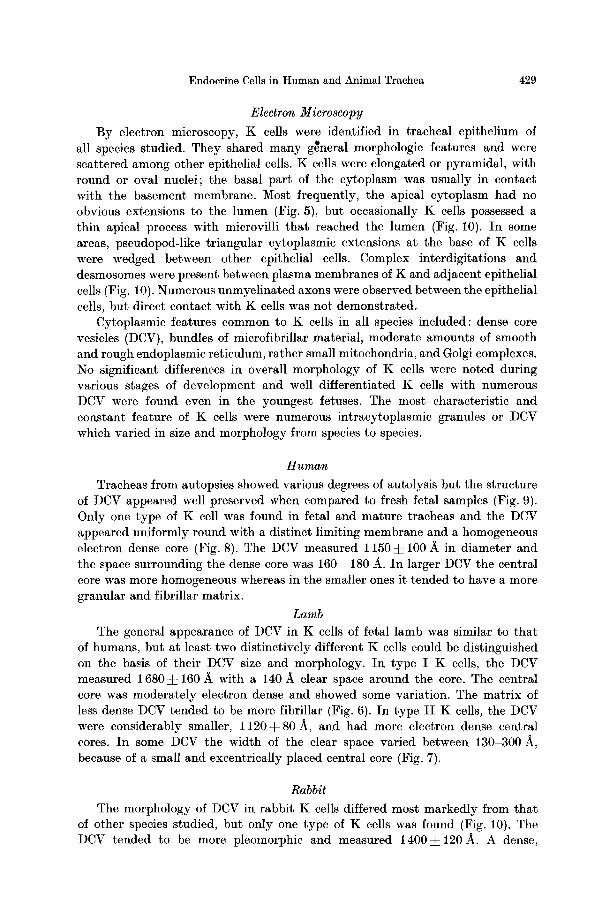
By electron microscopy, K cells were identified in tracheal epithelium of all species studied. They shared many gtneral morphologic features and were scattered among other epithelial cells. K cells were elongated or pyramidal, with round or oval nuclei; the basal part of the cytoplasm was usually in contact with the basement membrane. Most frequently, the apical cytoplasm had no obvious extensions to the lumen (Fig. 5), but occasionally K cells possessed a thin apical process with microvilli tha t reached the lumen (Fig. 10). In some areas, pseudopod-like triangular cytoplasmic extensions at the base of K cells were wedged between other epithelial cells. Complex interdigitations and desmosomes were present between plasma membranes of K and adjacent epithelial cells (Fig. 10). Numerous unmyelinated axons were observed between the epithelial cells, but direct contact with K cells was not demonstrated.
Cytoplasmic features common to K cells in all species included: dense core vesicles (DCV), bundles of microfibrillar material, moderate amounts of smooth and rough endoplasmic reticulum, rather small mitochondria, and Golgi complexes. No significant differences in overall morphology of K cells were noted during various stages of development and well differentiated K cells with numerous DCV were found even in the youngest fetuses. The most characteristic and constant feature of K cells were numerous intracytoplasmic granules or DCV which varied in size and morphology from species to species.
Hum, a n
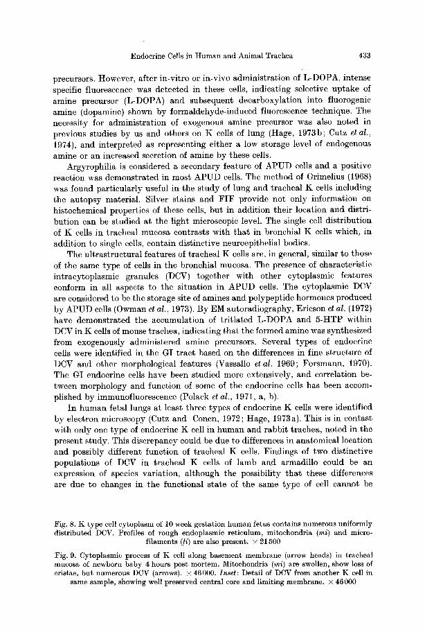
Tracheas from autopsies showed various degrees of autolysis but the structure of DCV appeared well preserved when compared to fresh fetal samples (Fig. 9). Only one type of K cell was found in fetal and mature tracheas and the DCV appeared uniformly round with a distinct limiting membrane and a homogeneous electron dense core (Fig. 8). The DCV measured 1 150 • 100 A in diameter and the space surrounding the dense core was 160--180 A. In larger DCV the central core was more homogeneous whereas in the smaller ones it tended to have a more granular and fibrillar matrix.
Lamb The general appearance of DCV in K cells of fetal lamb was similar to tha t
of humans, but at least two distinctively different K cells could be distinguished on the basis of their DCV size and morphology. In type I K cells, the DCV measured 1680:L 160 A with a 140/~ clear space around the core. The central core was moderately electron dense and showed some variation. The matr ix of less dense DCV tended to be more fibrillar (Fig. 6). In type I I K cells, the DCV were considerably smaller, 1 120• 80 A, and had more electron dense central cores. In some DCV the width of the clear space varied between 130-300 A, because of a small and excentrically placed central core (Fig. 7).
Rabbit
The morphology of DCV in rabbit K cells differed most markedly from that of other species studied, but only one type of K cells was found (Fig. 10). The DCV tended to be more pleomorphic and measured 1 4 0 0 • 120 A. A dense,

430 E. Cutz et al.

Endocrine Cells in Human and Animal Trachea 431
uniformly homogeneous central core had an almost indistinguishable halo measuring 80-100 A. (Fig. 10, inset.)
Armadillo
The appearance of DCV in armadillo K cells was generally similar to tha t in the human and lamb, hut the DCV tended to be larger. I n addition, two different types of K cells with distinct populat ions of DCV were found. DCV in K cells designated as type I (Fig. 11) measured 1 7 5 0 ~ 2 8 0 A and the homo- geneously dense central core filled the granule membrane almost entirely. The halo was 80-140 A wide. I n type I I K cells, the DCV were generally smaller, i.e., 1250-4- 130/~ in diameter. The well defined halo was 100-170 A.
Discussion
Previous ul t rastructural studies on tracheal epithelium described in detail the fine s tructure of ciliated and goblet cells (Rhodin and Dalhamn, 1956; Cireli, 1966). The brush cell, presumed to have a sensory function, was also reported (Luciano et al., t968). Ericson et al. (1972) have recently described enterochromaffin-like cells in tracheal mucosa of mice. These authors used a combinat ion of histo- chemical and EM methods and suggested tha t tracheal enterochromaffin-like cells could be par t of the APUD-endocr ine system. We believe tha t these entero- chromaffin-like cells are identical with the K cells described in this report. The occurrence of K cells within the tracheal mucosa can be expected because of the common entodermal origin with the gastrointestinal (GI) t ract in which endocrine cells are widely distributed. We consider the tracheal K cells to be a par t of the t racheobronchial system of endocrine cells belonging to the APUD-sys tem, because of histochemical and ul t ras t ructural similarities with the same type of cells in lung and A P U D cells elsewhere. The inclusion of tracheal K cells within the A P U D system is based on the demonstra t ion of amine handling properties in these cells, thus fulfilling the principal criterion of this system (Pearse, 1969).
The endogenous amine content of tracheal K cells appears to be low because of weak or absent specific fluorescence in samples not t reated with amine
:Fig. 5. EM low magnification view of fetal lamb tracheal epithelium showing usual location and shape of K cell (cytoplasm outlined in black). Cytoplasm at base in contact with basement membrane (arrow heads), but apical portion does not extend to lumen (Lu). Nucleus of K cell (K) at same level as neighbouring goblet (Go) and ciliated (Ci) cells. Basal cell (Ba). • 2900. Inset: Argyrophilic cell in paraffin section appears in identical position
as K cell shown in :Fig. 5. Grimelius stain. • 700
:Fig. 6. Higher magnification of basal cytoplasmic area of K cell shown in Fig. 5. Numerous large DCV with varying central core density characteristic of lamb type I K cells. Note
several small mitochondria (mi). • 33000
:Fig. 7. Cytoplasmic detail of Type I I K cell in fetal lamb trachea. DCV are smaller, have denser central core; several show excentrically placed central core (arrows), mi-mitochondria.
• 33000

432 E. Cutz et al.
Endocrine Cells in Human and Animal Trachea 433
precursors. However, after in-vitro or in-vivo administration of L-DOPA, intense specific fluorescence was detected in these cells, indicating selective uptake of amine precursor (L-DOPA) and subsequent deearboxylation into fluorogenic amine (dopamine) shown by formaldehyde-induced fluorescence technique. The necessity for administration of exogenous amine precursor was also noted in previous studies by us and others on K cells of lung (Hage, 1973b; Cutz et al., 1974), and interpreted as representing either a low storage level of endogenous amine or an increased secretion of amine by these cells.
Argyrophilia is considered a secondary feature of APUD cells and a positive reaction was demonstrated in most APUD cells. The method of Grimelius (1968) was found particularly useful in the study of lung and tracheal K cells including the autopsy material. Silver stains and F IF provide not only information on histochemical properties of these cells, but in addition their location and distri- bution can be studied at the light microscopic level. The single cell distribution of K cells in tracheal mucosa contrasts with that in bronchial K cells which, in addition to single cells, contain distinctive neuroepithelial bodies.
The ultrastructural features of tracheal K cells are, in general, similar to those of the same type of cells in the bronchial mucosa. The presence of characteristic intracytoplasmic granules (DCV) together with other cytoplasmic features conform in all aspects to the situation in APUD cells. The cytoplasmic DCV are considered to be the storage site of amines and polypeptide hormones produced by APUD cells (Owman et al., 1973). By EM autoradiography, Ericson et al. (1972) have demonstrated the accumulation of tritiated L-DOPA and 5-HTP within DCV in K cells of mouse trachea, indicating that the formed amine was synthesized from exogenously administered amine precursors. Several types of endocrine cells were identified in the GI tract based on the differences in fine structure of DCV and other morphological features (Vassallo et al. 1969; Forsmann, 1970). The GI endocrine cells have been studied more extensively, and correlation be- tween morphology and function of some of the endocrine cells has been accom- plished by immunofluorescence (Polack et al., 1971, a, b).
In human fetal lungs at least three types of endocrine K cells were identified by electron microscopy (Cutz and Conen, 1972; Huge, 1973a). This is in contast with only one type of endocrine K cell in human and rabbit trachea, noted in the present study. This discrepancy could be due to differences in anatomical location and possibly different function of tracheal K cells. Findings of two distinctive populations of DCV in tracheal K cells of lamb and armadillo could be an expression of species variation, although the possibility that these differences are due to changes in the functional state of the same type of cell cannot be
Fig. 8. K type cell cytoplasm of 10 week gestation human fetus contains numerous uniformly distributed DCV. Profiles of rough endoplasmic reticulum, mitochondria (mi) and micro-
filaments (/i) are also present, x 21500
Fig. 9. Cytoplasmic process of K cell along basement membrane (arrow heads) in tracheal mucosa of newborn baby 4 hours post mortem. Mitochondria (mi) are swollen, show loss of cristae, but numerous DCV (arrows). X 46000. Inset: Detail of DCV from another K cell in
same sample, showing well preserved central core and limiting membrane, x 46000

434 E. Cutz et al.
Endocrine Cells in Human and Animal Trachea 435
excluded. Conclusive evidence for the existence of different types of endocrine cells in trachea and lung requires further studies.
Most authors agree on the intraepithelial irmervation of tracheobronchial mucosa, but whether these nerve fibers terminate on specific epithelial cells remains controversial (Glorieux, 1963; Jeffery and Reid, 1973; Walsh and McLelland, 1974). We have also observed numerous intraepithelial axons scattered between epithelial cells, but could not convincingly demonstrate direct contact with K cells. In contrast, there is evidence that nerve fibers terminate in bronchial neuroepithelial bodies which are composed of epithelial cells with morphologic characteristics of K cells (Hung and Loosli, 1974; Cutz et al., 1974). Absence of neuroepithelial bodies in tracheal mucosa supports the previous suggestion that these structures may have a specialized, probable receptor function within the bronchial mucosa (Hung and Loosli, 1974).
The function of tracheal K cells is unknown, but certain speculations based on the morphologic data are possible. Presence of amine handling properties in APUD cells has been directly related to polypeptide hormone production by these cells (Owman, 1973). Therefore, if tracheal K cells produce polypeptide hormones in addition to amines, the former may be of physiological importance. Several hor- monal substances from the GI tract have been isolated and characterized (Pearse, 1974) and their role is thought to be directly or indirectly related to the function of the GI system (Hakanson, 1970).
Certain polypeptide substances of hormonal nature have been recently isolated from hog lungs and their possible role discussed (Said and Mutt, 1969; Said, 1973). Tracheal K cells most likely have a different function from those in the bronchial mucosa because of the rigidity and simpler anatomy of the trachea. The preferen- tial location of tracheal K cells close to the basement membrane, with concentra- tion of granules in the same region, might suggest that the secretions are directed to structures located below the basement membrane, such as blood vessels and smooth muscle coat. K cells with luminal contact could receive stimuli from the lumen.
The presence of fully differentiated K cells early in gestation suggests an important function during development. The occurrence of K cells in human fetuses prior to appearance of goblet cells might suggest a possible role in epithelial cell differentiation. The finding of increased numbers of argyrophilic
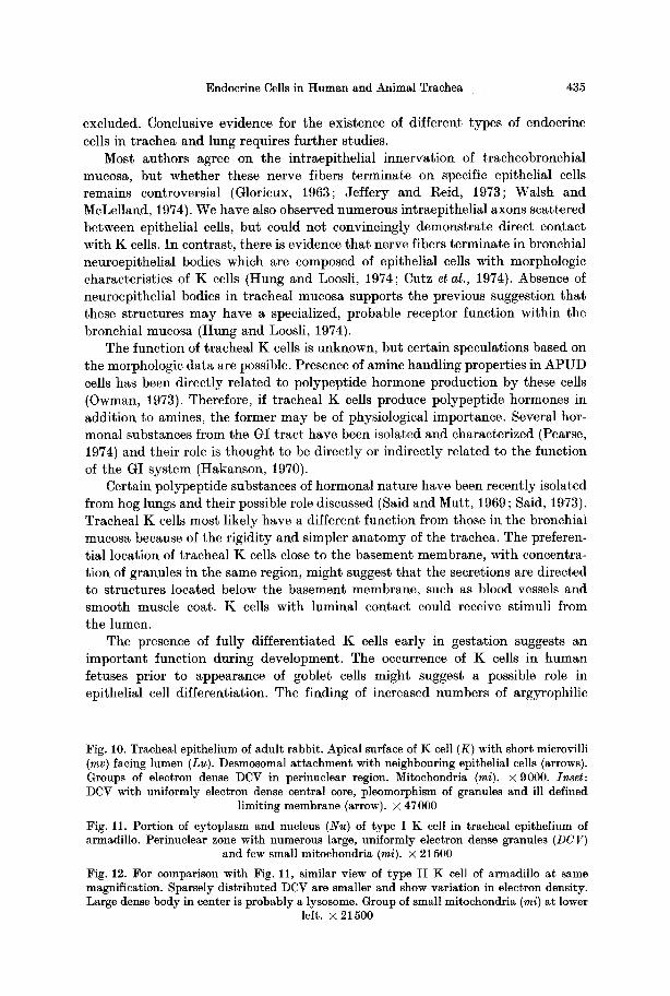
Fig. 10. Tracheal epithelium of adult rabbit. Apical surface of K cell (K) with short microvilli (mv) facing lumen (Lu). Desmosomal attachment with neighbouring epithelial cells (arrows). Groups of electron dense DCV in perinuclear region. Mitochondria (mi). x 9000. Inset: DCV with uniformly electron dense central core, pleomorphism of granules and ill defined
limiting membrane (arrow). x 47 000
Fig. 11. Portion of cytoplasm and nucleus (Nu) of type I K cell in tracheal epithelium of armadillo. Perinuclear zone with numerous large, uniformly electron dense granules (DCV)
and few small mitochondria (mi). • 21500
Fig. 12. For comparison with Fig. 11, similar view of type II K cell of armadillo at same magnification. Sparsely distributed DCV are smaller and show variation in electron density. Large dense body in center is probably a lysosome. Group of small mitochondria (mi) at lower
left. • 21500

436 E. Cutz et al.
cells in areas of goblet cell hyperplasia in bronchi supports this suggestion (Tateishi, 1973). I n t ima te relationships between endocrine cells and other epithelial cells in the s tomach have been commented on by R u b i n (1972).
I n order to elucidate the na ture and funct ion of tracheal endocrine cells, use of biochemical, pharmacologic and other methods would be necessary. Rela t ively good preservat ion of endocrine cells in h u m a n pos tmortem tissues will permit the s tudy of these cells in various pathologic conditions. The presence of K cells in the tracheas of various animal species as well as easier accessibility of tracheal mueosa, as compared to the l ining of bronchi, might serve as a model for fur ther experimental studies.
References
Bensch, K. G., Corrin, B., Pariente, R., Spencer, H.: Oat cell carcinoma of the lung. Its origin and relationship to bronchial carcinoid. Cancer (Philad.) 2~, 1163-1172 (1968)
Bensch, K. G., Gordon, G. B., Miller, L. R.: Studies on the bronchial counterpart of the Kultschitzky (argentaffin) cell and innervation of bronchial glands. J. Ultrastruet. Res. 12, 668-686 (1965)
Cireli, E. : Elektronenmikroskopisehe Analyse der pr~i- und postnatalen Differenzierung des Epithels der oberen Luftwege der Ratte. Z. mikr.-anat. Forsch. 74, 132 178 (1966)
Cutz, E., Chan, W., Wong, V., Conen, P. E. : Endocrine cells in rat fetal lungs. Ultrastructural and histochemical study. Lab. Invest. 30, 458 464 (1974)
Cutz, E., Conen, P. E.: Endocrine-like cells in human fetal lungs. Anat. Rec. 173, 115-122 (1972)
Ericson, L. E., Hs R., Larson, B., Owman, Ch., Sundler, F. : Fluorescence and electron microscopy of amine-stoling enterochromaffin-like cells in tracheal epithelium of mouse. Z. Zellforsch. 124, 532-545 (1972)
Forssman, W. G. : Ultrastructure of hormone-producing cells of the upper gastrointestinal tract. In: Origin, chemistry, physiology and pathophysiology of the gastrointestinal hormones (W. Creutzfeldt, ed.), p. 32-95. Stuttgart-New York: F. K. Schattauer 1970
Glorieux, R.: Les cellules argentaffines du poumon et leurs connexions avec le syst~me nerveux. Arch. Biol. (Liege) 74, 377-390 (1963)
Grimelius, L.: A silver nitrate stain for ~2 cells in human pancreatic islets. Acta Soc. Med. Uppsalien. 73, 243-270 (1968)
Hachmeister, U., Okorie, O.: Die Histogenese des kleinzelligen Bronchuscarcinoms. (Histo- genesis of the oat-cell carcinoma of the bronchus.) Verh. dtsch. Ges. Path. 55, 716-721 (1971)
Hage, E. : Endocrine cells in the bronchial mucosa of human foetuses. Acta path. mierobiol. scand., sect. A 80, 225-234 (1972)
Hage, E. : Electron microscopic identification of several types of endocrine cells in the bronchial epithelium of human foetuses. Z. Zellforsch. 141, 401412 (1973a)
Hage, E. : Amine-handling properties of APUD-cells in the bronchial epithelium of human fetuses and in the epithelium of main bronchi of human adults. Acta path. microbiol. scand., sect. A 81, 64-70 (1973b)
Hs R. : New aspects of the formation and function of histamine, 5-Hydroxytryptamine and dopamine in gastric mucosa. Acta physiol, scand., suppl. 340, 1-134 (1970)
Hattori, S., Matsuda, M., Tateishi, R., Nishihara, M., Horai, T. : Oat-cell carcinoma of the lung. Clinical and morphological studies in relation to its histogenesis. Cancer (Philad.) 30, 1014-1024 (1972)
Hung, K. S., Loosli, C. G. : Bronchiolar neuroepithelial bodies in the neonatal mouse lungs. Amer. J. Anat. 140, 191-200 {1974)
Jeffery, P., Reid, L. : Intraepithelial nerves in normal rat airways. A quantitative electron microscopic study. J. Anat. (Lond.) 114, 35-45 (1973)
Knowles, J. H., Smith, L. H., Jr. : Extrapulmonary manifestations of bronchogenic carcinoma. New Engl. J. Med. 262, 505-510 (1960)
Endocrine Cells in Human and Animal Trachea 437
Kreyberg, L., Jareg, E.: Combined staining method for Keratin and mucin-like substances. In: Histological typing of lung tumour. International histological classification of tumours (No. 1), Kreyberg, L. (ed.), p. 27-28. Geneva: World Health Organization 1967
Lauweryns, J. M., Cokelaere, M., Theunynck, P.: Neuroepithelial bodies in the respiratory mucosa of various mammals. A light optical, histochemical and ultrastructural investiga- tion. Z. Zellforsch. 185, 569-592 {1972)
Lauweryns, J. M., Peuskens, J . C. : Argyrophil (kinin and amine producing ?) cells in human infant airway epithelium. Life Sci. 8, 577-585 (1969)
Lauweryns, J. M., Peuskens, J. C. : Neuroepithelial bodies (neuroreceptor or secretory organs ?) in the human infant bronchial and bronchiolar epithelium. Anat. Rec. 172,471-482 (1972)
Lechago, J. , Bencosme, S. A. : The endocrine elements of the digestive system. In: Int. Rev. Exp. Path., Richter, G. W., Epstein, M. A. (ed.), vol. 12, 119-201. New York-London: Academic Press 1973
Luciano, L., Reale, E., Rnska, H. : t~ber einc ,,chemorezcptivc Sinneszelle in der Trachea der Ratte". Z. Zellforsch. 85, 350-375 (1968)
Omenn, G. S. : Ectopic polypeptide hormone production by tumors. Ann. intern. Med. 72, 136-138 (1970)
Owman, Ch., H&kanson, R., Sundler, F. : Occurrence and function of amines in endocrine cells producing polypeptide hormones. Fed. Proc. 32, 1785-1791 (1973)
Pearse, A. G. E.: The cytochemistry and ultrastructure of polypeptide hormone-producing cells of the APUD series and the embryologic, physiologic and pathologic implications of the concept. J. Histochem. Cytochem. 17, 303-313 (1969)
Pearse, A. G. E. : Cell migration and the alimentary system: endocrine contributions of the neural crest to the gut and its derivatives. Digestion 8, 372-385 (1973)
Pearse, A. G. E. : The gut as an endocrine organ. Brit. J. Hosp. Medicine (May) 697 704 (1974)
Polak, J .M. , Bloom, S., Coulling, I., Pearse, A. G .E . : Immunofluorescent localization of secretion in the canine duodenum. Gut 12, 605-610 (19719)
Polak, J. M., Bloom, S., Coulling, I., Pearse, A. G .E . : Immunofluorescent localization of enteroglucagon cells in the gastrointestinal tract of the dog. Gut 12, 311-318 (1971b)
Rubin, W. : An unusual intimate relationship between endocrine cells and other types of epithelial cells in the human stomach. J. Cell Biol. 52, 219-227 (1972)
Said, S. I. : The lung in relation to vasoactive hormones. Fed. Proc. 32, 1972-1976 (1973) Said, S. I., Mutt, V. : Long acting vasodilator peptide from lung tissue. Nature (Lond.) 224,
699-700 (1969) Singh, I. : A modification of the Masson-Hamperl method for staining of argcntaffin cells.
Anat. Anz. 115, 81-82 (1964) Tateishi, R. : Distribution of argyrophil cells in adult human lungs. Arch. Path. 96, 198 202
(1973) Vassallo, G., Solcia, E., Capella, C.: Light and electron microscopic identification of several
types of endocrine cells in the gastrointestinal mueosa of the cat. Z. Zellforsch. 98, 333-356 {1969)
Walsh, C., McLelland, J . : Intraepithelial axons in the avian trachea. Z. Zellforsch. 147, 209-217 (1974)