Buccal Mucosa as A Route for Systemic Drug Delivery
18
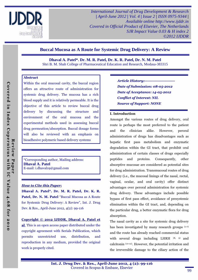
Buccal Mucosa as A Route for Systemic Drug Delivery: A Review Dhaval A. Patel*. Dr. M. R. Patel, Dr. K. R. Patel, Dr. N. M. Patel Shri B. M. Shah College of Pharmaceutical Education and Research, Modasa-383315 How to Cite this Paper: Dhaval A. Patel*. Dr. M. R. Patel, Dr. K. R. Patel, Dr. N. M. Patel “Buccal Mucosa as A Route for Systemic Drug Delivery: A Review”, Int. J. Drug Dev. & Res., April-June 2012, 4(2): 99-116 Copyright © 2012 IJDDR, Dhaval A. Patel et al. This is an open access paper distributed under the copyright agreement with Serials Publication, which permits unrestricted use, distribution, and reproduction in any medium, provided the original work is properly cited. I. Introduction Amongst the various routes of drug delivery, oral route is perhaps the most preferred to the patient and the clinician alike. However, peroral administration of drugs has disadvantages such as hepatic first pass metabolism and enzymatic degradation within the GI tract, that prohibit oral administration of certain classes of drugs especially peptides and proteins. Consequently, other absorptive mucosae are considered as potential sites for drug administration. Transmucosal routes of drug delivery (i.e., the mucosal linings of the nasal, rectal, vaginal, ocular, and oral cavity) offer distinct advantages over peroral administration for systemic drug delivery. These advantages include possible bypass of first pass effect, avoidance of presystemic elimination within the GI tract, and, depending on the particular drug, a better enzymatic flora for drug absorption. The nasal cavity as a site for systemic drug delivery has been investigated by many research groups (1-7) and the route has already reached commercial status with several drugs including LHRH (8, 9) and calcitonin (10-12) . However, the potential irritation and the irreversible damage to the ciliary action of the International Journal of Drug Development & Research | April-June 2012 | Vol. 4 | Issue 2 | ISSN 0975-9344 | Available online http://www.ijddr.in Covered in Official Product of Elsevier, The Netherlands SJR Impact Value 0.03 & H index 2 ©2012 IJDDR Abstract Within the oral mucosal cavity, the buccal region offers an attractive route of administration for systemic drug delivery. The mucosa has a rich blood supply and it is relatively permeable. It is the objective of this article to review buccal drug delivery by discussing the structure and environment of the oral mucosa and the experimental methods used in assessing buccal drug permeation/absorption. Buccal dosage forms will also be reviewed with an emphasis on bioadhesive polymeric based delivery systems *Corresponding author, Mailing address: Dhaval A. Patel E-mail: [email protected] Article History:------------------------ Date of Submission: 08-05-2012 Date of Acceptance: 14-05-2012 Conflict of Interest: NIL Source of Support: NONE Review Paper Covered in Index Copernicus with IC Value 4.68 for 2010 Int. J. Drug Dev. & Res., April-June 2012, 4 (2): 99-116 Covered in Scopus & Embase, Elsevier 99
-
Upload
khangminh22 -
Category
Documents
-
view
0 -
download
0
Transcript of Buccal Mucosa as A Route for Systemic Drug Delivery
Buccal Mucosa as A Route for Systemic Drug Delivery: A Review
Dhaval A. Patel*. Dr. M. R. Patel, Dr. K. R. Patel, Dr. N. M. Patel Shri B. M. Shah College of Pharmaceutical Education and Research, Modasa-383315
How to Cite this Paper:
Dhaval A. Patel*. Dr. M. R. Patel, Dr. K. R.
Patel, Dr. N. M. Patel “Buccal Mucosa as A Route
for Systemic Drug Delivery: A Review”, Int. J. Drug
Dev. & Res., April-June 2012, 4(2): 99-116
Copyright © 2012 IJDDR, Dhaval A. Patel et
al. This is an open access paper distributed under the
copyright agreement with Serials Publication, which
permits unrestricted use, distribution, and
reproduction in any medium, provided the original
work is properly cited.
I. Introduction
Amongst the various routes of drug delivery, oral
route is perhaps the most preferred to the patient
and the clinician alike. However, peroral
administration of drugs has disadvantages such as
hepatic first pass metabolism and enzymatic
degradation within the GI tract, that prohibit oral
administration of certain classes of drugs especially
peptides and proteins. Consequently, other
absorptive mucosae are considered as potential sites
for drug administration. Transmucosal routes of drug
delivery (i.e., the mucosal linings of the nasal, rectal,
vaginal, ocular, and oral cavity) offer distinct
advantages over peroral administration for systemic
drug delivery. These advantages include possible
bypass of first pass effect, avoidance of presystemic
elimination within the GI tract, and, depending on
the particular drug, a better enzymatic flora for drug
absorption.
The nasal cavity as a site for systemic drug delivery
has been investigated by many research groups (1-7)
and the route has already reached commercial status
with several drugs including LHRH (8, 9) and
calcitonin (10-12). However, the potential irritation and
the irreversible damage to the ciliary action of the
International Journal of Drug Development & Research | April-June 2012 | Vol. 4 | Issue 2 | ISSN 0975-9344 |
Available online http://www.ijddr.in Covered in Official Product of Elsevier, The Netherlands
SJR Impact Value 0.03 & H index 2 ©2012 IJDDR
Abstract
Within the oral mucosal cavity, the buccal region
offers an attractive route of administration for
systemic drug delivery. The mucosa has a rich
blood supply and it is relatively permeable. It is the
objective of this article to review buccal drug
delivery by discussing the structure and
environment of the oral mucosa and the
experimental methods used in assessing buccal
drug permeation/absorption. Buccal dosage forms
will also be reviewed with an emphasis on
bioadhesive polymeric based delivery systems
*Corresponding author, Mailing address:
Dhaval A. Patel E-mail: [email protected]
Article History:------------------------
Date of Submission: 08-05-2012
Date of Acceptance: 14-05-2012
Conflict of Interest: NIL
Source of Support: NONE
Re
vie
w P
ap
er
C
ov
er
ed
in
In
de
x C
op
er
nic
us
wit
h I
C V
alu
e 4
.68
fo
r 2
01
0
Int. J. Drug Dev. & Res., April-June 2012, 4 (2): 99-116 Covered in Scopus & Embase, Elsevier
99

nasal cavity from chronic application of nasal dosage
forms, as well as the large intra- and inter-subject
variability in mucus secretion in the nasal mucosa,
could significantly affect drug absorption from this
site. Even though the rectal, vaginal, and ocular
mucosae all offer certain advantages, the poor patient
acceptability associated with these sites renders them
reserved for local applications rather than systemic
drug administration. The oral cavity, on the other
hand, is highly acceptable by patients, the mucosa is
relatively permeable with a rich blood supply, it is
robust and shows short recovery times after stress or
damage (13-15), and the virtual lack of Langerhans cells
(16) makes the oral mucosa tolerant to potential
allergens. Furthermore, oral transmucosal drug
delivery bypasses first pass effect and avoids pre-
systemic elimination in the GI tract. These factors
make the oral mucosal cavity a very attractive and
feasible site for systemic drug delivery.
Within the oral mucosal cavity, delivery of drugs is
classified into three categories: (i) sublingual
delivery, which is systemic delivery of drugs through
the mucosal membranes lining the floor of the
mouth, (ii) buccal delivery, which is drug
administration through the mucosal membranes
lining the cheeks (buccal mucosa), and (iii) local
delivery, which is drug delivery into the oral cavity.
II. Overview of the Oral Mucosa
A. Structure
The oral mucosa is composed of an outermost layer
of stratified squamous epithelium (Figure 1). Below
this lies a basement membrane, a lamina propria
followed by the submucosa as the innermost layer.
The epithelium is similar to stratified squamous
epithelia found in the rest of the body in that it has a
mitotically active basal cell layer, advancing through
a number of differentiating intermediate layers to the
superficial layers, where cells are shed from the
surface of the epithelium (17). The epithelium of the
buccal mucosa is about 40-50 cell layers thick, while
that of the sublingual epithelium contains somewhat
fewer. The epithelial cells increase in size and become
flatter as they travel from the basal layers to the
superficial layers.
The turnover time for the buccal epithelium has been
estimated at 5-6 days (18), and this is probably
representative of the oral mucosa as a whole. The
oral mucosal thickness varies depending on the site:
the buccal mucosa measures at 500-800 µm, while
the mucosal thickness of the hard and soft palates,
the floor of the mouth, the ventral tongue, and the
gingivae measure at about 100-200 µm. The
composition of the epithelium also varies depending
on the site in the oral cavity. The mucosae of areas
subject to mechanical stress (the gingivae and hard
palate) are keratinized similar to the epidermis. The
mucosae of the soft palate, the sublingual, and the
buccal regions, however, are not keratinized (18). The
keratinized epithelia contain neutral lipids like
ceramides and acylceramides which have been
associated with the barrier function. These epithelia
are relatively impermeable to water. In contrast, non-
keratinized epithelia, such as the floor of the mouth
and the buccal epithelia, do not contain
acylceramides and only have small amounts of
ceramide (19-21). They also contain small amounts of
neutral but polar lipids, mainly cholesterol sulfate
and glucosyl ceramides. These epithelia have been
found to be considerably more permeable to water
than keratinized epithelia (18-20).
B. Permeability
The oral mucosae in general is a somewhat leaky
epithelia intermediate between that of the epidermis
and intestinal mucosa. It is estimated that the
permeability of the buccal mucosa is 4-4000 times
greater than that of the skin (22). As indicative by the
wide range in this reported value, there are
considerable differences in permeability between
different regions of the oral cavity because of the
diverse structures and functions of the different oral
Re
vie
w P
ap
er
C
ov
er
ed
in
In
de
x C
op
er
nic
us
wit
h I
C V
alu
e 4
.68
fo
r 2
01
0
Dhaval A. Patel et al: Buccal Mucosa as A Route for Systemic Drug Delivery: A Review
Int. J. Drug Dev. & Res., April-June 2012, 4 (2): 99-116 Covered in Scopus & Embase, Elsevier
100

mucosae. In general, the permeabilities of the oral
mucosae decrease in the order of sublingual greater
than buccal, and buccal greater than palatal (18). This
rank order is based on the relative thickness and
degree of keratinization of these tissues, with the
sublingual mucosa being relatively thin and non-
keratinized, the buccal thicker and non-keratinized,
and the palatal intermediate in thickness but
keratinized.
Epithelium
Lamina Propria
Submucosa
Figure 1: Structure of the oral mucosae. From reference (18) with permission.
It is currently believed that the permeability barrier
in the oral mucosa is a result of intercellular material
derived from the so-called ‘membrane coating
granules’ (MCG) (23). When cells go through
differentiation, MCGs start forming and at the apical
cell surfaces they fuse with the plasma membrane
and their contents are discharged into the
intercellular spaces at the upper one third of the
epithelium. This barrier exists in the outermost
200µm of the superficial layer. Permeation studies
have been performed using a number of very large
molecular weight tracers, such as horseradish
peroxidase (24) and lanthanum nitrate (25). When
applied to the outer surface of the epithelium, these
tracers penetrate only through outermost layer or
two of cells. When applied to the submucosal surface,
they permeate up to, but not into, the outermost cell
layers of the epithelium. According to these results, it
seems apparent that flattened surface cell layers
present the main barrier to permeation, while the
more isodiametric cell layers are relatively
permeable. In both keratinized and non-keratinized
epithelia, the limit of penetration coincided with the
level where the MCGs could be seen adjacent to the
superficial plasma membranes of the epithelial cells.
Since the same result was obtained in both
keratinized and non-keratinized epithelia,
keratinization by itself is not expected to play a
significant role in the barrier function (24). The
components of the MCGs in keratinized and non-
keratinized epithelia are different, however (19). The
MCGs of keratinized epithelium are composed of
lamellar lipid stacks, whereas the non-keratinized
epithelium contains MCGs that are non-lamellar. The
MCG lipids of keratinized epithelia include
sphingomyelin, glucosylceramides, ceramides, and
other nonpolar lipids, however for non-keratinized
epithelia, the major MCG lipid components are
cholesterol esters, cholesterol, and
glycosphingolipids (19). Aside from the MCGs, the
basement membrane may present some resistance to
permeation as well, however the outer epithelium is
still considered to be the rate limiting step to mucosal
penetration. The structure of the basement
membrane is not dense enough to exclude even
relatively large molecules.
C. Environment
The cells of the oral epithelia are surrounded by an
intercellular ground substance, mucus, the principle
components of which are complexes made up of
proteins and carbohydrates. These complexes may be
free of association or some maybe attached to certain
regions on the cell surfaces. This matrix may actually
play a role in cell-cell adhesion, as well as acting as a
lubricant, allowing cells to move relative to one
Re
vie
w P
ap
er
C
ov
er
ed
in
In
de
x C
op
er
nic
us
wit
h I
C V
alu
e 4
.68
fo
r 2
01
0
Dhaval A. Patel et al: Buccal Mucosa as A Route for Systemic Drug Delivery: A Review
Int. J. Drug Dev. & Res., April-June 2012, 4 (2): 99-116 Covered in Scopus & Embase, Elsevier
101

another (26). Along the same lines, the mucus is also
believed to play a role in bioadhesion of
mucoadhesive drug delivery systems (27). In stratified
squamous epithelia found elsewhere in the body,
mucus is synthesized by specialized mucus secreting
cells like the goblet cells, however in the oral mucosa,
mucus is secreted by the major and minor salivary
glands as part of saliva (26, 28). Up to 70% of the total
mucin found in saliva is contributed by the minor
salivary glands (26, 28). At physiological pH the mucus
network carries a negative charge (due to the sialic
acid and sulfate residues) which may play a role in
mucoadhesion. At this pH mucus can form a strongly
cohesive gel structure that will bind to the epithelial
cell surface as a gelatinous layer (17).
Another feature of the environment of the oral cavity
is the presence of saliva produced by the salivary
glands. Saliva is the protective fluid for all tissues of
the oral cavity. It protects the soft tissues from
abrasion by rough materials and from chemicals. It
allows for the continuous mineralisation of the tooth
enamel after eruption and helps in remineralisation
of the enamel in the early stages of dental caries (29).
Saliva is an aqueous fluid with 1% organic and
inorganic materials. The major determinant of the
salivary composition is the flow rate which in turn
depends upon three factors: the time of day, the type
of stimulus, and the degree of stimulation (26, 28). The
salivary pH ranges from 5.5 to 7 depending on the
flow rate. At high flow rates, the sodium and
bicarbonate concentrations increase leading to an
increase in the pH. The daily salivary volume is
between 0.5 to 2 liters and it is this amount of fluid
that is available to hydrate oral mucosal dosage
forms. A main reason behind the selection of
hydrophilic polymeric matrices as vehicles for oral
transmucosal drug delivery systems is this water rich
environment of the oral cavity.
III. Buccal Routes of Drug Absorption
They are two permeation pathways for passive drug
transport across the oral mucosa: paracellular and
transcellular routes. Permeants can use these two
routes simultaneously, but one route is usually
preferred over the other depending on the
physicochemical properties of the diffusant. Since the
intercellular spaces and cytoplasm are hydrophilic in
character, lipophilic compounds would have low
solubilities in this environment. The cell membrane,
however, is rather lipophilic in nature and
hydrophilic solutes will have difficulty permeating
through the cell membrane due to a low partition
coefficient. Therefore, the intercellular spaces pose as
the major barrier to permeation of lipophilic
compounds and the cell membrane acts as the major
transport barrier for hydrophilic compounds. Since
the oral epithelium is stratified, solute permeation
may involve a combination of these two routes. The
route that predominates, however, is generally the
one that provides the least amount of hindrance to
passage.
IV. Buccal Mucosa as a Site for Drug Delivery
As stated above in section I, there are three different
categories of drug delivery within the oral cavity (i.e.,
sublingual, buccal, and local drug delivery). Selecting
one over another is mainly based on anatomical and
permeability differences that exist among the various
oral mucosal sites. The sublingual mucosa is
relatively permeable, giving rapid absorption and
acceptable bioavailabilities of many drugs, and is
convenient, accessible, and generally well accepted
(18). The sublingual route is by far the most widely
studied of these routes. Sublingual dosage forms are
of two different designs, those composed of rapidly
disintegrating tablets, and those consisting of soft
gelatin capsules filled with liquid drug. Such systems
create a very high drug concentration in the
sublingual region before they are systemically
absorbed across the mucosa. The buccal mucosa is
considerably less permeable than the sublingual area,
Re
vie
w P
ap
er
C
ov
er
ed
in
In
de
x C
op
er
nic
us
wit
h I
C V
alu
e 4
.68
fo
r 2
01
0
Dhaval A. Patel et al: Buccal Mucosa as A Route for Systemic Drug Delivery: A Review
Int. J. Drug Dev. & Res., April-June 2012, 4 (2): 99-116 Covered in Scopus & Embase, Elsevier
102

and is generally not able to provide the rapid
absorption and good bioavailabilities seen with
sublingual administration. Local delivery to tissues of
the oral cavity has a number of applications,
including the treatment of toothaches (30),
periodontal disease (31, 32), bacterial and fungal
infections (33), aphthous and dental stomatitis (34),
and in facilitating tooth movement with
prostaglandins (35).
Even though the sublingual mucosa is relatively more
permeable than the buccal mucosa, it is not suitable
for an oral transmucosal delivery system. The
sublingual region lacks an expanse of smooth muscle
or immobile mucosa and is constantly washed by a
considerable amount of saliva making it difficult for
device placement. Because of the high permeability
and the rich blood supply, the sublingual route is
capable of producing a rapid onset of action making
it appropriate for drugs with short delivery period
requirements with infrequent dosing regimen. Due to
two important differences between the sublingual
mucosa and the buccal mucosa, the latter is a more
preferred route for systemic transmucosal drug
delivery (18, 23). First difference being in the
permeability characteristics of the region, where the
buccal mucosa is less permeable and is thus not able
to give a rapid onset of absorption (i.e., more suitable
for a sustained release formulation). Second being
that, the buccal mucosa has an expanse of smooth
muscle and relatively immobile mucosa which makes
it a more desirable region for retentive systems used
for oral transmucosal drug delivery. Thus the buccal
mucosa is more fitted for sustained delivery
applications, delivery of less permeable molecules,
and perhaps peptide drugs.
Similar to any other mucosal membrane, the buccal
mucosa as a site for drug delivery has limitations as
well. One of the major disadvantages associated with
buccal drug delivery is the low flux which results in
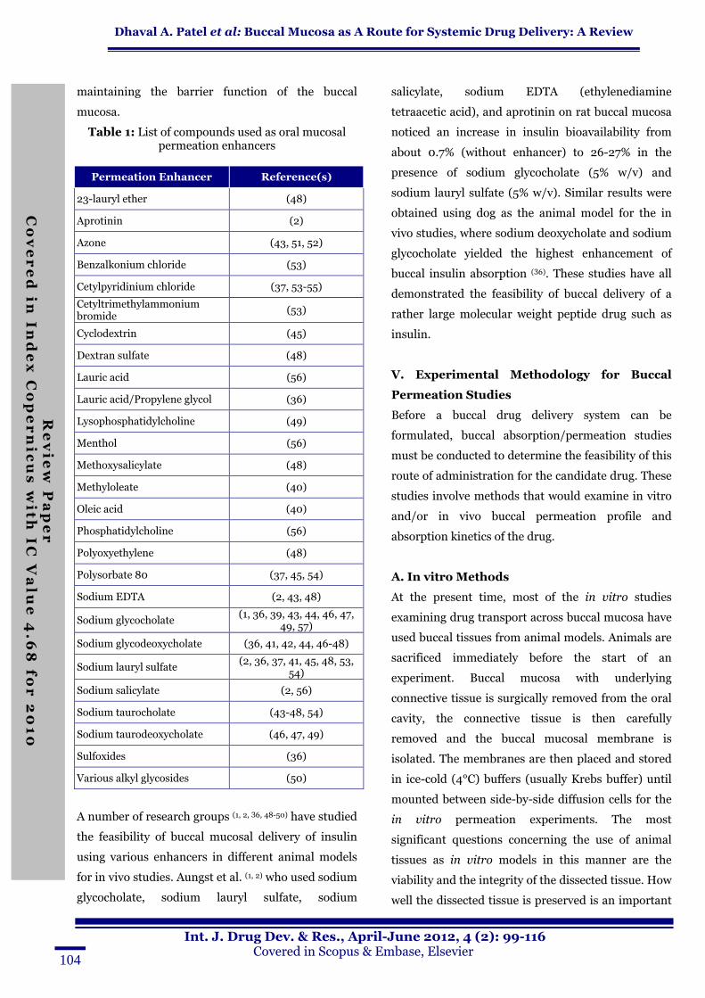
low drug bioavailability. Various compounds have
been investigated for their use as buccal penetration
enhancers in order to increase the flux of drugs
through the mucosa
(Table 1). Since the buccal epithelium is similar in
structure to other stratified epithelia of the body,
enhancers used to improve drug permeation in other
absorptive mucosae have been shown to work in
improving buccal drug penetration (36). Drugs
investigated for buccal delivery using various
permeation/absorption enhancers range in both
molecular weight and physicochemical properties.
Small molecules such as butyric acid and butanol (37),
ionizable low molecular weight drugs such as
acyclovir (38, 39), propranolol (40), and salicylic acid (41),
large molecular weight hydrophilic polymers such as
dextrans (42), and a variety of peptides including
octreotide (43), leutinizing hormone releasing
hormone (LHRH) (44), insulin (36), and a-interferon (45)
have all been studied.
A series of studies (42, 46, 47) on buccal permeation of
buserelin and fluorescein isothiocyanate (FITC)
labelled dextrans reported the enhancing effects of
di- and tri-hydroxy bile salts on buccal penetration.
Their results showed that in the presence of the bile
salts, the permeability of porcine buccal mucosa to
FITC increased by a 100-200 fold compared to FITC
alone. The mechanism of penetration enhancement
of FITC-labelled dextrans by sodium glycocholate
(SGC) was shown to be concentration dependent (47).
Below 10 mM SGC, buccal permeation was increased
by increasing the intercellular transport and at 10
mM and higher concentrations by opening up a
transcellular route. Gandhi and Robinson (41)
investigated the mechanisms of penetration
enhancement of transbuccal delivery of salicylic acid.
They used sodium deoxycholate and sodium lauryl
sulfate as penetration enhancers, both of which were
found to increase the permeability of salicylic acid
across rabbit buccal mucosa. Their results also
supported that the superficial layers and protein
domain of the epithelium may be responsible for
Re
vie
w P
ap
er
C
ov
er
ed
in
In
de
x C
op
er
nic
us
wit
h I
C V
alu
e 4
.68
fo
r 2
01
0
Dhaval A. Patel et al: Buccal Mucosa as A Route for Systemic Drug Delivery: A Review
Int. J. Drug Dev. & Res., April-June 2012, 4 (2): 99-116 Covered in Scopus & Embase, Elsevier
103
maintaining the barrier function of the buccal
mucosa.
Table 1: List of compounds used as oral mucosal permeation enhancers
Permeation Enhancer Reference(s)
23-lauryl ether (48)
Aprotinin (2)
Azone (43, 51, 52)
Benzalkonium chloride (53)
Cetylpyridinium chloride (37, 53-55)
Cetyltrimethylammonium bromide
(53)
Cyclodextrin (45)
Dextran sulfate (48)
Lauric acid (56)
Lauric acid/Propylene glycol (36)
Lysophosphatidylcholine (49)
Menthol (56)
Methoxysalicylate (48)
Methyloleate (40)
Oleic acid (40)
Phosphatidylcholine (56)
Polyoxyethylene (48)
Polysorbate 80 (37, 45, 54)
Sodium EDTA (2, 43, 48)
Sodium glycocholate (1, 36, 39, 43, 44, 46, 47,
49, 57)
Sodium glycodeoxycholate (36, 41, 42, 44, 46-48)
Sodium lauryl sulfate (2, 36, 37, 41, 45, 48, 53,
54)
Sodium salicylate (2, 56)
Sodium taurocholate (43-48, 54)
Sodium taurodeoxycholate (46, 47, 49)
Sulfoxides (36)
Various alkyl glycosides (50)
A number of research groups (1, 2, 36, 48-50) have studied
the feasibility of buccal mucosal delivery of insulin
using various enhancers in different animal models
for in vivo studies. Aungst et al. (1, 2) who used sodium
glycocholate, sodium lauryl sulfate, sodium
salicylate, sodium EDTA (ethylenediamine
tetraacetic acid), and aprotinin on rat buccal mucosa
noticed an increase in insulin bioavailability from
about 0.7% (without enhancer) to 26-27% in the
presence of sodium glycocholate (5% w/v) and
sodium lauryl sulfate (5% w/v). Similar results were
obtained using dog as the animal model for the in
vivo studies, where sodium deoxycholate and sodium
glycocholate yielded the highest enhancement of
buccal insulin absorption (36). These studies have all
demonstrated the feasibility of buccal delivery of a
rather large molecular weight peptide drug such as
insulin.
V. Experimental Methodology for Buccal
Permeation Studies
Before a buccal drug delivery system can be
formulated, buccal absorption/permeation studies
must be conducted to determine the feasibility of this
route of administration for the candidate drug. These
studies involve methods that would examine in vitro
and/or in vivo buccal permeation profile and
absorption kinetics of the drug.
A. In vitro Methods
At the present time, most of the in vitro studies
examining drug transport across buccal mucosa have
used buccal tissues from animal models. Animals are
sacrificed immediately before the start of an
experiment. Buccal mucosa with underlying
connective tissue is surgically removed from the oral
cavity, the connective tissue is then carefully
removed and the buccal mucosal membrane is
isolated. The membranes are then placed and stored
in ice-cold (4°C) buffers (usually Krebs buffer) until
mounted between side-by-side diffusion cells for the
in vitro permeation experiments. The most
significant questions concerning the use of animal
tissues as in vitro models in this manner are the
viability and the integrity of the dissected tissue. How
well the dissected tissue is preserved is an important
Re
vie
w P
ap
er
C
ov
er
ed
in
In
de
x C
op
er
nic
us
wit
h I
C V
alu
e 4
.68
fo
r 2
01
0
Dhaval A. Patel et al: Buccal Mucosa as A Route for Systemic Drug Delivery: A Review
Int. J. Drug Dev. & Res., April-June 2012, 4 (2): 99-116 Covered in Scopus & Embase, Elsevier
104

issue which will directly affect the results and
conclusion of the studies. To date, there are no
standard means by which the viability or the integrity
of the dissected tissue can be assessed. Dowty et al.
(58) studied tissue viability by using ATP levels in
rabbit buccal mucosa. Using ATP levels as an
indicator for tissue viability is not necessarily an
accurate measure, however. Dowty et al. reported a
50% drop in the tissue ATP concentration during the
initial 6 hours of the experiment without a
corresponding drop in tissue permeability. Despite
certain gradual changes, the buccal tissue seems to
remain viable for a rather long period of time.
Therefore, a decrease in ATP levels does not assure a
drop in permeability characteristics of the tissue. The
most meaningful method to assess tissue viability is
the actual permeation experiment itself, if the drug
permeability does not change during the time course
of the study under the specific experimental
conditions of pH and temperature, then the tissue is
considered viable.
Buccal cell cultures have also been suggested as
useful in vitro models for buccal drug permeation
and metabolism (25, 59-61). However, to utilize these
culture cells for buccal drug transport, the number of
differentiated cell layers and the lipid composition of
the barrier layers must be well characterized and
controlled. This has not yet been achieved with the
buccal cell cultures used thus far.
B. In vivo Methods
In vivo methods were first originated by Beckett and
Triggs (62) with the so-called buccal absorption test.
Using this method, the kinetics of drug absorption
were measured. The methodology involves the
swirling of a 25 ml sample of the test solution for up
to 15 minutes by human volunteers followed by the
expulsion of the solution. The amount of drug
remaining in the expelled volume is then determined
in order to assess the amount of drug absorbed. The
drawbacks of this method include salivary dilution of
the drug, accidental swallowing of a portion of the
sample solution, and the inability to localize the drug
solution within a specific site (buccal, sublingual, or
gingival) of the oral cavity. Various modifications of
the buccal absorption test have been carried out (63-66)
correcting for salivary dilution and accidental
swallowing, but these modifications also suffer from
the inability of site localization. A feasible approach
to achieve absorption site localization is to retain the
drug on the buccal mucosa using a bioadhesive
system (67-69). Pharmacokinetic parameters such as
bioavailability can then be calculated from the
plasma concentration vs. time profile.
Other in vivo methods include those carried out
using a small perfusion chamber attached to the
upper lip of anesthetized dogs (70, 71). The perfusion
chamber is attached to the tissue by cyanoacrylate
cement. The drug solution is circulated through the
device for a predetermined period of time and sample
fractions are then collected from the perfusion
chamber (to determine the amount of drug
remaining in the chamber) and blood samples are
drawn after 0 and 30 minutes (to determine amount
of drug absorbed across the mucosa).
C. Experimental Animal Species
Aside from the specific methodology employed to
study buccal drug absorption/permeation
characteristics, special attention is warranted to the
choice of experimental animal species for such
experiments. For in vivo investigations, many
researchers have used small animals including rats (1,
36, 37) and hamsters (51, 54, 72) or permeability studies.
However, such choices seriously limit the value of the
data obtained since, unlike humans, most laboratory
animals have an oral lining that is totally keratinized.
The rat has a buccal mucosa with a very thick,
keratinized surface layer. The rabbit is the only
laboratory rodent that has non-keratinized mucosal
lining similar to human tissue and has been
extensively utilized in experimental studies (48, 55, 58, 73,
Re
vie
w P
ap
er
C
ov
er
ed
in
In
de
x C
op
er
nic
us
wit
h I
C V
alu
e 4
.68
fo
r 2
01
0
Dhaval A. Patel et al: Buccal Mucosa as A Route for Systemic Drug Delivery: A Review
Int. J. Drug Dev. & Res., April-June 2012, 4 (2): 99-116 Covered in Scopus & Embase, Elsevier
105
74). The difficulty in using rabbit oral mucosa,
however, is the sudden transition to keratinized
tissue at the mucosal margins making it hard to
isolate the desired non-keratinized region (21). The
oral mucosa of larger experimental animals that has
been used for permeability and drug delivery studies
include monkeys (75), dogs (34, 57, 65, 70), and pigs (42, 47,
76-80). Due to the difficulties associated with
maintenance of monkeys, they are not very practical
models for buccal drug delivery applications. Instead,
dogs are much easier to maintain and considerably
less expensive than monkeys and their buccal mucosa
is non-keratinized and has a close similarity to that of
the human buccal mucosa. Pigs also have non-
keratinized buccal mucosa similar to that of human
and their inexpensive handling and maintenance
costs make them an equally attractive animal model
for buccal drug delivery studies. In fact, the oral
mucosa of pigs resembles that of human more closely
than any other animal in terms of structure and
composition (20, 81). However, for use in in vivo
studies pigs are not as ideal as dogs due to their rapid
growth which renders the animal handling rather
difficult. Miniature breeds of pigs can be used but
their high cost is a deterrent. For in vitro studies
though, because of easy availability and low cost
porcine tissue is more suited as compared to dog
buccal tissue.
VI. Buccal Drug Delivery Systems
Other than the low flux associated with buccal
mucosal delivery, a major limitation of the buccal
route of administration is the lack of dosage form
retention at the site of absorption. Consequently,
bioadhesive polymers have extensively been
employed in buccal drug delivery systems.
Bioadhesive polymers are defined as polymers that
can adhere onto a biological substrate. The term
mucoadhesion is applied when the substrate is
mucosal tissue (27). Polymers which can adhere to
either hard or soft tissue have been used for many
years in surgery and dentistry. Diverse classes of
polymers have been investigated for their potential
use as mucoadhesives. These include synthetic
polymers such as monomeric a cyanoacrylate (82),
polyacrylic acid (82), hydroxypropyl methylcellulose
(17), and poly methacrylate derivatives as well as
naturally occurring polymers such as hyaluronic acid
(84) and chitosan (85). Other synthetic polymers such
as polyurethanes, epoxy resins, polystyrene, and
natural-product cement have also been extensively
investigated (86).
In general, dosage forms designed for buccal
administration should not cause irritation and should
be small and flexible enough to be accepted by the
patient. These requirements can be met by using
hydrogels. Hydrogels are hydrophilic matrices that
are capable of swelling when placed in aqueous
media (87). Normally, hydrogels are crosslinked so
that they would not dissolve in the medium and
would only absorb water. When drugs are loaded into
these hydrogels, as water is absorbed into the matrix,
chain relaxation occurs and drug molecules are
released through the spaces or channels within the
hydrogel network. In a more broad meaning of the
term, hydrogels would also include water-soluble
matrices that are capable of swelling in aqueous
media, these include natural gums and cellulose
derivatives. These ‘pseudo-hydrogels’ swell infinitely
and the component molecules dissolve from the
surface of the matrix. Drug release would then occur
through the spaces or channels within the network as
well as through the dissolution and/or the
disintegration of the matrix. The use of hydrogels as
adhesive preparations for transmucosal drug delivery
has acquired considerable attention in recent years.
Table 2 summarizes the related research on
mucoadhesive polymers and delivery systems.
Re
vie
w P
ap
er
C
ov
er
ed
in
In
de
x C
op
er
nic
us
wit
h I
C V
alu
e 4
.68
fo
r 2
01
0
Dhaval A. Patel et al: Buccal Mucosa as A Route for Systemic Drug Delivery: A Review
Int. J. Drug Dev. & Res., April-June 2012, 4 (2): 99-116 Covered in Scopus & Embase, Elsevier
106

Table 2: Related research on mucoadhesive polymers and delivery systems
Bioadhesive Polymer(s) Studied Investigation Objectives Reference
HPC and CP Preferred mucoadhesive strength on CP, HPC, and HPC-CP combination
(57)
HPC and CP Measured Bioadhesive property using mouse peritoneal membrane (88)
CP, HPC, PVP, CMC Studied inter polymer complexation and its effects on bioadhesive strength
(89)
CP and HPMC Formulation and evaluation of buccoadhesive controlled release delivery systems
(90)
HPC, HEC, PVP, and PVA Tested mucosal adhesion on patches with two-ply laminates with an impermeable backing layer and hydrocolloid polymer layer
(91)
HPC and CP Used HPC-CP powder mixture as peripheral base for strong adhesion and HPC-CP freeze dried mixture as core base
(30)
CP, PIP, and PIB Used a two roll milling method to prepare a new bioadhesive patch formulation
(92)
Xanthum gum and Locust bean gum Hydrogel formation by combination of natural gums (93)
Chitosan, HPC, CMC, Pectin, Xantham gum, and Polycarbophil Evaluate mucoadhesive properties by routinely measuring the detachment force form pig intestinal mucosa
(85)
Hyaluronic acid benzyl esters, Polycarbophil, and HPMC Evaluate mucoadhesive properties (84)
Hydroxyethylcellulose Design and synthesis of a bilayer patch (polytef-disk) for thyroid gland diagnosis
(94)
Polycarbophil Design of a unidirectional buccal patch for oral mucosal delivery of peptide drugs
(70)
Poly(acrylic acid) and Poly(methacrylic acid) Synthesized and evaluated crosslinked polymers differing in charge densities and hydrophobicity
(82)
Number of Polymers including HPC, HPMC, CP, CMC. Measurement of bioadhesive potential and to derive meaningful information on the structural requirement for bioadhesion
(86)
Poly(acrylic acid-co-acrylamide) Adhesion strength to the gastric mucus layer as a function of crosslinking agent, degree of swelling, and carboxyl group density
(95)
Poly(acrylic acid) Effects of PAA molecular weight and crosslinking concentration on swelling and drug release characteristics
(96)
Poly(acrylic acid-co-methyl methacrylate) Effects of polymer structural features on mucoadhesion (83, 97)
Poly(acrylic acid-co- butylacrylate) Relationships between structure and adhesion for mucoadhesive polymers
(16)
HEMA copolymerized with Polymeg® (polytetramethylene glycol) Bioadhesive buccal hydrogel for controlled release delivery of buprenorphine
(98)
Cydot® by 3M (bioadhesive polymeric blend of CP and PIB) Patch system for buccal mucoadhesive drug delivery (69, 99)
Formulation consisting of PVP, CP, and cetylpyridinium chloride (as stabilizer)
Device for oramucosal delivery of LHRH - device containing a fast release and a slow release layer
(44)
CMC, Carbopol 974P, Carbopol EX-55, Pectin (low viscosity), Chitosan chloride,
Mucoadhesive gels for intraoral delivery (100)
CMC, CP, Polyethylene oxide, Polymethylvinylether/Maleic anhydride (PME/MA), and Tragacanth
Buccal mucoadhesive device for controlled release anticandidal device - CMC tablets yielded the highest adhesive force
(101)
HPMC and Polycarbophil (PC) Buccal mucoadhesive tablets with optimum blend ratio of 80:20 PC to HPMC yielding the highest force of adhesion
(102)
PVP, Poly(acrylic acid) Transmucosal controlled delivery of isosorbide dinitrate (103, 104)
Poly(acrylic acid-co-poly ethyleneglycol) copolymer of acrylic acid and poly ethyleneglycol monomethylether monomethacryalte
To enhance the mucoadhesive properties of PAA for buccal mucoadhesive drug delivery
(105-107)
Poly acrylic acid and poly ethylene glycol To enhance mucoadhesive properties of PAA by interpolymer complexation through template polymerization
(108)
Drum dried waxy maize starch (DDWM), Carbopol 974P, and sodium stearylfumarate
Bioadhesive erodible buccal tablet for progesterone delivery (109)
Abbreviations: CP = Carbopol 934P, HPC = Hydroxy propyl cellulose, PVP = Poly(vinyl pyrrolidone), CMC = Sodium carboxymethyl cellulose, HPMC = Hydroxy propyl methyl cellulose, HEC = Hydroxy ethyl cellulose,
PVA = Poly(vinyl alcohol), PIB = Poly(isobutylene), PIP = Poly(isoprene).
Nagai et al. (35) studied the applicability of
hydroxypropyl cellulose (HPC) as a mucoadhesive
agent, they found this high viscosity grade material to
be a suitable adhesive for topical mucus membranes.
They reported the combination of HPC and carbopol
934P (CP) to produce a preferable material for
mucoadhesive dosage forms. They examined directly
compressed tablets of these polymers by placing
them on an agar gel bed. HPC tablets showed a slight
adhesion but dissolved easily on the gel bed. On the
other hand, CP tablets showed strong adhesion but
the swollen CP tablets seemed too hard. The
combination of HPC and CP provided the
mucoadhesion and adequate softness to prepare the
tablets. Satoh et al. (88) measured the bioadhesive
property of tablets consisting of HPC and CP using
mouse peritoneal membrane. The adhesive force of
the HPC-CP tablet was affected by the mixing ratio of
Re
vie
w P
ap
er
C
ov
er
ed
in
In
de
x C
op
er
nic
us
wit
h I
C V
alu
e 4
.68
fo
r 2
01
0
Dhaval A. Patel et al: Buccal Mucosa as A Route for Systemic Drug Delivery: A Review
Int. J. Drug Dev. & Res., April-June 2012, 4 (2): 99-116 Covered in Scopus & Embase, Elsevier
1078

HPC and CP. The adhesion force showed a minimum
value at the mixing ratio of 3:2 (HPC:CP) due to the
formation of an inter-polymer complex between HPC
and CP in the acidic pH range.
Complex formation between CP and HPC seemed to
suppress the interaction between molecules of
hydrogel and the mucus membrane, and the
adhesion force was therefore most reduced at a
mixture ratio of 1:4 (HPC/CP). Inter-polymer
complexation and its effect on bioadhesive strength
was also studied by Gupta et al. (89). They reported
that CP shows strong complexation with poly(vinyl
pyrrolidone) and hydroxypropyl cellulose, but very
little with sodium carboxy methyl cellulose. The
degree of complexation was higher at acidic pH and
decreased with an increase in pH. Anlar et al. (90)
reported on formulation and evaluation of
buccoadhesive controlled release systems for the
delivery of morphine sulfate. They prepared tablets
by direct compression of carbomer and
hydroxypropyl methyl cellulose (HPMC) and found
the drug release behavior to be non-Fickian and also
confirmed interpolymer complex formation between
HPMC and carbomer in acidic pH medium.
Anders and Merkle (91), developed and evaluated
adhesive patches for buccal administration,
consisting of two-ply laminates of an impermeable
backing layer and a hydrocolloid polymer layer
containing the drug. The polymers used HPC, HEC,
PVP, and PVA. The integrity of the laminate was
based on adhesive bonds between the hydrocolloid
layer and an agarose layer grafted to one side of the
backing layer sheet. Their work showed that among
the cellulose ethers studied HEC and HPC possessed
superior mucosal adhesion. Ishida et al. (30) utilized
similar materials in a lidocaine delivery system for
toothache. They used an HPC-CP powder mixture as
a peripheral base aiming at strong adhesion, but an
HPC-CP freeze-dried mixture was used as a core
base. HPC and CP formed a complex during the
freeze drying process, and this contributed to the
ease of admixing by blockage of the functional group
of CP. Guo (92) used a two roll milling method to
prepare a new bioadhesive polymer patch
formulation for controlled drug delivery consisting of
CP, PIB, and PIP. It was found that the surface
properties of buccal patches were not only dependent
on the CP content but also dependent on the PIB:PIP
ratio. The strongest peel strength was found on
buccal patches with a CP:PIB:PIP ratio of
50:43.75:6.25.
Watanabe et al. (93) reported on hydrogels formed by
the combination of natural gums, xantham gum, and
locust bean gum, which are applicable in buccal
delivery systems. Xantham gum is a natural gum
obtained through fermentation of glucose by
Xanthamonas campestris. Locust bean gum and
xantham gum alone cannot form a hydrogel.
However, when a mixture of these gums is dissolved
in a neutral medium at 90°C and then cooled with ice
for 30 min, a clear, strong hydrogel is formed. The
mechanism of gel formation was reported to be the
formation of a three dimensional network by
interaction between the double helix structure of
xantham gum and the straight molecular chain of
locust bean gum. The gel strength of the hydrogels
was affected by the mixing ratio of the gums, and the
addition of sucrose improved the sustained release
properties of the hydrogels. The hydrogel consisting
of xantham gum and locust bean gum showed only a
low mucoadhesion, but it can be applied to a buccal
delivery system because of its safety, gel strength,
sustained release properties and good feel in the
mouth.
Anders et al. (94) designed a bilayer patch (polytef-
disk) consisting of protirelin for thyroid gland
diagnosis. The patch had a backing layer of teflon and
mucoadhesive layer of protirelin dispersed in
hydroxyethylcellulose. Veillard et al. (70) reported the
use of a unidirectional buccal patch which consisted
of three layers: an impermeable backing layer, a rate
limiting center membrane containing the drug, and a
Re
vie
w P
ap
er
C
ov
er
ed
in
In
de
x C
op
er
nic
us
wit
h I
C V
alu
e 4
.68
fo
r 2
01
0
Dhaval A. Patel et al: Buccal Mucosa as A Route for Systemic Drug Delivery: A Review
Int. J. Drug Dev. & Res., April-June 2012, 4 (2): 99-116 Covered in Scopus & Embase, Elsevier
108

mucoadhesive layer containing bioadhesive polymer
polycarbophil. The bioadhesive polymer swells,
creating a flexible network through which diffusion of
drug takes place. This patch was tested in dog buccal
mucosa and was shown to remain in place for up to
17 hours without any obvious discomfort.
Ch’ng et al. (82) synthesized and evaluated a series of
crosslinked, swellable polymers of acrylic acid and
methacrylic acid, differing in charge densities and
hydrophobicity. They found that an increase in the
number of hydrophobic groups in the polymer
structure reduced hydration whereas the density of
the polymer was unaffected. Furthermore, polymers
of acrylic acid loosely crosslinked (0.3 % w/w) with 3
different agents, divinyl glycol, 2,5-dimethyl-1,5-
hexadiene, and divinyl benzene, showed the same
degree of bioadhesion while poly(methacrylic acid)
crosslinked with divinyl benzene showed reduced
bioadhesion. The small percent of crosslinking agent,
irrespective of physicochemical properties, did not
contribute substantially to bioadhesion, whereas the
starting monomer had a large effect. The effect of pH
on the bioadhesion of poly(acrylic acid) crosslinked
with divinyl glycol was also studied at constant
temperature, ionic strength, and osmolality. It was
found that the polymer showed maximum adhesion
at pH 5 and 6 and a minimum at pH 7.
Park and Robinson (86) examined a large number of
polymers as to their bioadhesive potential and to
derive meaningful information on the structural
requirements for bioadhesion. They concluded that
charged carboxylated polyanions are good potential
bioadhesives for drug delivery. To understand the
role of the carboxyl groups in mucoadhesion, acrylic
acid-acrylamide random copolymers [P(AA-co-AM)]
were synthesized (95) and the adhesion strength of the
crosslinked polymers to the gastric mucus layer as a
function of pH, initial concentration of the
crosslinking agent, degree of swelling, and carboxyl
group density were examined. From the study on
mucoadhesion of various P(AA-co-AM), it was found
that at least 80% of the vinyl groups of the polymer
must possess carboxyl groups in the protonated
form. The dependence of mucoadhesion on pH and
carboxyl-group density suggests that mucoadhesion
occurs through hydrogen bonding. In addition, the
density of the crosslinking agent significantly affects
mucoadhesion. As the density of the crosslinking
agent is lowered, the mucoadhesive strength
increases, although the density of carboxyl groups in
the test surface area is reduced. It was concluded that
for mucoadhesion to occur, polymers must have
functional groups that are able to form hydrogen
bonds above the critical concentration (80% for vinyl
polymers), and the polymer chains should be flexible
enough to form as many hydrogen bonds as possible.
Similar results were achieved by Garcia-Gonzalez et
al. (96) who evaluated the effects of poly(acrylic acid)
(PAA) molecular weight and the concentration of
crosslinking agent (sucrose) on swelling and drug
(metoclopramide) release characteristics of PAA
(carbopol) hydrogels. They reported that both factors
and the interaction term had significant effects on
hydrogel swelling and drug release. In particular,
they found that increased sucrose concentration
(high crosslinking density) led to reduced swelling
and reduced drug release efficiency.
Leung and Robinson (83, 97) studied the contribution
of anionic polymer structural features to
mucoadhesion using 0.2% crosslinked copolymers of
acrylic acid and methyl methacrylate. Their results
showed that the expanded nature of both the
interacting mucus and polymer networks influences
the strength of mucoadhesion. They concluded that
mucoadhesive polymer with the desired
mucoadhesive strength can be designed by
controlling the percentage of charged groups and the
corresponding openness of the network.
Bodde et al. (16) investigated relationships between
structure and adhesion for mucoadhesive polymers.
Their study was based on an assumption that
bioadhesion should possess two properties: (i)
Re
vie
w P
ap
er
C
ov
er
ed
in
In
de
x C
op
er
nic
us
wit
h I
C V
alu
e 4
.68
fo
r 2
01
0
Dhaval A. Patel et al: Buccal Mucosa as A Route for Systemic Drug Delivery: A Review
Int. J. Drug Dev. & Res., April-June 2012, 4 (2): 99-116 Covered in Scopus & Embase, Elsevier
109

optimal polarity to make sure the polymer is "wetted"
by the mucus, and (ii) optimal fluidity to allow for the
mutual adsorption and interpenetration of polymer
and mucus to take place. They studied acrylic
polymer films, designed for buccal drug delivery. The
films were made either through mixing or
copolymerization of poly(butyl acrylate) and
poly(acrylic acid). In both cases satisfactory
mucoadhesion was found within a range of
compositions optimized for surface polarity and the
fluidity of the polymer film.
In an attempt to enhance the intrinsic mucoadhesive
properties of poly (acrylic acid), Shojaei and Li (105-107)
designed and formulated a series of novel copolymers
of acrylic acid and poly ethyleneglycol
monomethylether monomethacrylate [P(AA-co-
PEG)]. The addition of PEG into the polymer
increased the potential for hydrogen bond formation,
since the lone pair electrons of oxygen in the repeat
unit (CH2CH2O) of PEG served as hydrogen bond
acceptors (107). The surface properties of PAA for
mucoadhesion were also improved by the PEG
incorporation. Using these copolymers, a patch
device was prepared for buccal acyclovir delivery and
the feasibility of such delivery was proven in vitro
with the incorporation of sodium glycocholate as the
permeation enhancer (39).
Using copolymeric hydrogel discs of HEMA
(monomer) and Polymeg® (macromer) a buccal
mucoadhesive device for controlled release of
buprenorphine was developed (98). The hydrogel
containing a monomer:macromer ratio of 80:20
(w/w) yielded the best result both in terms of
adhesion and drug release. The device was applied
for a 3 hour application time and steady state levels
were maintained for the time of application.
Formulation of another buccal delivery system was
reported by DeGrande et al. (69, 99) from the 3M
company. The buccal patch device (Cydot; 3M
Pharmaceutical, St. Paul, MN) consists of a flexible
mucoadhesive matrix composed of a blend of
poly(acrylic acid) (Carbopol 934P; B.F. Goodrich,
Cleveland, OH) and poly(isobutylene) (Vistanex;
Exxon Chemical Company, Houston, TX). The patch
device is unidirectional with a polyurethane backing
layer. The patch is intended for application to the
upper gum and in vivo studies in human subjects
have revealed effective bioadhesive characteristics for
12 hours of application (69). Several investigators have
reported on the development of TmTs (transmucosal
therapeutic systems) devices with a field-shaped
bilayer design consisting of fast-release and
sustained-release layers (44, 103, 104). The fast release
layer contains PVP as the bioadhesive component
and is designed to adhere to the buccal mucosa and
the sustained release layer consists of a mixture of
PVP and poly (acrylic acid) and is intended to adhere
to the gingival mucosa (103). Most recently, a TmTs
formulation was reported for the buccal delivery of
LHRH (luetinizing hormone releasing hormone) (44)
with results indicating the feasibility of controlled
release transmucosal delivery of the peptide drug.
VI. Conclusion
The buccal mucosa offers several advantages for
controlled drug delivery for extended periods of time.
The mucosa is well supplied with both vascular and
lymphatic drainage and first-pass metabolism in the
liver and pre-systemic elimination in the
gastrointestinal tract are avoided. The area is well
suited for a retentive device and appears to be
acceptable to the patient. With the right dosage form
design and formulation, the permeability and the
local environment of the mucosa can be controlled
and manipulated in order to accommodate drug
permeation. Buccal drug delivery is a promising area
for continued research with the aim of systemic
delivery of orally inefficient drugs as well as a feasible
and attractive alternative for non-invasive delivery of
potent peptide and protein drug molecules. However,
the need for safe and effective buccal
permeation/absorption enhancers is a crucial
Re
vie
w P
ap
er
C
ov
er
ed
in
In
de
x C
op
er
nic
us
wit
h I
C V
alu
e 4
.68
fo
r 2
01
0
Dhaval A. Patel et al: Buccal Mucosa as A Route for Systemic Drug Delivery: A Review
Int. J. Drug Dev. & Res., April-June 2012, 4 (2): 99-116 Covered in Scopus & Embase, Elsevier
110
component for a prospective future in the area of
buccal drug delivery.
References
1) Aungst, B.J., Rogers, N.J., and Shefter, E.,
Comparison of nasal, rectal, buccal, sublingual and
intramuscular insulin efficacy and the effects of a
bile salt absorption promoter, The J. Pharmacol.
Exp. Ther., 244:23-27, 1988.
2) Aungst, B.J. and Rogers, N.J., Site dependence of
absorption-promoting actions of Laureth-9, Na
salicylate, Na2EDTA, and Aprotinin on rectal, nasal,
and buccal insulin delivery, Pharm. Res., 5:305-
308, 1988.
3) Lee, W.E., Permeation enhancers for the nasal
delivery of protein and peptide therapeutics, Bio
Pharm, 3:22-25, 1990.
4) Tengamnuay, P. and Mitra, A.K., Bile salt-fatty acid
mixed micelles as nasal absorption promoters of
peptides. I. Effects of ionic strength. adjuvant
composition, and lipid structure on the nasal
absorption of [D-Arg2]Kyotorphin, Pharm. Res.,
7:127-133, 1990.
5) Shao, Z. and Mitra, A.K., Nasal membrane and
intracellular protein and enzyme release by bile
salts and bile salt-fatty acid mixed micelles:
correlation with facilitated drug transport, Pharm.
Res., 9:1992, 1992.
6) Shao, Z. and Mitra, A.K., Bile salt fatty acid mixed
micelles as nasal absorption promoters. III. Effects
on nasal transport and enzymatic degradation of
acyclovir prodrugs, Pharm. Res., 11:243-250, 1994.
7) Soyani, A.P. and Chien, Y.W., Systemic delivery of
peptides and proteins across absorptive mucosae,
Crit. Rev. Therap. Drug Carrier Systems, 13:85-
184, 1996.
8) Adjei, A., Sundberg, D., Miller, J., and Chun, A.,
Bioavailability of leuprolide acetate following nasal
inhalation delivery to rats and healthy humans,
Pharm. Res., 9:244-249, 1992.
9) Shimamoto, T., Pharmaceutical aspects. Nasal and
depot formulations of leuprolide, J. Androl., 8:S14-
S16, 1987.
10) Dal Negra, R., Turco, P., Pomari, C., and Trevisan,
F., Calcitonin nasal spray in patienmts with chronic
asthma: a double-blind crossover study vs placebo,
Int. J. Clin. Pharmacol. Ther. Toxicol., 29:144-146,
1991.
11) Plosker, G.L. and McTavish, D., Intranasal
salcatonin (salmon calcitonin). A review of its
pharmacological properties and role in the
management of postmenopausal osteoporosis,
Drugs Aging, 8:378-400, 1996.
12) Reginster, J.Y. and Lecart, M.P., Efficacy and safety
of drugs for Paget's disease of bone, Bone, 17:485S-
488S, 1995.
13) Rathbone, M.J. and Hadgraft, J., Absorption of
drugs from the human oral cavity, Int. J. Pharm.,
74:9-24, 1991.
14) de Vries, M.E., Bodde, H.E., Verhoef, J.C., and
Junginger, H.E., Developments in buccal drug
delivery, Crit. Rev. Ther. Drug Carr. Sys., 8:271-
303, 1991.
15) Squier, C.A., The permeability of oral mucosa, Crit.
Rev. Oral Biol. Med., 2:13-32, 1991.
16) Bodde, H.E., De Vries, M.E., and Junginger, H.E.,
Mucoadhesive polymers for the buccal delivery of
peptides, structure-adhesiveness relationships, J.
Control. Rel., 13:225-231, 1990.
17) Gandhi, R.E. and Robinson, J.R., Bioadhesion in
drug delivery, Ind. J. Pharm. Sci., 50:145-152, 1988.
18) Harris, D. and Robinson, J.R., Drug delivery via the
mucous membranes of the oral cavity, J. Pharm.
Sci., 81:1-10, 1992. Reproduced with permission of
the American Pharmaceutical Association.
19) Wertz, P.W. and Squier, C.A., Cellular and
molecular basis of barrier function in oral
epithelium, Crit. Rev. Ther. Drug Carr. Sys., 8:237-
269, 1991.
20) Squier, C.A., Cox, P., and Wertz, P.W., Lipid content
and water permeability of skin and oral mucosa,
The J. Invest. Dermat., 96:123-126, 1991.
21) Squier, C.A. and Wertz, P.W. Structure and function
of the oral mucosa and implications for drug
delivery, in eds. M.J. Rathbone, Oral Mucosal Drug
Delivery, Marcel Dekker, Inc., New York, New York,
1-26, 1996.
22) Galey, W.R., Lonsdale, H.K., and Nacht, S., The in
vitro permeability of skin and buccal mucosa to
Re
vie
w P
ap
er
C
ov
er
ed
in
In
de
x C
op
er
nic
us
wit
h I
C V
alu
e 4
.68
fo
r 2
01
0
Dhaval A. Patel et al: Buccal Mucosa as A Route for Systemic Drug Delivery: A Review
Int. J. Drug Dev. & Res., April-June 2012, 4 (2): 99-116 Covered in Scopus & Embase, Elsevier
111

selected drugs and tritiated water, J. Invest.
Dermat., 67:713-717, 1976.
23) Gandhi, R.B. and Robinson, J.R., Oral cavity as a
site for bioadhesive drug delivery, Adv. Drug Del.
Rev., 13:43-74, 1994.
24) Squier, C.A. and Hall, B.K., The permeability of
mammalian non-keratinized oral epithelia to
horseraddish peroxidase applied in vivo and in
vitro, Arch. Oral Biol., 29:45-50, 1984.
25) Hill, M.W. and Squier, C.A., The permeability of
oral palatal mucosa maintained in organ culture, J.
Anat., 128:169-178, 1979.
26) Tabak, L.A., Levine, M.J., Mandel, I.D., and Ellison,
S.A., Role of salivary mucins in the protection of the
oral cavity, J. Oral Pathol., 11:1-17, 1982.
27) Peppas, N.A. and Buri, P.A., Surface, interfacial and
molecular aspects of polymer bioadhesion on soft
tissues, J. Control. Rel., 2:257-275, 1985.
28) Rathbone, M., Drummond, B., and Tucker, I., Oral
cavity as a site for systemic drug delivery, Adv.
Drug Del. Rev., 13:1-22, 1994.
29) Edgar, W.M., Saliva: its secretion, composition and
functions, Br. Dent. J., 172:305-312, 1992.
30) Ishida, M., Nambu, N., and Nagai, T., Mucosal
dosage form of lidocaine for toothache using
hydroxypropyl cellulose and carbopol, Chem.
Pharm. Bull., 30:980-984, 1982.
31) Collins, A.E.M., Deasy, P.B., Mac Carthy, D.J., and
Shanley, D.B., Evaluation of a controlled release
compact containing tetracycline hydrochloride
bonded to tooth for the treatment of periodontal
disease, Int. J. Pharm., 51:103-114, 1989.
32) Elkayam, R., Friedman, M., Stabholz, A., Soskolne,
A.w., Sela, M.N., and Golub, L., Sustained release
device containing minocycline for local treatment of
periodontal disease, J. Control. Rel., 7:231-236,
1988.
33) Samaranayake, L. and Ferguson, M., Delivery of
antifungal agents to the oral cavity, Adv. Drug Del.
Rev., 13:161-179, 1994.
34) Nagai, T., Adhesive topical drug delivery system, J.
Control. Rel., 2:121-134, 1985.
35) Nagai, T. and Machida, Y., Mucosal adhesive dosage
forms, Pharm. Int., 196-200, 1985.
36) Aungst, B.J. and Rogers, N.J., Comparison of the
effects of various transmucosal absorption
promoters on buccal insulin delivery, Int. J.
Pharm., 53:227-235, 1989.
37) Siegel, I.A. and Gordon, H.P., Surfactant-induced
increase of permeability of rat oral mucosa to non-
electolytes in vivo, Arch. Oral Biol., 30:43-47, 1985.
38) Shojaei, A.H. and Li, X., In vitro permeation of
acyclovir through porcine buccal mucosa,
Proceedings of International Symposium on
Controlled Release of Bioactive Materials, 23:507-
508, 1996.
39) Shojaei, A.H. and Li, X., Determination of transport
route of acyclovir across buccal mucosa, Proceed.
Int. Symp. Control. Rel. Bioact. Mater., 24:427-
428, 1997.
40) Manganaro, A.M. and Wertz, P.W., The effects of
permeabilizers on the in vitro penetration of
propranolol through porcine buccal epithelium, Mil.
Med., 161:669-672, 1996.
41) Gandhi, R. and Robinson, J., Mechanisms of
penetration enhancement for transbuccal delivery
of salicylic acid, Int. J. Pharm., 85:129-140, 1992.
42) Hoogstraate, A.J., Verhoef, J.C., Tuk, B., Pijpers, A.,
van leengoed, L.A.M.G., Vheijden, J.H.M.,
Junjinger, H.E., and Bodde, H.E., Buccal delivery of
fluorescein isothiocyanate-dextran 4400 and the
peptide drug buserelin with glycodeoxycholate as an
absorption enhancer in pigs, J. Control. Rel., 41:77-
84, 1996.
43) Wolany, G.J.M., Munzer, J., Rummelt, A., and
Merkle, H.P., Buccal absorption of Sandostatin
(octreotide) in conscious beagle dogs, Proceed.
Intern. Symp. Control. Rel. Bioact. Mater., 17:224-
225, 1990.
44) Nakane, S., Kakumoto, M., Yulimatsu, K., and
Chien, Y.W., Oramucosal delivery of LHRH:
Pharmacokinetic studies of controlled and
enhanced transmucosal permeation, Pharm. Dev.
Tech., 1:251-259, 1996.
45) Steward, A., Bayley, D.L., and Howes, C., The effect
of enhancers on the buccal absorption of hybrid
(BDBB) alpha-interferom, Int. J. Pharm., 104:145-
149, 1994.
Re
vie
w P
ap
er
C
ov
er
ed
in
In
de
x C
op
er
nic
us
wit
h I
C V
alu
e 4
.68
fo
r 2
01
0
Dhaval A. Patel et al: Buccal Mucosa as A Route for Systemic Drug Delivery: A Review
Int. J. Drug Dev. & Res., April-June 2012, 4 (2): 99-116 Covered in Scopus & Embase, Elsevier
112

46) Senel, S., Hoogstraate, A.J., Spies, F., Verhoef, J.C.,
Bos-van Geest, A., Junginger, H.E., and Bodde,
H.E., Enhancement of in vitro permeability of
porcine buccal mucosa by bile salts: kinetic and
histological studies, J. Control. Rel., 32:45-56, 1994.
47) Hoogstraate, A.J., Senel, S., Cullander, C., Verhoef,
J., Junginger, H.E., and Bodde, H.E., Effects of bile
salts on transport rates and routes of FTIC-labelled
compounds across porcine buccal epithelium in
vitro, J. Control. Rel., 40:211-221, 1996.
48) Oh, C.K. and Ritschel, W.A., Biopharmaceutic
aspects of buccal absorption of insulin, Meth. Find
Exp. Clin. Pharmacol., 12:205-212, 1990.
49) Zhang, J., Niu, S., Ebert, C., and Stanley, T.H., An in
vivo dog model for studying recovery kinetics of the
buccal mucosa permeation barrier after exposure to
permeation enhancers: apparent evidence of
effective enhancement without tissue damage, Int.
J. Pharm., 101:15-22, 1994.
50) Aungst, B.J., Site-dependence and structure-effect
relationships for alkylglycosides as transmucosal
absorption promoters for insulin, Int. J. Pharm.,
105:219-225, 1994.
51) Kurosaki, Y., Hisaichi, S., Hong, L., Nakayama, T.,
and Kimura, T., Enhanced permeability of
keratinized oral-mucosa to salicylic acid with 1-
dodecylacycloheptan-2-one (Azone). In vitro studies
in hamster cheek pouch, Int. J. Pharm., 49:47-55,
1989.
52) Kurosaki, Y., Hisaichi, S., Nakayama, T., and
Kimura, T., Enhancing effect of 1-
dodecylazacycloheptan-2-one (Azone) on the
absorption of salicyclic acid from keratinized oral
mucosa and the duration of enhancement in vivo,
Int. J. Pharm., 51:47-54, 1989.
53) Siegel, I.A. and Gordon, H.P., Effects of surfactants
on the permeability of canine oral mucosa in vitro,
Tox. Lett., 26:153-157, 1985.
54) Kurosaki, Y., Hisaichi, S., Hamada, C., Nakayama,
T., and Kimura, T., Effects of surfactants on the
absorption of salicylic acid from hamster cheek
pouch as a model of keratinized oral mucosa, Int. J.
Pharm., 47:13-19, 1988.
55) Siegel, I.A., Izutsu, K.T., and Watson, E.,
Mechanisms of non-electrolyte penetration across
dog and rabbit oral mucosa in vitro, Arch. Oral
Biol., 26:357-361, 1981.
56) Coutel-Egros, A., Maitani, Y., Veillard, M., Machida,
Y., and Nagai, T., Combined effects of pH, cosolvent
and penetration enhancers on the in vitro buccal
absorption of propranolol through excised hamster
cheek pouch, Int. J. Pharm., 84:117-128, 1992.
57) Ishida, M., Machida, Y., Nambu, N., and Nagai, T.,
New mucosal dosage form of insulin, Chem. Pharm.
Bull., 29:810-816, 1981.
58) Dowty, M.E., Knuth, K.E., Irons, B.K., and
Robinson, J.R., Transport of thyrotropin releasing
hormone in rabbit buccal mucosa in vitro, Pharm.
Res., 9:1113-1122, 1992.
59) Tavakoli-Saberi, M.R. and Audus, K.L., Cultured
buccal epithelium: an in vitro model derived from
the hamster pouch for studying drug transport and
metabolism, Pharm. Res., 6:160-162, 1989.
60) Tavakoli-Saberi, M.R., Williams, A., and Audus,
K.L., Aminopeptidase activity in human buccal
epithelium and primary cultures of hamster buccal
epithelium, Pharm. Res., 6:S-197, 1989.
61) Leipold, H.R. and Quadros, E., Nicotine permeation
through buccal cultures, Proceed. Int. Symp.
Control. Rel. Bioact. Mater., 20:242-243, 1993.
62) Beckett, A.H. and Triggs, E.J., Buccal absorption of
basic drugs and its application as an in vivo model
of passive drug transfer through lipid membranes,
J. Pharm. Pharmacol., 19:31S-41S, 1967.
63) Schurmann, W. and Turner, P., A membrane model
of the human oral mucosa as derived from buccal
absorption performance and physicochemical
properties of the beta-blocking drugs atenolol and
propranolol, J. Pharm. Pharmacol., 30:137-147,
1978.
64) Tucker, I.G., A method to study the kinetics of oral
mucosal drug absorption from solutions, J. Pharm.
Pharmacol., 40:679-683, 1988.
65) Barsuhn, C.L., Olanoff, L.S., Gleason, D.D., Adkins,
E.L., and Ho, N.F.H., Human buccal absorption of
flubriprofen, Clin. Pharmacol. Ther., 44:225-231,
1988.
66) Gonzalez-Younes, I., Wagner, J.G., and Gaines,
D.A., Absorption of flubriprofen through human
buccal mucosa, J. Pharm. Sci., 80:820-823, 1991.
Re
vie
w P
ap
er
C
ov
er
ed
in
In
de
x C
op
er
nic
us
wit
h I
C V
alu
e 4
.68
fo
r 2
01
0
Dhaval A. Patel et al: Buccal Mucosa as A Route for Systemic Drug Delivery: A Review
Int. J. Drug Dev. & Res., April-June 2012, 4 (2): 99-116 Covered in Scopus & Embase, Elsevier
113

67) Benes, L., et al. Plasma melatonin (M) and
sulfatoxymelatonin (aMT6s) kinetics after
transmucosal administration to humans, in eds. Y.
Touitou, J. Arendt, andP. Pevet, Melatonin and the
pineal gland-From basic science to clinical
application, Elsevier Science Publishers B.V., New
York, 347-350, 1993.
68) McQuinn, R.L., Kvam, D.C., Maser, M.J., Miller,
A.L., and Oliver, A., Sustained oral mucosal delivery
in human volunteers of buprenorphine from thin
non-eroding mucoadhesive polymeric disks, J.
Control. Rel., 34:243-250, 1995.
69) Benes, L., et al., Transmucosal, oral controlled-
release, transdermal drug administration in human
subjects: A crossover study with melatonin, Journal
of Pharm. Sci., 86:1115-1119, 1997.
70) Veillard, M.M., Longer, M.A., Martens, T.W., and
Robinson, J.R., Preliminary studies of oral mucosal
delivery of peptide drugs, J. Control. Rel., 6:123-
131, 1987.
71) Yamahara, H. and Lee, V.H., Drug Metabolism in
the Oral Cavity, Adv. Drug Del. Rev., 12:25-39,
1993.
72) Tanaka, M., Yanagibashi, N., Fukuda, H., and
Nagai, T., Absorption of salicylic through the oral
mucous membrane of hamster cheek pouch, Chem.
Pharm. Bull., 28:1056-1061, 1980.
73) Siegel, I.A. and Gordon, H.P., Surfactant-induced
alterations of permeability of rabbit oral mucosa in
vitro, Exp. Mol. Path., 44:132-137, 1986.
74) Dowty, M.E., Knuth, K.E., and Robinson, J.R.,
Enzyme characterization studies on the rate-
limiting barrier in rabbit buccal mucosa, Int. J.
Pharm., 88:293-302, 1992.
75) Mehta, M., Kemppainen, B.W., and Staffford, R.G.,
In vitro penetration of tritium-labelled water (THO)
and [3H]PbTx-3 (a red tide toxin) through monkey
buccal mucosa and skin, Tox. Lett., 55:185-194,
1991.
76) Lesch, C.A., Squier, C.A., Cruchley, A., Williams,
D.M., and Speight, P., The permeability of human
oral mucosa and skin to water, J. Dent. Res.,
68:1345-1349, 1989.
77) Quadros, E., Cassidy, J., Gniecko, K., and LeRoy, S.,
Buccal and colonic absorption of CGS 16617, a novel
ACE inhibitor, J. Control. Rel., 19:77-86, 1991.
78) Squier, C.A. and Hall, B.K., The permeability of skin
and oral mucosa to water and horseradish
peroxidase as related to the thickness of the
permeability barrier, The J. Invest. Dermat.,
84:1985.
79) Hoogstraate, A.J., Cullander, C., Nagelkerke, J.F.,
Verhoel, J., Junginger, H.E., and Bodde, H.E.,
Diffusion rates and transport pathways of FITC-
labelled model compounds through buccal
epithelium, Proceed. Int. Symp. Control. Rel.
Bioact. Mater., 20:234-235, 1993.
80) Hoogstraate, A.J., Bodde, H.E., Cullander, C., and
Junginger, H.E., Diffusion rates and transport
pathways of FITC-labelled model compounds
through buccal epithelium, Pharm. Res., 9:S-188,
1992.
81) Collins, P., Laffoon, J., and Squier, C.A.,
Comparative study of porcine oral epithelium, J.
Dent. Res., 60:543, 1981.
82) Ch'ng, H.S., Park, H., Kelly, P., and Robinson, J.R.,
Bioadhesive polymers as platforms for oral
controlled drug delivery II: Synthesis and
evaluation of some swelling, water-insoluble
bioadhesive polymers, J. Pharm. Sci., 74:399-405,
1985.
83) Leung, S.S. and Robinson, J.R., Polymer structure
features contributing to mucoadhesion: II., J.
Control. Rel., 12:187-194, 1990.
84) Sanzgiri, Y.D., Topp, E.M., Benedetti, L., and Stella,
V.J., Evaluation of mucoadhesive properties of
hyaluronic caid benzyl esters, Int. J. Pharm.,
107:91-97, 1994.
85) Lehr, C.M., Bouwstra, J.A., Schact, E.H., and
Junginger, H.E., In vitro evaluation of
mucoadhesive properties of chitosan and some
other natural polymers, Int. J. Pharm., 78:43-48,
1992.
86) Park, K. and Robinson, J.R., Bioadhesive polymers
as platforms for oral-controlled drug delivery:
method to study bioadhesion, Int. J. Pharm.,
19:107-127, 1984.
Re
vie
w P
ap
er
C
ov
er
ed
in
In
de
x C
op
er
nic
us
wit
h I
C V
alu
e 4
.68
fo
r 2
01
0
Dhaval A. Patel et al: Buccal Mucosa as A Route for Systemic Drug Delivery: A Review
Int. J. Drug Dev. & Res., April-June 2012, 4 (2): 99-116 Covered in Scopus & Embase, Elsevier
114

87) Nagai, T. and Machida, Y., Buccal delivery systems
using hydrogels, Adv. Drug Del. Rev., 11:179-191,
1993.
88) Satoh, K., Takayama, K., Machida, Y., Suzuki, Y.,
Nakagaki, M., and Nagai, T., Factors affecting the
bioadhesive property of tablets consisting of
hydroxypropyl cellulose and carboxyvinyl polymer.,
Chem. Pharm. Bull., 37:1366-1368, 1989.
89) Gupta, A., Garg, S., and Khar, R.K., Interpolymer
complexation and its effect on bioadhesion strength
and dissolution characteristics of buccal drug
delivery systems, Drug Dev. Ind. Pharm., 20:315-
325, 1994.
90) Anlar, S., Capan, Y., Guven, O., Gogus, A., Dlakara,
T., and Hincal, A.A., Formulation and in-vitro-in
vivo evaluation of buccoadhesive morphine sulfate
tablets, Pharm. Res., 11:231-236, 1994.
91) Anders, R. and Merkle, H., Evaluation of laminated
muco-adhesive patches for buccal drug delivery, Int.
J. Pharm., 49:231-240, 1989.
92) Guo, J.-H., Investigating the surface properties and
bioadhesion of buccal patches, J. Pharm.
Pharmacol., 46:647-650, 1994.
93) Watanabe, K., Yakou, S., Takayama, K., Machida,
Y., and Nagai, T., Drug release behaviors from
hydrogel prepared with water soluble dietary fibers,
J. Pharm. Sci. Techn. Jpn., 51:29-35, 1991.
94) Anders, R., Merkle, H.P., Schurr, W., and Ziegler,
R., Buccal absorption of protirelin: an effective way
to stimulate thyrotropin and prolactin, J. Pharm.
Sci., 72:1481-1483, 1983.
95) Park, H. and Robinson, J.R., Mechanisms of
mucoadhesion of poly(acrylic acid) hydrogels,
Pharm. Res., 4:457-464, 1987.
96) Garcia-Gonzalez, N., Kellaway, I., Blanco-Fuente,
H., Anguiano-Igea, S., Delgado-Charro, B., Otero-
Espinar, F., and Blanco-Mendez, J., Design and
evaluation of buccoadhesive metoclopramide
hydrogels composed of poly(acrylic acid)
crosslinked with sucrose, Int. J. Pharm., 100:65-70,
1993.
97) Leung, S. and Robinson, J.R., The contribution of
anionic polymer structural features to
mucoadhesion, J. Control. Rel., 5:223-231, 1988.
98) Cassidy, J.P., Landzert, N.M., and Quadros, E.,
Controlled buccal delivery of buprenorphine, J.
Control. Rel., 25:21-29, 1993.
99) DeGrande, G., Benes, L., Horriere, F., Karsenty, F.,
LaCoste, C., McQuinn, R., Guo, J., and Scherrer, R.
Specialized oral mucosal drug delivery systems:
Patches, in eds. M.J. Rathbone, Oral Mucosal Drug
Delivery, Marcel Dekker, Inc., New York, 285-318,
1996.
100) Nguyen-Xuan, T., Towart, R., Terras, A., Jacques,
Y., Buri, P., and Gurny, R., Mucoadhesive semi-
solid formulations for intraoral use containing
sucralfate, Eur. J. Pharm. Biopharm., 43:133-137,
1996.
101) Nair, M. and Chien, Y.W., Development of
anticandidal delivery systems: (II) Mucoadhesive
devices for prolonged drug delivery in the oral
cavity, Drug Dev. Ind. Pharm., 22:243-253, 1996.
102) Taylan, B., Yilmaz, C., Guven, O., Kes, S., and
Hincal, A.A., Design and evaluation of sustained-
release and buccal adhesive propranolol
hydrochloride tablets, J. Control. Rel., 38:11-20,
1996.
103) Yukimatsu, K., Nozaki, Y., Makumoto, M., and
Ohta, M., Development of a trans-mucosal
controlled-release device for systemic of antianginal
drugs pharmacokinetics and pharmacodynamics,
Drug Dev. Ind. Pharm., 20:503-534, 1994.
104) Nozaki, Y., Ohta, M., and Chien, Y.W.,
Transmucosal controlled systemic delivery of
isosorbide dinitrate: in vivo/in vitro correlation, J.
Control. Rel., 43:105-114, 1997.
105) Shojaei, A.H. and Li, X., Novel copolymers of acrylic
acid and poly ethylene glycol monomethylether
monomethacrylate for buccal mucoadhesion:
Preparation and surface characterization, Pharm.
Res., 12 (S1):S210, 1995.
106) Li, X. and Shojaei, A.H., Novel copolymers of acrylic
acid and poly ethylene glycol monomethylether
monomethacrylate for buccoadhesion: In vitro
assessment of buccoadhesion and analysis of
surface free energy, Proceed. Int. Symp. Control.
Rel. Bioact. Mater., 23:165-166, 1996.
107) Shojaei, A.H. and Li, X., Mechanisms of buccal
mucoadhesion of novel copolymers of acrylic acid
Re
vie
w P
ap
er
C
ov
er
ed
in
In
de
x C
op
er
nic
us
wit
h I
C V
alu
e 4
.68
fo
r 2
01
0
Dhaval A. Patel et al: Buccal Mucosa as A Route for Systemic Drug Delivery: A Review
Int. J. Drug Dev. & Res., April-June 2012, 4 (2): 99-116 Covered in Scopus & Embase, Elsevier
115

and polyethylene glycol monomethylether
monomethacrylate, J. Control. Rel., 47:151-161,
1997.
108) Choi, H.K., Kim, O.J., Jung, C.K., Cho, Y.J., and
Cho, C.S., Interpolymer complex prepared by
template polymerization for transmucosal drug
delivery, Proceed. Int. Symp. Control. Rel. Bioact.
Mater., 24:415-416, 1997.
109) Voorspoels, J., Comhaire, F., De Sy, W., and
Remon, J.P., Buccal absorption of progesterone in
the dog, Proceed. Int. Symp. Control. Rel. Bioact.
Mater., 24:185-186, 1997.
Re
vie
w P
ap
er
C
ov
er
ed
in
In
de
x C
op
er
nic
us
wit
h I
C V
alu
e 4
.68
fo
r 2
01
0
Dhaval A. Patel et al: Buccal Mucosa as A Route for Systemic Drug Delivery: A Review
Int. J. Drug Dev. & Res., April-June 2012, 4 (2): 99-116 Covered in Scopus & Embase, Elsevier
116