Tracheal Tear Caused by Extubation of a Double-Lumen Tube
15
CASE REPORTS Anesthesiology 2002; 97:1007– 8 © 2002 American Society of Anesthesiologists, Inc. Lippincott Williams & Wilkins, Inc. Tracheal Tear Caused by Extubation of a Double-Lumen Tube Jonathan L. Benumof, M.D.,* Dickson Wu, M.D.† A COMPLETE tear of the posterior membrane of the trachea due to extubation of a single-lumen (SLT) or double-lumen tube (DLT) has never been described. The facts of the case reported herein show that extubation of a DLT in a patient with persistent complete collapse of the left upper lobe likely caused a 7-cm long, clean, midline tear of the posterior membrane of the trachea. Case Report The patient was a 76-yr-old, 153-cm, 63-kg woman who had the appearance of two new 1-cm opacifications in the left lung and was scheduled for thoracoscopic lung biopsy. The patient was on no medications, was allergic to erythromycin, had a remote history of hysterectomy, and had no significant present medical problems. On physical examination, the patient appeared frail, but all organ symp- toms functioned within normal limits, and all laboratory values were within normal limits. General anesthesia was induced with 2 mg/kg intravenous propofol, 5 g/kg fentanyl, and 0.05 mg/kg midazolam and was maintained with 0.5–1.0% isoflurane. Paralysis was induced with 0.1 mg/kg vecuro- nium. After paralysis, a styletted, 8.0-mm inside diameter SLT was easily inserted (the tip of the stylet was 2 cm from the tip of the SLT and was entirely intraluminal) with the aid of conventional laryngoscopy to a depth of 20 cm at the incisors, and fiberoptic bronchoscopy (5.0-mm outside diameter) revealed a normal tracheobronchial tree. The distal 7 cm of the trachea was available for inspection. Next, a 35-French, left-sided, relatively blunt-tipped DLT (Catalog #95895; Mallinkrodt Medical, St. Louis, MO) with stylet was inserted. The stylet was entirely within the left lumen and was removed as soon as the left endobron- chial cuff passed the vocal cords and before the 90° counterclockwise rotation of the DLT. Unilateral clamping and auscultation maneuvers revealed normal breath sounds and cannulation of the left lung with the endobronchial lumen of the DLT. Fiberoptic bronchoscopy (4.0-mm outside diameter) through the tracheal lumen revealed that withdrawal of the DLT by 2 cm was required in order to place the blue endobronchial cuff just below the tracheal carina. With the blue endobronchial cuff just below the tracheal carina, the distal 4 cm of the trachea was visualized and was found to be normal. Both tracheal and endobronchial cuffs were inflated so that the pilot balloons were soft (significantly compressible between the fingers with minimal effort), and there was no air leak by either cuff at a pressure of 30 cm H 2 O, as demonstrated by auscultation. The patient was disconnected from the ventilator and then easily turned to the right lateral decubitus position without deflation of the softly inflated cuffs and without any movement of the head and neck. For the next 75 min, only the right lung was ventilated with a tidal volume of 500 ml, a respiratory frequency of 12 breaths/min, a peak inspiratory pressure of 30 cm H 2 O, and a fraction of inspired oxygen of 1.0. During these 75 min, the surgeon biopsied and stapled shut a site in the lingula and left lower lobe. Prior to withdrawing the thoraco- scope, an attempt was made to expand the collapsed left lung with a large tidal volume and sustained peak inspiratory pressure of 40 cm H 2 O for 10 s. However, the left upper lobe (LUL) remained completely atelectatic. The LUL remained collapsed after both conventional suc- tioning down the left lumen of the DLT and after saline lavage and suctioning of thick, bloody secretions in the LUL through a fiberoptic bronchoscope (4.0-mm outside diameter) in the left lumen of the DLT. Next, the patient was disconnected from the ventilator and easily turned to the supine position with both pilot balloons to the cuffs of the DLT still softly inflated and without any movement of the head and neck. The patient was then reventilated with manual positive pressure ventilation for 2 min. The cuffs of the DLT were then completely deflated, and the DLT was withdrawn. During extubation of the DLT, no resistance to pulling the DLT out was noted. An 8.0-mm inside diameter, styletted SLT was easily inserted once again to a 20-cm depth at the incisors, and the cuff was inflated so that the pilot balloon was soft and no leak auscultated over three manual breaths. Fiberoptic bronchoscopy (5.0-mm outside diameter) down the new and second SLT (in order to lavage and suction the LUL) very surprisingly revealed a 7-cm long, complete, midline, clean tear of the membrane of trachea that began 2 or 3 mm above the carina and ended 7 cm cephalad. The distal 1 cm of the tear veered slightly to the right. Along the entire length of the tear, the esophagus could be seen to be bulging slightly into the lumen of the trachea. The peak inspiratory pressure was reduced to 12 cm H 2 O, the respiratory frequency was increased to 20 breaths/min, and the LUL was quickly lavaged and suctioned with a larger fiberoptic bronchoscope. A right trapdoor thoracotomy with slight left lateral body tilt was performed, the SLT was guided both fiberoptically and from within the chest into the left mainstem bronchus, and the tracheal tear was repaired by primary suture and secondary reinforcement with a peri- cardial flap. During the repair of the trachea, the tissues of the trachea were felt to be of normal strength and integrity. At the end of the repair of the tracheal tear, the distal end of the SLT was positioned just proximal to the proximal end of the sutured tear, and the patient was sent to the intensive care unit for postoperative mechanical ventilation. Pathologic diagnosis of the biopsy specimens was benign granuloma. The patient was mechanically ventilated and bronchoscoped daily for 1 month prior to extubation. For the next month after the initial extubation, the patient had repeated bouts of aspiration and required reintubation and mechanical ventilation three times. After the third reintubation, the patient had a tracheostomy. Nevertheless, she died 4 months later, malnourished and septic (perhaps from infection around a jejunostomy feeding tube). Discussion The tracheal tear most likely occurred between the end of the thoracoscopy and the insertion of the second SLT. There are two very important reasons why the * Professor of Anesthesia, Department of Anesthesiology, University of Cali- fornia San Diego Medical Center. † Assistant Professor of Anesthesia, Depart- ment of Anesthesia, Rush North Shore Medical Center, Rush Medical College. Received from the Department of Anesthesiology, University of California San Diego Medical Center, San Diego, California; and the Department of Anesthesia, Rush North Shore Medical Center, Rush Medical College, Chicago, Illinois. Submitted for publication December 10, 2001. Accepted for publication March 19, 2002. Support was provided solely from institutional and/or departmental sources. Address reprint requests to Dr. Benumof: Department of Anesthesiology, University of California San Diego Medical Center, 402 Dickinson Street, San Diego, California 92103-8812. Address electronic mail to: [email protected]. Individual article reprints may be purchased through the Journal Web site, www.anesthesiology.org. Anesthesiology, V 97, No 4, Oct 2002 1007 Downloaded from http://pubs.asahq.org/anesthesiology/article-pdf/97/4/1011/406636/7i1002001007.pdf by guest on 04 June 2022
-
Upload
khangminh22 -
Category
Documents
-
view
1 -
download
0
Transcript of Tracheal Tear Caused by Extubation of a Double-Lumen Tube

� CASE REPORTS
Anesthesiology 2002; 97:1007–8 © 2002 American Society of Anesthesiologists, Inc. Lippincott Williams & Wilkins, Inc.
Tracheal Tear Caused by Extubation of a Double-Lumen TubeJonathan L. Benumof, M.D.,* Dickson Wu, M.D.†
A COMPLETE tear of the posterior membrane of thetrachea due to extubation of a single-lumen (SLT) ordouble-lumen tube (DLT) has never been described. Thefacts of the case reported herein show that extubation ofa DLT in a patient with persistent complete collapse ofthe left upper lobe likely caused a 7-cm long, clean,midline tear of the posterior membrane of the trachea.
Case Report
The patient was a 76-yr-old, 153-cm, 63-kg woman who had theappearance of two new 1-cm opacifications in the left lung and wasscheduled for thoracoscopic lung biopsy. The patient was on nomedications, was allergic to erythromycin, had a remote history ofhysterectomy, and had no significant present medical problems. Onphysical examination, the patient appeared frail, but all organ symp-toms functioned within normal limits, and all laboratory values werewithin normal limits.
General anesthesia was induced with 2 mg/kg intravenous propofol,5 �g/kg fentanyl, and 0.05 mg/kg midazolam and was maintained with0.5–1.0% isoflurane. Paralysis was induced with 0.1 mg/kg vecuro-nium. After paralysis, a styletted, 8.0-mm inside diameter SLT was easilyinserted (the tip of the stylet was 2 cm from the tip of the SLT and wasentirely intraluminal) with the aid of conventional laryngoscopy to adepth of 20 cm at the incisors, and fiberoptic bronchoscopy (5.0-mmoutside diameter) revealed a normal tracheobronchial tree. The distal7 cm of the trachea was available for inspection. Next, a 35-French,left-sided, relatively blunt-tipped DLT (Catalog #95895; MallinkrodtMedical, St. Louis, MO) with stylet was inserted. The stylet was entirelywithin the left lumen and was removed as soon as the left endobron-chial cuff passed the vocal cords and before the 90° counterclockwiserotation of the DLT. Unilateral clamping and auscultation maneuversrevealed normal breath sounds and cannulation of the left lung withthe endobronchial lumen of the DLT. Fiberoptic bronchoscopy(4.0-mm outside diameter) through the tracheal lumen revealed thatwithdrawal of the DLT by 2 cm was required in order to place the blueendobronchial cuff just below the tracheal carina. With the blueendobronchial cuff just below the tracheal carina, the distal 4 cm of thetrachea was visualized and was found to be normal. Both tracheal andendobronchial cuffs were inflated so that the pilot balloons were soft(significantly compressible between the fingers with minimal effort),and there was no air leak by either cuff at a pressure of 30 cm H2O, asdemonstrated by auscultation.
The patient was disconnected from the ventilator and then easilyturned to the right lateral decubitus position without deflation of thesoftly inflated cuffs and without any movement of the head and neck.For the next 75 min, only the right lung was ventilated with a tidalvolume of 500 ml, a respiratory frequency of 12 breaths/min, a peakinspiratory pressure of 30 cm H2O, and a fraction of inspired oxygen of1.0. During these 75 min, the surgeon biopsied and stapled shut a sitein the lingula and left lower lobe. Prior to withdrawing the thoraco-scope, an attempt was made to expand the collapsed left lung with alarge tidal volume and sustained peak inspiratory pressure of 40 cmH2O for 10 s. However, the left upper lobe (LUL) remained completelyatelectatic. The LUL remained collapsed after both conventional suc-tioning down the left lumen of the DLT and after saline lavage andsuctioning of thick, bloody secretions in the LUL through a fiberopticbronchoscope (4.0-mm outside diameter) in the left lumen of the DLT.
Next, the patient was disconnected from the ventilator and easilyturned to the supine position with both pilot balloons to the cuffs ofthe DLT still softly inflated and without any movement of the head andneck. The patient was then reventilated with manual positive pressureventilation for 2 min. The cuffs of the DLT were then completelydeflated, and the DLT was withdrawn. During extubation of the DLT,no resistance to pulling the DLT out was noted. An 8.0-mm insidediameter, styletted SLT was easily inserted once again to a 20-cm depthat the incisors, and the cuff was inflated so that the pilot balloon wassoft and no leak auscultated over three manual breaths. Fiberopticbronchoscopy (5.0-mm outside diameter) down the new and secondSLT (in order to lavage and suction the LUL) very surprisingly revealeda 7-cm long, complete, midline, clean tear of the membrane of tracheathat began 2 or 3 mm above the carina and ended 7 cm cephalad. Thedistal 1 cm of the tear veered slightly to the right. Along the entirelength of the tear, the esophagus could be seen to be bulging slightlyinto the lumen of the trachea. The peak inspiratory pressure wasreduced to 12 cm H2O, the respiratory frequency was increased to20 breaths/min, and the LUL was quickly lavaged and suctioned witha larger fiberoptic bronchoscope.
A right trapdoor thoracotomy with slight left lateral body tilt wasperformed, the SLT was guided both fiberoptically and from within thechest into the left mainstem bronchus, and the tracheal tear wasrepaired by primary suture and secondary reinforcement with a peri-cardial flap. During the repair of the trachea, the tissues of the tracheawere felt to be of normal strength and integrity. At the end of the repairof the tracheal tear, the distal end of the SLT was positioned justproximal to the proximal end of the sutured tear, and the patient wassent to the intensive care unit for postoperative mechanical ventilation.Pathologic diagnosis of the biopsy specimens was benign granuloma.
The patient was mechanically ventilated and bronchoscoped dailyfor 1 month prior to extubation. For the next month after the initialextubation, the patient had repeated bouts of aspiration and requiredreintubation and mechanical ventilation three times. After the thirdreintubation, the patient had a tracheostomy. Nevertheless, she died4 months later, malnourished and septic (perhaps from infectionaround a jejunostomy feeding tube).
Discussion
The tracheal tear most likely occurred between theend of the thoracoscopy and the insertion of the secondSLT. There are two very important reasons why the
* Professor of Anesthesia, Department of Anesthesiology, University of Cali-fornia San Diego Medical Center. † Assistant Professor of Anesthesia, Depart-ment of Anesthesia, Rush North Shore Medical Center, Rush Medical College.
Received from the Department of Anesthesiology, University of California SanDiego Medical Center, San Diego, California; and the Department of Anesthesia,Rush North Shore Medical Center, Rush Medical College, Chicago, Illinois.Submitted for publication December 10, 2001. Accepted for publication March19, 2002. Support was provided solely from institutional and/or departmentalsources.
Address reprint requests to Dr. Benumof: Department of Anesthesiology,University of California San Diego Medical Center, 402 Dickinson Street, SanDiego, California 92103-8812. Address electronic mail to: [email protected] article reprints may be purchased through the Journal Web site,www.anesthesiology.org.
Anesthesiology, V 97, No 4, Oct 2002 1007
Dow
nloaded from http://pubs.asahq.org/anesthesiology/article-pdf/97/4/1011/406636/7i1002001007.pdf by guest on 04 June 2022

tracheal tear could not have been present during and atthe end of the thoracoscopy. First, when the DLT wasfirst inserted in the supine position, the distal 4 cm of thetrachea appeared normal. Second, it is extremely un-likely that the patient could survive 75 min of positivepressure ventilation with a peak inspiratory pressure of30 cm H2O in the lateral decubitus position without anyair entering the mediastinum; indeed, the surgeon had agood view of the mediastinum, and no abnormality wasnoted.
The tracheal tear had to be present prior to the inser-tion of the second SLT. As soon as the second SLT waseasily inserted and after approximately three manualpositive pressure ventilations at a fraction of inspiredoxygen of 1.0, almost all of the tracheal tear was visual-ized below the tip of the second SLT.
Only two events occurred between the end of thethoracoscopy and the insertion of the SLT—the patientwas turned to the supine position, and the DLT wasextubated from the trachea. It is very unlikely that turn-ing the patient to the supine position caused the tear,because it was easily and gently accomplished, with nomovement of the head and neck, and the pilot balloonsof both cuffs were soft. Following the turn to the supineposition, no abnormalities in positive pressure ventila-tion through the DLT were noted for the next 2 min.
Consequently, from a timing point of view, the extu-bation of the DLT very likely caused the tracheal tear.We hypothesize that the loss of left hemithorax volumedue to LUL collapse pulled the posterior membrane ofthe tracheal tight in the carinal area so that the posteriormembrane lost its normal distensibility. During extuba-tion, the tip of the relatively blunt left endobronchiallumen most likely rotated posteriorly enough to pene-trate the now taut posterior membrane and slice its waythrough the membrane until the normal distensibility of
the membrane and rotation of the DLT allowed the tip ofthe left lumen to disengage from the membrane. Insupport of this hypothesis, a midline incision into aposterior membrane that is pulled to the left would beexpected to veer to the right after relaxation of theposterior membrane by the incision (and/or subsequentreexpansion of the LUL). Furthermore, the midline inci-sion appeared sharp and clean as opposed to the cuff-induced tears, which tend to appear ragged, bleed, andoccur at the right junction of the membrane and thetracheal cartilages.1
Heretofore, the parts of the DLT that have been con-sidered to be culprits for tracheobronchial tree damagehave been the endobronchial tip on intubation, a pro-jecting stylet from the endobronchial lumen, and over-inflation of either of the DLT cuffs (including diffusion ofnitrous oxide into the cuff).1–3 This case is importantbecause it suggests that extubation of a DLT in thepresence of a complete lobar collapse may also create arisk for a tracheal tear. This makes sense because itseems logical that the tip of the DLT would have just asmuch a chance of causing damage in an area of pathol-ogy going in one direction as in the other direction. Inthis case, the pathology (complete LUL collapse causingloss of distensibility of the posterior membrane in thecarinal region) was created post–DLT intubation andpre–DLT extubation. Experiments in animals underthese specific conditions will be necessary to more pre-cisely define the risks of such pathology.
References
1. Marty-Ane CH, Picard E, Jonquet O, Mary H: Membranous tracheal ruptureafter endotracheal intubation. Ann Thorac Surg 1995; 60:1367–71
2. Fitzmaurice BG, Brodsky JB: Airway rupture from double-lumen tubes.J Cardiothorac Vasc Anesth 1999; 13:322–9
3. Kaloud H, Smolle-Juettner FR, Prause G, Franz W: Iatrogenic ruptures of thetracheobronchial tree. Chest 1997; 112:774–8
1008 CASE REPORTS
Anesthesiology, V 97, No 4, Oct 2002
Dow
nloaded from http://pubs.asahq.org/anesthesiology/article-pdf/97/4/1011/406636/7i1002001007.pdf by guest on 04 June 2022
Anesthesiology 2002; 97:1009–11 © 2002 American Society of Anesthesiologists, Inc. Lippincott Williams & Wilkins, Inc.
Thoracic Motor Paralysis Secondary to Zoster Sine HerpeteRussell K. McAllister, M.D.,* Stanley E. Borum, M.D.,* Derek T. Mitchell, M.D.,† Timothy M. Bittenbinder, M.D.*‡
POSTHERPETIC neuralgia rarely presents as a motor def-icit.1–3 Even more unusual is its presentation without ahistory of the typical rash in the distribution of the pain(zoster sine herpete).4,5 We present what we believe tobe an extremely rare case of zoster sine herpete withsignificant motor involvement.
Case Report
A 63-yr-old white man presented to the chronic pain clinic forevaluation of left-sided midabdominal and flank pain. The pain hadbegun approximately 2 months prior, and the patient had noticed asmall “soft tissue mass” in his midabdominal area that had been grad-ually increasing in size. He complained of burning pain and hyperes-thesia in the distribution of the bulge. A surgeon had been consultedfor evaluation of what was initially thought to be a lipoma. The surgeonnoted that the area had the appearance of a surgically denervatedmuscle, but the patient had never had surgery in this region. Thepatient was referred to the chronic pain clinic for further evaluation.
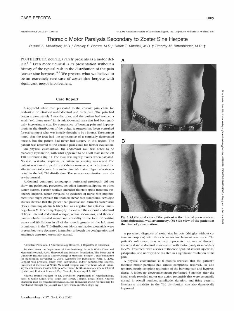
On physical examination, the abdominal wall was noted to bemarkedly asymmetric, with what appeared to be a soft mass in the leftT10 distribution (fig. 1). The mass was slightly tender when palpated.No rash, vesicular eruptions, or cutaneous scarring was noted. Thepatient was asked to perform a Valsalva maneuver, which caused theaffected area to become firm and to diminish in size. Hyperesthesia wasnoted in the left T10 distribution. The sensory examination was oth-erwise normal.
Abdominal computed tomography performed previously did notshow any pathologic processes, including hematoma, lipoma, or othertumor masses. Further workup included thoracic spine magnetic res-onance imaging, which revealed no evidence of nerve root impinge-ment that might explain the thoracic nerve root symptoms. Serologicstudies showed that the patient had positive anti–varicella-zoster virus(VZV) immunoglobulin G titers but was negative for anti-VZV immu-noglobulin M. Electromyelography to evaluate the external abdominaloblique, internal abdominal oblique, rectus abdominus, and thoracicparavertebrals revealed membrane irritability in the form of positivewaves and fibrillations in all of the muscle groups on the left, mostprominently in the T10 distribution. Motor unit action potentials werepresent but were decreased in number, although the configuration andamplitude appeared essentially normal. A presumed diagnosis of zoster sine herpete (shingles without cu-
taneous eruption) with thoracic motor involvement was made. Thepatient’s soft tissue mass actually represented an area of thoracicintercostal and abdominal musculature with motor paralysis secondaryto VZV. Treatment with a series of thoracic epidural steroid injections,gabapentin, and nortriptyline resulted in a significant resolution of hispain.
A physical examination at 6 months revealed that the patient’sthoracic motor paralysis had almost completely resolved. He alsoreported nearly complete resolution of the burning pain and hyperes-thesia. A follow-up electromyelogram performed 5 months after theinitial study revealed motor unit action potentials that were essentiallynormal in overall number, amplitude, duration, and firing pattern.Membrane irritability in the T10 distribution was also dramaticallyimproved.
* Assistant Professor, † Anesthesiology Resident, ‡ Department Chairman.
Received from the Department of Anesthesiology, Scott & White Clinic andMemorial Hospital, Scott, Sherwood, and Brindley Foundation, The Texas A& MUniversity Health Science Center College of Medicine, Temple, Texas. Submittedfor publication November 9, 2001. Accepted for publication April 4, 2002.Support was provided solely from institutional and/or departmental sources.Presented at the Scott & White Memorial Hospital and The Texas A& M Univer-sity Health Science Center College of Medicine Tenth Annual Anesthesia ClinicalUpdate and Resident Research Day, Temple, Texas, April 7, 2001.
Address reprint requests to Dr. McAllister: Department of Anesthesiology,Scott & White Clinic, 2401 South 31st Street, Temple, Texas 76508. Addresselectronic mail to: [email protected]. Individual article reprints may bepurchased through the Journal Web site, www.anesthesiology.org.
Fig. 1. (A) Frontal view of the patient at the time of presentation.Note abdominal wall asymmetry. (B) Side view of the patient atthe time of presentation.
1009CASE REPORTS
Anesthesiology, V 97, No 4, Oct 2002
Dow
nloaded from http://pubs.asahq.org/anesthesiology/article-pdf/97/4/1011/406636/7i1002001007.pdf by guest on 04 June 2022

Discussion
Varicella-zoster virus is responsible for chickenpox,during the primary infection, and shingles, during reac-tivation episodes later in life.6 Following primary infec-tion, the virus remains latent in the dorsal root ganglia ofthe nervous system. Its reactivation gives rise to shingles(herpes zoster), which is usually characterized by unilat-eral neuralgia followed by a vesiculobullous eruption ina dermatomal distribution.6 The incidence of shingles isknown to be higher in the elderly (� 55 yr old), in theimmunocompromised, and in those originally infectedprior to 1 yr of age.7
Rarely, patients experience shingles without the zos-teriform rash (zoster sine herpete). Lewis5 described120 patients that experienced “zoster-type” pain withouta rash in a dermatomal distribution distant from a der-matome with a rash. There have been few well-docu-mented cases of zoster sine herpete that have beenvirologically shown to be caused by VZV.4
Virologic evidence of herpes zoster is difficult to ob-tain due to a lack of specificity, and because most casesare diagnosed based on clinical features.6 Positive anti-VZV immunoglobulin G antibodies represent previousexposure. Positive anti-VZV immunoglobulin M antibod-ies are suggestive, but not specific, for herpes zoster.6
Sundqvist8 showed that increases in anti-VZV immuno-globulin M, due to zoster, are transient. In a study of25 patients, 21 were positive for VZV immunoglobulinM; however, only one patient was still positive 46 daysafter the illness. Therefore, it is not surprising that theimmunoglobulin M serologic results were negative inthis patient, since the delay in diagnosis resulted in theserologic results not being obtained until approximately70 days after the onset of his illness. Dahl et al.9 reportedthat the specificity of clinical diagnosis of zoster (with arash) is 95% and, therefore, laboratory confirmation isnot always required. The specificity of clinical diagnosisin zoster sine herpete is unclear.
Herpes zoster usually affects sensory nerves; however,it can also damage motor neurons. The incidence ofmotor involvement with VZV reactivation has been esti-mated to be 0.52,10 to 5%.1,3 The electromyelographicfindings seen with thoracic motor paralysis are identicalto those observed in our patient, with positive wavesand fibrillations seen in the affected muscle groups.11
Documented cases most commonly have involved cra-nial nerves; however, there are also reports of abdomi-nal, diaphragmatic, and lower extremity paralysis.12–15
Yaszay et al.16 describe unilateral left C5–C6 segmentalparesis associated with a zoster rash in an otherwisehealthy individual.
The true incidence of thoracic motor paralysis withherpes zoster is unknown. Thomas and Howard3 re-viewed 1,210 patients with herpes zoster, 61 of whomwere found to have zoster-induced segmental muscle
weakness. The thoracic dermatome was involved in onlytwo of these cases. Although the thoracic dermatomeseems to be the most frequently affected in herpes zos-ter, it appears to have the lowest incidence of motorparalysis.8 This apparent low incidence may be mislead-ing due to the fact that segmental weakness of intercos-tal and abdominal muscles is likely to pass unnoticed.8,10
Motor paralysis secondary to herpes zoster appears tohave a relatively good prognosis for recovery. Completeor nearly complete recovery has been reported in50–70% of cases.2,3,10 Our patient demonstrated nearlycomplete resolution of his sensory symptoms and motorparalysis over a 5-month period, as evidenced by resolu-tion of the abdominal wall asymmetry.
The differential diagnoses entertained in this case in-cluded soft tissue tumor mass (including lipoma or cyst),herniation of abdominal musculature, abdominal musclehematoma, and lower motor neuron lesion. Tumor mass,hematoma, and muscle herniation were excluded byphysical examination and abdominal computed tomog-raphy scan. Causes of lower motor neuron injury couldinclude surgical injury, trauma, cutaneous nerve entrap-ment, or thoracic nerve root injury due to neural forami-nal stenosis, compression fracture, or herniated nucleuspulposus. The patient had no history of surgery ortrauma to the area, and magnetic resonance imagingrevealed no evidence of thoracic nerve root injury. Cu-taneous nerve entrapment is most commonly found inpatients who have had previous surgery.17 Entrapmentof these cutaneous nerves is typically noted as intermit-tent pain in the lateral rectus abdominis musculaturewith a single painful trigger point on palpation.18 Aliterature search revealed no descriptions of cutaneousnerve entrapment in the external or internal obliquemusculature. As stated previously, electromyelographicevaluation of our patient revealed involvement of theinternal and external oblique, rectus abdominus, andthoracic paravertebral musculature, making cutaneousnerve entrapment an unlikely diagnosis.
As discussed, diagnoses of zoster sine herpete are mostoften made clinically given the difficulty of obtainingserologic evidence. Clinical evidence suggests that ourpatient had both zoster sine herpete and thoracic motorparalysis secondary to herpes zoster. A literature searchrevealed that no similar cases have been reported. In thiscase, serologic evidence is consistent with, but not di-agnostic of, herpes zoster. The clinical symptoms of painand thoracic motor involvement, as documented by elec-tromyelography, further strengthen the validity of thediagnosis. The patient’s rapid recovery over 5 months isalso consistent with the natural course of zoster motorparalysis.2,3,10 We believe that thoracic motor paralysissecondary to zoster sine herpete is a rare diagnosis.However, as mentioned previously, this diagnosis maybe more common than is apparent due to the difficultyin diagnosing each occurrence.8,10
1010 CASE REPORTS
Anesthesiology, V 97, No 4, Oct 2002
Dow
nloaded from http://pubs.asahq.org/anesthesiology/article-pdf/97/4/1011/406636/7i1002001007.pdf by guest on 04 June 2022

The authors thank John C. Hendricks, M.D. (Associate Professor, Departmentof Surgery, Scott & White Memorial Hospital, Texas A&M University, Temple,Texas), for referral; John A. Schuchmann, M.D. (Associate Professor and Chair-man, Department of Physical Medicine and Rehabilitation, Scott & White Memo-rial Hospital, Texas A&M University, Temple, Texas), for electromyelographicevaluation; and Jolene D. Bean-Lijewski, M.D. (Associate Professor, Departmentof Anesthesiology, Scott & White Memorial Hospital, Texas A&M University,Temple, Texas), and Jeff R. Gibson, M.D. (Assistant Professor, Department ofAnesthesiology, Scott & White Memorial Hospital, Texas A&M University, Tem-ple, Texas), for valuable comments in the preparation of this manuscript.
References
1. Grant BD, Rowe CR: Motor paralysis of the extremities in herpes zoster.J Bone Joint Surg Am 1961; 43A:885–96
2. Gupta SK, Helal BH, Kiely P: The prognosis in zoster paralysis. J Bone JointSurg Br 1969; 51:593–603
3. Thomas JE, Howard FM Jr: Segmental zoster paresis—a disease profile.Neurology 1972; 22:459–66
4. Gilden DH, Dueland AN, Devlin ME, Mahalingam R, Cohrs R: Varicella-zoster virus reactivation without rash. J Infect Dis 1992; 166(suppl 1):S30–4
5. Lewis GW: Zoster sine herpete. BMJ 1958; 2:418–216. Arvin AM: Varicella-zoster virus, Fields Virology, 3rd edition. Edited by
Fields BN, Knipe DM, Howley PM. Philadelphia, Lippincott–Raven, 1996, pp2547–85
7. Trannoy E, Berger R, Hollander G, Bailleux R, Heimendinger P, Vuillier D,Creusvaux H: Vaccination of immunocompetent elderly subjects with a live
attenuated Oka strain of varicella zoster virus: A randomized, controlled, dose-response trial. Vaccine 2000; 18:1700–6
8. Sundqvist V: Frequency and specificity of varicella zoster virus IgM re-sponse. J Virol Methods 1982; 5:219–27
9. Dahl H, Marcoccia J, Linde A: Antigen detection: The method of choice incomparison with virus isolation and serology for laboratory diagnosis of herpeszoster in human immunodeficiency virus-infected patients. J Clin Microbiol 1997;35:347–9
10. Akiyama N: Herpes zoster infection complicated by motor paralysis. JDermatol 2000; 27:252–7
11. Cioni R, Giannini F, Passero S, Paradiso C, Rossi S, Fimiani M, Battistini N:An electromyographic evaluation of motor complications in thoracic herpeszoster. Electromyogr Clin Neurophysiol 1994; 34:125–8
12. Brostoff J: Diaphragmatic paralysis after herpes zoster. BMJ 1966;2:1571–2
13. Furuta Y, Ohtani F, Mesuda Y, Fekuda S, Inuyama Y: Early diagnosis ofzoster sine herpete and antiviral therapy for the treatment of facial palsy. Neu-rology 2000; 55:708–10
14. Soler JJ, Perpina M, Alfaro A: Hemidiaphragmatic paralysis caused bycervical herpes zoster. Respiration 1996; 63:403–6
15. Spiers AS: Herpes zoster and its motor lesions, with a report of a case ofphrenic nerve paralysis. Med J Aust 1963; 50:850–3
16. Yaszay B, Jablecki CK, Safran MR: Zoster paresis of the shoulder. Casereport and review of the literature. Clin Orthop 2000; 377:112–8
17. Hall PN, Lee AP: Rectus nerve entrapment causing abdominal pain. Br JSurg 1988; 75:917
18. Gallegos NC, Hobsley M: Abdominal wall pain: An alternative diagnosis.Br J Surg 1990; 77:1167–70
Anesthesiology 2002; 97:1011–3 © 2002 American Society of Anesthesiologists, Inc. Lippincott Williams & Wilkins, Inc.
Osteogenesis Imperfecta, Perioperative Bleeding, andDesmopressin
Mark T. Keegan, M.B., M.R.C.P.I.,* Brett D. Whatcott, M.D.,† Barry A. Harrison, M.B.B.S., F.A.N.Z.C.A., F.R.A.C.P.‡
OSTEOGENESIS imperfecta (OI), also known as “brittle-bone disease,” is a genetic disorder of connective tissuethat causes susceptibility to fractures. In OI, there is a10–30% incidence of bleeding diathesis, and these individ-uals have an increased risk of bleeding when undergoingsurgery.1,2 The coagulation defect is related to the effect ofabnormal collagen on platelet–endothelial cell interactionsand capillary strength. By its action on capillary endothelialcells, desmopressin,3 a synthetic analog of antidiuretic hor-mone, may improve the coagulation abnormality seen inpatients with OI. We describe the use of desmopressin in apatient with OI who experienced excessive bleeding in thesetting of a major surgical procedure.
Case Report
A 72-yr-old man with adenocarcinoma of the prostate and transi-tional cell carcinoma of the bladder presented for cystoprostatectomy.
The patient had a history of OI tarda that had manifested as multiplefractures in childhood and reduced hearing on the right side. His pastmedical history was also significant for hypertension and bleedinghemorrhoids. There was no history of spontaneous bleeding, easybruising, or excessive surgical bleeding. The patient was not takingaspirin or nonsteroidal antiinflammatory drugs. Physical examinationrevealed the presence of blue sclera, a dorsal kyphosis, and bilateralreduction in hip mobility. His prostate gland was slightly enlarged andhad areas of induration at the bases and right apex. His prostate-specific antigen level was 6.9 ng/ml (reference, � 5.6). Preoperativecystoscopic examination with biopsy revealed an invasive grade 3transitional cell carcinoma of the bladder, and prostate biopsy resultswere positive for adenocarcinoma of the prostate. The cardiorespira-tory examination was unremarkable. The patient’s preoperative plate-let count was 220 � 109/l.
Standard American Society of Anesthesiologists monitors wereplaced. General anesthesia was induced with propofol and fentanyland maintained with isoflurane in air and oxygen. An inline fluidwarmer and Bair Hugger (Augustine Medical, Eden Prairie, MN) wereused. Muscle relaxation was achieved using cisatracurium, and inter-mittent doses of morphine were used for analgesia. Marked vascularityand friability of the vessels complicated the dissection, leading toexcessive, diffuse bleeding. Within 40 min of the incision, an estimated1500 ml of blood had been lost, causing the patient’s hemoglobinconcentration to decrease from 13 g/dl (as measured preoperatively)to 8.7 g/dl. In light of the patient’s hemodynamic instability andexcessive, ongoing blood loss, 4 units of packed erythrocytes and6 units of platelets were transfused. Despite this, the diffuse surgicalbleeding continued, but no other bleeding sites were identified.Twenty minutes after administration of the platelets, 0.3 �g/kg (totaldose, 22 �g) desmopressin acetate was administered in an effort toimprove platelet function and to decrease blood loss. The drug wasinfused over 20 min to avoid severe hypotension. Within minutes of
* Assistant Professor, Department of Anesthesiology, Mayo Clinic, Rochester,Minnesota. † Chief, Department of Anesthesiology, Crawford Memorial Hospi-tal, Van Buren, Arkansas. ‡ Assistant Professor, Department of Anesthesiology,Mayo Clinic, Jacksonville, Florida.
Received from the Department of Anesthesiology, Mayo Clinic, Rochester,Minnesota. Submitted for publication January 4, 2002. Accepted for publicationApril 10, 2002. Support was provided solely from institutional and/or departmen-tal sources.
Address reprint requests to: Dr. Keegan: Department of Anesthesiology, MayoClinic, Charlton 1145, 200 1st Street Southwest, Rochester, Minnesota 55905.Address electronic mail to: [email protected]. Individual article reprintsmay be purchased through the Journal Web site, www.anesthesiology.org.
1011CASE REPORTS
Anesthesiology, V 97, No 4, Oct 2002
Dow
nloaded from http://pubs.asahq.org/anesthesiology/article-pdf/97/4/1011/406636/7i1002001007.pdf by guest on 04 June 2022

the infusion, the excessive, diffuse bleeding stopped. No further ab-normal bleeding occurred, and the patient remained hemodynamicallystable for the remainder of the procedure. In total, the patient received6 units of packed erythrocytes, 6 units of platelets, 500 ml of 5%albumin, 500 ml hetastarch, and 5,600 ml lactated Ringer’s solution.At the end of the case, the patient’s hemoglobin concentration was11.8 g/dl, and 4 h later his hemoglobin concentration was 11.2 g/dl,his platelet count was 199 � 109/l, his prothrombin time was 11.9 s(reference range, 8.4–12.0 s), and his activated partial thromboplastintime was 25 s (reference range, 21–33 s). Prior to the onset of bleeding,the patient’s temperature was 35.9°C, and it increased to 36.6°C afterthe bleeding episode. The patient’s recovery and postoperative coursewere uneventful.
Discussion
Osteogenesis imperfecta is a heterogenous group ofinherited disorders of collagen type I caused by muta-tions of the COL1A1 or COL1A2 genes.4 Although theclassic clinical description of OI is of a patient withbrittle bones, blue sclera, and premature deafness, otherorgan systems are affected. The disease may cause car-diac valvular lesions, kidney stones, neurologic abnor-malities, and metabolic derangements. In addition, thedefect in collagen synthesis can cause excessive bruisingand generalized oozing from wound sites. Surgical pro-cedures performed in patients with OI carry a higher riskof bleeding complications. Laboratory investigations ofpatients with OI have demonstrated increased capillaryfragility, decreased platelet retention, decreased levels offactor VIII, and deficient collagen–induced platelet ag-gregation.5,6 It is believed that the collagen defect resultsin friable tissues, small blood vessels that are unable toconstrict adequately, and a defect in the platelet aggre-gation response around exposed subendothelium. Thebleeding may occur despite normal results of coagula-tion studies and bleeding times, making predictionsabout intraoperative bleeding difficult.7 Coagulopathywith sudden development of widespread petechiae hasbeen reported,8 as has fatal hemorrhage secondary toaccidental rib fractures during spinal fusion surgery.9
The evaluation of platelet function and the role ofplatelet transfusion in OI are difficult to assess. BecauseOI is due to an impairment of the platelet–endothelialcell interaction, the thromboelastogram may not be use-ful. Indeed, we could not find a published case reportregarding the use of the thromboelastogram in OI. TheAmerican Society of Anesthesiologists transfusion guide-lines state, “platelet transfusion may be indicated despitean apparently adequate platelet count if there is knownplatelet dysfunction and microvascular bleeding.”10
Bleeding time and platelet aggregation tests are not use-ful in the operating room,10 and “there is an urgent needfor the development of clinically relevant measures of invivo platelet function and bleeding risk to guide therational use of platelet transfusion.”11
Desmopressin is a synthetic analog of antidiuretic hor-
mone. It has a significant antidiuretic effect mediated viaV2 receptors and little or no vasoconstrictive effect(V1).3,12 The drug, mediated via V2 effects, causes en-dothelial cells to release von Willebrand factor, tissuetype plasminogen activator, and factor VIII:C. Desmo-pressin may be used in patients with uremia, chronicliver disease, and certain types of hemophilia, in whichincreased release of von Willebrand factor promotesplatelet adhesiveness to the vascular endothelium and,hence, increases hemostatic activity. There are conflict-ing reports as to the utility of desmopressin in the re-duction of blood loss during operations that involvesubstantial bleeding. Studies involving patients undergo-ing coronary artery bypass grafting, spinal fusion, andhip surgery have varied in their methodology and out-come.13–16 The adverse effects associated with the use ofdesmopressin are few.3 They include facial flushing,headache, hypotension, tachycardia, and the possibilityof thrombosis. In addition, the antidiuretic effect mayincrease the difficulty of assessing the intravascular vol-ume status. There have been a few reports of hypona-tremia and seizures, mostly in children.
Desmopressin works in congenital and acquired plate-let disorders by increasing plasma concentrations of fac-tor VIII, von Willebrand factor, and tissue plasminogenactivator and by increasing platelet adhesiveness.3,12 Thehemostatic effect is almost immediate after administer-ing an intravenous dose of 0.3 �g/kg.3 Desmopressinmay be useful for improving hemostasis and ultimatelydecreasing transfusion requirements in the surgical pa-tient with OI. By increasing platelet deposition ontovascular subendothelium, desmopressin would seem tobe an excellent adjunct to the perioperative manage-ment of patients with OI.
We cannot say definitively that desmopressin stoppedthe bleeding in this case, but circumstantial evidencesupports this hypothesis. A review of the literature failedto reveal any previous reports of the use of desmopressinto treat the coagulopathy associated with OI. Althoughfurther investigation is warranted, due to the low inci-dence of OI and the difficulty in documenting the co-agulopathy associated with OI, randomized, controlledtrials will be difficult to perform. Qualitative tests ofplatelet function, performed before and after the admin-istration of desmopressin, may help to elucidate thedrug’s role in this setting. However, given the relativelybenign side effect profile of desmopressin and the the-oretical basis for its effectiveness, consideration shouldbe given to the use of desmopressin in patients with OIundergoing major surgery when complicated by signifi-cant microvascular bleeding.
References
1. Morton ME: Excessive bleeding after surgery in osteogenesis imperfecta.Br J Oral Maxillofac Surg 1987; 25:507–11
1012 CASE REPORTS
Anesthesiology, V 97, No 4, Oct 2002
Dow
nloaded from http://pubs.asahq.org/anesthesiology/article-pdf/97/4/1011/406636/7i1002001007.pdf by guest on 04 June 2022

2. Siegel BM, Friedman A, Schwartz SO: Hemorrhagic disease in osteogenesisimperfecta. Am J Med 1957; 22:315–21
3. Lethagen S: Desmopressin (DDAVP) and hemostasis. Ann Hematol 1994;69:173–80
4. Marini JC: Osteogenesis imperfecta—managing brittle bones. N Engl J Med1998; 339:986–7
5. Evensen SA, Myhre L, Stormorken H: Haemostatic studies in osteogenesisimperfecta. Scand J Haematol 1984; 33:177–9
6. Hathaway WE, Solomons CC, Ott JE: Platelet function and pyrophosphatesin osteogenesis imperfecta. Blood 1972; 39:500–9
7. Wong RS, Follis FM, Shively BK, Wernly JA: Osteogenesis imperfecta andcardiovascular diseases. Ann Thorac Surg 1995; 60:1439–43
8. Edge G, Okafor B, Fennelly ME, Ransford AO: An unusual manifestation ofbleeding diathesis in a patient with osteogenesis imperfecta. Eur J Anaesthesiol1997; 14:215–9
9. Sperry K: Fatal intraoperative hemorrhage during spinal fusion surgery forosteogenesis imperfecta. Am Forensic Med Pathol 1989; 10:54–9
10. Practice Guidelines for blood component therapy: A report by the Amer-
ican Society of Anesthesiologists Task Force on Blood Component Therapy.ANESTHESIOLOGY 1996; 84:732–47
11. Norfolk DR, Ancliffe PJ, Contreras M, Hunt BJ, Machin SJ, Murphy WG,Williamson LM: Consensus Conference on Platelet Transfusion, Royal College ofPhysicians of Edinburgh, 27–28 November 1997. Br J Haematol 1998; 101:609–14
12. Stoelting RK: Pharmacology and Physiology in Anesthetic Practice, 3rdedition. Philadelphia, Lippincott–Raven, 1999, p 424
13. Theroux MC, Corddry DH, Tietz AE, Miller F, Peoples JD, Kettrick RG: Astudy of desmopressin and blood loss during spinal fusion for neuromuscularscoliosis. ANESTHESIOLOGY 1997; 87:260–7
14. Frankville DD, Harper GB, Lake CL, Johns RA: Hemodynamic conse-quences of desmopressin administration after cardiopulmonary bypass. ANESTHE-SIOLOGY 1991; 74:988–96
15. Mongan PD, Hosking MP: The role of desmopressin acetate in patientsundergoing coronary artery bypass surgery. ANESTHESIOLOGY 1992; 77:38–46
16. Guay J, Reinberg C, Poitras B, David M, Mathews S, Lortie L, Rivard GE: Atrial of desmopressin to reduce blood loss in patients undergoing spinal fusion foridiopathic scoliosis. Anesth Analg 1992; 75:405–10
Anesthesiology 2002; 97:1013–4 © 2002 American Society of Anesthesiologists, Inc. Lippincott Williams & Wilkins, Inc.
Processed Electroencephalographic Changes Associated withHypoglycemia during the Resection of an Insulinoma
Laurence M. Hausman, M.D.
THE Patient State Analyzer (PSA 4000; Physiometrix,North Billerica, MA) is an FDA-approved device thatquantifies regional electroencephalographic data andconverts them into a linear scale (“score”) from 1 to 100.This score has been shown to correlate with depth ofanesthesia.1 In this report, I describe a case in which thedevice was instrumental in alerting the anesthesiologistto hypoglycemia.
Case Report
A 60-yr-old, 98-kg, white man presented to the operating room for alaparoscopic resection of an insulinoma. His past medical history wassignificant for multiple episodes of altered mental status, associatedwith confusion and disorientation, over a 3 to 4 month period. On eachoccasion, his blood glucose concentration was 30–40 mg/dl. He ex-perienced neither seizures nor coma during these periods of profoundhypoglycemia. His only medication at the time was fluvastatin (acholesterol-lowering drug). The initial diagnostic workup revealed aninappropriately high concentration of insulin after a prolonged fast andan increased concentration of C peptide. Magnetic resonance imagingand computed tomography scan of the abdomen showed no abnor-mality. Despite this preoperative inability to localize the source ofexcess insulin, the patient was scheduled to undergo an exploratorylaparoscopy, with possible resection of an insulin-secreting tumor.
Other significant past medical history included occasional gastro-esophageal reflux disease and mild multiple sclerosis, causing minimal
right-sided weakness. Significant laboratory findings on the morning ofsurgery included a blood glucose concentration of 61 mg/dl and apotassium concentration of 4.8 mEq/l. His heart rate was 82 beats/min,and his blood pressure was 135/75 mmHg.
On arrival to the preoperative holding area, the PSA 4000 array wasplaced on the patient’s head, and 30 ml sodium bicitrate was givenorally. He was brought into the operating room, where intravenousaccess was established, and an infusion of 5% dextrose in 0.9% sodiumchloride was started at 100 ml/h. The device was activated, and thepatient’s initial PSA value was 97. The standard American Society ofAnesthesiologists monitors were applied. Metoclopramide, 10 mg, andmidazolam, 2 mg, were administered intravenously. After preoxygen-ation, a modified rapid sequence induction with 2 mg/kg propofol and0.25 mg/kg cisatracurium was performed while maintaining cricoidpressure. The trachea was intubated approximately 90 s later.
After induction, the patient’s blood pressure and heart rate remainedwithin normal limits, and his PSA score decreased to 19. Anesthesiawas maintained with 50% oxygen, 50% air, 0.2% isoflurane, and neu-romuscular blockade. Several minutes later, his score increased to 38,as the effect of the induction dose of propofol diminished. His heartrate and blood pressure remained unchanged. On insertion of theurinary catheter by the surgeons, the patient’s heart rate increased to90 beats/min, and his blood pressure increased to 160/88 mmHg.Fentanyl, 250 �g, was administered intravenously. This resulted in adecrease in both heart rate and blood pressure to their baseline value.However the patient’s processed electroencephalography scan re-mained essentially unchanged. An arterial catheter and a second intra-venous catheter were placed.
Over the next 15 min, during preparation and draping of the patient,the PSA value gradually decreased to 17, and the isoflurane was dis-continued. Despite this adjustment, his electroencephalography valueremained essentially unchanged. During this time, there were nosignificant changes in heart rate or blood pressure, and the patientremained normothermic. An arterial blood sample was drawn, theanalysis of which revealed a blood glucose concentration of 40 mg/dl,an arterial oxygen tension of 171 mmHg, and an arterial carbon dioxidetension of 32 mmHg. Intravenous dextrose, 12.5 g, was given rapidly.Approximately 2 to 3 min later, his PSA value increased to 48, and hisblood glucose concentration was 235 mg/dl. The isoflurane was re-started at 0.2%, while the surgeons continued to prepare and drape thepatient. Several minutes prior to incision, the isoflurane was titrated up
Assistant Professor of Anesthesiology.
Received from the Department of Anesthesiology, The Mount Sinai School ofMedicine, New York, New York. Submitted for publication December 10, 2001.Accepted for publication April 15, 2002. The PSA 4000 machine and the headarrays were supplied by Physiometrix, North Billerica, Massachusetts. No otherfunds were received.
Address reprint requests to Dr. Hausman: Department of Anesthesiology, TheMount Sinai School of Medicine, 1 Gustave L. Levy Place, Box 1010, New York,New York 10029. Address electronic mail to: [email protected] article reprints may be purchased through the Journal Web site,www.anesthesiology.org.
1013CASE REPORTS
Anesthesiology, V 97, No 4, Oct 2002
Dow
nloaded from http://pubs.asahq.org/anesthesiology/article-pdf/97/4/1011/406636/7i1002001007.pdf by guest on 04 June 2022

to 1.9%. The patient tolerated the incision, and the isoflurane wasgradually decreased. The patient remained stable throughout the pro-cedure and was maintained with 50% oxygen, 50% air, and isofluranebetween 0.9 and 1.5%.
An infusion of 10% dextrose in water was administered intraopera-tively to maintain a blood glucose concentration between 100 and200 mg/dl. Frequent blood gas analyses were performed to direct thisgoal. Laparoscopy revealed a tumor of the pancreatic islet cells. Afterenucleation of the tumor, the patient’s blood glucose concentrationincreased to 359 mg/dl. The 10% dextrose in water and 5% dextrose in0.9% sodium chloride infusions were discontinued, and an infusion ofa balanced salt solution was started. At the conclusion of the surgicalprocedure, the patient awoke, the trachea was extubated, and he wastransferred to the postanesthesia care unit. In the postanesthesia careunit, his blood glucose concentration stabilized, and he was transferredto the inpatient ward later that evening. He was discharged home onthe third postoperative day. The patient denied any intraoperativeawareness or memory.
Discussion
There has been much controversy over glucose man-agement during the resection of an insulinoma.2–4 Al-though a continuous intravenous infusion of glucose isnot absolutely indicated, it is commonly used to achievethe goal of maintaining a blood glucose concentrationgreater than 50–60 mg/dl.4–6 Frequent intraoperativeblood glucose measurements are necessary.7
Hypoglycemia can cause permanent cerebral damage.5
The signs of hypoglycemia include hypertension, tachy-cardia, sweating, change in mental status, and even bra-dycardia, which may be secondary to an increase in vagaltone.6 Many of these signs are masked by general anes-thesia.4,7 Those available are nonspecific and have mul-tiple etiologies. In this patient, the heart rate and bloodpressure were unchanged. The only indication of possi-ble hypoglycemia was his low PSA score. His heart rateand blood pressure remained at their baseline. The PSAscore of 17 reflected a very deep anesthetic state, despitean end-tidal isoflurane of zero. The patient’s blood glu-cose concentration at that time was 40 mg/dl, and, atthis concentration, he was known to be symptomatic.Another indication that the score was affected by bloodglucose concentration was its rapid increase to 48 fol-lowing treatment of hypoglycemia.
The PSA 4000 monitor utilizes regional quantitativeelectroencephalographic data. When a general anes-thetic is administered, there are regional changes inelectroencephalographic activity that occur regardlessof the anesthetic used.8 These regional electroencepha-
lographic changes can then be quantified. A score of 100corresponds with the awake state, whereas a score ofzero corresponds with complete electroencephalo-graphic suppression. Hypnosis during general anesthesiais reflected by a score of 25–50.
In this case, the patient’s score decreased immediatelyon induction of anesthesia from 97 to 19. His scorepromptly increased to 38, indicating an adequate depthof anesthesia. However, during the period of preparationand draping, his score began to decrease to very lowlevels without any measurable end-tidal volatile anes-thetic. The end-tidal isoflurane had been stable at 0.0%,but his score continued to decrease to well below 25.Hypoglycemia was considered as a possible cause, andthe patient’s blood glucose concentration was checked,confirming this etiology. When a bolus of glucose wasgiven, the score promptly increased. Of note, hyperten-sion and tachycardia, common signs of hypoglycemia inthe anesthetized patient, were not seen. Whether thesechanges in vital signs would have ultimately occurred isnot known.
In this case, a hypoglycemic patient had significantquantitative electroencephalographic changes, as re-flected in the PSA 4000 score, while all vital signs re-mained within normal limits. Further studies are neededto determine if there are consistent electroencephalo-graphic changes seen in the hypoglycemic patient, andwhether these changes routinely precede alterations inhemodynamic variables.
References
1. Drover DR, Lemmens HJ, Pierce ET, Plourde G, Loyd G, Ornstein E, PrichepLS, Chabot RJ, Gugino L: Patient state index titration of delivery and recoveryfrom propofol, alfentanil, and nitrous oxide anesthesia. ANESTHESIOLOGY 2002;97:82–9
2. Tutt GO, Edes AJ, Servic FJ, van Heerden JA: Plasma glucose monitoringduring operations for insulinoma. A critical reappraisal. Surgery 1980; 88:351–6
3. Schwartz SS, Horwitz DL, Zehfus B, Langer BG, Kaplan E: Continuousmonitoring and control of plasma glucose during operation for removal ofinsulinomas. Surgery 1979; 85:702–7
4. Muir JJ, Endres SM, Offord K, van Heerden JA, Tinker JH: Glucose manage-ment in patients undergoing operation for insulinoma removal. ANESTHESIOLOGY
1983; 59:371–55. Lamont AS, Jones D: Anaesthetic management of insulinoma. Anaesth In-
tensive Care 1978; 6:261–26. Chari P, Pandit SK, Kataria RN, Singh H, Baheti DK, Wig J: Anaesthetic
management of insulinoma. Anaesthesia 1977; 32:261–47. Roizen MF: Diseases of the endocrine system, Anesthesia and Uncommon
Diseases. Edited by Benumof JL. Philadelphia, WB Saunders, 1998, pp 259–628. John ER, Prichep LS, Valdes-Sosa P, Bosch J, Aubert E, Gugino LD, Kox W,
Tom M, di Michele F: Invariant reversible QEEG effects of anesthetics. ConsciousCogn 2001; 10:165–83
1014 CASE REPORTS
Anesthesiology, V 97, No 4, Oct 2002
Dow
nloaded from http://pubs.asahq.org/anesthesiology/article-pdf/97/4/1011/406636/7i1002001007.pdf by guest on 04 June 2022

Anesthesiology 2002; 97:1015–9 © 2002 American Society of Anesthesiologists, Inc. Lippincott Williams & Wilkins, Inc.
Anesthesia and Airway Management of Pediatric Patients withSmith–Lemli–Opitz Syndrome
Zenaide M. N. Quezado, M.D.,* Judith Veihmeyer, M.D.,† Lynnae Schwartz, M.D.,† Ngozi A. Nwokoro, M.D., Ph.D.,‡Forbes D. Porter, M.D., Ph.D.§
SMITH–LEMLI–OPITZ syndrome (SLOS) is an autosomalrecessive disorder with an incidence of between 1 in26,500 pregnancies in Canada and 1 in 50,000 pregnan-cies in the United States.1,2 The syndrome results froman inborn error of cholesterol biosynthesis involving adeficiency of 3�-hydroxysterol �7–reductase, the en-zyme that catalyzes the reduction of 7-dehydrocholes-terol to cholesterol.3,4 As a result, patients with SLOShave abnormal embryonic development, high plasmaconcentrations of 7-dehydrocholesterol, and usually de-creased plasma concentrations of cholesterol.
This biochemical defect is associated with a broadspectrum of clinical manifestations with potentially pro-found anesthetic implications. Patients with SLOS canhave severe growth failure, congenital anomalies affect-ing most organ systems, early death, developmental de-lay, and self-injurious and ritualistic behavior.5–9 Of specialconcern to anesthesiologists are the typical dysmorphicfacial features, including micrognathia, cleft palate, and asmall and abnormally hard tongue, which can present achallenge in airway management of patients with SLOS.5,8
The broad spectrum of congenital anomalies and po-tential increase in lifespan with dietary cholesterol re-placement therapy make it very likely that diagnostic andtherapeutic interventions requiring anesthesia will beneeded in patients with SLOS. However, since the dis-covery of the biochemical defect in SLOS, little has beenreported on the anesthetic management of these pa-
tients.10 In this report, we describe the administration of20 anesthetics in 14 patients with confirmed biochemi-cal diagnosis of SLOS. Our focus was on airway manage-ment, and because of previous reports of difficult intuba-tions in some of our patients, we chose to use fiberopticlaryngoscopy as the initial technique to achieve trachealintubation.
Report of Cases
We reviewed the anesthetic management of 14 children who wereenrolled in ongoing investigations of the clinical, molecular, and bio-chemical features of SLOS and received 20 general anesthetics fromJuly 2000 to February 2002. The investigation protocol was approvedby the National Institutes of Child Health and Development Institu-tional Review Board. Clinical diagnosis was confirmed biochemically inall patients (table 1).
Demographics, including the SLOS severity score11 of the 14 chil-dren, are listed in table 1. Additional details on the patients andanesthetics reported here are available on the ANESTHESIOLOGY Web site,http://www.anesthesiology.org. Figure 1 depicts a 9-month-old infantwith SLOS and features of Pierre Robin sequence (micrognathia, glos-soptosis, and high-arched and cleft soft and hard palates) who wasanesthetized at 3, 9, and 15 months of age. Six patients had a history ofgastroesophageal reflux predominantly associated with feedings (table1). However, once on metoclopramide, H2 blocker, and H�–K�
ATPase inhibitor, these patients had no clinical signs of gastric reflux.In addition, despite the history of gastric reflux, no episode of aspira-tion pneumonia was recorded in any of our patients. Eight patients hadgastric tubes through which they were fed exclusively (table 1). Pa-tient 9 had a history of hypertension that was treated with a calciumchannel blocker. Five patients (table 1) had a documented history ofdifficult intubation, as described by anesthesiologists who had previ-ously performed direct laryngoscopies. In patient 9, a previous anes-thetic was aborted because we were unable intubate after induction ofanesthesia.
In this series, one anesthesiologist administered all of the anestheticsand performed all of the endotracheal intubations, with the exceptionof one (table 2). All patients had solids or formula discontinued 8 hbefore induction of anesthesia, but clear liquids were allowed for up to3 h before induction of anesthesia. Only patient 4, an 8-yr-old boy withaggressive behavior, required premedication with oral midazolam(0.4 mg/kg) and ketamine (10 mg/kg), which yielded effective sedativeeffects. In patients with gastric tubes, the stomach was emptied im-mediately prior to induction of anesthesia.
The ability to mask ventilate was established, and intravenous accesswas obtained. One patient had intravenous induction with sodiumthiopental. All patients had documented easy mask ventilation withoutoropharyngeal airways. During fiberoptic laryngoscopy and endotra-cheal intubation, most patients were spontaneously ventilating, andthe inspired concentration of oxygen was increased to 100%. Overall,during induction of anesthesia and tracheal intubations, oxygen satu-ration was maintained above 99% in all patients. In two patients(patients 2 and 13), mivacurium was given just prior to the insertion ofthe endotracheal tube in the trachea. Succinylcholine was adminis-tered to treat one episode of laryngospasm that was evident during
Additional material related to this article can be found on theAnesthesiology Web site. Go to the following address, click onthe Enhancements Index, and then scroll down to find theappropriate article and link. http://www.anesthesiology.org
�
*Associate Professor of Anesthesia, Children’s National Medical Center,George Washington University, and Staff Physician, Department of Anesthesiaand Surgical Services, Warren Magnuson Clinical Center. †Staff Physician, De-partment of Anesthesia and Surgical Services, Warren Magnuson Clinical Center,‡Clinical Fellow, §Investigator, Heritable Disorders Branch, National Institute ofChild Health and Human Development, National Institutes of Health.
Received from the Department of Anesthesia, Children’s National MedicalCenter, George Washington University, Washington, DC; and the Department ofAnesthesia and Surgical Services, Warren Magnuson Clinical Center, and theHeritable Disorders Branch, National Institute of Child Health and Human Devel-opment, National Institutes of Health, Bethesda, Maryland. Submitted for publi-cation December 14, 2001. Accepted for publication April 16, 2002. Supportedby intramural funding from the National Institutes of Health.
Address reprint requests to Dr. Quezado: Department of Anesthesia andSurgical Services, Warren Magnuson Clinical Center, National Institutes ofHealth, Building 10, Room 2C624, 10 Center Drive, MSC 1512, Bethesda,Maryland 20892. Address electronic mail to: [email protected]. Individual articlereprints may be purchased through the Journal Web site, www.anesthesiology.org.
1015CASE REPORTS
Anesthesiology, V 97, No 4, Oct 2002
Dow
nloaded from http://pubs.asahq.org/anesthesiology/article-pdf/97/4/1011/406636/7i1002001007.pdf by guest on 04 June 2022

fiberoptic laryngoscopy (patient 5). All endotracheal intubations wereelectively performed with fiberoptic bronchoscopes (model BF-XP40[outside diameter, 2.8 mm] for endotracheal tubes sized 3.5 or aboveand model LF-P [outside diameter, 2.2 mm] for those sized 3 andbelow, Olympus America, Melville, NY). In patients undergoing oraland dental examinations (six anesthetics), the endotracheal tube wasinserted nasally, whereas in those undergoing only imaging studies, itwas inserted orally (14 anesthetics). We used cuffed endotrachealtubes for 8 anesthetics and uncuffed tubes for the remaining 12anesthetics. Most children required a smaller sized endotracheal tubethan that predicted for their age (table 2).
In 19 anesthetics, fiberoptic laryngoscopy was the technique chosenfor tracheal intubation from the outset. In one anesthetic, four directlaryngoscopies were performed, and an otolaryngologist was con-sulted to perform indirect laryngoscopy and thereby intubate the
trachea. All intubations were performed successfully and withouttrauma to the airway. The agents used in the maintenance phase of theanesthetics are listed in table 2. At the completion of all procedures,the patients were extubated. Only patient 7, when administered thefirst anesthetic, presented airway obstruction after extubation that wasrelieved with a nasopharyngeal airway. There were no episodes ofoxygen desaturation, bradycardia, witnessed aspiration, or aspirationpneumonia.
Discussion
Given the relatively high incidence, presence of mul-tiple congenital malformations, expansion of the pheno-
Table 1. Demographics, Congenital Anomalies, and Diagnostic Plasma Sterol Levels in a Series of Patients with Smith-Lemli-OptizSyndrome
Patient(Gender)
Age*(mo)
Weight, Kg(percentile)
SeverityScore†
CongenitalHeart
Disease GERDGastricTube Hypotonia
CleftPalate Micrognathia
Plasma Sterol Levels(normal values, mg/dL)
Cholesterol(100–200)
7-DHC (0.01� 0.005)
1 (F) 7 5 (�3rd) 28 PDA Y Y Y Y 56 27.32 (F) 29 10 (�3rd) 17 PDA, Aorta
coarctationY Y Y 70 13.5
3 (M) 13 8 (�3rd) 11 Pulmonarystenosis
Y Y 65 12.6
4 (M) 96 25 (50th) 11 195 0.075 (M) 18 7 (�3rd) 6 Y Y 89 12.26 (M) 4 6 (25th) 11 Y Y Y Y 72 8.57 (F) 3 3 (�3rd) 33 Y Y Y Y 8 23.78 (M) 9 6 (�3rd) 6 Y Y Y 36 12.89 (F) 25 6 (�3rd) 31 ASD Y Y Y 83 15
10 (M) 27 10 (�3rd) 6 Y Y 114 5.411 (M) 8 7 (�3rd) 33 Y Y Y Y 31 13.912 (F) 11 5 (�3rd) 11 Y Y Y Y 20 7.813 (M) 39 12 (3rd) 11 Y Y 73 6.214 (F) 26 8 (�3rd) 39 ASD Y Y Y Y Y 21 13.7
* Age at the first anesthetic in this series. † Severity scores are based on the presence of malformations in each of 10 embryologically distinct areas.11
Scores greater than 50 reflect severe disease, 25–50 reflect moderate or typical disease, and less than 25 reflect mild disease.
F, female; M, male; PDA, patent ductus arteriosus; ASD, atrial septal defect; GERD, gastroesophageal reflux disease; Y, yes (showing that the characteristic ispresent).
Fig. 1. (A) Nine-month-old infant withSmith–Lemli–Opitz syndrome presentingfeatures associated with Pierre Robin se-quence, including micrognathia, cleftpalate (B), and pseudomacroglossia.
1016 CASE REPORTS
Anesthesiology, V 97, No 4, Oct 2002
Dow
nloaded from http://pubs.asahq.org/anesthesiology/article-pdf/97/4/1011/406636/7i1002001007.pdf by guest on 04 June 2022

typic spectrum to include patients with milder cases,biochemical confirmation of the clinical diagnosis, andthe use of dietary cholesterol replacement therapy forSLOS, the probability that an anesthesiologist will carefor patients with SLOS is likely to increase. However, theliterature on the anesthetic management of SLOS isscarce. Since the discovery of the biochemical defectassociated with SLOS in 1994, there have been only smallcase series reported in the literature.10,12,13 In this re-port, we describe a series of 20 anesthetics in 14 patientswith biochemically confirmed SLOS. Because of priorhistory of difficult intubation in 5 of our 14 patients, weprospectively decided to use fiberoptic laryngoscopy asthe initial technique for tracheal intubation in the anes-thetics described here. We have shown that, in skilledhands, fiberoptic intubation can be performed safely,efficiently, and without complications, and it can beused as an initial technique for airway management inpatients with SLOS.
When planning to administer an anesthetic in a patientwith SLOS, the anesthesiologist will have to addressseveral issues pertinent to this complex syndrome. First,facial dysmorphic features in patients with SLOS, includ-ing those associated with Pierre Robin sequence (micro-gnathia and palatal anomalies) and prominent incisors,can be associated with difficulties in airway manage-
ment. In the literature, there are several reports of diffi-cult intubation and abnormal laryngoscopic view in pa-tients with SLOS.10,13,14 In our institution, we had toabort two procedures because of inability to intubateSLOS patients with direct laryngoscopy. In one patient inour series, an otolaryngologist performed fiberoptic in-tubation of the trachea after a pediatric anesthesiologisthad attempted four direct laryngoscopies. We chose toperform fiberoptic intubation of the trachea as the initialtechnique for 19 of the anesthetics in this series. Wewere able to maintain spontaneous ventilation and ade-quate oxygenation and to intubate all patients withoutrepeated direct laryngoscopies or trauma to the airway.Therefore, our experience suggests that in patients withSLOS, tracheal intubation by fiberoptic laryngoscopy isan excellent alternative for the airway management ofthese pediatric patients. Evidently, in order to make thisa viable alternative, fiberoptic scopes that accommodateendotracheal tubes smaller than size 5.5 are required,and the staff must be trained in the technique of fiber-optic intubation.
Second, the potential for a difficult airway, when asso-ciated with the presence of gastroesophageal reflux, canbe particularly concerning for the anesthesiologist. Al-though a common problem in patients with SLOS, gas-troesophageal reflux is often a result of the combination
Table 2. Series of 20 Anesthetics in Patients with Smith-Lemli-Optiz Syndrome
PatientHistory of Difficult
Intubation* Procedures Induction AgentsEndotracheal
Tube Size Maintenance Agents Comments
1 Y Eye muscle surgery ThiopentalVecuronium
3.5 Uncuffed N2O, Desflurane Tracheal intubation byan otolaryngologist
1 Y Brain MRI, spectroscopy N2O, Sevoflurane 3.5 Uncuffed N2O, Isoflurane1 Y Brain MRI, spectroscopy N2O, Sevoflurane 3.5 Uncuffed N2O, Propofol2 Brain MRI, spectroscopy N2O, Sevoflurane 4.0 Uncuffed N2O, Isoflurane3 Brain MRI, spectroscopy N2O, Sevoflurane 4.0 Uncuffed N2O, Isoflurane
Dental rehabilitation4 Brain MRI, spectroscopy N2O, Sevoflurane 5.0 Cuffed N2O, Isoflurane5 Brain MRI, spectroscopy N2O, Sevoflurane 4.0 Uncuffed N2O, Propofol Laryngospasm during
intubation6 Brain MRI, spectroscopy N2O, Sevoflurane 3.5 Uncuffed N2O, Propofol6 Brain MRI, spectroscopy N2O, Sevoflurane 3.5 Cuffed N2O, Propofol7 Y Brain MRI, spectroscopy N2O, Sevoflurane 3.0 Uncuffed N2O, Isoflurane7 Y Brain MRI, spectroscopy N2O, Sevoflurane 3.0 Uncuffed N2O, Isoflurane7 Y Brain MRI, spectroscopy
eye EUAN2O, Sevoflurane 3.5 Uncuffed N2O, Propofol
8 Brain MRI, spectroscopy N2O, Sevoflurane 3.5 Uncuffed N2O, Propofol9 Y Brain MRI, spectroscopy
Dental rehabilitationN2O, Sevoflurane 3.5 Cuffed N2O, Propofol Prior procedure
cancellationbecause of inabilityto intubate
10 Brain MRI, spectroscopy N2O, Sevoflurane 4.0 Cuffed N2O, PropofolDental rehabilitation
11 Y Brain MRI, spectroscopy N2O, Sevoflurane 3.5 Uncuffed N2O, Propofol12 Brain MRI, spectroscopy N2O, Sevoflurane 3.5 Cuffed N2O, Propofol13 Brain MRI, spectroscopy N2O, Sevoflurane 4.0 Cuffed N2O, Propofol
Dental rehabilitation14 Y Brain MRI, spectroscopy N2O, Sevoflurane 3.5 Cuffed N2O, Propofol
Dental rehabilitation
* Difficult intubation as described by anesthesiologists who had previously performed direct laryngoscopies.
Y, yes; MRI, magnetic resonance imaging.
1017CASE REPORTS
Anesthesiology, V 97, No 4, Oct 2002
Dow
nloaded from http://pubs.asahq.org/anesthesiology/article-pdf/97/4/1011/406636/7i1002001007.pdf by guest on 04 June 2022

of small stomachs and intestinal dysmotility.5 After care-ful history taking, it was clear that, in most of ourpatients with SLOS, gastroesophageal reflux was pre-dominantly associated with feedings. We felt that rapidsequence inductions were not warranted. Preopera-tively, we prescribed an 8-h fast for formula and 3-h forclear liquids. Those patients on H2 blockers and meto-clopramide were given their doses 3 h prior to theiranesthetics. Just prior to induction, we applied gentlesuction to the gastric tube in patients who had a gastros-tomy. We observed no episodes of regurgitation or aspi-ration of gastric contents with this regimen. Therefore,our experience with patients with SLOS suggests that ifthe history indicates that gastroesophageal reflux is pre-dominantly associated with feedings and if strict fastingguidelines are followed, rapid sequence induction maynot be necessary in all patients, and inhalation inductioncan be performed safely.
Another issue that anesthesiologists must keep in mind isthe possible association of muscle rigidity and the use ofinhalation agents in patients with SLOS. Petersen andCrouch12 reported a 4-yr-old child with SLOS who exhib-ited muscle rigidity after the use of halothane. In that case,muscle rigidity was associated with a mild elevation intemperature but no elevation of creatinine kinase concen-trations. In our series of 20 anesthetics, all patients receivedhalogenated agents, and no episodes of muscle rigiditywere observed. Furthermore, we used succinylcholine inone of these patients without any problems. Although nodefinitive conclusion can be made, in our experience, theuse of halogenated anesthetics in this population was notassociated with muscle rigidity.
Behavioral abnormalities are also issues that need to beaddressed in the preoperative assessment of patientswith SLOS. SLOS is known to be associated with behav-ioral disorders, including autism. In a study of patientswith SLOS, 9 of the 17 patients without hearing deficit(53%) met the diagnostic criteria for autistic disorder,and 50 of 56 (89%) had aggressive behavior that waseither self-injurious or directed against others.9 In thatstudy, patients younger than 22 months did not showbehavioral dysregulation.9 In addition, in a large series ofpatients with documented SLOS, the use of sedativeswas reportedly ineffective.8 Certainly, such a high inci-dence of abnormal and aggressive behavior coupledwith a lack of response to sedatives can pose a challengefor the pediatric anesthesiologist. In our series, only fiveanesthetics were administered in children older than 22months, and only one of these children exhibited aggres-sive behavior. Although we did not observe any difficultyin sedating or anesthetizing our patients with SLOS, onecould postulate that abnormalities of cellular membranesterol composition in SLOS patients could alter theirresponse to sedatives and anesthetic agents.
Some animal studies support this hypothesis. In a geneticmouse model of SLOS with biochemical and phenotypic
similarities to the human syndrome, the neurophysiologicresponse of frontal cortex neurons to the excitatory aminoacid glutamate was significantly impaired.15 These findingsin animals may suggest that in patients with SLOS, anabnormal sterol cellular membrane composition could im-pact the physiologic response of neurotransmitters. In turn,these abnormalities could impact the response to sedativesand anesthetic agents. Therefore, it is not surprising thatpatients with SLOS reportedly have an abnormal responseto sedatives.8 From a clinical standpoint, all of our anesthet-ics were rather uncomplicated. However, further studiesare needed to fully characterize the neuropharmacologicimpact of the biochemical abnormalities of patients withSLOS.
In summary, we present a series of 20 anestheticsadministered to 14 patients with SLOS. In our series,although patients with SLOS can have difficult intuba-tions, mask airway was always adequate, and fiberopticintubation of the trachea was a safe and reliable optionas an initial technique in airway management. Despite ahistory of gastroesophageal reflux disease predominantlyassociated with feedings, we saw no cases of aspirationof gastric contents. Our findings suggest that althoughSLOS patients can present with a broad spectrum ofphenotypes and congenital abnormalities that are poten-tially challenging for the anesthesiologist, they usuallyrequire rather routine anesthetic management.
The authors thank Mavra Diavatis, R.N. (Department of Anesthesia and SurgicalServices, Warren Magnuson Clinical Center, National Institutes of Health, Be-thesda, Maryland), for technical assistance during the anesthesia, and the mem-bers of the Department of Anesthesia and Surgical Services, Warren MagnusonClinical Center, National Institutes of Health, Bethesda, Maryland, for their helpin the care of our patients.
References
1. Nowaczyk MJ, McCaughey D, Whelan DT, Porter FD: Incidence of Smith-Lemli-Opitz syndrome in Ontario, Canada. Am J Med Genet 2001; 102:18–20
2. Kelley RI: A new face for an old syndrome. Am J Med Genet 1997; 68:251–63. Irons M, Elias ER, Salen G, Tint GS, Batta AK: Defective cholesterol biosyn-
thesis in Smith-Lemli-Opitz syndrome (letter). Lancet 1993; 341:14144. Tint GS, Irons M, Elias ER, Batta AK, Frieden R, Chen TS, Salen G: Defective
cholesterol biosynthesis associated with the Smith-Lemli-Opitz syndrome. N EnglJ Med 1994; 330:107–13
5. Kelley RI, Hennekam RC: The Smith-Lemli-Opitz syndrome. J Med Genet2000; 37:321–35
6. Lin AE, Ardinger HH, Ardinger RH Jr, Cunniff C, Kelley RI: Cardiovascularmalformations in Smith-Lemli-Opitz syndrome. Am J Med Genet 1997; 68:270–8
7. Linck LM, Hayflick SJ, Lin DS, Battaile KP, Ginat S, Burlingame T, GibsonKM, Honda M, Honda A, Salen G, Tint GS, Connor WE, Steiner RD: Fetal demisewith Smith-Lemli-Opitz syndrome confirmed by tissue sterol analysis and theabsence of measurable 7-dehydrocholesterol Delta(7)–reductase activity in cho-rionic villi. Prenat Diagn 2000; 20:238–40
8. Ryan AK, Bartlett K, Clayton P, Eaton S, Mills L, Donnai D, Winter RM, BurnJ: Smith-Lemli-Opitz syndrome: A variable clinical and biochemical phenotype.J Med Genet 1998; 35:558–65
9. Tierney E, Nwokoro NA, Porter FD, Freund LS, Ghuman JK, Kelley RI:Behavior phenotype in the RSH/Smith-Lemli-Opitz syndrome. Am J Med Genet2001; 98:191–200
10. Choi PT, Nowaczyk MJ: Anesthetic considerations in Smith-Lemli-Opitzsyndrome. Can J Anaesth 2000; 47:556–61
11. Witsch-Baumgartner M, Fitzky BU, Ogorelkova M, Kraft HG, Moebius FF,Glossmann H, Seedorf U, Gillessen-Kaesbach G, Hoffmann GF, Clayton P, KelleyRI, Utermann G: Mutational spectrum in the Delta7-sterol reductase gene andgenotype-phenotype correlation in 84 patients with Smith-Lemli-Opitz syndrome.Am J Hum Genet 2000; 66:402–12
1018 CASE REPORTS
Anesthesiology, V 97, No 4, Oct 2002
Dow
nloaded from http://pubs.asahq.org/anesthesiology/article-pdf/97/4/1011/406636/7i1002001007.pdf by guest on 04 June 2022

12. Petersen WC, Crouch ER Jr: Anesthesia-induced rigidity, unrelated tosuccinylcholine, associated with Smith-Lemli-Opitz syndrome and malignant hy-perthermia. Anesth Analg 1995; 80:606–8
13. Haji-Michael PG, Hatch DL: Smith-Lemli-Opitz syndrome and malignanthyperthermia (letter). Anesth Analg 1996; 83:200
14. Butler MG, Hayes BG, Hathaway MM, Begleiter ML: Specific genetic dis-
eases at risk for sedation/anesthesia complications. Anesth Analg 2000; 91:837–55
15. Wassif CA, Zhu P, Kratz L, Krakowiak PA, Battaile KP, Weight FF, GrinbergA, Steiner RD, Nwokoro NA, Kelley RI, Stewart RR, Porter FD: Biochemical,phenotypic and neurophysiological characterization of a genetic mouse model ofRSH/Smith–Lemli–Opitz syndrome. Hum Mol Genet 2001; 10:555–64
Anesthesiology 2002; 97:1019–21 © 2002 American Society of Anesthesiologists, Inc. Lippincott Williams & Wilkins, Inc.
Severe Rhabdomyolysis after Laparoscopic Surgery forAdenocarcinoma of the Rectum in Two Patients Treated with
StatinsFrançois Forestier, M.D.,* Yannick Breton, M.D.,† Emmanuel Bonnet, M.D.,* Gerard Janvier, M.D., Ph.D.‡
RHABDOMYOLYSIS is a clinical and biochemical syn-drome resulting from skeletal muscle injury with therelease of muscle contents into the plasma. It resultseither from a direct muscle injury or from an alteredmetabolic relationship between energy production andconsumption in muscle. Crush syndrome, extensiveburns, electric shock, and prolonged immobilization arethe main causes of direct muscle injury. Strenuous phys-ical exercise, malignant hyperthermia, muscle ischemiaresulting from compression or vascular injury, infec-tions, myopathies, toxins, and drugs, including alcohol,which accounts for 20% of the cases, can cause nontrau-matic rhabdomyolysis. This syndrome has been de-scribed with fibrates and statin lipid-lowering drugs, cul-minating in the recent removal of cerivastatin from themarket because of a number of rhabdomyolysis-relateddeaths.1 Severe cases were reported in elderly patientsand in patients taking lipid-lowering drugs. We observedtwo consecutive cases of severe postoperative rhabdo-myolysis in patients who underwent elective laparo-scopic surgery for adenocarcinoma of the rectum, andfor whom cumulative risk factors were identified inretrospect: long-term treatment with statins and laparo-scopic surgery with prolonged immobilization undergeneral anesthesia.
Case Reports
Case 1
A 52-yr-old male patient (body mass index, 26.6) was scheduled toundergo elective surgery for adenocarcinoma of the rectum. He had ahistory of myocardial infarction, moderate hypertension, pulmonaryembolism, appendectomy, hypercholesterolemia, and diabetes melli-tus. His usual medications included aspirin, fosinopril, atenolol,
glimepiride, and pravastatin, which were discontinued the day beforesurgery. Preoperative evaluation of the patient did not show anybiologic disturbances, and his chest radiograph was normal. The pa-tient was premedicated with 50 mg hydroxyzine the evening beforethe surgery and 100 mg hydroxyzine 2 h before his admission to theoperating room. Antibiotic prophylaxis (cefoxitin, 2 g) was givenintravenously before the induction of anesthesia with propofol (3 mg/kg), sufentanil (0.5 �g/kg), and cisatracurium (0.15 mg/kg) for musclerelaxation. Anesthesia was maintained with isoflurane in an oxygen–airmixture supplemented with sufentanil (1.8 �g · kg�1 · h�1) andadditional doses of cisatracurium as needed. The support points wereprotected by gelatin patches and checked at each change of position.The rectotomy lasted 6 h in a lithotomy position. The patient’s systolicblood pressure remained above 80 mmHg during the surgery, and hisperioperative blood loss was less than 500 ml. Volume replacementwas ensured by administering 500 ml of a hydroxylethyl starch solution(200,000/0.5/6%/5) and 4,300 ml of crystalloids. The patient wasextubated 1 h after arrival in the intensive care unit. On the first dayafter the surgery, the patient complained about his calves despiteintravenous morphine (patient-controlled analgesia). No compartmentsyndrome sign was observed, and his abdomen was soft. The serumcreatine phosphokinase (CPK) concentration was 7,103 IU/ml (fig. 1).On the second day, the patient’s temperature increased (38°C), bloodgases showed metabolic acidosis, his serum lactate concentration was4.75 mM, and a chest radiograph revealed mixed alveolar–interstitialinfiltrates. Ventilatory assistance was required as a result of acuterespiratory failure. A laparotomy failed to show a surgical complica-tion. On the third day after the surgery, the patient developed oliguricacute renal failure with a serum creatinine concentration of 272 �M
and a creatinine clearance of 26 ml/min (fig. 1). Laboratory investiga-tions revealed that his CPK concentration was 2,191 IU/l, his aspartateaminotransferase concentration was 234 IU/l, and his serum myoglo-bin concentration was 1.98 ng/ml. Continuous venovenous hemofil-tration was initiated, and the patient’s renal and respiratory functionrecovered slowly, as did the clinical sign of rhabdomyolysis. SerumCPK reached the maximum concentration 10 days after admission tothe intensive care unit and returned to normal values 4 days later. Thepatient left the intensive care unit 30 days after his admission withoutapparent sequelae.
Case 2
A 63-yr-old male patient (body mass index, 32.8) was scheduled forthe same surgery. His medical history revealed that he had undergonecardiac surgery (coronary artery bypass grafting) and an appendec-tomy, and that he had glaucoma and hypercholesterolemia. His usualmedications included diltiazem, losartan, a potassium-sparing diuretic(furosemide–amiloride), acenocoumarol (discontinued 5 days beforesurgery), and fluvastatin (discontinued the day before surgery). Elec-trocardiogram, chest radiograph, complete blood count, coagulation
* Senior Lecturer, † Resident, ‡ Professor and Chairman.
Received from the Département d’ Anesthésie-Réanimation II, Centre Hospi-talier Universitaire de Bordeaux, France. Submitted for publication December 17,2001. Accepted for publication April 16, 2002. Support was provided solely frominstitutional and/or departmental sources.
Address reprint requests to Dr. Forestier: Département d’ Anesthésie-RéanimationII, Centre Hospitalier Universitaire de Bordeaux, Avenue de Magellan, 33604 PessacCedex, France. Address electronic mail to: [email protected]. Indi-vidual article reprints may be purchased through the Journal Web site,www.anesthesiology.org.
1019CASE REPORTS
Anesthesiology, V 97, No 4, Oct 2002
Dow
nloaded from http://pubs.asahq.org/anesthesiology/article-pdf/97/4/1011/406636/7i1002001007.pdf by guest on 04 June 2022

variables, and serum biochemistry results were normal. Premedicationand anesthesia, type of surgery, position during surgery, duration ofsurgery, blood loss, volume replacement, and time to tracheal extuba-tion were similar to those encountered in the previous case. On thefirst day after the surgery, the patient felt the same kind of pain he hadfor the past 2 months, in his left calf and his right arm, withoutneurologic deficit or sign of ischemia. On the second day, the patienthad a raised temperature and a swollen left calf. Doppler ultrasoundexamination ruled out phlebitis. Nonoliguric acute renal failure waspresent, with a serum CPK concentration of 17,300 IU/l (fig. 2). Thefollowing day, the patient had decreased contraction of the anteroex-ternal muscles, which led us to perform an aponeurotomy on the leftleg. The patient recovered rapidly. His serum CPK concentration de-creased steadily, and the renal failure disappeared in 8 days. However,the left common peroneal nerve paralysis persisted at discharge fromthe intensive care unit.
Discussion
Statins are a class of drugs (HMG Co-A reductase inhib-itors) that lower cholesterol and reduce the risk of cor-onary heart disease.2,3 Although they have the samemechanism of action, their pharmacokinetics and effecton plasma lipids are different. They are metabolized inthe liver via different isoforms of the cytochrome P450
enzyme system. Drugs that are metabolized by the samepathways (especially cytochrome P450 3A4), such as cy-
closporin, itraconazole, ketoconazole, and erythromy-cin, may interact with statins and increase the risk ofmyopathy.4 All statins have been associated with veryrare reports of rhabdomyolysis.5 Fatal cases have beenreported more frequently for cerivastatin, especiallywhen it was used at higher doses, in elderly patients, orin combination with gemfibrozil, another lipid loweringdrug.6 Pravastatin does not undergo significant hepaticmetabolism, and fluvastatin is metabolized by more thanone cytochrome P450 isoenzyme. Thus, among thestatins, fluvastatin and pravastatin are the least prone tointeractions. In this report, however, our patients devel-oped severe postoperative rhabdomyolysis with thesedrugs. Their usual medications did not include drugs thatare likely to interact with these statins. Classic risk fac-tors for rhabdomyolysis, such as myopathy in the familyor personal history, endocrine or metabolic disorders,malignant hyperthermia, chronic renal failure, and airembolism, were absent. There was no preoperative orpostoperative anemia, perioperative hypotensive epi-sode, or sepsis. Blood volume and urine output weremaintained during the surgery. Other risk factors formuscular injuries were identified: a history of atheroscle-rotic disease, a high body mass index in the second case,and a long duration of surgery in a lithotomy position.7,8
The choice of a laparoscopic approach in the surgicalmanagement of colorectal cancer could have contrib-uted to the incidence of muscular injuries in our pa-tients. In a survey of neuromuscular injuries associatedwith urologic laparoscopic surgery, Wolf et al.9 reportedsix cases of clinical rhabdomyolysis (0.4%) among a totalof 1,651 procedures completed by 18 urologists from 15institutions in the United States. Patients with rhabdo-myolysis were heavier and underwent longer proce-dures. As in our report, this complication occurred inpatients who were not in an extreme lithotomy position.The investigators, however, did not mention their pa-tients’ usual medications. In retrospect, we believe thatour patients had an increased risk of muscular injuries asa result of (1) a major surgery with a prolonged immo-bilization under general anesthesia, (2) a laparoscopicapproach with hemodynamic consequences such as de-creased regional blood flow and increased systemic vas-cular resistances10, and (3) prolonged treatment with aHMG Co-A reductase inhibitor. This risk was probablyeven higher in the second patient, who had preoperativeclinical signs of myopathy.
We retrieved the case notes of 53 other patients whohad undergone a similar procedure over the last year inour department. We found three other instances of statinuse and no other case of rhabdomyolysis. The associa-tion of rhabdomyolysis and statin use was significant(Fisher exact test, P � 0.0067). Eighty percent of thepatients, including the two cases of rhabdomyolysis, hadreceived propofol during induction of anesthesia, a drugthat could additionally inhibit cytochrome P450 3A4.11
Fig. 1. Evolution in the intensive care unit of serum creatinephosphokinase concentration and creatinine clearance in case 1.
Fig. 2. Evolution in the intensive care unit of serum creatinephosphokinase concentration and creatinine clearance in case 2.
1020 CASE REPORTS
Anesthesiology, V 97, No 4, Oct 2002
Dow
nloaded from http://pubs.asahq.org/anesthesiology/article-pdf/97/4/1011/406636/7i1002001007.pdf by guest on 04 June 2022

However, this should not have impaired the metaboliza-tion of fluvastatin and pravastatin.
In this particular context, and apart from the usualmeasures to prevent muscular injuries, physiciansshould systematically look for clinical and biologic signsof rhabdomyolysis in the postoperative period. The ques-tion of the preoperative discontinuation of statins shouldalso be raised. In our department, patients usuallystopped taking statins the day before surgery. Althoughthe half-life of statins is short (0.5–3 h), and this periodseems to be reasonable, the literature on the potentialinteractions of statin metabolites with drugs adminis-tered during or after anesthesia and on possible muscularaccumulation of these drugs is sorely lacking. Consider-ing that these drugs are used for long-term prevention,stopping the drug for a few weeks before surgery wouldnot significantly decrease the cardiovascular protection.We suggest much earlier discontinuation of these drugsin the hope of decreasing the risk of rhabdomyolysis.
The authors thank the Department of Pharmacology of the Centre HospitalierUniversitaire de Bordeaux, France, for assistance.
References
1. Omar MA, Wilson JP, Cox TS: Rhabdomyolysis and HMG-CoA reductaseinhibitors. Ann Pharmacother 2001; 35:1096–107.
2. Sacks F, Pfeffer M, Moye L, Rouleau J, Rutherford J, Cole T, Braunwald E:The effect of pravastatin on coronary events after myocardial infarction inpatients with average cholesterol levels. N Engl J Med 1996; 335:1001–9
3. Scandinavian Simvastatin Survival Study Group: Randomised trial of choles-terol lowering in 4444 patients with coronary heart disease: The ScandinavianSimvastatin Survival Study (4S). Lancet 1994; 344:1383–9
4. Farmer JA, Torre-Amione G: Comparative tolerability of the HMG-CoAreductase inhibitors. Drug Saf 2000; 23:197–213
5. Ucar M, Mjorndal T, Dahlqvist R: HMG-Co A reductase inhibitors andmyotoxicity. Drug Saf 2000; 22:441–57
6. Lau T, Leachman D, Lufschanowski R: Severe rhabdomyolysis associatedwith the cerivastatin-gemfibrozil combination therapy: Report of a case. TexHeart Inst J 2001; 28:142–5
7. Gabrielli A, Caruso L: Postoperative acute renal failure secondary torhabdomyolysis from exaggerated lithotomy position. J Clin Anesth 1999;11:257– 63.
8. Biswas S, Gnanasekaran I, Ivatury RR, Simon R, Patel AN: Exaggeratedlithotomy position-related rhabdomyolysis. Am Surg 1997; 63:361–4.
9. Wolf JS Jr, Marcovich R, Gill IS, Kavoussi LR, Clayman RV, McDougall EM,Shalhav A, Dunn MD, Afane JS, Moore RG, Parra RO, Winfield HN: Survey ofneuromuscular injuries to the patient and surgeon during urologic laparoscopicsurgery. Urology 2000; 55:831–6
10. Struthers AD, Cuschieri A: Cardiovascular consequences of laparoscopicsurgery. Lancet 1998; 352:568–70
11. Hamaoka N, Oda Y, Hase I, Mizutani K, Nakamoto T, Ishizaki T, Asada A:Propofol decreases the clearance of midazolam by inhibiting CYP3A4: An in vivoand in vitro study. Clin Pharmacol Ther 1999; 66:110–7
1021CASE REPORTS
Anesthesiology, V 97, No 4, Oct 2002
Dow
nloaded from http://pubs.asahq.org/anesthesiology/article-pdf/97/4/1011/406636/7i1002001007.pdf by guest on 04 June 2022