NAADP induces pH changes in the lumen of acidic Ca2+ stores
10
Biochem. J. (2007) 402, 301–310 (Printed in Great Britain) doi:10.1042/BJ20060759 301 NAADP induces pH changes in the lumen of acidic Ca 2+ stores Anthony J. MORGAN 1 and Antony GALIONE Department of Pharmacology, University of Oxford, Mansfield Road, Oxford OX1 3QT, U.K. NAADP (nicotinic acid–adenine dinucleotide phosphate)-in- duced Ca 2+ release has been proposed to occur selectively from acidic stores in several cell types, including sea urchin eggs. Using fluorescence measurements, we have investigated whether NAADP-induced Ca 2+ release alters the pH L (luminal pH) within these acidic stores in egg homogenates and observed their prompt, concentration-dependent alkalinization by NAADP (but not β - NAD + or NADP). Like Ca 2+ release, the pH L change was de- sensitized by low concentrations of NAADP suggesting it was se- condary to NAADP receptor activation. Moreover, this was a direct effect of NAADP upon the acidic stores and not second- ary to increases in cytosolic Ca 2+ as it was not mimicked by IP 3 (inositol 1,4,5-trisphosphate), cADPR (cyclic adenine diphos- phoribose), ionomycin, thapsigargin or by direct addition of Ca 2+ , and was not blocked by EGTA. The results of the present study further support acidic stores as targets for NAADP and for the first time reveal an adjunct role for NAADP in regulating the pH L of intracellular organelles. Key words: Acridine Orange, calcium signalling, luminal pH (pH L ), inositol 1,4,5-trisphosphate (IP 3 ), cyclic ADP-ribose (cADPR), sea urchin egg. INTRODUCTION The ER (endoplasmic reticulum) is established as the primary Ca 2+ store for the intracellular messengers IP 3 (inositol 1,4,5-tris- phosphate) and cADPR (cyclic ADP-ribose) [1,2]. Its properties have been well defined with respect to protein synthesis and ionic movements, particularly in relation to Ca 2+ [1,2], but more recently, other subcellular organelles have been ascribed a dy- namic Ca 2+ storage role; both the Golgi [3] and acidic/lysosome- related organelles [4–8] release Ca 2+ in response to IP 3 and NAADP (nicotinic acid–adenine dinucleotide phosphate) res- pectively. However, such organelle selectivity of these second messengers is not universally accepted, and some controversy surrounds whether acidic vesicles represent a unique NAADP- sensitive store [9–12]. While the ER and Golgi accumulate Ca 2+ as a direct con- sequence of P-type Ca 2+ -ATPase activities {e.g. SERCA (sarco- endoplasmic reticulum Ca 2+ -ATPase) and secretory pathway Ca 2+ -ATPase [3]}, the NAADP-sensitive acidic stores are refilled in a more indirect manner, driven by the proton gradient generated by a V-type H + -ATPase (and speculatively supported by Ca 2+ /H + exchange in animals [6,13,14], by analogy with plants and yeast [15,16]). Inhibitors of the V-type H + -ATPase such as bafilo- mycin A thereby abrogate NAADP-sensitive Ca 2+ release upon dissipation of the H + gradient, a situation also mimicked by protonophores [5–7,17,18]. The proposed acidic nature of the NAADP-sensitive Ca 2+ store prompted us to investigate whether the pH of these stores changes during Ca 2+ signalling. We have used fluorescence techniques in sea urchin egg homogenates, an excellent model system for investigating acidic vesicles and Ca 2+ stores [6], to show, for the first time, that NAADP is unique in increasing the pH L (luminal pH) of acidic stores directly, independent of changes in cytosolic Ca 2+ . The results of the present paper strengthen the argument for acidic stores as a target for NAADP and reveal a novel role for NAADP in their pH regulation. MATERIALS AND METHODS Reagents NAADP was enzymatically synthesized [19] or purchased from Sigma–Aldrich. IP 3 and bafilomycin A1 were from LC Lab- oratories. cADPR, GPN (glycyl-L-phenylalanine β -naphthyl- amide), nigericin, FCCP [carbonyl cyanide 4-(trifluoromethoxy)- phenylhydrazone], thapsigargin and valinomycin were all obtained from Sigma–Aldrich. Acridine Orange, LysoSensor TM Green DND-189 and Fluo-3 (K + salt) were from Molecular Probes (Invitrogen). Ionomycin was from Calbiochem- Novabiochem (Merck Biosciences). Homogenate preparation Sea urchin eggs from Lytechinus pictus were harvested by intracoelomic injection of 0.5 M KCl, collected in ASW (artificial sea water: 435 mM NaCl; 40 mM MgCl 2 ; 15 mM MgSO 4 ; 11 mM CaCl 2 ; 10 mM KC1; 2.5 mM NaHCO 3 and 20 mM Tris base, pH 8.0) and de-jellied by passage through 100 µm nylon mesh (Millipore). Egg homogenates were prepared at 4 ◦ C and used as described previously [19]. Essentially, eggs were washed four times in Ca 2+ -free ASW (the first two washes containing 1 mM EGTA) and then washed in GluIM (gluconate intracellular- like medium: 250 mM potassium gluconate; 250 mM N- methylglucamine; 20 mM Hepes and 1 mM MgCl 2 , pH 7.2). Eggs were then homogenized in GluIM supplemented with 2 mM Mg-ATP, 20 units/ml creatine phosphokinase, 20 mM phosphocreatine and a Complete TM EDTA-free protease inhibitor tablet (Roche). The homogenate (50 %, v/v) was centrifuged at 13 000 g at 4 ◦ C for 10 s and the supernatant stored at −70 ◦ C. On the day of use, an aliquot (500 µl) of homogenate was sequen- tially diluted in equal volumes of GluIM (containing the ATP regenerating system above) over a period of 3 h at 17 ◦ C to give a 2.5 % (v/v) final concentration. Abbreviations used: ASW, artificial sea water; cADPR, cyclic adenine diphosphoribose; ER, endoplasmic reticulum; FCCP, carbonyl cyanide 4-(trifluoromethoxy)phenylhydrazone; GPN, glycyl-L-phenylalanine β-naphthylamide; GluIM, gluconate intracellular-like medium; IP 3 , inositol 1,4,5- trisphosphate; NAADP, nicotinic acid–adenine dinucleotide phosphate; NICR, NAADP-induced Ca 2+ release; pH L , luminal pH; RFU, relative fluorescence units; SERCA, sarco-endoplasmic reticulum Ca 2+ -ATPase. 1 To whom correspondence should be addressed (email [email protected]). c 2007 Biochemical Society
Transcript of NAADP induces pH changes in the lumen of acidic Ca2+ stores

Biochem. J. (2007) 402, 301–310 (Printed in Great Britain) doi:10.1042/BJ20060759 301
NAADP induces pH changes in the lumen of acidic Ca2+ storesAnthony J. MORGAN1 and Antony GALIONEDepartment of Pharmacology, University of Oxford, Mansfield Road, Oxford OX1 3QT, U.K.
NAADP (nicotinic acid–adenine dinucleotide phosphate)-in-duced Ca2+ release has been proposed to occur selectively fromacidic stores in several cell types, including sea urchin eggs.Using fluorescence measurements, we have investigated whetherNAADP-induced Ca2+ release alters the pHL (luminal pH) withinthese acidic stores in egg homogenates and observed their prompt,concentration-dependent alkalinization by NAADP (but not β-NAD+ or NADP). Like Ca2+ release, the pHL change was de-sensitized by low concentrations of NAADP suggesting it was se-condary to NAADP receptor activation. Moreover, this was adirect effect of NAADP upon the acidic stores and not second-
ary to increases in cytosolic Ca2+ as it was not mimicked by IP3
(inositol 1,4,5-trisphosphate), cADPR (cyclic adenine diphos-phoribose), ionomycin, thapsigargin or by direct addition of Ca2+,and was not blocked by EGTA. The results of the present studyfurther support acidic stores as targets for NAADP and for thefirst time reveal an adjunct role for NAADP in regulating the pHL
of intracellular organelles.
Key words: Acridine Orange, calcium signalling, luminal pH(pHL), inositol 1,4,5-trisphosphate (IP3), cyclic ADP-ribose(cADPR), sea urchin egg.
INTRODUCTION
The ER (endoplasmic reticulum) is established as the primaryCa2+ store for the intracellular messengers IP3 (inositol 1,4,5-tris-phosphate) and cADPR (cyclic ADP-ribose) [1,2]. Its propertieshave been well defined with respect to protein synthesis andionic movements, particularly in relation to Ca2+ [1,2], but morerecently, other subcellular organelles have been ascribed a dy-namic Ca2+ storage role; both the Golgi [3] and acidic/lysosome-related organelles [4–8] release Ca2+ in response to IP3 andNAADP (nicotinic acid–adenine dinucleotide phosphate) res-pectively. However, such organelle selectivity of these secondmessengers is not universally accepted, and some controversysurrounds whether acidic vesicles represent a unique NAADP-sensitive store [9–12].
While the ER and Golgi accumulate Ca2+ as a direct con-sequence of P-type Ca2+-ATPase activities {e.g. SERCA (sarco-endoplasmic reticulum Ca2+-ATPase) and secretory pathwayCa2+-ATPase [3]}, the NAADP-sensitive acidic stores are refilledin a more indirect manner, driven by the proton gradient generatedby a V-type H+-ATPase (and speculatively supported by Ca2+/H+
exchange in animals [6,13,14], by analogy with plants and yeast[15,16]). Inhibitors of the V-type H+-ATPase such as bafilo-mycin A thereby abrogate NAADP-sensitive Ca2+ release upondissipation of the H+ gradient, a situation also mimicked byprotonophores [5–7,17,18].
The proposed acidic nature of the NAADP-sensitive Ca2+ storeprompted us to investigate whether the pH of these stores changesduring Ca2+ signalling. We have used fluorescence techniques insea urchin egg homogenates, an excellent model system forinvestigating acidic vesicles and Ca2+ stores [6], to show, for thefirst time, that NAADP is unique in increasing the pHL (luminalpH) of acidic stores directly, independent of changes in cytosolicCa2+. The results of the present paper strengthen the argument foracidic stores as a target for NAADP and reveal a novel role forNAADP in their pH regulation.
MATERIALS AND METHODS
Reagents
NAADP was enzymatically synthesized [19] or purchased fromSigma–Aldrich. IP3 and bafilomycin A1 were from LC Lab-oratories. cADPR, GPN (glycyl-L-phenylalanine β-naphthyl-amide), nigericin, FCCP [carbonyl cyanide 4-(trifluoromethoxy)-phenylhydrazone], thapsigargin and valinomycin were allobtained from Sigma–Aldrich. Acridine Orange, LysoSensorTM
Green DND-189 and Fluo-3 (K+ salt) were from MolecularProbes (Invitrogen). Ionomycin was from Calbiochem-Novabiochem (Merck Biosciences).
Homogenate preparation
Sea urchin eggs from Lytechinus pictus were harvested byintracoelomic injection of 0.5 M KCl, collected in ASW (artificialsea water: 435 mM NaCl; 40 mM MgCl2; 15 mM MgSO4; 11 mMCaCl2; 10 mM KC1; 2.5 mM NaHCO3 and 20 mM Tris base,pH 8.0) and de-jellied by passage through 100 µm nylon mesh(Millipore). Egg homogenates were prepared at 4 ◦C and usedas described previously [19]. Essentially, eggs were washedfour times in Ca2+-free ASW (the first two washes containing1 mM EGTA) and then washed in GluIM (gluconate intracellular-like medium: 250 mM potassium gluconate; 250 mM N-methylglucamine; 20 mM Hepes and 1 mM MgCl2, pH 7.2).Eggs were then homogenized in GluIM supplemented with2 mM Mg-ATP, 20 units/ml creatine phosphokinase, 20 mMphosphocreatine and a CompleteTM EDTA-free protease inhibitortablet (Roche). The homogenate (50 %, v/v) was centrifuged at13000 g at 4 ◦C for 10 s and the supernatant stored at −70 ◦C. Onthe day of use, an aliquot (500 µl) of homogenate was sequen-tially diluted in equal volumes of GluIM (containing the ATPregenerating system above) over a period of 3 h at 17 ◦C to give a2.5% (v/v) final concentration.
Abbreviations used: ASW, artificial sea water; cADPR, cyclic adenine diphosphoribose; ER, endoplasmic reticulum; FCCP, carbonyl cyanide4-(trifluoromethoxy)phenylhydrazone; GPN, glycyl-L-phenylalanine β-naphthylamide; GluIM, gluconate intracellular-like medium; IP3, inositol 1,4,5-trisphosphate; NAADP, nicotinic acid–adenine dinucleotide phosphate; NICR, NAADP-induced Ca2+ release; pHL, luminal pH; RFU, relative fluorescenceunits; SERCA, sarco-endoplasmic reticulum Ca2+-ATPase.
1 To whom correspondence should be addressed (email [email protected]).
c© 2007 Biochemical Society

302 A. J. Morgan and A. Galione
Fluorimetry
All fluorimetry was conducted at 17 ◦C in a microcuvettecontaining a magnetic stir bar mounted in a PerkinElmer LS-50Bfluorimeter. Ca2+ release was routinely measured in homogen-ates with 3 µM Fluo-3 (excitation 506 +− 3 nm, emission 526 +−4 nm) which was calibrated using the standard equation [Ca2+] =Kd × (F − Fmin)/(Fmax−F), using a Kd of 0.4 µM; Fmin (where F isfluorescence) and Fmax were determined by the addition of 0.5 mMEGTA and 10 mM Ca2+ respectively at the end of each run [20].
To assess whether Acridine Orange loading of homogenatesaffected NAADP-induced Ca2+ release, we measured Ca2+ withthe spectrally-distinct Fura-2 (3 µM; excitation 340 +− 3 nm,emission 520 +− 3 nm) and using a Kd of 0.224 µM. In preliminaryin vitro experiments in GluIM, excitation and emission spectraof 3 µM Fura-2 were generated in the absence or presence of� 10 µM Acridine Orange in order to verify optimal wavelengthsfor minimal interference. In homogenate, the Fura-2 signal wascalibrated on every run as for Fluo-3 (see above). Note that basalFmin and Fmax values were unaffected by the presence of even10 µM of Acridine Orange, with the observations being 98 +−6%, 96 +− 6% and 93 +− 5% of the experiments without AcridineOrange (n = 7; P > 0.2).
To routinely assess a vesicular pH gradient, 1–10 µM AcridineOrange was added to each cuvette immediately before each runand allowed to equilibrate (5–10 min), while the dye partitionedinto acidic vesicles, as indicated by a gradual fall in fluorescence(see Figure 5A). Acquisition wavelengths were the same as forFluo-3. In some experiments, 10 µM LysosensorTM Green DND-189 was used as an alternative to Acridine Orange (excitation443 +− 4 nm and emission 505 +− 3 nm). The ordinate of eachrepresentative trace is scaled as RFU (relative fluorescence units),except those for Figures 6(C) and 6(D), which were normalizedto their minimum and maximum fluorescence changes.
In vitro concentration–fluorescence curves for Acridine Orangeand LysosensorTM Green DND-189 were generated in GluIM atpH 5.0, which more closely mimics the pH of acidic vesicles.Excitation and emission wavelengths were as described above.Fluorescein was used as a control dye as it does not readilyundergo self-quenching, a phenomenon inferred from a non-linearrelationship between concentration and fluorescence. Fluoresceinfluorescence was centred upon the same wavelengths as those usedfor Acridine Orange. For each dye, the results were normalized tothe fluorescence of a 1 µM dose, which offered a good signal-to-noise ratio.
Confocal laser scanning microscopy
Intact sea urchin eggs adhering to poly-lysine-coated glass cover-slips were loaded with 10 µM Acridine Orange for 15–20 minat room temperature (20 ◦C) and placed on the stage of a ZeissLSM 510 Meta confocal microscope equipped with a 40× oilimmersion objective (NA 1.3), and maintained at room temper-ature in ASW. Micropipettes for pressure injection were back-filled with 0.5 M KCl containing 200 µM AlexaFluor 647 Dextranas an injection marker either without (control) or with 100 µMNAADP (∼1 µM intracellular concentration). The excitation/emission wavelengths for acquisition were 488 nm/505–530 nm (Acridine Orange) 633 nm/645–719 nm (AlexaFluor 647Dextran). The whole-cell mean Acridine Orange fluorescence wasnormalized to the initial basal value (F0) and the results expressedas the �F/F0.
Statistical analysis
Results are expressed as means +− S.E.M. Where two data setswere compared, Student’s t test was used, whereas multiple groups
were analysed using ANOVA and a Tukey–Kramer post-test.Results were paired where appropriate and significance assumedat P < 0.05.
RESULTS
Characterization of pHL dyes
Changes in the pHL within acidic vesicles can be monitored in seaurchin egg homogenates using fluorescent dyes that selectivelypartition into these stores as a function of pHL. Acridine Orange isa weak base that accumulates in these vesicles and is well knownto undergo self-quenching upon aggregation [21], whereas theproperties of LysosensorTM Green DND-189 are less well defined.Therefore, we first investigated the relationship between dyeconcentration and fluorescence in vitro; dyes that do not readilyself-quench exhibit a near linear relationship, as exemplified byfluorescein which was used as a control (r2 = 0.988; P < 0.001;Figure 1A, main panel). By contrast, Acridine Orange markedlydeparted from linearity, and displayed a bell-shaped curve overthe range used in situ, as did LysosensorTM Green DND-189,consistent with both pHL dyes undergoing self-quenching (Fig-ure 1A). Excitation and emission spectra with different con-centrations of Acridine Orange or LysosensorTM Green DND-189 also confirmed that this non-linearity was independent ofwavelength (results not shown).
Before investigating pHL changes, we first checked thatAcridine Orange-loading of acidic vesicles did not interfere withNAADP-sensitive Ca2+ responses, therefore we measured NICR(NAADP-induced Ca2+ release) in the presence or absence ofthe pHL dye. As Figures 1(B) and 1(C) show, NAADP evokeda substantial Ca2+ release (Figure 1B, black trace). Addition of10 µM Acridine Orange (the highest concentration used) did notaffect basal Fura-2 fluorescence, and while there was a tendencyfor NICR to be slightly reduced after Acridine Orange loading(Figure 1B, grey trace), this did not attain statistical significance(n = 7, P > 0.05). Indeed, we have shown previously that theclosely related compound 9-amino acridine had only a small effectupon NICR even at 100 µM [22]. We therefore conclude thatAcridine Orange-loading does not overly perturb the NAADP-sensitive Ca2+ stores.
NAADP-induced pHL changes in acidic vesicles
Using these dyes to measure pHL changes in homogenates, 1 µMNAADP elicited a prompt, but sustained, alkalinization indicatedby an increase (dequenching) of Acridine Orange fluorescencewhen compared with water vehicle (Figures 2A and 2C; as apercentage of the maximal 10 mM NH4Cl response: vehicle,4.5 +− 1.0; NAADP, 19.5 +− 1.6; P < 0.001; n = 36–45). Qualitati-vely similar results were seen with both 1 and 10 µM AcridineOrange. This was not a dye artefact, as similar results wereobtained with 10 µM LysosensorTM Green DND-189 (Figure 2B),where NAADP elicited a response that was 26 +− 9% of themaximum (n = 3) compared with the lack of response observedwith vehicle (4 +− 3%, P < 0.05 compared with NAADP).However, the superior Acridine Orange signal-to-noise ratio wasroutinely used. The prompt alkalinization was apparently selectivefor NAADP as the same concentration of other related nucleotidesβ-NAD+ and NADP failed to reproduce the effect (Figure 2D).
The observation that changes in Acridine Orange fluorescencewere truly emanating from an acidic Ca2+ store was supportedfurther by additional data. First, the increase evoked by NH4Clwas not merely a consequence of vesicular disruption whichwould be detected as an increase in [Ca2+] and which was barely
c© 2007 Biochemical Society

Organelle pH and NAADP 303
Figure 1 Characterization of pHL dyes in vitro and effects upon NAADP-dependent Ca2+ release
(A) Increasing concentrations of fluorescein (�), Acridine Orange (�) and LysosensorTM
Green DND-189 (�) were added to GluIM (pH 5.0) in a cuvette and the fluorescence wasmeasured. Fluorescence at each concentration is expressed as a fraction of the fluorescence atthe 1 µM value for each dye, and the inset depicts a magnified view of the Acridine Orangeand LysosensorTM Green DND-189 values with the same units for the ordinate and abscissa.Results are expressed as means +− S.E.M. for six separate determinations. (B) Effect of AcridineOrange upon NAADP-induced Ca2+ release in homogenates, as measured with Fura-2. NAADPresponses in the absence (black trace) or presence (grey trace) of 10 µM Acridine Orange (AO)added where indicated. (C) Bar graph summarizing the results of (B), showing means +− S.E.M.of seven experiments.
detectable with Fluo-3 (�[Ca2+] of 6 +− 1 nM; n = 7). Secondly,fluorescence did not arise from mitochondrial vesicles (e.g. thoseinverted during homogenization), because there was no effectof 5 µg/ml oligomycin with or without 2 µg/ml antimycin A(results not shown). Thirdly, the K+/H+ ionophore, nigericin,evoked an increase in fluorescence in both Acridine Orange- andLysosensorTM Green DND-189-loaded homogenates (Figure 2A,
Figure 2 Characterization of the pHL of acidic Ca2+ stores
Sea urchin egg homogenates were loaded with 1–10 µM Acridine Orange (A,C and D) or10 µM LysosensorTM Green DND-189 (B). Homogenates were treated with 1 µM NAADP,1 µM β-NAD+, 1 µM NADP, 100 µM GPN, 10 mM NH4Cl or 2 µM nigericin (Nig) whereindicated. Traces are representative of at least three preparations.
2 µM, 70 +− 5%, n = 3; Figure 2B, 95 +− 5%, n = 3). The increasein fluorescence induced by the cathepsin C substrate and lyso-somotropic peptide GPN (75 +− 3%, n = 9; Figures 2A and 2C)additionally confirmed the labelling of a lysosome-related organ-elle. Responses were non-additive (Figures 2A–2C).
In contrast to nigericin, the electrogenic protonophore, FCCP(2 µM), failed to evoke much of an alkalinization (Figures 3A and3C). However, a high concentration (20 µM) slowly increased theAcridine Orange signal (up to 74 +− 11% after approx. 45 min;n = 2; results not shown). However, this weak protonophore effectcan be attributed to the membrane potential of the organelle, wellknown to limit the H+ movements facilitated by the electrogenicV-ATPase and by FCCP [23,24]. In naı̈ve homogenate, the K+
ionophore valinomycin, presumably by dissipating the inhibitorymembrane potential, promoted further vesicle acidification anda fall in Acridine Orange fluorescence (Figure 3B), which hasbeen observed in other systems [24]. Subsequent addition of2 µM FCCP now evoked a rapid, robust increase in fluorescence(Figure 3B), which was significantly larger and 45 +− 9-fold fasterthan in the absence of valinomycin (P < 0.05; Figures 3A and 3C),and the combination was effective at alkalinizing when added ineither order (cf. Figure 3) [24]. We conclude that Acridine Orangedetects pHL changes from well-sealed acidic vesicles.
Role of V-ATPase in regulating pHL in sea urchin egg
The proposed importance of V-ATPase activity for the NAADPCa2+ response [6] prompted us to more directly investigate itsrole in regulating pHL. To our surprise, a maximal concentrationof the V-ATPase inhibitor, bafilomycin A1, had little effect uponresting Acridine Orange fluorescence over a period of 8–11 min
c© 2007 Biochemical Society

304 A. J. Morgan and A. Galione
Figure 3 Valinomycin is required for an FCCP-induced alkalinization
FCCP (2 µM) and valinomycin (10 µM) were added to Acridine Orange-loaded homogenates where indicated (A and B). (C) Summarizes the fluorescence changes in (A) and (B) and shows themeans +− S.E.M. for 7 or 8 experiments. Open bars refer to the protocol in (A) (FCCP addition first), whereas the closed bars refer to the protocol in (B) (FCCP added second).
Figure 4 Effect of bafilomycin A1 upon NAADP-evoked pHL changes
Bafilomycin A1 (1 µM), NAADP (1 µM) and NH4Cl (10 mM) were added to Acridine Orange-labelled homogenates where indicated (A and B). Traces are drawn on a common scale depicted in (A).(C) Summarizes the changes in fluorescence (�F) in (A) and (B), and are shown as the means +− S.E.M. of 9–12 determinations expressed as a percentage of the maximal NH4Cl response. BafPostinc, responses when bafilomycin A1 was added after NAADP; Baf Preinc, responses when bafilomycin A1 was added before NAADP.
(Figures 4B and 4C) when we had expected to observe the un-masking of an H+-leak pathway cf. [25,26]. Nonetheless, bafilo-mycin A1 was apparently active because it significantly enhancedthe subsequent NAADP-induced response (P < 0.001; Figure 4Bcompared with Figure 4A), consistent with NAADP acting upona bafilomycin A1-sensitive compartment [6]. Addition of bafilo-mycin A1 after NAADP (Figure 4A) had a marginally larger effectthan bafilomycin A1 in resting homogenate (P < 0.05).
To understand why bafilomycin A1 had a minimal effect uponpHL when added alone, we first hypothesized that the vesicularmembrane potential acted as a brake upon passive H+ efflux, as ithad upon protonophore-induced efflux (Figure 3). Therefore, weadded bafilomycin A1 after a 3 min treatment with valinomycin(Figure 5B). Reassuringly, bafilomycin A1 immediately arrestedthe valinomycin-induced acidification, confirming that thisdecrease was mediated by a V-ATPase, but, in spite of this block,there was still no substantial increase in fluorescence (Figures 5Band 5D) and the increase that there was only occurred at a slowrate (Figure 5E).
We therefore speculated that these acidic vesicles are very‘tight’ and do not express a sizeable H+-leak pathway; we over-came this by introducing an exogenous, low-level leak pathwaywith a minimal amount of FCCP (100 nM). A 20-fold lowerconcentration than used in the experiments shown in Figure 3 hadlittle effect upon Acridine Orange fluorescence in naı̈ve homo-genate (Figures 5A, 5D open bar and 5E open bar) or uponthe valinomycin-evoked acidification rate (Figures 5C and 5Ehatched bars; note that FCCP was added 3 min after valinomycincf. bafilomycin A1). The latter result, in particular, confirmed that100 nM FCCP is sufficiently low so as not to offset the V-ATPase.
It was only under these conditions (collapsed membrane potentialwith low-level H+ leak) that bafilomycin A1 could now evokea substantial alkalinization (Figures 5C–5E). This alkalinizationwas observed only when all three agents were added together, inwhich ever order (Figure 5), although the absolute kinetics variedsomewhat. These experiments strongly suggest that naı̈ve (basal)sea urchin egg acidic vesicles are very ‘tight’ with respect to H+.Moreover, the magnitude of the fluorescence increase confirmedthat bafilomycin A1-sensitive acidic vesicles account for at least80% of the Acridine Orange labelling (Figure 5D) calculated asa percentage of the NH4Cl response; Figure 5(A) illustrates theequivalence of the NH4Cl response and the degree of AcridineOrange quenching upon uptake into acidic vesicles. This also heldfor Figures 5(B) and 5(C) and results not shown, such that NH4Clattained 104 +− 0%, 108 +− 1% and 107 +− 2% of the initialAcridine Orange fluorescence respectively for Figures 5(A – C).
Properties of NAADP-induced pHL changes
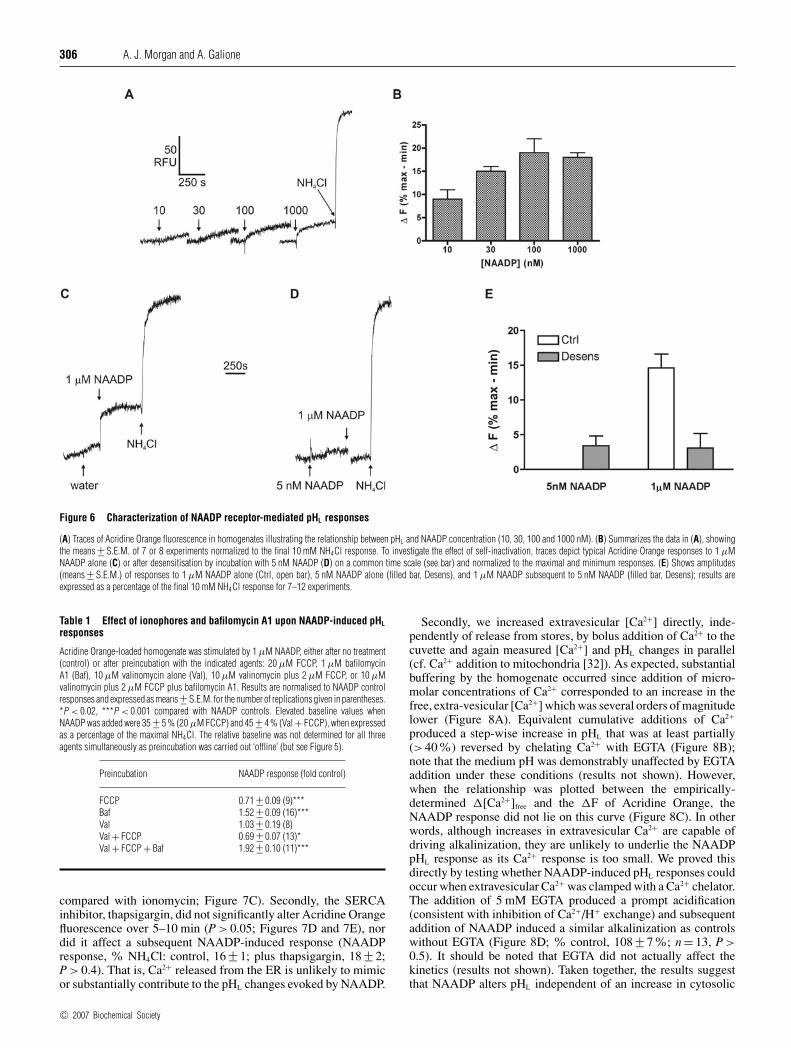
To characterize more thoroughly the NAADP pHL response, weinvestigated its concentration-response relationship. Increasingthe concentration of NAADP resulted in a graded increase influorescence (Figures 6A and 6B). The relatively small signal-to-noise ratio precluded an exact determination of the EC50, but anestimate of 10–30 nM agrees well with the EC50 of NAADP forCa2+ release in sea urchin egg homogenates [27,28].
In sea urchin egg, NAADP-induced Ca2+ release shows aprofound self-inactivation when preincubated with low NAADPconcentrations [4], so we next assessed whether Acridine Orangechanges also exhibited this phenomenon. In parallel, we first
c© 2007 Biochemical Society

Organelle pH and NAADP 305
Figure 5 Effect of membrane potential, H+ leak and V-ATPase activity on pHL
(A, B and C) Fluorescence traces of homogenates labelled with 10 µM Acridine Orange (AO). Addition of AO, 100 nM FCCP (F), 1 µM bafilomycin A1 (B or Baf), 10 µM valinomycin (V or Val)and 10 mM NH4Cl were effected where shown. The bar charts show means +− S.E.M. for 8–10 experiments for the amplitudes (D) and initial kinetics (E) of the respective responses. Amplitudes areexpressed as the change in fluorescence (�F) as a percentage of the maximum change with NH4Cl and rates as fluorescence units/s (U/s).
confirmed that application of 5 nM NAADP for 5–10 mininhibited the Ca2+ response elicited by 1 µM NAADP in the samepreparations (control: 197 +− 12 nM; desensitized: 53 +− 10 nM;P < 0.001; n = 4–5; note the small response in the desensitizedhomogenate reflects Ca2+ contamination of the NAADP andtherefore underestimates the degree of inactivation). Accordingly,5 nM NAADP elicited a very small response itself, but profoundlyreduced that to a subsequent addition of 1 µM NAADP whencompared with the control (Figures 6C–6E). This stronglysuggests that NAADP is affecting pHL changes via activationof the NAADP receptor, for which self-inactivation is diagnostic.
NAADP-induced pHL responses and transmembraneion movements
To go some way towards examining the role of ion fluxes acrossthe vesicle membrane during NAADP-induced pHL changes, weinvestigated the effect of several ionophore and bafilomycin A1combinations (Table 1). Note that the inevitable non-linearity ofthe Acridine Orange signal means that one must exercise cautionin comparing the absolute fluorescence changes when NAADPis added upon an elevated baseline (i.e. an apparent decrease inthe Acridine Orange change with NAADP does not necessarilyreflect a real decrease in the alkalinization response).
NAADP still elicited a response in the presence of a high con-centration of FCCP, albeit slightly reduced, and whilst valino-mycin alone had no effect upon the magnitude of the NAADPresponse (Table 1; P > 0.9), it did enhance the kinetics (time-to-peak: NAADP alone, 179 +− 29 s; NAADP plus valinomycin,72 +− 7 s; P < 0.01). When valinomycin was included with FCCP,the subsequent NAADP response was essentially the same aswith FCCP alone (with no effect upon kinetics; results notshown). One possible explanation for these results is that NAADPpredominantly alters pHL by a mechanism other than by changing
H+ leaks or K+ fluxes. By contrast, bafilomycin A1 enhancedthe NAADP response (Figure 4 and Table 1) and even more soin the presence of valinomycin and FCCP (conditions where V-ATPase activity is enhanced; Figure 3). These results confirmthat the bafilomycin A1-sensitive V-ATPase is a major player inmodulating dynamic pHL changes, but is unlikely to be a targetfor NAADP.
Cytosolic Ca2+ and pHL changes
We next asked whether NAADP-induced changes in pHL were re-lated to a direct effect of NAADP upon vesicular Ca2+ fluxes, e.g.a fall in the luminal [Ca2+] [29] and/or an increase in Ca2+ at thecytosolic face [30] when Ca2+ is released from the acidic store. Asa first step, we elevated the extravesicular [Ca2+] (i.e. the cytosolicface) in different ways, initially by comparing NAADP withother Ca2+-releasing messengers that release Ca2+ from mostlyneutral stores [5,6]. In spite of the fact that the Ca2+ responsesto messengers were not significantly different when measured inparallel {�[Ca2+] (% NAADP): cADPR, 64 +− 16; IP3, 74 +− 23;P > 0.1; ANOVA; n = 3}, neither IP3 nor cADPR could mimicthe alkalinization induced by NAADP (P < 0.001; Figures 7A, 7Band 7E) and, indeed, there was no significant difference betweenthe vehicle, cADPR and IP3 responses (P > 0.05).
We also released Ca2+ from the ER in other ways. First, theCa2+ ionophore, ionomycin (which preferentially releases Ca2+
from neutral but not acidic stores [31]), which evoked a substantialCa2+ response in homogenate that was 203 +− 10% (n = 6) of thecorresponding NAADP response; however, this robust increasein extravesicular Ca2+ did not translate into a substantial pHL
response (Figures 7C and 7E; P < 0.001 compared with NAADP),and was not significantly different from IP3 or cADPR (P >0.05), whereas a subsequent addition of NAADP evoked thecharacteristic alkalinization (12 +− 1% of NH4Cl, P < 0.001
c© 2007 Biochemical Society
306 A. J. Morgan and A. Galione
Figure 6 Characterization of NAADP receptor-mediated pHL responses
(A) Traces of Acridine Orange fluorescence in homogenates illustrating the relationship between pHL and NAADP concentration (10, 30, 100 and 1000 nM). (B) Summarizes the data in (A), showingthe means +− S.E.M. of 7 or 8 experiments normalized to the final 10 mM NH4Cl response. To investigate the effect of self-inactivation, traces depict typical Acridine Orange responses to 1 µMNAADP alone (C) or after desensitisation by incubation with 5 nM NAADP (D) on a common time scale (see bar) and normalized to the maximal and minimum responses. (E) Shows amplitudes(means +− S.E.M.) of responses to 1 µM NAADP alone (Ctrl, open bar), 5 nM NAADP alone (filled bar, Desens), and 1 µM NAADP subsequent to 5 nM NAADP (filled bar, Desens); results areexpressed as a percentage of the final 10 mM NH4Cl response for 7–12 experiments.
Table 1 Effect of ionophores and bafilomycin A1 upon NAADP-induced pHL
responses
Acridine Orange-loaded homogenate was stimulated by 1 µM NAADP, either after no treatment(control) or after preincubation with the indicated agents: 20 µM FCCP, 1 µM bafilomycinA1 (Baf), 10 µM valinomycin alone (Val), 10 µM valinomycin plus 2 µM FCCP, or 10 µMvalinomycin plus 2 µM FCCP plus bafilomycin A1. Results are normalised to NAADP controlresponses and expressed as means +− S.E.M. for the number of replications given in parentheses.*P < 0.02, ***P < 0.001 compared with NAADP controls. Elevated baseline values whenNAADP was added were 35 +− 5 % (20 µM FCCP) and 45 +− 4 % (Val + FCCP), when expressedas a percentage of the maximal NH4Cl. The relative baseline was not determined for all threeagents simultaneously as preincubation was carried out ‘offline’ (but see Figure 5).
Preincubation NAADP response (fold control)
FCCP 0.71 +− 0.09 (9)***Baf 1.52 +− 0.09 (16)***Val 1.03 +− 0.19 (8)Val + FCCP 0.69 +− 0.07 (13)*Val + FCCP + Baf 1.92 +− 0.10 (11)***
compared with ionomycin; Figure 7C). Secondly, the SERCAinhibitor, thapsigargin, did not significantly alter Acridine Orangefluorescence over 5–10 min (P > 0.05; Figures 7D and 7E), nordid it affect a subsequent NAADP-induced response (NAADPresponse, % NH4Cl: control, 16 +− 1; plus thapsigargin, 18 +− 2;P > 0.4). That is, Ca2+ released from the ER is unlikely to mimicor substantially contribute to the pHL changes evoked by NAADP.
Secondly, we increased extravesicular [Ca2+] directly, inde-pendently of release from stores, by bolus addition of Ca2+ to thecuvette and again measured [Ca2+] and pHL changes in parallel(cf. Ca2+ addition to mitochondria [32]). As expected, substantialbuffering by the homogenate occurred since addition of micro-molar concentrations of Ca2+ corresponded to an increase in thefree, extra-vesicular [Ca2+] which was several orders of magnitudelower (Figure 8A). Equivalent cumulative additions of Ca2+
produced a step-wise increase in pHL that was at least partially(>40%) reversed by chelating Ca2+ with EGTA (Figure 8B);note that the medium pH was demonstrably unaffected by EGTAaddition under these conditions (results not shown). However,when the relationship was plotted between the empirically-determined �[Ca2+]free and the �F of Acridine Orange, theNAADP response did not lie on this curve (Figure 8C). In otherwords, although increases in extravesicular Ca2+ are capable ofdriving alkalinization, they are unlikely to underlie the NAADPpHL response as its Ca2+ response is too small. We proved thisdirectly by testing whether NAADP-induced pHL responses couldoccur when extravesicular Ca2+ was clamped with a Ca2+ chelator.The addition of 5 mM EGTA produced a prompt acidification(consistent with inhibition of Ca2+/H+ exchange) and subsequentaddition of NAADP induced a similar alkalinization as controlswithout EGTA (Figure 8D; % control, 108 +− 7%; n = 13, P >0.5). It should be noted that EGTA did not actually affect thekinetics (results not shown). Taken together, the results suggestthat NAADP alters pHL independent of an increase in cytosolic
c© 2007 Biochemical Society

Organelle pH and NAADP 307
Figure 7 Ca2+ release from the ER does not mimic NAADP-induced pHL responses
pHL was measured in Acridine Orange-labelled homogenates in response to application of 10 µM cADPR (A), 4 µM IP3 (B), 5 µM ionomycin (Iono) (C) or 1 µM thapsigargin (Tg) (D). NAADP wassubsequently applied at 1 µM (C and D). (E) Shows the means +− S.E.M. of responses to the agents alone as indicated; all observations were normalized to 10 mM NH4Cl for 5–28 experiments.
Ca2+ and is due to a direct effect of NAADP upon the vesicle itself(e.g. via a fall in the luminal [Ca2+]).
We next verified that the changes we were seeing in homogenatewere relevant for the intact egg. To that end, we labelledeggs with Acridine Orange in order to measure pHL changes asreported previously [21] and then microinjected the centre of theegg with or without NAADP (approx. 1 µM intracellular concen-tration). Whilst control injections failed to alter Acridine Orangefluorescence (�F/F0, 0.00 +− 0.01; n = 13; P > 0.6), NAADP in-jection elicited a rapid, robust alkalinization (�F/F0, 0.25 +− 0.03;n = 16; P < 0.001; P < 0.001 compared with the control). How-ever, it should be noted that the NAADP response was nothomogeneous, being greater in the egg periphery, in contrast with10 mM NH4Cl, which enhanced fluorescence globally (resultsnot shown; cf. [21]). Nonetheless, it confirms that the NAADPresponses we observe in homogenates are present in the intactegg.
DISCUSSION
Properties of pHL-sensitive dyes
Both Acridine Orange and LysosensorTM Green DND-189 wereused to monitor pHL changes in sea urchin egg homogenates,and both gave increases in fluorescence upon alkalinization.While this was to be expected for Acridine Orange [21], weanticipated that LysosensorTM Green DND-189 would undergoa reduction in signal as its fluorescence is weaker at alkalinepH. This paradox can be explained by the hitherto unknown
fact that LysosensorTM Green DND-189 also self-quenches atmicromolar concentrations in vitro, levels which could easily beexceeded in situ with net accumulation (although we have nofirm idea of the luminal concentration of LysosensorTM GreenDND-189 or Acridine Orange). Hence, we propose that, as withAcridine Orange, the alkalinization of acidic organelles reducesthe accumulation of LysosensorTM Green DND-189, and theloss of some of the dye from the lumen results in the partialrelief of self-quenching from that remaining, and an increasein fluorescence ensues under our experimental conditions. Weestimated that bafilomycin A1-sensitive vesicles account for atleast 80% of the Acridine Orange labelling, although this is likelyto be a low estimate owing to the incomplete alkalinization evenover long time periods, as well as the inevitable non-linearity ofthe Acridine Orange signal. Overall, we confirm that the dyes area reliable index of pHL in sea urchin egg acidic stores [21].
NAADP-induced changes in pHL
NAADP-sensitive stores are physically separated from thoseresponding to other messengers [8] and are acidic organelles ina number of systems [5,7,12,18] including sea urchin egg, wherethey probably correspond to the yolk platelets [6]. However, thenotion of an acidic NAADP-sensitive store has been challenged[9–11], although subsequently partially resolved in exocrinepancreas [12]. Since Ca2+ release from acidic vesicles (e.g.secretory granules [33,34]) has repercussions for pHL [29], wereasoned that NAADP might affect pHL of acidic Ca2+ stores insea urchin egg homogenates. This preparation has not only beeninstrumental in the discovery and characterization of NAADP
c© 2007 Biochemical Society

308 A. J. Morgan and A. Galione
Figure 8 Relationship between extravesicular [Ca2+] and pHL
(A) Sequential boluses of 1–100 µM Ca2+ were applied directly to homogenates in the cuvette and extravesicular [Ca2+] changes were measured with Fluo-3. (B) Corresponding pHL trace measuredin homogenates labelled with Acridine Orange in response to incremental Ca2+, 5 mM EGTA and 10 mM NH4Cl. (C) Relationship between [Ca2+] changes determined with Fluo-3 in (A) andcorresponding pHL changes measured with Acridine Orange in (B). Open circles (�) represent bolus additions of Ca2+, the grey triangle those associated with 1 µM NAADP in the same preparation.Results are means +− S.E.M. for 8 (NAADP) and 31 experiments (Ca2+). (D) clamping cytosolic Ca2+ with 5 mM EGTA produces a fall in pHL, but does not block the alkalinization induced by 1 µMNAADP (n = 13). (B and D) Inset panels show hypothetical Ca2+ and H+ movements for orientation.
itself [35], but also proven to be an experimentally ‘cleaner’system, since the normal coupling between Ca2+-releasing secondmessengers [36] i.e. “channel chatter” [37], is disrupted uponhomogenization.
Using two fluorescent dyes in egg homogenates, we haverevealed that the luminal pH of NAADP-sensitive acidic Ca2+-storing organelles is coupled to Ca2+ release as a prompt alkalin-ization. The pHL response bore all the hallmarks of activationof the NAADP receptor as judged by a number of criteria: thenucleotide selectivity, the EC50 for NAADP (10–30 nM) andthe desensitization protocol, which inhibited both Ca2+ releaseand the pHL increase. This is the first demonstration of NAADPaltering pHL and also reveals that desensitization is not driven bypHL changes as 5nM NAADP barely altered pHL. The fact thatan increase in Acridine Orange fluorescence is observed in intacteggs upon NAADP microinjection confirms that the alkalinizationis not an artefact of homogenization.
H+ pumps and leaks
The pHL of vesicles is strongly influenced by the balance betweenH+ pumping into and H+ efflux (leaks) from them [23]. Ex-periments with ionophores were instructive because they revealedthat acidic vesicles in our preparation were not very leaky withrespect to H+ owing to both an inhibitory membrane potential aswell as a negligible H+ leak pathway (known to decrease duringsecretory/acidic vesicle maturation [23]). Indeed, the very factthat FCCP by itself causes little change in pHL also implies that
the movement of other possible counter ions is negligible in these‘tight’ vesicles.
Because passive H+ efflux contributed little to the resting pHL,bafilomycin A1 treatment alone did not substantially alterpHL. The same cannot be said under stimulated conditions whenV-ATPase inhibition actually enhanced the NAADP pHL response,from which we can draw several conclusions: first, this offersfurther support for V-ATPase expression on NAADP-sensitivestores [6]; secondly, it eliminates the V-ATPase as a targetfor NAADP action; thirdly, it is consistent with the V-ATPaseacting as a modulator of the NAADP-induced response suchthat alleviating the acidifying brake increases the alkalinizationresponse.
Our previous results in sea urchin eggs [6] revealed thatNAADP-sensitive stores did not readily leak Ca2+ upon bafilo-mycin A1 application, which apparently differs from mammaliancells [5,7,18]. In the light of the results of the present paper, wewould suggest that the primary reason for this discrepancy is thatthe pHL gradient in sea urchin egg acidic stores does not readilycollapse upon bafilomycin A1 treatment (although this clearlydoes not exclude an additional Ca2+ leak).
NAADP and transmembrane ion movements
In view of the tightness of these vesicles, it is interesting thatNAADP induces an alkalinization and raises the question ofwhether NAADP alters H+ leakiness and/or the veside membranepotential (as it does in the starfish oolemma [38]). The majority
c© 2007 Biochemical Society

Organelle pH and NAADP 309
of our results are not easily reconciled with NAADP increasing aH+ leak: (a) the NAADP response largely persists in the presenceof 20 µM FCCP, which already confers a substantial H+ leak;and (b) even when the H+ leak is facilitated by valinomycin, themagnitude of the NAADP response is not changed considerably,even when additionally combined with FCCP. The only pieceof data consistent with an H+ efflux is the enhancement of theNAADP response kinetics by valinomycin alone. In summary,we suggest that most of our results do not support H+ efflux as amajor mechanism, but any that does occur is likely to be secondaryto other transport processes.
If NAADP was only stimulating a Ca2+ efflux from the acidicstores, this cation efflux would serve to dissipate the membranepotential that inhibits the V-ATPase and promote acidification(cf. valinomycin), which is the opposite of what we observe. Bythe same token, the NAADP-induced efflux of cations other thanCa2+ (e.g. K+) would also be expected to acidify and so can alsobe discounted. Therefore, NAADP is working via some otheradditional (or adjunct) mechanism to alter pHL.
Overall, our data are most consistent with the electrogenicV-ATPase establishing a membrane potential in the NAADP-sensitive acidic Ca2+ store, inside positive as observed in otheracidic vesicles [39,40]. However, until we have a better idea ofion transporters and the ion concentrations on either side of theacidic store membrane, this remains speculative. In the context ofNAADP, it is interesting that K+ channel blockers inhibit NICR[22], either as a result of their effect upon membrane potential or,indeed, as a consequence of their being ammonium analogues,which could directly affect pHL.
Cytosolic Ca2+ and pHL
We then undertook a basic characterization of how Ca2+ move-ments interact with the NAADP-induced pHL response. Ourunderstanding of how pHL is regulated by Ca2+ in egg acidicvesicles is minimal compared with that of secretory vesicles,which are prototypic acidic Ca2+ stores [5,12,29,30], and were firstcharacterised in terms of their IP3-dependent responses [29,41].The influence of counterions and membrane potential [23,42,43]adds to the complexity, but we chose to focus on how Ca2+
movements affect pHL.As already alluded to, one direct model describes how mes-
senger-induced Ca2+ release results in a fall in luminal [Ca2+],thereby producing a decrease in the free [H+] (alkalinization) asa consequence of their competitive binding to a sequestering,polyanionic matrix [29,41]. In an alternative indirect model,increases in cytosolic (extravesicular) [Ca2+] lead to alkalinizationbecause of Ca2+ uptake via a putative Ca2+/H+ exchanger in thevesicle membrane [6,30]. We tested which of these models betterapplies to NAADP-induced pHL changes.
To mimic NICR, increases in extravesicular (cytosolic) Ca2+
were generated in two ways, either by release from non-acidicCa2+ stores, or by direct addition of Ca2+ to the cuvette. Ca2+
release induced by the other second messengers, IP3 or cADPR,failed to mimic NAADP despite the Ca2+ responses to all threebeing similar. This profile mirrors their organellar selectivity in seaurchin egg [6] and other systems ([5,7,18], but see [12]) with IP3
and cADPR releasing from non-acidic ER. Similarly, ionomycingave a larger Ca2+ signal than NAADP, but still had no effectupon pHL. Under these conditions, ionomycin will only releaseCa2+ from neutral stores such as the ER because the low pHL ofthe acidic stores blocks the ionophore Ca2+/H+ exchange activity[31,44–47]. Finally, the SERCA inhibitor, thapsigargin, did notsignificantly alter pHL or block the NAADP-induced pHL response(which mirrors the thapsigargin-insensitivity of NICR [4]). We
conclude that Ca2+ release from the ER itself does not result inpHL changes nor is it likely to be the driving force underlying theNAADP-induced alkalinization.
Manipulating cytosolic Ca2+ independently of release from theER involved additions of Ca2+ or EGTA to the cuvette. Cumulativeincreases in the [Ca2+] corresponded to cumulative increases inpHL, plausibly via a Ca2+/H+ exchanger. However, the affinityof this exchanger for Ca2+ was too low to mediate the NAADPpHL response whose corresponding global Ca2+ signal was 8–10-fold too low. One criticism of this conclusion is that the local[Ca2+] induced by NAADP might reach levels high enough to berelevant, similar to the ER–mitochondrion interface [32]. This wasnot the case, however, since EGTA did not affect the NAADP-induced alkalinization, even though 5 mM would be more thansufficient to swamp the Ca2+ concentration (1–2 µM) required tomimic the NAADP pHL response. One puzzling result was that theaddition of EGTA after the bolus addition of Ca2+ only resultedin an incomplete reversal to basal Acridine Orange fluorescencewhen the predicted [Ca2+] would be in the nanomolar range. Wehave no definitive explanation for this result, but it is conceivablethat the previous Ca2+ exposure affected H+ movements (ormembrane potential), since acidic vesicle transporters are knownto be affected by cytosolic Ca2+[48], luminal Ca2+ loading [49]or pHL [15,49]. Taken together, these data confirmed that theNAADP pHL response was independent of cytosolic Ca2+.
If an increase in cytosolic Ca2+ does not drive alkalinization,then we are left with some variant of the direct model whereNAADP binds to, and modulates the pHL of, the target organelle.One possibility is that the fall in the luminal [Ca2+] allowsH+ to bind to vacated sites on a luminal anionic matrix, inmuch the same way as has been suggested for IP3-dependentacidic stores [29,41]. We have attempted to test this directlyby lowering the luminal [Ca2+] with membrane-permeant Ca2+
chelators such as TPEN and oxalate, and while we did observe arobust alkalinization consistent with our hypothesis, we couldnot unequivocally exclude an artefact of their own acid/baseproperties (results not shown).
Our data show that NAADP alters pHL in its own acidic Ca2+
stores, independently of cytosolic Ca2+ changes. This providesfurther evidence in support of the somewhat controversial ideathat acidic Ca2+ stores are unique targets for NAADP since IP3
and cADPR could not mimic the alkalinization. The ramificationsof an increased pHL in acidic stores is unknown at present,but in terms of NAADP Ca2+ store function, an alkalinizationmight reduce Ca2+ reloading and prolong the refractory periodto NAADP. Sea urchin eggs have proven an excellent modelfor higher organisms [4], and our observations may wellhave implications for mammalian cells which utilize NAADP-sensitive acidic vesicles, e.g. alkalinization of secretory vesiclesin pancreatic acinar cells [12] could alter their exocytotic threshold[50], while lysosomal pH in smooth muscle cells [7] might affectproteolysis [25].
This work was supported by the Wellcome Trust, U.K. We would like to thank Dr NigelEmptage, Dr Stevan Rakovic and Dr Grant Churchill from our department for helpfulsuggestions or critically reading this manuscript.
REFERENCES
1 Papp, S., Dziak, E., Michalak, M. and Opas, M. (2003) Is all of the endoplasmic reticulumcreated equal? The effects of the heterogeneous distribution of endoplasmicreticulum Ca2+-handling proteins. J. Cell Biol. 160, 475–479
2 Burdakov, D., Petersen, O. H. and Verkhratsky, A. (2005) Intraluminal calciumas a primary regulator of endoplasmic reticulum function. Cell Calcium 38,303–310
c© 2007 Biochemical Society

310 A. J. Morgan and A. Galione
3 Missiaen, L., Raeymaekers, L., Dode, L., Vanoevelen, J., Van Baelen, K., Parys, J. B.,Callewaert, G., De Smedt, H., Segaert, S. and Wuytack, F. (2004) SPCA1 pumps andHailey–Hailey disease. Biochem. Biophys. Res. Commun. 322, 1204–1213
4 Galione, A. and Ruas, M. (2005) NAADP receptors. Cell Calcium 38, 273–2805 Yamasaki, M., Masgrau, R., Morgan, A. J., Churchill, G. C., Patel, S., Ashcroft, S. J. and
Galione, A. (2004) Organelle selection determines agonist-specific Ca2+ signals inpancreatic acinar and beta cells. J. Biol. Chem. 279, 7234–7240
6 Churchill, G. C., Okada, Y., Thomas, J. M., Armando, A., Genazzani, Patel, S. andGalione, A. (2002) NAADP mobilizes Ca2+ from reserve granules, a lysosome-relatedorganelle, in sea urchin eggs. Cell 111, 703–708
7 Kinnear, N. P., Boittin, F. X., Thomas, J. M., Galione, A. and Evans, A. M. (2004)Lysosome-sarcoplasmic reticulum junctions. A trigger zone for calcium signaling bynicotinic acid adenine dinucleotide phosphate and endothelin-1. J. Biol. Chem. 279,54319–54326
8 Lee, H. C. (2004) Multiplicity of Ca2+ messengers and Ca2+ stores: a perspective fromcyclic ADP-ribose and NAADP. Curr. Mol. Med. 4, 227–237
9 Gerasimenko, J. V., Maruyama, Y., Yano, K., Dolman, N. J., Tepikin, A. V., Petersen, O. H.and Gerasimenko, O. V. (2003) NAADP mobilizes Ca2+ from a thapsigargin-sensitivestore in the nuclear envelope by activating ryanodine receptors. J. Cell Biol. 163,271–282
10 Navazio, L., Bewell, M. A., Siddiqua, A., Dickinson, G. D., Galione, A. and Sanders, D.(2000) Calcium release from the endoplasmic reticulum of higher plants elicited by theNADP metabolite nicotinic acid adenine dinucleotide phosphate. Proc. Natl.Acad. Sci. U.S.A. 97, 8693–8698
11 Mandi, M., Toth, B., Timar, G. and Bak, J. (2006) Ca2+ release triggered by NAADP inhepatocyte microsomes. Biochem. J. 395, 233–238
12 Gerasimenko, J. V., Sherwood, M., Tepikin, A. V., Petersen, O. H. and Gerasimenko, O. V.(2006) NAADP, cADPR and IP3 all release Ca2+ from the endoplasmic reticulum and anacidic store in the secretory granule area. J. Cell Sci. 119, 226–238
13 Christensen, K. A., Myers, J. T. and Swanson, J. A. (2002) pH-dependent regulation oflysosomal calcium in macrophages. J. Cell Sci. 115, 599–607
14 Srinivas, S. P., Ong, A., Goon, L. and Bonanno, J. A. (2002) Lysosomal Ca2+ stores inbovine corneal endothelium. Invest. Ophthalmol. Vis. Sci. 43, 2341–2350
15 Pittman, J. K., Shigaki, T. and Hirschi, K. D. (2005) Evidence of differential pH regulationof the Arabidopsis vacuolar Ca2+/H+ antiporters CAX1 and CAX2. FEBS Lett. 579,2648–2656
16 Pozos, T. C., Sekler, I. and Cyert, M. S. (1996) The product of HUM1, a novel yeast gene,is required for vacuolar Ca2+/H+ exchange and is related to mammalian Na+/Ca2+
exchangers. Mol. Cell Biol. 16, 3730–374117 Lopez, J. J., Camello-Almaraz, C., Pariente, J. A., Salido, G. M. and Rosado, J. A. (2005)
Ca2+ accumulation into acidic organelles mediated by Ca2+- and vacuolar H+-ATPasesin human platelets. Biochem. J. 390, 243–252
18 Brailoiu, E., Hoard, J. L., Filipeanu, C. M., Brailoiu, G. C., Dun, S. L., Patel, S. and Dun,N. J. (2005) Nicotinic acid adenine dinucleotide phosphate potentiates neurite outgrowth.J. Biol. Chem. 280, 5646–5650
19 Morgan, A. J., Churchill, G. C., Masgrau, R., Ruas, M., Davis, L. C., Billington, R. A.,Patel, S., Yamasaki, M., Thomas, J. M., Genazzani, A. A. and Galione, A. (2006) Methodsin cADPR and NAADP research. In Methods in Calcium Signalling (Putney, J. W., Jr, ed.),pp. 265–334, CRC Press, Boca Raton
20 Morgan, A. J. and Thomas, A. P. (1999) Single cell and subcellular measurement ofintracellular Ca2+ concentration ([Ca2+]i). Methods Mol. Biol. 114, 93–123
21 Lee, H. C. and Epel, D. (1983) Changes in intracellular acidic compartments in sea urchineggs after activation. Dev. Biol. 98, 446–454
22 Genazzani, A. A., Mezna, M., Dickey, D. M., Michelangeli, F., Walseth, T. F. andGalione, A. (1997) Pharmacological properties of the Ca2+-release mechanism sensitiveto NAADP in the sea urchin egg. Br. J. Pharmacol. 121, 1489–1495
23 Demaurex, N. (2002) pH homeostasis of cellular organelles. News Physiol. Sci. 17,1–5
24 Rodrigues, C. O., Scott, D. A. and Docampo, R. (1999) Characterization of a vacuolarpyrophosphatase in Trypanosoma brucei and its localization to acidocalcisomes.Mol. Cell Biol. 19, 7712–7723
25 Busch, G. L., Lang, H. J. and Lang, F. (1996) Studies on the mechanism ofswelling-induced lysosomal alkalinization in vascular smooth muscle cells.Pflugers Arch. 431, 690–696
26 Milani, G., Schereiber, A. Z. and Vercesi, A. E. (2001) Ca2+ transport into an intracellularacidic compartment of Candida parapsilosis. FEBS Lett. 500, 80–84
27 Lee, H. C. and Aarhus, R. (1995) A derivative of NADP mobilizes calcium storesinsensitive to inositol trisphosphate and cyclic ADP-ribose. J. Biol. Chem. 270,2152–2157
28 Genazzani, A. A., Empson, R. M. and Galione, A. (1996) Unique inactivation properties ofNAADP-sensitive Ca2+ release. J. Biol. Chem. 271, 11599–11602
29 Quesada, I., Chin, W. C. and Verdugo, P. (2003) ATP-independent luminal oscillationsand release of Ca2+ and H+ from mast cell secretory granules: implications for signaltransduction. Biophys. J. 85, 963–970
30 Moreno, A., Lobaton, C. D., Santodomingo, J., Vay, L., Hernandez-SanMiguel, E.,Rizzuto, R., Montero, M. and Alvarez, J. (2005) Calcium dynamics incatecholamine-containing secretory vesicles. Cell Calcium 37, 555–564
31 Fasolato, C., Zottini, M., Clementi, E., Zacchetti, D., Meldolesi, J. and Pozzan, T. (1991)Intracellular Ca2+ pools in PC12 cells. Three intracellular pools are distinguished by theirturnover and mechanisms of Ca2+ accumulation, storage, and release. J. Biol. Chem.266, 20159–20167
32 Csordas, G., Thomas, A. P. and Hajnoczky, G. (1999) Quasi-synaptic calcium signaltransmission between endoplasmic reticulum and mitochondria. EMBO J. 18, 96–108
33 Mitchell, K. J., Lai, F. A. and Rutter, G. A. (2003) Ryanodine receptor type I and nicotinicacid adenine dinucleotide phosphate receptors mediate Ca2+ release frominsulin-containing vesicles in living pancreatic beta-cells (MIN6). J. Biol. Chem. 278,11057–11064
34 Petersen, O. H. (1996) Can Ca2+ be released from secretory granules or synapticvesicles? Trends Neurosci. 19, 411–413
35 Lee, H. C. and Aarhus, R. (1995) A derivative of NADP mobilizes calcium storesinsensitive to inositol trisphosphate and cyclic ADP-ribose. J. Biol. Chem. 270,2152–2157
36 Churchill, G. C. and Galione, A. (2000) Spatial control of Ca2+ signaling by nicotinicacid–adenine dinucleotide phosphate diffusion and gradients. J. Biol. Chem. 275,38687–38692
37 Patel, S., Churchill, G. C. and Galione, A. (2001) Coordination of Ca2+ signalling byNAADP. Trends Biochem. Sci. 26, 482–489
38 Moccia, F., Lim, D., Kyozuka, K. and Santella, L. (2004) NAADP triggers the fertilizationpotential in starfish oocytes. Cell Calcium 36, 515–524
39 Beyenbach, K. W. and Wieczorek, H. (2006) The V-type H+ ATPase: molecular structureand function, physiological roles and regulation. J. Exp. Biol. 209, 577–589
40 Hedrich, R., Kurkdjian, A., Guern, J. and Flugge, U. I. (1989) Comparative studies on theelectrical properties of the H+ translocating ATPase and pyrophosphatase of thevacuolar-lysosomal compartment. EMBO J. 8, 2835–2841
41 Nguyen, T., Chin, W. C. and Verdugo, P. (1998) Role of Ca2+/K+ ion exchange inintracellular storage and release of Ca2+. Nature 395, 908–912
42 Thevenod, F. (2002) Ion channels in secretory granules of the pancreas and their role inexocytosis and release of secretory proteins. Am. J. Physiol. Cell Physiol. 283,C651–C672
43 Wang, J. and Zhang, G. J. (2005) Influence of membrane physical state on lysosomalpotassium ion permeability. Cell Biol. Int. 29, 393–401
44 Martinez, J. R., Sugita, K., Mork, A. C., Helmke, R. J. and Zhang, G. H. (1998) Alterationsin Ca2+ storage and mobilization in submandibular acinar cells of reserpine-treated rats.Comp. Biochem. Physiol. C Pharmacol. Toxicol. Endocrinol. 120, 201–209
45 Yagodin, S., Pivovarova, N. B., Andrews, S. B. and Sattelle, D. B. (1999) Functionalcharacterization of thapsigargin and agonist-insensitive acidic Ca2+ stores in Drosophilamelanogaster S2 cell lines. Cell Calcium 25, 429–438
46 Marchesini, N., Luo, S., Rodrigues, C. O., Moreno, S. N. and Docampo, R. (2000)Acidocalcisomes and a vacuolar H+-pyrophosphatase in malaria parasites. Biochem. J.347, 243–253
47 Ruiz, F. A., Lea, C. R., Oldfield, E. and Docampo, R. (2004) Human platelet dense granulescontain polyphosphate and are similar to acidocalcisomes of bacteria and unicellulareukaryotes, J. Biol. Chem. 279, 44250–44257
48 Cunningham, K. W. and Fink, G. R. (1996) Calcineurin inhibits VCX1-dependentH+/Ca2+ exchange and induces Ca2+ ATPases in Saccharomyces cerevisiae.Mol. Cell Biol. 16, 2226–2237
49 Yamaguchi, T., Aharon, G. S., Sottosanto, J. B. and Blumwald, E. (2005) VacuolarNa+/H+ antiporter cation selectivity is regulated by calmodulin from within the vacuole ina Ca2+- and pH-dependent manner. Proc. Natl. Acad. Sci. U.S.A. 102, 16107–16112
50 Renstrom, E., Ivarsson, R. and Shears, S. B. (2002) Inositol 3,4,5,6-tetrakisphosphateinhibits insulin granule acidification and fusogenic potential. J. Biol. Chem. 277,26717–26720
Received 23 May 2006/9 November 2006; accepted 22 November 2006Published as BJ Immediate Publication 22 November 2006, doi:10.1042/BJ20060759
c© 2007 Biochemical Society