Trigeminal ganglion innervates the auditory brainstem
15
Trigeminal Ganglion Innervates the Auditory Brainstem SUSAN E. SHORE, 1,2 * ZOLTAN VASS, 3 NOEL L. WYS, 1 AND RICHARD A. ALTSCHULER 1 1 Kresge Hearing Research Institute, University of Michigan, Ann Arbor, Michigan 48109-0506 2 Department of Otolaryngology, Medical College of Ohio, Toledo, Ohio 43699 3 Department of Otolaryngology, Albert Szent-Gyorgyi Medical University, Szeged, Hungary ABSTRACT A neural connection between the trigeminal ganglion and the auditory brainstem was investigated by using retrograde and anterograde tract tracing methods: iontophoretic injec- tions of biocytin or biotinylated dextran-amine (BDA) were made into the guinea pig trigem- inal ganglion, and anterograde labeling was examined in the cochlear nucleus and superior olivary complex. Terminal labeling after biocytin and BDA injections into the ganglion was found to be most dense in the marginal cell area and secondarily in the magnocellular area of the ventral cochlear nucleus (VCN). Anterograde and retrograde labeling was also seen in the shell regions of the lateral superior olivary complex and in periolivary regions. The labeling was seen in the neuropil, on neuronal somata, and in regions surrounding blood vessels. Retrograde labeling was investigated using either wheatgerm agglutinin- horseradish peroxidase (WGA-HRP), BDA, or a fluorescent tracer, iontophoretically injected into the VCN. Cells filled by retrograde labeling were found in the ophthalmic and mandib- ular divisions of the trigeminal ganglion. We have previously shown that these divisions project to the cochlea and middle ear, respectively. This study provides the first evidence that the trigeminal ganglion innervates the cochlear nucleus and superior olivary complex. This projection from a predominantly somatosensory ganglion may be related to integration mechanisms involving the auditory end organ and its central targets. J. Comp. Neurol. 419: 271–285, 2000. © 2000 Wiley-Liss, Inc. Indexing terms: pathways; cochlear nucleus; lateral superior olivary complex; olivocochlear; somatosensory It is well established that somatosensory stimuli evoke responses in auditory neurons (Aitkin et al., 1978, 1981). Such multisensory integration may play a role in localiz- ing the body in space, once attributed only to higher cen- ters such as the superior colliculus where auditory, so- matosensory, and visual inputs converge (Drager and Hubel, 1976; Chalupa and Rhoades, 1977). The trigeminal system is one major component of this integration, con- tributing a major source of somatosensory input to the superior colliculus from the brainstem trigeminal complex (Killackney and Erzurumlu, 1981). More recent neuroana- tomical data indicate that somatosensory and auditory information converge at more peripheral sites. First-order relay somatosensory neurons, which subserve tactile and kinesthetic sensations, send direct projections to the co- chlear nucleus: for example, the dorsal column nuclei and interpolar and caudal spinal trigeminal nuclei send fibers directly to the dorsal cochlear nucleus (DCN) and the granule cell region of the ventral cochlear nucleus (Itoh et al., 1987; Weinberg and Rustioni, 1987). A projection from the cuneate nucleus gives rise to mossy fiber terminals in the dorsal DCN and granule cell domains of the cochlear nucleus (Wright and Ryugo, 1996). These projections may be excitatory or inhibitory (Saade et al., 1989; Young et al., 1995) as demonstrated by electrical stimulation and tac- Grant sponsor: NIH; Grant numbers: 5RO1DC00383, RO1 DC 000105; Grant sponsor: National Organization of Hearing Research (NOHR); Grant sponsor: Fogerty International; Grant number: TW 00502. *Correspondence to: S.E. Shore, Ph.D., Kresge Hearing Research Insti- tute, University of Michigan, 1301 E. Ann St., Ann Arbor, MI 48109. E-mail: [email protected] Received 2 August 1999; Revised 2 November 1999; Accepted 2 Decem- ber 1999 THE JOURNAL OF COMPARATIVE NEUROLOGY 419:271–285 (2000) © 2000 WILEY-LISS, INC.
-
Upload
independent -
Category
Documents
-
view
1 -
download
0
Transcript of Trigeminal ganglion innervates the auditory brainstem

Trigeminal Ganglion Innervates theAuditory Brainstem
SUSAN E. SHORE,1,2* ZOLTAN VASS,3 NOEL L. WYS,1AND
RICHARD A. ALTSCHULER1
1Kresge Hearing Research Institute, University of Michigan, Ann Arbor,Michigan 48109-0506
2Department of Otolaryngology, Medical College of Ohio, Toledo, Ohio 436993Department of Otolaryngology, Albert Szent-Gyorgyi Medical University,
Szeged, Hungary
ABSTRACTA neural connection between the trigeminal ganglion and the auditory brainstem was
investigated by using retrograde and anterograde tract tracing methods: iontophoretic injec-tions of biocytin or biotinylated dextran-amine (BDA) were made into the guinea pig trigem-inal ganglion, and anterograde labeling was examined in the cochlear nucleus and superiorolivary complex. Terminal labeling after biocytin and BDA injections into the ganglion wasfound to be most dense in the marginal cell area and secondarily in the magnocellular areaof the ventral cochlear nucleus (VCN). Anterograde and retrograde labeling was also seen inthe shell regions of the lateral superior olivary complex and in periolivary regions.The labeling was seen in the neuropil, on neuronal somata, and in regions surroundingblood vessels. Retrograde labeling was investigated using either wheatgerm agglutinin-horseradish peroxidase (WGA-HRP), BDA, or a fluorescent tracer, iontophoretically injectedinto the VCN. Cells filled by retrograde labeling were found in the ophthalmic and mandib-ular divisions of the trigeminal ganglion. We have previously shown that these divisionsproject to the cochlea and middle ear, respectively. This study provides the first evidence thatthe trigeminal ganglion innervates the cochlear nucleus and superior olivary complex. Thisprojection from a predominantly somatosensory ganglion may be related to integrationmechanisms involving the auditory end organ and its central targets. J. Comp. Neurol. 419:271–285, 2000. © 2000 Wiley-Liss, Inc.
Indexing terms: pathways; cochlear nucleus; lateral superior olivary complex; olivocochlear;
somatosensory
It is well established that somatosensory stimuli evokeresponses in auditory neurons (Aitkin et al., 1978, 1981).Such multisensory integration may play a role in localiz-ing the body in space, once attributed only to higher cen-ters such as the superior colliculus where auditory, so-matosensory, and visual inputs converge (Drager andHubel, 1976; Chalupa and Rhoades, 1977). The trigeminalsystem is one major component of this integration, con-tributing a major source of somatosensory input to thesuperior colliculus from the brainstem trigeminal complex(Killackney and Erzurumlu, 1981). More recent neuroana-tomical data indicate that somatosensory and auditoryinformation converge at more peripheral sites. First-orderrelay somatosensory neurons, which subserve tactile andkinesthetic sensations, send direct projections to the co-chlear nucleus: for example, the dorsal column nuclei andinterpolar and caudal spinal trigeminal nuclei send fibers
directly to the dorsal cochlear nucleus (DCN) and thegranule cell region of the ventral cochlear nucleus (Itoh etal., 1987; Weinberg and Rustioni, 1987). A projection fromthe cuneate nucleus gives rise to mossy fiber terminals inthe dorsal DCN and granule cell domains of the cochlearnucleus (Wright and Ryugo, 1996). These projections maybe excitatory or inhibitory (Saade et al., 1989; Young et al.,1995) as demonstrated by electrical stimulation and tac-
Grant sponsor: NIH; Grant numbers: 5RO1DC00383, RO1 DC 000105;Grant sponsor: National Organization of Hearing Research (NOHR); Grantsponsor: Fogerty International; Grant number: TW 00502.
*Correspondence to: S.E. Shore, Ph.D., Kresge Hearing Research Insti-tute, University of Michigan, 1301 E. Ann St., Ann Arbor, MI 48109.E-mail: [email protected]
Received 2 August 1999; Revised 2 November 1999; Accepted 2 Decem-ber 1999
THE JOURNAL OF COMPARATIVE NEUROLOGY 419:271–285 (2000)
© 2000 WILEY-LISS, INC.
tile manipulation of the pinna (Young et al., 1995). Be-cause changes in pinna position provide important spec-tral cues for sound localization (Rice et al., 1992),somatosensory input to the cochlear nucleus regardingpinna position may aid in sound localization performed byDCN cells. Somatosensory innervation of the cochlear nu-cleus or superior olivary complex may also be involved inmechanisms related to neck movements, which play a rolein orientation to acoustic stimuli.
The present investigation demonstrates that second-order auditory neurons in the cochlear nucleus are inner-vated by both the ophthalmic and mandibular divisions ofthe trigeminal ganglion, which also project to the cochleaand middle ear, respectively (Vass et al., 1997, 1998b).The ophthalmic division of the trigeminal ganglion alsoinnervates the extraocular muscles (Aigner et al., 1997),raising the interesting possibility of interactions amongthese three systems. A second major finding indicates thata reciprocal connection exists between neurons in the shellregions of the lateral superior olivary complex and thetrigeminal ganglion. Together with the trigeminal gan-glion innervation of the small cell cap region of the an-teroventral cochlear nucleus (AVCN), this projection mayform part of the olivocochlear feedback system.
MATERIALS AND METHODS
All experiments were carried out in accordance with theNIH Guide For the Care and Use of Laboratory Animals.Twelve pigmented guinea pigs (250–350 g; NIH Outbredstrain, Murphy Breeding Laboratory, Plainfield, IN) wereused in this study. The guinea pigs were anesthetizedwith ketamine hydrochloride (Ketaset; 80 mg/kg) and xy-lazine (Rompun; 4 mg/kg) and placed in a stereotaxicframe (David Kopf, Tujunga, CA), then a longitudinalincision was made in the scalp. Periodic drug supplementswere used to maintain anesthetic levels throughout theprocedure.
Anterograde tracing
Injections using biocytin and BDA. Biocytin and bi-otinylated dextran-amine (BDA) injections were madeinto the trigeminal ganglion, and anterograde labelingwas examined in the cochlear nucleus, a light microscopicevaluation in five animals and an electron microscopicevaluation in two animals. The location of the trigeminalganglion was identified by using stereotaxic coordinates(0.37 cm caudal to bregma, 0.45 cm lateral from midline,1.3 cm ventral to bregma), a small hole was drilled in theskull without disturbing the meninges, and a glass mi-cropipette (tip diameter 25–30 mm) was lowered 13 mm bymicromanipulator into the left trigeminal ganglion. Ionto-phoretic injections of biocytin or BDA were delivered via aglass microelectrode by using a constant current genera-tor (Stoelting, 3 mA alternating for 20 minutes). Biocytin(Sigma Chemical Co., St. Louis, MO; B-4261) was pre-pared as a 5% solution in 0.05 M Tris buffer (pH 7.6). Thesolution could be stored at 4°C for up to 7 days with nonoticeable loss of activity. Dental cement was used to sealthe opening, the overlying skin was sutured, and the an-imals were allowed to recover.
Light microscopic assessment. Twenty-four hoursafter the biocytin and 4–6 days after BDA injections,animals were euthanized. Each guinea pig was anesthe-tized with nembutal (15 mg/kg, i.p.) and ketamine (40mg/kg, i.m.) and perfused through the left ventricle of theheart with 200 ml of 0.1 M pH 7.4 phosphate buffer,followed by 200 ml of 1% paraformaldehyde and 1.5%glutaraldehyde in 0.1 M phosphate buffer, pH 7.2–7.4. Theflow of the fixative to the trigeminal ganglia and brain-stem by way of the common carotid arteries was maxi-mized by occluding the brachial arteries and the descend-ing aorta. Following perfusion-fixation, both the brain andtrigeminal ganglia were isolated and placed in fresh fixa-tive (2% paraformaldehyde and 2.5% glutaraldehyde in0.1 M phosphate buffer, pH 7.2–7.4) for 2–4 hours at 4°C.The tissues were then incubated overnight at 4°C with30% sucrose in 0.1 M phosphate buffer.
Each specimen was frozen with dry ice in 100% ethanoland frozen transverse sections obtained serially at 30-mmincrements on a sliding microtome. For BDA and biocytin,the sections were first incubated for 2 hours in Avidin-D-horseradish peroxidase (HRP; Vector Laboratories, Inc.,Burlingame, CA; #A-2004), in 0.1 M phosphate buffer with1% Triton X-100 (Sigma 9002-93-1), pH 7.4, and thenreacted with 3,31-diaminobenzidine tetrahydrochloride(DAB). Slides were counterstained for 3.5 minutes in 1%aqueous neutral red (J.T. Baker Inc., Phillipsburg, NJ, R747-03; Mesulam, 1978), buffered at pH 4.8 with acetatebuffer. The slides were washed in distilled H2O, dehy-drated in graded EtOH, cleared in xylene, coverslipped byusing Permount, and studied using a Leitz Dialux micro-scope equipped with a drawing tube.
Slides were examined by using brightfield microscopy tolocate and map labeled neurons located in the trigeminalganglion injection site and anterograde labeling of neuronpuncta in the auditory brainstem. Photomicrographs andcamera lucida drawings were digitized and imported toAdobe Photoshop for labeling and contrast adjustment.
Electron microscopic evaluation. Three guinea pigswere processed for electron microscopy, two 24 hours afterbiocytin and one 4–6 days after BDA injections were madeinto ventral cochlear nucleus (VCN) as described above.
Abbreviations
AVCN anteroventral cochlear nucleusAVII accessory facial nucleusBDA biotinylated dextran amineDAB 3,39-diaminobenzidine tetrahydrochlorideDCN dorsal cochlear nucleusLNTB lateral nucleus of the trapezoid bodyLPO lateral periolivary regionLSO lateral superior olivary complexMand mandibular divisionMax maxillary divisionMSO medial superior olivary complexMTB medial nucleus of the trapezoid bodyM5 motor nucleus of the fifth cranial (trigeminal) nerveOCA octopus cell areaOph ophthalmic divisionPN pontine nucleusPR5 principal trigeminal nucleusPVCN posteroventral cochlear nucleusRN raphe nucleusRPO rostral periolivary regionR7 root of the 7th cranial nerveSCC small cell cap of the VCNST5 spinal trigeminal nucleusTZ trapezoid bodyVCN ventral cochlear nucleusVNTB ventral nucleus of the trapezoid bodyWGA-HRP wheatgerm agglutinin-horseradish peroxidase
272 S.E. SHORE ET AL.
Guinea pigs were deeply anesthetized with an intraperi-toneal injection of sodium pentobarbital (75 mg/kg) andperfused transcardially with 600–800 ml of 3% parafor-maldehyde and 0.5% glutaraldehyde in 0.15 M sodiumcacodylate buffer (pH 7.35) at the rate of 50–70 ml/min.The brainstem was removed and immersed in the samefixative for 2–4 hours at 4°C, followed by an overnightwash in cold phosphate-buffered saline (PBS), pH 7.2–7.4.Transverse sections through the auditory brainstem wereobtained at a thickness of 60 mm with a Vibratome (Pelco101, St. Louis, MO). For enzymatic reaction to revealbiocytin and BDA label, the sections were first incubatedfor 2 hours in Avidin-D-HRP (Vector Laboratories, Inc.#A-2004), dissolved in 1% Triton X-100 (Sigma 9002-93-1)in 0.1 M phosphate buffer, pH 7.4. The sections were thenincubated for 8 minutes in 0.075% DAB and 0.005% H2O2(Sigma, H-1009). The reacted sections were postfixed atroom temperature for 1 hour in 0.2 % osmium tetroxide in0.15 M cacodylate buffer, pH 7.35. Following osmication,the sections were washed in three 5-minute changes ofcacodylate buffer, dehydrated in graded ethanols, and flat-embedded in epoxy resin between sheets of ACLAR (TedPella, Redding CA; Embed 812, EMSciences, Fort Wash-ington, PA). After polymerization, subdivisions of the co-chlear nucleus were dissected from the plastic sectionsand glued to blank resin blocks with a cyanoacrylate ad-hesive (Borden Wonderbond, Borden, Columbus, OH).Ninety-five-nm sections were cut with an ultramicrotome(Reichert Ultracut, Vienna, Austria), mounted onto pre-cleaned nickel grids, counterstained with lead citrate anduranyl acetate, and examined with a JEOL EM-1200 elec-tron microscope.
Retrograde tracing using WGA-HRPand BDA
Injections. For wheatgerm agglutinin-horseradishperoxidase (WGA-HRP; n 5 5), BDA (n 5 2), and fluores-cent tracer (n 5 4) injections, the left cochlear nucleus wasapproached from the posterior fossa and visualized byaspirating a small part of the overlying cerebellum. Aglass micropipette filled with a solution of 2% WGA-HRP,or 10 % BDA (MW 10,000, Molecular Probes, Eugene, OR)in phosphate-buffered saline (pH 7.4), was placed undervisual control on the lateral surface of the DCN. After themicropipette placement, the brain was covered with warmmineral oil to prevent tissue desiccation and reduce brainpulsation. Evoked potentials in response to click stimula-tion were recorded as the electrode was advanced in arostral-ventral direction (Shore and Nuttall, 1985). At adepth corresponding to the maximum-amplitude evokedpotential, positive current (3–5 mA, continuous for WGA-HRP, fluorogold and fast blue, and pulsed 7 seconds on, 7seconds off for BDA) was passed through the micropipettefor 2–5 minutes. After the micropipette was removed,some neck muscle was applied to replace the volume ofaspirated brain, dental cement was used to seal the open-ing, and the skin was sutured. The animal was allowed torecover.
Retrograde labeling. After the injections, 24 hoursfor WGA-HRP and 4–6 days for BDA were allowed foradequate neuron labeling. In some animals in which onefluorescent tracer was injected into the VCN, the skinoverlying the jaw was injected with a second fluorescenttracer in order to label sensory cells in the trigeminalganglion for comparison with cells labeled by VCN injec-
tions. Animals were then perfused and tissue processed asdescribed above for the light microscopic analysis of bio-cytin anterograde transport for HRP, or by incubatingBDA-containing sections with avidin-conjugated fluores-cent markers (Vector Labs, 1:50 in 0.1 M PBS, pH 8.2).
Neurons labeled by retrograde transport of WGA-HRPor BDA in the trigeminal ganglion were mapped by usinga brightfield microscope equipped with a camera lucidadrawing tube. The labeled cells in each section werecounted manually. Cells with a distinctly labeled outlineand either a relatively clear central nuclear zone or analmost homogeneous black labeling of the cytoplasm,which are criteria indicating that the major part of the cellwas included in the section, were selected for mapping.For labeled cells, soma shape, size, and location in theganglion were recorded. Soma size was measured by usinga calibrated eyepiece micrometer without correction fortissue shrinkage which occurs during processing. Stu-dent’s t-test was used to determine the significance level ofcell size differences in different divisions of the ganglion.Fluorescent labeled cells were viewed by using a Nikonepifluorescence microscope with the appropriate filters forfluorogold, fast blue, rhodamine, and fluoroscine isothio-cyanate (FITC) emission.
Controls for endogenous peroxidase activity. To controlfor the presence of endogenous peroxidase activity in tri-geminal afferent neurons, trigeminal ganglia from twoanimals were fixed by vascular perfusion, sectioned seri-ally at 30 mm, and processed according to the DAB tech-nique without exposure to reagents containing exogenousHRP. Sections were then examined under bright- anddarkfield illumination. No labeling was seen under theseconditions.
Controls for nonspecific labeling. To control for thepossibility that some of the visualized reaction productmay have resulted from the nonspecific deposition of in-cubation medium components, several sections from eachexperimental animal were treated with medium lackingthe chromogen DAB. To rule out the possibility that thevisualized reaction product resulted from the nonenzy-matic hydrolysis of an incubation medium component,several sections from each experiment were heated for15–20 minutes at 90–100°C to inactivate the enzymesprior to incubation in the complete medium. No labelingwas seen under these conditions.
RESULTS
Anterograde labeling following injectionsinto the trigeminal ganglion
The biocytin and BDA injection sites in the trigeminalganglion were characterized by a central core of labeledneurons that appeared to be completely filled with agent.Most cells around the injection site exhibited dense label-ing. The reaction product was present in the form of small,evenly distributed cytoplasmic granules. The nucleus wasclearly visible in most labeled neurons. Labeled axonsemerged from the central core of the injection site.
Cochlear nucleus. Following survival periods of 24hours for biocytin and 4–6 days for BDA injections, lightmicroscopic examination revealed numerous labeledpuncta or synaptic terminals in the ipsilateral magnocel-lular regions of VCN, and in the surrounding marginal cellareas. The most dense concentration of puncta was in the
273TRIGEMINAL INNERVATION OF AUDITORY BRAINSTEM

Fig. 1. Terminals from thin axons of the trigeminal ganglion endin the small cell cap (SCC) region of the anteroventral cochlear nu-cleus (AVCN). A transverse section (inset) of the ventral cochlearnucleus (VCN) is drawn, indicating the location of terminals by stip-pling. Large arrow points to an expanded drawing of some of these
terminals at upper left. Small arrow points to photomicrograph atlower right, showing an expanded view of some of the terminals. Theaxons typically form boutons de passage. D, dorsal; M, medial. Largescale bar 5 5 mm; small scale bar 5 10 mm.
274 S.E. SHORE ET AL.

marginal cell area (or small cell cap) of the VCN in theform of beads or varicosities (Fig. 1). Labeled axons in thisarea were thin (around 1 mm), suggesting slow conductionvelocities, and typically formed boutons de passage (Fig.1). Puncta were also located on the lateral edges of thegranule cell area.
In the magnocellular regions of anteroventral and pos-teroventral cochlear nucleus (AVCN and PVCN), labeledterminals tended to cluster towards the edges of the nu-cleus both medially and laterally. Puncta were seen bothon cell bodies (Fig. 2A) and in the proximity of, or sur-rounding blood vessels (Fig. 2B). Labeling in the vicinity ofblood vessels was also observed in the marginal cell area.Occasional labeling in the vicinity of blood vessels wasalso observed in the contralateral VCN.
EM examination of labeled terminals in the cochlearnucleus
Marginal cell area of VCN. Densely labeled terminalswere observed synapsing on dendrites in the marginal cellarea of the VCN (Fig. 3A,B). The terminals were oftenapposed to two or more dendrites (Fig. 3B). In some cases,there was evidence for asymmetric junctional thickening(Fig. 3A,B). Quantitative measurements of vesicles in onelabeled terminal yielded average areas of 1,396 nm2, withan average roundness value of 0.92. On this basis, vesicleswere judged to be small, round, consistent with the clas-sification of Cant and Morest (1979) and Cant (1993). Nocorrection factors were applied for the outer margins of thevesicles being sometimes obscured by reaction product.
Magnocellular cell area of VCN. Two types of associa-tions were observed in the magnocellular regions of VCN.Many labeled terminals were seen in close associationwith blood vessels. Less numerous labeled terminals wereobserved making axodendritic contacts, often on spines, oraxosomatic contacts onto spherical bushy or globularbushy cells.
Trigeminal nucleus. Biocytin and BDA injections intothe trigeminal ganglion resulted in dense anterogradelabeling in the ipsilateral principal trigeminal nucleusand the sensory root of the trigeminal nerve. The diame-ters of labeled axons and terminal arborizations in thetrigeminal nucleus were considerably larger (greater than2 mm) than those labeled in the marginal cell area. Label-ing in the trigeminal nucleus was not associated withblood vessels.
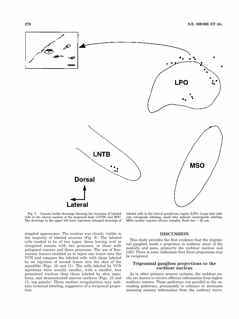
Superior olivary complex. After BDA injections intothe trigeminal ganglion, terminal labeling was observedin ipsilateral “shell” regions (Vetter and Mugnaini, 1992;Warr et al., 1997) of the lateral superior olivary complex(LSO; Fig. 4), the lateral nucleus of the trapezoid body(LNTB), and the lateral periolivary area (LPO) rostral toLSO (Fig. 5).
Fig. 2. A: Brightfield micrograph of biocytin-labeled puncta on acell in anteroventral cochlear nucleus (AVCN) 24 hours after aninjection was made into the left trigeminal ganglion. This cell waslocated towards the medial border of AVCN. B: Brightfield micro-graph of biocytin-labeled puncta surrounding the lumen of a bloodvessel in the magnocellular region of posteroventral cochlear nucleus(PVCN). Scale bar 5 10 mm for A; 20 mm for B.
275TRIGEMINAL INNERVATION OF AUDITORY BRAINSTEM

Fig. 3. Trigeminal ganglion fibers synapse on dendrites in themarginal cell area of anteroventral cochlear nucleus (AVCN). A: Thiselectron micrograph shows asymmetric junctional thickening in abiotinylated dextran amine (BDA)-labeled terminal (arrowhead), ap-posed to a dendrite. The terminal contains mainly small, round ves-
icles (arrowhead). B: A terminal apposed to two dendrites (D1 andD2) is shown. Asymmetric junctional thickening (small arrow) isevident. The terminals contained primarily small, round vesicles,although occasional, larger vesicles were seen (large arrow). Scalebars 5 250 nm.
276 S.E. SHORE ET AL.

Retrograde labeling following injectionsinto the trigeminal ganglion
Cells exhibiting reaction product throughout the cyto-plasm and dendrites were observed primarily in the ipsi-lateral motor trigeminal nucleus, raphe nuclei, and pon-tine nuclei of both sides. Fewer labeled cells were observedin the ipsilateral cochlear nucleus, superior olivary com-plex, and principal trigeminal nucleus (Figs. 4–6). La-beled cells in the cochlear nucleus were usually located inthe ventral regions of AVCN and PVCN towards the cen-ter, whereas those in the LSO were located on the mar-gins, in the “shell” regions (Figs. 4 and 6).
Retrograde labeling following injectionsinto the cochlear nucleus
Histologic examination confirmed that WGA-HRP andBDA injections were localized to the VCN. As we reportedpreviously (Shore et al., 1991, 1992; Shore and Moore,1998), retrogradely labeled cells projecting to the VCNwere found throughout the brainstem. In this investiga-tion, we focused on labeled cells in the trigeminal gan-glion.
Retrogradely labeled cells in the trigeminal gan-
glion. Injections of WGA-HRP, BDA, or fluorescent
tracers into the VCN produced robust labeling of cellsprimarily in the ipsilateral trigeminal ganglion, butlabeled cells were also observed on the contralateralside. The heaviest labeling was seen in the medial por-tion of the ganglion and at the origin of the ophthalmicnerve. Labeled cells were observed mostly on the medialside of the ophthalmic nerve. There were also numerouslabeled cells laterally, in the posterior portion of themandibular division of the ganglion. Fewer labeled cellswere seen in the mid-medial and posteromedial portionsof the trigeminal ganglia (Fig. 7A,B). Labeling after acochlear injection with WGA-HRP (Fig. 7C,D), is re-stricted to the ophthalmic division.
The distribution of HRP-WGA- and BDA-labeled cellsaccording to their maximal diameter indicates that thelabeled cells in the ophthalmic division (Fig. 8A) werelarger than in the mandibular division (Fig. 8B, P ,.05). Labeled cells in the ophthalmic division had max-imal diameters ranging from 10 to 45 mm, with a meanof 20 mm. In contrast, labeled cells in the mandibulardivision had maximal diameters ranging from 10 to 28mm, with a mean of 12 mm. Most cells were denselylabeled, with the reaction products appearing as small,evenly distributed cytoplasmic granules that created a
Fig. 4. Camera lucida drawings showing the locations of labeledcells in the “shell,” or surrounding regions of the lateral superiorolivary complex (LSO). Data from three sections are included. Some ofthe labeled cells (inset, color micrograph) contain reaction product in
their cytoplasm suggestive of retrograde uptake. Numbered cells areenlarged drawings of indicated labeled cells. Large dots indicate ret-rograde labeling; small dots indicate anterograde labeling; asterisksindicate labeled puncta around blood vessels. Scale bar 5 20 mm.
277TRIGEMINAL INNERVATION OF AUDITORY BRAINSTEM
stippled appearance. The nucleus was clearly visible inthe majority of labeled neurons (Fig. 9). The labeledcells tended to be of two types: those having oval orelongated somata with two processes, or those withpolygonal somata and three processes. The use of fluo-rescent tracers enabled us to inject one tracer into theVCN and compare the labeled cells with those labeledby an injection of second tracer into the skin of themandible (Figs. 10 and 11). The cells labeled by VCNinjections were usually smaller, with a smaller, lessprominent nucleus than those labeled by skin injec-tions, and demonstrated uneven surfaces (Figs. 10 and11, top panels). These surface irregularities may indi-cate terminal labeling, suggestive of a reciprocal projec-tion.
DISCUSSION
This study provides the first evidence that the trigemi-nal ganglion sends a projection to auditory areas of themedulla and pons, primarily the cochlear nucleus andLSO. There is some indication that these projections maybe reciprocal.
Trigeminal ganglion projections to thecochlear nucleus
As in other primary sensory systems, the cochlear nu-clei are known to receive efferent information from higherauditory centers. These pathways run parallel to the as-cending pathways, presumably to enhance or attenuateincoming sensory information from the auditory nerve.
Fig. 5. Camera lucida drawings showing the locations of labeledcells in the lateral nucleus of the trapezoid body (LNTB) and RPO.The drawings in the upper left inset represent enlarged drawings of
labeled cells in the lateral periolivary region (LPO). Large dots indi-cate retrograde labeling; small dots indicate anterograde labeling.MSO, medial superior olivary complex. Scale bar 5 20 mm.
278 S.E. SHORE ET AL.

Because many of the neurons projecting out of the VCNreceive descending information from their target neurons(Covey et al., 1984; Spangler and Warr, 1991; Winter etal., 1989; Spangler et al., 1987; Shore et al., 1991, 1992),some consider these projections to have a feedback func-tion. Whereas modulatory interactions of heterosensorypathways have been described in higher nuclei of thecentral nervous system (Aitkin et al., 1978), only in recentyears has it become apparent that input from nonauditorysensory systems also impinges on first-order auditory neu-rons. In the present study, injections of anterograde trac-ers into the trigeminal ganglion produced the most denselabeling in the marginal cell (including small cell cap)region of the cochlear nucleus. The labeled axons in thisarea were thin (around 1 mm), suggesting slow conductionvelocities. Their axodendritic terminals, containing smallspherical vesicles, are likely to be excitatory (Ostapoff andMorest, 1991). Other excitatory inputs to this area include
the cuneate nucleus, dorsal column nuclei, interpolar andcaudal spinal trigeminal nuclei (Itoh et al., 1987; Wolffand Kunzle, 1997), which form possible glutamatergicsynapses with granule cells (Wright and Ryugo, 1996).Immunocytochemical studies of the cochlear nucleus alsoreveal puncta that stain for substance P, a known sensoryneurotransmitter (Adams, 1993), whose locations coincideterminations of projections from the somatosensory re-gions described by Itoh et al. (1986), thus implicatingsubstance P as another possible neurotransmitter of thoseprojections. Substance P is involved in the cochlear pro-jection from the trigeminal ganglion (Vass et al., 1997,1998b) and thus may also be the neurotransmitter of thetrigeminal ganglion-cochlear nucleus projection.
Injections of retrograde tracer into one cochlear nucleusproduced labeling in both trigeminal ganglia, although thecontralateral side was sparsely labeled. Contralateral in-nervation has also been reported for cerebral blood vessels
Fig. 6. Camera lucida reconstructions of transverse sectionsthrough the brainstem of one guinea pig following a biotinylateddextran amine (BDA) injection into the left trigeminal ganglion. An-terograde labeling is shown by stippling; retrograde labeling is shownby large dots; asterisks indicate anterograde labeling around bloodvessels. Examples of retrograde labeling (large dots) are seen primar-
ily in pontine nucleus (PN), motor nucleus of the fifth cranial (trigem-inal) nerve (M5), and raphe nucleus (RN). More sparse retrogradelabeling is seen in principal trigeminal nucleus (PR5), lateral superiorolivary complex (LSO), and anteroventral cochlear nucleus (AVCN).For other abbreviations, see list.
279TRIGEMINAL INNERVATION OF AUDITORY BRAINSTEM

Figure 7
280 S.E. SHORE ET AL.

(Edvinsson et al., 1989) and cochlear blood supply (Vass etal., 1997, 1998b). As shown for the projection from thetrigeminal ganglion to the cochlea by using WGA-HRP(Vass et al., 1997), two types of cells, bipolar and multi-polar, comprise the projection from the trigeminal gan-glion to the cochlear nucleus. Both cell types are located inthe same region of the ganglion (ophthalmic) that projectsto the cochlea, as well as a restricted part of the mandib-ular division. Cells of similar size and shape, with two orthree processes, have also been observed to innervate ce-rebral blood vessels (Moskowitz, 1984).
The cells labeled by mandibular skin and cochlear nu-cleus injections in this study indicate that the cells pro-jecting to the cochlear nucleus are smaller than the sen-
sory cells innervating the skin overlying the mandible.The presence of terminal labeling on the surface of thecochlear nucleus-projecting cells indicates the possibilityof this projection being reciprocal.
Trigeminal ganglion projections to the LSO
The trigeminal ganglion, in contrast to other somato-sensory pathways, innervates both neurons and blood ves-
Fig. 8. Distribution of wheatgerm agglutinin-horseradish peroxi-dase (WGA-HRP) and biotinylated dextran amine (BDA)-labeled cellsin the trigeminal ganglion according to their maximal soma diame-ters. These data represent a random sample taken from 5 ganglia.A: Ophthalmic division; n 5 222. B: Mandibular division; n 5 148.
Fig. 7. Schematic of horizontal sections through the trigeminalganglia of one representative animal after a 2% wheatgermagglutinin-horseradish peroxidase (WGA-HRP) injection was madeinto the ventral cochlear nucleus (VCN). The injection site is shown atupper left. A: Ipsilateral (Ipsi) trigeminal ganglion. B: Contralateral(Contra) trigeminal ganglion. Each dot represents a single labeledneuron, and data are taken from 3 consecutive sections. C: Schematichorizontal section through the ipsilateral trigeminal ganglion, and(D) the contralateral trigeminal ganglion after a 2% WGA-HRP injec-tion was made into the cochlea (reproduced from Vass et al., 1997,with permission). For abbreviations, see list.
Fig. 9. Brightfield photomicrograph of wheatgerm agglutinin-horseradish peroxidase (WGA-HRP)-labeled cell bodies in the oph-thalmic division of the ipsilateral trigeminal ganglion after injectioninto the cochlear nucleus. The cell bodies shown here are polygonalwith three processes (top), or elongated with two processes (middleand bottom). Scale bar 5 20 mm.
281TRIGEMINAL INNERVATION OF AUDITORY BRAINSTEM

sels in the cochear nucleus, and “shell” regions of LSOwhich are known to send projections to the cochlea (Vetterand Mugnaini, 1992; Warr et al., 1997). The small cell capregion of the cochlear nucleus, which is heavily innervatedby the trigeminal ganglion, is also innervated by type IIspiral ganglion cells. It is also innervated by collaterals ofthe olivocochlear system which project to cochlear haircells and type I auditory nerve fibers (Brown and Benson,1992; Brown, 1993; Benson et al., 1996; Ye et al., 1999). Inaddition, the trigeminal ganglion innervates both the au-ditory and vestibular portions of the labyrinth (Vass et al.,1997, 1998). Because the trigeminal ganglion projects toolivocochlear regions of the LSO as well as to the small cellcap region, which forms a reciprocal connection with olivo-cochlear neurons (Brown and Benson, 1992; Brown, 1993;
Benson et al., 1996; Ye et al., 1999), we may speculate thatthis projection could form part of the olivocochlear feed-back system.
Function of somatosensory input to theauditory brainstem
Physiological studies demonstrate that electrical stim-ulation of the dorsal column and trigeminal nuclei usuallyinhibits, but can also excite cells in the superficial anddeep layers of DCN and VCN (Saade et al., 1989; Young etal., 1995). The inhibition to DCN neurons is producedthrough excitation of cells in the granular regions, whichin turn modify (inhibit) the activity of principal cells in theDCN (Young et al., 1995). Tactile manipulation of thepinna likewise can inhibit DCN neurons, suggesting that
Fig. 10. Photomicrographs of injection sites (insets) and individ-ual trigeminal ganglion somata filled by retrograde labeling (mainbody of micrograph) . The neurons were labeled 1 week after fluoro-gold and fast-blue were injected into the cochlear nucleus and the skinoverlying the mandible, respectively. The fluorogold-labeled neuron
projecting to the cochlear nucleus (top) has an irregular surface,perhaps indicating terminal endings, whereas the fast-blue-labeledneuron (bottom) projecting to the mandible has a prominent, enlargednucleus. Scale bar 5 25 mm.
282 S.E. SHORE ET AL.

some of the cells stimulated electrically are those whichrepresent the pinna and back of the head (Young et al.,1995). Thus, brainstem somatosensory input concerningpinna position may provide sound localization informationto DCN cells.
Furthermore, the ophthalmic portion of the trigeminalganglion also innervates extraocular muscles (Aigner etal., 1997). This raises another interesting possibility thatthese connections might be involved in vestibulo-ocularreflexes. Direct connections from the vestibular end or-gans to the cochlear nucleus have indeed been demon-strated (Kvetter and Perachio, 1989), as well as directprimary projections from the trigeminal ganglion to thelateral and superior vestibular nuclei (Pfaller and Arvids-son, 1988). Pfaller and Arvidsson (1988), by using HRP-WGA injections into the trigeminal and dorsal root gan-glia of the rat, observed terminal labeling along the medialedge of the VCN after injections into the C2 dorsal rootganglion but not after injections into the trigeminal gan-
glion. It is possible that species, tracer, and survival-timedifferences could account for the disparity in findings.
The present study demonstrates that the regions of thetrigeminal ganglion, which innervate the cochlear nu-cleus, overlap the regions that innervate both the cochleaand the middle ear: The ophthalmic division innervatesthe cochlea, and the mandibular region innervates themiddle ear (Vass et al., 1997). The intriguing possibilitytherefore exists of a role for the trigeminal ganglion in themiddle ear reflex. Support for this hypothesis is providedby the finding that cutaneous stimulation of the perior-bital and external ear regions can evoke the middle earreflex (Moller, 1975). The ascending limb of this reflex arcmay be trigeminal input from the external auditory me-atus (Folan-Curran et al., 1994). The first central relaystation of the middle ear reflex is the VCN, which sends aprojection bilaterally to the motor neurons of the tensortympani and stapedius muscles located in the trigeminaland facial nuclei, respectively (Itoh et al., 1986; Rouiller et
Fig. 11. An individual trigeminal ganglion neuron (red cell, top,right) is filled by retrograde labeling 6 days after an injection offluororuby into the posteroventral cochlear nucleus (left column ofmicrograph). The injection site (arrow) shows deposits of fluororuby inlateral posteroventral cochlear nucleus (PVCN) with an adjacent oc-topus cell to the right which was filled by spread of the injection. Theblue cell shown on bottom right is a cell filled by retrograde labeling
6 days after the skin overlying the mandible was injected with fastblue (injection site not shown). The fluororuby-labeled neuron project-ing to the PVCN has an irregular surface, similar to that shown inFig. 10, perhaps indicating terminal endings, whereas the fast-blue-labeled neuron, projecting to the mandible, has a prominent, enlargednucleus. Scale bar 5 20 mm.
283TRIGEMINAL INNERVATION OF AUDITORY BRAINSTEM

al., 1989). Thus, the trigeminal ganglion, activated bycutaneous stimulation in the region of the external ear,may modulate the activity of neurons in the VCN, whichin turn project to motor neurons of the tensor tympani andstapedius. Because we have shown that the trigeminalganglion projects to the middle ear, the cochlea and theVCN (Vass et al., 1997, 1998, and present study), modu-lation of the middle ear reflex could occur through directtrigeminal ganglion projections to the VCN and middleear muscles. The projection may also be involved in cen-tral or “somatic” tinnitus, which can be initiated or mod-ified by somatic stimulation of the head and neck region(Lockwood et al., 1998).
Finally, the observation, after biocytin injections intothe trigeminal ganglion, that many of the labeled punctain the VCN and LSO were located around the lumina ofblood vessels, suggests an involvement of this pathway inthe regulation of blood flow or metabolism in the cochlearnucleus and superior olivary complex. Stimulation of thetrigeminal ganglion results in changes in cochlear and incerebral blood flow (Vass et al., 1995; Goadsby et al., 1997;Lambert et al., 1997). Additionally, in peripheral and cen-tral blood vessels, administration of capsaicin, which stim-ulates release of substance P, produces sustained vasodi-latation, increased vascular permeability (Jansco, 1992),and an increase in cochlear blood flow (Vass et al., 1994).In cerebral circulation, substance P-containing neuronsmay be associated with headache pain or reactions totrauma such as breakdown of the blood-brain barrier(Duckles and Buck, 1982). In the cochlea, the trigeminalganglion may play a role in the pathophysiology of Me-nieres disease (Vass et al., 1998b).
Trigeminal input to the cochlear nucleus and other au-ditory brainstem neurons could have a great impact on themetabolism and function of cochlear nucleus neurons if ittargets both the neurons and their vascular supply. Ouranatomical characterization of this projection will set thestage for detailed functional studies of this potentiallyimportant projection. Our findings provide a unique op-portunity to study the relationship between cochlear andcochlear nucleus innervation because tracer injectionsinto the trigeminal ganglion also produce labeling in thecochlear vasculature (Vass et al., 1997). Because changesin peripheral hearing structures that occur with deafnessmay affect the structure of central auditory neurons, thenpathological changes in innervation from peripheral so-matosensory structures could also profoundly affect audi-tory functions requiring multisensory input, such as thelocalization of the body in space.
ACKNOWLEDGMENTS
We thank Tom Carey for many helpful suggestions overthe course of this study and Don Godfrey for valuablecomments on the manuscript.
LITERATURE CITED
Adams JC. 1993. Non-primary inputs to the cochlear nucleus visualizedusing immunocytochemistry. In: Merchan MA, Juiz JM, Godfrey DA,Mugnaini E, editors. The mammalian cochlear nuclei: organization andfunction. New York: Plenum.
Aigner M, Lukas JR, Denk M, Mayr R. 1997. Sensory innervation of theguinea pig extraocular muscles: a 1,10-dioctadecyl-3,3,3030-tetra-methylindocarbocyanine perchlorate tracing and calcitonin gene-
related peptide immunohistochemical study. J Comp Neurol 380:16–22.
Aitkin LM, Dickhaus H, Schult W, Zimmerman M. 1978. External nucleusof inferior colliculus: auditory and spinal somatosensory afferents andtheir interactions. J Neurophysiol 41:837–847.
Aitkin LM, Kenyon CE, Philpott P. 1981. The representation of the audi-tory and somatosensory systems in the external nucleus of the catinferior colliculus. J Comp Neurol 196:25–40.
Benson TE, Berglund AM, Brown MC. 1996. Synaptic input to cochlearnucleus dendrites that receive medial olivocochlear synapses. J CompNeurol 365:27–41.
Brown MC. 1993. Fiber pathways and branching patterns of biocytin-labeled olivocochlear neurons in the mouse brainstem. J Comp Neurol337:600–613.
Brown MC, Benson TE. 1992.Transneuronal labeling of cochlear nucleusneurons by HRP-labeled auditory nerve fibers and olivocochlearbranches in mice. J Comp Neurol 321:645–665.
Chalupa LM, Rhoades RW. 1977. Responses of visual, somatosensory, andauditory neurones in the golden hamster’s superior colliculus. J Physiol(Lond) 270:595–626.
Covey E, Jones DR, Casseday JH. 1984. Projections from the superiorolivary complex to the cochlear nucleus in the tree shrew. J CompNeurol 226:289–305.
Drager UC, Hubel DH. 1976. Topography of visual and somatosensoryprojections to mouse superior colliculus. J Neurophysiol 39:91–101.
Duckles SP, Buck SH. 1982. Substance P in the cerebral vasculature:depletion by capsaicin suggests a sensory role. Brain Res 245:171–174.
Edvinsson L, Hara H, Uddman R. 1989. Retrograde tracing of nerve fibersto the rat middle cerebral artery with True Blue: co-localization withdifferent peptides. J Cerebr Blood Flow Metab 9:212–218.
Folan-Curran J, Kickey K, Monkhouse WS. 1994. Innervation of the ratexternal auditory meatus: a retrograde tracing study. Somatosen Mo-tor Res 11:65–68.
Goadsby PJ, Knight YE, Hoskin KL, Butler P. 1997. Stimulation of anintracranial trigeminally-innervated structure selectively increases ce-rebral blood flow. Brain Res 751:247–252.
Itoh K, Nomura S, Konishi A, Yasui Y, Sugimoto T, Mizuno N. 1986. Amorphological evidence of direct connections from the cochlear nuclei totensor tympani motoneurons in cat: a possible afferent limb of theacoustic middle ear reflex pathways. Brain Res 375:214–219.
Itoh K, Kamiya H, Mitani A, Yasui Y, Takada M, Mizuno N. 1987. Directprojections from the dorsal column nuclei and the spinal trigeminalnuclei to the cochlear nuclei in the cat. Brain Res 400:145–150.
Jansco G. 1992. Pathobiological reactions of C-fibre primary sensory neu-rones to peripheral nerve injury. Exp Physiol 77:405–431.
Killackney HP, Erzurumlu RS. 1981. Trigeminal projections to the supe-rior colliculus of the rat. J Comp Neurol 201:221–242.
Kvetter GA, Perachio AA. 1989. Projections from the sacculus to thecochear nuclei in the Mongolian gerbil. Brain Behav Evol 34:193–200.
Lambert GA, Michalicek J, Regaglia F. 1997. Responses of the duralcirculation to electrical stimulation of the trigeminal ganglion in thecat. Clin Exp Pharm Physiol 24:377–390.
Lockwood AH, Salvi RJ, Coad Ml, Towsley ML, Wack DS, Murhpy BW.1998. The functional neuroanatomy of tinnitus: evidence for limbicsystem links and neural plasticity. Neurology 50:114–120.
Mesulam MM. 1978. Tetramethyl benzidine for horseradish peroxidaseneurochemistry: a non-carcinogenic blue reaction-product with a supe-rior sensitivity for visualizing neural afferents and efferents. J Histo-chem Cytochem 26:106–117.
Møller A. 1975. The acoustic middle ear reflex. In: Keidel WD, Neff WD,editors. Handbook of sensory physiology: auditory system, vol VII.Berlin: Springer. p 519–548.
Moskowitz MA. 1984. The neurobiology of vascular head pain. Ann Neurol16:157–168.
Ostapoff EM, Morest DK. 1991. Synaptic organization of globular bushycells in the ventral cochlear nucleus of the cat: a quantitative study.J Comp Neurol 314:598–613.
Pfaller K, Arvidsson J. 1988. Central distribution of trigeminal and uppercervical primary afferents in the rat studied by anterograde transportof horseradish peroxidase conjugated to wheat germ-agglutinin.J Comp Neurol 268:91–108.
Rice JJ, May BJ, Spirou GA, Young ED. 1992. Pinna-based spectral cuesfor sound localization in cat. Hearing Res 58:132–152.
Rouiller EM Capt M, Dolivo M, De Ribaupierre F. 1989. Neuronal organi-
284 S.E. SHORE ET AL.
zation of the stapedius reflex pathways in the rat: a retrograde HRPand viral transneuronal tracing study. Brain Res 476:21–28.
Saade NE, Frangieh AS, Atweh SF, Jabbur SJ. 1989. Dorsal column inputto cochlear neurons in decerebrate-decerebellate cats. Brain Res 486:399–402.
Shore SE, Moore JK. 1998. Sources of input to the cochlear granule cellregion in guinea pig. Hear Res 116:33–42.
Shore SE, Nuttall AL. 1985. The effects of cochlear hypothermiaon compound action potential tuning. J Acoust Soc Am 77:590 –598.
Shore SE, Helfert RH, Bledsoe SC Jr, Altschuler RA, Godfrey DA. 1991.Descending projections to the guinea pig cochlear nucleus. Hearing Res52:255–268.
Shore SE, Helfert RH, Bledsoe SC Jr, Altschuler RA, Godfrey DA. 1992.Connections between the cochlear nuclei in the guinea pig . HearingRes 62:16–26.
Spangler KM, Warr WB. 1991. The descending auditory system. In:Altschuler RA, Bobbin RP, Clopton BM, Hoffman DW, editors. Neu-robiology of hearing. The central auditory system. New York: RavenPress. p 27– 45.
Spangler KM, Cant, NB, Henkel CK, Farley GR, Warr WB. 1987. Descend-ing projections from the superior olivary complex to the cochlear nu-cleus of the cat. J Comp Neurol 259:452–465.
Vass Z, Bari F, Jansco G. 1994. Possible involvement of capsaicin-sensitivesensory nerves in the regulation of cochlear blood flow in the guineapig. Acta Otolaryngol (Stockh) 114:156–161.
Vass Z, Nuttall AL, Coleman JKM, Miller JM. 1995. Capsaicin-inducedrelease of substance P increases cochlear blood flow in the guinea pig.Hear Res 89:86–92.
Vass Z, Shore SE, Nuttall AL, Miller JM, Brechtelsbauer PB. 1997. Tri-
geminal ganglion innervation of the cochlea—a retrograde transportstudy. Neuroscience 79:605–615.
Vass Z, Shore SE, Nuttall AL, Miller JM. 1998a. Direct evidence of tri-geminal innervation of the cochlear blood vessels. Neuroscience 84:559–567.
Vass Z, Shore SE, Nuttall AL, Miller JM. 1998b. Endolymphatic hydropsreduces trigeminal innervation to inner ear vasculature. Exp Neurol151:241–248.
Vetter DE, Mugnaini E. 1992. Distribution and dendritic reatures of threegroups of rat olivocochlear neurons. Anat Embryol 185:1–16.
Warr WB, Beck Boche J, Neely ST. 1997. Efferent innervation of the innerhair cell region: origins and terminations of two lateral olivocochlearsystems. Hear Res 108:89–111.
Weinberg RJ, Rustioni A. 1987. A cuneocochlear pathway in the rat.Neuroscience 20:209–219.
Winter IM, Robertson D, Cole KS. 1989. Descending projections fromauditory brainstem nuclei to the cochlea and cochlear nucleus of theguinea pig. J Comp Neurol 280:143–157.
Wolff A, Kunzle H. 1997. Cortical and medullary somatosensory projec-tions to the cochlear nucleus complex in the hedgehog tenrec. NeurosciLet 221:125–128.
Wright DD, Ryugo DK. 1996. Mossy fiber projections from the cuneatenucleus to the cochlear nucleus in the rat. J Comp Neurol 365:159–172.
Ye Y, Machado DG, Kim DO. 1999. Projections of the marginal shell of theanteroventral cochlear nucleus to olivocochlear neurons in the cat.Abstract 22nd ARO, 584, p. 148.
Young ED, Nelken I, Conley RA. 1995. Somatosensory effects on neuronsin dorsal cochlear nucleus. J Neurophysiol 73:743–765.
285TRIGEMINAL INNERVATION OF AUDITORY BRAINSTEM