A new model of brainstem ischemia by embolization technique in cats
Upload
wzhospitalCategory
view
0download
0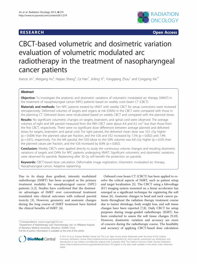
Jin et al. Radiation Oncology 2013, 8:279http://www.ro-journal.com/content/8/1/279
RESEARCH Open Access
CBCT-based volumetric and dosimetric variationevaluation of volumetric modulated arcradiotherapy in the treatment of nasopharyngealcancer patientsXiance Jin1, Weigang Hu2, Haijiao Shang3, Ce Han1, Jinling Yi1, Yongqiang Zhou1 and Congying Xie1*
Abstract
Objective: To investigate the anatomic and dosimetric variations of volumetric modulated arc therapy (VMAT) inthe treatment of nasopharyngeal cancer (NPC) patients based on weekly cone beam CT (CBCT).
Materials and methods: Ten NPC patients treated by VMAT with weekly CBCT for setup corrections were reviewedretrospectively. Deformed volumes of targets and organs at risk (OARs) in the CBCT were compared with those inthe planning CT. Delivered doses were recalculated based on weekly CBCT and compared with the planned doses.
Results: No significant volumetric changes on targets, brainstem, and spinal cord were observed. The averagevolumes of right and left parotid measured from the fifth CBCT were about 4.4 and 4.5 cm3 less than those fromthe first CBCT, respectively. There were no significant dose differences between average planned and delivereddoses for targets, brainstem and spinal cord. For right parotid, the delivered mean dose was 10.5 cGy higher(p = 0.004) than the planned value per fraction, and the V26 and V32 increased by 7.5% (p = 0.002) and 7.4%(p = 0.01), respectively. For the left parotid, the D50 (dose to the 50% volume) was 8.8 cGy higher (p = 0.03) thanthe planned values per fraction, and the V26 increased by 8.8% (p = 0.002).
Conclusion: Weekly CBCTs were applied directly to study the continuous volume changes and resulting dosimetricvariations of targets and OARs for NPC patients undergoing VMAT. Significant volumetric and dosimetric variationswere observed for parotids. Replanning after 30 Gy will benefit the protection on parotids.
Keywords: CBCT-based dose calculation, Deformable image registration, Volumetric modulated arc therapy,Nasopharyngeal cancer, Adaptive replanning
Due to its sharp dose gradient, intensity modulatedradiotherapy (IMRT) has been accepted as the primarytreatment modality for nasopharyngeal cancer (NPC)patients [1,2]. Studies have confirmed that the dosimet-ric advantages of IMRT over conventional treatmenttranslated into clinical outcomes with reduced parotidtoxicity [3]. However, geometry and anatomic changesduring the long course of IMRT treatment have limitedthe clinical benefits of IMRT [4].
* Correspondence: [email protected] of Radiotherapy and Chemotherapy, the 1st Affiliated Hospitalof Wenzhou Medical University, Wenzhou 325000, ChinaFull list of author information is available at the end of the article
© 2013 Jin et al.; licensee BioMed Central Ltd.Commons Attribution License (http://creativecreproduction in any medium, provided the orwaiver (http://creativecommons.org/publicdomstated.
Onboard cone beam CT (CBCT) has been applied to re-solve the critical aspects of IMRT, such as patient setupand target localization [5]. The CBCT using a kilovoltage(kV) imaging system mounted on a linear accelerator hasemerged as a significant technique for registering the softtissue [6]. Anatomic changes in head and neck cancer pa-tients throughout the radiation therapy treatment coursedue to tumor shrinkage, body weight loss, and soft tissuechanges have been reported [7,8]. Daily CBCT for setuppurposes during image-guided radiotherapy (IGRT) hasbeen conducted to assess the soft tissue changes [9,10].However, dosimetric variation and accuracy are moreof concern during the radiotherapy course. The feasibilityand accuracy of applying CBCT-based dose calculation
This is an Open Access article distributed under the terms of the Creativeommons.org/licenses/by/2.0), which permits unrestricted use, distribution, andiginal work is properly cited. The Creative Commons Public Domain Dedicationain/zero/1.0/) applies to the data made available in this article, unless otherwise
Jin et al. Radiation Oncology 2013, 8:279 Page 2 of 7http://www.ro-journal.com/content/8/1/279
are still under investigation due to the severe scatter prob-lem of CBCT images [11].Weekly computed tomography images during IMRT in
the treatment of head and neck patients have beenconducted to study the spatial variability and dosimetricdifferences between planned and delivered dose [12,13].However, rescanning and replanning with weekly CT arenot favored because of the time consuming and additionalmachine occupancy [14]. Weekly CBCT employed directlyfor dosimetric verification for IMRT or VMAT in thetreatment of NPC patients is a promising solution.In a previous study, we had achieved reasonable dose
calculation accuracy for head-and-neck cancer patientsbased on CBCT with a region of interest (ROI) mappingmethod [15]. The purpose of this study is to evaluate theanatomic changes and related dosimetric effect based onweekly CBCT directly for NPC patients undergoingvolumetric modulated arc therapy (VMAT) treatment.
Materials and methodsPatient characteristics and planningThis study was approved by the Institutional Review Boardand performed at the 1st Affiliated Hospital of WenzhouMedical University. We retrospectively reviewed 10 con-secutive NPC patients treated by dual arc VMAT betweenJanuary 2011 and November 2012 with weekly CBCT forsetup error corrections. All the patients had diagnosedNPC with various AJCC stages, as summarized in Table 1.Five patients received induction chemotherapy with pacli-taxel and cisplatin. One was treated by concurrent chemo-therapy with paclitaxel, and the other four were treatedby radiotherapy only. Patients were immobilized with athermoplastic head mask and scanned on a planning kilo-voltage CT scan (Philips Medical Systems, Eindhoven, TheNetherlands) with a 3-mm slice thickness.Target and normal tissue delineations have been re-
ported in our previous study and generalized here onlybriefly [16]. Gross tumor volume (GTV) was delineatedas the mass shown in the enhanced CT images and/or
Table 1 Characteristics of NPC patients
Patients Sex Age Stages
1 M 48 T4N1M0
2 M 63 T2bN2M0
3 M 71 T3N0M0
4 M 59 T3N1M0
5 M 71 T2bN1M0
6 F 66 T1N1M0
7 M 55 T2bN1M0
8 M 39 T3N2M0
9 M 48 T2bN3bM0
10 M 62 T1N1M0
MRI images, including the nasopharyngeal tumor, retro-pharyngeal lymphadenopathy, and enlarged neck nodes.The clinical target volume (CTV) was defined as theGTV plus a margin of potential microscopic spread,encompassing the inferior sphenoid sinus, clivus, skullbase, nasopharynx, ipsilateral parapharyngeal space, andposterior third of the nasal cavity and maxillary sinuses.High-risk nodal regions, including the bilateral upperdeep jugular nodes, submandibular nodes, jugulodigas-tric, mid-jugular, low jugular, and supraclavicular nodesand the posterior cervical nodes were included. Theplanning target volume (PTV) was created by adding a3 mm margin to the CTV to account for setup variabil-ity. Prescription doses were 70 Gy and 56 Gy for GTVand CTV in 28 fractions, respectively. OARs consistingof the brainstem, spinal cord, left and right parotids wereconstrained for optimization.Dual arc VMAT plans were generated on Philips
Pinnacle3 treatment planning system (TPS) (clinical ver-sion 9.2; Philips, Fichburg, WI,USA). Optimization param-eters and process have been reported in our previousstudy [17]. Briefly, the first arc rotates clockwise with astart angle of 181° and a stop angle of 180°, and the secondarc rotates counterclockwise from 180° to 181°. Duringthe optimization, leaf motion of 0.46 cm/deg and a finalarc space degree of 4 were employed.
CBCT imaging and number to density curve calculationVMAT plans were delivered on an Elekta Synergy linac(Elekta Ltd., Crawley, UK) which integrated an onboardkV-CBCT. CBCT images were acquired at the first treat-ment day (CBCT 1) with patients in the treatmentposition prior to radiation delivery, and then performedweekly after. The acquisition parameters were 120 kV,25 mA, 40 ms per projection with F0 filter. A total ofabout 650 projections were acquired for a full rotationin about 2 min. S20 collimator cassette was used on allpatients giving a nominal irradiated scan length at theisocenter of approximately 26 cm.
Chemotherapy GTV (cm3) CTV (cm3)
NA 32.8 638.6
NA 92.8 668.0
NA 89.7 652.1
Induction 26.7 561.3
Induction 212.0 725.6
Induction 93.1 603.8
Induction 37.6 443.0
Concurrent 37.2 458.0
Induction 28.2 719.4
NA 21.4 299.6

Jin et al. Radiation Oncology 2013, 8:279 Page 3 of 7http://www.ro-journal.com/content/8/1/279
A region of interest (ROI) CT number mapping methodwas used to generate the CBCT number to physical elec-tron density conversion curve for the dose calculationwith a phantom, Catphan-600 module CTP503 (PhantomLaboratory, NY) [18]. This process has been reported inour previous study [15] and summarized here: (1) registerthe planning CT images and kV-CBCT images in thePinnacle TPS; (2) map the ROIs from conventional CTdataset to the CBCT dataset, and record the mean CBCTnumber values of these ROIs, and (3) Generate the kV-CBCT numbers to physical electron density calibrationcurve based on the density values measured on the con-ventional CT. The typical CT number to density curvesfor CT and CBCT were presented in Figure 1.
Volumetric and dosimetric evaluationVolumetric changes and resulting dosimetric effects basedon CBCT images were investigated using the RaystationTPS (version 3.5, RaySearch, Stockholm, Sweden). TheRaystation TPS was commissioned with the same beamdata as the Pinnacle system. The dose deviations betweenRaystation and Pinnacle were within 1.5% during the com-mission process. All VMAT plans with initial CT datawere exported from Pinnacle TPS to Raystation TPSthrough DICOM service and the dose distributions wererecalcuated based on the same CT number to density cali-bration curve. Weekly CBCT images were also importedinto the Raystation TPS through DICOM service. For eachpatient, each weekly CBCT image was rigidly registered tothe planning CT individually. The rigid registration wasperformed automatically and final manual adjustment wasused for better alignment. After the rigid registration, adeformable registration was also performed automaticallyusing vertex-vertex correspondence between the referenceimage set and the target image sets. That is the user canconvert an region of interest (ROI) with contour shape toa new ROI with triangle mesh shape. The new ROI can be
Figure 1 HU to density calibration curves for CT and CBCT.
used as controlling ROI, which means that it has the samenumber of vertices in all image sets and that it has point-to-point correspondence for the vertices. As a result, eachweekly CBCT image had one rigid and one deformedregistration to the original planning CT. Auto contourswere conducted for target volumes and OARs on weeklyCBCTs by mapping the contours in the planning CT toCBCT with the deformable registrations. A physician care-fully evaluated all contours and corrections were per-formed if necessary.For each patient, the beam arrangements and opti-
mization parameters in the initial treatment plan on theplanning CT was directly applied to the weekly CBCTs.Using the CBCT number to density calibration curve, thefractional dose based on the weekly CBCT were recalcu-lated. To compare the planned dose in the initial planningCT and the delivered dose on weekly CBCT, the dose to95% (D95) and 90% (D90) of the GTV and CTV, and thevolume of CTV irradiated by 110% of the prescriptiondose (V110) were recorded and compared. The dose to1% (D1) of brainstem and spinal cord, the dose to 50%(D50) of parotids, the mean dose (Dmean), the volume ofparotids receiving 26 Gy (V26) and 32 Gy (V32) were alsorecorded and compared.
Statistical analysisDescriptive statistics were calculated to characterize thedosimetric and volumetric changes of targets and OARs.Comparisons between the planned dose in the initial CTand recalculated dose based on weekly CBCTs were ana-lyzed using one-way ANOVA. When an overall signifi-cant difference was observed, the post hoc Tukey testwas used to determine which pairwise comparisons dif-fered. All statistical analyses were conducted with SPSS17.0 software (spss Inc., Chicago, IL). Differences wereconsidered statistically significant when p < 0.05.
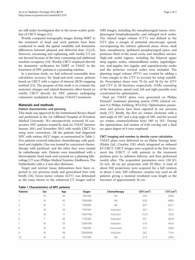
ResultsFigure 2 shows the typical planning CT with manualcontours and the weekly CBCT with deformed contours.Detailed average volume changes for targets and OARswere listed in Table 2. The average volumes of CTV onthe first CBCT were smaller than those in the planningCT, however, no statistical difference of volume changeswere observed during the treatment course. There werealso no general trend and significant volume changes forGTV and brainstem. The average volumes of parotids onthe first CBCT (CBCT 1) before the radiotherapy werealso close to those in the planning CT, but the averagevolumes of parotids decreased continuously during thetreatment course. The average volume of the right par-otid and left parotid measured from the fifth CBCT(CBCT 5) were about 4.4 and 4.5 cm3 less than thosemeasured from the CBCT 1, respectively. Individual
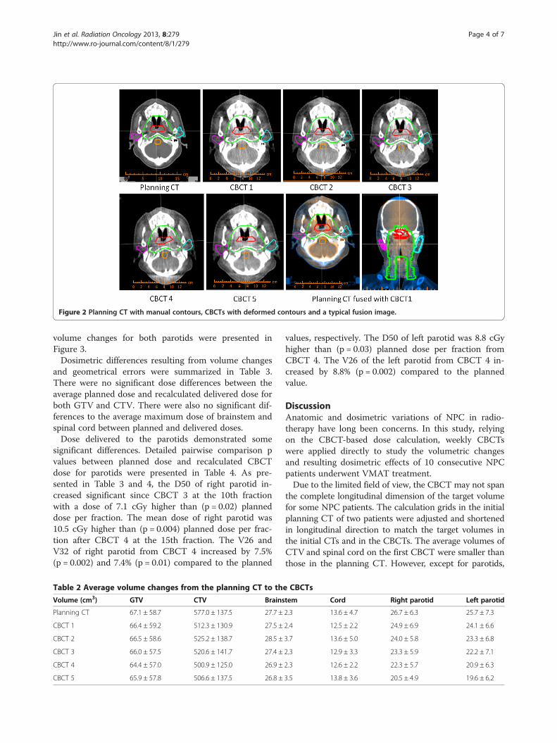
Figure 2 Planning CT with manual contours, CBCTs with deformed contours and a typical fusion image.
Jin et al. Radiation Oncology 2013, 8:279 Page 4 of 7http://www.ro-journal.com/content/8/1/279
volume changes for both parotids were presented inFigure 3.Dosimetric differences resulting from volume changes
and geometrical errors were summarized in Table 3.There were no significant dose differences between theaverage planned dose and recalculated delivered dose forboth GTV and CTV. There were also no significant dif-ferences to the average maximum dose of brainstem andspinal cord between planned and delivered doses.Dose delivered to the parotids demonstrated some
significant differences. Detailed pairwise comparison pvalues between planned dose and recalculated CBCTdose for parotids were presented in Table 4. As pre-sented in Table 3 and 4, the D50 of right parotid in-creased significant since CBCT 3 at the 10th fractionwith a dose of 7.1 cGy higher than (p = 0.02) planneddose per fraction. The mean dose of right parotid was10.5 cGy higher than (p = 0.004) planned dose per frac-tion after CBCT 4 at the 15th fraction. The V26 andV32 of right parotid from CBCT 4 increased by 7.5%(p = 0.002) and 7.4% (p = 0.01) compared to the planned
Table 2 Average volume changes from the planning CT to th
Volume (cm3) GTV CTV Brains
Planning CT 67.1 ± 58.7 577.0 ± 137.5 27.7 ±
CBCT 1 66.4 ± 59.2 512.3 ± 130.9 27.5 ±
CBCT 2 66.5 ± 58.6 525.2 ± 138.7 28.5 ±
CBCT 3 66.0 ± 57.5 520.6 ± 141.7 27.4 ±
CBCT 4 64.4 ± 57.0 500.9 ± 125.0 26.9 ±
CBCT 5 65.9 ± 57.8 506.6 ± 137.5 26.8 ±
values, respectively. The D50 of left parotid was 8.8 cGyhigher than (p = 0.03) planned dose per fraction fromCBCT 4. The V26 of the left parotid from CBCT 4 in-creased by 8.8% (p = 0.002) compared to the plannedvalue.
DiscussionAnatomic and dosimetric variations of NPC in radio-therapy have long been concerns. In this study, relyingon the CBCT-based dose calculation, weekly CBCTswere applied directly to study the volumetric changesand resulting dosimetric effects of 10 consecutive NPCpatients underwent VMAT treatment.Due to the limited field of view, the CBCT may not span
the complete longitudinal dimension of the target volumefor some NPC patients. The calculation grids in the initialplanning CT of two patients were adjusted and shortenedin longitudinal direction to match the target volumes inthe initial CTs and in the CBCTs. The average volumes ofCTV and spinal cord on the first CBCT were smaller thanthose in the planning CT. However, except for parotids,
e CBCTs
tem Cord Right parotid Left parotid
2.3 13.6 ± 4.7 26.7 ± 6.3 25.7 ± 7.3
2.4 12.5 ± 2.2 24.9 ± 6.9 24.1 ± 6.6
3.7 13.6 ± 5.0 24.0 ± 5.8 23.3 ± 6.8
2.3 12.9 ± 3.3 23.3 ± 5.9 22.2 ± 7.1
2.3 12.6 ± 2.2 22.3 ± 5.7 20.9 ± 6.3
3.5 13.8 ± 3.6 20.5 ± 4.9 19.6 ± 6.2

Figure 3 Volume changes measured with weekly CBCT of a) right parotid and b) left parotid.
Jin et al. Radiation Oncology 2013, 8:279 Page 5 of 7http://www.ro-journal.com/content/8/1/279
no significant volume changes of targets and OARs wereobserved based on CBCT 1 in this study. The volumechanges of parotids were unique for each individuals asshown in Figure 3. According to Table 2, the averageweekly shrinkage of right and left parotids were 4.4% and4.7%, respectively. This was close to the reported glandsshrinkage of 4.9%/wk in the study of Robar et al., in whichweekly CT was applied to study the spatial variability ofOAR and the resultant dosimetric effects during IMRT for15 head and neck patients [12].
Table 3 Dosimetric differences between planned dose and de
Planned dose CBCT1 CBCT2
GTV
D95 (cGy) 228.6 ± 3.8 230.1 ± 5.0 227.8 ± 6.8
D90 (cGy) 233.2 ± 3.6 235.0 ± 5.1 231.6 ± 7.5
CTV
V110 29.3 ± 13.0 32.0 ± 14.0 28.9 ± 14.3
D95 (cGy) 188.6 ± 3.0 189.9 ± 4.5 188.1 ± 4.7
D90 (cGy) 192.8 ± 2.7 194.5 ± 3.7 192.7 ± 5.4
D1 (cGy)
Brainstem 153.6 ± 11.2 150.1 ± 11.2 150.1 ± 15.8
Cord 130.3 ± 10.4 130.3 ± 6.3 133.7 ± 16.2
Right parotid
D50 (cGy) 74.5 ± 3.4 79.9 ± 5.0 77.8 ± 4.5
Dmean (cGy) 95.6 ± 5.4 101.9 ± 5.3 99.1 ± 6.4
V26 36.5 ± 3.0 41.5 ± 3.8 38.9 ± 4.3
V32 29.3 ± 4.9 33.7 ± 4.7 31.2 ± 5.2
Left parotid
D50 (cGy) 74.9 ± 2.6 80.0 ± 3.7 77.3 ± 5.7
Dmean (cGy) 94.8 ± 8.1 101.4 ± 10.2 99.6 ± 8.2
V26 34.8 ± 3.3 39.8 ± 5.3 39.0 ± 2.7
V32 27.5 ± 5.5 31.2 ± 6.7 31.2 ± 5.7
Currently, the design of onboard CBCT is far from op-timal and its quality is adversely influenced by many fac-tors, such as scatter, beam hardening and intra-scanningorgan motion. The question of whether CBCT imagescan be used directly for radiation dose calculation hasbeen raised and investigated. Based on reliable CBCTHU and density calibration curve, studies have demon-strated the reliability and accuracy of CBCT-based dosecalculation [19,20]. Our previous study also demon-strated that ROI mapping method was an effective and
livered dose calculating from CBCT
CBCT3 CBCT4 CBCT5 p
226.6 ± 6.3 228.5 ± 7.1 230.3 ± 9.9 0.83
224.6 ± 23.3 232.9 ± 7.1 227.1 ± 23.4 0.57
28.8 ± 13.9 30.3 + 14.7 30.1 + 14.2 1.00
187.5 ± 4.5 188.0 ± 4.5 189.1 ± 5.0 0.86
193.1 ± 4.9 193.0 ± 4.8 194.6 ± 3.3 0.83
151.6 ± 17.4 149.7 ± 16.6 152.1 ± 16.8 1.00
133.4 ± 11.6 130.5 ± 13.6 137.3 ± 16.0 0.79
81.6 ± 5.3 82.8 ± 6.2 82.2 ± 3.7 0.002
102.6 ± 6.8 106.1 ± 8.0 101.2 ± 4.4 0.012
41.3 ± 5.0 44.0 ± 4.5 42.3 ± 3.5 0.003
35.3 ± 5.2 36.7 ± 4.6 36.0 ± 4.0 0.006
76.4 ± 9.2 83.7 ± 7.3 82.0 ± 6.3 0.017
101.9 ± 7.6 104.6 ± 9.8 103.3 ± 7.6 0.18
40.5 ± 5.5 43.6 ± 6.4 42.9 ± 4.9 0.02
32.7 ± 5.9 35.1 ± 7.2 33.6 ± 7.6 0.17

Table 4 Comparison between planned parotid dose and delivered parotid dose calculated using CBCT
p Planned vs CBCT 1 Planned vs. CBCT 2 Planned vs. CBCT 3 Planned vs. CBCT 4 Planned vs. CBCT 5
Right parotid
D50 (cGy) 0.13 0.64 0.02 0.004 0.01
Dmean (cGy) 0.22 0.80 0.13 0.004 0.34
V26 0.09 0.78 0.11 0.002 0.03
V32 0.32 0.94 0.07 0.01 0.03
Left parotid
D50 (cGy) 0.45 0.95 1.00 0.03 0.13
V26 0.21 0.38 0.11 0.002 0.01
Jin et al. Radiation Oncology 2013, 8:279 Page 6 of 7http://www.ro-journal.com/content/8/1/279
simple method for the CBCT-based dose calculation[15]. Therefore, we applied the same method in thisstudy to investigate the dosimetric effects during VMATbased on weekly CBCT directly.There were no significant differences between the
planned and delivered doses for GTV and CTV. This wasconsistent with the study of Zhang et al., in which a plan-ning CT and weekly repeat CT were scanned to study theactual dose variability of targets and OARs for 11 NPC pa-tients during IMRT [21]. The average maximum doses rep-resented by D1 for the brainstem and spinal cord were notsignificant different between planned and delivered doses.However, patient 1 demonstrated a 16.8% and 26.0% doseincrease for brainstem and spinal cord on CBCT 5, re-spectively. This indicated a random dosimetric variabilityfor brainstem and spinal cord similar to the reported re-sults in the study of Robar et al. [12]. The increased doseon CBCT 5 could be caused by a dramatic anatomicchanges resulting from a sharp volume shrinkage of pa-rotids, as shown in Figure 3.Both parotids shifted towards a greater delivered dose
during the VMAT treatment, which was consistent withthe studies based on weekly CT during IMRT treatment[12,21]. Compared to the planned dose, the delivereddose on D50, Dmean, V26 and V32 of parotids were in-creased significantly after CBCT 4, which was obtainedat the 15th fraction with a dose of 30 Gy and 37.5 Gyfor CTV and GTV, respectively. This implies a replan-ning after 30 Gy will benefit the parotid protection forNPC patients during VMAT treatment. However, thetime trend for the dosimetric changes of parotids wasless obvious compared to the time trend of their volumechanges, as shown in Table 3. The small dosimetric fluc-tuations among CBCTs could be caused by the errors ofCBCT based dose calculation [15]. Another explanationcould be the limited number of patients included in thisstudy. Additional work on a larger patient population iswarranted to decide the proper adaptive replanning timefor NPC patients.CBCT and megavoltage CT (MVCT) [4] have been
widely employed during the radiotherapy for geometric
error corrections. Direct dose calculation based onCBCT and MVCT will certainly provide a more conveni-ent and straightforward way than weekly repeated CTimages for adaptive replanning [12,13]. However, due tothe intrinsic limitations of CBCT, extensive work onthe reliability and accuracy of CBCT-based dose calcula-tion is also warranted to evaluate whether our findingswere accurate enough and could actually translate intoguidelines.
ConclusionCBCT-based dose calculation was applied directly tostudy the anatomic changes and resulting dosimetricvariations for NPC patients undergoing VMAT treat-ment. Continuous volume changes in the parotids wereobserved with weekly CBCT. Significant dosimetric vari-ations in the parotids were presented at 15th fractionwith CBCT 4. No significant difference in volumetricand dosimetric variations were observed for other OARsand targets. A replanning after 30 Gy may be useful inthe VMAT for NPC.
ConsentWritten informed consent was obtained from the patientfor the publication of this report and any accompanyingimages.
Competing interestsThe authors declare that they have no competing interests.
Authors’ contributionsEach author has participated sufficiently in the work to take publicresponsibility for appropriate portions of the content. XJ, CX designed thestudy. WH did the CBCT calculation, HS and YJ performed the study andanalysis. YZ provided the patients’ images. The manuscript was written by XJ,all other authors helped and finally approved the final manuscript.
AcknowledgmentsThe study was supported by Wenzhou Science and Technology BureauFunding (Y20120137) and the Scientific Research Foundation for theReturned Overseas Chinese Scholars (604090656/037).
Author details1Department of Radiotherapy and Chemotherapy, the 1st Affiliated Hospitalof Wenzhou Medical University, Wenzhou 325000, China. 2Department ofOncology, Shanghai Medical College, Fudan University, Shanghai, China.
Jin et al. Radiation Oncology 2013, 8:279 Page 7 of 7http://www.ro-journal.com/content/8/1/279
3Research department of IPTA(Beijing) Investment Adminstration Co.,Ltd,Beijing, China.
Received: 24 September 2013 Accepted: 25 November 2013Published: 1 December 2013
References1. Lee N, Kramer A, Xia P: RTOG 0225: a phase II study of intensity modulated
radiation therapy (IMRT)+/− chemotherapy for nasopharyngeal cancer.Philadelphia, PA: Radiation Therapy Oncology Group; 2003.
2. Lee TF, Ting HM, Chao PJ, Fang FM: Dual arc volumetric-modulated arcradiotherapy (VMAT) of nasopharyngeal carcinomas: a simultaneousintegrated boost treatment plan comparison with intensity-modulatedradiotherapies and single arc VMAT. Clin Oncol 2012, 24:196–207.
3. Nutting CM, Morden JP, Harrington KJ, Urbano TG, Bhide SA, Clark C, MilesEA, Miah AB, Newbold K, Tanay M, Adab F, Jefferies SJ, Scrase C, Yap BK,A’Hern RP, Sydenham MA, Emson M, Hall E: PARSPORT trial managementgroup: parotid-sparing intensity modulated versus conventionalradiotherapy in head and neck cancer (PARSPORT): a phase 3multicentre randomised controlled trial. Lancet Oncol 2011, 12:127–136.
4. Han CH, Chen YJ, Liu AL, Schultheiss TE, Wong JY: Actual dose variation ofparotid glands and spinal cord for nasopharyngeal cancer patientsduring radiotherapy. Int J Radiat Oncol Biol Phys 2008, 70:1256–1262.
5. Ding GX, Duggan DM, Coffey CW, Deeley M, Hallahan DE, Cmelak A,Malcolm A: A study on adaptive IMRT treatment planning using kVcone-beam CT. Radiother Oncol 2007, 85:116–125.
6. Lawson JD, Schreibmann E, Jani AB, Fox T: Quantitative evaluation of aconebeam computed tomography–planning computed tomographydeformable image registration method for adaptive radiation therapy.J Appl Clin Med Phys 2007, 8(4):2432.
7. Barker JL Jr, Garden AS, Ang KK, O’Daniel JC, Wang H, Court LE, MorrisonWH, Rosenthal DI, Chao KS, Tucker SL, Mohan R, Dong L: Quantification ofvolumetric and geometric changes occurring during fractionatedradiotherapy for head-and-neck cancer using an integrated CT/linearaccelerator system. Int J Radiat Oncol Biol Phys 2004, 59:960–970.
8. Kreppel M, Drebber U, Eich HT, Dreiseidler T, Zoller JE, Muller RP, Scheer M:Combined-modality treatment in advanced oral squamous cellcarcinoma: primary surgery followed by adjuvant concomitantradiochemotherapy. Strahlenther Onkol 2011, 187:555–560.
9. Boda-Heggemann J, Lohr F, Wenz F, Flentje M, Guckenberger M: kVcone-beam CT-based IGRT: a clinical review. Strahlenther Onkol 2011,187:284–291.
10. Sterzing F, Herfarth K, Debus J: IGRT with helical tomotherapy-effort andbenefit in clinical routine. Strahlenther Onkol 2007, 183(2):35–37.
11. Fotina I, Hopfgartner J, Stock M, Steininger T, Lutgendorf-Caucig C, Georg D:Feasibility of CBCT-based dose calculation: comparative analysis of HUadjustment techniques. Radiother Oncol 2012, 104:249–256.
12. Robar JL, Day A, Clancey J, Kelly R, Yewondwossen M, Hollenhorst H,Rajaraman M, Wilke D: Spatial and dosimetric variability of organs at riskin head-and-neck intensity-modulated radiotherapy. Int J Radiat OncolBiol Phys 2007, 68(4):112–1130.
13. Wu Q, Chi Y, Chen PY, Krauss DJ, Yan D, Martinez A: Adaptive replanningstrategies accounting for shrinkage in head and neck IMRT. Int J RadiatOncol Biol Phys 2009, 75(3):924–932.
14. Hong TS, Tome WA, Chappell RJ, Harari PM: Variations in targetdelineation for head and neck IMRT: an international multiinstitutionalstudy. Int J Radiat Oncol Biol Phys 2004, 60(Suppl. 1):S157–S158.
15. Hu W, Ye J, Wang J, Ma X, Zhang Z: Use of kilovoltage X-ray volumeimaging in patient dose calculation for head-and-neck and partial brainradiation therapy. Radiat Oncol 2010, 5:29–38.
16. Wu SX, Xie CY, Jin XC, Zhang P: Simultaneous modulated acceleratedradiation therapy in the treatment of nasopharyngeal cancer: a localcenter’s experience. Int J Radiat Oncol Biol Phys 2006, 66:s40–s46.
17. Jin X, Yi J, Zhou Y, Yan H, Han C, Xie C: Comparison of whole fieldsimultaneous integrated boost VMAT and IMRT in the treatment ofnasopharyngeal cancer. Med Dosim 2013, 38:418–423.
18. Richter A, Hu Q, Steglich D, Baier K, Wilbert J, Guckenberger M, Flentje M:Investigation of the usability of cone beam CT data sets for dosecalculation. Radiat Oncol 2008, 3:42. doi:10.1186/1748-717X-3-42.
19. Yang Y, Schreibmann E, Li T, Wang C, Xing L: Evaluation of on-board kVcone beam CT (CBCT)-based dose calculation. Phys Med Biol 2007,52:685–705.
20. Guan H, Dong H: Dose calculation accuracy using cone-beam CT (CBCT)for pelvic adaptive radiotherapy. Phys Med Biol 2009, 54(20):6239–6250.
21. Zhang X, Li M, Cao J, Luo JW, Xu GZ, Gao L, Yi J, Huang X, Xiao J, Li S, Dai J:Dosimetric variations of target volumes and organs at risk innasopharyngeal carcinoma intensity-modulated radiotherapy. Br J Radiol2012, 85:e506–e513.
doi:10.1186/1748-717X-8-279Cite this article as: Jin et al.: CBCT-based volumetric and dosimetricvariation evaluation of volumetric modulated arc radiotherapy in thetreatment of nasopharyngeal cancer patients. Radiation Oncology2013 8:279.
Submit your next manuscript to BioMed Centraland take full advantage of:
• Convenient online submission
• Thorough peer review
• No space constraints or color figure charges
• Immediate publication on acceptance
• Inclusion in PubMed, CAS, Scopus and Google Scholar
• Research which is freely available for redistribution
Submit your manuscript at www.biomedcentral.com/submit
Copyright © 2022 FDOKUMEN