Limited intravascular coupling in the rodent brainstem and retina supports a role for glia in...
15
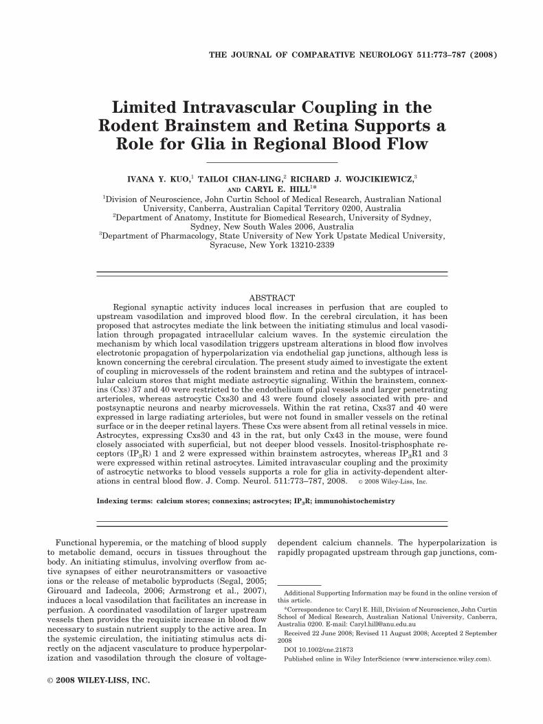
Limited Intravascular Coupling in the Rodent Brainstem and Retina Supports a Role for Glia in Regional Blood Flow IVANA Y. KUO, 1 TAILOI CHAN-LING, 2 RICHARD J. WOJCIKIEWICZ, 3 AND CARYL E. HILL 1 * 1 Division of Neuroscience, John Curtin School of Medical Research, Australian National University, Canberra, Australian Capital Territory 0200, Australia 2 Department of Anatomy, Institute for Biomedical Research, University of Sydney, Sydney, New South Wales 2006, Australia 3 Department of Pharmacology, State University of New York Upstate Medical University, Syracuse, New York 13210-2339 ABSTRACT Regional synaptic activity induces local increases in perfusion that are coupled to upstream vasodilation and improved blood flow. In the cerebral circulation, it has been proposed that astrocytes mediate the link between the initiating stimulus and local vasodi- lation through propagated intracellular calcium waves. In the systemic circulation the mechanism by which local vasodilation triggers upstream alterations in blood flow involves electrotonic propagation of hyperpolarization via endothelial gap junctions, although less is known concerning the cerebral circulation. The present study aimed to investigate the extent of coupling in microvessels of the rodent brainstem and retina and the subtypes of intracel- lular calcium stores that might mediate astrocytic signaling. Within the brainstem, connex- ins (Cxs) 37 and 40 were restricted to the endothelium of pial vessels and larger penetrating arterioles, whereas astrocytic Cxs30 and 43 were found closely associated with pre- and postsynaptic neurons and nearby microvessels. Within the rat retina, Cxs37 and 40 were expressed in large radiating arterioles, but were not found in smaller vessels on the retinal surface or in the deeper retinal layers. These Cxs were absent from all retinal vessels in mice. Astrocytes, expressing Cxs30 and 43 in the rat, but only Cx43 in the mouse, were found closely associated with superficial, but not deeper blood vessels. Inositol-trisphosphate re- ceptors (IP 3 R) 1 and 2 were expressed within brainstem astrocytes, whereas IP 3 R1 and 3 were expressed within retinal astrocytes. Limited intravascular coupling and the proximity of astrocytic networks to blood vessels supports a role for glia in activity-dependent alter- ations in central blood flow. J. Comp. Neurol. 511:773–787, 2008. © 2008 Wiley-Liss, Inc. Indexing terms: calcium stores; connexins; astrocytes; IP 3 R; immunohistochemistry Functional hyperemia, or the matching of blood supply to metabolic demand, occurs in tissues throughout the body. An initiating stimulus, involving overflow from ac- tive synapses of either neurotransmitters or vasoactive ions or the release of metabolic byproducts (Segal, 2005; Girouard and Iadecola, 2006; Armstrong et al., 2007), induces a local vasodilation that facilitates an increase in perfusion. A coordinated vasodilation of larger upstream vessels then provides the requisite increase in blood flow necessary to sustain nutrient supply to the active area. In the systemic circulation, the initiating stimulus acts di- rectly on the adjacent vasculature to produce hyperpolar- ization and vasodilation through the closure of voltage- dependent calcium channels. The hyperpolarization is rapidly propagated upstream through gap junctions, com- Additional Supporting Information may be found in the online version of this article. *Correspondence to: Caryl E. Hill, Division of Neuroscience, John Curtin School of Medical Research, Australian National University, Canberra, Australia 0200. E-mail: [email protected] Received 22 June 2008; Revised 11 August 2008; Accepted 2 September 2008 DOI 10.1002/cne.21873 Published online in Wiley InterScience (www.interscience.wiley.com). THE JOURNAL OF COMPARATIVE NEUROLOGY 511:773–787 (2008) © 2008 WILEY-LISS, INC.
-
Upload
independent -
Category
Documents
-
view
4 -
download
0
Transcript of Limited intravascular coupling in the rodent brainstem and retina supports a role for glia in...
Limited Intravascular Coupling in theRodent Brainstem and Retina Supports a
Role for Glia in Regional Blood Flow
IVANA Y. KUO,1 TAILOI CHAN-LING,2 RICHARD J. WOJCIKIEWICZ,3
AND CARYL E. HILL1*1Division of Neuroscience, John Curtin School of Medical Research, Australian National
University, Canberra, Australian Capital Territory 0200, Australia2Department of Anatomy, Institute for Biomedical Research, University of Sydney,
Sydney, New South Wales 2006, Australia3Department of Pharmacology, State University of New York Upstate Medical University,
Syracuse, New York 13210-2339
ABSTRACTRegional synaptic activity induces local increases in perfusion that are coupled to
upstream vasodilation and improved blood flow. In the cerebral circulation, it has beenproposed that astrocytes mediate the link between the initiating stimulus and local vasodi-lation through propagated intracellular calcium waves. In the systemic circulation themechanism by which local vasodilation triggers upstream alterations in blood flow involveselectrotonic propagation of hyperpolarization via endothelial gap junctions, although less isknown concerning the cerebral circulation. The present study aimed to investigate the extentof coupling in microvessels of the rodent brainstem and retina and the subtypes of intracel-lular calcium stores that might mediate astrocytic signaling. Within the brainstem, connex-ins (Cxs) 37 and 40 were restricted to the endothelium of pial vessels and larger penetratingarterioles, whereas astrocytic Cxs30 and 43 were found closely associated with pre- andpostsynaptic neurons and nearby microvessels. Within the rat retina, Cxs37 and 40 wereexpressed in large radiating arterioles, but were not found in smaller vessels on the retinalsurface or in the deeper retinal layers. These Cxs were absent from all retinal vessels in mice.Astrocytes, expressing Cxs30 and 43 in the rat, but only Cx43 in the mouse, were foundclosely associated with superficial, but not deeper blood vessels. Inositol-trisphosphate re-ceptors (IP3R) 1 and 2 were expressed within brainstem astrocytes, whereas IP3R1 and 3were expressed within retinal astrocytes. Limited intravascular coupling and the proximityof astrocytic networks to blood vessels supports a role for glia in activity-dependent alter-ations in central blood flow. J. Comp. Neurol. 511:773–787, 2008. © 2008 Wiley-Liss, Inc.
Indexing terms: calcium stores; connexins; astrocytes; IP3R; immunohistochemistry
Functional hyperemia, or the matching of blood supplyto metabolic demand, occurs in tissues throughout thebody. An initiating stimulus, involving overflow from ac-tive synapses of either neurotransmitters or vasoactiveions or the release of metabolic byproducts (Segal, 2005;Girouard and Iadecola, 2006; Armstrong et al., 2007),induces a local vasodilation that facilitates an increase inperfusion. A coordinated vasodilation of larger upstreamvessels then provides the requisite increase in blood flownecessary to sustain nutrient supply to the active area. Inthe systemic circulation, the initiating stimulus acts di-rectly on the adjacent vasculature to produce hyperpolar-ization and vasodilation through the closure of voltage-
dependent calcium channels. The hyperpolarization israpidly propagated upstream through gap junctions, com-
Additional Supporting Information may be found in the online version ofthis article.
*Correspondence to: Caryl E. Hill, Division of Neuroscience, John CurtinSchool of Medical Research, Australian National University, Canberra,Australia 0200. E-mail: [email protected]
Received 22 June 2008; Revised 11 August 2008; Accepted 2 September2008
DOI 10.1002/cne.21873Published online in Wiley InterScience (www.interscience.wiley.com).
THE JOURNAL OF COMPARATIVE NEUROLOGY 511:773–787 (2008)
© 2008 WILEY-LISS, INC.
posed of connexins (Cxs) 37, 40, and 43, along the axiallyoriented endothelial cells (Hill et al., 2001; Segal, 2005).
In contrast to the situation in the systemic circulation,within the central nervous system the close association ofastrocytes to neurons and arterioles has led to the propo-sition that signaling via astrocytes may play a significantrole in neuronally induced functional hyperemia (Harderet al., 1998). Subsequent studies in brain slices haveshown that activation of single astrocytes causes the dila-tion of nearby arterioles, whereas stimulation of neuronsinduces intracellular calcium waves within astrocytes,which is followed by vasodilation (Zonta et al., 2003;Filosa et al., 2004; Takano et al., 2006). Other investiga-tions have established that astrocyte-mediated signalscan also initiate vasoconstriction (Mulligan and MacVicar,2004; Metea and Newman, 2006). The collective signalingamong neurons, astrocytes, and neighboring blood vesselshas therefore been termed neurovascular coupling. How-ever, little information is available on the mechanism bywhich this local increase in perfusion is coupled to up-stream increases in cerebral blood flow, although mecha-nisms involving flow-mediated vasodilation seem unlikely(Girouard and Iadecola, 2006).
On the local level, increases in intracellular calcium andthe subsequent production of calcium waves in astrocytesare integral to the coupling of regional blood flow to neu-ronal activity within the central nervous system (Scemes,2000). Inter-astrocytic calcium waves have been shown tobe propagated by two mechanisms involving either intra-cellular movement of second messengers through gapjunctions formed primarily of Cxs30 and 43, or the extra-cellular release of ATP and activation of purinergic recep-tors on adjacent astrocytes (Cornell-Bell et al., 1990; New-man, 2001; Zahs et al., 2003). Pharmacological studieshave shown that the calcium waves themselves involve aninositol-trisphosphate receptor (IP3R) pathway (Filosa etal., 2004), although the exact subtype of IP3Rs has not yetbeen determined (Yamamoto-Hino et al., 1995; Sharp etal., 1999; Holtzclaw et al., 2002, Cotrina and Nedergaard,2005). Although the three IP3R subtypes have similarchannel properties, calcium release from the various sub-types can be differentially modulated by calcium, ATP,and phosphorylation (Foskett et al., 2007; Soulsby andWojcikiewicz, 2007), thus modulating the resultant cal-cium wave.
Larger supplying vessels in the pial circulation are con-nected to parenchymal capillaries by penetrating arte-rioles that have been demonstrated to conduct responsesover distances of some 650 !m, following exogenous ap-plication of vasoactive factors, such as ATP and potassium(Dietrich et al., 1996; Horiuchi et al., 2002; Ngai andWinn, 2002). However, a recent study has provided evi-dence that glial cells and not vascular endothelial cells areresponsible for conduction of vasodilation from intracere-bral areas to superficial pial vessels (Xu et al., 2008).These data suggest that intravascular coupling via Cxsmay be confined to more superficially located vessels andabsent from the bulk of intracerebral vessels.
The aim of the present study was therefore first toinvestigate whether the endothelial cells of intracranialmicrovessels are coupled via gap junctions expressingCxs37, 40, and 43 and second, to determine which intra-cellular calcium stores might be involved in signalingthrough astrocytic networks. We chose to investigate therodent auditory brainstem, which contains the medial nu-
cleus of the trapezoid body (MNTB), the second synapticrelay in the auditory pathway. Within the MNTB, which islocated bilaterally near the ventral surface, synapses arediscretely localized to the neuronal soma in the form of thelarge calyces of Held, which cover most of the surface areaof the postsynaptic cell bodies (Satzler et al., 2002). Studyof the auditory brainstem therefore enabled us to investi-gate Cx expression in blood vessels at the site of theinitiating stimulus for hyperemic responses. We also ex-tended our study to the rodent retina, often referred to as“nature’s brain slice,” as this preparation has been thesubject of recent functional investigations examining pos-sible neurovascular pathways involving either astrocytesor pericytes (Metea and Newman, 2006; Peppiatt et al.,2006).
MATERIALS AND METHODSAnimals
Juvenile (14–17 days) and adult (6–8 weeks) Wistarrats (Animals Services Division, ANU, Canberra, Austra-lia) and juvenile (18–21 days) and adult (6–8 weeks) CBAmice (Animal Services Division, Canberra, Australia andAnimal Resource Center, Murdoch, WA, Australia) of ei-ther sex were anesthetized with ether and decapitated,according to a protocol approved by the Australian Na-tional University Animal Ethics Committee.
Brainstem dissectionTo ensure tissue integrity, the hind brain was rapidly
dissected in modified artificial cerebrospinal fluid (aCSF,in mM: 130 NaCl, 3 KCl, 5 MgCl2, 1 CaCl2, 1.25 NaH2PO4,26.2 NaHCO3, 10 glucose), equilibrated with 95% O2/5%CO2. The auditory brainstem was sliced (700 !m) trans-versely in ice-cold modified aCSF using a tissue cutter(Campden Instruments, Leicester, UK). Brain sectionswere fixed in either 2% paraformaldehyde (PFA; BDHBiochemicals, Poole, UK) for 10 minutes at room temper-ature (RT), or ice-cold acetone for 30 minutes.
Brain slices were washed in phosphate-buffered saline(PBS; pH 7.4), immersed in 30% sucrose, embedded inOCT medium (Tissue Tek, Torrance, CA), and cryosec-tioned at 25 !m by using a Vibratome (Leica, Heidelberg,Germany). Cryosections containing the MNTB were col-lected on microscopic slides coated with 2% silane andvacuum dried for 25 minutes over phosphorus pentoxide(Unilab, Tarzana, CA) to ensure maximum adhesion tothe slides.
Retinal dissectionEyes were enucleated and the retina dissected in PBS as
previously described (Chan-Ling, 1997). Briefly, an inci-sion was made to the front of the eye, and the iris, cornea,and lens were carefully removed. Any vitreous humourclinging to the retina was gently lifted off, and the eyecupwas cut away. Isolated retinae were fixed in 2% PFA for 10minutes. Following washing in PBS, the retinae wereready for immunostaining. Other retinae were processedfor cryosectioning as described above.
ImmunohistochemistryWholemount retinae or cryosections of brain and reti-
nae were preincubated in 2% bovine serum albumin (BSA;Bovogen Biochemicals, Melbourne, Victoria, Australia),
The Journal of Comparative Neurology
774 I.Y. KUO ET AL.

and 0.2% Triton X-100 in PBS for 1 hour at RT, followedby incubation in primary antibodies diluted in the samesolution. Details of the primary antibodies used to detectspecific connexins and intracellular calcium stores can befound in Table 1. All Cx and IP3R antibodies were incu-bated for 1–2 hours at 37°C followed by overnight incuba-tion at RT.
To label astrocytes in the brainstem and retina, but notMuller glia, rabbit anti-glial fibrillary acidic protein(GFAP; 1:500, DAKO, Carpinteria, CA, Z-0334, Lot00005193) was applied overnight at RT. Presynaptic neu-rons in the brainstem were labeled with guinea pig anti-vesicular glutamate transporter (VGlut-1; 1:600, Sigma,St. Louis, MO, V 0389), applied overnight at RT. Thepostsynaptic bodies of the MNTB neurons were observabledue to autofluorescence. Three markers, anti-myosin,anti-desmin, and biotinylated lectin from Bandeiraea(Griffonia) simplicifolia (BS-lectin) were used to identifyarterioles in the brainstem and retina. Mouse anti-desmin(1:400, Sigma, D-8281) and rabbit anti-myosin (gift fromDr. Groschel-Stewart, Technische Hochschule Darmstadt,Germany; Paul et al., 1994) labeled smooth muscle cells invessels; anti-desmin also immunolabeled pericytes. BS-lectin was used (1:500, Sigma, L2140) to label the vascu-lature in retinae; however, BS-lectin immunoreactivitywas less successful in the brainstem. All vascular markerswere applied overnight at RT. After washing, slices wereincubated with the appropriate secondary antibody di-luted in 0.01% Triton X-100 in PBS for 1 hour at RT andmounted in buffered glycerol.
For slice preparations, the secondary antibodies usedwere Alexa Fluor (AF) 488 goat anti-rabbit IgG (1:600;Molecular Probes, Eugene, OR), AF 488 donkey anti-goatIgG (1:800, Molecular Probes), AF 488 goat anti-mouseIgG (1:800; Molecular Probes), AF 488 streptavidin (1:
1,000, Molecular Probes), Cy3 donkey anti-rabbit IgG (1:500; Jackson Labs, Bar Harbor, ME), Cy3 donkey anti-goat IgG (1:600, Jackson Labs), AF 568 donkey anti-mouse IgG (1:800, Molecular Probes), AF 633 donkey anti-rabbit IgG (1:500, Molecular Probes), AF 633 goat anti-guinea pig IgG (1:800, Molecular Probes), and AF 633streptavidin (1:500; Molecular Probes). Secondary anti-bodies used in wholemount preparations were applied atthree to fourfold higher concentrations than in the condi-tions used for slice preparations.
For multiple labeling with primary antibodies raised inthe same species, sections were incubated sequentially ineach primary antibody followed by its appropriate second-ary antibody with an intervening incubation in unlabeledanti-rabbit IgG Fab fragment (1:100, Jackson Labs) for 1hour at RT. Erroneous binding of subsequent secondaryantibodies to the initial primary antibody was checked byomitting the subsequent primary antibodies.
To ensure specificity of the primary antibodies, controlsamples were incubated with primary antibodies that hadbeen preincubated in the presence of the appropriate im-munizing peptide (10-fold excess concentration) for 1 hourat RT. The immunizing peptide for Cx30 was kindly pro-vided by Zymed (San Francisco, CA, Cx30-241, Lot10013287). For all of the antibodies listed in Table 1,except for anti-Cx26, for which antigenic peptides wereunavailable, this preincubation resulted in the abolition ofimmunoreactive staining. Specificity of the secondary an-tibodies was checked by omitting the primary antibody.
Confocal microscopyImmunostained preparations were imaged with a laser
scanning confocal microscope (TCS 4D Leica [Fig. 1] orAxioskop II, Zeiss Instruments, Heidelberg, Germany).Sequential scanning was used to detect secondary anti-
TABLE 1. Connexin and Intracellular Calcium Store Antibodies Used in This Study
Antibody and source Species and immunogen Antibody host and working dilutions Characterization
Cx26 Zymed, 51-2800 Lot: 41191512 Mouse 13 AA from the C-terminus Affinity-purified Rabbit polyclonal 1:100 allpreparations
Antibody recognizes 29-kDa protein bandand does not cross-react with Cx30(Nagy et al., 2001).
Cx30 Zymed, 71-2200 Lot: 40487644 Mouse 22 AA from last 100 AAs of C-terminus
Affinity-purified Rabbit polyclonal 1:100 allpreparations
Antibody detects a single band of 30 kDaon Western blots; abolished afterpreadsorption with the peptide antigen(Nagy et al., 1999)
Cx37 IMVS1 Rat AA 266–281 of C-terminus Affinity-purified2 Sheep polyclonal 1:100 allpreparations
Antibody detects unphosphorylated andphosphorylated forms in Cos-7 cellstransfected with Cx37, but not Cx40, 43,or45; staining abolished with peptideantigen (Rummery et al., 2005)
Cx40 IMVS1 Rat AA 254–270 of C-terminus Affinity-purified2 Sheep polyclonal 1:100 allpreparations
Antibody detects unphosphorylated andphosphorylated forms in Cos-7 cellstransfected with Cx40, but not Cx37, 43,or 45; staining abolished with peptideantigen (Rummery et al., 2005)
Cx43 Zymed, 71-0700 Lot: 60605865 Rat AA 346–363 of C-terminus Affinity-purified Rabbit polyclonal 1:100 allpreparations
Antibody detects unphosphorylated andphosphorylated forms in Cx43-expressing tissues and Cos-7 cellstransfected with Cx43, but not Cx37, 40,and 45; abolished with the peptideantigen (Rummery et al., 2002).
IP3R1 Upstate3 Rat AA 2,731–2,749 of C-terminus Affinity-purified Rabbit polyclonal 1:100sections 1:50 wholemount retinae
Antibody detects immunopurified IP3R1standards, with a single band(Wojcikiewicz, 1995)
IP3R2 Upstate3 Rat AA 2,685–2,701 of C-terminus Affinity-purified Rabbit polyclonal 1:40sections 1:10 wholemount retinae
Antibody detects immunopurified IP3R2standards, with a single band(Wojcikiewicz, 1995)
IP3R3 Upstate3 Rat AA 2,658–2,670 of C-terminus Affinity-purified Rabbit polyclonal 1:10 allpreparations
Antibody detects immunopurified IP3R3standards. (Wojcikiewicz, 1995)
1Institute of Medical and Veterinary Sciences, Adelaide, Australia.2Mimotopes Pty Ltd, Victoria, Australia.3Upstate Medical University, SUNY, NY.
The Journal of Comparative Neurology
775CELL COUPLING IN ASTROCYTES AND BLOOD VESSELS

body fluorescence by using excitation wavelengths fromArKr and HeNe laser sources of 488 nm, 543 nm, and 633nm and corresponding emission wavelengths of 515–530nm, 570–615 nm band passes, and long pass " 650 nm,respectively. Images were taken with 16#, 40#, and 100#oil objective lenses by using Scanware software (Leica), or40# and 100# Plan Apo oil objective lenses by usingPascal software (Zeiss Instruments). Optical z-axis serialsections on 40# and 100# objectives were collected at 1-and 0.5-!m intervals with the Leica microscope and at0.8- and 0.4-!m intervals with the Zeiss microscope. Im-ages taken with the 100# objective on the Zeiss micro-scope were collected at both 1# and 2# zoom. All controlslides were scanned using the same settings for laserpower and detection gain as for the corresponding exper-imental samples. For each reported outcome for a partic-ular age group and rodent strain, images were taken fromn $ 4–7 animals.
Collected serial sections through the thickness of ves-sels were projected as a single image (Aim Image Browser,Zeiss Instruments) and brightness, contrast, and pseudo-coloring were adjusted (Adobe [San Jose, CA] PhotoshopCS2). All control images were adjusted to the same extentas their matched experimental samples.
RESULTSUnless stated otherwise, all results were consistent
among the juvenile and adult rats and mice.
Auditory brainstemThe basilar artery and side branches were located on
the ventral surface of the auditory brainstem, in which theMNTB could be found on either side of the midline (Fig.1A). Within the MNTB, astrocytes, identified with anti-GFAP reactivity, could be observed surrounding the neu-rons, which were identified by autofluorescence (Fig.1A,B). Branching penetrating arterioles that extendedfrom the pial surface were delineated by astrocytes (Fig.1A, pink arrows, C). In addition to the large vessels(greater than 15 !m in diameter), numerous smaller ves-sels that were anti-myosin reactive and varied between 7and 10 !m in diameter were found within the MNTB (Fig.1D).
Expression of vascular connexins 37 and 40. Thevascular Cxs37 and 40 were restricted to the basilar ar-tery and pial vessels of the brainstem in adult mice as wellas juvenile and adult rats. Immunoreactivity for bothCxs37 and 40 was typified by linear punctate staining thatran in the longitudinal direction of the vessel and waslocalized to the endothelial layer. In juvenile mice, similarstaining was found in the endothelial layer of penetratingarterioles, but not within the smooth muscle cell layer norin astrocytes (Fig. 1E,F). The smaller vessels presentwithin the MNTB, which were myosin positive, were notimmunoreactive for either Cxs37 or 40 (Fig. 1G,I, whitearrows; compare H,J). Cxs43 and 30 were not detectedwithin the endothelial or the smooth muscle cell layer ofany arterioles within the brainstem (Fig. 2A,B, labeled A).
Expression of astrocytic connexins 30 and 43.Strong punctate immunoreactivity for Cxs30 and 43 wasfound within the MNTB of the brainstem (Fig. 2A,B) andless intensely external to the MNTB (Table 2). ApplyingCxs30 and 43 antibodies sequentially to the same prepa-ration demonstrated colocalization of the punctate stain-
ing of the two Cxs to astrocytic processes that surroundedneurons and blood vessels (Fig. 2A–C). Cxs30 and 43 werealso colocalized to astrocytes outside the MNTB (Table 2).
The association of the astrocytic connexins with theMNTB neurons was examined more closely by utilizing anantibody against the presynaptic membrane transporterVGlut-1. VGlut-1 localized to the cell membranes of thecalyces of Held that surrounded the principal MNTB neu-rons (blue, Fig. 2D). Although Cxs30 and 43 puncta colo-calized with each other and were closely associated withVGlut-1, no colocalization was observed with VGlut-1 (Fig.2D–F). Cxs30 and 43 were also not observed on the prin-cipal MNTB neurons, inside the VGlut-1 staining (labeledN, Fig. 2D–F).
Because anti-Cxs30 and 43 were both raised in rabbit,control experiments were conducted in which the secondprimary antibody (anti-Cx43) was not applied followingincubation with the nonlabeled IgG Fab fragment. Thesecontrols showed that the subsequent secondary antibodyused to detect anti-Cx43, AF 488 (Fig. 2G,H), did notproduce any staining, that is, AF 488 did not erroneouslybind to anti-Cx30. Control experiments were undertakento ensure that the Cx antibodies (Cxs30, 37, 40, and 43)were specific by demonstrating that staining was abol-ished after preincubation with the immunizing peptide(Fig. 2I for Cx30 antibody and peptide).
Cx26 has been previously reported to be present withinastrocytes (Nagy et al., 2001); however, although stainingwas detected in the surrounding pia mater, no immuno-staining with the anti-Cx26 antibody within astrocyteswas detected (data not shown).
Distribution of intracellular calcium stores. Withinthe penetrating arterioles, IP3R1 immunoreactivity wasfound to colocalize with the smooth muscle cell markerdesmin. IP3R1 staining was typified by dense staining ofthe smooth muscle cells (Fig. 3A). IP3R1 staining orien-tated perpendicular to the smooth muscle cell layer wasdiagnostic of endothelial cell staining (Fig. 3A, inset,white arrowhead). Punctate staining for IP3R1 was alsofound to be associated with the astrocytic processes sur-rounding vessels, as indicated by colocalization with anti-GFAP (Fig. 3A and inset, white arrows). Astrocytes notassociated with vessels were also immunoreactive forIP3R1, with punctate staining localizing to the soma and
Fig. 1. Distribution of astrocytes, vessels, and vascular connexinsin the auditory brainstem. A: Montage of a juvenile CBA mouseauditory brainstem stained with anti-GFAP showing the distributionof astrocytes within the MNTB (red dashed line), and in associationwith arterioles (pink arrows). Montages were prepared in Photoshopand the final image inverted to black on white. B: Astrocytes (green)surround neurons (red autofluoresence) in the MNTB. C: Astrocyticprocesses (green) encompass a vessel marked with anti-myosin (red)in the auditory brainstem. D: Small arterioles between 7 and 10 !m,marked with anti-myosin (red arrows) were seen within the MNTB.Image inverted to black on white. E,F: Immmunoreactivity (red) forCxs37 (E) and 40 (F) was present within the endothelium of penetrat-ing arterioles surrounded by astrocytes (green) in juvenile CBA mice.G–J: Cxs37 (G,H) and 40 (I,J) were absent from the small vessels(green, H,J, white arrows) that existed within the MNTB. Stacksizes $ 5 !m in B; 8 !m in C; 6 !m in E; 4 !m in F. A,G–J are singleoptical sections. Figure 1 is also provided as a Supplementary file, inwhich the red fluorescence has been converted to magenta. Scalebar $ 300 !m in A; 20 !m in B,F (that in F also applies to E); 50 !min C,D,J (also applies to G–I).
The Journal of Comparative Neurology
776 I.Y. KUO ET AL.

Figure 1
The Journal of Comparative Neurology
777CELL COUPLING IN ASTROCYTES AND BLOOD VESSELS

Figure 2
The Journal of Comparative Neurology
778 I.Y. KUO ET AL.

processes (Fig. 3B). To ensure that the anti-GFAP stain-ing was not due to binding of the anti-rabbit AF 633secondary antibody to the IP3R1 antibody, control exper-iments were carried out with the addition of the secondprimary antibody, anti-GFAP, omitted. Whereas IP3R1immunoreactivity was detected with Cy3-conjugated anti-rabbit IgG, no staining was observed when AF 633 anti-rabbit IgG was applied.
IP3R2 was found to be the most dominant receptorexpressed in astrocytes within the auditory brainstem.Within the penetrating arterioles, anti-IP3R2 immuno-reactivity was detected in the surrounding astrocyticprocesses (Fig. 3C and inset). No immunoreactivity wasdetected within the smooth muscle cell layer or theendothelial cell layer (Fig. 3C and inset). Astrocytes notassociated with blood vessels were also immunoreactivefor IP3R2, with discrete punctate labeling observed tocolocalize with the soma and processes (Fig. 3D). Omis-sion of the second rabbit primary antibody, anti-GFAP,demonstrated that there was no nonspecific binding ofthe IP3R2 antibody with AF 633-conjugated anti-rabbitIgG.
In contrast to the other two IP3R subtypes, IP3R3 im-munoreactivity was localized solely to the endotheliallayer as dense, linearly arranged puncta (Fig. 3E). Nocolocalization was detected with the smooth muscle celllayer identified with anti-desmin (Fig. 3E and inset) norwith astrocytes surrounding vessels or those not associ-ated with vessels (Fig. 3E,F).
The data are summarized in Table 2.
RetinaThe retinal blood supply consists of alternating arte-
rioles and veins radiating out from the optic nerve. Whole-mount retinal preparations showed that the astrocytesformed a network between vessels (Fig. 4A,B), as previ-ously observed (Bushong et al., 2002). Arterioles on thesurface of the retina were also closely ensheathed by as-trocytic processes (Fig. 4A,B).
Expression of vascular connexins 37 and 40. Inmouse retinae neither Cxs37 nor 40 were detected in anyarterioles (Table 2). In contrast, in rat retinae, Cxs37 and40 were found in the endothelial layer of the arteriolesradiating from the optic nerve, typified by parallel stain-ing running the length of the vessel (Fig. 4C,D, arrows).Staining was found to decrease after the branching oflarger vessels and was absent from the smaller vessels(Fig. 4C,D, arrowheads) and veins (V). Expression wasrestricted to arterioles on the retinal surface and did notextend into vessels, which descended into other cell layers.No immunoreactivity for either Cxs37 or 40 was detectedin astrocytes or in the smooth muscle layer of the arte-rioles (Fig. 4E,F; note gap between the red Cx stainingand the blue astrocytic staining).
Expression of astrocytic connexins 30 and 43. Ex-amination of the astrocytic Cxs30 and 43 revealed species-dependent expression. In the rat retina, punctate stainingfor both Cxs30 and 43 was found in GFAP-positive astro-cytic processes located in the nerve fiber layer, but it wasnot associated with retinal ganglion cell bodies or otherretinal layers (Fig. 4G,H,L). As observed in the brainstem,neither Cxs30 nor 43 were associated with the smoothmuscle or endothelial layers of vessels in the retina, al-though both were expressed in the astrocytic processes
Fig. 2. Astrocytic connexin distribution within the MNTB. A–C:Cxs30 (red, A) and 43 (green, B) colocalized with each other (C) andwith astrocytic processes (blue, A–C) that surrounded the neurons ofthe MNTB (white N) and arterioles (white A). Cxs30 and 43 were notexpressed within arterioles. D–F: Cxs30 (red, D) and 43 (green, E)puncta did not colocalize with the marker to the presynaptic calyx ofHeld, VGlut-1 (blue, D–F) or to the principal MNTB neurons (whiteN). G,H: Primary antibodies made in the same host animal wereapplied sequentially with an intervening application of a nonlabeledanti-rabbit IgG fraction. Slices were incubated first with rabbit anti-Cx30 antibody, detected with anti-rabbit Cy3 (G). To ensure thatthere was no nonspecific binding of the second secondary antibody(e.g., anti-rabbit AF 488) to the first primary, the subsequent rabbitprimary antibody was omitted (H). I: Preincubation with an immu-nizing peptide to Cx30 blocked Cx30 immunoreactivity. All imagesare single optical sections. Figure 2 is also provided as a Supplemen-tary file, in which the red fluorescence has been converted to magenta.Scale bar $ 50 !m in C (also applies to A,B) and I (also applies toG,H); 20 !m in F (also applies to D,E).
TABLE 2. Connexin and IP3R Immunoreactivity in the Auditory Brainstem and Retina of Juvenile and Adult CBA Mice and Wistar Rats
Cx26 Cx30 Cx37 Cx40 Cx43 IP3R1 IP3R2 IP3R3
Brainstem Vessels Penetratingarterioles
Smooth muscle % % % % % & % %
Endothelium % % %2 %2 % & % &Small vessels
('15 !m)Smooth muscle % % % % % & % %
Endothelium % % % % % & % &Astrocytes Around
vesselsSomatic region % & % % & & & %
Processes % & % % & & & %In the
brainstemSomatic region % & % % & & & %
Processes % & % % & & & %In the MNTB % & % % & & & %
Retina Vessels Primaryarterioles
Smooth muscle % % % % % & % %
Endothelium % % %3 %3 % & & &Branching
arteriolesSmooth muscle % % % % % & % %
Endothelium % % % % % & & &Astrocytes % &4 % % & & % &
1&, staining abolished by preincubation with peptide antigen; %, no detectable expression. MNTB, medial nucleus of the trapezoid body.2Cxs37 and 40 were only detected in the penetrating arterioles of juvenile CBA mice auditory brainstem.3Cxs37 and 40 were only present in rat retinae.4Cx30 was only present in rat retinae.
The Journal of Comparative Neurology
779CELL COUPLING IN ASTROCYTES AND BLOOD VESSELS

Figure 3
The Journal of Comparative Neurology
780 I.Y. KUO ET AL.
ensheathing retinal arterioles (Fig. 4I,J, Table 2). Appli-cation of both Cxs30 and 43 antibodies to the same retinalslice demonstrated colocalization of Cxs30 and 43 punctain single optical sections. Cxs30 and 43 were also presentover the surface of the smaller blood vessels within thesuperficial retinal plexus (Fig. 4K, wholemount prepara-tion) but were not detected in association with any vesselslocated in the deeper retinal layers (Fig. 4L, arrow, retinalsection).
In the mouse retina, Cx43 was also expressed in astro-cytes and in association with the superficial blood vessels(Fig. 4M,N), but, in contrast to the rat, Cx30 was notdetected in either location (Fig. 4O,P).
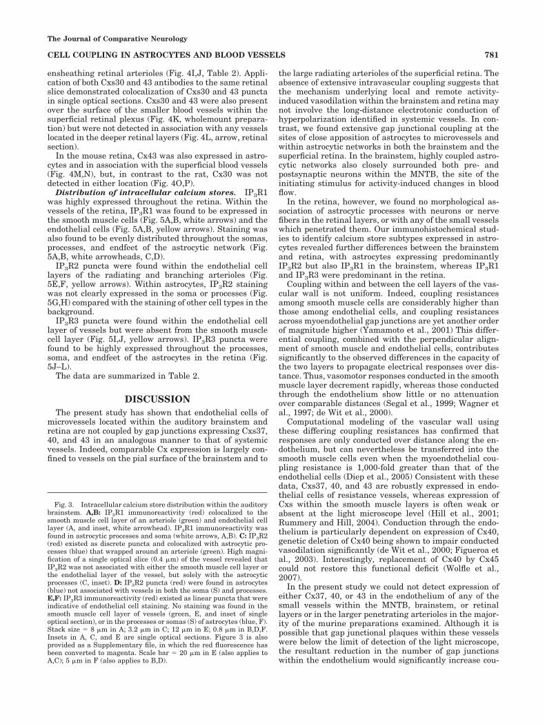
Distribution of intracellular calcium stores. IP3R1was highly expressed throughout the retina. Within thevessels of the retina, IP3R1 was found to be expressed inthe smooth muscle cells (Fig. 5A,B, white arrows) and theendothelial cells (Fig. 5A,B, yellow arrows). Staining wasalso found to be evenly distributed throughout the somas,processes, and endfeet of the astrocytic network (Fig.5A,B, white arrowheads, C,D).
IP3R2 puncta were found within the endothelial celllayers of the radiating and branching arterioles (Fig.5E,F, yellow arrows). Within astrocytes, IP3R2 stainingwas not clearly expressed in the soma or processes (Fig.5G,H) compared with the staining of other cell types in thebackground.
IP3R3 puncta were found within the endothelial celllayer of vessels but were absent from the smooth musclecell layer (Fig. 5I,J, yellow arrows). IP3R3 puncta werefound to be highly expressed throughout the processes,soma, and endfeet of the astrocytes in the retina (Fig.5J–L).
The data are summarized in Table 2.
DISCUSSIONThe present study has shown that endothelial cells of
microvessels located within the auditory brainstem andretina are not coupled by gap junctions expressing Cxs37,40, and 43 in an analogous manner to that of systemicvessels. Indeed, comparable Cx expression is largely con-fined to vessels on the pial surface of the brainstem and to
the large radiating arterioles of the superficial retina. Theabsence of extensive intravascular coupling suggests thatthe mechanism underlying local and remote activity-induced vasodilation within the brainstem and retina maynot involve the long-distance electrotonic conduction ofhyperpolarization identified in systemic vessels. In con-trast, we found extensive gap junctional coupling at thesites of close apposition of astrocytes to microvessels andwithin astrocytic networks in both the brainstem and thesuperficial retina. In the brainstem, highly coupled astro-cytic networks also closely surrounded both pre- andpostsynaptic neurons within the MNTB, the site of theinitiating stimulus for activity-induced changes in bloodflow.
In the retina, however, we found no morphological as-sociation of astrocytic processes with neurons or nervefibers in the retinal layers, or with any of the small vesselswhich penetrated them. Our immunohistochemical stud-ies to identify calcium store subtypes expressed in astro-cytes revealed further differences between the brainstemand retina, with astrocytes expressing predominantlyIP3R2 but also IP3R1 in the brainstem, whereas IP3R1and IP3R3 were predominant in the retina.
Coupling within and between the cell layers of the vas-cular wall is not uniform. Indeed, coupling resistancesamong smooth muscle cells are considerably higher thanthose among endothelial cells, and coupling resistancesacross myoendothelial gap junctions are yet another orderof magnitude higher (Yamamoto et al., 2001) This differ-ential coupling, combined with the perpendicular align-ment of smooth muscle and endothelial cells, contributessignificantly to the observed differences in the capacity ofthe two layers to propagate electrical responses over dis-tance. Thus, vasomotor responses conducted in the smoothmuscle layer decrement rapidly, whereas those conductedthrough the endothelium show little or no attenuationover comparable distances (Segal et al., 1999; Wagner etal., 1997; de Wit et al., 2000).
Computational modeling of the vascular wall usingthese differing coupling resistances has confirmed thatresponses are only conducted over distance along the en-dothelium, but can nevertheless be transferred into thesmooth muscle cells even when the myoendothelial cou-pling resistance is 1,000-fold greater than that of theendothelial cells (Diep et al., 2005) Consistent with thesedata, Cxs37, 40, and 43 are robustly expressed in endo-thelial cells of resistance vessels, whereas expression ofCxs within the smooth muscle layers is often weak orabsent at the light microscope level (Hill et al., 2001;Rummery and Hill, 2004). Conduction through the endo-thelium is particularly dependent on expression of Cx40,genetic deletion of Cx40 being shown to impair conductedvasodilation significantly (de Wit et al., 2000; Figueroa etal., 2003). Interestingly, replacement of Cx40 by Cx45could not restore this functional deficit (Wolfle et al.,2007).
In the present study we could not detect expression ofeither Cx37, 40, or 43 in the endothelium of any of thesmall vessels within the MNTB, brainstem, or retinallayers or in the larger penetrating arterioles in the major-ity of the murine preparations examined. Although it ispossible that gap junctional plaques within these vesselswere below the limit of detection of the light microscope,the resultant reduction in the number of gap junctionswithin the endothelium would significantly increase cou-
Fig. 3. Intracellular calcium store distribution within the auditorybrainstem. A,B: IP3R1 immunoreactivity (red) colocalized to thesmooth muscle cell layer of an arteriole (green) and endothelial celllayer (A, and inset, white arrowhead). IP3R1 immunoreactivity wasfound in astrocytic processes and soma (white arrows, A,B). C: IP3R2(red) existed as discrete puncta and colocalized with astrocytic pro-cesses (blue) that wrapped around an arteriole (green). High magni-fication of a single optical slice (0.4 !m) of the vessel revealed thatIP3R2 was not associated with either the smooth muscle cell layer orthe endothelial layer of the vessel, but solely with the astrocyticprocesses (C, inset). D: IP3R2 puncta (red) were found in astrocytes(blue) not associated with vessels in both the soma (S) and processes.E,F: IP3R3 immunoreactivity (red) existed as linear puncta that wereindicative of endothelial cell staining. No staining was found in thesmooth muscle cell layer of vessels (green, E, and inset of singleoptical section), or in the processes or somas (S) of astrocytes (blue, F).Stack size $ 8 !m in A; 3.2 !m in C; 12 !m in E; 0.8 !m in B,D,F.Insets in A, C, and E are single optical sections. Figure 3 is alsoprovided as a Supplementary file, in which the red fluorescence hasbeen converted to magenta. Scale bar $ 20 !m in E (also applies toA,C); 5 !m in F (also applies to B,D).
The Journal of Comparative Neurology
781CELL COUPLING IN ASTROCYTES AND BLOOD VESSELS

Figure 4
The Journal of Comparative Neurology
782 I.Y. KUO ET AL.

pling resistance among endothelial cells and attenuate thedistance over which vasodilatory responses could propa-gate. Modeling data confirm these predictions (Diep et al.,2005). We did not study Cx45 expression, as previousstudies have not demonstrated Cx45 protein in endothe-lial cells; expression, if present, is instead confined tosmooth muscle cells (Kruger et al., 2000; Li and Simard,2001; Wolfle et al., 2007), where effects on conductionwould be minimal, as discussed above.
The close proximity of astrocytes to both neurons andvessels within the auditory brainstem, the absence ofCxs37, 40, or 43 expression within adjacent vessels, andthe ubiquitous expression of Cxs30 and 43 in astrocyticnetworks suggest that these glia are well placed to medi-ate local changes in perfusion in response to synapticactivity, and perhaps to initiate more remote upstreamalterations in blood flow. Our data concord well with arecent study of the rat cortex in which pial arteriolardilation in response to two forms of intracerebral neuronalactivation was prevented by damage to the glia limitans,but not by damage to the intravascular endothelium (Xuet al., 2008). Although it is possible that responses couldstill be propagated through the intact vascular smoothmuscle, conduction along this pathway would be limited torelatively short distances (Wagner et al., 1997; Segal etal., 1999). These data were obtained in spite of conductionof vasomotor responses being reported in penetrating ar-terioles of the rat cortex (Dietrich et al., 1996). In contrast,in the auditory brainstem of the present study, Cx expres-sion within penetrating arterioles was confined to juvenilemice.
Our anatomical data demonstrated intense expressionof Cxs30 and 43 on astrocytic processes surrounding bothMNTB neurons and the large presynaptic calyces of Held.Although this may reflect appreciable coupling among as-trocytic processes, it is possible that some of the plaquesrepresent hemichannels through which astrocytes couldrelease neurotransmitters and modulate synaptic trans-mission. Indeed, expressed Cx43 hemichannels have re-cently been shown to be permeable to ATP, and channelswith similar properties have been recorded from hip-pocampal astrocytes (Kang et al., 2008). However, becausethere is little evidence that hemichannels open underphysiological conditions (Spray et al., 2006) and only 14hemichannels were recorded in experiments involving 700patches from hippocampal astrocytes (Kang et al., 2008),the possibility that hemichannels are involved in neuro-transmitter release from astrocytes in vivo remains aspeculation.
In contrast to the brainstem, the stratified arrangementof cells within the retina and the confinement of astrocytesto the innermost fiber layer preclude a role for astrocytesat the site of initiation of activity-induced changes inperfusion. Instead, Muller glia, which traverse the retinalcell layers and contact retinal ganglion cells (Hollander etal., 1991), have been shown to generate calcium transientsin response to light stimulation (Newman, 2005) and canmodulate neuronal activity and effect vasomotor re-sponses of surface vessels (Newman, 2003; Peppiatt et al.,2006). In addition, Muller glia and astrocytes are electri-cally coupled (Ceelen et al., 2001) and can communicatethrough ATP release (Newman, 2001). Therefore, neuro-vascular coupling in the rodent retina appears to be in-herently more complex, potentially involving a combina-tion of Muller glia and astrocytes to coordinate changes inblood flow.
In addition to differences in anatomical relationshipsfound between the retina and brainstem, there was vari-ation in the distribution of intracellular calcium receptorsubtypes between the two areas. Astrocytes within thebrainstem expressed IP3R2, and to a lesser extent IP3R1,consistent with the immunohistochemical studies ofSharp et al. (1999) and Holtzclaw et al. (2002). Animportant role for IP3R2 in calcium mobilization hasbeen established in recent physiological studies demon-strating that IP3R2 knockout mice are incapable of ex-hibiting astrocytic calcium increases in hippocampalslices upon metabolic receptor activation (Fiacco et al.,2007). However, the contribution of IP3R1 cannot beruled out as IP3Rs are capable of forming heterotetram-ers (Foskett et al., 2007), and such a formation betweenIP3R1 and R2 is also possible in the brainstem, althoughthe biophysical properties of heterotetramers are yet tobe characterized.
Interestingly, within the retina, IP3R3, and not IP3R2,was abundantly expressed within the retinal astrocytes,along with IP3R1. As IP3R3 has a much lower bindingaffinity for IP3 than do P3R1 and IP3R2 (Wojcikiewicz andLuo, 1998; Taylor and Laude, 2002), and the receptors arealso differentially regulated (Foskett et al., 2007; Soulsbyand Wojcikiewicz, 2007), the calcium released from IP3Rstores in retinal and brainstem astrocytes to a given stim-ulus may have potentially different spatial and/or tempo-ral characteristics. In support of this suggestion, intracel-lular calcium oscillations induced by acetylcholine invascular myocytes were dependent on the expression ofIP3R2, but not IP3R1 or IP3R3 (Morel et al., 2003). Al-though the consequences of this variation in IP3R sub-
Fig. 4. Vascular and astrocytic connexin distribution in the retina.A,B: Wholemount preparations from a central region of a rat retinashowed a subset of astrocytes (blue, A,B) closely ensheathed branch-ing vessels identified with BS-lectin (green, B). C,D: Cxs37 (red, C)and 40 (red, D) immunoreactivity was present within the endotheliallayer of the main arterioles found in wholemount preparations of ratretina. Cxs37 and 40 were also detected in the major branches ofarterioles (white arrow), but both were absent from the smallerbranches (white arrowhead) and veins (yellow V). Neither Cxs37 nor40 were observed in the smooth muscle layer of arterioles (green, C,D;also E,F, gap between blue astrocytes and red staining). E,F: Cxs37(E) and 40 (F) were not detected within astrocytic processes (blue) incross sections of the retina. G–L:. In the rat retina, Cxs30 (G,I) and 43(H,J) colocalized with astrocytic processes (blue, G,H), but both wereabsent from arterioles (green, I,J). The retinal section in I and J was
triple labeled with Cx30, 43, and lectin and the resulting image laterpseudocoloured. Cx43 (red, K wholemount, L retinal section) waslocalized in the nerve fiber layer (NFL) on the surface of the retina inassociation with astrocytes and superficial vessels (green, K), but wasnot found associated with vessels in deeper retinal layers (L, arrow).M–P: In wholemount preparations of the mouse retina, Cx43 (red,M,N) was associated with superficial vessels (green, M) and astrocyticprocesses (blue, N). No Cx30 immunoreactivity (O,P) was found ineither astrocytes (blue) or vessels (green, P) of wholemount mouseretinae. GCL, ganglion cell layer; IPL, inner plexiform layer; INL,inner nuclear layer. Stack size $ 6 !m in A,B; 8 !m in C,D,H; E $ 8.4!m; F $ 12 !m; G $ 20 !m; 2.4 !m in I,J; 4 !m in K; 15 !m in L; 0.8!m in M–P. Figure 4 is also provided as a Supplementary file, inwhich the red fluorescence has been converted to magenta. Scalebar $ 50 !m in A–D, O,P; 20 !m in E–H, K–N; 10 !m in I,J.
The Journal of Comparative Neurology
783CELL COUPLING IN ASTROCYTES AND BLOOD VESSELS

Fig. 5. Intracellular calcium store staining on the inner surface ofwholemount rat retina. A–D: Anti-IP3R1 (red) puncta were localizedwithin the smooth muscle cells (white arrows) and endothelial celllayer (A,B, yellow arrow). IP3R1 was also found in the soma (S) andprocesses of astrocytes (green, C,D). E–H: Anti-IP3R2 (red) punctawere present in the endothelial cell layer (yellow arrow) of vessels(blue, E,F) but were absent from smooth muscle cells. IP3R2 was notclearly expressed in either the soma or processes of astrocytes (green,
G,H). I–L: Anti-IP3R3 (red) puncta were present in the endothelialcell layer (yellow arrow) of vessels (blue, I,J) but were absent fromsmooth muscle cells. IP3R3 was found throughout the soma andprocesses of astrocytes (green, K,L). Stack size $ 12 !m in A,B; 2.4 !min C,D; 19.2 !m in E,F; 3.6 !m in G,H; 8 !m in I,J; 4.2 !m in K,L.Figure 5 is also provided as a Supplementary file, in which the redfluorescence has been converted to magenta. Scale bar $ 20 !m in J(also applies to A,B,E,F,I); 5 !m in L (also applies to C,D,G,H,K).
The Journal of Comparative Neurology

types on communication between astrocytes and bloodvessels is at present purely speculative, it should be notedthat the shape of complex calcium signals may also havemore long-term regulatory effects on gene transcription(Foskett et al., 2007). In comparison with the variations inIP3R distribution in astrocytes from the brainstem andretina, the distribution of IP3R subtypes within the arte-rioles of the retina and brainstem was similar to thatfound previously in the basilar and mesenteric arteries(Grayson et al., 2004), with the exception that IP3R2 wasnot detected at all in the brainstem vessels.
The implications of our anatomical results for neuro-vascular signaling mechanisms in the brainstem andretina are summarized in the models presented in Fig.6. In the brainstem, neurotransmitters released duringsynaptic transmission activate IP3R1 and R2 stores insurrounding astrocytes to produce intracellular calciumwaves. These waves spread through neighboring astro-cytes, perhaps via the gap junctions comprised of Cxs30and 43, which electrically couple them, and vasodilatoryfactors are released from perivascular endfeet on tolocal and perhaps more remote blood vessels to elicit anincrease in regional perfusion and blood flow. Intrace-rebral spatial restriction of the augmented blood flow tothe specifically activated area, for example, tonotopi-cally located neurons within the MNTB, may occurthrough subpopulations of selectively coupled astro-cytes, as has been recently demonstrated in the barrelcortex (Houades et al., 2008). In the pial circulation and
in some penetrating arterioles, vascular responses arerelayed to larger vessels and conducted by electrotonicpropagation through endothelial gap junctions com-prised of Cxs37 and 40.
A more complicated model is presented for the retina(Fig. 6B), whereby stimuli from active synapses are re-layed to the vasculature by Muller glia and astrocytes(Metea and Newman, 2006). Calcium waves involvingIP3R1 and R3 stores are propagated through the extensiveastrocytic network, leading to the release of vasoeffectorsfrom perivascular endfeet. Blood flow may be augmentedthrough additional intracellular gap junctional mecha-nisms in the radiating arterioles of the rat, but not themouse, retina.
In conclusion, we suggest that astrocytes are in an idealposition to mediate functional hyperemia in the brain-stem, where they are in close contact to neurons andvessels. In contrast, within the retina, astrocytes are onlylocated near the surface arterioles, suggesting that theyrepresent just one element in a multicellular cascade thatserves the more complex anatomical and functional ar-rangement of the retina.
LITERATURE CITEDArmstrong ML, Dua AK, Murrant CL. 2007. Potassium initiates vasodila-
tation induced by a single skeletal muscle contraction in hamstercremaster muscle. J Physiol 581:841–852.
Bushong EA, Martone ME, Jones YZ, Ellisman MH. 2002. Protoplasmic
Fig. 6. Vascular and astrocytic coupling in the brainstem andretina. A: Neurovascular coupling in the brainstem is activated fol-lowing neurotransmitter spillover from activated calyces of Held (1).Regenerative astrocytic calcium waves following activation of IP3R1and R2 stores spread through the astrocytic syncytium via gap junc-tions comprised of Cxs30 and 43. Astrocytic endfeet abutting vesselsrelease vasodilatory factors onto the smooth muscle of either local (2)or remote vessels (3). Rapid communication to the upstream pialarteries is propagated via electrotonic coupling of the endothelial celllayer by Cxs37 and 40. Dilation of the pial vessels results in improved
blood flow to downstream vessels. B: Responses to neuronal activity(1) in the retina are communicated by the ascending Muller glia andthen astrocytes via calcium waves involving IP3R1 and R3. The ex-tensive astrocytic network on the surface of the retina is coupled byCxs30 and 43 in the rat, but only 43 in the mouse. Release of vasodi-lators at local sites by astrocytic endfeet (2) occurs in both rodentspecies; however, augmentation of blood flow through endothelial gapjunctions is only present in the rat (3). MNTB, medial nucleus of thetrapezoid body.
The Journal of Comparative Neurology
785CELL COUPLING IN ASTROCYTES AND BLOOD VESSELS

astrocytes in CA1 stratum radiatum occupy separate anatomical do-mains. J Neurosci 22:183–192.
Ceelen PW, Lockridge A, Newman EA. 2001. Electrical coupling betweenglial cells in the rat retina. Glia 35:1–13.
Chan-Ling T. 1997. Glial, vascular, and neuronal cytogenesis in whole-mounted cat retina. Microsc Res Tech 36:1–16.
Cornell-Bell AH, Finkbeiner SM, Cooper MS, Smith SJ. 1990. Glutamateinduces calcium waves in cultured astrocytes: long-range glial signal-ing. Science 247:470–473.
Cotrina ML, Nedergaard M. 2005. Intracellular calcium control mecha-nisms in glia. In: Kettenmann H, Ransom B, editors. Neuroglia, 2nd ed.Oxford: Oxford University Press. p 229–239.
de Wit C, Roos F, Bolz SS, Kirchhoff S, Kruger O, Willecke K, Pohl U. 2000.Impaired conduction of vasodilation along arterioles in connexin40-deficient mice. Circ Res 86:649–655.
Diep HK, Vigmond EJ, Segal SS, Welsh DG. 2005. Defining electricalcommunication in skeletal muscle resistance arteries: a computationalapproach. J Physiol 568:267–281.
Dietrich HH, Kajita Y, Dacey RG, Jr. 1996. Local and conducted vasomotorresponses in isolated rat cerebral arterioles. Am J Physiol 271:H1109–1116.
Fiacco TA, Agulhon C, Taves SR, Petravicz J, Casper KB, Dong X, Chen J,McCarthy KD. 2007. Selective stimulation of astrocyte calcium in situdoes not affect neuronal excitatory synaptic activity. Neuron 54:611–626.
Figueroa XF, Paul DL, Simon AM, Goodenough DA, Day KH, Damon DN,Duling BR. 2003. Central role of connexin40 in the propagation ofelectrically activated vasodilation in mouse cremasteric arterioles invivo. Circ Res 92:793–800.
Filosa JA, Bonev AD, Nelson MT. 2004. Calcium dynamics in corticalastrocytes and arterioles during neurovascular coupling. Circ Res 95:e73–81.
Foskett JK, White C, Cheung KH, Mak DO. 2007. Inositol trisphosphatereceptor Ca2& release channels. Physiol Rev 87:593–658.
Girouard H, Iadecola C. 2006. Neurovascular coupling in the normal brainand in hypertension, stroke, and Alzheimer disease. J Appl Physiol100:328–335.
Grayson TH, Haddock RE, Murray TP, Wojcikiewicz RJ, Hill CE. 2004.Inositol 1,4,5-trisphosphate receptor subtypes are differentially distrib-uted between smooth muscle and endothelial layers of rat arteries. CellCalcium 36:447–458.
Harder DR, Alkayed NJ, Lange AR, Gebremedhin D, Roman RJ. 1998.Functional hyperemia in the brain: hypothesis for astrocyte-derivedvasodilator metabolites. Stroke 29:229–234.
Hill CE, Phillips JK, Sandow SL. 2001. Heterogeneous control of blood flowamong different vascular beds. Med Res Rev 21:1–60.
Hollander H, Makarov F, Dreher Z, van Driel D, Chan-Ling TL, Stone J.1991. Structure of the macroglia of the retina: sharing and division oflabour between astrocytes and Muller cells. J Comp Neurol 313:587–603.
Holtzclaw LA, Pandhit S, Bare DJ, Mignery GA, Russell JT. 2002. Astro-cytes in adult rat brain express type 2 inositol 1,4,5-trisphosphatereceptors. Glia 39:69–84.
Horiuchi T, Dietrich HH, Hongo K, Dacey RG, Jr. 2002. Mechanism ofextracellular K&-induced local and conducted responses in cerebralpenetrating arterioles. Stroke 33:2692–2699.
Houades V, Koulakoff A, Ezan P, Seif I, Giaume C. 2008. Gap junction-mediated astrocytic networks in the mouse barrel cortex. J Neurosci28:5207–5217.
Kang J, Kang N, Lovatt D, Torres A, Zhao Z, Lin J, Nedergaard M. 2008.Connexin 43 hemichannels are permeable to ATP. J Neurosci 28:4702–4711.
Kruger O, Plum A, Kim JS, Winterhager E, Maxeiner S, Hallas G, Kirch-hoff S, Traub O, Lamers WH, Willecke K. 2000. Defective vasculardevelopment in connexin 45-deficient mice. Development (CambridgeEng) 127:4179–4193.
Li X, Simard JM. 2001. Connexin45 gap junction channels in rat cerebralvascular smooth muscle cells. Am J Physiol Heart Circ Physiol 281:H1890–1898.
Metea MR, Newman EA. 2006. Glial cells dilate and constrict bloodvessels: a mechanism of neurovascular coupling. J Neurosci 26:2862–2870.
Morel JL, Fritz N, Lavie JL, Mironneau J. 2003. Crucial role of type 2inositol 1,4,5-trisphosphate receptors for acetylcholine-induced Ca2&
oscillations in vascular myocytes. Arterioscler Thromb Vasc Biol 23:1567–1575.
Mulligan SJ, MacVicar BA. 2004. Calcium transients in astrocyte endfeetcause cerebrovascular constrictions. Nature 431:195–199.
Nagy JI, Patel D, Ochalski PA, Stelmack GL. 1999. Connexin30 in rodent,cat and human brain: selective expression in gray matter astrocytes,co-localization with connexin43 at gap junctions and late developmen-tal appearance. Neuroscience 88:447–468.
Nagy JI, Li X, Rempel J, Stelmack G, Patel D, Staines WA, Yasumura T,Rash JE. 2001. Connexin26 in adult rodent central nervous system:demonstration at astrocytic gap junctions and colocalization with con-nexin30 and connexin43. J Comp Neurol 441:302–323.
Newman EA. 2001. Propagation of intercellular calcium waves in retinalastrocytes and Muller cells. J Neurosci 21:2215–2223.
Newman EA. 2003. Glial cell inhibition of neurons by release of ATP.J Neurosci 23:1659–1666.
Newman EA. 2005. Calcium increases in retinal glial cells evoked bylight-induced neuronal activity. J Neurosci 25:5502–5510.
Ngai AC, Winn HR. 2002. Pial arteriole dilation during somatosensorystimulation is not mediated by an increase in CSF metabolites. Am JPhysiol Heart Circ Physiol 282:H902–907.
Paul ER, Christian AL, Franke R, Groschel-Stewart U. 1994. Embryonicchicken gizzard: smooth muscle and non-muscle myosin isoforms. CellTissue Res 276:381–386.
Peppiatt CM, Howarth C, Mobbs P, Attwell D. 2006. Bidirectional controlof CNS capillary diameter by pericytes. Nature 443:700–704.
Rummery NM, Hill CE. 2004. Vascular gap junctions and implications forhypertension. Clin Exp Pharmacol Physiol 31:659–667.
Rummery NM, Hickey H, McGurk G, Hill CE. 2002. Connexin37 is themajor connexin expressed in the media of caudal artery. ArteriosclerThromb Vasc Biol 22:1427–1432.
Rummery NM, Grayson TH, Hill CE. 2005. Angiotensin-converting en-zyme inhibition restores endothelial but not medial connexin expres-sion in hypertensive rats. J Hypertens 23:317–328.
Satzler K, Sohl LF, Bollmann JH, Borst JG, Frotscher M, Sakmann B,Lubke JH. 2002. Three-dimensional reconstruction of a calyx of Heldand its postsynaptic principal neuron in the medial nucleus of thetrapezoid body. J Neurosci 22:10567–10579.
Scemes E. 2000. Components of astrocytic intercellular calcium signaling.Mol Neurobiol 22:167–179.
Segal SS. 2005. Regulation of blood flow in the microcirculation. Microcir-culation 12:33–45.
Segal SS, Welsh DG, Kurjiaka DT. 1999. Spread of vasodilatation andvasoconstriction along feed arteries and arterioles of hamster skeletalmuscle. J Physiol 516:283–291.
Sharp AH, Nucifora FC, Jr., Blondel O, Sheppard CA, Zhang C, Snyder SH,Russell JT, Ryugo DK, Ross CA. 1999. Differential cellular expressionof isoforms of inositol 1,4,5-triphosphate receptors in neurons and gliain brain. J Comp Neurol 406:207–220.
Soulsby MD, Wojcikiewicz RJ. 2007. Calcium mobilization via type IIIinositol 1,4,5-trisphosphate receptors is not altered by PKA-mediatedphosphorylation of serines 916, 934, and 1832. Cell Calcium 42:261–270.
Spray DC, Ye ZC, Ransom BR. 2006. Functional connexin “hemichannels”:a critical appraisal. Glia 54:758–773.
Takano T, Tian GF, Peng W, Lou N, Libionka W, Han X, Nedergaard M.2006. Astrocyte-mediated control of cerebral blood flow. Nat Neurosci9:260–267.
Taylor CW, Laude AJ. 2002. IP3 receptors and their regulation by calmod-ulin and cytosolic Ca2&. Cell Calcium 32:321–334.
Wagner AJ, Holstein-Rathlou NH, Marsh DJ. 1997. Internephron couplingby conducted vasomotor responses in normotensive and spontaneouslyhypertensive rats. Am J Physiol 272:F372–379.
Wojcikiewicz RJ. 1995. Type I, II, and III inositol 1,4,5-trisphosphatereceptors are unequally susceptible to down-regulation and are ex-pressed in markedly different proportions in different cell types. J BiolChem 270:11678–11683.
Wojcikiewicz RJ, Luo SG. 1998. Differences among type I, II, and IIIinositol-1,4,5-trisphosphate receptors in ligand-binding affinity influ-ence the sensitivity of calcium stores to inositol-1,4,5-trisphosphate.Mol Pharmacol 53:656–662.
The Journal of Comparative Neurology
786 I.Y. KUO ET AL.

Wolfle SE, Schmidt VJ, Hoepfl B, Gebert A, Alcolea S, Gros D, de Wit C.2007. Connexin45 cannot replace the function of connexin40 in con-ducting endothelium-dependent dilations along arterioles. Circ Res101:1292–1299.
Xu HL, Mao L, Ye S, Paisansathan C, Vetri F, Pelligrino DA. 2008.Astrocytes are a key conduit for upstream signaling of vasodilationduring cerebral cortical neuronal activation in vivo. Am J PhysiolHeart Circ Physiol 294:H622–632.
Yamamoto-Hino M, Miyawaki A, Kawano H, Sugiyama T, Furuichi T,Hasegawa M, Mikoshiba K. 1995. Immunohistochemical study of ino-
sitol 1,4,5-trisphosphate receptor type 3 in rat central nervous system.Neuroreport 6:273–276.
Yamamoto Y, Klemm MF, Edwards FR, Suzuki H. 2001. Intercellularelectrical communication among smooth muscle and endothelial cellsin guinea-pig mesenteric arterioles. J Physiol 535:181–195.
Zahs KR, Kofuji P, Meier C,Dermietzel R. 2003. Connexin immunoreactiv-ity in glial cells of the rat retina. J Comp Neurol 455:531–546.
Zonta M, Angulo MC, Gobbo S, Rosengarten B, Hossmann KA, Pozzan T,Carmignoto G. 2003. Neuron-to-astrocyte signaling is central to thedynamic control of brain microcirculation. Nat Neurosci 6:43–50.
The Journal of Comparative Neurology
787CELL COUPLING IN ASTROCYTES AND BLOOD VESSELS