Selectively targeting pain in the trigeminal system
12
Selectively targeting pain in the trigeminal system Hyun Yeong Kim a , Kihwan Kim a , Hai Ying Li a , Gehoon Chung a , Chul-Kyu Park a , Joong Soo Kim a , Sung Jun Jung b , Min Kyung Lee c , Dong Kuk Ahn c , Se Jin Hwang d , Youngnam Kang e , Alexander M. Binshtok f , Bruce P. Bean g , Clifford J. Woolf f , Seog Bae Oh a, * a National Research Laboratory for Pain, Dental Research Institute and Department of Physiology School of Dentistry, Seoul National University, Seoul 110-749, Republic of Korea b Department of Physiology School of Medicine, Hanyang University, Seoul 133-791, Republic of Korea c Department of Oral Physiology and Neurobiology School of Dentistry, Kyungpook National University, Daegu 700-412, Republic of Korea d Department of Anatomy and Cell Biology College of Medicine, Hanyang University, Seoul 133-791, Republic of Korea e Department of Neuroscience and Oral Physiology, Osaka University Graduate School of Dentistry, Osaka 565-0871, Japan f Neural Plasticity Research Group, Massachusetts General Hospital & Harvard Medical School, Charlestown, MA 02129, USA g Department of Neurobiology, Harvard Medical School, Boston, MA 02115, USA article info Article history: Received 21 August 2009 Received in revised form 5 December 2009 Accepted 9 February 2010 Available online xxxx Keywords: Action potentials Local anesthetics QX-314 Trigeminal system TRPV1 Voltage-gated sodium channels abstract We tested whether it is possible to selectively block pain signals in the orofacial area by delivering the permanently charged lidocaine derivative QX-314 into nociceptors via TPRV1 channels. We examined the effects of co-applied QX-314 and capsaicin on nociceptive, proprioceptive, and motor function in the rat trigeminal system. QX-314 alone failed to block voltage-gated sodium channel currents (I Na ) and action potentials (APs) in trigeminal ganglion (TG) neurons. However, co-application of QX-314 and capsaicin blocked I Na and APs in TRPV1-positive TG and dental nociceptive neurons, but not in TRPV1-negative TG neurons or in small neurons from TRPV1 knock-out mice. Immunohistochemistry revealed that TRPV1 is not expressed by trigeminal motor and trigeminal mesencephalic neurons. Capsa- icin had no effect on rat trigeminal motor and proprioceptive mesencephalic neurons and therefore should not allow QX-314 to enter these cells. Co-application of QX-314 and capsaicin inhibited the jaw-opening reflex evoked by noxious electrical stimulation of the tooth pulp when applied to a sensory but not a motor nerve, and produced long-lasting analgesia in the orofacial area. These data show that selective block of pain signals can be achieved by co-application of QX-314 with TRPV1 agonists. This approach has potential utility in the trigeminal system for treating dental and facial pain. Ó 2010 International Association for the Study of Pain. Published by Elsevier B.V. All rights reserved. 1. Introduction Local anesthetics (LAs) abolish the transmission of nociceptive information to the central nervous system (CNS) by blocking volt- age-gated sodium channels (VGSCs) and thereby prevent the gen- eration and propagation of action potentials (APs) [6]. Most LAs in clinical use are tertiary amines that under physiological conditions exist in a mixture of protonated and uncharged base forms [5]. The uncharged hydrophobic form of LAs penetrates through the mem- brane of all neurons, so that in addition to blocking pain signals, LAs produce general numbness from the block of low-threshold sensory nerves, as well deficits in motor function, and a block of autonomic nerves [3,4,15,30]. Selective block of pain signals might be possible either by tar- geting certain sodium channels that are present only in pain-sens- ing neurons [7,12,20] or by delivering sodium channel blockers selectively to pain-sensing neurons. One strategy is to deliver a permanently charged sodium channel blocker such as QX-314 (N-ethyl-lidocaine) by entry through large-pore ion channels selec- tively expressed in nociceptive neurons [33]. The transient recep- tor potential vanilloid 1 (TRPV1) channel is a primary nociceptive transducer in pain-sensing neurons, activated by noxious heat (>43 °C), capsaicin, protons and endocannabinoids [8,36]. A recent study demonstrated that the pore of TRPV1 channels, when opened by the TRPV1 channel agonist capsaicin, might be large enough to deliver QX-314 or other large molecule selectively into nociceptive neurons [11,22]. Binshtok et al. demonstrated that QX-314, a 0304-3959/$36.00 Ó 2010 International Association for the Study of Pain. Published by Elsevier B.V. All rights reserved. doi:10.1016/j.pain.2010.02.016 Abbreviations: APs, action potentials; CNS, central nervous system; dEMG, digastric electromyogram; DiI, 1,1 0 -dioctadecyl-3,3,3 00 3 0 -teramethylindo-carbocya- nine perchlorate; DRG, dorsal root ganglia; I Na , voltage-gated sodium channel currents; LAs, local anesthetics; TG, trigeminal ganglion; TRPV1, transient receptor potential vanilloid 1; VGSCs, voltage-gated sodium channels; Vmes, trigeminal mesencephalic nucleus; Vmot, trigeminal motor nucleus; V rest , resting membrane potential. * Corresponding author. Address: Department of Physiology, School of Dentistry, Seoul National University, 28-2 Yeongeon-Dong, Chongno-Ku, Seoul 110-749, Republic of Korea. Tel.: +82 2 740 8656; fax: +82 2 7625107. E-mail address: [email protected] (S.B. Oh). PAIN Ò xxx (2010) xxx–xxx www.elsevier.com/locate/pain ARTICLE IN PRESS Please cite this article in press as: Kim HY et al. Selectively targeting pain in the trigeminal system. PAIN Ò (2010), doi:10.1016/j.pain.2010.02.016
Transcript of Selectively targeting pain in the trigeminal system
ARTICLE IN PRESS
PAIN�
xxx (2010) xxx–xxx
w w w . e l se v i e r . c o m / l o c a t e / p a i n
Selectively targeting pain in the trigeminal system
Hyun Yeong Kim a, Kihwan Kim a, Hai Ying Li a, Gehoon Chung a, Chul-Kyu Park a, Joong Soo Kim a,Sung Jun Jung b, Min Kyung Lee c, Dong Kuk Ahn c, Se Jin Hwang d, Youngnam Kang e,Alexander M. Binshtok f, Bruce P. Bean g, Clifford J. Woolf f, Seog Bae Oh a,*
a National Research Laboratory for Pain, Dental Research Institute and Department of Physiology School of Dentistry, Seoul National University, Seoul 110-749, Republic of Koreab Department of Physiology School of Medicine, Hanyang University, Seoul 133-791, Republic of Koreac Department of Oral Physiology and Neurobiology School of Dentistry, Kyungpook National University, Daegu 700-412, Republic of Koread Department of Anatomy and Cell Biology College of Medicine, Hanyang University, Seoul 133-791, Republic of Koreae Department of Neuroscience and Oral Physiology, Osaka University Graduate School of Dentistry, Osaka 565-0871, Japanf Neural Plasticity Research Group, Massachusetts General Hospital & Harvard Medical School, Charlestown, MA 02129, USAg Department of Neurobiology, Harvard Medical School, Boston, MA 02115, USA
a r t i c l e i n f o a b s t r a c t
Article history:Received 21 August 2009Received in revised form 5 December 2009Accepted 9 February 2010Available online xxxx
Keywords:Action potentialsLocal anestheticsQX-314Trigeminal systemTRPV1Voltage-gated sodium channels
0304-3959/$36.00 � 2010 International Associationdoi:10.1016/j.pain.2010.02.016
Abbreviations: APs, action potentials; CNS, centdigastric electromyogram; DiI, 1,10-dioctadecyl-3,3,30
nine perchlorate; DRG, dorsal root ganglia; INa, vocurrents; LAs, local anesthetics; TG, trigeminal ganglipotential vanilloid 1; VGSCs, voltage-gated sodiummesencephalic nucleus; Vmot, trigeminal motor nucpotential.
* Corresponding author. Address: Department of PhSeoul National University, 28-2 Yeongeon-Dong, CRepublic of Korea. Tel.: +82 2 740 8656; fax: +82 2 7
E-mail address: [email protected] (S.B. Oh).
Please cite this article in press as: Kim HY et al
We tested whether it is possible to selectively block pain signals in the orofacial area by delivering thepermanently charged lidocaine derivative QX-314 into nociceptors via TPRV1 channels. We examinedthe effects of co-applied QX-314 and capsaicin on nociceptive, proprioceptive, and motor function inthe rat trigeminal system. QX-314 alone failed to block voltage-gated sodium channel currents (INa)and action potentials (APs) in trigeminal ganglion (TG) neurons. However, co-application of QX-314and capsaicin blocked INa and APs in TRPV1-positive TG and dental nociceptive neurons, but not inTRPV1-negative TG neurons or in small neurons from TRPV1 knock-out mice. Immunohistochemistryrevealed that TRPV1 is not expressed by trigeminal motor and trigeminal mesencephalic neurons. Capsa-icin had no effect on rat trigeminal motor and proprioceptive mesencephalic neurons and thereforeshould not allow QX-314 to enter these cells. Co-application of QX-314 and capsaicin inhibited thejaw-opening reflex evoked by noxious electrical stimulation of the tooth pulp when applied to a sensorybut not a motor nerve, and produced long-lasting analgesia in the orofacial area. These data show thatselective block of pain signals can be achieved by co-application of QX-314 with TRPV1 agonists. Thisapproach has potential utility in the trigeminal system for treating dental and facial pain.
� 2010 International Association for the Study of Pain. Published by Elsevier B.V. All rights reserved.
1. Introduction
Local anesthetics (LAs) abolish the transmission of nociceptiveinformation to the central nervous system (CNS) by blocking volt-age-gated sodium channels (VGSCs) and thereby prevent the gen-eration and propagation of action potentials (APs) [6]. Most LAs inclinical use are tertiary amines that under physiological conditionsexist in a mixture of protonated and uncharged base forms [5]. The
for the Study of Pain. Published by
ral nervous system; dEMG,030-teramethylindo-carbocya-ltage-gated sodium channelon; TRPV1, transient receptor
channels; Vmes, trigeminalleus; Vrest, resting membrane
ysiology, School of Dentistry,hongno-Ku, Seoul 110-749,
625107.
. Selectively targeting pain in t
uncharged hydrophobic form of LAs penetrates through the mem-brane of all neurons, so that in addition to blocking pain signals,LAs produce general numbness from the block of low-thresholdsensory nerves, as well deficits in motor function, and a block ofautonomic nerves [3,4,15,30].
Selective block of pain signals might be possible either by tar-geting certain sodium channels that are present only in pain-sens-ing neurons [7,12,20] or by delivering sodium channel blockersselectively to pain-sensing neurons. One strategy is to deliver apermanently charged sodium channel blocker such as QX-314(N-ethyl-lidocaine) by entry through large-pore ion channels selec-tively expressed in nociceptive neurons [33]. The transient recep-tor potential vanilloid 1 (TRPV1) channel is a primary nociceptivetransducer in pain-sensing neurons, activated by noxious heat(>43 �C), capsaicin, protons and endocannabinoids [8,36]. A recentstudy demonstrated that the pore of TRPV1 channels, when openedby the TRPV1 channel agonist capsaicin, might be large enough todeliver QX-314 or other large molecule selectively into nociceptiveneurons [11,22]. Binshtok et al. demonstrated that QX-314, a
Elsevier B.V. All rights reserved.
he trigeminal system. PAIN�
(2010), doi:10.1016/j.pain.2010.02.016

2 H.Y. Kim et al. / PAIN�
xxx (2010) xxx–xxx
ARTICLE IN PRESS
charged membrane-impermeant lidocaine derivative, can be tar-geted selectively into nociceptors when co-administrated withcapsaicin, resulting in selective blocking of sodium channels andinhibition of excitability in nociceptors [3].
The orofacial area is mainly innervated by the trigeminal nervethat consists of both sensory and motor fibers [18,31]. LAs arewidely used in the trigeminal system, including nerve block forgeneral dental treatment, trigeminal neuralgia or trigger pointinjection for myofacial pain syndrome [32]. However, side-effectssuch as numbness or systemic toxicity following accidental intra-vascular injection following LA injection [32] limit the use of cur-rent LAs. A strategy for targeting delivery of sodium channelblockers only to nociceptors could minimize many adverse effects.In this study, we tested whether the charged lidocaine derivativeQX-314 can be selectively targeted to nociceptors in the trigeminalsystem via TRPV1 channels, using a combination of in vitro, in vivoand behavioral studies. The trigeminal system lends itself to studythe selectivity of nerve block and we found that the effects pro-duced by co-application of QX-314 and capsaicin are restricted toTRPV1-expressing trigeminal nociceptive neurons, with no effecton proprioceptive neurons or motor neurons. We suggest that thisapproach should be applicable for producing pain-selective localanesthesia in the trigeminal system.
2. Materials and methods
2.1. Animals
All surgical and experimental procedures were reviewed andapproved by the Institutional Animal Care and Use Committee (IA-CUC) at the School of Dentistry, Seoul National University. Animaltreatments were performed according to the Guidelines of theInternational Association for the Study of Pain. Male Sprague–Daw-ley (SD) rats (OrientBio, Korea) were housed at a temperature of23 ± 2 �C with a 12-h light–dark cycle (light on 08:00 to 20:00),and fed food and water ad libitum. The animals were allowed tohabituate to the housing facilities for 1 week before the experi-ments, and efforts were made to limit distress to the animals.
2.2. Preparation of TG neurons
TG neurons from adult SD rats (weighing approximately 150–200 g) were prepared as previously described [13]. TG neuronswere harvested and transferred to cold (4 �C) HBSS (Life Technolo-gies, USA). The cells were incubated in 3 ml HBSS containing 2.5%trypsin (Sigma–Aldrich, USA) at 37 �C for 45 min. After enzymedigestion, the suspension was washed in DMEM and trituratedwith a flame-polished Pasteur pipette to separate the cells. Subse-quently, the cells were centrifuged through 15% BSA (Sigma–Al-drich, USA), resuspended and plated on coverslips coated withpoly-L-ornithine (0.5 mg/ml; Sigma–Aldrich, USA). TG neuronswere incubated in Neurobasal medium (Gibco, USA) supplementedwith penicillin and streptomycin (1%; Gibco, USA), B-27 supple-ment (invitrogen, USA), L-glutamine (1 mM; Invitrogen, USA),nerve growth factor (NGF 2.5s, 50 ng/ml; invitrogen, USA), GDNF(2 ng/ml; Sigma–Aldrich, USA) and Ara-C (10 lM; Sigma–Aldrich,USA) at 37 �C under 5% CO2. Electrophysiological recordings wereperformed within 24 h of plating.
2.3. Retrograde labeling of dental primary afferent neurons andtrigeminal motoneurons
Dental primary afferent TG neurons were retrograde labeledwith the fluorescent dye, 1,10-dioctadecyl-3,3,30 030-teramethylin-do-carbocyanine perchlorate (DiI; Molecular Probes, USA) as previ-ously described [27]. Briefly, DiI was placed in the cavity in the
Please cite this article in press as: Kim HY et al. Selectively targeting pain in t
upper molar teeth of adult SD rats (150–200 g) under anesthesiawith sodium pentobarbital (30 mg/kg body weight, intraperitonealinjection). After 2 weeks, TG neurons were acutely isolated as men-tioned above. Trigeminal motoneurons were also retrograde la-beled with DiI as previously described [24].
2.4. Electrophysiological recordings
Whole cell voltage and current clamp recording for TG neuronswere performed at room temperature to measure INa and APs,respectively, with EPC-9 amplifier and Pulse 8.30 software (bothfrom HEKA, Germany). Patch pipettes were pulled from borosili-cate capillaries (Chase Scientific Glass Inc., Rockwood, USA). Whenfilled with the pipette solution, the resistance of the pipettes was4–7 MX (always less than 10 MX). The recoding chamber (volume500 ll) was continuously superfused (1–2 ml/min). Data were lowpass filtered at 2 kHz, sampled at 10 kHz, and acquired using Pulseprogram (HEKA, Germany). The pipette solution for voltage clampexperiments was composed of 110 mM CsCl, 2 mM MgCl2, 1 mMCaCl2, 11 mM EGTA, 10 mM HEPES, and pH adjusted to 7.4 withCsOH. The external solution was 60 mM NaCl, 60 mM choline chlo-ride, 4 mM KCl, 2 mM CaCl2, 1 mM MgCl2, 0.1 mM CdCl2, 15 mMtetraethylammonium chloride, 5 mM 4-aminopyridine, 10 mMglucose, 10 mM HEPES, and pH adjusted to 7.4 with NaOH. Sodiumcurrent was recorded during 30 ms voltage clamp steps from aholding potential of �70 mV to a test potential of 0 mV every10 s. The pipette solution for current clamp recordings was com-posed of 135 mM potassium gluconate, 2 mM MgCl2, 6 mM KCl,10 mM HEPES, 5 mM MgATP, 0.5 mM Li2GTP, and pH adjusted to7.4 with KOH, and an external solution of 145 mM NaCl, 5 mMKCl, 1 mM MgCl2, 2 mM CaCl2, 10 mM HEPES, 10 mM glucose,and pH adjusted to 7.4 with NaOH. APs were evoked by 3 ms depo-larizing current pulses with 250 pA amplitude.
Electrophysiological recordings from trigeminal motoneuronsand trigeminal mesencephalic neurons were performed withwhole cell patch clamp recording methods similar to the previousstudies [16]. Briefly, coronal slices of 200 lm thickness were madefrom the brain stem of Wistar rats (6–14 days postnatal). The arti-ficial cerebrospinal fluid used as bath solution was composed of124 mM NaCl, 26 mM NaHCO3, 1.8 mM KCl, 1.2 mM KH2PO4,2.5 mM CaCl2, 1.3 mM MgCl2, and 10 mM Glucose. The internalsolution of the patch pipettes had 123 mM K-gluconate, 18 mMKCl, 10 mM NaCl, 3 mM MgCl2, 2 mM ATP-Na2, 0.3 mM GTP-Na3,and 10 mM HEPES, pH 7.4 adjusted with KOH. Mesencephalic tri-geminal nucleus neurons and trigeminal motor nucleus neuronswere easily recognized under microscope (BX-50WI, Olympus, Ja-pan) by their location and large oval shape. All recordings wereperformed with Axopatch 200B amplifier and Digidata 1322A dataacquisition system (Molecular Devices, CA). Data were low-pass fil-tered at 5–10 kHz, digitized at a sampling rate of 10 kHz.
2.5. Immunofluorescent staining
Following whole cell patch clamp recordings, DiI-labeled TGneurons were washed in PBS and then kept in blocking solutioncontaining 5% normal donkey serum, 2% BSA, 2% FBS, and 0.1% Tri-ton X-100 for 1 h at RT. The cells were incubated overnight at 4 �Cwith a mixture of guinea pig anti-TRPV1 antibody (1:500; Chem-icon, USA) and washed in PBS. The slides were then incubated for1 h at RT with FITC-conjugated guinea pig IgG antibody (1:200;Jackson ImmunoResearch, USA). For the staining of Vmot, the ratswere perfused with physiological saline and sequentially with 4%paraformaldehyde in 0.1 M phosphate buffer (pH 7.4). Whole ratbrain was removed and immersed in the post-fixative at 4 �C over-night and then transferred to 10% and 30% sucrose in PB for 48 h.Serial frozen transverse sections (30 lm thickness) were collected
he trigeminal system. PAIN�
(2010), doi:10.1016/j.pain.2010.02.016
H.Y. Kim et al. / PAIN�
xxx (2010) xxx–xxx 3
ARTICLE IN PRESS
in cold PBS. Free-floating sections were incubated with guinea piganti-TRPV1 antibody (1:500; Chemicon, USA) and processed as de-scribed above. All immunohistochemical procedures were per-formed at room temperature unless otherwise stated. The sliceswere mounted with VectaShield (Vector Laboratories, USA) andvisualized using a confocal microscope using appropriate filter sets(FV-300, Olympus, Japan). To study the expression of TRPV1 in theinferior alveoloar nerve, the nerve was double immunostained forTRPV1 and PGP9.5, a marker for both myelinated and unmyeli-nated nerve fibers. The rats were anesthetized with ethyl etherand perfused with heparinized saline followed by 4% paraformal-dehyde in phosphate buffer (PB, 0.1 M, pH 7.4). The inferior alveo-lar nerve was removed, post-fixed for 3 h in the same fixative, andcryoprotected in 30% sucrose in PB. Fifteen micrometer thick longi-tudinal sections of the nerves were cut on a cryostat, blocked with10% normal donkey serum (NDS, Jackson Immunolabs, USA) inphosphate-buffered saline (PBS, 0.01 M, pH 7.2) for 10 min, andincubated overnight with primary antibodies. For double immuno-fluorescence, the sections were incubated with a mixture of poly-clonal rabbit anti-human PGP 9.5 (1:500; UltraClone, UK) andgoat anti-TRPV1 (1:1000; Neuromics, USA). The sections wererinsed and incubated in 2% NDS for 10 min and then incubatedwith a combination of Cy3-conjuaged donkey anti-goat (1:200,Jackson Immunolabs, USA) and Alexa 488-conjugated donkey antirabbit (1:200, Invitrogen, USA) in PBS for 3 h. After several rinses,the sections were mounted on slides, coverslipped with Vecta-Shield (Vector laboratories, USA), and observed on a Leica DMR
Capsaicin1 2
A B
- 70 mV0 mV
30 ms
21
60 s1 nA
QX-314 + Capsaicin 1 2
C
2
1
60 s2 nA
D
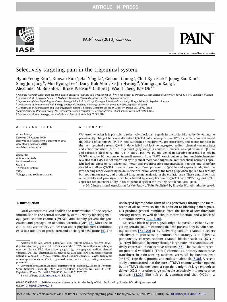
Fig. 1. Effects of QX-314 and capsaicin on voltage-gated sodium currents (INa) in capsaextracellular application of capsaicin alone (1 lM, 1 min) (A), co-application of QX-31application of QX-314 and capsaicin (1 min) (C). Upper panel: Long time-base recordingstest potential of 0 mV delivered every 10 s. Each vertical deflection indicates INa measuredindicated by the horizontal bar. Lower panel: fast time-base recordings of superimposedCollected results for the effects of QX-314, capsaicin, or co-application of both on INa in ccells tested. Results are means ± S.E.M. *P < 0.05 compared with the control (INa peak be
Please cite this article in press as: Kim HY et al. Selectively targeting pain in t
microscope. Images were acquired with a CCD camera (Fluoview,SIS, Germany) attached to the microscope, and contrast and bright-ness were adjusted with Photoshop (CS2, Adobe Systems Inc., USA).
2.6. dEMG recording of jaw-opening reflex
The rats, weighing between 250 and 300 g, were initially anes-thetized with ethyl ether, followed by urethane solution (120 mg/100 g body weight, intraperitoneal injection). Once the rats werestably anesthetized, a dental cavity was made on the left mandib-ular incisor by using a dental drill. For electrical stimulation, one ofthe stimulating wires (Belden, USA) was inserted into the cavitymade on the left mandibular incisor and sealed with acrylic dentalcement and the other reference wire was hypodermically locatednear the lip. Another pair of copper wires was placed in the leftanterior belly of the digastric muscle. Electrical stimulation,300 ls in duration, was generated with a stimulator (Pulse trainmodule 1831 connected to Pulse interval module 1830, World Pre-cision Instruments, USA) at 30 s intervals. Once the threshold wasdetermined, EMG amplitudes to the electrical nociceptive stimula-tion (two and a half times to the threshold, �2.5T) were recordedfrom the digastric muscle. The EMG leads were connected to anamplifier (DAM80, World Precision Instruments, USA), and thenmonitored and recorded with IGOR Pro (Ver. 4.0, WaveMetricsInc., USA). Drugs were applied onto either the exposed IAN or themylohyoid motor nerve. Capsaicin (0.5 lg/ll) was prepared with20% ethanol, 5% Tween 20 and 75% normal saline solution and
0
0.4
0.8
1.2
2
3
1
QX-314 Capsaicin
1 32
60 s
1 nA
∗∗ ∗
QX-314 + Capsaicin
Rel
ativ
e pe
ak I N
a
(12) (10)
(15)(15) (12)
icin-sensitive TG neurons from adult rats. Representative recordings following the4 (5 mM) and capsaicin (1 min) after pretreatment by QX-314 alone (B) and co-of INa measured during 30-ms voltage steps from a holding potential of �70 mV to aevery 10 s. Capsaicin, QX-314 or QX-314 plus capsaicin was applied during the timesodium current evoked by test pulse at the points indicated in the upper panel. (D)apsaicin-sensitive TG neurons. The numbers in parentheses indicate the number offore drug application).
he trigeminal system. PAIN�
(2010), doi:10.1016/j.pain.2010.02.016

4 H.Y. Kim et al. / PAIN�
xxx (2010) xxx–xxx
ARTICLE IN PRESS
QX-314, and lidocaine was directly dissolved in physiological sal-ine. The solutions were freshly made on the day of the experimentand solubilized in an ultrasonic vibrator (Model 3510, BransonUltrasonic Co., USA) for 30 min prior to application.
2.7. Behavioral studies
For behavioral observation, the rats (weighing between 250and 300 g) were placed in a customized cylinder-type acrylic ro-dent restrainer for evaluation of thermal pain nociceptive re-sponse as described previously [26]. The cage has a hole in thetop so that the head could receive thermal stimulation and pro-duce withdrawal action. Each cage was placed in a darkened andnoise-free room and the animals were habituated for at least30 min before experiment. Applications of heat stimuli were per-formed by infrared thermal stimulator (Infrared Diode Laser, LVI-808-10, Korea). Power and current of thermal stimulator wereadjusted at 11 W and 18.1 A, respectively. This intensity of ther-mal stimuli produced stable head flick or head withdrawal re-sponse with a distance of 10 cm from heat source to vibrissapad. Each rat received 2 stimuli and the interstimulus intervalfor each trial was at least 2 min. A cut-off time of 20 s was usedin the experiments to prevent possible tissue damage. Beforedrug injection, latencies of withdrawal responses were deter-mined from all animals. The vehicle and drugs were adminis-tered subcutaneously into the left vibrissa pad followed bycapsaicin injection (1 lg/10 ll, n = 11) 10 min later, latencies of
B
QX-314 + capsaicin
C D
Capsaicin1 2
A
60 s1.5 nA
1 2
- 70 mV0 mV
30 ms
1 2
1 nA60 s
1 2
Fig. 2. Effects of QX-314 and capsaicin on INa in capsaicin-insensitive rat TG neurons. R(1 lM) (A), co-application of QX-314 (5 mM) and capsaicin with the pretreatment of QX-3recordings of INa measured during 30-ms voltage steps from a holding potential of �70 mINa measured every 10 s. Capsaicin, QX-314 or QX-314 plus capsaicin was applied duringsuperimposed sodium current evoked by test pulse at the points indicated in the upperboth on INa in capsaicin-sensitive TG neurons. The numbers in parentheses indicate the nu(INa peak before drug application).
Please cite this article in press as: Kim HY et al. Selectively targeting pain in t
withdrawal responses were determined at 10, 30, 60, 120, 180,240, 300, 360 and 420 min after injection.
The experimenter was blind to the treatment group in allbehavioral experiments. For behavioral experiments, capsaicinwas dissolved in 10% ethanol, 10% Tween 80 and 80% normal salinesolution. QX-314 was dissolved in normal saline.
2.8. Chemicals
For in vitro study, capsaicin stock solutions were made in etha-nol and stored at �20 �C. QX-314 was dissolved in distilled waterand stored at �20 �C. All drugs were purchased from Sigma–Al-drich. The drugs were diluted to their final concentration usingthe external solution, and then were applied to cells by local perfu-sion through a capillary tube positioned near the cell of interest.The drug solution flow was driven by gravity (flow rate, 1–2 ml/min) and controlled by miniature solenoid valves.
2.9. Statistics
All data are presented as means ± standard error (SEM). Forin vitro studies, an unpaired Student’s t-test was used to determinethe differences (SigmaPlot, SPSS, USA). Statistical analyses of ther-mal hyperalgesia were carried out using a repeated measures AN-OVA followed by multiple group comparisons using the LSD posthoc test. Differences were considered to be significant when P va-lue was less than 0.05 or 0.001.
QX-314 Capsaicin
1 2 3
31 2
60 s3 nA
0
0.4
0.8
1.2
Rel
ativ
e pe
ak IN
a (11) (20) (11)
epresentative recordings following the extracellular application of capsaicin alone14 (B) and co-application of QX-314 and capsaicin (C). Upper panel: Long time-baseV to a test potential of 0 mV delivered every 10 s. Each vertical deflection indicatesthe time indicated by the horizontal bar. Lower panel: fast time-base recordings of
panel. (D) Collected results for the effects of QX-314, capsaicin, or co-application ofmber of cells tested. Results are means ± S.E.M. *P < 0.05 compared with the control
he trigeminal system. PAIN�
(2010), doi:10.1016/j.pain.2010.02.016

H.Y. Kim et al. / PAIN�
xxx (2010) xxx–xxx 5
ARTICLE IN PRESS
3. Results
3.1. Co-application of QX-314 and capsaicin blocks INa only in TGnociceptor neurons
We first tested whether QX-314 applied either alone or togetherwith capsaicin can inhibit voltage-gated sodium current (INa) elic-ited by a depolarizing step pulse from a holding potential of�70 mV in dissociated TG neurons, a preparation which includesboth capsaicin-sensitive and capsaicin-insensitive neurons. Toavoid the contamination of capsaicin-induced inward currents dur-ing the measurement of INa, we measured INa after the holding cur-rent at �70 mV returned to the control level following capsaicinexposure (time points 2 and 3 in Fig. 1A and B). Through this ap-proach, we could also test whether the block persists after washout(5 min) of capsaicin and QX-314, which is expected if the block re-flects TRPV1-mediated entry of QX-314 inside the cells. Capsaicinalone (1 lM, 1 min) produced little effect on INa after washout(0.91 ± 0.08, P = 0.6, n = 10) (Fig. 1A); as expected, it evoked inwardcurrents in most small neurons (<25 lm in diameter). However,when QX-314 (5 mM) was applied followed by capsaicin (1 lM,1 min) or applied together with capsaicin (1 lM, 1 min), INa wasdramatically inhibited (0.32 ± 0.07, P < 0.05, n = 15) (Fig. 1B andC) and the inhibitory effects became larger as the application timeincreased (Fig. 1D). We observed that the blockade of INa was main-tained throughout our recordings up to at least 45 min of washout.QX-314 (5 mM, 5 min) applied alone had marginal effects on INa
D
QX-314A
-60
0
60
30
-30Vm (m
V)
B
QX-314+ CapsaicinC
60 s
-60
0
60
30
-30Vm (m
V)
1 2
1 2
1 2
1 2
Fig. 3. Effects of QX-314 and capsaicin on action potentials in capsaicin-sensitive TG neurQX-314 (5 mM, 5 min) (A), capsaicin (1 lM, 5 min) (B) or co-application of capsaicin and Q3-ms depolarizing current pulses with 250 pA amplitude. Note that membrane depolarcapsaicin-sensitive TG neurons. Lower panel: APs recorded at the time points indicated atcapsaicin-sensitive TG neurons. (D) Collected results for the effects of QX-314, capsaicinnumbers in parentheses indicate the number of cells tested. Results are means ± S.E.M.
Please cite this article in press as: Kim HY et al. Selectively targeting pain in t
(0.93 ± 0.07, P = 0.1, n = 12), consistent with the previous data ob-tained from DRG neurons [3] (Fig. 1B and D).
In capsaicin-insensitive TG neurons (>40 lm in diameter), cap-saicin neither elicited inward currents (Fig. 2) nor affected INa
(Fig. 2A) (0.96 ± 0.03, P = 4.2, n = 20). QX-314, either alone or ap-plied together with capsaicin, also had little effect on INa
(0.94 ± 0.02, P = 0.5, n = 11 and 0.93 ± 0.05, P = 0.2, n = 11, respec-tively) (Fig. 2B–D). Our data suggest that QX-314, entering throughTRPV1 channels, blocks INa only in capsaicin-sensitive nociceptiveTG neurons and only when co-applied with capsaicin.
3.2. Co-application of QX-314 and capsaicin selectively blocks actionpotentials in TG nociceptor neurons
We next examined the effects of QX-314 on action potentials(APs) in TG neurons. As previously reported [26], capsaicin-sensi-tive neurons had a prominent shoulder in the falling phase of theaction potential and in these cells capsaicin elicited a membranedepolarization and then robust AP firing (Fig. 3). Because APs areblocked by capsaicin due to the inactivation of VGSCs followingthe membrane depolarization elicited by capsaicin (data notshown), we examined APs evoked by current injection (250 pA,3 ms) 5 min after washout of capsaicin, after the membrane poten-tial returned to baseline (time point 2 in Fig. 3). By this approach,we could again test whether the effect of QX-314 inside the cellsremained even after the washout of capsaicin and QX-314. Capsa-icin-sensitive TG neurons were small sized (<25 lm in diameter)
0
50
100
AP a
mpl
itude
(mV) (11) (21)
(14)
Capsaicin
-60
0
60
30
-30Vm (m
V)
1 2
1 2
∗
ons. Representative current-clamp recordings over time following the application ofX-314 (5 min) (C). Upper panel: action potentials (APs) were elicited by injection of
ization and transient action potential discharges were evoked by capsaicin in theeach upper panel. Arrow indicates the typical hump in the falling phase of AP in the, or co-application of both on AP amplitude in capsaicin-sensitive TG neurons. The*P < 0.05 compared with the control (AP amplitude before drug application).
he trigeminal system. PAIN�
(2010), doi:10.1016/j.pain.2010.02.016

6 H.Y. Kim et al. / PAIN�
xxx (2010) xxx–xxx
ARTICLE IN PRESS
with an average resting membrane potential (Vrest) of�53.1 ± 1.1 mV (n = 46). QX-314 (5 mM, 5 min) alone had no effecton either Vrest or APs (99.6 ± 2.1%, P = 0.9, n = 11) (Fig. 3A). Capsai-cin (1 lM, 5 min) alone decreased AP amplitude during membranedepolarization (data not shown) but APs at 5 min after washoutwere nearly identical to control (96.3 ± 4.5%, P = 0.4, n = 21;Fig. 3B). However, co-application (5 min) of QX-314 (5 mM) andcapsaicin (1 lM) completely abolished the generation of action po-tential at 5 min after washout (1 ± 0.1%, P < 0.05, n = 14) (Fig. 3C).As in the INa experiments, we again observed that the blockade ofAP firing lasted for 45 min of washout. The duration of the effectafter washout suggests that the QX-314 remains in nociceptor neu-rons after its entry via TRPV1 channels.
In capsaicin-insensitive TG neurons (>40 lm in diameter, withVrest of �53.5 ± 3.4 mV, n = 29), there was no hump in the fallingphase of the APs [26] (see Supplementary Fig. 1) and applicationof either QX-314 or capsaicin alone had no effect on the generationof APs (100.5 ± 1.1%, P = 0.9, n = 8 and 98.8 ± 0.7%, P = 0.8, n = 10,respectively) (Supplementary Fig. 1A and B). Co-application of cap-saicin and QX-314 together also had no effect (96.7 ± 3.4%, P = 0.3,n = 11) (Supplementary Fig. 1C). Our data show that the blockingeffect of APs by QX-314 and capsaicin, like that on sodium currents,is restricted to capsaicin-sensitive neurons.
We further tested whether TRPV1 is required for the inhibitoryeffects of QX-314 on the INa and APs by studying small-sized noci-ceptive TG neurons (<25 lm in diameter, n = 34) with a hump in
B
A
QX-314 + Capsaicin 1 2
2 n60 s
a
21
- 70 mV0 mV
30 ms
QX-314+ Capsaicin
a
-60
0
60
30
-30Vm (m
V)
1 2
1 2
60 s
Fig. 4. Effect of co-application of capsaicin and QX-314 on INa and APs in TRPV1 knock-oco-application of QX-314 (5 mM) and capsaicin (1 lM, 5 min) in the TRPV1 knock-out mindicated in upper panel. (b) Summary of the effects of QX-314, capsaicin, or co-applicatrepresentative recording following the application or co-application of QX-314 (5 mM) anthe upper panel. (b) Collected results for the effects of QX-314, capsaicin, or co-applicati(<25 lm in diameter) with a resting membrane potential (Vres) of �54.2 ± 4.6 mV. The nu
Please cite this article in press as: Kim HY et al. Selectively targeting pain in t
the falling phase of the APs in TRPV1 knock-out mice. As shownin Fig. 4, QX-314 applied together with capsaicin consistentlyfailed to affect both INa (0.93 ± 0.05, P = 0.2, n = 14) (Fig. 4A) andAPs (93.3 ± 5.8%, P = 0.3, n = 12) (Fig. 4B) in TG neurons from thesemice. These results suggest that it is specifically the presence ofTRPV1 in nociceptors that is required for QX-314 entry and block.
3.3. Co-application of QX-314 and capsaicin blocks INa and APs indental nociceptors
Pain is the only sensation perceived by humans when noxiousstimulus is applied to the teeth [34]. Thus, retrograde labelingthe cells that innervates tooth pulp identifies a nearly pure popu-lation of nociceptors; dental primary afferent neurons [27]. Wetherefore examined next whether the co-application of QX-314and capsaicin can block INa and APs in nociceptive dental primaryafferent neurons. Consistent with our previous report [27], DiI-la-beled neurons represented �15% of the total cultured TG neurons,and were almost exclusively below 40 lm in diameter. The major-ity of dental primary afferent neurons responded to capsaicin(n = 37/45), indicating a prominent expression of TRPV1 in dentalnociceptive neurons. Co-application (1 min) of capsaicin (1 lM)and QX-314 (5 mM) blocked INa (0.41 ± 0.02, P < 0.05, n = 14) andAPs in the DiI-labeled TG neurons (0.6 ± 0.2%, P < 0.05, n = 14)(Fig. 5A and B). Expression of TRPV1 in dental primary afferentneurons was verified by immunoreactivity in DiI-labeled neurons
0
0.4
0.8
1.2
0
50
100
b
AP a
mpl
itude
(mV)
A
b
Rel
ativ
e pe
ak IN
a
(11) (14) (14)
(11) (12)(11)
ut mice. (A) (a) Upper panel: a representative recording following the application orice TG neurons. Lower panel: superimposed INa evoked by test pulse at the points
ion of both on INa in TG neurons from TRPV1 knock-out mice. (B) (a) Upper panel: Ad capsaicin (1 lM, 5 min). Lower panel: APs recorded at the time points indicated at
on of both on AP amplitude in TRPV1 knock-out mice. TG neurons were small sizedmbers in parentheses indicate the number of cells tested. Results are means ± S.E.M.
he trigeminal system. PAIN�
(2010), doi:10.1016/j.pain.2010.02.016

H.Y. Kim et al. / PAIN�
xxx (2010) xxx–xxx 7
ARTICLE IN PRESS
(Fig. 5Ac). We found that only a few dental primary afferent neu-rons were not responsive to capsaicin (n = 8/45) (data not shown),and these were the only neurons in which the combined applica-tion of QX-314 and capsaicin failed to block INa or APs.
3.4. Trigeminal mesencephalic neurons and motoneurons lackfunctional expression of TRPV1
In addition to transmitting pain the trigeminal nerve also con-veys proprioceptive sensation to the CNS and has motor axons.Proprioceptive neurons are located in the trigeminal mesence-phalic nucleus (Vmes) and motor neurons in the trigeminal motornucleus (Vmot) [16,21]. To verify the nociceptor-specific effects ofthe combination of QX-314 and capsaicin, we determined whetherTRPV1 is expressed in the trigeminal mesencephalic and motorneurons using slice whole cell recordings and immunohistochem-istry. We identified trigeminal mesencephalic neurons by observ-ing large Ih currents (Fig. 6Aa) and typical delayed firing of actionpotential (Fig. 6Ab) [16]. In these Vmes neurons, the applicationof capsaicin (2 lM) did not induce inward currents (n = 12)(Fig. 6Ac) and we also could not detect TRPV1-immunoreactivity(Fig. 6Ad). Capsaicin (2 lM) also failed to evoke inward currents(n = 7) (Fig. 6Ba) and TRPV1-immunoreactivity was absent in Vmotneurons identified by retrograde labeling following DiI injection
30 s
Ab
aB
QX-314+ Capsaicin
QX-314 + Capsaicin 1 2
60 s3 nA
a
Rel
ativ
e pe
ak IN
a
-60
0
60
30
-30Vm (m
V)
2
1
- 70 mV0 mV
30 ms 0
0.4
0.8
1.2(1
1
1
Fig. 5. Effect of co-application of capsaicin and QX-314 on INa and APs in nociceptivefollowing the application or co-application of QX-314 (5 mM) and capsaicin (1 lM, 1 minpulse at the points indicated at the upper panel. (b) Collected results for the effects of Qprimary afferent TG neurons. (c) Dental primary afferent neurons were identified byphotograph of TG neurons shows TRPV1-immunoreactivity (green), DiI (red), DAPI (blufollowing the application or co-application of QX-314 (5 mM) and capsaicin (1 lM, 5 mSummary of the effects of QX-314, capsaicin, or co-application of both on AP amplitude inin diameter) with the resting membrane potential (Vres) of �55.6 ± 3.2 mV. The numb*P < 0.05 compared with the control (the AP amplitude before drug application).
Please cite this article in press as: Kim HY et al. Selectively targeting pain in t
into the masseter muscle (Fig. 6Bb). These results suggest thatco-application of QX-314 and capsaicin would not affect proprio-ceptive sensation or motor function, and is therefore truly painselective.
3.5. Co-application of QX-314 and capsaicin inhibits the jaw-openingreflex when applied to sensory but not motor nerves
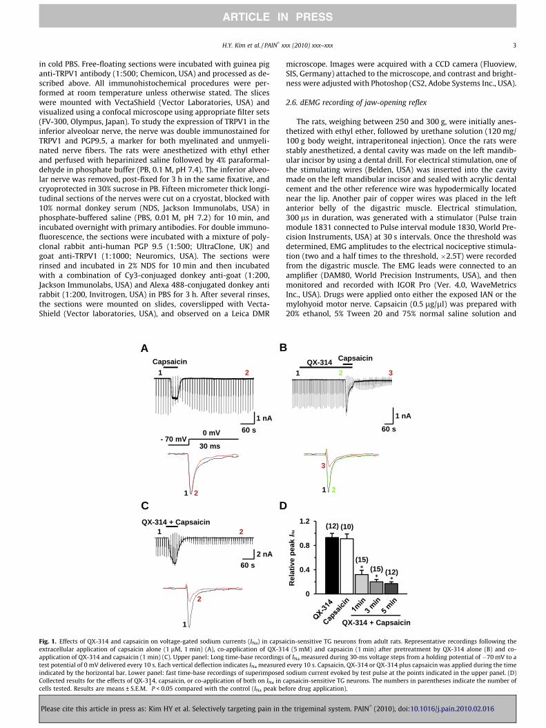
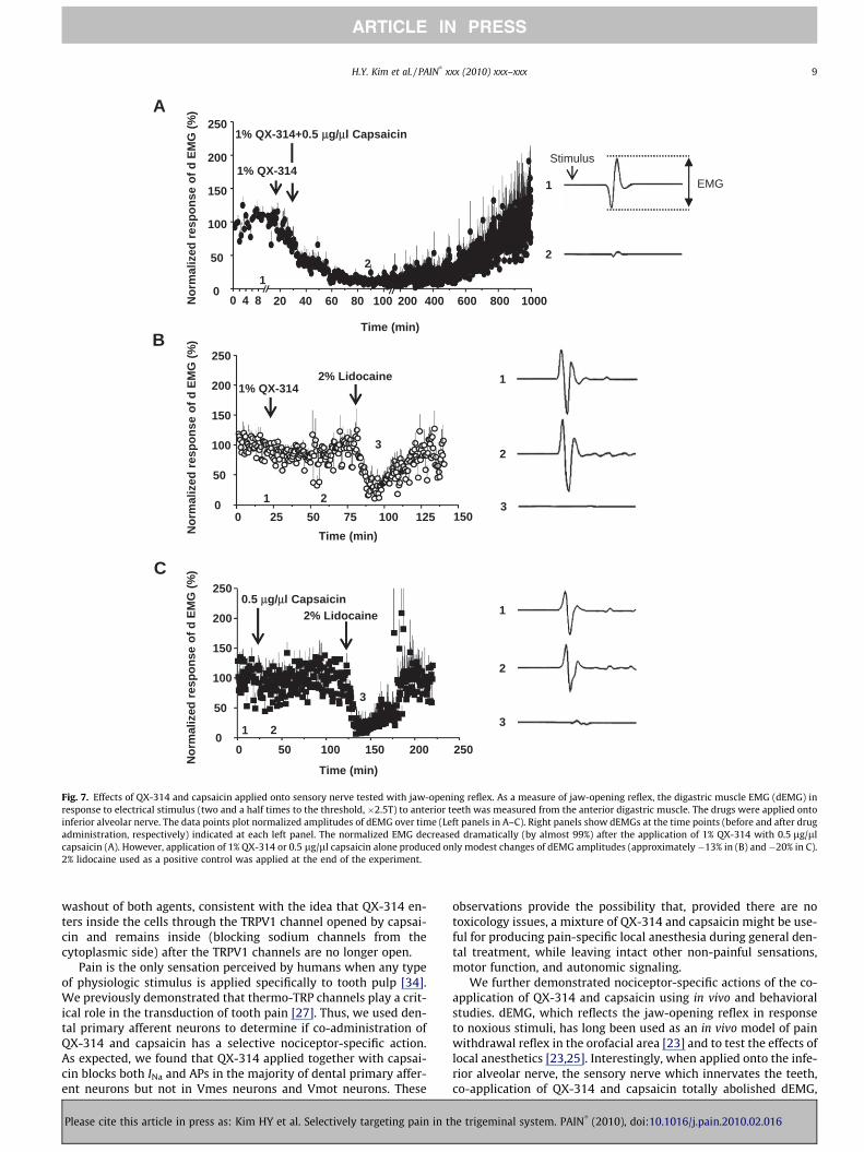
We next examined whether co-application of QX-314 and cap-saicin specifically blocks conduction of pain-related signals in vivoby measuring the reflex activation of the digastric muscle in re-sponse to electrical stimulation of teeth as part of the nociceptivejaw-opening reflex. Co-application (50 ll) of QX-314 (1%) and cap-saicin (0.5 lg/ll) onto the inferior alveolar nerve (the sensorynerve that innervates teeth and is the afferent limb of the reflex)totally blocked the electromyogram of digastric muscle (dEMG)with a slow onset and gradual recovery to base line over severalhours (n = 6) (Fig. 7A). Application of either QX-314 (1%) (n = 4)(Fig. 7B) or capsaicin (0.5 lg/ll) alone (n = 4) (Fig. 7C) producedno effect. In contrast, lidocaine alone (2%, 100 ll) completelyblocked the dEMG within a few minutes, with a block lasting aboutan hour (Fig. 7B and C). The inhibition of dEMG produced by co-administration of QX-314 and capsaicin was much longer than thatof lidocaine alone (12 h versus 1 h).
0
50
100
TRPV1
DAPI Merge
DiIc
AP a
mpl
itude
(mV)
b (12) (11)
∗
∗
QX-314 + Capsaicin
2) (15)
(14)
(11)
2
2
(14)∗
dental primary afferent neurons. (A) (a) Upper panel: a representative recordings) in dental primary afferent neurons. Lower panel: superimposed INa evoked by testX-314, capsaicin, or co-application of both compounds on INa in nociceptive dentalretrograde labeling with a fluorescent dye, DiI, placed into the molar teeth. Thee) and merged. Scale bar = 50 lm. (B) (a) Upper panel: a representative recordingin). Lower panel: APs recorded at the time points indicated at the upper panel. (b)nociceptive dental primary afferent neurons. TG neurons were small sized (<25 lm
ers in parentheses indicate the number of cells tested. Results are means ± S.E.M.
he trigeminal system. PAIN�
(2010), doi:10.1016/j.pain.2010.02.016

Fig. 6. Both trigeminal mesencephalic (proprioceptive) neurons and motoneurons lack functional expression of TRPV1. (A) Trigeminal mesencephalic neurons were identifiedby their typical Ih current (a) and AP shape (b). Capsaicin failed to activate inward current at Vh, �60 mV (c). Immuno-staining did not detect TRPV1; Mesencephalictrigeminal nucleus visualized under FITC filter and overlay with DIC (d). (B) No currents were evoked by capsaicin (applied at �60 mV) in trigeminal motoneurons (a).Trigeminal motoneurons were identified by retrograde labeling with DiI into the exposed masseter muscle. A representative photograph shows immunoreactivity of TRPV1(FITC filter, green) and DiI (DiI filter, red) image in motor trigeminal nucleus (b). Scale bar = 200 lm (A); 100 lm (B).
8 H.Y. Kim et al. / PAIN�
xxx (2010) xxx–xxx
ARTICLE IN PRESS
We then examined whether co-application of QX-314 and cap-saicin produces a reduction in the reflex activity in the digastricmuscle upon application to the mylohyoid nerve, which innervatesthe anterior belly of the digastric muscle and is the efferent limb ofthe nociceptive jaw-opening reflex. Application of QX-314 and cap-saicin (50 ll), either separately or together, failed to inhibit the re-flex (n = 6, 5 and 5, respectively) (Fig. 8A–C). In contrast, lidocaine(10%, 20 ll) administration totally blocked the activity for about anhour in all the rats tested (n = 4) (Fig. 8B and C), as expected for thenon-selective blocking action on all types of nerve fibers.
3.6. Co-application of QX-314 and capsaicin inhibits thermalhyperalgesia in the orofacial area
We next investigated whether co-application of QX-314 andcapsaicin can reduce orofacial pain. After QX-314, either alone ortogether with capsaicin, was administered subcutaneously intothe left vibrissa pad, we measured latencies of withdrawal re-sponses following thermal stimulation. When QX-314 (1%) was in-jected together with capsaicin (1 lg), thermal latency significantlyincreased at 60 min after injection and this anesthetic effect wasmaintained throughout the full 7 h observation time(F(1, 20) = 33.657, P < 0.001), compared with the absence of anyanalgesia with QX-314 (1%) or capsaicin (1 lg) alone (Fig. 9).
4. Discussion
Our results suggest that pain-selective local anesthesia can beproduced in the orofacial system by the strategy of selectivelydelivering charged sodium channel blockers into pain-sensing neu-rons without blocking other types of neurons. The trigeminalnerve, the largest of the cranial nerves, is primarily a sensory nerve
Please cite this article in press as: Kim HY et al. Selectively targeting pain in t
in the face and head, but it also has certain motor functions such asmastication and swallowing [31]. The sensory function of the tri-geminal nerve is to provide the tactile, proprioceptive and painsensation of the face and mouth. The trigeminal ganglion (TG),analogous to the dorsal root ganglia (DRG) of the spinal cord, con-tains the cell bodies of incoming sensory nerve fibers, except forproprioceptor fibers, which have their cell bodies in the trigeminalmesencephalic nucleus (Vmes). Motor branches of the trigeminalnerve are distributed in the mandibular nerve that originates inthe motor nucleus of the trigeminal nerve (Vmot) [14,31]. Wefound that co-administration of QX-314 and capsaicin blocks so-dium channels and thereby inhibits excitability only in TRPV1-expressing nociceptive TG neurons and not in Vmes and Vmot neu-rons, which we also find do not express TRPV1. Consistent with thespecificity for action to TRPV1-expressing nociceptive neurons, thejaw-opening reflex, a withdrawal reflex in the orofacial area, wasinhibited by the co-application of QX-314 and capsaicin only ontothe peripheral sensory nerve, but not onto the peripheral motornerve. Moreover, from behavioral studies, we found that co-appli-cation of QX-314 and capsaicin produced long-lasting anti-noci-ceptive effects.
Inhibitory effects on INa and APs produced by co-application ofQX-314 and capsaicin were restricted to capsaicin-sensitive TGneurons, and neither INa nor APs were blocked by co-applicationof QX-314 and capsaicin to neurons from TRPV1 knock-out mice.Under in vitro conditions, QX-314 co-applied with capsaicin pro-duced the block of INa or APs in TRPV1-expressing trigeminal gan-glion nociceptors within a minute, similar to DRG neurons [11].Previous experiments in DRG neurons showing block of INa andAPs by co-applied QX-314 and capsaicin [3] did not examine thetime-course of the effect after washout; we now find that the ef-fects of QX-314 and capsaicin persist for at least 45minutes after
he trigeminal system. PAIN�
(2010), doi:10.1016/j.pain.2010.02.016
1
2
1% QX-314+0.5 μμg/μl Capsaicin
Nor
mal
ized
resp
onse
of d
EM
G (%
)
0
50
100
150
200
250
0 4 8 20 40 60 80 100 200 400 600 800 1000
1
1% QX-314
2
Stimulus
EMG
A
1% QX-3142% Lidocaine
0
50
100
150
200
250
0 25 50 75 100 125 1501 2
3
3
2
1
B
2% Lidocaine0.5 μg/μl Capsaicin
0
350
100
150
200
250
0 50 100 150 200 250
Time (min)
1 2
1
2
3
C
Nor
mal
ized
resp
onse
of d
EM
G (%
)N
orm
aliz
ed re
spon
se o
f d E
MG
(%)
Time (min)
Time (min)
Fig. 7. Effects of QX-314 and capsaicin applied onto sensory nerve tested with jaw-opening reflex. As a measure of jaw-opening reflex, the digastric muscle EMG (dEMG) inresponse to electrical stimulus (two and a half times to the threshold, �2.5T) to anterior teeth was measured from the anterior digastric muscle. The drugs were applied ontoinferior alveolar nerve. The data points plot normalized amplitudes of dEMG over time (Left panels in A–C). Right panels show dEMGs at the time points (before and after drugadministration, respectively) indicated at each left panel. The normalized EMG decreased dramatically (by almost 99%) after the application of 1% QX-314 with 0.5 lg/llcapsaicin (A). However, application of 1% QX-314 or 0.5 lg/ll capsaicin alone produced only modest changes of dEMG amplitudes (approximately �13% in (B) and�20% in C).2% lidocaine used as a positive control was applied at the end of the experiment.
H.Y. Kim et al. / PAIN�
xxx (2010) xxx–xxx 9
ARTICLE IN PRESS
washout of both agents, consistent with the idea that QX-314 en-ters inside the cells through the TRPV1 channel opened by capsai-cin and remains inside (blocking sodium channels from thecytoplasmic side) after the TRPV1 channels are no longer open.
Pain is the only sensation perceived by humans when any typeof physiologic stimulus is applied specifically to tooth pulp [34].We previously demonstrated that thermo-TRP channels play a crit-ical role in the transduction of tooth pain [27]. Thus, we used den-tal primary afferent neurons to determine if co-administration ofQX-314 and capsaicin has a selective nociceptor-specific action.As expected, we found that QX-314 applied together with capsai-cin blocks both INa and APs in the majority of dental primary affer-ent neurons but not in Vmes neurons and Vmot neurons. These
Please cite this article in press as: Kim HY et al. Selectively targeting pain in t
observations provide the possibility that, provided there are notoxicology issues, a mixture of QX-314 and capsaicin might be use-ful for producing pain-specific local anesthesia during general den-tal treatment, while leaving intact other non-painful sensations,motor function, and autonomic signaling.
We further demonstrated nociceptor-specific actions of the co-application of QX-314 and capsaicin using in vivo and behavioralstudies. dEMG, which reflects the jaw-opening reflex in responseto noxious stimuli, has long been used as an in vivo model of painwithdrawal reflex in the orofacial area [23] and to test the effects oflocal anesthetics [23,25]. Interestingly, when applied onto the infe-rior alveolar nerve, the sensory nerve which innervates the teeth,co-application of QX-314 and capsaicin totally abolished dEMG,
he trigeminal system. PAIN�
(2010), doi:10.1016/j.pain.2010.02.016

0
50
100
150
200
0 20 40 60 80
1% QX-314
1% QX-314+ 0.5 μμg/μl capsaicin
1 2
0
50
100
150
200
0 20 40 60 80
0.5 μg/μl Capsaicin
1 2
1
2
C
0
50
100
150
200
0 20 40 60 80 100
1 2
10% lidocaine
3
1
2
3
Time (min)
D
Nor
mal
ized
resp
onse
of d
EM
G (%
)
2
1
Stimulus
EMG
A
0
50
100
150
200
0 20 40 60 80
1% QX-314
1 2
1
2
B Time (min)
Time (min)
Time (min)
Nor
mal
ized
resp
onse
of d
EM
G (%
)N
orm
aliz
ed re
spon
se o
f d E
MG
(%)
Nor
mal
ized
resp
onse
of d
EM
G (%
)
Fig. 8. Effects of QX-314 and capsaicin applied onto motor nerve tested with jaw-opening reflex. QX-314, capsaicin or both were applied onto mylohyoid nerve, the motornerve innervating digastric muscle, while measuring dEMGs in response to electrical stimulus (�2.5T). Right panels show the representative dEMGs at the time pointsindicated in the left panels. The normalized EMG had no significant change in amplitude after 1% QX-314 with 0.5 lg/ll capsaicin (A), application of 1% QX-314 (B) and0.5 lg/ll capsaicin (C). As a positive control, the dEMG amplitude decreased dramatically upon the application of 10% lidocaine (20 ll) onto mylohyoid nerve (D).
10 H.Y. Kim et al. / PAIN�
xxx (2010) xxx–xxx
ARTICLE IN PRESS
an effect that lasted several hours. This suggests that TRPV1 chan-nels are present on the axons of the sensory nerve fibers in theinferior alveolar nerve at sufficient density to allow QX-314 entry.
Please cite this article in press as: Kim HY et al. Selectively targeting pain in t
TRPV1 is expressed on peripheral somatosensory nerve fibers (e.g.in sciatic nerve) [2], and we also now find that TRPV1 is expressedon the unmyelinated axons of inferior alveolar nerve (see Supple-
he trigeminal system. PAIN�
(2010), doi:10.1016/j.pain.2010.02.016

Time (min)0 120 240 360
Ther
mal
late
ncy
(sec
)
0
10
15
20 ** * * *
*
*
Capsaicin (1 μg)QX-314 (1%) + Capsaicin (1 μg)
QX-314 (1%)
Fig. 9. Effects of subcutaneously applied QX-314 and capsaicin on pain behaviors inorofacial area. QX-314 alone, capsaicin alone, or together with QX-314 wasadministered subcutaneously into the left vibrissa pad and latencies of headwithdrawal responses to thermal stimulation were determined at 10, 30, 60, 120,180, 240, 300, 360 and 420 min. The number of animals was 11. *P < 0.001 versusQX-314.
H.Y. Kim et al. / PAIN�
xxx (2010) xxx–xxx 11
ARTICLE IN PRESS
mentary Fig. 2). When applied onto the mylohyoid nerve, the mo-tor nerve that innervates the anterior belly of digastric muscle, co-application of QX-314 and capsaicin failed to block dEMG, presum-ably reflecting the lack of TRPV1 channels in the motor axons. Con-duction of C-fiber and, to a lesser extent, Ad-fiber sensorycomponents of the compound action potential is decreased by per-ineural application of a high dose of capsaicin (1%) [1,10,28]. Wefound in the present study that dEMG was not significantly de-creased by a much lower concentration of capsaicin (0.05%), whenapplied by itself.
The blocking effect of co-application of QX-314 and capsaicinapplied to the inferior alveolar nerve in dEMG study was muchlonger than lidocaine. Similarly, co-application of QX-314 and cap-saicin produced much longer anti-nociceptive effects in responseto thermal stimulation, compared to lidocaine. Thus, the applica-tion of QX-314 together with capsaicin may be clinically usefulas long-lasting, nociceptive-specific local anesthesia. It seemslikely that charged QX-314 remains in the axon longer than lido-caine, which likely readily crosses the membrane in its uncharged(unprotonated) form. Eventually, trapped intracellular QX-314might be slowly exocytosed from axons or directly metabolisedwithin neurons. In hepatocytes, most lidocaine is N-deethylatedvia cytochrome P4503A4 (CYP3A4) enzymes [37]. Hence, a meta-bolic pathway from the quaternary amid QX-314 to lidocaine itselfmediated by the same enzyme seems to be plausible. However,nothing is known about CYP-enzymes in sensory neurons or QX-314 metabolism in general.
Orofacial pain-selective local anesthesia could be very useful intreating dental pain, trigeminal neuralgia, pain from temporoman-dibular joint disorder, and other conditions. Capsaicin is probablynot an ideal TRPV1 agonist for clinical use, because it activatestransient action potential firing in nociceptors. TRPV1 agonistswith less pungent or burning characteristics may be better. Re-cently, we demonstrated that eugenol, which evokes robustTRPV1-mediated inward currents that are blocked by capsazepine,produces a slow depolarization that can fail to trigger APs, proba-bly because of sodium channel inactivation [26]. This result sug-gests that the initial irritable action of eugenol might benegligible compared to capsaicin, but it may still allow QX-314to enter nociceptive neurons to block excitability. As recently dem-onstrated, lidocaine is also a TRPV1 agonist, and can be used there-
Please cite this article in press as: Kim HY et al. Selectively targeting pain in t
fore in the place of capsaicin to allow the entry of QX-314 throughTRPV1 channels [4,17]. QX-314 itself is likely to have little toxicitywith local administration; although QX-314 can inhibit cardiac so-dium channels acting from the outside of the membrane [29], thiseffect should be no greater (and probably less) than that producedby systemic availability of lidocaine after local administration.
TRPV1 is not expressed by all nociceptors. Thus, selective entryof QX-314 into TRPV1-expressing nociceptors may not necessarilyguarantee total block of pain sensation in surgery or with neuro-pathic pain conditions. Other channels in addition to TRPV1 mayhave large enough pores to allow QX-314 entry; recently, TRPA1,but not TRPM8, was demonstrated to undergo pore dilatationwhen activated by agonists [9,11]. As TRPA1 is also expressed bynociceptors [27], this channel could be another target for QX-314application. Also, P2X2, receptors expressed by a subpopulationof sensory neurons as heteromers with P2X3 receptors [19] and di-lated by the application of appropriate agonists [35], might be an-other possibility.
Although several issues remain to be addressed before clinicaluse of this strategy, the delivery of permanently charged localanesthetic compounds into nociceptors by combining them withan agonist for large-pore ion channels specifically expressed onthese cells has provided a new possibility for producing pain-selec-tive local anesthesia. Orofacial pain may be an especially attractivetarget for this strategy since our results show a very high degree ofselectivity for the block of TRPV1-expressing nociceptive TG neu-rons, with no effects on trigeminal proprioceptive sensory neuronsand trigeminal motor neurons. In addition to selectivity for painsignals, the much longer lasting (>7 h) block produced by QX-314when co-applied with TRPV1 agonists compared with lidocaine(�1 h) would be a major advantage in many clinical situations.
Acknowledgements
This research was supported by Grant (R0A-2008-000-20101-0)from National Research Laboratory Program and a Grant(2009K001256) from the Brain Research Center of the 21st CenturyFrontier Research Program funded by the Ministry of Education,Science and Technology, Republic of Korea. S.B.O was supportedby Alice and Joseph Brooks Fund in the Department of Neurobiol-ogy, Harvard Medical School and LG Yonam Foundation during partof this work. Drs. Bean and Woolf are inventors on a filed patentrelated to targeting impermeant sodium channel blockers intonociceptors that has been licensed by Endo Pharmaceuticals fromHarvard University.
Appendix A. Supplementary data
Supplementary data associated with this article can be found, inthe online version, at doi:10.1016/j.pain.2010.02.016.
References
[1] Baranowski R, Lynn B, Pini A. The effects of locally applied capsaicin onconduction in cutaneous nerves in four mammalian species. Br J Pharmacol1986;89:267–76.
[2] Bernardini N, Neuhuber W, Reeh PW, Sauer SK. Morphological evidence forfunctional capsaicin receptor expression and calcitonin gene-related peptideexocytosis in isolated peripheral nerve axons of the mouse. Neuroscience2004;126:585–90.
[3] Binshtok AM, Bean BP, Woolf CJ. Inhibition of nociceptors by TRPV1-mediatedentry of impermeant sodium channel blockers. Nature 2007;449:607–10.
[4] Binshtok AM, Gerner P, Oh SB, Puopolo M, Suzuki S, Roberson DP, Herbert T,Wang CF, Kim D, Chung G, Mitani AA, Wang GK, Bean BP, Woolf CJ.Coapplication of lidocaine and the permanently charged sodium channelblocker QX-314 produces a long-lasting nociceptive blockade in rodents.Anesthesiology 2009;111:127–37.
[5] Butterworth J, Oxford GS. Local anesthetics: a new hydrophilic pathway for thedrug-receptor reaction. Anesthesiology 2009;111:12–4.
he trigeminal system. PAIN�
(2010), doi:10.1016/j.pain.2010.02.016

12 H.Y. Kim et al. / PAIN�
xxx (2010) xxx–xxx
ARTICLE IN PRESS
[6] Butterworth JFt, Strichartz GR. Molecular mechanisms of local anesthesia: areview. Anesthesiology 1990;72:711–34.
[7] Caterina MJ, Julius D. Sense and specificity: a molecular identity fornociceptors. Curr Opin Neurobiol 1999;9:525–30.
[8] Caterina MJ, Schumacher MA, Tominaga M, Rosen TA, Levine JD, Julius D. Thecapsaicin receptor: a heat-activated ion channel in the pain pathway. Nature1997;389:816–24.
[9] Chen J, Kim D, Bianchi BR, Cavanaugh EJ, Faltynek CR, Kym PR, Reilly RM. Poredilation occurs in TRPA1 but not in TRPM8 channels. Mol Pain 2009;5:3.
[10] Chung JM, Lee KH, Hori Y, Willis WD. Effects of capsaicin applied to aperipheral nerve on the responses of primate spinothalamic tract cells. BrainRes 1985;329:27–38.
[11] Chung MK, Guler AD, Caterina MJ. TRPV1 shows dynamic ionic selectivityduring agonist stimulation. Nat Neurosci 2008;11:555–64.
[12] Cummins TR, Sheets PL, Waxman SG. The roles of sodium channels innociception: implications for mechanisms of pain. Pain 2007;131:243–57.
[13] Fang Z, Park CK, Li HY, Kim HY, Park SH, Jung SJ, Kim JS, Monteil A, Oh SB, MillerRJ. Molecular basis of Ca(v)2.3 calcium channels in rat nociceptive neurons. JBiol Chem 2007;282:4757–64.
[14] Fried K, Bongenhielm U, Boissonade FM, Robinson PP. Nerve injury-inducedpain in the trigeminal system. Neuroscientist 2001;7:155–65.
[15] Gold MS, Reichling DB, Hampl KF, Drasner K, Levine JD. Lidocaine toxicity inprimary afferent neurons from the rat. J Pharmacol Exp Ther 1998;285:413–21.
[16] Kang Y, Saito M, Sato H, Toyoda H, Maeda Y, Hirai T, Bae YC. Involvement ofpersistent Na+ current in spike initiation in primary sensory neurons of the ratmesencephalic trigeminal nucleus. J Neurophysiol 2007;97:2385–93.
[17] Leffler A, Fischer MJ, Rehner D, Kienel S, Kistner K, Sauer SK, Gavva NR, ReehPW, Nau C. The vanilloid receptor TRPV1 is activated and sensitized by localanesthetics in rodent sensory neurons. J Clin Invest 2008;118:763–76.
[18] Leston JM. Functional anatomy of the trigeminal nerve. Neurochirurgie2009;55:99–112.
[19] Lewis C, Neidhart S, Holy C, North RA, Buell G, Surprenant A. Coexpression ofP2X2 and P2X3 receptor subunits can account for ATP-gated currents insensory neurons. Nature 1995;377:432–5.
[20] McCleskey EW, Gold MS. Ion channels of nociception. Annu Rev Physiol1999;61:835–56.
[21] McDavid S, Verdier D, Lund JP, Kolta A. Electrical properties of interneuronsfound within the trigeminal motor nucleus. Eur J Neurosci 2008;28:1136–45.
[22] Meyers JR, MacDonald RB, Duggan A, Lenzi D, Standaert DG, Corwin JT, CoreyDP. Lighting up the senses: FM1-43 loading of sensory cells throughnonselective ion channels. J Neurosci 2003;23:4054–65.
Please cite this article in press as: Kim HY et al. Selectively targeting pain in t
[23] Miyoshi T, Aida H, Kaneko Y. Comparative study on anesthetic potency ofdental local anesthetics assessed by the jaw-opening reflex in rabbits. AnesthProg 2000;47:35–41.
[24] Oh SB, Piao ZG, Shin SS, Ren D, Park K, Kim JS. GABAergic and serotonergicmodulation of calcium currents in rat trigeminal motoneurons. BiochemBiophys Res Commun 2003;309:58–65.
[25] Ohkado S, Ichinohe T, Kaneko Y. Comparative study on anesthetic potencydepending on concentrations of lidocaine and epinephrine: assessment ofdental local anesthetics using the jaw-opening reflex. Anesth Prog2001;48:16–20.
[26] Park CK, Kim K, Jung SJ, Kim MJ, Ahn DK, Hong SD, Kim JS, Oh SB. Molecularmechanism for local anesthetic action of eugenol in the rat trigeminal system.Pain 2009;144:84–94.
[27] Park CK, Kim MS, Fang Z, Li HY, Jung SJ, Choi SY, Lee SJ, Park K, Kim JS, Oh SB.Functional expression of thermo-transient receptor potential channels indental primary afferent neurons: implication for tooth pain. J Biol Chem2006;281:17304–11.
[28] Petsche U, Fleischer E, Lembeck F, Handwerker HO. The effect of capsaicinapplication to a peripheral nerve on impulse conduction in functionallyidentified afferent nerve fibres. Brain Res 1983;265:233–40.
[29] Qu Y, Rogers J, Tanada T, Scheuer T, Catterall WA. Molecular determinants ofdrug access to the receptor site for antiarrhythmic drugs in the cardiac Na+
channel. Proc Natl Acad Sci USA 1995;92:11839–43.[30] Rigler ML, Drasner K, Krejcie TC, Yelich SJ, Scholnick FT, DeFontes J, Bohner D.
Cauda equina syndrome after continuous spinal anesthesia. Anesth Analg1991;72:275–81.
[31] Sessle BJ. Acute and chronic craniofacial pain: brainstem mechanisms ofnociceptive transmission and neuroplasticity, and their clinical correlates. CritRev Oral Biol Med 2000;11:57–91.
[32] Stanley FM. Handbook of local anesthesia 5e. New York, USA: Elsevier Inc.;1994.
[33] Suzuki S, Gerner P, Colvin AC, Binshtok AM. C-fiber-selective peripheral nerveblockade. Open Pain J 2009;2:24–9.
[34] Taddese A, Nah SY, McCleskey EW. Selective opioid inhibition of smallnociceptive neurons. Science 1995;270:1366–9.
[35] Virginio C, MacKenzie A, Rassendren FA, North RA, Surprenant A. Pore dilationof neuronal P2X receptor channels. Nat Neurosci 1999;2:315–21.
[36] Wang H, Woolf CJ. Pain TRPs. Neuron 2005;46:9–12.[37] Wang JS, Backman JT, Taavitsainen P, Neuvonen PJ, Kivisto KT. Involvement of
CYP1A2 and CYP3A4 in lidocaine N-deethylation and 3-hydroxylation inhumans. Drug Metab Dispos 2000;28:959–65.
he trigeminal system. PAIN�
(2010), doi:10.1016/j.pain.2010.02.016