Thermal characteristics of sapphire contact probe delivery systems for laser angioplasty
11
Lasers in Surgery and Medicine 10:234-244 (1990) Thermal Characteristics of Sapphire Contact Probe Delivery Systems for Laser Ang ioplasty S. Ashley, FRCS, FICA, S.G. Brooks, FRCS, A.A. Gehani, MRCP, FACA, R.C. Kester, MCh, MD, FRCS, and M.R. Rees, FRCR, MRCP, FICA Cardiac Research Unit, Killingbeck Hospital United Kingdom (S.A., S.G. B., A.A.G., M. R. R.); Department of Vascular Surgery, St. James’s University Hospital, Leeds LS9 7TF, United Kingdom (R.C.K.) Contact probes made from synthetic sapphire crystal, designed for general laser surgery, are currently being evaluated for use in laser angioplasty. Their mode of action and safety in the context of arterial recanalisation is unknown, particularly with respect to the degree of probe and catheter heating. Infrared thermal imaging was used to investigate the surface temperature rise of various rounded sapphire probes during emission of continuous wave Nd-YAG (1,064 nm) laser energy. Catheter safety was ad- dressed by analyzing the temperature of the metal interface be- tween the optical fiber and sapphire, as well as the catheter prox- imal to this junction. Transmission of Nd-YAG energy through each probe was also measured. Five rounded probes of 1.8-3.0 mm diameter (three supplied by Surgical Laser Technologies [SLTI, two by Living Technology [LT]), along with their respective optical catheters, were com- pared. There was a large temperature gradient between the front and rim of the probes. The maximum surface temperature rise of the sapphire (at 20 W, 5-second exposure) was 314-339°C (SLT) and 90-108°C (LT) [P<O.OOl, 3-way ANOVAI. The reason for this difference may be related to “crazing” of the front surface of the SLT sapphires. At all energy levels sapphire temperatures were considerably lower than attained by metal laser thermal angio- plasty probes. Forward transmission was slightly higher in the SLT probes (7585%)than the LT sapphires (54-69%). With fiber perfusion at 2 ml/minute, a minor degree of heating of the metal sapphire holders was recorded (maximum rise 35”C), but heating of the catheter proximal to this was negligible. Therefore, it would appear that the risk of tip detachment or arterial injury due to heating of the connecting metal interface is extremely low. Without perfusion, however, there was a greater degree of inter- face heating in the LT delivery system suggestive of more laser backscattering by these sapphires compared with the SLT probes [P<O.OOl, one-way ANOVAI. The SLT system is, therefore, potentially safer in this respect. These results suggest that some Accepted for publication January 30, 1990. Address reprint requests to Mr. Simon Ashley FRCS, Cardiac Research Unit, Killingbeck Hospital, York Road, Leeds. LS14 6UQ, UK. 0 1990 Wiley-Liss, Inc.
-
Upload
independent -
Category
Documents
-
view
3 -
download
0
Transcript of Thermal characteristics of sapphire contact probe delivery systems for laser angioplasty
Lasers in Surgery and Medicine 10:234-244 (1990)
Thermal Characteristics of Sapphire Contact Probe Delivery Systems for
Laser Ang ioplasty S. Ashley, FRCS, FICA, S.G. Brooks, FRCS, A.A. Gehani, MRCP, FACA,
R.C. Kester, MCh, MD, FRCS, and M.R. Rees, FRCR, MRCP, FICA
Cardiac Research Unit, Killingbeck Hospital United Kingdom (S.A., S.G. B., A.A.G., M. R. R.); Department of Vascular Surgery, St. James’s University Hospital,
Leeds LS9 7TF, United Kingdom (R.C. K.)
Contact probes made from synthetic sapphire crystal, designed for general laser surgery, are currently being evaluated for use in laser angioplasty. Their mode of action and safety in the context of arterial recanalisation is unknown, particularly with respect to the degree of probe and catheter heating. Infrared thermal imaging was used to investigate the surface temperature rise of various rounded sapphire probes during emission of continuous wave Nd-YAG (1,064 nm) laser energy. Catheter safety was ad- dressed by analyzing the temperature of the metal interface be- tween the optical fiber and sapphire, as well as the catheter prox- imal to this junction. Transmission of Nd-YAG energy through each probe was also measured.
Five rounded probes of 1.8-3.0 mm diameter (three supplied by Surgical Laser Technologies [SLTI, two by Living Technology [LT]), along with their respective optical catheters, were com- pared. There was a large temperature gradient between the front and rim of the probes. The maximum surface temperature rise of the sapphire (at 20 W, 5-second exposure) was 314-339°C (SLT) and 90-108°C (LT) [P<O.OOl, 3-way ANOVAI. The reason for this difference may be related to “crazing” of the front surface of the SLT sapphires. At all energy levels sapphire temperatures were considerably lower than attained by metal laser thermal angio- plasty probes. Forward transmission was slightly higher in the SLT probes (7585%) than the LT sapphires (54-69%). With fiber perfusion at 2 ml/minute, a minor degree of heating of the metal sapphire holders was recorded (maximum rise 35”C), but heating of the catheter proximal to this was negligible. Therefore, it would appear that the risk of tip detachment or arterial injury due to heating of the connecting metal interface is extremely low. Without perfusion, however, there was a greater degree of inter- face heating in the LT delivery system suggestive of more laser backscattering by these sapphires compared with the SLT probes [P<O.OOl, one-way ANOVAI. The SLT system is, therefore, potentially safer in this respect. These results suggest that some
Accepted for publication January 30, 1990. Address reprint requests to Mr. Simon Ashley FRCS, Cardiac Research Unit, Killingbeck Hospital, York Road, Leeds. LS14 6UQ, UK.
0 1990 Wiley-Liss, Inc.
Sapphire Contact Probes in Laser Angioplasty degree of surface heating of contact probes due to energy absorp- tion within the sapphire does occur, but is localised to the front of the probe. This effect may contribute to the process of arterial recanalisation with this device. However, variation in the ther- mal and optical properties of sapphires from different source- shas been demonstrated. The influence of these properties on plaque ablation, and ultimately the clinical performance of dif- ferent contact probe systems, requires further investigation.
235
Key words: laser recanalisation, Nd-YAG, sapphire probe, thermal energy
INTRODUCTION sequelae of the thermal injury thus created is
The use of modified distal fibertips has im- proved the overall safety and results of laser an- gioplasty in the treatment of occlusive arterial disease by reducing the incidence of mechanical and laser-mediated vessel wall perforation [ 11. The majority of clinical experience in the treat- ment of peripheral arterial occlusions has been achieved with the Metal Laser Probe (Trime- dyne-“Hot-tip”) [2,31. These metal capped probes convert all the laser energy into heat without any transmission of laser light to the tissue, thereby producing a thermal as opposed to “laser” angio- plasty. Experiments with thermocouples attached t o the metal probe have demonstrated tempera- tures above 600°C in air and blood 141, which may produce an excessive degree of thermal damage to the surrounding arterial wall. Clinical success with this type of contact probe suggests that heat alone is an effective means of ablating atheroma and recanalising arteries, although the long-term
presently unknown. An alternative delivery device suitable for
use with continuous wave Nd-YAG lasers is the sapphire contact probe. This is an optical probe developed for use in general surgical contact laser applications 151, its use for laser angioplasty be- ing a recent development [61. It has potential ad- vantages over metallic tips 171. Transmission of continuous wave Nd-YAG energy through the sapphire produces photo-thermal vaporisation of at,heroma 181. In addition, surface and internal scattering effects will enhance energy absorption within the sapphire and cause probe heating, but the magnitude of this effect and whether it con- tributes to the process of recanalisation by melt- ing and thermally compressing atheroma directly in contact is presently unknown C91. Furthermore, a concern regarding the use of sapphire probes for arigioplasty has been potential back-heating of the connecting metal interface between fiber and sapphire due to reflected energy. This could po- tentiate thermal damage to surrounding arterial
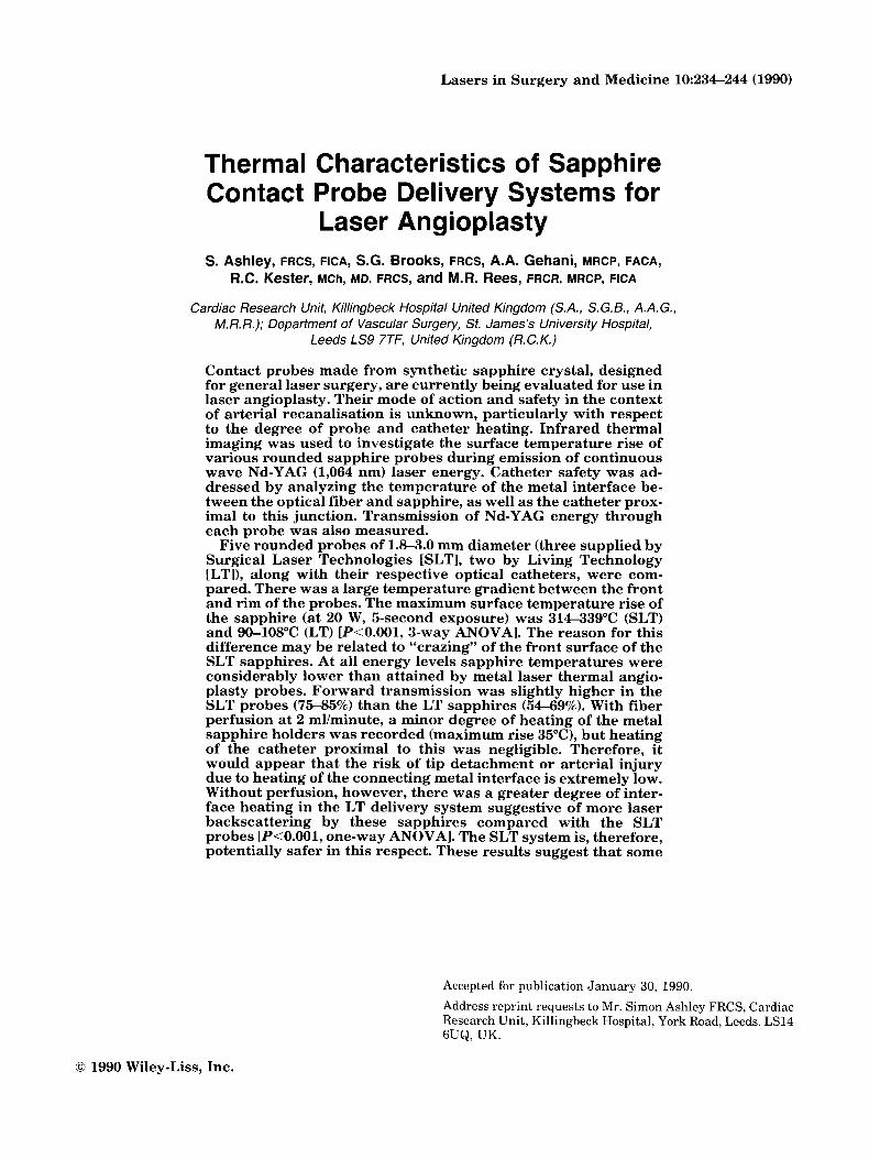
Fig. 1. The five rounded sapphire contact probes studied (left t o right Surgical Laser Technologies [SLTl 3.0 mm (MTRL), SLT 2.2 mm (MTR), SLT 1.8 mm (SMTR), Living Technology 2.2 mm, Osada 2.2 mm. Each sapphire crystal is mounted in a metal holder within which there is a small hole to emit the saline perfusate. threads.
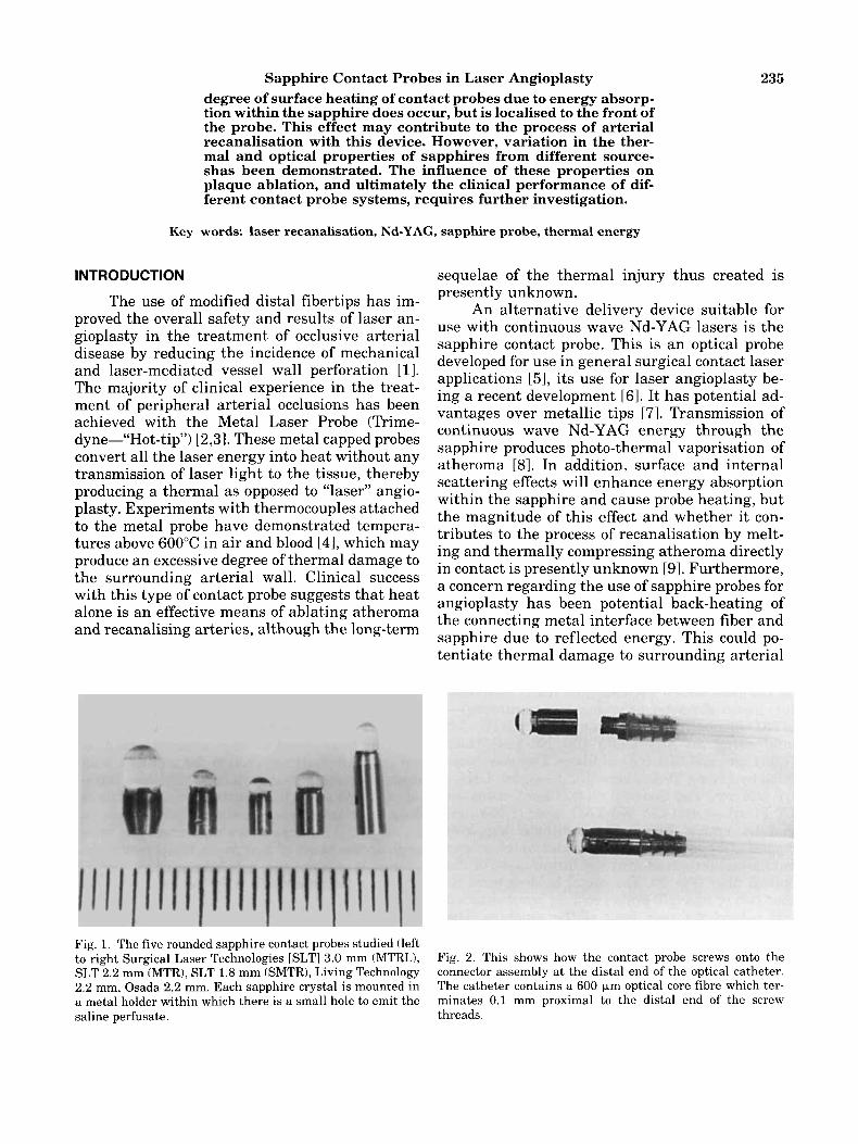
Fig. 2. This shows how the contact probe screws onto the connector assembly at the distal end of the optical catheter. The catheter contains a 600 km optical core fibre which ter- minates 0.1 mm proximal to the distal end of the screw
236 Ashley et al. Rear facet o f sapphire
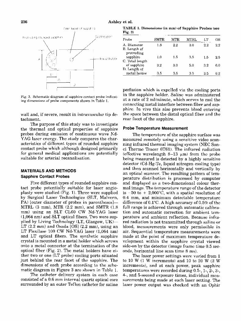
I TABLE 1. Dimensions (in mm) of Sapphire Probes (see Fig. 3)
A
Fig. 3. Schematic diagram of sapphire contact probe indicat- ing dimensions of probe components shown in Table 1.
wall and, if severe, result in intravascular tip de- tachment.
The purpose of this study was to investigate the thermal and optical properties of sapphire probes during emission of continuous wave Nd- YAG laser energy. The study compares the char- acteristics of different types of rounded sapphire contact probe which although designed primarily for general medical applications are potentially suitable for arterial recanalisation.
MATERIALS AND METHODS Sapphire Contact Probes
Five different types of rounded sapphire con- tact probe potentially suitable for laser angio- plasty were studied (Fig. 1). Three were supplied by Surgical Laser Technologies (SLT, Malvern, PA) [outer diameter of probes in parenthesesl- MTRL (3 mm), MTR (2.2 mm), and SMTR (1.8 mm) using an SLT CL60 CW Nd-YAG laser (1,064 nm) and SLT optical fibers. Two were sup- plied by Living Technology (LT, Glasgow, UK)- LT (2.2 mm) and Osada [OS] (2.2 mm), using an LT Flexilase 100 CW Nd-YAG laser (1,064 nm) and LT optical fibers. The synthetic sapphire crystal is mounted in a metal holder which screws onto a metal connector at the termination of the optical fiber (Fig. 2). The metal holders have ei- ther two or one (LT probe) cooling ports situated just behind the rear facet of the sapphire. The dimensions of each probe according to the sche- matic diagram in Figure 3 are shown in Table 1.
The catheter delivery system in each case consisted of a 0.6 mm internal quartz optical core surrounded by an outer Teflon catheter for saline
Probe SMTR MTR MTRL LT 0s A. Diameter 1.8 2.2 3.0 2.2 2.2 B. Length of
protruding sapphire 1.0 1.5 3.5 1.5 2.5
C. Total length of sapphire 3.2 3.0 5.0 3.2 6.0
D. Length of metal holder 3.5 3.5 3.5 3.5 6.2
perfusion which is expelled via the cooling ports in the sapphire holder. Saline was administered at a rate of 2 mllminute, which serves to cool the connecting metal interface between fiber and sap- phire. In vivo this also prevents blood entering the space between the distal optical fiber and the rear facet of the sapphire.
Probe Temperature Measurement
The temperature of the sapphire surface was measured remotely using a sensitive video scan- ning infrared thermal imaging system (NEC San- ei Thermo Tracer 6T61). The infrared radiation (effective wavelength 8-13 pm) from the probe being measured is detected by a highly sensitive detector (Cd-Hg-Te, liquid nitrogen cooling type) and then scanned horizontally and vertically by an optical scanner. The resulting pattern of tem- perature distribution is processed by computer and displayed as a two-dimensional colour ther- mal image. The temperature range of the detector is -50 to + 2,O0O0C, with a spatial resolution of 0.4 mm, and minimum detectable temperature difference of 0.1"C. A high accuracy of 0.5% of the full range is achieved through automatic calibra- tion and automatic correction for ambient tem- perature and ambient reflection. Because infra- red radiation is not transmitted through saline or blood, measurements were only permissible in air. Sequential temperature measurements were made at the point of maximum temperature de- velopment within the sapphire crystal viewed side-on by the detector (image frame time 0.5 sec- onds, horizontal line scan time 8 ms).
The laser power settings were varied from 1 to 10 W (1 W increments) and 10 to 20 W (2 W increments), and at each power, peak sapphire temperatures were recorded during 0.5-, 1-, 2-, 3-, 4-, and 5-second exposure times, individual mea- surements being made at each laser setting. The laser power output was checked with an Ophir
Sapphire Contact Probes in Laser Angioplasty 237 , Sapphire Contact Probe mounted in black body
In f ra red thermal imaging camera l inked to computer
Heat gun uni form heat source
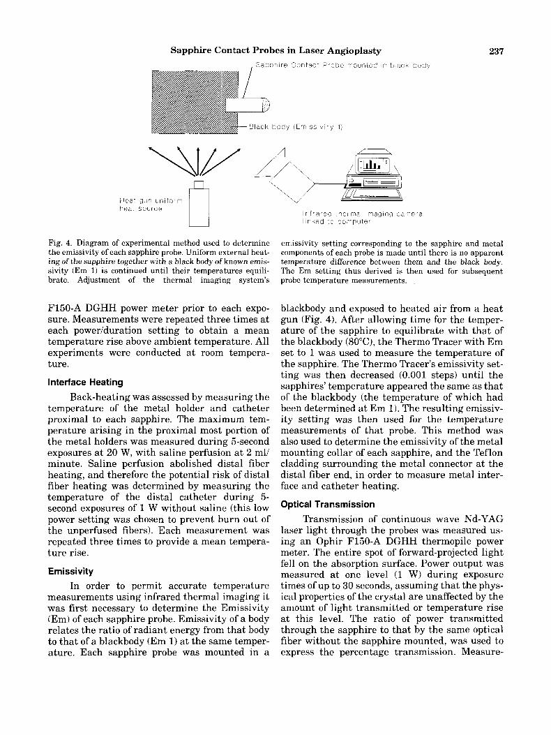
Fig. 4. Diagram of experimental method used to determine the emissivity of each sapphire probe. Uniform external heat- ing of the sapphire together with a black body of known emis- sivity (Em 1) is continued until their temperatures equili- brate. Adjustment of the thermal imaging system’s
F150-A DGHH power meter prior to each expo- sure. Measurements were repeated three times at each power/duration setting to obtain a mean temperature rise above ambient temperature. All experiments were conducted a t room tempera- ture.
Interface Heating
Back-heating was assessed by measuring the temperature of the metal holder and catheter proximal to each sapphire. The maximum tem- perature arising in the proximal most portion of the metal holders was measured during 5-second exposures at 20 W, with saline perfusion at 2 ml/ minute. Saline perfusion abolished distal fiber heating, and therefore the potential risk of distal fiber heating was determined by measuring the temperature of the distal catheter during 5- second exposures of 1 W without saline (this low power setting was chosen to prevent burn out of the unperfused fibers). Each measurement was repeated three times to provide a mean tempera- ture rise.
Emissivity In order to permit accurate temperature
measurements using infrared thermal imaging it was first necessary to determine the Emissivity (Em) of each sapphire probe. Emissivity of a body relates the ratio of radiant energy from that body to that of a blackbody (Em 1) at the same temper- ature. Each sapphire probe was mounted in a
en-iissivity setting corresponding to the sapphire and metal components of each probe is made until there is no apparent temperature difference between them and the black body. The Em setting thus derived is then used for subsequent probe temperature measurements.
blackbody and exposed to heated air from a heat gun (Fig. 4). After allowing time for the temper- ature of the sapphire to equilibrate with that of the blackbody (8OoC), the Thermo Tracer with Em set to 1 was used to measure the temperature of the sapphire. The Thermo Tracer’s emissivity set- ting was then decreased (0.001 steps) until the sapphires’ temperature appeared the same as that of the blackbody (the temperature of which had been determined a t Em 1). The resulting emissiv- ity setting was then used for the temperature measurements of that probe. This method was also used to determine the emissivity of the metal mounting collar of each sapphire, and the Teflon cladding surrounding the metal connector at the distal fiber end, in order to measure metal inter- face and catheter heating.
Optical Transmission
Transmission of continuous wave Nd-YAG laser light through the probes was measured us- ing an Ophir F150-A DGHH thermopile power meter. The entire spot of forward-projected light fell on the absorption surface. Power output was measured at one level (1 W) during exposure times of up to 30 seconds, assuming that the phys- ical properties of the crystal are unaffected by the amount of light transmitted or temperature rise at this level. The ratio of power transmitted through the sapphire to that by the same optical fiber without the sapphire mounted, was used to express the percentage transmission. Measure-
238 Ashley et al. TABLE 2. Emissivity (Em) of Sapphires and Proximal Metal Holders
Probe Samhire Em Metal holder Em SMTR 0.74 0.35 MTR 0.76 0.35 MTRL 0.77 0.35 LT 0.76 0.40 0s 0.73 0.16
ments were made with and without saline perfu- sion of the fibers. This method was chosen rather than using an integrating sphere, as it only mea- sures the antegrade optical transmission, as op- posed to the total transmission including laterally scattered light. These data therefore give an in- dication of how much light is either absorbed by the probe, scattered outside the probe, or reflected back up the optical fiber.
Scanning Electron Microscopy (SEM) of Sapphire Probe Surface
The front surface of each type of sapphire probe was examined by scanning electron micros- copy. New probes were examined before any ex- posure to laser energy. Each probe was secured onto an aluminium stub with adhesive. Electrical contact was made with Carbon Dag (suspension of carbon in organic medium). The probes were sput- ter coated with gold using an Emscope SC 500 Gold Sputter Coater and scanning electron mi- croscopy performed with a Cam Scan S.E.M. (Se- ries 3 ) .
RESULTS Emissivity
The emissivity of the sapphires and their metal holders is shown in Table 2. The outer clad- ding of all delivery catheters tested had an emis- sivity of 1.
Temperature of Sapphire Contact Probes
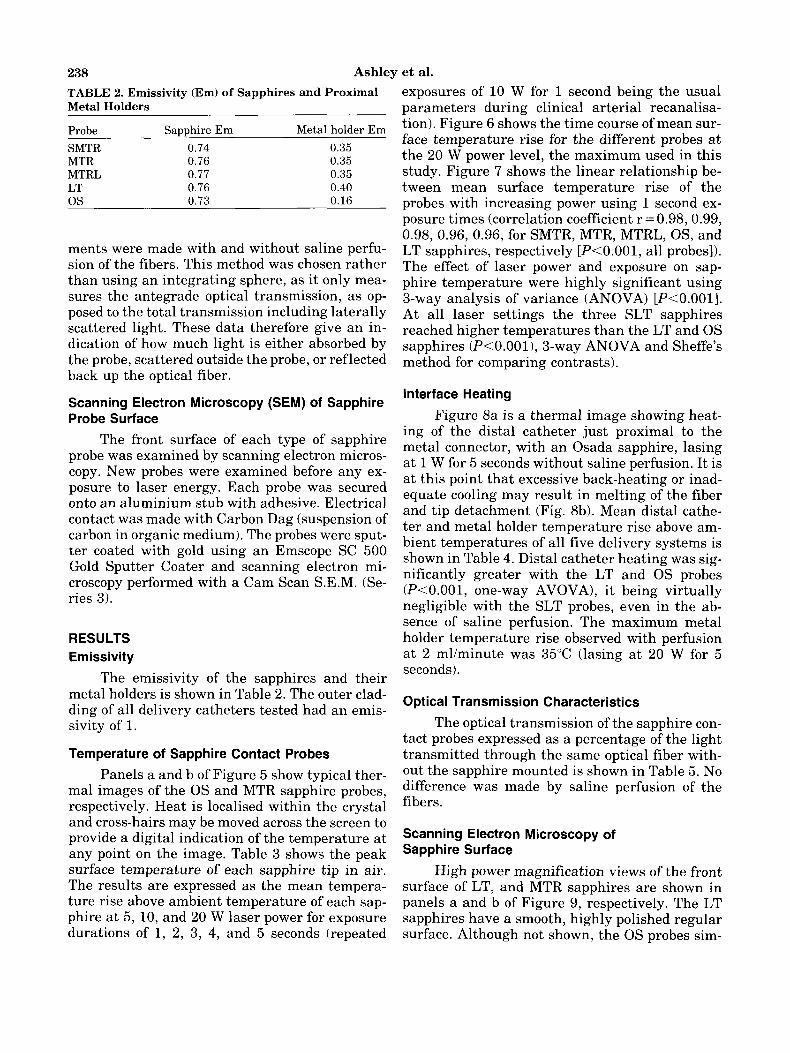
Panels a and b of Figure 5 show typical ther- mal images of the 0s and MTR sapphire probes, respectively. Heat is localised within the crystal and cross-hairs may be moved across the screen to provide a digital indication of the temperature at any point on the image. Table 3 shows the peak surface temperature of each sapphire tip in air. The results are expressed as the mean tempera- ture rise above ambient temperature of each sap- phire a t 5, 10, and 20 W laser power for exposure durations of 1, 2, 3 , 4, and 5 seconds (repeated
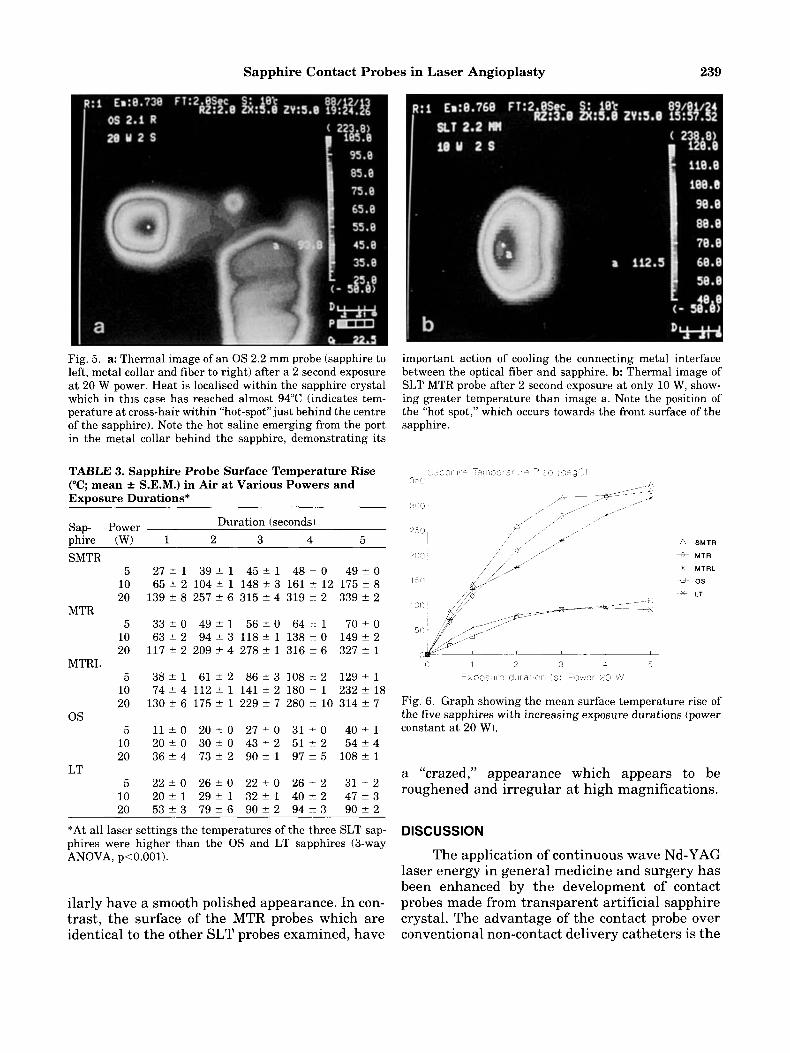
exposures of 10 W for 1 second being the usual parameters during clinical arterial recanalisa- tion). Figure 6 shows the time course of mean sur- face temperature rise for the different probes at the 20 W power level, the maximum used in this study. Figure 7 shows the linear relationship be- tween mean surface temperature rise of the probes with increasing power using 1 second ex- posure times (correlation coefficient r = 0.98,0.99, 0.98, 0.96, 0.96, for SMTR, MTR, MTRL, OS, and LT sapphires, respectively [ P < O . O O l , all probes]). The effect of laser power and exposure on sap- phire temperature were highly significant using 3-way analysis of variance (ANOVA) [P<O.OOl] . At all laser settings the three SLT sapphires reached higher temperatures than the LT and 0s sapphires ( P < O . O O l ) , 3-way ANOVA and Sheffe’s method for comparing contrasts).
Interface Heating
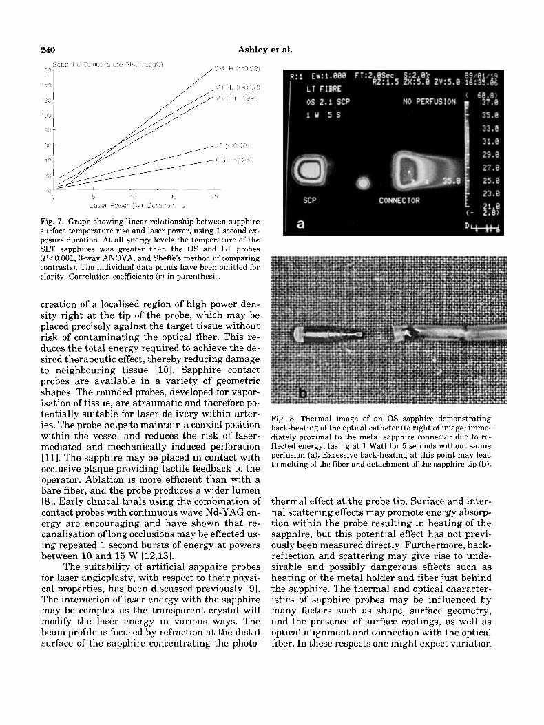
Figure 8a is a thermal image showing heat- ing of the distal catheter just proximal to the metal connector, with an Osada sapphire, lasing at 1 W for 5 seconds without saline perfusion. It is at this point that excessive back-heating or inad- equate cooling may result in melting of the fiber and tip detachment (Fig. 8b). Mean distal cathe- ter and metal holder temperature rise above am- bient temperatures of all five delivery systems is shown in Table 4. Distal catheter heating was sig- nificantly greater with the LT and 0s probes ( P < O . O O l , one-way AVOVA), it being virtually negligible with the SLT probes, even in the ab- sence of saline perfusion. The maximum metal holder temperature rise observed with perfusion at 2 ml/minute was 35°C (lasing a t 20 W for 5 seconds).
Optical Transmission Characteristics
The optical transmission of the sapphire con- tact probes expressed as a percentage of the light transmitted through the same optical fiber with- out the sapphire mounted is shown in Table 5. No difference was made by saline perfusion of the fibers.
Scanning Electron Microscopy of Sapphire Surface
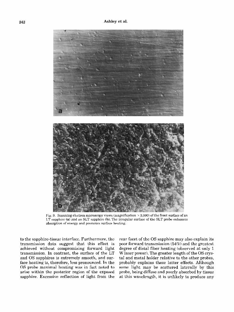
High power magnification views of the front surface of LT, and MTR sapphires are shown in panels a and b of Figure 9, respectively. The LT sapphires have a smooth, highly polished regular surface. Although not shown, the 0s probes sim-
Sapphire Contact Probes in Laser Angioplasty 239
Fig. 5. a: Thermal image of an 0s 2.2 mm probe (sapphire to left, metal collar and fiber to right) after a 2 second exposure at 20 W power. Heat is localised within the sapphire crystal which in this case has reached almost 94°C (indicates tem- perature at cross-hair within “hot-spot”just behind the centre of the sapphire). Note the hot saline emerging from the port in the metal collar behind the sapphire, demonstrating its
important action of cooling the connecting metal interface between the optical fiber and sapphire. b: Thermal image of SLT MTR probe after 2 second exposure at only 10 W, show- ing greater temperature than image a. Note the position of the “hot spot,” which occurs towards the front surface of the sapphire.
TABLE 3. Sapphire Probe Surface Temperature Rise (“C; mean f S.E.M.) in Air at Various Powers and Exposure Durations*
Sap- Power
SMTR
Duration (seconds~ phire (W) 1 2 3 4 5
5 2 7 2 1 3 9 2 1 4 5 2 1 4810 4 9 2 0 10 65 f 2 104 f 1 148 2 3 161 z 12 175 ? 8 20 139 2 8 257 2 6 315 f 4 319 z 2 339 2 2
5 3 3 t 0 4 9 2 1 5 6 2 0 6 4 - 1 7 0 2 0 10 63 2 2 94 2 3 118 2 1 138 z 0 149 f 2 20 1 1 7 k 2 209 t 4 278 2 1 316 = 6 327 2 1
5 3 8 2 1 6 1 2 2 8 6 2 3 1 0 8 ~ 2 1 2 9 2 1 10 74 2 4 112 2 1 141 2 2 180 I 1 232 2 18 20 130 2 6 175 2 1 229 2 7 280 2 10 314 2 7
5 1 1 2 0 2 0 t O 2 7 2 0 3110 4 0 2 1 10 2 0 2 0 3 0 2 0 4 3 2 2 5 1 ~ 2 5 4 f 4 20 3 6 2 4 7 3 2 2 9 0 2 1 9 7 ~ 5 1 0 8 2 1
5 2 2 2 0 2 6 2 0 2 2 t O 2 6 ~ 2 3 1 2 2 10 2 0 f 1 2 9 f 1 3 2 2 1 4 0 - 2 2 4 7 2 3 20 5 3 2 3 7 9 2 6 9 0 2 2 9 4 - 2 3 9 O k 2
MTR
MTRL
0s
LT
A SMTR
* MTR
* MTRL
iz- 0 s
* LT
0 1 1 3 5 E X W s J r p t ldrat L I IS, L h e r 2:) W
Fig. 6. Graph showing the mean surface temperature rise of the five sapphires with increasing exposure durations (power constant at 20 W).
a “crazed,” appearance which appears to be roughened and irregular at high magnifications.
*At all laser settings the temperatures of the three SLT sap- phires were higher than the 0s and LT sapphires (3-way ANOVA, p< 0 .OO 1).
ilarly have a smooth polished appearance. In con- trast, the surface of the MTR probes which are identical to the other SLT probes examined, have
DISCUSSION
The application of continuous wave Nd-YAG laser energy in general medicine and surgery has been enhanced by the development of contact probes made from transparent artificial sapphire crystal. The advantage of the contact probe over conventional non-contact delivery catheters is the
240 Ashley et al.
3 ,5 10 It 20 Laser Power (a ) DJratior 1 s
Fig. 7. Graph showing linear relationship between sapphire surface temperature rise and laser power, using 1 second ex- posure duration. At all energy levels the temperature of the SLT sapphires was greater than the 0s and LT probes ( P < O . O O l , 3-way ANOVA, and Sheffe's method of comparing contrasts). The individual data points have been omitted for clarity. Correlation coeficients (r) in parenthesis.
creation of a localised region of high power den- sity right at the tip of the probe, which may be placed precisely against the target tissue without risk of contaminating the optical fiber. This re- duces the total energy required to achieve the de- sired therapeutic effect, thereby reducing damage to neighbouring tissue [lo]. Sapphire contact probes are available in a variety of geometric shapes. The rounded probes, developed for vapor- isation of tissue, are atraumatic and therefore po- tentially suitable for laser delivery within arter- ies. The probe helps to maintain a coaxial position within the vessel and reduces the risk of laser- mediated and mechanically induced perforation [l l] . The sapphire may be placed in contact with occlusive plaque providing tactile feedback to the operator. Ablation is more efficient than with a bare fiber, and the probe produces a wider lumen [8] . Early clinical trials using the combination of contact probes with continuous wave Nd-YAG en- ergy are encouraging and have shown that re- canalisation of long occlusions may be effected us- ing repeated 1 second bursts of energy at powers between 10 and 15 W [12,131.
The suitability of artificial sapphire probes for laser angioplasty, with respect to their physi- cal properties, has been discussed previously [9]. The interaction of laser energy with the sapphire may be complex as the transparent crystal will modify the laser energy in various ways. The beam profile is focused by refraction at the distal surface of the sapphire concentrating the photo-
Fig. 8. Thermal image of an 0s sapphire demonstrating back-heating of the optical catheter (to right of image) imme- diately proximal to the metal sapphire connector due to re- flected energy, lasing at 1 Watt for 5 seconds without saline perfusion (a). Excessive back-heating at this point may lead to melting of the fiber and detachment of the sapphire tip (b).
thermal effect at the probe tip. Surface and inter- nal scattering effects may promote energy absorp- tion within the probe resulting in heating of the sapphire, but this potential effect has not previ- ously been measured directly. Furthermore, back- reflection and scattering may give rise to unde- sirable and possibly dangerous effects such as heating of the metal holder and fiber just behind the sapphire. The thermal and optical character- istics of sapphire probes may be influenced by many factors such as shape, surface geometry, and the presence of surface coatings, as well as optical alignment and connection with the optical fiber. In these respects one might expect variation
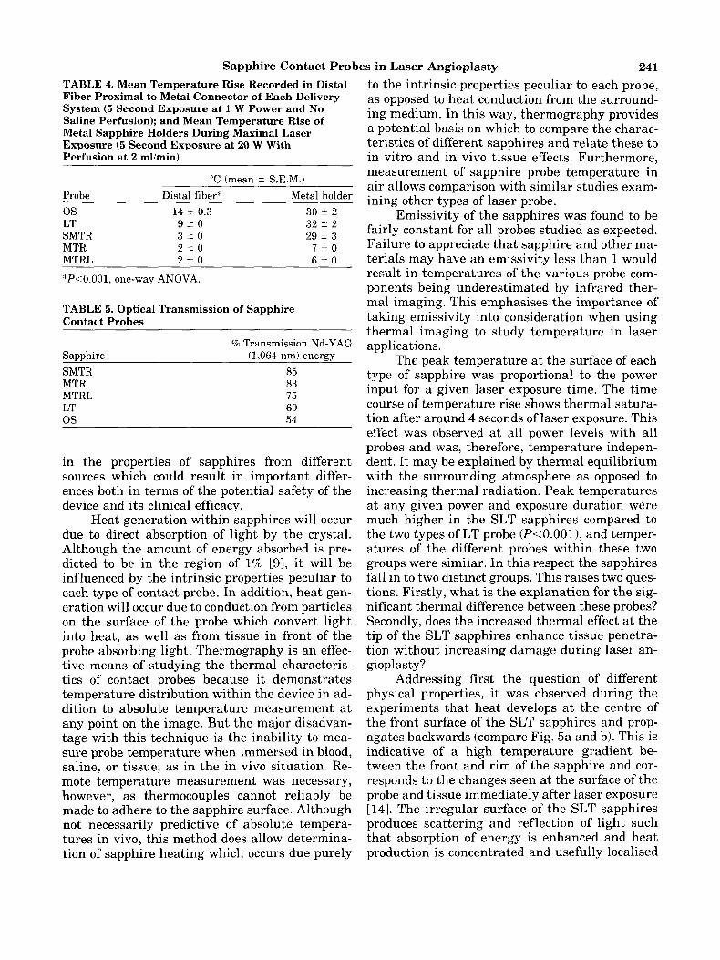
Sapphire Contact Probes in Laser Angioplasty 24 1 TABLE 4. Mean Temperature Rise Recorded in Distal Fiber Proximal to Metal Connector of Each Delivery System (5 Second Exposure at 1 W Power and No Saline Perfusion); and Mean Temperature Rise of Metal Sapphire Holders During Maximal Laser Exposure (5 Second Exposure at 20 W With Perfusion at 2 mumin)
“C (mean 2 S.E.M.) Probe Distal fiber* Metal holder 0s LT SMTR MTR MTRL
14 ? 0.3 9 5 0 3 4 0 2 2 0 2 2 0
30 2 2 32 +- 2 29 ? 3
7 * 0 6 k O
*P<O.OOl, one-way ANOVA.
TABLE 5. Optical Transmission of Sapphire Contact Probes
% Transmission Nd-YAG Sapphire (1,064 nm) energy SMTR 85 MTR 83 MTRL 75 LT 69 0s 54
in the properties of sapphires from different sources which could result in important differ- ences both in terms of the potential safety of the device and its clinical efficacy.
Heat generation within sapphires will occur due to direct absorption of light by the crystal. Although the amount of energy absorbed is pre- dicted to be in the region of 1% [9], it will be influenced by the intrinsic properties peculiar to each type of contact probe. In addition, heat gen- eration will occur due to conduction from particles on the surface of the probe which convert light into heat, as well as from tissue in front of the probe absorbing light. Thermography is an effec- tive means of studying the thermal characteris- tics of contact probes because it demonstrates temperature distribution within the device in ad- dition to absolute temperature measurement a t any point on the image. But the major disadvan- tage with this technique is the inability to mea- sure probe temperature when immersed in blood, saline, or tissue, as in the in vivo situation. Re- mote temperature measurement was necessary, however, as thermocouples cannot reliably be made to adhere to the sapphire surface. Although not necessarily predictive of absolute tempera- tures in vivo, this method does allow determina- tion of sapphire heating which occurs due purely
to the intrinsic properties peculiar to each probe, as opposed to heat conduction from the surround- ing medium. In this way, thermography provides a potential basis on which to compare the charac- tteristics of different sapphires and relate these to in vitro and in vivo tissue effects. Furthermore, measurement of sapphire probe temperature in air allows comparison with similar studies exam- ining other types of laser probe.
Emissivity of the sapphires was found to be €airly constant for all probes studied as expected. Failure to appreciate that sapphire and other ma- terials may have an emissivity less than 1 would result in temperatures of the various probe com- ponents being underestimated by infrared ther- mal imaging. This emphasises the importance of taking emissivity into consideration when using thermal imaging to study temperature in laser applications.
The peak temperature at the surface of each type of sapphire was proportional to the power input for a given laser exposure time. The time course of temperature rise shows thermal satura- tion after around 4 seconds of laser exposure. This effect was observed at all power levels with all probes and was, therefore, temperature indepen- dent. It may be explained by thermal equilibrium with the surrounding atmosphere as opposed to increasing thermal radiation. Peak temperatures at any given power and exposure duration were much higher in the SLT sapphires compared to the two types of LT probe (P<O.OOl), and temper- atures of the different probes within these two groups were similar. In this respect the sapphires fall in to two distinct groups. This raises two ques- tions. Firstly, what is the explanation for the sig- nificant thermal difference between these probes? Secondly, does the increased thermal effect at the tip of the SLT sapphires enhance tissue penetra- tion without increasing damage during laser an- gioplasty?
Addressing first the question of different physical properties, it was observed during the experiments that heat develops at the centre of the front surface of the SLT sapphires and prop- agates backwards (compare Fig. 5a and b). This is indicative of a high temperature gradient be- tween the front and rim of the sapphire and cor- responds to the changes seen a t the surface of the probe and tissue immediately after laser exposure [14]. The irregular surface of the SLT sapphires produces scattering and reflection of light such that absorption of energy is enhanced and heat production is concentrated and usefully localised
242 Ashley et al.
Fig. 9. Scanning electron microscopy views (magnification X 3,000) of the front surface of an LT sapphire (a) and an SLT sapphire (b). The irregular surface of the SLT probe enhances absorption of energy and promotes surface heating.
to the sapphire-tissue interface. Furthermore, the transmission data suggest that this effect is achieved without compromising forward light transmission. In contrast, the surface of the LT and 0s sapphires is extremely smooth, and sur- face heating is, therefore, less pronounced. In the 0s probe maximal heating was in fact noted to arise within the posterior region of the exposed sapphire. Excessive reflection of light from the
rear facet of the 0s sapphire may also explain its poor forward transmission (54%) and the greatest degree of distal fiber heating (observed at only 1 W laser power). The greater length of the 0s crys- tal and metal holder relative to the other probes, probably explains these latter effects. Although some light may be scattered laterally by this probe, being diffuse and poorly absorbed by tissue at this wavelength, it is unlikely to produce any
Sapphire Contact Probes in Laser Angioplasty 243 significant lateral tissue vaporisation. The slightly lower forward transmission observed in the LT probe compared with the SLT sapphires may be explained by the presence of an anti-re- flective coating on the rear facet of the latter probes [91 (this is unknown).
With regard to the second question, we have demonstrated in vitro that the SLT sapphires pro- duce greater penetration of vascular tissue and more efficient tissue ablation than the LT and 0s probes [ X I . Moreover, we have attempted two clinical recanalisations with the 0s sapphires. These were non-calcified femoral occlusions in which recanalisation failed due to lack of penetra- tion of the probe through the lesion at powers be- tween 10 and 15 W. This difficulty has not been experienced during subsequent treatment of over 30 similar lesions using the SLT probes.
In view of potential lateral damage to the vessel wall during recanalisation, heating of the connecting metal interface behind the sapphires is important. With saline perfusion at 2 mll minute (the minimum imposed by the integral fluid pump on the SLT laser), a laser exposure of 20 W for 5 seconds (the maximum in the study) produced a peak temperature rise of 35°C in one collar. These results are similar to those obtained using a thermocouple to measure the temperature rise of the metal connector during lasing in blood [141. It probably occurs due to conduction of heat from the sapphire, as well as due to back-scat- tered energy, and will be related to the mass and dimensions of the probe (hence the relatively high temperature rise in the 1.8 mm SLT holder com- pared to the larger SLT probes). Thermal energy does, therefore, appear to be localised to the sap- phire tip.
With respect to the potential risk of tip de- tachment due to melting of the fiber immediately proximal to the connector assembly, distal fiber heating was measured with each probe. Distal fi- ber heating was greater in the unperfused 0s and LT probes than with the SLT probes (P<O.OOl), in which it was virtually negligible. It relates to the degree of back-reflected energy from the rear facet of the sapphire crystal, as well as lateral scattering of laser light at the point where the optical fiber inserts into the metal connector. On the basis of this, it would seem that the SLT sys- tem is potentially safer. However, saline perfu- sion abolished heating of the distal fiber with all probes, and therefore the potential risk of tip de- tachment with any of the delivery devices is low.
The high temperature gradient produced by
the combination of forward photo-thermal effects in addition to heating of the front of the sapphire promotes axial tissue ablation, and in these re- spects this study suggests that the properties of rounded SLT sapphires are more suitable for laser angioplasty than the other contact probes tested. In contrast, metal laser probes produce circumfer- ential thermal energy distribution and, in this re- spect, sapphire contact probes may cause less thermal injury to the surrounding vessel wall. Supporting this hypothesis are preliminary in vitro studies demonstrating that metal probes produce a greater volume of thermal necrosis than sapphires probes [7]. Also, percutaneous an- gioscopic inspection of occluded femoral arteries after laser angioplasty with continuous wave Nd- YAG energy and SLT sapphires has revealed min- imal charring [16], further supporting the loca- lised nature of heat transfer to tissue by this device.
ACKNOWLEDGMENTS
We wish to thank Mr G.A. Davies (Director, Cardiac Research Unit) and the National Heart Research Fund for providing facilities and con- tributing towards the funding of this project. We also thank Miss Anne Schofield for research as- sistance.
REFERENCES
1.
2.
3.
.4.
5.
6.
‘7.
8.
!3.
Borst C: Percutaneous recanalisation of arteries: Status and prospects of laser angioplasty with modified fiber tips. Lasers Med Sci 1987; 2:137-151. Cumberland DC, Sandborn TA, Taylor DI, et al.: Percu- taneous laser thermal angioplasty: Initial clinical results with a laser probe in patients with total peripheral arte- rial occlusions. Lancet 1986; i:1457-59. Sandborn TA, Cumberland DC, Greenfield AJ, et al.: Per- cutaneous laser thermal angioplasty: Initial results and one year follow up in 129 femoro-popliteal lesions. Radi- ology 1988;168:121-5. Verdaasdonk RM, Borst C, Boulanger L, et al.: Laser an- gioplasty with a metal laser probe (‘hot tip’): Probe tem- perature in blood. Lasers Med Sci 1987; 2:153-158. Joffe SN, Schroder T: Lasers in general surgery. Adv. Surg. 1987;20:125-154. Fourrier JL, Brunetand JM, Prat A, et al.: Percutaneous laser angioplasty with sapphire tips. Lancet 1987; i: 105. Lammer J, Kleinert R, Pilger E, et al.: Contact probes for intravascular laser recanalization. Experimental evalu- ation. Invest Radio1 1989; 3:190-195. Geschwind HJ, Blair JD, Mongkolsmai D, et al.: Devel- opment and experimental application of contact probe catheter for laser angioplasty. JACC 1987; 9:lOl-7. Verdaasdonk RM, Cross FW, Borst C: Physical properties

244 Ashley et al. of sapphire fibertips for laser angioplasty. Lasers Med Sci 1987; 21183-188.
10. Daikuzono N: Contact delivery systems and accessories. In: Joffe SN, Oguro Y, (eds): “Advances in Nd-YAG Laser Surgery,” New York: Springer-Verlag, 1988, pp 19-29.
11. Bowker TJ, Cross FW, Bown SG, et al.: Reduction of ves- sel wall perforation by the use of sapphire tipped optical fibers in laser angioplasty. Br Heart J 1987; 57:88.
12. Pilger E, Lammer J, Kleinert R, et al.: Laser angioplasty with a contact probe for the treatment of peripheral vas- cular disease. Cardiovas Res 1988; 22:149-153.
13. Lammer J, Karnel F: Percutaneous transluminal laser angioplasty with contact probes. Radiology 1988; 168: 133-737.
14. Verdaasdonk RM, Rienks R, van Erven L, et al.: Sapphire and metal tip recanalisation: Implications for safety. (Proc. First plenary workshop on safety and laser-tissue interaction, Berlin 1988) Lasers Med Sci 1989; (Suppl): 119-127.
15. Ashley S, Brooks SG, Gehani AA, et al.: In vitro applica- tion of synthetic sapphire contact probes for laser re- canalisation with continuous wave Nd-YAG energy. Ra- diology 1989; 173(P):431.
16. Rees MR, Gehani AA, Ashley S, et al. Percutaneous video angioscopy in peripheral vascular disease. Clin Radio1 1989; 40:347-351.