Contracted Interlayer Distance in Graphene/Sapphire ...
11
Contracted Interlayer Distance in Graphene/Sapphire Heterostructure Shiro Entani 1 ( ), Liubov Yu. Antipina 2,3 , Pavel V. Avramov 1,4 , Manabu Ohtomo 1 , Yoshihiro Matsumoto 1 , Norie Hirao 5 , Iwao Shimoyama 5 , Hiroshi Naramoto 1 , Yuji Baba 5 , Pavel B. Sorokin 1,2,6 ( ) and Seiji Sakai 1 1 Advanced Science Research Center, Japan Atomic Energy Agency, Tokai, Ibaraki 319-1195, Japan 2 Technological Institute for Superhard and Novel Carbon Materials, Troitsk, Moscow 142190, Russian Federation 3 Moscow Institute of Physics and Technology, Dolgoprudny, Moscow 141700, Russian Federation 4 Siberian Federal University, 79 Svobodniy pr., Krasnoyarsk 660041, Russian Federation 5 Quantum Beam Science Directorate, Japan Atomic Energy Agency, Tokai, Ibaraki 319-1195 Japan 6 National University of Science and Technology MISiS, Moscow 119049, Russian Federation Received: 29 July 2014 Revised: 09 November 2014 Accepted: 13 November 2014 © Tsinghua University Press and Springer-Verlag Berlin Heidelberg 2014 KEYWORDS graphene, sapphire, chemical vapor deposition, graphene/insulator interface ABSTRACT Direct growth of graphene on insulators is expected to yield significant improvements in performance of graphene-based electronic and spintronic devices. In this study, we successfully reveal the atomic arrangement and electronic properties of a coherent heterostructure of single-layer graphene and α-Al 2 O 3 (0001). The analysis of the atomic arrangement of single-layer graphene on α-Al 2 O 3 (0001) revealed an apparentcontradiction. The in-plane analysis shows that single-layer graphene grows not in a single-crystalline epitaxial manner, but rather in polycrystalline form, with two strongly pronounced preferred orientations. This suggests relatively weak interfacial interactions are operative. However, we demonstrate that unusually strong physical interactions between graphene and α-Al 2 O 3 (0001) exist, as evidenced by the small separation between the graphene and the α-Al 2 O 3 (0001) surface. The interfacial interaction is shown to be dominated by the electrostatic forces involved in the graphene π-system and the unsaturated electrons of the topmost O layer of α-Al 2 O 3 (0001), rather than the van der Waals interactions. Such features causes graphene hole doping and enable the graphene to slide on the α-Al 2 O 3 (0001) surface with only a small energy barrier despite the strong interfacial interactions. 1 Introduction Graphene has attracted considerable research attention in recent years for potential applications in nano- electronics and spintronics due to properties including quantum electronic transport, tunable band gap, and the extremely large charge carrier mobility [1–3]. A method of direct chemical vapor deposition (CVD) Nano Research DOI 10.1007/s12274-014-0640-7 Address correspondence to Shiro Entani, [email protected]; Pavel B. Sorokin, [email protected]
-
Upload
khangminh22 -
Category
Documents
-
view
0 -
download
0
Transcript of Contracted Interlayer Distance in Graphene/Sapphire ...

Contracted Interlayer Distance in Graphene/Sapphire Heterostructure
Shiro Entani1 (), Liubov Yu. Antipina2,3, Pavel V. Avramov1,4, Manabu Ohtomo1, Yoshihiro Matsumoto1,
Norie Hirao5, Iwao Shimoyama5, Hiroshi Naramoto1, Yuji Baba5, Pavel B. Sorokin1,2,6 () and Seiji Sakai1
1 Advanced Science Research Center, Japan Atomic Energy Agency, Tokai, Ibaraki 319-1195, Japan 2 Technological Institute for Superhard and Novel Carbon Materials, Troitsk, Moscow 142190, Russian Federation 3 Moscow Institute of Physics and Technology, Dolgoprudny, Moscow 141700, Russian Federation 4 Siberian Federal University, 79 Svobodniy pr., Krasnoyarsk 660041, Russian Federation 5 Quantum Beam Science Directorate, Japan Atomic Energy Agency, Tokai, Ibaraki 319-1195 Japan 6 National University of Science and Technology MISiS, Moscow 119049, Russian Federation
Received: 29 July 2014
Revised: 09 November 2014
Accepted: 13 November 2014
© Tsinghua University Press
and Springer-Verlag Berlin
Heidelberg 2014
KEYWORDS
graphene,
sapphire,
chemical vapor deposition,
graphene/insulator interface
ABSTRACT
Direct growth of graphene on insulators is expected to yield significant
improvements in performance of graphene-based electronic and spintronic
devices. In this study, we successfully reveal the atomic arrangement and
electronic properties of a coherent heterostructure of single-layer graphene and
α-Al2O3(0001). The analysis of the atomic arrangement of single-layer graphene
on α-Al2O3(0001) revealed an apparentcontradiction. The in-plane analysis shows
that single-layer graphene grows not in a single-crystalline epitaxial manner,
but rather in polycrystalline form, with two strongly pronounced preferred
orientations. This suggests relatively weak interfacial interactions are operative.
However, we demonstrate that unusually strong physical interactions between
graphene and α-Al2O3(0001) exist, as evidenced by the small separation between
the graphene and the α-Al2O3(0001) surface. The interfacial interaction is shown
to be dominated by the electrostatic forces involved in the graphene π-system
and the unsaturated electrons of the topmost O layer of α-Al2O3(0001), rather
than the van der Waals interactions. Such features causes graphene hole doping
and enable the graphene to slide on the α-Al2O3(0001) surface with only a small
energy barrier despite the strong interfacial interactions.
1 Introduction
Graphene has attracted considerable research attention
in recent years for potential applications in nano-
electronics and spintronics due to properties including
quantum electronic transport, tunable band gap, and
the extremely large charge carrier mobility [1–3]. A
method of direct chemical vapor deposition (CVD)
Nano Research
DOI 10.1007/s12274-014-0640-7
Address correspondence to Shiro Entani, [email protected]; Pavel B. Sorokin, [email protected]
| www.editorialmanager.com/nare/default.asp
2 Nano Res.
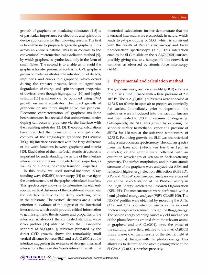
growth of graphene on insulating substrates [4-8] is
of particular importance for electronic and spintronic
device applications for the following reasons. The first
is to enable us to prepare large-scale graphene films
across an entire substrate. This is in contrast to the
conventional micromechanical exfoliation method [9],
by which graphene is synthesized only in the form of
small flakes. The second is to enable us to avoid the
graphene transfer process, in contrast to CVD graphene
grown on metal substrates. The introduction of defects,
impurities, and cracks into graphene, which occurs
during the transfer process, leads to significant
degradation of charge and spin transport properties
of devices, even though high-quality [10] and highly
uniform [11] graphene can be obtained using CVD
growth on metal substrates. The direct growth of
graphene on insulators might solve this problem.
Electronic characterization of graphene–insulator
heterostructures has revealed that unintentional carrier
doping can occur in graphene via the interface with
the insulating substrates [12, 13]. Theoretical calculations
have predicted the formation of a charge-transfer
complex at the single-layer graphene (SLG)/rutile
TiO2(110) interface associated with the large difference
of the work functions between graphene and titania
[13]. Elucidation of the interfacial atomic structures is
important for understanding the nature of the interface
interactions and the resulting electronic properties, as
well as for tailoring the charge transport properties.
In this study, we used normal-incidence X-ray
standing wave (NIXSW) spectroscopy [14] to investigate
the atomic structure of the graphene/insulator interface.
This spectroscopy allows us to determine the element-
specific vertical distances of the constituent atoms near
the interface relative to the X-ray scattering plane
in the substrate. The vertical distances are a useful
criterion to evaluate of the degree of the interfacial
interactions, which could provide critical information
to gain insight into the structures and properties of the
interface. Analysis of the contrasted standing wave
(SW) profiles [15] obtained from the SLG/c-plane
sapphire (α-Al2O3(0001)) substrate prepared by the
direct CVD growth, shows the remarkably small
vertical distance between SLG and α-Al2O3(0001) at the
interface, suggesting the existence of stronger interfacial
interactions than van der Waals interactions. Ab initio
theoretical calculations further demonstrate that the
interfacial interactions are electrostatic in nature, which
leads to p-type doping of SLG, which is consistent
with the results of Raman spectroscopy and X-ray
photoelectron spectroscopy (XPS). This interaction
enables the SLG to slide on the α-Al2O3(0001) surface,
possibly giving rise to a honeycomb-like network of
wrinkles, as observed by atomic force microscopy
(AFM).
2 Experimental and calculation method
The graphene was grown on an α-Al2O3(0001) substrate
in a quartz tube furnace with a base pressure of 2
10–6 Pa. The α-Al2O3(0001) substrates were annealed at
1,173 K for 60 min in open air to prepare an atomically
flat surface. Immediately prior to deposition, the
substrates were introduced into the vacuum furnace
and then heated to 873 K in vacuum for degassing.
Subsequently, the SLG was grown by exposing the
sapphire surface to methanol vapor at a pressure of
350 Pa for 120 min at the substrate temperature of
1,273 K. Following growth, the SLG was characterized
using a micro-Raman spectrometry. The Raman spectra
from the laser spot (which was less than 1 m in
diameter) on the sample were collected with an
excitation wavelength of 488 nm in back-scattering
geometry. The surface morphology and in-plane atomic
structure of the graphene were analyzed via AFM and
reflection high-energy electron diffraction (RHEED).
XPS and NIXSW spectroscopic analyses were carried
out at the BL-27A station of the Photon Factory in
the High Energy Accelerator Research Organization
(KEK-PF). The measurements were performed with a
hemispherical energy analyzer (VSW CLASS-100). The
NIXSW profiles were obtained by recording the Al 1s,
O 1s, and C 1s photoelectron yields as the incident
photon energy was scanned through the Bragg energy.
The photon energy scanning causes a yield-modulation
of the photoelectrons emitted from the relevant atoms
in graphene and α-Al2O3(0001), since the phase of
the standing wave field relative to the α-Al2O3(0001)
Bragg planes (i.e., the intensity of the electric field at
these atoms) changes with the photon energy. This
allows us to determine the atomic arrangement at the
SLG/α-Al2O3(0001) interface precisely.

www.theNanoResearch.com∣www.Springer.com/journal/12274 | Nano Research
3 Nano Res.
The ab initio calculations of the SLG/α-Al2O3(0001)
heterostructure were performed using density func-
tional theory (DFT) [16, 17] with the local density
approximation (LDA) for the exchange-correlation
functional [18] with periodic boundary conditions
using the Vienna Ab initio Simulation Package [19–21].
Vanderbilt ultrasoft pseudopotentials (PP) [22] were
used along with a plane wave basis set, with an energy
cutoff of 396 eV. To calculate the equilibrium atomic
structures, the Brillouin zone was sampled according
to the Monkhorst–Pack scheme [23], which was used
with an 8 × 8 × 1 mesh of k-space points (and 8 × 8 × 3
in the test bulk case given below). To avoid spurious
interactions between neighboring structures in a
tetragonal supercell, a vacuum layer of 10 Å was
included in the non-periodic direction. Structural
relaxation was performed until the forces acting on each
atom become less than 0.05 eV/Å. The α-Al2O3(0001)
substrate was simulated as a 18-Å thick slab with an
oxygen-terminated upper surface taking into account
of the NIXSW results (see below). The hexagonal
graphene unit cell was multiplied by 2 × 2 (giving a
total of 8 carbon atoms) to coincide with the substrate
unit cell.
The accuracy of the approach was confirmed by
calculation of the corresponding characteristics of
bulk sapphire. The structural parameters of the Al2O3
system were calculated within error of less than 0.05%
(compared with the experimental data taken from
Ref. [24]; i.e., acalc = 4.7623 Å and aexp = 4.7602(4) Å,
ccalc = 12.9906 Å and cexp = 12.9933 Å). This comparison
between experimentally measured XPS spectra and
the distribution of the partial density of states [25]
demonstrates good accuracy of the simulated electronic
properties. The calculated band gap of the material
was Ecalc = 6.1 eV compared with a measured value of
Eexp = 7.5–9.5 eV [26–28]; however, this does not impact
the results of our study.
3 Results and discussion
Figures 1(a) and 1(b) show a set of Raman spectra of
SLG in the SLG/α-Al2O3(0001) heterostructure. The
two peaks that appear around 1,600 cm–1 and 2,700 cm–1
are characteristic of the graphitic structure; i.e., the
so-called G and 2D peaks, respectively. The Raman
Figure 1 Raman spectra for the (a) D, G and (c) 2D bands of SLG/-Al2O3(001). (c) Plot of Pos(2D)vs. Pos(G). The data-points were measured at randomly selected areas on the SLG/-Al2O3(0001) sample and on SLG/SiO2 [30] obtained by micromechanical exfoliation. The symbols ■(black) and ●(red) denote the experi-mental data obtained from Raman spectra of SLG/SiO2 and SLG/-Al2O3(0001), respectively. The yellow line is plotted assuming linear extrapolation from the data points shown by the black squares.
spectra collected at different positions on the sample
typically show the single component of the 2D peak
as in the figure. This indicates that the whole surface
of the α-Al2O3(0001) substrate was covered uniformly
with the SLG [29]. The spectra also show the disorder-
related peaks at around 1,360 cm–1 and 2,950 cm–1;
i.e., the so-called D peak and D + G peaks. Figure 1(c)
shows a plot of the positions of G and 2D peaks
(Pos(G) and Pos(2D)) collected from the randomly
selected areas on the SLG/α-Al2O3(0001) and on the
SLG flakes on a SiO2/Si substrate (SLG/SiO2) prepared
by micromechanical exfoliation for comparison. Pos(G)
and Pos(2D) were distributed in the range 1,600–
1,612 cm–1 and 2,706-2,721 cm–1, respectively, in the
SLG/α-Al2O3(0001) (red circles), and are in the ranges
of 1,582-1,593 and 2,687-2,703 cm–1, respectively, in the
SLG/SiO2 (black squares). The distribution of Pos(2D)
and Pos(G) exhibited a linear relationship for SLG/
SiO2, which is attributed to the carrier (i.e., hole) con-
centration due to the unintentional doping from the
substrate [30, 31].

| www.editorialmanager.com/nare/default.asp
4 Nano Res.
The larger wavenumbers for Pos(G) and Pos(2D) in
the SLG/α-Al2O3(0001) exhibited anapproximately
linear relationship, which is similar to the SLG/SiO2
interface, but the deviation from the extrapolated
linear fit suggestsa higher hole concentration. There
are two factors that may influence the locations of the
peaks in addition to doping. The first is the intro-
duction of defects, such as grain boundaries [32–34],
and another is chemical interactions with the substrate
[35]. It has been reported that the appearance of a
new peak (D’ peak) at around 1,620 cm–1 results in an
upward shift of the G peak in nanocrystalline graphitic
structures [33], whereas no obvious upward shift has
been reported for the 2D peak in nanocrystalline
graphene. The second is that it has been reported that
chemical interactions at the interfaces lead to con-
siderable deviation of Pos(2D) and Pos(G) from the
linear relationship for SLG/SiO2 [35]. This second effect,
however, can be ruled out based on the NIXSW results
(see below). It is clear that a higher hole concentration
and greater defect density in SLG are responsible for
the higher Pos(G) and Pos(2D) in the SLG/α-Al2O3(0001),
which follows form considering the appearance of
the intense D and D + G peaks.
Figure 2(a) shows RHEED patterns before and after
the SLG growth on the α-Al2O3(0001) substrate. The
directions of the incident electron beam were parallel
to the [101_
0] (i and iii) and [112_
0] (ii and iv) azimuths
of the α-Al2O3(0001) substrate, respectively. Following
the SLG growth, two sets of streaks appear in the
RHEED pattern (see the arrows in Fig. 2(a) (iii) and
(iv)). These streaks are attributed to the [101_
0] and
(112_
0) reflection from SLG, which are equivalent to
the corresponding reflections of the three dimensional
graphitic structure, because the distance between the
neighboring carbon atoms calculated from the spacing
of the streak was 1.40 Å, which is consistent with the
reported value for graphene obtained by microme-
chanical exfoliation (1.42 Å) [36]. Figure 2(b) shows
an intensity profile along the lines (see Fig. 2(a))
parallel to the shadow edge for the two incident
directions of [101_
0] and [112_
0] of α-Al2O3(0001). By
comparing the line profiles taken before and after the
SLG growth, the component from the SLG can be
clearly distinguished from the component of the
Figure 2 (a) RHEED patterns (i, ii) from -Al2O3(0001) substrate,
and (iii, iv) from SLG/-Al2O3(0001). The electrons were incident
parallel to the [101_
0] (left column) and [112_
0] (right column),
respectively. (b) An intensity profile along the dotted lines parallel
to the shadow edge for the two incident directions of [101_
0] and
[112_
0] of -Al2O3(0001). (c) Streak intensities of [101_
0] and
[112_
0] reflections as a function of the azimuthal angle with respect to the [101
_
0] direction of -Al2O3(0001).
α-Al2O3(0001). The streak spacing due to SLG was
constant irrespective of the incident direction, as
shown by the solid lines for the (101_
0) and (112_
0)
streaks at the two azimuths. A clear change was
observed in the streak intensity depending on the
incident direction. In Fig. 2(c), the (101_
0) and (112_
0)
streak intensities, which were obtained by subtracting
a smoothed background from the line profiles, are
shown plotted as a function of the azimuth angle
with respect to the [101_
0] direction of α-Al2O3(0001).
Both the (101_
0) and (112_
0) streaks show the maximum
intensity at the angles that coincide with the [112_
0]
and [101_
0] azimuth of α-Al2O3(0001). This indicates
that SLG is preferably grown with the epitaxial
orientations of [101_
0]SLG//[112_
0]α-Al2O3(0001), and [101_
0]SLG//[101_
0]α-Al2O3(0001).
Figure 3(a) shows an AFM image of SLG/
α-Al2O3(0001). One can see a honeycomb-like network
of wrinkles with mesh sizes of several 100 nm. The
height of each wrinkle was less than 0.4 nm, as shown
in Fig. 3(b), which is considerably lower than that of
graphene/metal heterostructures [37]. It has been

www.theNanoResearch.com∣www.Springer.com/journal/12274 | Nano Research
5 Nano Res.
Figure 3 (a) An AFM image of SLG/-Al2O3(0001) and (b) the line profile along the white dotted line in (a). (c) Histogram (the numbers of intersection points) obtained from the AFM analysis, showing the angles of the intersection points of the wrinkles on SLG. The data are from Fig. 3(a).
reported that,with graphene/metal heterostructures,
wrinkles are formed due to the difference of the
thermal expansion coefficients between graphene and
the metal substrate and/or due to the surface roughness
of the metal substrate [37]. Note that the angles at the
intersections of the wrinkles had a narrow distribution
centered at 120, as shown in Fig. 3(c), which implies
long-range stress relaxation in SLG accompanied by
wrinkle formation.
Figures 4(a), 4(b), and 4(c) show the standing wave
(SW) profiles for the Al, O, and C atomic layers in the
SLG/α-Al2O3(0001) heterostructure. The Bragg energy
was determined as 2,938.5 eV. This energy corresponds
to the Bragg diffraction condition from the one sixth of
the c-axis of α-Al2O3 with a lattice spacing of d = 2.17 Å.
The SW profile is described by I(E)/I0 = 1 + R(E) +
2F[R(E)]1/2cos[2dH(E)], where E is the photon
energy, R(E) is the reflectivity of the substrate, (E) is
the energy-dependent phase modulation caused by
the X-ray standing wave, F is the structure factor, and
dH is the coherent position of the atoms measured
from the X-ray scattering plane [14]. The red solid
lines in the figures are the best fits to this equation for
the respective profiles. These fits give dH = 0.1, dH = 0.5
and dH = 1.7 for Al, O and C, respectively. Thus, the
vertical distances of the Al, O and C atoms above the
scattering plane, which are obtained by multiplying
Figure 4 (left panels) NIXSW profiles from the (a) Al 1s, (b) O 1s and (c) C 1s core level emission. The calculated intensity profiles (solid lines) are also included in the figure. (right panels) XPS spectra for the (d) Al 1s, (e) O 1s and (f) C 1s core level regions taken using 3,000 eV photons. The inset in (f) shows the C 1s shake-up spectrum corresponding to the higher binding energy side of the mainC 1s peak, where the horizontal axis represents the relative energy with respect to the main peak position.

| www.editorialmanager.com/nare/default.asp
6 Nano Res.
dH by d, were calculated to be 0.2, 1.1, and 3.7 Å,
respectively.
Table 1 lists a summary of these data with the DFT
calculation results (see below). Two possible arrange-
ments of the Al and O atomic layers in the interfacial
region can be considered in accordance with these
values, which are the structures corresponding to the
oxygen or aluminum termination on the surface of
α-Al2O3(0001). We estimate that the former and latter
arrangements have the vertical distances of 2.6 Å and
3.9 Å between SLG and the topmost O and Al layer
of α-Al2O3(0001), respectively. The latter possibility,
however, can be eliminated since the distance is much
larger than the interlayer distance of graphite (3.356 Å)
[38] and the vertical distance between SLGIr(111)
(3.38 Å) [39] is expected for weak interfacial interactions
due to van der Waals forces. The SLGα-Al2O3(0001)
distance (2.6 Å) is larger than the SLGNi(111) distance
(2.15 Å) [40] with covalent interactions; however, is
much smaller than the separations with van der Waals
interactions.
Figures 4(d), 4(e), and 4(f) show the Al 1s, O 1s, and
C 1s XPS spectra of the SLG/α-Al2O3(0001) heteros-
tructure, respectively. The C 1s shake-up spectrum is
also shown in the inset of Fig. 4(f). The binding
energies of the Al 1s and O 1s core levels associated
with the α-Al2O3(0001) substrate were almost identical
to the previously reported values for sapphire [41]. In
contrast, the C 1s binding energy of SLG (283.1 eV)
was shifted to the lower energies by 1.3 eV compared
with that of graphite (284.4 eV [42]). The C 1s binding
energy of SLG has been reported to show a considerable
dependence on the substrate; i.e., 285.1 eV for SLG/
Ni(111) [43], 284.0 eV for SLG/Pt(111) [44] and 284.2 eV
for SLG/Ir(111) [45]; however, the energy shifts from
that in graphite are much smaller than 1 eV. In the C
1s shake-up spectrum, a single peak is seen at around
5 eV (indicated by arrow in the inset of Fig. 4(f)).
This peak is attributed to the π-plasmon excitation of
the graphitic structure [46] and carbide formation can
be ruled out. Therefore, we may conclude that the
unusually large negative C 1s binding energy shift
observed in this study is due to nonchemical interac-
tions between SLG and sapphire. From the above
discussion on the Pos(2D) vsPos(G) relationship in
the Raman spectra (see Fig. 1(c)) and on the C 1s
binding energy shift in XPS, it is clear that SLG on the
sapphire surface is p-doped and chemical interactions
are not dominant at the SLG/α-Al2O3(0001) interface.
The DFT calculations provide additional insight
into the atomic and electronic structure of the SLG/
α-Al2O3(0001) interface and the nature of the strong
interfacial interactions. In the calculations, the top
and bottom surfaces of the sapphire slab were assumed
to be terminated by O atoms, considering the atomic
arrangements elucidated by NIXSW spectra. Taking
account of the RHEED measurements shown in Fig. 2,
two atomic arrangements of SLG on α-Al2O3(0001) were
adopted for the calculations: [101_
0]SLG//[112_
0]α-Al2O3(0001)
and [101_
0]SLG//[101_
0]α-Al2O3(0001). The distance between
the neighboring carbon atoms was altered to match
the lattice of α-Al2O3(0001) substrate. In the first
atomic rearrangement, the C–C distance was 1.38 Å,
whereas in the second it was 1.51 Å. This approach
is justified by a report [47] of a model with strained
graphene lattice, which reproduced qualitatively the
Table 1 Vertical distances of the atomic layers of the constituent elements above the X-ray scattering plane (for the NIXSW results) and above the first Al layer (for the DFT results) in the SLG/-Al2O3(0001) interfacial region, and the vertical distances between the SLG film and the -Al2O3(0001) surface estimated from the NIXSW spectroscopy and DFT calculations, respectively
NIXSW DFT
Interfacial region; vertical distance from scattering plane (Å)
Interfacial region; vertical distance from first Al layer (Å)
[101_
0]SLG//[112_
0]α-Al2O3(0001) [101_
0]SLG//[101_
0]α-Al2O3(0001)
Al 0.2 0 0
O 1.1 1.2 1.1
C 3.7 4.1 3.8
SLG/-Al2O3(0001) 2.6 2.9 2.7

www.theNanoResearch.com∣www.Springer.com/journal/12274 | Nano Research
7 Nano Res.
main features of the surface electronic structure.
Figures 5 (a) and 5(b) show the calculated atomic
structure of SLG/α-Al2O3(0001). The calculations give the
vertical distance of 2.9 Å for [101_
0]SLG//[112_
0]α-Al2O3(0001)
(Fig. 5(a)) and 2.7 Å for [10 1_
0]SLG//[10 1_
0]α-Al2O3(0001)
(Fig. 5(b)) between SLG and the topmost O layer of
α-Al2O3(0001), which is consistent with the experi-
mentally measured value of 2.6 Å.
The vertical distance between SLG and the first Al
layer were found to be 4.1 Å and 3.8 Å and that
between the topmost O layer and the first Al layer
was found to be 1.2 Å and 1.1 Å for the two atomic
arrangements, respectively. These are consistent with
the experimentally measured values of 3.9 Å and 1.3 Å,
respectively (see Table 1). Note that the interactions
with SLG lead to a slight decrease in the vertical
distance between the topmost O layer and the first Al
layer from 1.23 Å to 1.1–1.2 Å, which suggests that
relaxation of the interfacial structure has occurred.
Calculations of the charge density in the SLG/
α-Al2O3(0001) system are shown in Figs. 5(c) and 5(e),
which allow us to gain insight into the nature of the
interfacial interactions. Figures 5(d) and 5(f) show
the difference in the charge density between the SLG/
α-Al2O3(0001) heterostructure and the freestanding
sapphire and freestanding SLG; these data show that
the interactions between the SLG and sapphire are
physical in nature, and originate from the electrostatic
interactions between graphene and the topmost O
atoms of α-Al2O3(0001). The electrostatic interactions
between SLG and α-Al2O3(0001) for both arrangements
can be understood form Fig. 6(a), which shows the
partial densities of states of SLG and sapphire in
SLG/α-Al2O3(0001) compared with those of the frees-
tanding SLG and sapphire slabs. In SLG/α-Al2O3(0001),
the Dirac point of graphene is shifted to 1.1–1.3 eV
Figure 5 (a,b) Atomic structure, (c,e) spatial charge density distribution, and (d,f) the difference in the spatial charge density distribution between the SLG/-Al2O3(0001) system and the free-standing SLG and sapphire slabs for SLG/-Al2O3(0001) for[101
_
0]SLG//[112_
0]α-Al2O3(0001), and [101_
0]SLG//[101_
0]α-Al2O3(0001) arrangements. C, O, and Al atoms are marked by dark green, red, andpink, respectively. In (a) and (b) the experimental data are shown in parentheses. In (d) and (f), the loss and the gain of the charge are shown by the yellowish and bluish colors, respectively.

| www.editorialmanager.com/nare/default.asp
8 Nano Res.
relative to the freestanding case (see the insets of
Fig. 6(a)). This result is consistent with p-type doping
of the graphene structure due to the interaction with
the substrate. The magnitude of the shift is com-
parable with that reported for graphene on oxygen-
terminated SiO2 (i.e., 1 eV) [12].
The distortion of carbon pz orbitals in the region of
–1 eV, where the surface oxygen pz orbitals are located,
indicates the presence of strong electrostatic interactions
between SLG and α-Al2O3(0001), which are attributed
mainly to coupling between the graphene system
and the unsaturated pz electrons of the topmost O
layer. The adhesion energy of SLG on α-Al2O3(0001)
was estimated to be 0.11 and 0.13 eV/carbon for
[101_
0]SLG//[101_
0]α-Al2O3(0001) and [101_
0]SLG//[112_
0]α-Al2O3(0001),
respectively. These energies are comparable to those
reported for SLG on metals, i.e., 0.16–0.18 eV/carbon
for Co [48, 49], 0.12–0.13 eV/carbon for Ni [48–51], and
0.14 eV/carbon for Ru [51].
The calculations of the adhesion energy depend on
the lateral position of the SLG on α-Al2O3(0001), and
allow the construction of a potential energy surface,
as shown in Fig. 6(b). This provides information about
SLG migration on α-Al2O3(0001), including an energy
barrier for migration. The electrostatic nature of the
interfacial interactions associated with the relatively
uniform system of graphene without the influence of
chemical bonding leads to a small migration barrier
of around 0.03 eV from the energetically favorable
state (see Fig. 5(a)), as shown in Fig. 6(b).
Figure 6 (a) Partial density of states of pz orbitals of graphene (pink), the first Al layer (green) and the topmost O layer, which isdecomposed into pxy and pz orbitals (red and purple, respectively), for SLG/-Al2O3(0001) (solid lines) and freestanding -Al2O3(0001) (dotted lines). The two insets show the partial density of states of pz orbitals of graphene in SLG/-Al2O3(0001) (pink solid line) and in the free-standing SLG (pink dotted line). The calculated results shown in the left and right panels reflect the atomic arrangements of[101
_
0]SLG//[112_
0]α-Al2O3(0001) and [101_
0]SLG//[101_
0]α-Al2O3(0001), respectively. (b) Potential energy surface for SLG migration on -Al2O3(0001). The arrows represent [101
_
0] and [112_
0] directions. The two-dimensional changes in the energy are shown via color-index.

www.theNanoResearch.com∣www.Springer.com/journal/12274 | Nano Research
9 Nano Res.
The small migration barrier for SLG/α-Al2O3(0001)
allows the SLG to “slide” on the surface, despite the
strong interfacial interactions, as indicated by the
small vertical distance between SLG and α-Al2O3(0001).
It has been demonstrated that the atomic arrangement
of SLG on α-Al2O3(0001) is strongly dependent on the
growth temperature [4, 5, 8]. Fanton et al. pointed out
that 1,550 °C is required for van der Waals epitaxy [5].
Hwang et al. have demonstrated that a single dominant
SLG crystal can be obtained via a two-step growth
process, which consists of a low-temperature nucleation
at 1,250–1,350 °C followed by growth at higher tem-
perature of 1,450–1,650 °C [8]. In this work, although
single-crystalline epitaxial growth was not realized in
SLG/α-Al2O3(0001), two preferred orientations were
clearly observed even at the lower growth temperature
of 1,000 °C. This is attributed to the growth mechanism
of SLG on α-Al2O3(0001), which is affected by the
interfacial interactions. This might also enable relaxation
of the thermal stressesover a large area, which results
in the honeycomb-like network of wrinkles, possibly
during the cooling of the sample following CVD
growth. For comparison, the migration barrier of SLG
has been reported to be as large as 0.08 eV for SLG/
Ni(111), where the SLG exhibits epitaxial growth on
the Ni(111) surface [11] and gives rise to much larger
and more irregularly distributed wrinkles [37], which
is in contrast to the SLG/α-Al2O3(0001) system.
4 Conclusion
The atomic structure and electronic properties of SLG
directly grown on α-Al2O3(0001) have been investigated
experimentally and theoretically. The analysis of the
in-plane atomic arrangement of SLG on α-Al2O3(0001)
shows that SLG grows with two distinct preferred
orientations, i.e., [101_
0]SLG//[112_
0]α-Al2O3(0001) and [101_
0]SLG//
[10 1_
0]α-Al2O3(0001) in the polycrystalline grains. The
graphene atomic planes face the oxygen atoms, which
constitute the topmost layer of α-Al2O3(0001) at the
interface. The vertical distance between the graphene
layer and the α-Al2O3(0001) was found to be 2.6 Å,
which is considerably smaller than that expected for
van der Waals interactions, and indicates that the
system exhibits strong interfacial interactions.
The electronic properties, measured via micro-
Raman spectroscopy and XPS, also support the above
strong interactions due to the evidence for p-type
doping in SLG. The DFT calculations were able to
reproduce the experimental results accurately, and
provide evidence for the electrostatic interactions
between the graphene π system and the unsaturated
pz electrons of the topmost O layer, whichleads strong
interfacial interactions at the SLG/α-Al2O3(0001)
interface. The small migration barrier originating
from the electrostatic interactions may give rise to
the appearance of two atomic arrangements, and
a honeycomb-like network of wrinkles in the SLG
film at the macroscopic scale through easy sliding
of SLG.
This work provides an important basis for a com-
prehensive understanding of the growth mechanism
of graphene on sapphire substrates, as well as precise
control of the electrical properties of graphene, which
is required to manufacture high performance electronic
and spintronic devices.
Acknowledgements
We are grateful to the ‘Chebishev’ and ‘Lomonosov’
supercomputers of Moscow State University for
providing the chance of using a cluster computer for
quantum-chemical calculations. S.E. thanks Prof. H.
Kondo (Keio University) and Prof. T. Shimada
(Hirosaki University) for NIXSW measurements. This
work was partly supported by Grants-in-Aid for Young
Scientists B (Grant No. 22760033) from the Japan Society
for the Promotion of Science. The present work has
been performed under the approval of the Photon
Factory Program Advisory Committee (PF PAC Nos.
2010G660 and 2012G741). P.V.A., P.B.S. and L.Y.A.
acknowledge the support from the Russian Science
Foundation (project No. 14-13-00139).
References
[1] Novoselov, K. S.; Geim, A. K.; Morozov, S. V.; Jiang, D.;
Zhang, Y.; Dubonos, S. V.; Grigorieva, I. V.; Firsov, A. A.
Electric field effect in atomically thin carbon films. Science
2004, 306, 666669.

| www.editorialmanager.com/nare/default.asp
10 Nano Res.
[2] Zhang, Y. B.; Tan, Y. W.; Stormer, H. L.; Kim, P. Experi-
mental observation of the quantum Hall effect and Berry’s
phase in graphene. Nature 2005, 438, 201204.
[3] Heersche, H. B.; Jarillo-Herrero, P.; Oostinga, J. B.;
Vandersypen, L. M. K.; Morpurgo, A. F. Bipolar supercurrent
in graphene. Nature 2007, 446, 5659.
[4] Hwang, J.; Shields, V. B.; Thomas, C. I.; Shivaraman, S.;
Hao, D.; Kim, M.; Woll, A. R.; Tompa, G. S.; Spencer, M.
G. Epitaxial growth of graphitic carbon on C-face SiC and
sapphire by chemical vapor deposition (CVD). J. Cryst.
Growth 2010, 312, 32193224.
[5] Fanton, M. A.; Robinson, J. A.; Puls, C.; Liu, Y.; Hollander,
M. J.; Weiland, B. E.; LaBella, M.; Trumbull, K.; Kasarda,
R.; Howsare, C. et al. Characterization of graphene films
and transistors growth on sapphire by metal-free chemical
vapor deposition. ACS Nano 2011, 5, 80628069.
[6] Song, H. J.; Son, M.; Park, C.; Lim, H.; Levendorf, M. P.;
Tsen, A. W.; Park, J.; Choi, H. C. Large scale metal-free
synthesis of graphene on sapphire and transfer-free device
fabrication. Nanoscale 2012, 4, 30503054.
[7] Nakamura, A.; Miyasaka, Y.; Temmyo, J. Direct growth
properties of graphene layers on sapphire substrate by
alcohol-chemical vapor deposition. Jpn. J. Appl. Phys. 2012,
51, 04DN03.
[8] Hwang, J.; Kim, M.; Campbell, D.; Alsalman, H. A.; Kwak,
J. Y.; Shivaraman, S.; Woll, A. R.; Singh, A. K.; Hennig,
R. G.; Gorantla, S.et al. Van der waals epitaxial growth of
graphene on sapphire by chemical vapor deposition without
a metal catalyst. ACS Nano 2013, 7, 385395.
[9] Novoselov, K. S.; Jiang, D.; Schedin, F.; Booth, T. J.;
Kohtkevich, V. V.; Morozov, S. V.; Geim, A. K. Two-
dimensional atomic crystals. PNAS 2005, 102, 10451
10453.
[10] Li, X. S.; Cai, W. W.; An, J. H.; Kim, S.; Nah, J.; Yang, D.
X.; Piner, R.; Velamakanni, A.; Jung, I.; Tutuc, E.; et al.
Large-area synthesis of high-quality and uniform graphene
films on copper foils. Science 2009, 324, 13121314.
[11] Entani, S.; Matsumoto, Y.; Ohtomo, M.; Avramov, P. V.;
Naramoto, H.; Sakai, S. Precise control of single- and bi-layer
graphene growths on epitaxial Ni(111) thin film. J. Appl.
Phys. 2012, 111, 064324.
[12] Chen, K.; Wang, X. M.; Xu, J. B. Electronic properties of
graphene altered by substrate surface chemistry and externally
applied electric field. J. Phys. Chem. C2012, 116, 62596267.
[13] Du, A. J.; Ng, H. H.; Bell, N. J.; Zhu, Z. H.; Amal, R.;
Smith, S. C. Hybrid graphene/titaniananocomposite: interface
charge transfer, hole doping, and sensitization for visible
light response. J. Phys. Chem. Lett. 2011, 2, 894899.
[14] Woodruff, D. P.; Seymour, D. L.; McConvill, C. F.; Riley,
C. E.; Crapper, M. D.; Prince, N. P.; Jones, R. G. Simple
X-ray standing-wave technique and its application to the
investigation of the Cu(111) (33)R30-Cl structure. Phys.
Rev. Lett. 1987, 58, 14601462.
[15] Baba, Y.; Narita, A.; Sekiguchi, T.; Shimoyama, I.; Hirao, N.;
Entani, S.; Sakai, S. Structure determination of self-assembled
monolayer on oxide surface by soft-X-ray standing wave. e-J.
Surf. Sci. Nanotech. 2012, 10, 69-73.
[16] Hohenberg, P.; Koh, W. Inhomogeneous electron gas. Phys.
Rev. 1964, 136, B864B871.
[17] Kohn, W.; Sham, L. J. Self-consistent equations including
exchange and correlation effects. Phys. Rev. 1965, 140,
A1133A1138.
[18] Ceperley, D. M.; Alder, B. J. Ground state of the electron gas
by a stochastic method. Phys. Rev. Lett. 1980, 45, 566569.
[19] Kresse, G.; Hafner, J. Ab initio molecular dynamics for liquid
metals. Phys. Rev. B1993, 47, 558561.
[20] Kresse, G.; Hafner, J. Ab initio molecular-dynamics simulation
of the liquid-metal-amorphous-semiconductor transition in
germanium. Phys. Rev. B1994, 49, 1425114269.
[21] Kresse, G.; Furthmüller, J. Efficient iterative schemes for
ab initio total-energy calculations using a plane-wave basis
set. Phys. Rev. B1996, 54, 1116911186.
[22] Vanderbilt, D. Soft self-consistent pseudopotentials in a
generalized eigenvalue formalism. Phys. Rev. B1990, 41,
78927895.
[23] Monkhorst, H. J.; Pack, J. D. Special points for Brillouin-
zone integrations. Phys. Rev. B1976, 13, 51885192.
[24] Lewis, J.; Schwarzenbach, D.; Flack, H. D. Electric field
gradients and charge density in corundum, -Al2O3. Acta
Cryst. 1982, A38, 733739.
[25] Perevalov, T. V.; Shaposhnikov, A. V.; Gritsenko, V. A.;
Wong, H.; Han, J. H.; Kim, C. W. Electronic structure
of -Al2O3: ab initio simulations and comparison with
experiment. JETP Lett. 2007, 85, 165168.
[26] Arakawa, E. T.; Williams, M. W. Optical properties of
aluminum oxide in the vacuum ultraviolet. J. Phys. Chem.
Solids. 1968, 29, 735744.
[27] Balzarotti, A.; Bianconi, A. Electronic structure of aluminium
oxide as determined by X-ray photoemission. Phys. Status.
Solidi B 1976, 76, 689694.
[28] French, R. H. Electronic band structure of Al2O3, with
comparison to AlON and AlN. J. Am. Ceram. Soc. 1990, 73,
477489.
[29] Ferrari, A. C.; Meyer, J. C.; Scardaci, V.; Casiraghi, C.;
Lazzeri, K.;, Marui, F.; Piscanec, S.; Jiang, D.; Novoselov,
K. S.; Roth, S. et al. Raman spectrum of graphene and
graphene layers. Phys. Rev. Lett. 2006, 97, 187401.

www.theNanoResearch.com∣www.Springer.com/journal/12274 | Nano Research
11 Nano Res.
[30] Yan, J.; Zhang, Y. B.; Kim, P.; Pinczuk, A. Electric field
effect tuning of electron-phonon coupling in graphene.Phys.
Rev. Lett. 2007, 98, 166802.
[31] Pisana, S.; Lazzeri, M.; Casiraghi, C.; Novoselov, K. S.;
Geim, A. K.; Ferrari, A. C.; Mauri, F. Breakdown of the
adiabatic Born-Oppenheimer approximation in graphene. Nat.
Mater. 2007, 6, 198201.
[32] Lespade, P.; Marchand, A.; Couzi, M; Cruege, F.
Caracterisation de materiaux carbones par microspectro-
metrieraman. Carbon 1984, 22, 375385.
[33] Ferrari, A. C.; Robertson, J. Interpretation of raman spectra
of disordered and amorphous carbon. Phys. Rev. B2000, 61,
14095.
[34] Casiraghi, C.; Pisana, S.; Novoselov, K. S.; Geim, A. K.;
Ferrari, A. C. Raman fingerprint of charged impurities in
graphene. Appl. Phys. Lett. 2007, 91, 233108.
[35] Entani, S.; Sakai, S.; Matsumoto, Y.; Naramoto, H.; Hao,
T.; Maeda, Y. Interface properties of metal/graphene
heterostructures studied by micro-raman spectroscopy. J.
Phys. Chem. C2010, 114, 2004220048.
[36] Gass, M. H.; Bangert, U.; Bleloch, A. L.; Wang, P.; Nair, R. R.;
Geim, A. K. Free-standing graphene at atomic resolution.
Nat. Nanotechnol. 2008, 3, 676681.
[37] Chae, S. J.; Günes, F.; Kim, K. K.; Kim, E. S.; Han, G. H.;
Kim, S. M.; Shin, H.; Yoon, S.; Choi, Y.; Park, M. H. et al.
Synthesis of large-area graphene layers on poly-nickel
substrate by chemical vapor deposition: Wrinkle formation.
Adv. Mater. 2009, 21, 23282333.
[38] Trucano, P.; Chen, R. Structure of graphite by neutron
diffraction. Nature 1975, 258, 136137.
[39] Busse, C.; Lazić, P.; Djemour, R.; Coraux, J.; Gerber, T.;
Atodirescei, N.; Caciuc, V.; Brako, R.; N'Diaye, A. T.;
Blügel, S. et al. Graphene on Ir(111): Physisorption with
chemical modulation. Phys. Rev. Lett. 2011, 107, 036101.
[40] Gamo, Y.; Nagashima, A.; Wakabayashi, M.; Terai, M.;
Oshima, C. Atomic structure of monolayer graphite formed
on Ni(111). Surf. Sci. 1997, 374, 6164.
[41] Wagner, C. D. X-ray photoelectron spectroscopy with x-ray
photons of higher energy. J. Vac. Sci. Technol. 1978, 15,
518523.
[42] Hamrin, K.; Johansson, G.; Gelius, U.; Nordling, C.; Siegbahn,
K. Valence bands and core levels of the isoelectronic series
LiF, BeO, BN, and graphite studied by ESCA. Phys. Scripta.
1970, 1, 277280.
[43] Nagashima, A.; Tejima, N.; Oshima, C. Electronic states of
the pristine and alkali-metal-intercalated monolayer graphite/
Ni(111) systems. Phys. Rev. B1994, 50, 1748717495.
[44] Entani, S.; Ikeda, S.; Kiguchi, M.; Saiki, K.; Yoshikawa, G.;
Nakai, I.; Kondoh, H.; Ohta, T. Growth of nanographite on
Pt(111) and its edge state. Appl. Phys. Lett. 2006, 88, 153126.
[45] Preobrajenski, A. B.; Ng, N. L.; Vinogradov, A. S.;
Mårtensson, N. Controlling graphene corrugation on lattice-
mismatched substrates.Phys. Rev. B2008, 78, 073401.
[46] Eberlein, T.; Bangert, U. B.; Nair, R. R.; Jones, R.; Gass, M.;
Bleloch, A. L.; Novoselov, K. S.; Geim, A.; Briddon, P. R.
Plasmon spectroscopy of free-standing graphene films. Phys.
Rev. B2008, 77, 233406.
[47] Stradi, D.; Barja, S.; Díaz, C.; Barnica, M.; Borca, B.;
Hinarejos, J. J.; Sánchez-Portal, D.; Alcamí, M.; Arnau, A.;
Vázquez de Parga, A. L.; Miranda, R.; Martín, F. Lattice-
matched versus lattice-mismatched models to describe epitaxial
monolayer graphene on Ru(0001). Phys. Rev. B2013, 88,
245401.
[48] Giovannetti, G.; Khomyakov, P. A.; Brocks, G.; Karpan, V.
M.; van den Brink, J.; Kelly, P. J. Doping graphene with
metal contacts. Phys. Rev. Lett. 2008, 101, 026803.
[49] Vanin, M.; Mortensen, J. J.; Kelkkanen, A. K.; Garcia-
Lastra, J. M.; Thygesen, K. S.; Jacobsen, K. W. Graphene
on metals: A van der Waals density functional study. Phys.
Rev. B2010, 81, 081408.
[50] Kozlov, S. M.; Viñes, F.; Görling, A. Bonding mechanisms
of graphene on metal surfaces. J. Phys. Chem. C2012, 116,
736.
[51] Gong, C.; Lee, G.; Shan, B.; Vogel, E. M.; Wallace, R. M.;
Cho, K. First-principles study of metal-graphene interface.
J. Appl. Phys. 2010, 108, 123711.