Successful arterial embolization of a giant pseudoaneurysm of ...
8
Radiology Case. 2012 Feb; 6(2):9-16 Interventional Radiology: Successful arterial embolization of a giant pseudoaneurysm of the gastroduodenal artery secondary to chronic pancreatitis with literature review Klauß et al. Journal of Radiology Case Reports www.RadiologyCases.com 9 Successful arterial embolization of a giant pseudoaneurysm of the gastroduodenal artery secondary to chronic pancreatitis with literature review Miriam Klauß 1 , Tobias Heye 1 , Ulrike Stampfl 1 , Lars Grenacher 1 , Boris Radeleff 1* 1. Department of Diagnostic and Interventional Radiology, University hospital, Heidelberg, Germany * Correspondence: Boris Radeleff, Department of Diagnostic and Interventional Radiology, Im Neuenheimer Feld 110, 69120 Heidelberg, Germany ( [email protected]) Radiology Case. 2012 Feb; 6(2):9-16 :: DOI: 10.3941/jrcr.v6i2.919 ABSTRACT We report a case of an uncommon giant pseudoaneurysm of the gastroduodenal artery secondary to chronic pancreatitis. It presented with a perfused volume of 17.3 cm³ close to the branch-off of the right hepatic artery. Superselective transcatheter embolization including interlocking detachable coils and a mixture of Ethibloc and Lipiodol was our technique of choice. Following the procedure, the patient was in hemodynamically stable condition. At that time, he was free of any clinical symptoms and showed no further signs of bleeding or ischaemia. Additionally, we present an overview of the relevant literature. CASE REPORT A 47 year-old patient, with a history of chronic pancreatitis due to long-lasting alcohol abuse, presented with acute upper abdominal pain. On initial presentation, his haemoglobin level was 7.3 g/dl (normal range 13-17 g/dl). Esophagogastroduodenoscopy revealed an erosive duodenitis without any signs of active or recent bleeding. An ultrasound examination revealed a cystic lesion in the pancreatic head, which pulsated when viewed in doppler mode. Subsequently, a contrast-enhanced (150 ml Imeron 300, Altana, Germany) computer tomography (Definition, Siemens, Erlangen, Germany) was performed. In the arterial phase it detected a giant pseudoaneurysm of the proximal gastroduodenal artery (GDA). The perfused part measured 3.7 x 2.5 x 4.1 cm. The total pseudoaneurysm dimension including the thrombosed part was 4.5 x 5.7 x 6.5 cm (Fig. 1, 2). CT volumetry of the perfused part of the pseudoaneurysm identified a volume of 17.3 cm3. The hepatic, gastroduodenal and splenic artery were well patent and lead to good organ perfusion. The pancreatic parenchyma exhibited scattered calcifications indicating chronic pancreatitis without any signs indicative of an acute pancreatitis. A surgical approach was deemed unfeasible due to the high risk of rupture and consecutive massive intraabdominal bleeding. Furthermore, the surgical access to an area marked with extensive scar tissue arising out of chronic infection would have proven to be extremely difficult. As a result, the patient was referred to angiography for occlusion of the pseudoaneurysm. An emergency angiography was performed. Celiac trunk angiography employing a selective F4 cobra catheter in DSA-technique including the injection of 30 ml contrast media (Imeron 300, Altana, Germany) with a flow of 5 ml/sec revealed that the GDA arises from the right hepatic artery. A giant pseudoaneurysm with a small neck was identified at the proximal GDA, very close to the branch-off of the right hepatic artery (Fig. 1, 2, 3). A F2.7 microcatheter (Progreat , Terumo, USA) was advanced into the GDA distal to the pseudoaneurysm, where three interlocking detachable-coils (Boston Scientific, USA) were deployed: two 8 mm in diameter / 10 cm long coils and one 6 mm / 10 cm coil. Angiographic control confirmed the complete occlusion of the GDA distal to the pseudoaneurysm's neck (Fig. 4). Secondly, the F2.7 microcatheter was advanced via the small neck into the pseudoaneurysm and 18 interlocking CASE REPORT
-
Upload
khangminh22 -
Category
Documents
-
view
4 -
download
0
Transcript of Successful arterial embolization of a giant pseudoaneurysm of ...

Radiology Case. 2012 Feb; 6(2):9-16
Interventional Radiology: Successful arterial embolization of a giant pseudoaneurysm of the gastroduodenal artery secondary to chronic pancreatitis with literature review
Klauß et al.
Jou
rnal
of
Rad
iolo
gy
Cas
e R
epo
rts
ww
w.R
adio
logyC
ases.com
9
Successful arterial embolization of a giant
pseudoaneurysm of the gastroduodenal artery
secondary to chronic pancreatitis with literature review
Miriam Klauß1, Tobias Heye
1, Ulrike Stampfl
1, Lars Grenacher
1, Boris Radeleff
1*
1. Department of Diagnostic and Interventional Radiology, University hospital, Heidelberg, Germany
* Correspondence: Boris Radeleff, Department of Diagnostic and Interventional Radiology, Im Neuenheimer Feld 110, 69120
Heidelberg, Germany
Radiology Case. 2012 Feb; 6(2):9-16 :: DOI: 10.3941/jrcr.v6i2.919
ABSTRACT
We report a case of an uncommon giant pseudoaneurysm of the
gastroduodenal artery secondary to chronic pancreatitis. It presented with a
perfused volume of 17.3 cm³ close to the branch-off of the right hepatic
artery. Superselective transcatheter embolization including interlocking
detachable coils and a mixture of Ethibloc and Lipiodol was our technique of
choice. Following the procedure, the patient was in hemodynamically stable
condition. At that time, he was free of any clinical symptoms and showed no
further signs of bleeding or ischaemia. Additionally, we present an overview
of the relevant literature.
CASE REPORT
A 47 year-old patient, with a history of chronic
pancreatitis due to long-lasting alcohol abuse, presented with
acute upper abdominal pain. On initial presentation, his
haemoglobin level was 7.3 g/dl (normal range 13-17 g/dl).
Esophagogastroduodenoscopy revealed an erosive duodenitis
without any signs of active or recent bleeding.
An ultrasound examination revealed a cystic lesion in the
pancreatic head, which pulsated when viewed in doppler
mode. Subsequently, a contrast-enhanced (150 ml Imeron
300, Altana, Germany) computer tomography (Definition,
Siemens, Erlangen, Germany) was performed. In the arterial
phase it detected a giant pseudoaneurysm of the proximal
gastroduodenal artery (GDA). The perfused part measured 3.7
x 2.5 x 4.1 cm. The total pseudoaneurysm dimension including
the thrombosed part was 4.5 x 5.7 x 6.5 cm (Fig. 1, 2). CT
volumetry of the perfused part of the pseudoaneurysm
identified a volume of 17.3 cm3. The hepatic, gastroduodenal
and splenic artery were well patent and lead to good organ
perfusion. The pancreatic parenchyma exhibited scattered
calcifications indicating chronic pancreatitis without any signs
indicative of an acute pancreatitis.
A surgical approach was deemed unfeasible due to the
high risk of rupture and consecutive massive intraabdominal
bleeding. Furthermore, the surgical access to an area marked
with extensive scar tissue arising out of chronic infection
would have proven to be extremely difficult. As a result, the
patient was referred to angiography for occlusion of the
pseudoaneurysm. An emergency angiography was performed.
Celiac trunk angiography employing a selective F4 cobra
catheter in DSA-technique including the injection of 30 ml
contrast media (Imeron 300, Altana, Germany) with a flow of
5 ml/sec revealed that the GDA arises from the right hepatic
artery. A giant pseudoaneurysm with a small neck was
identified at the proximal GDA, very close to the branch-off of
the right hepatic artery (Fig. 1, 2, 3).
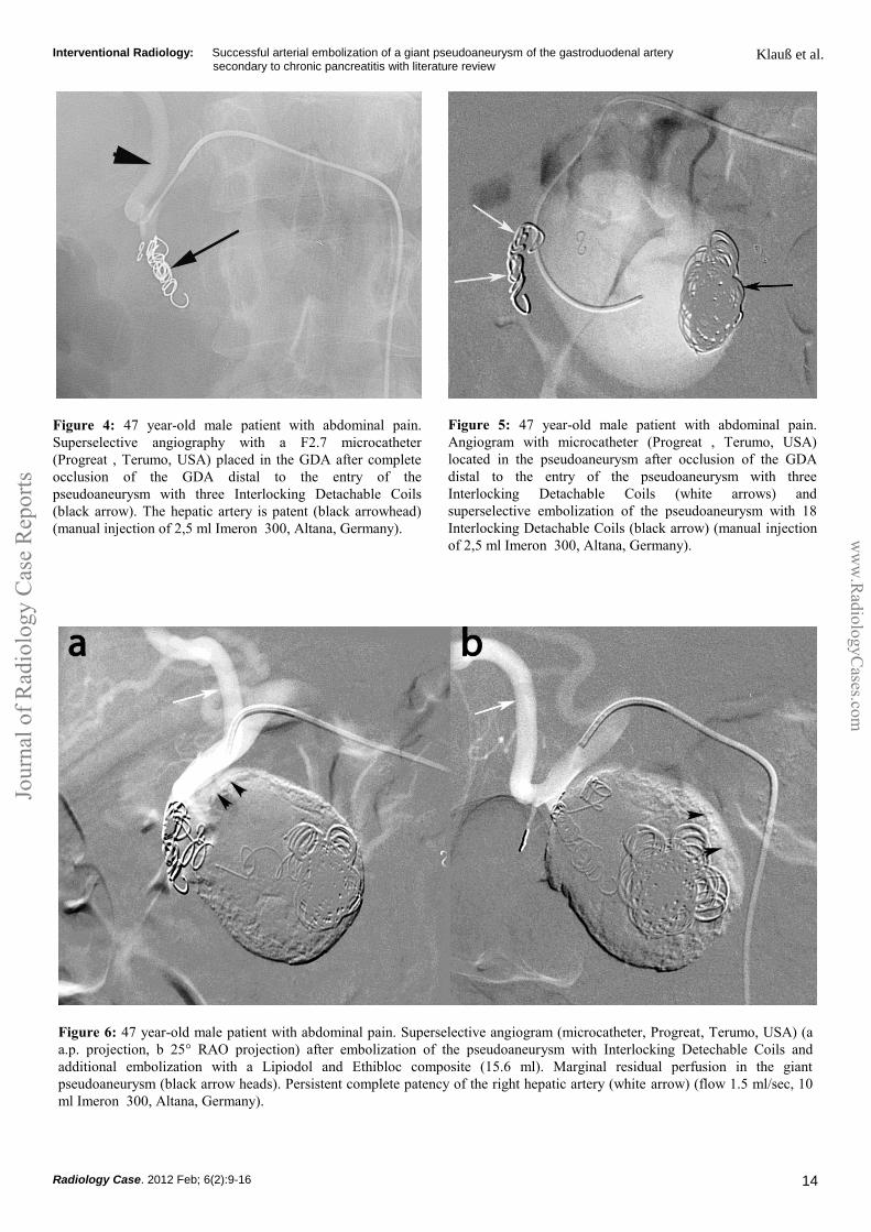
A F2.7 microcatheter (Progreat , Terumo, USA) was
advanced into the GDA distal to the pseudoaneurysm, where
three interlocking detachable-coils (Boston Scientific, USA)
were deployed: two 8 mm in diameter / 10 cm long coils and
one 6 mm / 10 cm coil. Angiographic control confirmed the
complete occlusion of the GDA distal to the pseudoaneurysm's
neck (Fig. 4).
Secondly, the F2.7 microcatheter was advanced via the
small neck into the pseudoaneurysm and 18 interlocking
CASE REPORT

Radiology Case. 2012 Feb; 6(2):9-16
Interventional Radiology: Successful arterial embolization of a giant pseudoaneurysm of the gastroduodenal artery secondary to chronic pancreatitis with literature review
Klauß et al.
Jou
rnal
of
Rad
iolo
gy
Cas
e R
epo
rts
ww
w.R
adio
logyC
ases.com
10
detachable-coils (Boston Scientific, USA) were deployed: four
10 mm in diameter / 20 cm long, six 9 mm / 20 cm coils, three
8 mm / 20 cm, and three 6 mm / 10 cm (Fig. 4). After the
placement of 16 coils, an angiographic control uncovered only
a marginal filling of the pseudoaneurysm (Fig. 5). As a
consequence, an additional embolization using the liquid
embolic agent Lipiodol/Ethibloc -mixture (Ethicon,
Norderstedt, Germany) was performed in sandwich-technique:
following the injection of 2 ml of 40% glucose, we applied 26
separate portions of 0.6 ml of the embolic-mixture (ratio of 15
ml Ethibloc/10 ml Lipiodol) resulting in a total volume of 15.6
ml liquid embolic agent.
Finally, two interlocking detachable coils (Boston
Scientific, USA, 6 mm in diameter / 10 cm long) were
deployed. While retracking the superselective catheter that
way, one half of the coil remained inside the pseudoaneurysm
and the other half was left in the GDA proximal to the
pseudoaneurysm. A final angiographic control with selective
catheter in the common hepatic artery showed an only discrete
residual perfusion in the upper part of the pseudoaneurysm.
Because the residual perfusion was only very discrete we
expected that it would completely thrombose and no further
embolization was required. The right and left hepatic arteries
were completely visible (Fig. 5).
The patient's ensuing hospital stay was uneventful and he
could be discharged after 6 days without any signs of bleeding
or intestinal ischaemia. Laboratory results showed stable
haemoglobin and hematocrit values. Pancreatic enzymes were
not elevated following the embolization.
The clinical condition remained stable over time, which
was confirmed by a follow-up conducted after twelve months.
A contrast-enhanced follow-up CT scan (6 weeks post
embolization) verified a complete occlusion without any
reperfusion of the pseudoaneurysm (Fig. 7).
Pseudoaneuryms of visceral arteries are uncommon (5-
10%), but occur as critical complications following pancreatic
surgery and pancreatitis [1-3, 8]. Therefore, early diagnosis
and adequate therapeutic interventions are imperative. Arterial
hemorrhage and/or ruptured pseudoaneurysms of the
gastroduodenal artery (GDA) are uncommonly reported and
studies investigating diagnostic and therapeutic algorithms are
rare [8]. Patients with ruptured pseudoaneurysms of the GDA
may constitute poor candidates for emergency surgery due to
hemodynamic instability and critical general conditions.
However, interventional radiology such as superselective
embolization offers less invasive treatment methods.
The exact pathogenesis of pseudoaneurysm formation is
still unclear, but, to date, three pathogenic mechanisms are
being discussed: (1) severe inflammation and enzymatic
autodigestion of a pancreatic or peripancreatic artery may
cause a disruption of the artery; (2) an established pseudocyst
eroding a visceral artery, thereby converting the pseudocyst
into a large pseudoaneurysm, and (3) a pseudocyst may erode
the bowel wall with bleeding from the mucosal surface itself
[4]. Pseudocysts or infected fluid collections are also
frequently considered to be associated with the formation of a
pseudoaneurysm [6, 7].
Another important mechanism might be an iatrogenic
trauma to visceral arteries during pancreaticoduodenectomy
e.g. during extensive regional lymphadenectomy or radical
resection at the site of the primary tumor [5].
The vast majority of patients with pseudoaneurysms
evolving in the setting of pancreatitis present with abdominal
pain. However, they do not experience hypotension derived by
acute blood loss (62%). The pain is often described as
"crescendo" and different from the pain characteristic of
pancreatitis [7-9].
Bergert et al reported a 5% prevalence of bleeding
pseudoaneurysms for patients with chronic pancreatitis and a
12% prevalence for patients with necrotizing pancreatitis [8].
The most commonly affected arteries were the splenic,
intrahepatic and gastroduodenal arteries [8].
Ruptures of pseudoaneurysms of the splenic (about 31%),
gastroduodenal (about 24%), pancreaticoduodenal (about
21%), superior mesenteric, hepatic, or gastric arteries are
reported with declining incidence [2, 6].
In a systemic review of 214 patients with pancreatitis-
associated vascular complications, the splenic artery was most
frequently involved, followed by the gastroduodenal,
pancreaticoduodenal, and the hepatic arteries [2, 6]. Acute
hemorrhage from a pseudoaneurysm is the most rapidly fatal
complication of chronic pancreatitis. The mortality rate of
untreated patients reaches 90 to even a 100%. Even with the
most aggressive treatment, the mortality is still at 12- 50%
[10].
Mortality rates after surgical repair of bleeding visceral
arteries have been reported in patients with chronic
pancreatitis and pseudocysts. Mortality in patients treated with
arterial ligation was 43 % for pseudoaneurysms located in the
head of the pancreas and 15% involving the body and tail [2].
Insufficient ligation of a vessel with a recurrence of the
bleeding due to surrounding tissue infection or insufficient
control of bleeding may occur. Additionally, these patients
may not be hemodynamically stable enough for surgery and
anaesthesia due to their generally poor condition [11].
The detection of an aneurysm mostly depends on CT or
angiography [12]. CT should include an arterial phase with
thin slices (1 mm) in axial and coronal plane. Pseudoaneuryms
appear as sharply delineated lesion with homogenous and
intense arterial enhancement and an anastomosis to an artery.
Angiography allows for a precise detection of bleeding arteries
and pseudoaneurysms. As a result, the decision making,
whether an embolization is technically feasible and safe, is
facilitated [1, 13]. Interventional techniques are an accepted
and safe method in the treatment of arterial complications
following pancreatitis [6, 14-16]. Boughene et al reported
treatment success rates of embolization therapy alone to reach
DISCUSSION

Radiology Case. 2012 Feb; 6(2):9-16
Interventional Radiology: Successful arterial embolization of a giant pseudoaneurysm of the gastroduodenal artery secondary to chronic pancreatitis with literature review
Klauß et al.
Jou
rnal
of
Rad
iolo
gy
Cas
e R
epo
rts
ww
w.R
adio
logyC
ases.com
11
approximately 78% [17]. In their study involving 35 patients
with pseudoaneurysms associated with pancreatitis Bergert et
al arrived at a success rate of 88% and a mortality rate of 19%
in 16 embolized patients [8]. The systemic appraisal of the
management of major vascular complications of pancreatitis
by Balachandra reported that angiographic embolization
proved successful in achieving hemostasis in 74% [6].
The angiographic principle of occluding the artery
upstream and downstream (front- and backdoor concept) from
the origin of the lesion should be adopted. Therefore, it is
necessary to perform embolization proximally and distal to the
pseudoaneurysm, as well as, within the pseudoaneurysm itself.
This is done to exclude it from both arterial backflow and
antegrade flow [17, 18].
As chronic pancreatitis is an ongoing inflammatory
process, Boughene et al argue that a definitive surgical
solution should follow embolization as soon as possible [17].
The results of Bergert et al suggest that the mortality rate is not
affected by the mode of treatment in chronic pancreatitis. As a
consequence, a successful embolization requires no further
treatment [8]. Therefore, angiography represents the diagnostic
and, along with embolization, therapeutic procedure of choice.
That is, if the patient is hemodynamically stable, and surgery
as backup in cases of ruptured pseudoaneurysms is available
[4, 8, 13].
A giant pseudoaneurysm is defined as a pseudoaneurysm
equal to, or greater than, 5 cm in size [19]. In our case, the
embolization of the giant pseudoaneurysm was critical, as the
origin was in close proximity to the branch-off from the right
hepatic artery. With regards to the arterial embolization, we
faced the therapeutic dilemma of not wanting to occlude the
hepatic artery, but to still sufficiently control bleeding from the
pseudoaneurysm. It was decided to occlude the GDA first to
stop the backflow into the pseudoaneurysm. Secondly, the
occlusion of the pseudoaneurysm was achieved by filling it
with numerous coils and finally with a mixture of
Ethibloc/Lipiodol. Then two coils were positioned partially
inside the pseudoaneurysm and partially into the GDA
proximal the pseudoaneurysm to close the entry. The
angiography performed after embolization (Fig. 6) confirmed
the enduring complete patency of the right hepatic artery.
Efficacy for transcatheteral arterial embolization in
pseudoaneurysms caused by pancreatitis ranges from 67 to
100%. Mortality rates have been reported to be 0-14% along
with a morbidity of 14-25% [7, 20]. In most published reports
involving embolization of bleeding sites, such as,
pseudoaneurysms or arteries, coil deployment or a
combination of coils with additional occluding material is used
to achieve successful occlusion [1, 2, 11, 15]. We believe that
the choice of occlusion material depends on the type and the
location of the bleeding source. In small pseudoaneurysms coil
embolization alone can be performed whereas in large
pseudoaneurysms additional embolization with a liquid
embolic agent might be necessary.
In conclusion transcatheteral embolization can effectively
control bleeding from pseudoaneurysms. Emergency surgery
should be limited to cases, where angiography cannot bring
about the desired results due to the failed catheterization of
vessels or insufficient occlusion of the source of bleeding.
In the reported case, interventional embolization was able
to successfully occlude a giant pseudoaneurym, thus
controlling the risk of massive intraabdominal bleeding
without the need for surgery.
Interventional embolization is able to successfully occlude
a giant pseudoaneurym (defined as pseudoaneurysms equal, or
greater than, 5 cm in size) of the gastroduodenal artery
secondary to chronic pancreatitis even with a wide neck and
the origin being in close proximity to the branch-off from the
right hepatic artery. At first, one should occlude the GDA
distal to the entry of the pseudoaneurysm to stop the backflow.
Then one can occlude the pseudoaneurym. Finally, coils can
be partially placed inside the GDA to close the entry and to
control the risk of massive intraabdominal bleeding. Complete
occlusion of the pseudoaneurysm can be achieved without
surgery.
1. Sato N, Yamaguchi K, Shimizu S, et al. Coil embolization
of bleeding visceral pseudoaneurysms following
pancreatectomy: the importance of early angiography. Arch
Surg 1998; 133:1099-1102. PMID: 9790208
2. Stabile BE, Wilson SE, Debas HT. Reduced mortality from
bleeding pseudocysts and pseudoaneurysms caused by
pancreatitis. Arch Surg 1983; 118:45-51. PMID: 6848076
3. Balthazar EJ, Fisher LA. Hemorrhagic complications of
pancreatitis: radiologic evaluation with emphasis on CT
imaging. Pancreatology 2001; 1:306-313. PMID: 12120199
4. Chen HL, Chang WH, Shih SC, Wang TE, Yang FS, Lam
HB. Ruptured pancreaticoduodenal artery pseudoaneurysm
with chronic pancreatitis presenting as recurrent upper
gastrointestinal bleeding. Dig Dis Sci 2007; 52:3149-3153.
PMID: 17404854
5. Thakker RV, Gajjar B, Wilkins RA, Levi AJ. Embolisation
of gastroduodenal artery aneurysm caused by chronic
pancreatitis. Gut 1983; 24:1094-1098. PMID: 6605284
6. Balachandra S, Siriwardena AK. Systematic appraisal of the
management of the major vascular complications of
pancreatitis. Am J Surg 2005; 190:489-495. PMID:
16105542
7. de Perrot M, Berney T, Buhler L, Delgadillo X, Mentha G,
Morel P. Management of bleeding pseudoaneurysms in
patients with pancreatitis. Br J Surg 1999; 86:29-32. PMID:
10027355
REFERENCES
TEACHING POINT
TEACHING POINT

Radiology Case. 2012 Feb; 6(2):9-16
Interventional Radiology: Successful arterial embolization of a giant pseudoaneurysm of the gastroduodenal artery secondary to chronic pancreatitis with literature review
Klauß et al.
Jou
rnal
of
Rad
iolo
gy
Cas
e R
epo
rts
ww
w.R
adio
logyC
ases.com
12
Figure 1: 47 year-old male patient with abdominal pain. a) Axial contrast enhanced CT (arterial phase) and b) coronal maximum
intensity projection (MIP) of the upper abdomen show a giant pseudoaneurysm (white arrow) 4.5 x 5.7 cm in size and
originating from the gastroduodenal artery. As a sign of the underlying chronic pancreatitis, there are calcifications in the
pancreatic body (black arrow) (protocol: 120 kV, 250 mAs, 3 mm slice thickness, MIP 20 mm slice thickness, 150 ml Imeron
300, Altana, Germany).
8. Bergert H, Hinterseher I, Kersting S, Leonhardt J,
Bloomenthal A, Saeger HD. Management and outcome of
hemorrhage due to arterial pseudoaneurysms in pancreatitis.
Surgery 2005; 137:323-328. PMID: 15746787
9. Zyromski NJ, Vieira C, Stecker M, et al. Improved
outcomes in postoperative and pancreatitis-related visceral
pseudoaneurysms. J Gastrointest Surg 2007; 11:50-55.
PMID: 17390186
10. Law NM, Freeman ML. Emergency complications of acute
and chronic pancreatitis. Gastroenterol Clin North Am 2003;
32:1169-1194, ix. PMID: 14696302
11. Okuno A, Miyazaki M, Ito H, et al. Nonsurgical
management of ruptured pseudoaneurysm in patients with
hepatobiliary pancreatic diseases. Am J Gastroenterol 2001;
96:1067-1071. PMID: 113161148
12. Mendelson RM, Anderson J, Marshall M, Ramsay D.
Vascular complications of pancreatitis. ANZ J Surg 2005;
75:1073-1079. PMID: 16398814
13. Vujic I. Vascular complications of pancreatitis. Radiol
Clin North Am 1989; 27:81-91. PMID: 2642279
14. Mansueto G, Cenzi D, D'Onofrio M, et al. Endovascular
treatment of arterial bleeding in patients with pancreatitis.
Pancreatology 2007; 7:360-369. PMID: 17703083
15. Vujic I, Andersen BL, Stanley JH, Gobien RP. Pancreatic
and peripancreatic vessels: embolization for control of
bleeding in pancreatitis. Radiology 1984; 150:51-55. PMID:
6689787
16. Kalva SP, Yeddula K, Wicky S, Fernandez del Castillo C,
Warshaw AL. Angiographic Intervention in Patients with a
Suspected Visceral Artery Pseudoaneurysm Complicating
Pancreatitis and Pancreatic Surgery. Arch Surg 2011; 146
(6): 647-652 PMID: 21339414
17. Boudghene F, L'Hermine C, Bigot JM. Arterial
complications of pancreatitis: diagnostic and therapeutic
aspects in 104 cases. J Vasc Interv Radiol 1993; 4:551-558.
PMID: 8353353
18. Smith RE, Fontanez-Garcia D, Plavsic BM.
Gastrointestinal case of the day. Pseudoaneurysm of the left
gastric artery as a complication of acute pancreatitis.
Radiographics 1999; 19:1390-1392. PMID: 10489192
19. Gupta V, Kumar S, Kumar P, Chandra A. Giant
pseudoaneurysm of the splenic artery. JOP 2011; 12:190-
193. PMID: 21386651
20. Carr JA, Cho JS, Shepard AD, Nypaver TJ, Reddy DJ.
Visceral pseudoaneurysms due to pancreatic pseudocysts:
rare but lethal complications of pancreatitis. J Vasc Surg
2000; 32:722-730. PMID: 11013036
FIGURES

Radiology Case. 2012 Feb; 6(2):9-16
Interventional Radiology: Successful arterial embolization of a giant pseudoaneurysm of the gastroduodenal artery secondary to chronic pancreatitis with literature review
Klauß et al.
Jou
rnal
of
Rad
iolo
gy
Cas
e R
epo
rts
ww
w.R
adio
logyC
ases.com
13
Figure 2: 47 year-old male patient with abdominal pain. The volume rendering technique (VRT) of the contrast enhanced CT
(arterial phase) in ventral (a) and dorsal (b) view demonstrate that the neck of the giant pseudoaneurysm (black arrow head) is
very close of its outlet from the hepatic artery (white arrow) (protocol: 120 kV, 250 mAs, 150 ml Imeron 300, Altana,
Germany).
Figure 3: 47 year-old male patient with abdominal pain. Celiac trunk angiography (arterial phase) using a selective F4
sidewinder-catheter (a) in DSA-technique with injection of 30 ml contrast media at a flow of 5 ml/sec revealed that the
gastroduodenal artery (GDA) arises from the right hepatic artery. Superselective angiography with a F2.7 microcatheter
(Progreat , Terumo, USA) in the GDA (b) (injection of 12 ml contrast media with a flow of 2 ml/sec; Imeron 300, Altana,
Germany) reveals the giant pseudoaneurysm (white arrow) with a small neck at the proximal GDA very close to the branch-off
of the right hepatic artery.
Radiology Case. 2012 Feb; 6(2):9-16
Interventional Radiology: Successful arterial embolization of a giant pseudoaneurysm of the gastroduodenal artery secondary to chronic pancreatitis with literature review
Klauß et al.
Jou
rnal
of
Rad
iolo
gy
Cas
e R
epo
rts
ww
w.R
adio
logyC
ases.com
14
Figure 6: 47 year-old male patient with abdominal pain. Superselective angiogram (microcatheter, Progreat, Terumo, USA) (a
a.p. projection, b 25° RAO projection) after embolization of the pseudoaneurysm with Interlocking Detechable Coils and
additional embolization with a Lipiodol and Ethibloc composite (15.6 ml). Marginal residual perfusion in the giant
pseudoaneurysm (black arrow heads). Persistent complete patency of the right hepatic artery (white arrow) (flow 1.5 ml/sec, 10
ml Imeron 300, Altana, Germany).
Figure 4: 47 year-old male patient with abdominal pain.
Superselective angiography with a F2.7 microcatheter
(Progreat , Terumo, USA) placed in the GDA after complete
occlusion of the GDA distal to the entry of the
pseudoaneurysm with three Interlocking Detachable Coils
(black arrow). The hepatic artery is patent (black arrowhead)
(manual injection of 2,5 ml Imeron 300, Altana, Germany).
Figure 5: 47 year-old male patient with abdominal pain.
Angiogram with microcatheter (Progreat , Terumo, USA)
located in the pseudoaneurysm after occlusion of the GDA
distal to the entry of the pseudoaneurysm with three
Interlocking Detachable Coils (white arrows) and
superselective embolization of the pseudoaneurysm with 18
Interlocking Detachable Coils (black arrow) (manual injection
of 2,5 ml Imeron 300, Altana, Germany).

Radiology Case. 2012 Feb; 6(2):9-16
Interventional Radiology: Successful arterial embolization of a giant pseudoaneurysm of the gastroduodenal artery secondary to chronic pancreatitis with literature review
Klauß et al.
Jou
rnal
of
Rad
iolo
gy
Cas
e R
epo
rts
ww
w.R
adio
logyC
ases.com
15
Figure 7: 47 year old male after embolization of a giant pseudoaneurysm in the pancreas head with coils and Lipiodol and
Ethibloc composite. Contrast-enhanced computed tomography (portalvein phase) (a upper slice, b lower slice) 6 weeks after
embolization shows no relevant perfusion of the pseudoaneurysm in the arterial, portalvein or delayed phase (protocol: 120 kV,
290 mAs, 3 mm slice thickness, 150 ml Imeron 300, Altana, Germany).
Figure 8: Diagrammatic scheme to manage visceral pseudoaneurysms

Radiology Case. 2012 Feb; 6(2):9-16
Interventional Radiology: Successful arterial embolization of a giant pseudoaneurysm of the gastroduodenal artery secondary to chronic pancreatitis with literature review
Klauß et al.
Jou
rnal
of
Rad
iolo
gy
Cas
e R
epo
rts
ww
w.R
adio
logyC
ases.com
16
CT MRI Angiography US
Pseudoaneurys
m
Sharply delineated
lesion with homogenous
enhancement and
intense enhancement in
the arterial phase,
anastomosis to an artery
On T1 homogeneous
hypointense, on T2
homogeneous
hyperintense lesion,
intense enhancement in
arterial phase
Sharply
delineated, highly
perfused lesion
with anastomosis
to an artery
Hypoechogenic,
cystic, in the
Doppler mode
pulsating lesion
Neuroendocrine
pancreatic
tumor
Hyperattenuating, well-
defined lesion in the
arterial and venous
phase without
anastomosis to a vessel
On T1 hypointense, on
T2 hyperintense lesion,
hyperattenuating after
administration of contrast
medium
Well-defined
lesion with high
contrast medium
enhancement
Sharply defined
lesion,
hypoechogenic
Hypervascular
metastases
One or more lesions
with heterogeneous
enhancement,
hyperdense in the
arterial phase
On T1 hypo- or
isointense, on T2 hyper-
or isointense lesion,
hyperattenuating in
arterial phase
Heterogeneous
lesion with higher
contrast-medium
enhancement than
surrounding tissue
One or multiple
hypoechogenic
lesions
Table 2: Differential table of pseudoaneurysm
Etiology Pseudoaneurysm secondary to chronic pancreatitis
Incidence Bleeding pseudoaneurysms in 5% of patients with chronic pancreatitis
Gender ratio No gender predilection
Age predilection No age predilection
Risk factors Chronic pancreatitis secondary to alcohol abuse, smoking, hereditary chronic pancreatitis
Treatment Interventional embolization of the pseudoaneurysm or surgical resection
Prognosis The mortality is 12-50% for an acute hemorrhage of a pseudoaneurysm
Findings on imaging CT and MRI: Sharply delineated highly perfused lesion with anastomosis to an artery,
homogenous enhancement, intense enhancement in arterial phase
MRI: On T1 homogeneous hypointense, on T2 homogeneous hyperintense lesion.
Table 1: Summary table of pseudoaneurysm secondary to chronic pancreatitis
CT = computed tomography
GDA = gastroduodenal artery
Angiography; embolization; pseudoaneurysm; interventional
radiology; chronic pancreatitis
Online access This publication is online available at:
www.radiologycases.com/index.php/radiologycases/article/view/919
Peer discussion Discuss this manuscript in our protected discussion forum at:
www.radiolopolis.com/forums/JRCR
Interactivity This publication is available as an interactive article with
scroll, window/level, magnify and more features. Available online at www.RadiologyCases.com
Published by EduRad
www.EduRad.org
ABBREVIATIONS
KEYWORDS