Exercise Training and Peripheral Arterial Disease
85
Exercise Training and Peripheral Arterial Disease Tara L. Haas, 1 Pamela G. Lloyd, 2 Hsiao-Tung Yang, 3 and Ronald L. Terjung * 4 ABSTRACT Peripheral arterial disease (PAD) is a common vascular disease that reduces blood flow capacity to the legs of patients. PAD leads to exercise intolerance that can progress in severity to greatly limit mobility, and in advanced cases leads to frank ischemia with pain at rest. It is estimated that 12 to 15 million people in the United States are diagnosed with PAD, with a much larger popu- lation that is undiagnosed. The presence of PAD predicts a 50% to 1500% increase in morbidity and mortality, depending on severity. Treatment of patients with PAD is limited to modification of cardiovascular disease risk factors, pharmacological intervention, surgery, and exercise ther- apy. Extended exercise programs that involve walking approximately five times per week, at a significant intensity that requires frequent rest periods, are most significant. Preclinical studies and virtually all clinical trials demonstrate the benefits of exercise therapy, including improved walking tolerance, modified inflammatory/hemostatic markers, enhanced vasoresponsiveness, adaptations within the limb (angiogenesis, arteriogenesis, and mitochondrial synthesis) that en- hance oxygen delivery and metabolic responses, potentially delayed progression of the disease, enhanced quality of life indices, and extended longevity. A synthesis is provided as to how these adaptations can develop in the context of our current state of knowledge and events known to be orchestrated by exercise. The benefits are so compelling that exercise prescription should be an essential option presented to patients with PAD in the absence of contraindications. Obviously, selecting for a lifestyle pattern that includes enhanced physical activity prior to the advance of PAD limitations is the most desirable and beneficial. C 2012 American Physiological Society. Compr Physiol 2:2933-3017, 2012. Introduction Peripheral arterial disease (PAD) is a fairly common de- generative vascular condition that leads to inadequate blood flow (BF), typically in the legs. PAD is due to atheroscle- rosis that causes chronic narrowing of arteries, which can precipitate acute thrombotic events. This atherosclerotic con- dition often affects a large primary conduit artery (e.g., il- iofemoral/femoral artery region), but it can also be multilevel and diffuse, causing complex and generally more severe com- plications. The initial narrowing of an artery reduces the flow capacity to the limb. The loss of this BF reserve seems be- nign until the flow demands of the limb muscles require a BF that exceeds the reduced flow capacity. At this time, exer- cise tolerance becomes limited, with a significant but limited fraction of the patients (10-35%) exhibiting pain on exertion with an altered gait typical of intermittent claudication, while approximately 50% describe atypical symptoms that limit ex- ercise (181, 336, 388). Upon resting, the pain or discomfort goes away, but returns with renewed exertion. Unfortunately, the vascular lesions often progress, leading to a greater loss of flow reserve, resulting in an even greater limitation to mobil- ity. In its extreme, BF can become limiting at rest, leading to frank ischemia, ulcerations, pathological changes, gangrene, and, all too often, amputation of the distal tissues (388). As illustrated in Figure 1, the prevalence of intermittent claudi- cation increases markedly with age, with a generally higher rate in men than women (180, 661). There is some evidence that the prevalence of PAD is influenced by race/ethnicity, with a higher rate among African American men and women and a lower rate among Hispanic women and Chinese men (601) and that heritability of PAD is real but limited (20-45%) after adjusting for other risk factors (656). It is estimated that approximately 12 to 15 million people in the United States are diagnosed with PAD, with a much larger number that are not diagnosed (180). Since there is such a strong influence of age on the prevalence of PAD, and the population of older in- dividuals in the US has increased disproportionally in the past 10 years, the number with PAD must be much greater. In one study, with a population base in southern California, between 2% and 20% of individuals between the ages 38 to 82 years exhibited BF deficits in large vessel(s) of the limb (181). This is similar to the prevalence of PAD observed in other studies, again increasing dramatically with age (cf., Fig. 1) (661). * Correspondence to [email protected] 1 Angiogenesis Research Group, Muscle Health Research Centre, Faculty of Health, York University, Toronto, Ontario, Canada 2 Department of Physiological Sciences, Oklahoma State University, Stillwater, Oklahoma 3 Department of Biomedical Sciences, University of Missouri, Columbia, Missouri 4 Departments of Biomedical Sciences and Medical Pharmacology and Physiology, Dalton Cardiovascular Research Center, University of Missouri, Columbia, Missouri Published online, October 2012 (comprehensivephysiology.com) DOI: 10.1002/cphy.c110065 Copyright C American Physiological Society Volume 2, October 2012 2933
Transcript of Exercise Training and Peripheral Arterial Disease

Exercise Training and Peripheral Arterial DiseaseTara L. Haas,1 Pamela G. Lloyd,2 Hsiao-Tung Yang,3 and Ronald L. Terjung*4
ABSTRACTPeripheral arterial disease (PAD) is a common vascular disease that reduces blood flow capacityto the legs of patients. PAD leads to exercise intolerance that can progress in severity to greatlylimit mobility, and in advanced cases leads to frank ischemia with pain at rest. It is estimated that12 to 15 million people in the United States are diagnosed with PAD, with a much larger popu-lation that is undiagnosed. The presence of PAD predicts a 50% to 1500% increase in morbidityand mortality, depending on severity. Treatment of patients with PAD is limited to modificationof cardiovascular disease risk factors, pharmacological intervention, surgery, and exercise ther-apy. Extended exercise programs that involve walking approximately five times per week, at asignificant intensity that requires frequent rest periods, are most significant. Preclinical studiesand virtually all clinical trials demonstrate the benefits of exercise therapy, including improvedwalking tolerance, modified inflammatory/hemostatic markers, enhanced vasoresponsiveness,adaptations within the limb (angiogenesis, arteriogenesis, and mitochondrial synthesis) that en-hance oxygen delivery and metabolic responses, potentially delayed progression of the disease,enhanced quality of life indices, and extended longevity. A synthesis is provided as to how theseadaptations can develop in the context of our current state of knowledge and events known to beorchestrated by exercise. The benefits are so compelling that exercise prescription should be anessential option presented to patients with PAD in the absence of contraindications. Obviously,selecting for a lifestyle pattern that includes enhanced physical activity prior to the advance ofPAD limitations is the most desirable and beneficial. C© 2012 American Physiological Society.Compr Physiol 2:2933-3017, 2012.
IntroductionPeripheral arterial disease (PAD) is a fairly common de-generative vascular condition that leads to inadequate bloodflow (BF), typically in the legs. PAD is due to atheroscle-rosis that causes chronic narrowing of arteries, which canprecipitate acute thrombotic events. This atherosclerotic con-dition often affects a large primary conduit artery (e.g., il-iofemoral/femoral artery region), but it can also be multileveland diffuse, causing complex and generally more severe com-plications. The initial narrowing of an artery reduces the flowcapacity to the limb. The loss of this BF reserve seems be-nign until the flow demands of the limb muscles require aBF that exceeds the reduced flow capacity. At this time, exer-cise tolerance becomes limited, with a significant but limitedfraction of the patients (10-35%) exhibiting pain on exertionwith an altered gait typical of intermittent claudication, whileapproximately 50% describe atypical symptoms that limit ex-ercise (181, 336, 388). Upon resting, the pain or discomfortgoes away, but returns with renewed exertion. Unfortunately,the vascular lesions often progress, leading to a greater loss offlow reserve, resulting in an even greater limitation to mobil-ity. In its extreme, BF can become limiting at rest, leading tofrank ischemia, ulcerations, pathological changes, gangrene,and, all too often, amputation of the distal tissues (388). Asillustrated in Figure 1, the prevalence of intermittent claudi-cation increases markedly with age, with a generally higherrate in men than women (180, 661). There is some evidence
that the prevalence of PAD is influenced by race/ethnicity,with a higher rate among African American men and womenand a lower rate among Hispanic women and Chinese men(601) and that heritability of PAD is real but limited (20-45%)after adjusting for other risk factors (656). It is estimated thatapproximately 12 to 15 million people in the United Statesare diagnosed with PAD, with a much larger number that arenot diagnosed (180). Since there is such a strong influence ofage on the prevalence of PAD, and the population of older in-dividuals in the US has increased disproportionally in the past10 years, the number with PAD must be much greater. In onestudy, with a population base in southern California, between2% and 20% of individuals between the ages 38 to 82 yearsexhibited BF deficits in large vessel(s) of the limb (181). Thisis similar to the prevalence of PAD observed in other studies,again increasing dramatically with age (cf., Fig. 1) (661).
*Correspondence to [email protected] Research Group, Muscle Health Research Centre,Faculty of Health, York University, Toronto, Ontario, Canada2Department of Physiological Sciences, Oklahoma State University,Stillwater, Oklahoma3Department of Biomedical Sciences, University of Missouri,Columbia, Missouri4Departments of Biomedical Sciences and Medical Pharmacologyand Physiology, Dalton Cardiovascular Research Center, University ofMissouri, Columbia, Missouri
Published online, October 2012 (comprehensivephysiology.com)
DOI: 10.1002/cphy.c110065
Copyright C© American Physiological Society
Volume 2, October 2012 2933

Exercise Training and Peripheral Arterial Disease Comprehensive Physiology
8
7
6
5
4
3
2
1
0
24
21
18
15
PAD
pre
vale
nce
(%)
Inte
rmitt
ent c
laud
icat
ion
prev
alen
ce (
%)
12
9
6
3
030-34 35-39 40-44 45-49 50-54
Age55-59 60-64 65-69 70-74
Figure 1 Prevalence of peripheral arterial disease, and the subsetof patients with intermittent claudication, increases markedly with age.Reproduced from Norgren et al. (661) with permission.
Since PAD is an atherosclerotic disease, the risk fac-tors are numerous, predictable, and common to cardiovas-cular diseases in general, and associated with inflammation(94, 348, 829). Thus, the typical risk profile of smoking, dys-lipidemia, hypertension, diabetes, obesity, and physical in-activity raise the prospects that numerous comorbidities arefrequent with PAD. Figure 2 (55, 661) illustrates the haz-ardous odds ratios for developing symptomatic PAD, as afunction of various risk factors, with diabetes and smoking asthe strongest modifiable risk factors. Smoking and diabetesare particularly noteworthy risks, as the ischemic limitations
Figure 2 The risk factors for peripheral arterial disease are numer-ous, as illustrated by these hazard ratios. Figure adapted from Norgrenet al. (661), with permission, and added concept from Booth et al. (86).
and dysfunction are more exaggerated as compared to theirabsence (9,315). Not considered by this Study Group [Trans-Atlantic Inter-Society Consensus Document on Managementof Peripheral Arterial Disease (TASC II)] is the impact ofphysical inactivity. In view of the evidence that physicalinactivity is a major risk factor for coronary heart disease(CHD) (511, 706), with significant impact of associated riskfactors of atherosclerotic diseases (86), it stands to reasonthat physical activity should have a major influence in theprimary prevention of PAD. Thus, physical inactivity shouldbe listed as an additional modifiable risk factor for PAD, al-though the precise quantitative impact has not been studied.In addition, it is becoming recognized that PAD invokes in-flammatory responses (94) that exhibit themselves as elevatedbiomarkers of the inflammatory process, such as C-reactiveprotein (CPR) (483, 603). The coincidence of CHD in pa-tients with PAD is fairly high, generally ranging from 35%to 60% of patients based upon clinical history and electrocar-diogram (656, 829); however, when a more sensitive criteriaof angiographic-defined coronary stenosis of more than 50%was applied, approximately 90% of PAD patients were iden-tified to have CHD (309). Similarly, the coincidence of PADand cerebrovascular disease (CBVD) is extensive, with up to20%, up to 50%, or up to 80% of PAD patients exhibitingCBVD, based upon criteria of clinical history, bruits, or ul-trasonic evaluation, respectively (309). Diabetes engenders a1.5-fold to fourfold increased risk of developing PAD (55).This comorbidity is especially difficult, since large vessel dis-ease occurs earlier in life and appears more aggressive (661).Further, PAD patients with diabetes tend to experience morecomplex distal obstructions, have revascularization interven-tions that are less successful, and have higher rates of peri-operative complications and death (470). Thus, while PADcan have serious consequences, independent of other chronicdiseases, it is particularly ominous when exacerbated by thepresence of CHD, CBVD, and/or diabetes.
PAD leads to a reduced mobility, to a significant loss inthe quality of life (QoL), and to premature death. The im-pact of PAD can be overwhelming, as depression occurs ata high frequency among affected patients and is associatedwith reduced success of surgical intervention and recurrenceof symptomatic PAD (148). Nonetheless, PAD rarely presentsas the cause of death. Rather, CHD and CBVD account for thevast majority (∼65%) of deaths, with other vascular diseasesaccounting for an additional 10%, leaving the remainder of25% due to nonvascular diseases (213). There is an increasein mortality based upon the severity of PAD. Premature deathdue to cardiovascular disease increases 50% to 1500%, de-pending upon the severity of PAD (182). As illustrated inFigure 3, the 10-year survival of patients with intermittentclaudication is well below that of the normal population,but further exceeded by patients with critical limb ischemia(661). Even the diagnostic measure of the ankle-brachial in-dex (ABI) provides clear evidence for the severity of PAD.This is illustrated in Figure 4 by the increase in cardiovascularand all-cause mortality as the ABI approaches and declines
2934 Volume 2, October 2012

Comprehensive Physiology Exercise Training and Peripheral Arterial Disease
Figure 3 The increase in mortality with peripheral arterial diseaseis related to its severity. Reproduced from Norgren et al. (661), withpermission.
below 0.60 (596). As would be expected from the above,the presence of PAD also predicts an increase in morbidity,independent of other comorbidities, as seen by increased car-diovascular events and complications. This high prevalenceof PAD and its dire predictions of increased morbidity andmortality places an importance on primary care for detec-tion and management of such patients (387). Interestingly,the presence of PAD, in the absence of CHD, is a more pow-erful predictor of cardiovascular events than is the presenceof CHD, in the absence of PAD (309). As shown in Figure4, even patients that present with borderline PAD, as definedby an ABI less than 1.0 but greater than 0.9, experience anincreased risk of all-cause mortality (266). In the face of thisrisk and the high prevalence of approximately 10% of theseborderline individuals in the general population older than40 years of age (499), has placed greater emphasis on clini-cal recognition of borderline PAD in the primary care setting
Figure 4 The increased mortality of peripheral arterial disease ispredicted by the decline in the ankle-brachial artery pressure ratio.Reproduced from Resnick et al. (749), with permission.
(596). Thus, since PAD represents such a major health hazardthat, unfortunately, leads to increased frequency of medicalevents and premature death, it is critical to establish early dis-ease detection and appropriate treatment, even for secondaryprevention measures (264).
Treatment of PeripheralArterial DiseaseThere has been a relatively small arsenal available to managepatients with PAD. Typical management includes treatmentfor general cardiovascular risk factors, cessation of smoking,loss of body weight, pharmacological interventions, increasedphysical activity and, in certain candidates, surgical interven-tion (304, 375, 388, 661, 752); however, there have been no“breakthroughs” to reverse or eliminate the disease. Therehas been success in managing patients with PAD by pharma-cological treatments to inhibit phosphodiesterase III (782) andinfluence blood rheology and hemostasis (618). However, themagnitude of benefit is not as great as that observed with par-ticipation in a supervised exercise program (304,618). Whilesurgical intervention can provide a marked improvement indistal perfusion, with critical benefit to tissue oxygenation(557), the success rate has been less than optimal (689), espe-cially if there is early surgical failure of the procedure (768)and the long-term outlook has been guarded (728), owing tothe complex and progressive nature of the disease. Interest-ingly, while vascular surgery imparts an advantage to patients,as compared to an exercise program at 6 months postinter-vention (154), a long-term benefit was observed with exercisetraining to increase claudication and maximal walking dis-tance, especially in patients with superficial femoral arteryobstructions (710). Indeed, surgical patients can gain an addi-tional benefit by participation in an exercise training program(557). Thus, the treatment of enhanced physical activity is aworthy means of managing patients with PAD.
Influence of Exercise Training inPeripheral Arterial InsufficiencyThere are several comprehensive meta analyses (69, 88, 290,304, 326, 738, 985), exhibiting some overlap in studies eval-uated, and a host of excellent reviews presenting various as-pects of exercise training in patients with PAD that shouldbe consulted (108, 127, 133, 290, 304, 336, 578, 597, 618, 699,728,743,744,748,764,886,887,888,889,985). The wealth ofthis information and attention reveals the extensive clinicalinterest in the myriad of biological responses to exercise thatcan impart potential benefits to patients with PAD. These in-clude: enhanced 6-min walking distance, increased walk timeto pain onset, increased walk time to maximal pain, improvedself-defined QoL index, improved muscle function, enhancedmetabolic response, improved inflammatory/hemostatic func-tion, reduced morbidity and mortality risk, and possibly areduced rate of disease progression.
Volume 2, October 2012 2935

Exercise Training and Peripheral Arterial Disease Comprehensive Physiology
Virtually all studies that have evaluated the impact ofexercise training in patients with PAD have demonstrateda benefit in exercise tolerance, a primary outcome measure(69, 88, 290, 738, 985). Exercise tolerance has been evaluatedusing the time of exercise or the distance walked during fairlystandard treadmill conditions where the patient must conformto a defined exercise protocol. Further, the duration of walkingto the onset of pain, as well as the duration of walking untilmaximum pain causes cessation of the exercise, have beenused as valuable parameters of walking tolerance. A recentanalysis has identified that progressive treadmill tests providethe best reliability for patient evaluation (658). These involvewalking at a given speed and then progressively increasingthe grade of the treadmill over time (291, 377). As you canappreciate, the patients must conform to the progressive in-tensity of the task until the onset of pain and/or until maximaltolerance. Relying on these walking tests, the improvementsin exercise tolerance with training are substantial, with typicalincreases in walking until the onset of pain of approximately180% and increases in maximal walking of approximately120%, as compared to before exercise training began. In ad-dition, numerous studies have evaluated less standard exerciseconditions, for example, where patients walk for a 6-min pe-riod at their own pace, or walk at their selected pace until theonset of pain or maximal pain. As illustrated in Figure 5, theduration of walking that can achieved at the patient’s selectedpace can be far greater than that observed in the more rigor-ous conditions of a laboratory treadmill test. Thus, while thestandard treadmill test is most useful for quantification of thepatients’ capacity, the “free walking” ability likely better char-acterizes the real impact of increased mobility that translatesto an improved QoL for the patient. The actual improvement
Figure 5 Typical increase in exercise tolerance, measured during adefined treadmill protocol and during free-pace walking, that was ob-served in patients with peripheral arterial disease who participatedin an exercise program. Data taken, with permission, from Carteret al. (132).
in exercise tolerance realized from participation in a trainingprogram depends upon a number of parameters in the study,including: patient population, mode of exercise, intensity ofexercise, duration of each exercise bout, frequency of exer-cise periods per week, duration of the training program, theexercise setting, and compliance with the exercise program.
Patient population and exerciseprogram complianceThe population of patients with PAD is rather heterogeneous,presenting from single large vessel to multiple-level vascu-lar involvement (181). This varying degree of vascular ob-struction leads to varied presentation of symptoms, from anoticeable limit to mobility during taxing locomotion, to asubstantial impairment in walking tolerance, to an extremelylimited mobility associated with rest ischemia. Thus, studiesevaluating the influence of exercise training involve those pa-tients who are at least mobile and able to achieve the demandsof the exercise program, even when the walking task is maderelative to each individual patient. The presence of comor-bidities, such as diabetes or risk biomarkers of the metabolicsyndrome, is associated with worsened PAD, physical func-tion, and peripheral circulation (285), although for the samedisease presentation in diabetics a poorer exercise tolerancewas attributed to obesity (315). It is well recognized that dueto the nature of PAD, patients select for a much reduced levelof leisure-time physical activity (288). Indeed, the amountof leisure-time activity declines directly with the severity ofthe disease, as reflected in the ankle-brachial pressure index(281). For these patients, barriers to walking include the walk-ing surfaces, uncertainty about the outcome of walking, theneed to take rest breaks, and the concern about leg pain (278).Thus, it can be expected that PAD patients enrolled in con-trolled studies represent the near extreme of inactive individu-als. This should optimize the opportunity to realize a response,should increased physical activity impart any change. Further,as nearly all patients enrolled in studies exhibit intermittentclaudication, it is relatively easy to establish changes in perfor-mance. However, it is important to recall that the majority ofpatients with PAD do not exhibit intermittent claudication, butrather experience a rather nondescript feeling of impaired mo-bility. Interestingly, McDermott and co-workers (598) haveobserved that the impairment in walking performance, thatscales with the reduction in ABI, is similar between PAD pa-tients that exhibit intermittent claudication [10-35% of total(181, 336, 388)] and those that do not. This raises the ex-pectation that the physiological response to training by PADpatients with intermittent claudication reasonably well char-acterize the responses of all PAD patients in the general case.
Supervised exercise therapy provides a significant andclinically relevant improvement of maximal treadmill walk-ing distance, compared with nonsupervised exercise programs(69). Obvious benefits of supervised exercise programs in-clude potential advantages as available instruction, oversightand accountability for compliance, favorable facilities, and
2936 Volume 2, October 2012

Comprehensive Physiology Exercise Training and Peripheral Arterial Disease
the availability of social interactions. However, the most suc-cessful programs are likely to be those that combine regular,supervised exercise with daily home exercise (738). This maybe attributed to the development of behavior patterns thatencourage exercise compliance and continuation of increasedactivity long after supervision has ended. Further, while short-term supervised programs typically achieve better programmanagement, in a 2-year follow-up the adherence rate to theprogram was only 36%, compared to 68% in a home-basedexercise program (37). As mentioned above, concern aboutthe onset of pain is one reported reason for reduced leisure-time activity; however, pain intensity was not considered adominant factor influencing walking behavior. Rather, it wasthe individual’s proclivity for planned behavior that favoredactivity (277). This implies that individual motivation for ex-ercise and a conviction toward its merits is a critical factor incompliance to an exercise program.
Training program: Type, intensity, and durationof exercise boutMost studies evaluating exercise prescription have utilizedwalking as the primary or sole activity. Training by walkinghas been shown to impart greater increases in performance,as compared to mixed or alternative activity programs (290)that have included cycling (799) and resistance-type exercise(380). This could be due, in part, to the well-known speci-ficity of training, wherein the best means of training is toemploy the activity of the measurement outcome—walkingperformance. On the other hand, exercise performance can bemarkedly influenced by the muscle mass available to performthe task (790). Thus, the muscle atrophy that accompanies ag-ing, which can be compounded by inactivity in PAD patients,could provide a significant impediment to patient mobility.One strategy to address this potential problem has been toutilize resistance training as the exercise prescription. Whilea 12-week “weight” training program increased the strengthof the lower limb muscle (12-17%) in patients, there wasonly a modest 36% increase in maximal walking time, com-pared to the 74% increase observed in the group of patientswho trained by treadmill walking (380). Further, introducingstrength training during a subsequent 12-week period of walk-ing training did not further increase walking performance.Thus, while the rationale to minimize muscle atrophy wasreasonable, it appears that conditioning by walking remainsthe most important feature of exercise to improve the condi-tion of PAD patients. This places emphasis on endurance-typeexercise training, which primarily enhances the duration ofperforming exercise at a reasonable intensity.
PAD patients with intermittent claudication who per-formed any amount of physical activity, beyond light inten-sity, have a lower mortality rate than similar patients whowere effectively sedentary (286). This reduced risk of mor-tality remained evident even when the findings were adjustedfor age, disease severity (ABI), and obesity (body mass index)(286). While these findings illustrate the importance of being
physically active, in the general case, there can be differentviewpoints as to what intensity of exercise should be proposedfor patients with PAD. There is unanimity in the need to rec-ommend an exercise intensity that is tailored to the capacity ofeach individual patient. Obviously, even a modest intensity ofexercise for a normal individual would be overwhelming forPAD patients with limited mobility. Thus, recommendationstypically involve walking until the onset of pain or to the limitof when the severe pain stops exercise. In analyzing 33 earlierstudies evaluating training responses in patients with PAD,Gardner and Poehlman (290) identified intensity of exerciseas the most important factor that determines the improvementin walking tolerance after training. Exercise programs thatinvolved repeated walking to the limit of maximal pain couldaccount for 55% and 40% of the total variance in the improvedperformance, marked either as the onset of pain or maximalwalking tolerance, respectively. This emphasis on exercise in-tensity is consistent with accepted training rationale, even innormal healthy people. The higher the intensity of exercise,the greater the cardiovascular responses approach their limit(781), the more encompassing motor unit recruitment occurswithin the active muscles (797), and the greater the metabolicresponses are stressed (612). These typically lead to quanti-tatively greater adaptations. In the absence of severe centralcardiovascular disease, patients with PAD are typically lim-ited by vascular problems within the limbs. Thus, walkingto the limit of pain may not challenge all physiological re-sponses equally, resulting in a heterogeneity of adaptations.However, there has been some concern for PAD patients whoexercise to the pain limit and induce an extensive inflamma-tory response that may exacerbate the their condition (930).While this is true and can affect other tissues [e.g., coro-nary endothelium (95)], it is generally recognized that theinflammatory response to the very intense exercise attenuateswith continuation in the exercise program (714). Nonethe-less, the stress for the individual patient can be overwhelmingdue to the leg pain. This can be counterproductive, as therecan be intolerable discomfort that reduces compliance withthe exercise program. Rather, placing emphasis on temperingthe intensity of the training bouts by, for example, walkingonly until the onset of pain could result in more successfulphysical activity. While this may not provide for an optimalstimulus for training adaptations, it can foster greater successin the exercise program. Further, even modest intensity train-ing can enhance exercise performance. This improvement inwalking performance can permit extended walking, whichcan in turn impart greater benefit. Thus, while performingintense exercise that may provide an optimal training stimu-lus and outcome is desirable for patients with PAD, a moretempered intensity of walking may be the best, since somebenefit is derived and success with the exercise program maybe superior. In time, an increase in the intensity of exercisemay be better tolerated, as the patient’s capacity for walkingimproves.
The duration of exercise performed each day is also animportant determinant of the training outcome. For example,
Volume 2, October 2012 2937

Exercise Training and Peripheral Arterial Disease Comprehensive Physiology
over time the muscle adaptation of an increase in mitochon-drial content reaches its asymptote with exercise bout du-rations of approximately 20 to 60 min, depending upon theintensity of exercise (217). In PAD patients, walking for 30min or greater duration per session results in greater increasesin exercise tolerance than walking for less than 30 min per ses-sion (290). Since patients with PAD have a limited ability towalk continuously (e.g., ∼5-12 min), they must rest to permitthe pain to abate. Thus, the means to extend the walking du-ration has been to perform repeated walking bouts, separatedby sufficient rest periods. This makes it possible to achieve atleast 30 min of exercise, the desired duration that is often pre-scribed. As exercise tolerance improves, some PAD patientsincrease their total walking time or sometimes introduce twowalking periods, morning and afternoon.
Training program: Exercise frequency andprogram durationIt is generally recognized that physical activity at leastthree times per week is essential to realize the benefits ofa training program. This should be considered a minimum,as patients that exercised three or more times per weekexhibited improvements in walking tolerance far greater thanthose patients who exercised less than three times per week(290). It is likely that benefits, other than simply increasedwalking performance, can be achieved with an exercisefrequency greater than three times per week. For example,the activity-induced benefit of improved glucose regulation,which is needed by inactive people, is an exercise adaptationthat is relatively short lived, lost within 48 h following anexercise bout (757). Thus, the improved insulin responsive-ness would be lost with long intervals between daily exercisesessions. Thus, exercise programs with exercise frequencyapproaching 5 d/wk are highly advisable.
The duration of the exercise program is also an impor-tant determinant for a successful outcome. An improvementin exercise tolerance can be observed within 3 months afterinitiating the exercise prescription. However, involvement inexercise programs greater than 6 months proved greater im-provements in exercise tolerance than those programs thatwere less than 6 months. Indeed, length of the training pro-gram was the second most important determinant of outcome,with 22% to 28% of the variance of improved walking, de-pending on the time to the onset of pain or the maximal paintolerance (290). Thus, patients who participate in an exer-cise program should view their involvement as long term,with benefits clearly realized in 6 months, and with contin-ued improvement by 12 months. Further, they should viewtheir exercise prescription as a lifestyle pattern, participationin which would sustain their enhanced mobility.
Training versus interventional therapytheray(endovascular angioplasty or surgery)There have been a number of randomized control trials to eval-uate the merits of exercise therapy compared to surgical recon-
struction and endovascular therapies [percutaneous translumi-nal angioplasty (PTA)] (178,298,557,593,646,710,711,880),including a combination of both PTA plus exercise ther-apy (316, 505, 557, 593). These have been nicely summa-rized in reports by meta-analyses (11, 154, 998) and re-views (267, 985). In general, these trials indicate that, insuccessful outcomes, surgical and endovascular interven-tions in patients with PAD lead to improvements in dis-tal BF (557), distal perfusion pressure and thereby ABI(178, 393, 505), no change (593) or improvements (646)in QoL indices, and increases in walking performance(298, 316, 393, 505, 557, 593, 646, 668, 710, 880, 991, 992)but not always (178). While these benefits are quite demon-strable in the early months following treatment, the prolon-gation of these effects have been less than optimal (689,880),especially if there is early failure of the procedure (e.g.,graft) (768), and the long-term outlook has been guarded(728), likely owing to the complex and progressive natureof the disease. However, this pattern has not always beenseen, as significant longer term (up to 2 years) benefits canbe realized by PTA therapy (710, 991). This variable pat-tern of response contributes, in part, to mixed conclusionswhen compared to exercise therapy. For example, some stud-ies conclude that surgery and PTA provide a better out-come as compared to exercise therapy (298, 393, 557, 880),whereas others conclude that supervised exercise prescrip-tion is better than PTA (178, 646, 710). Comparisons of ef-fects are further obfuscated by studies that have not utilizedsupervised exercise prescription but have only provided ad-vice on the benefits of exercise to the patients (666, 668,991, 992). It is well establish that supervised exercise pre-scription leads to significantly greater improvements in walk-ing performance, as compared with nonsupervised exerciseprograms (69). Thus, these trials with an intent-to-treat forexercise prescription do not likely provide the power toassess the comparison to exercise prescription. There haseven been an assessment of the costs of PTA versus super-vised exercise prescription, showing no difference in out-come measures (QoL, performance), but at a higher cost forPTA (879).
The improved walking performance with exercise pre-scription typically persists as long as participation in the ex-ercise program continues. On the other hand, any loss inclinical benefit from vascular interventions over time wouldundermine the comparison to exercise prescription. Whatseems clear, however, is that PTA, in combination with su-pervised exercise prescription, results in the greatest benefitto the patient (316,505,557,593). While vascular reconstruc-tion and PTA procedures and supervised exercise prescrip-tion can both impart clinical benefit to the PAD patient, itis presently not possible to provide a definitive conclusionas to which may be superior, owing to the limited numberof total patients enrolled in the published studies to date.Thus, there has been a call for larger, more encompassingclinical trials to be conducted to provide a more definitiveassessment (10).
2938 Volume 2, October 2012

Comprehensive Physiology Exercise Training and Peripheral Arterial Disease
Improved Quality of Life with ExercisePrescriptionAlthough indices of QoL vary by the focus of the question-naire, there is general consensus that patients with PAD ex-hibit deficits in numerous QoL parameters. These are mosteasily identified as those domains related to physical health,level of independence, pain and discomfort, energy and fa-tigue, mobility, and activities of daily living (90). Thus, pa-tients with PAD exhibit substantial impairment, often relatedto the severity of disease, in: physical index, including mo-bility, recreation, and work deficits; body care; sleep and rest;psychosocial index, social interactions; and even a small im-pact on depression (326,881,900). The dominance of reducedQoL index based primarily upon physical condition, with theresultant impact that can have on mobility, leisure time activi-ties, level of independence, fatigue, and potential social inter-actions, raises the expectation that improved activity toleranceinduced by exercise training can have a major influence on theoverall QoL of the patients with PAD. Indeed, participationin an exercise program establishes significant improvementin the overall health related QoL (481).
Improved Inflammatory/HemostaticFunction with TrainingIt is well recognized that risks of cardiovascular diseases aregreater in the presence of abnormal inflammatory/hemostaticbiomarkers (637, 657), including those related to: (i) in-flammation: elevations in circulating monocyte chemotrac-tant protein-1 (MCP-1), interleukin-6, CPR, soluble forms ofvascular cell adhesion molecule-1 (sVCAM-1), and intracel-lular adhesion molecule-1 (sICAM-1); and (ii) coagulationand fibrinolysis: enhanced coagulation, platelet aggregation,and increased plasma fibrinogen, tissue plasminogen activa-tor (tPA), and PA-inhibitor-1 (PAI-1) concentrations. Whilenot all of these parameters have been measured in any singlestudy, each one is related to enhanced risk of cardiovasculardisease. Since PAD is a general atherosclerotic/inflammatorydisease, with comorbidities of cardiac and CBVD, there isalso strong evidence that these biomarkers provide insightinto the risks of PAD (94,603). Inflammatory markers such asMCP-1 and IL-6 are significantly associated with the extentof atherosclerosis, as assessed by angiographic score, and themaximum treadmill walking distance in patients with PAD(667). In addition to the risk prediction of PAD (348), ele-vated MCP-1, D-dimer (fibrin degradation product), CRP, IL-6, sVCAM-1, and sICAM-1, are associated with poorer 6-minwalk performance (599, 602). Further, platelet aggregationand sensitivity for platelet activation, which could portendunwanted thromboembolic events, inversely correlate withthe ABI (869). These conditions raise the potential to acceler-ate the atherosclerotic and hemostatic processes, which couldexacerbate the condition of the patients with PAD. Thus, a
number of these parameters have been proposed as usefulbiomarkers of PAD and its severity (603).
There is a seeming paradox in the circulating inflamma-tory markers, observed following an acute bout of exercise,and the inflammatory state of the individual following re-peated bouts of exercise, as observed with participation inan exercise training program. On one hand, an acute boutof prolonged strenuous exercise can increase some inflam-matory markers, even in healthy young athletes, while at thesame time participation in an exercise training program pro-vides a long-term “anti-inflammatory” effect (475). This acuteresponse in healthy individuals typically requires prolongedstrenuous exercise, as it may or may not be observed in lessdemanding exercise. Thus, it is generally believed that theintensity of exercise, and its resultant stress, is an importantdeterminant of this acute phase response (24, 475). The sit-uation with patients with PAD is complicated because theirexercise tolerance is so limited and by the potential of an is-chemia/reperfusion response that can occur in the legs whenexercise is performed until the onset of pain and certainlywhen continued and ultimately limited by claudicant pain.Thus, even at rather slow walking conditions that are nonethe-less strenuous for patients with PAD, it has been repeatedlyobserved that inflammatory biomarkers are elevated in theserum following exercise to claudication or to the limit ofpain tolerance (24, 93, 225, 488, 655, 929, 930, 943, 1003). Itis probable that ischemia/reperfusion within the active mus-cle contributes significantly to this response. Neumann et al.(655) exercised a group of patients with unilateral PAD to thetime of pain limitation and observed an increase in neutrophilcount and neutrophil activation in the venous blood fromthe affected limb with exercise, compared to the contralat-eral limb. Similarly, Nawaz et al., (652) observed increases inmarkers of neutrophil activation with leg exercise, but not armexercise, in patients with claudication. Neutrophil activation,which may be related to the elevated IL-8 (488), could con-tribute to the increase in ROS that is typically observed in thesepatients (67, 930, 943). In accordance with these global indi-cators of muscle oxidative stress, capillary swelling within themicrovasculature of the ischemic muscle is more pronouncedand leukocyte adherence to venules is augmented in rodentmuscles activated by electrical stimulation (381, 427). Whilethe capillary swelling and leukocyte adherence are seen asnegative effects (i.e., capillary blockage and reduced distribu-tion of flow), it is clear that, overall, electrical stimulation en-hances recovery of muscle function. Thus, additional positiveeffects of the muscle activation, which may include improvedarteriolar dilatation and angiogenesis, overcome any delete-rious effects of the enhanced inflammatory response. How-ever, the extent of activity must be low, as more strenuousmuscle activity is associated with aggravated muscle injurywithout an enhancement of muscle BF recovery (422). Theseacute responses to demanding exercise could exacerbate theinflammatory risk profile that already exists in patients withPAD and potentially contribute to endothelial dysfunction,progression of atherosclerosis, and thromboembolic events.
Volume 2, October 2012 2939

Exercise Training and Peripheral Arterial Disease Comprehensive Physiology
Thus, there has been some discussion on the advisability ofpromoting exercise in patients with PAD (930). However, asdiscussed in the next paragraph, chronic physical activity canproduce anti-inflammatory effects (475, 705, 706, 714, 930).
It is generally recognized that physically active, as com-pared to sedentary older adults, exhibit an enhanced immu-nity (857). There is a well-characterized inverse relationshipbetween markers of inflammation and the level of physicalactivity or aerobic capacity of individuals (63). This relation-ship is observed, independent of obesity, as a major contri-bution to chronic inflammatory state. Indeed, the reductionin inflammatory/hemostatic markers associated with higherlevels of physical activity accounted for a major portion ofthe reduced risk of cardiovascular disease in these individuals(637). This inverse relationship implies that repeated boutsof exercise may have a direct effect on the expression of in-flammatory markers. A number of studies have not observedmodifications in inflammatory markers in healthy subjectswith training, possibly related to a modest exercise program(652). However, other studies observed reductions not onlyin healthy subjects, but especially when the markers are ini-tially elevated, as for example in patients with chronic disease(63,929,1004). Exercise training can lessen the magnitude ofthe acute phase response (e.g., neutrophil activation, free rad-ical production, and lipid peroxidation) to an exercise bout(67, 101, 943). The enhanced antioxidant capacity, inducedwithin the active muscle and vasculature (518), should pro-vide a greater buffer to free radical production and contributeto these observed training effects. In addition, the advancingwork of Pedersen and co-workers have provided significantinsight into the integrated processes brought about by ex-ercise training. Muscle can release copious amounts of IL-6during exercise (705, 706), which is in turn anti-inflammatoryby fostering an increase in anti-inflammatory cytokines (IL-1 receptor antagonists and IL-10) and a reduction in TNFα
and IL-1β (705, 706, 714). Thus, a compelling case is madethat exercise is anti-inflammatory to low-grade inflammation.This benefit of chronic exercise can also be realized by pa-tients with cardiovascular disease (977,1004), including PAD(929, 930). Since inflammatory/hemostatic markers are pre-dictive of disease severity, morbidity, and mortality (94,603),any reduction of these makers should provide a benefit to thesepatients and could contribute to the realized improvement inPAD patients who participate in an exercise program.
Training Improves WalkingEfficiency in PAD Patients DuringExtended WalkingAltered gait caused by PAD has been most well character-ized in patients with intermittent claudication because it isrelatively easy to identify when the limits of activity are ap-proached by the onset of pain. These patients exhibit an alteredgait (986) that can be characterized by temporal-spatial gait
parameters and gait kinematics (183), especially at the ankle(136). The altered gait is seen prior to, but exacerbated by,pain onset (501) and evident in both limbs, even with uni-lateral PAD (502, 1011). Thus, the gait pattern of claudicantsmay include some entrainment based upon history, since it canbe evident before flow limits become manifest. However, painslows walking velocity and increases gait asymmetry (287).While there is a shortened gait that develops in the elderly,possibly related to vibrotactile sensitivity (206), the alteredgait in PAD patients can be viewed as a consequence of thelimited BF experienced during exercise. This has been nicelydemonstrated by the induction of a gait change during exer-cise with cuff occlusion of the legs, even in normal healthyyoung subjects (647). There has been the suggestion to eval-uate PAD patients using a cycle ergometer to avoid the gaitproblems during treadmill walking. However, the outcomeof limited performance and pain onset was similar to thatachieved during walking, although a higher cardiopulmonaryresponse could be elicited (940). Treatment of PAD patientswith pentoxifylline or cilostazol, which improves exercise tol-erance, does not improve gait abnormalities (429, 430). Thus,it appears that gait abnormality is an inherent feature in thesequelae of PAD.
The altered gait mechanics of claudicants would be ex-pected to increase the energy costs of walking, especiallyafter the onset of pain, since there is the marked potentialfor a modified muscle recruitment that could add inefficiency.This would be particularly seen with the utilization of an in-ordinate muscle mass or if relatively high energy-cost motorunits were to become recruited, to support some fatigue inrelatively low energy-cost motor units. Certainly a shortenedstride length increases the energy cost of walking when the ve-locity of walking is kept constant (409). However, claudicantstypically slow their velocity of walking at the onset of pain(287) and this is expected to slightly reduce the energy costsof walking (39). Thus, it is presently unclear whether the en-ergy cost of walking is inherently greater (i.e., at the onset ofsteady-state oxygen consumption 3 to 5 min after the start ofsubmaximal walking), simply due to an altered gait. Steady-state oxygen cost of walking in PAD patients, measured nearthe onset of exercise, has been found to decrease slightly (378)or remain unchanged (380,1005) after exercise training. How-ever, gait abnormalities were not improved by 12 months ofexercise training, even though there was an enhanced walkingperformance and a delay in the onset of pain (183).
There can be a marked consequence of PAD on the energycost of walking, however, if the patient attempts an extendedduration of walking at a rate that is challenging but withintheir capability. It is well recognized in healthy individualsthat oxygen consumption increases gradually with time whenthe exercise intensity is fairly demanding. Muscle fatigue ofsome motor units is a requisite (123) and the increased cost ofexercise is thought to be due to the recruitment of relativelyhigh energy cost of contraction, fast-twitch motor units to sup-port the relatively low energy cost of contraction, slow-twitchmotor units that presumably have fatigued somewhat (955).
2940 Volume 2, October 2012

Comprehensive Physiology Exercise Training and Peripheral Arterial Disease
Since even a modest walking pace is rather challenging for aperson with PAD, the potential for fatigue of the motor unitsinitially recruited during exercise becomes exaggerated, com-pared to healthy individuals. Womack and colleagues (1005)performed an interesting study in which PAD patients walkedat 2 mph for nearly 20 min or until fatigue. This challengingeffort was followed by a significant increase in oxygen con-sumption (∼10%) at the end of exercise, as compared to nearthe beginning of exercise (at 3 min). Thus, the exercise effortwas performed in a less efficient manner over time. It is easyto imagine how this increase in oxygen consumption couldplace the distal muscle at even greater risk of ischemia andlead to the cessation of activity. However, following a pro-longed exercise training program of 4 months, there was noincrease in oxygen consumption over the exercise time dur-ing the same walking task, whereas the initial energy cost wasunchanged from before exercise training (1005). Thus, the pa-tients performed the prolonged exercise bout more efficientlyafter training than before. Since these patients realized a sig-nificant increase in maximal oxygen consumption (∼12%)and a marked improvement in endurance time (130% increaseto the onset of pain and 67% increase to maximal duration), itis likely that muscle fatigue was less profound after training ascompared to before training. This could lead to a lesser needto recruit additional motor units as time progressed, therebycontributing to the unchanged oxygen consumption. Thus,exercise training can meaningfully improve the physiologicalresponses of muscle function in patients with PAD.
Improved Endothelial-MediatedVessel Dilation with TrainingDilation of the large conduit arteries occurs with muscle activ-ity and serves to reduce the upstream resistance to optimallyperfuse active muscle. It develops in response to a reduc-tion in the downstream resistance within the active muscle,resulting in an increase in flow through the conduit artery. Ab-sence of this dilatation can impede flow to the active muscles.Flow-mediated dilation (FMD), observed experimentally asthe increase in diameter of conduit arteries established by is-chemia reperfusion, is thought to primarily reflect endothelialvasodilatation (518). This measure of endothelial “health,”typically obtained from the brachial or femoral artery, hasbecome a useful index of cardiovascular health, as there isa significant reduction in FMD in patients with chronic car-diovascular diseases and it is an independent predictor ofincreased risk of coronary artery disease (307). Similarly, alow FMD is an independent predictor of PAD (95, 96). In-deed, most patients with PAD exhibit a significantly lowerFMD as compared to healthy individuals. This could con-tribute to the slower rate of perfusion (447), altered Hb sat-uration kinetics (58, 289), and metabolic adjustments withinthe active muscle (317) observed in patients with PAD. Thereduced FMD is associated with both the severity and extent
of atherosclerosis in the lower limb arteries of PAD patientsand predicts a worsened health outcome (95). Further, FMDdeficits, in a relatively small group of PAD patients, wererelated to the presence of the comorbidity, coronary arterydisease, as evaluated by myocardial perfusion imaging (712).This reduction in FMD in patients with PAD is likely re-lated to an inadequate bioavailability of nitric oxide (NO), apotent endothelial-mediated dilator of arteries. Indirect evi-dence comes from the experiment of Boger and colleagues(84) who administered arginine, a precursor of NO, to PADpatients. Walking time, measured to the onset of pain andthe maximum tolerated, increased along with an improvedFMD. However, it is possible that other dilatory processes areimportant, since administration of prostaglandin E1, whichis expected to lead to relaxation of vascular smooth muscle,also improved walking tolerance of these same PAD patients,although it did not change FMD (84). In addition, an increasein sympathetic output, thought to be associated with PAD(cf., Section “Cardiovascular Control in Patients with PAD”),could be a contributor to the impaired FMD, acting via an ex-aggerated α-sympathetic stimulation (383). The increase inthe potent vasoconstrictor endothelin-1, which occurs in thecirculation following exercise in PAD patients (576), couldalso confound the dilatory response during FMD. Interest-ingly, PAD patients who exercise to the maximal limit of tol-erance exhibit a further reduction in FMD (23, 95, 469, 856)that is relatively short lived, with a recovery over 4 h (469). Anextreme effort to maximum exercise tolerance is apparentlyneeded, as submaximal exercise does not alter FMD (856).This distinction is thought to be due to the accompanyingincrease in reactive oxygen species (ROS), observed duringmaximal exercise (856), which can reduce NO bioavailabil-ity. Indeed, experimentally providing the antioxidant vitaminC eliminated the exercise-induced reduction in FMD (856).Thus, considerable evidence indicates that there is a dysfunc-tion in FMD in the arteries of patients with PAD that likelycontributes to functional limitations in muscle performance.
Exercise training can ameliorate the reduced FMD ob-served in patients with PAD. Supervised training programsimproved exercise tolerance (time to pain onset and maximaleffort) and FMD in the brachial artery (16, 23, 92); however,an unsupervised activity program was not effective (16), prob-ably related to lack of compliance. The improvement in timeto the onset of pain, established by exercise training, was cor-related with the increase in plasma nitrite flux, an index ofNO metabolism (16). There is a general association betweenhigher levels of physical activity and the FMD responsivenessin a selected population of patients with PAD, even when ad-justing for age, sex, race, ABI, cardiovascular risk factors,and other potential confounders (704). This is similar to thegeneral improvement in vasoresponsiveness observed withexercise training, even in healthy subjects (518). Thus, an im-provement in vasoresponsiveness of the supply arteries likelycontributes to the improvement in muscle perfusion, therebyenhancing walking tolerance in PAD patients who participatein an exercise training program.
Volume 2, October 2012 2941

Exercise Training and Peripheral Arterial Disease Comprehensive Physiology
Training Adaptations Within theActive Muscle: Increased CapillarityOne of the hallmark adaptations induced within active skeletalmuscle by endurance-type exercise training is an increase incapillarity of the active muscle brought about by the processof angiogenesis (21,100,441). This increase in capillary den-sity should enhance the nutritional BF within the contractingmuscle by increasing red blood cell transit time to exchangeoxygen, by shortening the diffusion path length, and by in-creasing the capillary surface area for diffusion. While it isapparent that the shortened average diffusion path length foroxygen should provide an advantage, Hepple and co-workers(371) provided evidence that the greater capillarity imparts anadvantage due to an enhanced capillary-to-tissue surface area,which is thought to be a major site of resistance for oxygendiffusion (411). Regardless of the precise physiological basis,an enhanced muscle capillarity is expected to result in greateroxygen extraction and muscle performance (245,1022,1023).Such adaptations with training could be most significant inpatients with PAD where optimizing utilization of the limitedoxygen delivery to the distal muscles would be an advan-tage, as illustrated years ago by Zetterquist (1036) and Sorlieand Myhre (877). Even in the absence of training, Askewand co-workers (38) found a correlation between the area ofhigh-oxidative, high-capillarity fibers in the calf muscle, in-dicative of well-functioning mitochondria (415), and exercisetolerance in patients with PAD. A reduced capillary density isfound in the gastrocnemius muscle of PAD patients who expe-rience intermittent claudication, and in this population, capil-lary density correlates significantly with several indicators ofexercise tolerance, such as peak oxygen consumption, peakwalking time, and claudication onset time (763). Furthermore,an exercise training program can induce increases in capillarydensity within gastrocnemius muscle of PAD patients, whichprecedes improvements in peak oxygen consumption (221).Thus, it is likely that an enhanced muscle capillarity, typicalof endurance-type exercise training in normal individuals, isalso an important contributor to the improvement of exercisetolerance in patients with PAD. As such, it is important tobetter understand the process of angiogenesis and its control.
Skeletal muscle capillary morphologyThe capillaries within skeletal muscle are composed of a layerof thin endothelial cells, having an average cell thickness of0.3 μm, except at the nuclear region. These cells are tightlyopposed to each other, often with overlapping or interwo-ven junctional regions. The endothelial cells are surroundedon the abluminal side by a continuous basement membrane,composed predominantly of the extracellular matrix proteinstype IV collagen and laminin. Pericytes, which are locatedwithin the basement membrane, form processes that extendaround the capillary and, at variable regions, extend directlythrough the basement membrane inner leaflet to form tight
junctions with the abluminal endothelial cell surface (389).The advential region surrounding the basement membrane iscomposed predominantly of fibrillar interstitial collagens, aswell as elastin fibers and some amorphous matrix materials.Perivascular cells (mast cells, macrophages, and fibroblasts)are also localized intermittently within this matrix (106).
Capillaries within skeletal muscle are oriented preferen-tially, in parallel with muscle fibers. Krogh’s pioneering stud-ies of oxygen transport in skeletal muscle (504), whose modelof oxygen diffusion often is referred to as the “Krogh cylin-der,” resulted in the widely accepted portrayal of skeletal cap-illaries as straight, unbranched structures. However, detailedmorphological analyses of skeletal muscle microcirculationusing corrosion casting and scanning electron microscopyreveals a highly complex capillary geometry, characterizedby the presence of anastamoses formed by lateral branch-ing of capillaries, and a high degree of capillary tortuosity(451, 486, 587). Furthermore, geometry of the capillary net-work is not static, because capillary orientation (degree oftortuosity) varies substantially with sarcomere length, provid-ing an indication that skeletal muscle capillaries are subjectedroutinely to mechanical perturbations. Capillaries are tetheredto the surrounding tissue by extracellular matrix. These teth-ers serve to transmit load to the abluminal capillary wall whenthe muscle fibers change orientation (i.e., during contraction,relaxation, or lengthening) (237, 318).
Assessment of capillarity in muscleInherent in the capacity to quantify changes in capillary num-ber is the ability to accurately detect all capillaries withinthe muscle. Early studies of capillary number in muscle uti-lized India ink-infusion to identify capillaries. However, thistechnique identified only the perfused vessels and, thus, wouldlead to underrepresentation of the anatomical number of capil-laries if the perfusion pressure was not adequate. Direct stain-ing of the endothelium utilizing a periodic acid Schiff reaction,or utilizing colorimetric substrates for alkaline phosphatase(an enzyme that is enriched within capillary endothelium ofmany animals) facilitates the detection of all capillaries withina muscle. However, alkaline phosphatase is present in all ves-sel types in humans, thus cannot be used reliably to detect cap-illaries in human tissue samples. Some types of plant-derivedlectin bind with high affinity to glycoproteins on the surfaceof endothelial cells and, thus, are useful tools for detection ofcapillaries. However, lectin affinities vary between species;for instance, Griffonia simplicifolia agglutinin I-B4 (whichhas affinity for α-d-galactosyl and N-acetyl galactosaminylresidues) interacts strongly with glycoproteins on the surfaceof rodent skeletal muscle endothelial cells, but only with hu-man endothelial cells from subjects having the B blood group(342, 509, 713). Ulex europaeus lectin interacts strongly onlywith endothelial cells of human origin (18, 410).
The second issue of concern regards the presentationof capillary number. Data commonly are reported as capil-lary density (number of capillaries per square millimeter of
2942 Volume 2, October 2012

Comprehensive Physiology Exercise Training and Peripheral Arterial Disease
tissue), capillaries around a muscle fiber, capillary to musclefiber ratio, or capillary to fiber perimeter ratio (586). Capillarydensity may provide a realistic value of the oxygen distribu-tion to a certain size region. However, capillary density isdependent on myofiber size, and thus changes in capillarydensity do not necessarily correspond to events of capillarygrowth or rarefaction. Measurements of capillary density alsovary, dependent on the fixation technique utilized, as variablelevels of tissue shrinkage will skew the density values. Capil-lary to fiber ratio, by normalizing total number of capillaries tothe number of whole myofibers within a field of view, avoidsthe effects of changes in myofiber size or tissue shrinkage,and can more accurately represent changes in the structure ofthe capillary network. However, an implicit limitation in pre-sentation of this normalized value is that information aboutcapillary spacing is lacking.
By combining detection of alkaline phosphatase (for cap-illaries) and cytochrome oxidase (to distinguish myofibertype), Romanul (773) made the seminal observation that thedensity of capillaries around a muscle fiber is proportionalto the oxidative activity of the fiber. This result fuelled dis-cussion of the concept that structural adaptations generate anonuniform capillary network within muscle, specialized toensure matching of oxygen delivery with cellular metabolicdemand. However, modeling of oxygen delivery in a way thattakes into account the nonhomogeneous layout of the capil-lary network remains a challenge (226).
Early observations of capillary remodeling inskeletal muscle of animals and humansThe earliest studies that indicated exercise-induced increasesin muscle capillary number, conducted by Vanotti and Magi-day and Petren and colleagues in the 1930s, relied on detectionof capillaries by dye infusion or by the presence of red bloodcells (as reviewed in references 330 and 421) and thus, theirresults may have reflected differences in flow to the mus-cle rather than an anatomical difference in capillary number.Similarly, Carrow et al. (131) reported an increase in capillarynumber relative to muscle fiber number following 35 days ofeither voluntary or forced exercise, observing also that thegreatest increase in capillary number was detected in associa-tion with white, rather than red, muscle fiber. Their detectionof capillaries utilized infusion of India ink, and thus, theyconcluded that their data provided evidence of the opening ofprecapillary sphincters to allow more flow to specific areas ofthe muscle.
Myrhage and Hudlicka, using a combination of histolog-ical and “real time” intravital recordings, provided concreteevidence of capillary angiogenesis (i.e., new capillary sprout-ing) in response to increased muscle activity induced by elec-trical stimulation (648). The type of sprouting they describedconcurred with descriptions of capillary growth made fromother model systems (41, 159), suggesting the existence of aconserved process.
Adolfsson (8) showed that endurance training (swim-ming) of rats could induce significant increases in capillaryto fiber number. The pattern of capillary growth varies in as-sociation with the type of motor unit recruitment. Mai andcolleagues observed that endurance training of guinea pigsevoked more capillary growth around oxidative muscle fibers(569). Conversely, the greatest amount of capillary growth inrat muscle activated by electrical stimulation was observedto occur around glycolytic muscle fibers (45), consistent withrecruitment of these muscle fibers by neural stimulation.
Examining the musculature of humans, Hermansen andcolleagues (100, 373) reported a correlation between levelof endurance training (as indicated by VO2max) and capil-lary to muscle fiber ratio by cross-sectional comparison ofuntrained and trained individuals. In concordance with ani-mal studies, Andersen and Henriksson (21) first showed thata training program induced significant increases in capillarynumber in human skeletal muscle, postulating growth of newcapillaries in response to the exercise stimulus. Ingjer (442)replicated and extended this observation by reporting that theexercise-induced change in capillary number varied relativeto the associated muscle fiber type, with the greatest increasesoccurring around type I fibers, and the smallest response as-sociated with type IIb fibers (442).
Sprouting angiogenesisThe conventional process of angiogenesis is understood tooccur via a series of well-orchestrated cellular events [as re-viewed in references (128,265,708)]. Angiogenic stimuli ac-tivate the endothelium, and through a cascade of intracellu-lar signals, first cause increased endothelial cell permeabilitythrough dissolution of adherens junctions. Endothelial cellproliferation occurs early in the angiogenesis process, andcontinues to occur in specific locations as the new capillarysprout elongates. Proteolysis of basement membrane matrixcomponents is necessary to promote endothelial sprout inva-sion into the surrounding interstitial matrix. Cellular migra-tion is triggered and the sprouting tip of the endothelial cellproceeds into the interstitium, utilizing filopodia or lamel-lipodia extensions to explore the interstitial matrix. Lumenformation occurs as the sprout forms a multicell structure.The new capillary channel forms an anastamosis with a pre-existing capillary, creating a new patent capillary. Ultimately,the nascent capillary is stabilized through the construction ofbasement membrane matrix proteins, reestablishment of ad-herens junctions, and cessation of endothelial cell activation.Each of these stages is described below in more detail.
Increased capillary permeability
Angiogenic factors commonly induce alterations in en-dothelial cell permeability. This occurs via reorganization ofthe adherens junctions, which form the major permeabilitybarrier throughout the majority of the vascular system [asreviewed in references (199, 200)]. The adherens junction
Volume 2, October 2012 2943

Exercise Training and Peripheral Arterial Disease Comprehensive Physiology
is formed by a dimer of vascular endothelial (VE)-cadherinproteins that interact with each other through extracellulardomains at sites of cell-cell contact. The cytoplasmic domainof VE-cadherin links with adaptor proteins (p120, β-catenin,and plakoglobin), which in turn form a bridge betweenVE cadherin and the anchoring actin cytoskeleton, throughassociation with actin-binding proteins such as α-actinin. Ty-rosine phosphorylation of VE cadherin, p120, and β-cateninmay occur in response to growth-factor stimulation. Thisphosphorylation is likely mediated by src kinase, thoughinhibition of specific phosphatases such as VE-proteintyrosine phosphatase will promote similar end effects. Phos-phorylation modifies protein-protein affinities, destabilizingthe binding between VE-cadherin proteins as well as betweenVE-cadherin-catenin complexes. As a result, the adherensjunction loosens, widening the gap between adjacent endothe-lial cells, which promotes enhanced filtration of fluids andmacromolecules from plasma to the interstitial space. Perme-ability may also be regulated by enhanced clathrin-dependentinternalization of VE cadherin or by proteolytic cleavage ofthe extracellular domain of VE cadherin. It is postulated thatthese changes in permeability assist in promoting subsequentevents in the angiogenic cascade. Plasma components thatfilter into the interstitial matrix (i.e., plasminogen andfibrinogen) may activate adhesion proteins and growth factorreceptors on the cell surface, which further stimulate theendothelial cell proliferative and migratory phenotype. Fur-thermore, intracellular signals triggered by the changes to theadherens junction promote cell proliferation, migration, andinvasion. While it is clear that substantive increases in perme-ability occur in wound healing and tumor angiogenesis, theextent of change in capillary permeability that occurs duringactivity-induced angiogenesis has not been determined.
Proliferation of endothelial cells
Under normal conditions within the adult, proliferation ofcapillary endothelial cells is extremely limited. The estimatedcapillary endothelial cell half-life is 1000 days (244). How-ever, endothelial cell proliferation does occur within capillar-ies that have been exposed to an angiogenic stimulus. Switch-ing from a quiescent to a proliferative phenotype requirescellular transition from Go to G1 of the cell cycle, which of-ten is stimulated by phosphatidylinositol 3 kinase (PI3K)/Aktand ras/mitagen-activated protein kinase (MAPK) signal path-ways. MAPK (ERK1/2, JNK1/2) transmit the proliferativesignals associated with growth factor stimulation of endothe-lial cells (691). MAPK, through activation of transcriptionfactors such as c-fos, c-jun, and c-myc promote simultane-ous upregulation of cyclins and cyclin-dependent kinases anddownregulation of inhibitory proteins such as p27. Electri-cal stimulation of muscle (10 Hz, 8 h/d) induces significantincreases in endothelial cell proliferation within 3 days, asindicated directly by bromodeoxyuridine (BrdU) labeling orindirectly by immunodetection of markers of cell cycle pro-
gression such as proliferating cell nuclear antigen (PCNA) orKi-67 (228, 424).
Proteolysis of basement membrane andinterstitial matrix
The basement membrane is an uninterrupted layer of matrixproteins surrounding the capillary. Integrin-mediated adhe-sion of endothelial cells to these matrix components con-tributes structurally to capillary integrity and biochemically,via activation of intracellular survival signal pathways. Dele-tion or mutation of basement membrane proteins, such aslaminin α4, causes development of weak, leaky vessels, re-sulting in embryonic lethality (926). During the process ofangiogenesis, the production, secretion, and activation of en-zymes facilitate the cleavage of adhesion proteins and matrixproteins, enabling endothelial cells to be released from the sta-bilizing influence of the basement membrane (472). Sproutsprotrude through breaks in the extracellular matrix (334,341).It has been postulated that sprouts occur most frequently in thelocations that are closest to perivascular cells (pericytes and fi-broblasts) (229). While the process of angiogenesis in tumorsor wound healing appears to involve complete dissolution ofthe substantial regions of basement membrane, sprouting ofskeletal muscle capillaries involves circumspect proteolysisthat is limited to the tip region of the sprout, while the base-ment membrane remains intact throughout the remainder ofthe capillary (1045).
Enzymes associated with basement membrane proteol-ysis include matrix metalloproteinases (MMPs) and PAs(333, 709). Chronic electrical stimulation or muscle overloadinduces expression of MMP-2 and MT1-MMP in endothe-lial cells (334,762). MMPs also are produced by perivascularcells and myocytes. Inhibition of MMP activity is sufficientto block the angiogenic response to chronic electrical stim-ulation, although the endothelial cell proliferation responsewas not affected (334). This finding suggests that proteolysisof matrix bound growth factors is not required to initiate theprocess of endothelial cell proliferation, but that proteolysisis required to permit sprout formation.
Migration and extension of the sprout
Extension of the proximal end of the sprout is led by the“tip” cells, which form long filopodia and are enriched withreceptors for vascular endothelial growth factor A (VEGFA)and other growth factors. These filopodia are highly dynamic,and may undergo either rapid formation or regression as they“feel” for directional cues. Deposition of VEGFA165 into thematrix appears to play a predominant role in providing guid-ance cues for the migrating tip cell. In the retina of animalsexpressing only the soluble form, VEGF120, filopodia forma-tion occurs less frequently and in a disorganized way, with lossof polarity (300). Tip cells have a differential pattern of geneexpression compared to stalk cells. Tip cells are characterizedby enrichment in VEGFR2 and VEGFR3, platelet-derived
2944 Volume 2, October 2012

Comprehensive Physiology Exercise Training and Peripheral Arterial Disease
growth factor-B (PDGFB), Unc5b and, dll4 and jagged(7, 796). Phenotypically, these cells are migratory rather thanproliferative, forming more extensive filopodia/lamellipodiathan can be observed on stalk cells (400). This differentialphenotype is in part attained through dll4/Notch signaling.Signals from dll4 (expressed on tip cells) activate Notch on ad-jacent stalk cells, which represses their sprouting (366). Stalkcells express another Notch ligand, Jagged1, which has onlyweak capacity to activate Notch. Therefore, Jagged1 com-petes with dll4 for binding to Notch receptors on adjacent tipcells, and effectively silences Notch signaling. This reciprocalactivation and inhibition of Notch helps to establish the tip cellselection (71). Cells treated with dll4 show a reduced sensi-tivity to VEGFA, as evidenced by reduced capacity to activateERK1/2 in response to VEGF165 (345). This may be a resultof Notch-dependent downregulation of VEGFR2 (916) or itscoreceptor neuropilin-1 (993) or by upregulation of VEGFR1(345). VEGFA itself induces the expression of dll4 in tipcells (548, 892). Thus, cells in which extending filopodia en-counter matrix-bound VEGFA165 will be stimulated to pro-duce dll4, which will promote the maintenance of the tip cellphenotype.
It is likely that the same guidance molecules are uti-lized for sprouting angiogenesis in the majority of tissues.For instance, blockade of dll4 inhibits tumor growth becauseit promotes deregulated formation of nonfunctional vessels(660,758). However, an important issue to consider in extrap-olating the findings from models of sprouting in zebrafish orretina to understanding sprouting in skeletal muscle is that ofthe role of tissue density in determining permissive sproutingpathways. The myocytes create considerable spatial constraintthat limits the possible routes for sprouting (226). The tightlyregulated spatial organization of capillaries within the musclesuggests that new sprouts are directed in a nonrandom pro-cess. The role of VEGF gradients and guidance moleculesremain to be investigated in this microenvironment.
Lumen formation and stabilization
The growing sprout must form a patent lumen to establish anew functional flow pathway. The process by which a lumenforms is not well established, though several potential mecha-nisms have been described through combination of cell cultureand electron microscopy observations [reviewed in references(232, 444)]. Coalescence of intracellular vesicles is one mech-anism by which lumena form. This is substantiated by in vivoobservations of “seamless” capillaries (i.e., growing sproutsthat are formed by a single endothelial cell). Sprouts formedby 2 to 3 comigrating endothelial cells are also observed, andthese sprouts may form intercellular lumena. In both cases,it appears likely that fusion of intracellular vesicles with theplasma membrane assists in the progressive enlargement ofthe lumen. It is also feasible that both events may occur withina single growing sprout, with the tip cell acting differently thanthose cells forming the stalk of the sprout.
Integrins, particularly α2β1, are required for successfullumen formation in three-dimensional cultured endothelialcells (190). Ccm1, a gene product associated with cerebralcavernous malformations, regulates lumen formation throughactivation of Rac1 GTPase (541). Ccm1 induces productionof extracellular matrix proteins and activates the dll4/notchsignal pathway (1012). Ccm1 may contribute to stabilizationof the newly formed sprout, through inhibition of prolifera-tion and migration while protecting cells from apoptosis viaactivation of Akt (1012).
Alternate forms of capillary angiogenesis:Intussusception and luminal splittingThe concept of capillary growth without sprout formation wasfirst proposed by Short (849), who postulated that capillarygrowth in the lung occurred through the insertion of inter-stitial tissue columns into the lumen of preexisting vessels.Caduff et al. (117) extended these observations, noting thatthe tiny “holes” in plaster casts of the postnatal rat lung wereactually tissue posts inserted into the lumen of the vessel. Thisform of angiogenesis was termed intussusceptive microvas-cular growth, literally meaning that the growth occurred with“in-itself.” Since then, intussusceptive microvascular growthhas been described in a variety of embryonic and adult tis-sues, including the chick chorioallantoic membrane, the my-ocardium, and tumors (109, 702, 960). Intussusceptive divi-sion of capillaries is thought to occur in response to elevatedhemodynamic forces. It is initiated by the inward protrusionof perivascular cells, which pinches the capillary wall, caus-ing the opposing walls of the capillary to come into contactwith each other, leading to the formation of new intercellularjunctions (571, 891). The endothelial bilayer then appears tobe perforated by the interstitial tissue, which creates a pillarsurrounded by the endothelial cells. These cross-luminal pil-lars are composed of cellular projections of myofibroblastsand/or pericytes, as well as collagen fibrils and other matrixcomponents (571).
Rigorous investigation of skeletal muscle capillary mor-phology by transmission electron microscopy led to the de-scription of an alternative mechanism of capillary networkexpansion, through the luminal division or “splitting” of cap-illaries (229). In this process, filopodial extensions are seento protrude into the lumen rather than from the abluminalsurface. These filopodia often, but not exclusively, form atsites of cell-cell junctions. The extending filopodia connectwith the opposite surface of the capillary, forming a cellu-lar bridge across the lumen. This bridge continues to extendalong the length of the lumen, effectively creating two par-allel flow channels (1046). The internal wall between thetwo lumena remodels, which allows for physical separationof the channels and results in two distinct capillaries. Cap-illary networks remodeled by this means are characterizedby fewer lateral branches, and a preferentially longitudinalorientation of the capillaries (229). This process has been ob-served to occur only under conditions in which capillary BF
Volume 2, October 2012 2945

Exercise Training and Peripheral Arterial Disease Comprehensive Physiology
is elevated, indicating that it is a response to altered hemo-dynamics. It is postulated that this process occurs as a meansto facilitate self-regulation of shear stress within the micro-circulatory network (435). The addition of parallel flow pathswill result in redistribution of flow, and lower shear stresswithin individual capillaries. During luminal splitting, en-dothelial cells exhibit indications of activation (cell thicken-ing, increased number of cytoplasmic vacuoles, and irregu-lar lumen surface). However, endothelial cell proliferation ismodest (229, 622). There is no upregulation of MMPs (762),and the basement membrane remains intact throughout thisprocess (1046).
It remains possible that luminal splitting and intussuscep-tion describe different aspects of the same process, or theymay be two distinct processes, both capable of remodelingthe microvascular network in response to changes in flow.Ultimately, capillary remodeling by intussusception or by lu-minal splitting will generate two parallel capillaries from oneinitial capillary. One key advantage of this type of networkgrowth is that it conserves energy, as there is little energyexpended on cellular proliferation or migration.
Stimulation of angiogenesis in skeletal muscleAngiogenic stimuli are generally identified as metabolic,hypoxic, or mechanical, although there can be considerableinteraction among these factors. For example, metabolicdemand, typical of what occurs in active muscle, requirescardiovascular adjustments to increase BF (oxygen deliv-ery) to maintain tissue oxygenation. However, even whenoxygenation of normal muscle is adequate for the metabolicdemand, there is a reduction in pO2 within the muscle (65,66, 753, 754). This relatively low muscle pO2, however, iswell within the range to serve as an important signal forangiogenesis by the upregulation of VEGF (651, 850), apowerful angiogenic regulator. This could contribute to thecapillary proliferation that occurs in the active muscles ofnormal individuals after exercise training (cf., Section “Earlyobservations of capillary remodeling in skeletal muscle ofanimals and humans”). If hypoxia is established (e.g., byinspiring 8% O2), the increases in VEGF can be amplified(910). Similarly, a limited BF, as can occur in PAD, causesdifficulty in maintaining a sufficient pO2 of the muscle, if themetabolic demands of the activity surpass what can supportedaerobically. Thus, inadequate BF, ischemia, is expected toexacerbate the stimuli for angiogenesis (971). Hypoxemiawould also contribute, if there were attendant pulmonarydysfunction to reduce oxygen saturation or a reduced inspiredpO2 associated with altitude. Thus, exacerbating the declinein muscle pO2 can enhance the stimulus for angiogenesis, aswell as alter metabolic and mechanical stimuli that might nor-mally occur with contractions. While we will separate thesefactors in the discussion below, it is apparent that a low pO2
(hypoxia), metabolic factors, and mechanical stress shouldbe viewed as a comprehensive set of stimuli for angiogenesis.
Metabolic factors that induce angiogenesis
Metabolites produced in exercising muscle are attractive toconsider as potential inducers of angiogenesis because theycould directly couple the elevation in cellular metabolic ac-tivity with signals for capillary growth (6). To address theimpact of metabolic stress on skeletal muscle adaptation toexercise in humans, researchers have utilized an exercise pro-tocol conducted under flow-restricted conditions, in whichflow to the exercising leg is reduced by 15% to 20% (81).Venous O2 saturation decreases and plasma lactate increases,indicative of a reduction in oxygen availability (331,896). Ex-ercise training under these flow-restricted conditions inducesa greater angiogenic response than comparative exercise un-der nonrestricted conditions, indicated by increased labelingof proliferating cells (332) and enhanced capillary to fibernumber after 4 weeks (896).
Of the metabolites produced and released by exercisingskeletal muscle, adenosine has received the greatest atten-tion as an angiogenic factor. Other metabolites such as lac-tate, pyruvate, hydrogen, or potassium ion provide excellentindicators of metabolic status, but either have no direct ef-fect or they exert a negative effect on endothelial cell an-giogenic behavior or on the stimulated release of angiogenicfactors from other cell types (111, 462). As a result, dis-cussion will be limited to the evidence for adenosine as anangiogenic stimulus. Key to the hypothesis that adenosinemediates activity-induced angiogenesis is whether levels ofadenosine increase in sufficient quantities, and in an appropri-ate time frame, within the active muscle. Extracellular levelsof adenosine increase in the heart and other tissues duringexercise and tissue ischemia (6). Interstitial adenosine accu-mulates in muscle during exercise, with the amount detectedproportional to workload (362).
Extracellular adenosine is derived mainly from cellularrelease of adenosine triphosphate (ATP). Extracellular ATPis converted rapidly on the endothelial surface to adenosine,first through the enzymatic activity of the ecto-apyrase[CD39, which converts ATP to adenosine monophosphate(AMP)] and then by the ecto-5′-nucleotidase (CD73, whichconverts AMP to adenosine) [as reviewed in reference (549)].The affinity of 5′ nucleotidase for AMP increases when pHdecreases from 7.4 to 6.8, resulting in greater conversionof ATP to adenosine (145). Multiple cellular sources ofATP/adenosine exist within skeletal muscle. Elevatedextracellular adenosine is detectable in cultures of primaryrat skeletal muscle fibers activated by electrical stimulation(361, 364, 562). Lactic acid and low intracellular pH areboth associated with elevated release of adenosine fromskeletal muscle cells (50, 51). Endothelial cells also releaseadenosine (207, 208). Furthermore, ATP is released fromtransiting red blood cells when the cells are exposed to lowoxygen tension [as reviewed in reference (238)]. Conversely,adenosine can be rapidly cleared from extracellular fluidthrough passive or active uptake by nucleoside transporters.Equilibrative nucleoside transporter 1 and 2 (ENT1,2) are
2946 Volume 2, October 2012

Comprehensive Physiology Exercise Training and Peripheral Arterial Disease
expressed at high levels on vascular endothelial cells, whileskeletal myocytes predominantly express ENT1,2,4 (549).Adenosine is degraded rapidly to inosine by the action of theextracellular ecto-enzyme adenosine deaminase (240). Thus,extracellular adenosine concentration is regulated not only bycellular production, but also by the activity of ecto-enzymesand transporters.
In support of the role of adenosine as an angiogenic medi-ator, adenosine receptor blockers interfere with vascular de-velopment in chick embryos (6), and with neovascularizationassociated with hind limb ischemia (792). Chronic infusionof adenosine leads to increases of capillary numbers in rab-bit fast twitch skeletal muscle (1047). Adenosine stimulatesendothelial cell proliferation (609), which may involve pro-duction of VEGF (321). Infusion of adenosine into muscleinterstitium results in the enhanced interstitial VEGFA pro-tein levels (399). On the other hand, intra-arterial administra-tion of adenosine in intact rat skeletal muscle did not enhanceVEGFA expression despite initiating a strong vasodilator re-sponse (72). This may reflect the high levels of adenosinedeaminase present on the endothelial surface, which couldprevent elevation of interstitial adenosine levels to induceskeletal muscle VEGFA production. Thus, while adenosinehas angiogenic properties, the contribution of adenosine toexercise-induced angiogenesis requires further investigation.
Hypoxia induces angiogenesis
N. Ashton (36) first suggested that “endothelial cells them-selves are in some way directly sensitive to oxygen-multiplying at low O2 levels, resting at normal O2 levels, anddying at high O2 concentrations.” Myrhage and Hudlicka pos-tulated that relative lack of oxygen may provoke the growthof new capillaries in skeletal muscle (648). Considering thataddition of new capillaries should enhance oxygen delivery tothe muscle, it would be logical to conclude that low tissue pO2
is a key homeostatic regulator of the process of angiogenesis.Measurements of oxygen tension in exercising muscle in-
dicate that tissue pO2 is reduced after a brief delay with theonset of exercise and remains low or recovers slightly, de-pending upon the fiber type (66, 604). Poole and co-workersmade simultaneous assessments of VO2, BF, and red bloodcell O2 saturation. They found that capillary red blood cellvelocity increased after just a single muscle contraction, co-inciding with the increase in VO2 (487). Similarly, using ofnuclear magnetic resonance spectroscopy to assess myoglobinoxygen saturation in humans, researchers found that intra-muscular pO2 levels were reduced significantly during exer-cise (754). Myoglobin desaturation occurred rapidly (within20 s) after initiation of exercise, and accordingly, returnedto preexercise value within 45 s of exercise cessation. Sur-prisingly, the degree of myoglobin desaturation was similaracross a range of workload intensities. However, researcherswho employed a different type of exercise protocol to as-sess myoglobin desaturation were able to demonstrate thatdesaturation was proportional to VO2max (635), indicating a
progressive drop in myocyte pO2 with increased workload.The initial detection of capillary growth to glycolytic fibers(426), where greater declines in muscle pO2 are expected(66, 604), has been cited as evidence to support the role ofhypoxia in stimulating exercise-induced angiogenesis.
Adaptive cellular responses to hypoxia are complex, asthey differ dependent on whether the stimulus is intermittentor continuous. Furthermore, there are both acute (rapid) andchronic (delayed) phases to the cellular responses that areinitiated to adapt to the hypoxic environment [as reviewedin references (830, 832)]. The hypoxia inducible transcrip-tion factor (HIF)1 is considered to be a master regulator ofthe adaptive responses to hypoxia. HIF1, first identified bySemenza and Wang (833), is a heterodimeric protein thatconsists of α- and β-subunits aryl hydrocarbon receptor nu-clear translocator (ARNT). HIF2α (also called also calledEPAS-1/HRF/HLF/MOP2) is regulated by hypoxia throughsimilar mechanisms as HIF1α, and also dimerizes with theβ-subunit ARNT. In contrast to the universal expression ofHIF1α, HIF2α is expressed preferentially, although not ex-clusively, in endothelial cells during development, as well asin the adult (242, 262).
Under normoxic conditions, HIFα-subunits are rapidlyand efficiently targeted for degradation through the actionof prolyl hydroxylase domain (PHD) proteins (76). Prolylhydroxylation facilitates binding of von Hippel-Lindau pro-tein, which in turn promotes the ubiquitination and protea-somal degradation of the HIF protein [reviewed in reference(471)]. Reduction in tissue pO2 reduces the enzymatic ac-tivity of the PHD proteins, resulting in an extended half-lifeof the HIFα-subunits, enabling dimerization with HIFβ andinteraction with coactivators Creb binding protein (CBP) andp300, to drive transcription of a series of oxygen sensitivegene products. Factor inhibiting HIF (FIH-1) hydroxylatesan asparagine residue on HIF, which reduces its capacityto interact with p300 and CBP, thus reducing its transcrip-tional activity. This enzyme is also inhibited under hypoxicconditions (512). Because both HIF1 total protein level andDNA binding/transcriptional activity are enhanced rapidlyupon reduction in tissue pO2, substantial changes in HIFtarget gene expression occur within a short period of time.“Hypoxia-sensitive” genes include growth factors (erythro-poietin, VEGFA), connective tissue growth factor, stem cellfactor, transcription factors (Ets1, Id2), proteins involvedin redox signaling [NO species (NOS), hemoxygenase, cy-tochrome C oxidase, and cyclooxygenase], and proteins in-volved in metabolic pathways (Glut1, PGK1, and PFK) [re-viewed in reference (832)].
Deletion of either HIF1α or HIF2α results in embry-onic lethality, characterized by severe vascular defects(171, 457, 707), indicating a nonredundant function ofthese two transcriptional regulators. Genes required forglycolysis are exclusively regulated by HIF1α, but VEGFAis regulated by both HIF1α and HIF2α (417). HIF2α
also enhances transcription of other endothelial cell genesrequired for angiogenesis, such as VEGFR2 (241) and Tie2
Volume 2, October 2012 2947

Exercise Training and Peripheral Arterial Disease Comprehensive Physiology
(927). Endothelial cell-specific deletion of HIF1α impairs theproliferative response to hypoxia, reduces wound-healing andtumor angiogenesis, and modifies expression of VEGFR1 and2 (911). On the other hand, endothelial cell-specific deletionof HIF2α results in loss of blood vessel integrity, high perme-ability, and reduced expression of VEGFR1, VEGFR2, Ang2,and Dll4 (but not VEGFA) in response to a hypoxic challenge(860). However, the role of HIF1 in skeletal muscle subjectedto chronic hypoxia has been questioned. Researchers havefound minimal change in HIF1α protein level in musclebiopsies from individuals exposed to environmental hypoxiaeither briefly or for a period of days (966).
Angiogenic effects of hypoxia also may be exertedthrough the influence of adenosine signaling. Endothelial cellENT1 and 2 mRNA and protein are downregulated by hy-poxia, likely involving transcriptional repression by HIF1α
(239). Reduced levels of ENT would potentially raise inter-stitial adenosine concentration. Consistent with this effect,systemic hypoxia has been reported to increase the concen-tration of skeletal muscle interstitial adenosine in healthy hu-man subjects (567). Hypoxia also induces a shift in humanendothelial and smooth muscle cell expression of adenosinereceptors from A2A to the more angiogenic receptor, A2B,which is associated with hypoxia response elements (HRE)-independent upregulation of VEGFA expression (255). Fur-thermore, hypoxic stimulation of VEGF mRNA productionin cultured endothelial cells can be blocked by treating cellswith the adenosine receptor blocker, 8-PT (259). Thus, hy-poxia facilitates greater responsiveness to adenosine.
Hypoxia stimulates endothelial cell proliferation (111).Hypoxia is a stimulus for increased capillary density in skele-tal muscle of hatchling Canada geese (870) and is a po-tent stimulus for capillary growth in chicken/quail embryos(222, 890, 1032). In contrast, numerous studies using rodentshave reported that skeletal muscle capillary density is un-changed by hypoxic conditions (79, 854, 871, 872). Morerecently, however, Deveci et al. reported that significant cap-illary growth can be observed in rat skeletal muscle follow-ing exposure to chronic hypoxia conditions, noting a con-siderably stronger response in oxidative muscle (diaphragm,soleus) compared to glycolytic muscles (EDL, TA) (209).Thus, the degree to which hypoxia stimulates angiogene-sis remains controversial, and varies substantially betweenspecies and between tissue types. The variability in respon-siveness to hypoxia suggests that the local or intracellularenvironment provides contextual information that substan-tially impacts capillary endothelial responsiveness. Impor-tantly, while sustained systemic exposure to low oxygen mayhelp to explain environmental adaptations of muscle vascula-ture, it does not adequately explain the response to exercise.Scenarios in which the extent of capillary growth does notvary proportionally with the decline in tissue pO2 are re-ported under both physiological and pathological conditions(274, 421). Recently, a longitudinal training study reportedthat capillary density and capillary to fiber ratio increasedto the same extent (approximately 16-18% above pretraining
levels) regardless of whether subjects were engaged in a lowamount, moderate-intensity or a high amount, high-intensitytraining program (220). This finding provides support for thehypothesis that workload-dependent alterations in metabolicor hypoxia-related factors on their own cannot sufficientlyaccount for the angiogenic response to exercise.
Mechanical stimuli that induce angiogenesis:Tensional forces/stretch and shear stress
Experiments in which cultured endothelial cells are exposedto cyclic/static stretch or to shear stress demonstrate the directresponsiveness of these cells to tensional forces. Mechanicalstimuli activate a wide range of sensors on the endothelial cellsurface, referred to as “mechanosensors,” which include in-tegrins, cell-cell adhesion proteins, tyrosine kinase receptors,G proteins and G protein-coupled receptors, ion channels,and glycocalyx components (150). Mechanosensor activationis followed by recruitment of intracellular adaptor proteins(Shc, Grb2) and/or activation of signal cascades, often involv-ing a combination of kinases (PKC, ERK1/2, JNK1/2, p38,and Akt) and Rho family GTPases (42, 189, 851). Mechan-otransduction is not a feature exclusive to endothelial cells,as fibroblasts, resident immune cells, and skeletal myocytesall respond to mechanical activation (102, 440). In endothe-lial cells, major outcomes include rearrangement of actin cy-toskeleton, modulation of proliferation and migration, and al-tered gene expression. The specific mechanosensors involved,as well as the types of intracellular signaling pathways andresultant shifts in cellular phenotype, vary dependent on thetype of mechanical stimulus.
Capillary dimensions are altered significantly withchanges in muscle sarcomere length (237). Attachments tothe extracellular matrix effectively tether capillaries to the sur-rounding tissue and act to transmit load to the abluminal capil-lary wall when the muscle fibers change orientation (i.e., dur-ing contraction, relaxation, or lengthening). Prolonged mus-cle overload, evoked experimentally by removal of a synergistmuscle, results in lengthening of myofibers (as indicated byincreased sarcomere length) and induces capillary growth viaabluminal sprouting (230, 1045). Overload induces expres-sion of VEGF, which correlates with increased proliferationof endothelial cells, as well as increased production and acti-vation of MMP-2 and MT1-MMP, correlating with basementmembrane degradation and abluminal sprout formation (762).Evidence in humans that passive muscle stretch is sufficientto evoke increased adenosine and VEGF (363) provides addi-tional support to the role of altered conformation of the tissuein transducing angiogenic signals.
Application of stretch to cultured endothelial cells elicitsan angiogenic phenotype of the cells. Stretch enhances prolif-eration and migration of endothelial cells (659). Productionof ROS via NADPH oxidase increases with endothelial cellstrain, which may modulate endothelial cell proliferation, sur-vival, and migration pathways (500). Specific integrins (i.e.,α2β1) are required to transmit stretch signals that promote
2948 Volume 2, October 2012

Comprehensive Physiology Exercise Training and Peripheral Arterial Disease
alterations in cell shape (386). Angiogenic genes inducedby exposure of cultured endothelial cells to stretch includeMMP-2, VEGF, (627), VEGFR2, VEGFR1, and Tie2 (627,1044). Endothelial cell stretch modulates the levels of proan-giogenic transcriptional regulators such as c-jun, HIF1α, andHIF2α. These factors are also elevated in response to muscleoverload, and their inhibition prevents overload-inducedangiogenesis (621). These findings are consistent with thehypothesis that tensile forces acting on endothelial cellscontribute to the overload-induced angiogenic response.
Shear stress is generated by the flow of blood past theluminal surface of the endothelium. Both blood velocity andvessel diameter play significant physiological roles in deter-mining shear stress. Arteries and arterioles actively modu-late vessel diameter to manage elevated shear stress associ-ated with increased BF, a feedback mechanism referred to asFMD. Long-term adaptations to elevated flow involve out-ward remodeling of the arterial wall (815, 949). However,capillaries lack the capacity to substantially modulate diam-eter. Under resting conditions, it is estimated that close to100% of capillaries within the skeletal muscle circulation areperfused (690, 871). Thus, arteriolar vasodilation results inincreased flux through the capillary network. Capillary redblood cell velocity increases 1.5-fold to twofold within 500ms of initiation of a skeletal muscle contraction (487). Theelevation in capillary shear stress is not limited to the dura-tion of the muscle contraction itself, but remains significantlyelevated between episodes of muscle activity (228). Adap-tation to chronic elevation in capillary shear stress involvesaddition of new capillaries via the process of luminal split-ting (as described earlier). Chronic administration of the va-sodilator dipyridamole induces endothelial cell proliferationand increases capillary number in skeletal and cardiac muscle(575,591,932). The alpha adrenergic receptor antagonist pra-zosin, which induces vasodilatation of skeletal muscle arteri-oles, stimulates significant capillary growth in skeletal muscle(192). Other vasodilators (adenosine, xanthine analogs) alsoshow capacity to induce capillary growth in skeletal muscle(1047). In contrast, passive hyperperfusion (1 h) of the doghindlimb has not been shown to increase VEGF or fibroblastgrowth factor 2 (FGF2) mRNA, while there was a slight in-crease in transforming growth factor β (TGFβ) mRNA (770).It is possible that the time frame of stimulation or the shearstress magnitude within the microcirculation was not compa-rable to the vasodilator treatments.
Shear stress is sensed by luminal surface molecules,as well as those at the apical and basal surfaces of thecell. This broad response is credited to the arrangementof the cytoskeletal proteins within the cell, which forms anetwork that links transmembrane proteins to the nucleus.Shear sensors include glycocalyx, receptor tyrosine kinases(VEGFR2), G protein coupled receptors, cell adhesionmolecules [platelet-endothelial cell adhesion molecule 1(PECAM-1)], and integrins (839). Elevated shear stress ofcultured cells induces re-organization of junctional proteins,reducing endothelial cell permeability (595, 827). VEGFR2
itself acts as a shear sensor, with its autophosphorylationstimulated by exposure to shear stress, which leads to recruit-ment of downstream signal molecules such as Gab1, Akt,and p38MAPK (297, 463, 464). Activation of endothelialNO synthase (eNOS) also occurs downstream of shearstress-induced VEGFR2 activation (463).
While some of the signaling intermediaries associatedwith shear sensitivity are now established, there is less knownabout the specific changes in gene expression that are re-quired to support the process of shear stress-induced angio-genesis. Shear stress exposure leads to increased productionof VEGFR2 in cultured endothelial cells (4) and in skele-tal muscle capillaries subjected to chronic increases in shearstress (996). VEGF production is increased in response to pra-zosin treatment, and can be localized to capillary endothelialcells (620, 762). The shear stress-induced increase in VEGFproduction is regulated via activation of VEGFR2 and p38MAPK (297). Blockade of p38MAPK signaling is sufficientto repress skeletal muscle angiogenesis induced by prazosinadministration, perhaps due to the reduction in VEGF produc-tion (297). Shear stress increases NO production, which playsa key role in shear stress-induced angiogenesis. Reductionof NO levels, either through pharmacological inhibition ofNOS or by genetic knockout of eNOS, inhibits new capillarygrowth (59, 425). Shear stress inhibits production of MMP-2(626) and MT1-MMP (1033) and also enhances productionof protease inhibitors including PAI-1, TIMP1, and TIMP3(630), all of which may contribute to capillary stabilizationand the prevention of abluminal sprouting.
Angiogenic role of vascular endothelial cellgrowth factor-AAmong the known growth factors present in mammals,VEGFA is considered to play the predominant role in pro-moting angiogenesis. Alternate splicing results in productionof multiple forms of VEGFA protein, with amino acid lengthsof 121, 145, 148, 165, 183, 189, or 201 (529, 692, 928).These isoforms have conserved N-terminal domains, whichinclude sites for dimerization and for binding to VEGFR1or VEGFR2. They differ predominantly in the C-terminalportion (exons 6 and 7), which contains domains that enableheparin binding, and sites for binding to coreceptor neu-ropilin 1, affecting the capacity of VEGFA protein to interactwith extracellular matrix components and with VEGFR2(1006). The predominant proangiogenic splice forms ofVEGFA are VEGF121 and VEGF165. The significantdifference between these isoforms is that VEGF121 is freelydiffusible, while VEGF165 is predominantly sequestered tothe matrix through interaction with heparin sulphate groupson extracellular matrix and cell surface proteoglycans. Somestudies have indicated that VEGF121 appears to be lessangiogenic than VEGF165 (does not stimulate proliferation,weakly activates ERK1/2, but strongly induces increasesin endothelial cell permeability through activation of p-src)(1040). However, other researchers have not observed
Volume 2, October 2012 2949

Exercise Training and Peripheral Arterial Disease Comprehensive Physiology
significant differences in the proliferative capacity of thesetwo isoforms (1039). Nonetheless, mice that were deficientin exons 6 and 7 (resulting in expression of only VEGF121and no larger isoforms) were shown to be reduced in capacityfor angiogenesis (130, 883). mRNA levels of Tie2 andneuropilin-1 were reduced in the muscle and myocardium ofVEGF120/120 mice. While VEGF120/120 embryos couldundergo vasculogenesis and angiogenesis, the resultantnetworks were characterized by large caliber vessels withexcessive numbers of endothelial cells, and with fewerbranch points. This led to the conclusion that heparin-boundforms of VEGF are required to appropriately direct filopodialextensions (784). Interestingly, these investigators alsoobserved that expression of only a heparin binding form ofVEGFA (VEGF188/188) generates vascular abnormalities,characterized by thin vessels with many, but disorganized,branches. Thus, both soluble and bound forms of VEGFAappear to contribute significantly to appropriate angiogenesis.
More recently, a distal splice variant of exon 8 was re-ported that gives rise to VEGFAxxxb isoforms (57, 1007).These isoforms differ from their VEGFAxxxa counterpartsonly in the final 6 amino acids of the C-terminus. They canbind to VEGFR2, but have reduced capacity to activate thereceptor, which may be the result of impaired binding to neu-ropilin 1. Thus, these isoforms act as competitors for VEG-FAxxxa isoforms. It has been postulated that the presence ofVEGFxxxb isoforms in the adult helps to maintain endothe-lial cells in a quiescent state, and that disruption of the ratiobetween VEGFxxxa and xxxb isoforms correlates with dis-ease progression (tumor growth, diabetic retinopathy) (1006).The VEGFA165b isoform was reported to be undetectable inbiopsies from healthy human skeletal muscle (329). At thistime, there is no evidence that the VEGFAxxxb isoforms con-tribute to the homeostatic regulation of capillary networks inthe skeletal muscle microcirculation.
Heparin sulfate-bound isoforms of VEGFA can be re-leased from the matrix as a soluble protein through cleavageby the serine protease plasmin (416) and by matrix metallo-proteineases (74). Plasmin cleavage results in approximately17 kDa forms of VEGF165 and VEGF188. Likewise, cleav-age of these isoforms by MMP-3, 7,9,19 also generates asmall approximately 16 kDa fragment (VEGF113). This frag-ment can bind to, and induce, VEGFR2 phosphorylation, butis not as efficient at stimulating tumor growth as VEGF165(523). Interestingly, VEGF165 promotes a migratory pheno-type (sprouting and branching), while VEGF113 stimulationprimarily results in increased cellular proliferation.
An emergent theme is that the means by which VEGFAis presented to endothelial cells significantly factors into thephenotypic response of the endothelial cells, thus modulat-ing the resultant angiogenic response. This is dependent inpart on which isoforms are being produced within the localenvironment. Isoform expression varies from tissue to tissue.However, shifts in isoform production within a tissue mayfavor pathological angiogenesis (1006). Interestingly, the mi-croenvironment may also regulate VEGFA presentation to the
endothelial cells. For example, VEGFA binding to heparinsulfates and to fibronectin is pH dependent. As pH decreases,there is enhanced binding of VEGFA to cell surface HSPGand to fibronectin (305, 306).
Regulation of VEGFA production: Transcription,splicing, and translation
Transcription of VEGFA increases substantially when cellsare subjected to hypoxic conditions (631, 692). This regula-tion is dependent in part on the presence of HRE in both 5′ and3′ flanking regions of the coding sequence, which facilitatethe binding of HIF1 or HIF2 transcriptional regulators (632).Despite the strong evidence of the requirement for these sitesbased on in vitro studies (850), researchers found that deletionof the HRE sites in the VEGFA promoter does not result insubstantial physiological reduction in VEGFA production invivo (685). Mice expressing this mutant do not have globalvascular defects, and skeletal muscle VEGF levels are unaf-fected in normoxia (685). In fact, this study reported reducedVEGFA mRNA levels in skeletal muscle of both wild-type andHRE-mutant mice upon exposure to hypoxia. These findingsindicate that HIF is not a prominent physiological regulatorof VEGFA expression in muscle.
Numerous cis-elements aside from the HRE regulateVEGFA expression under both normoxic and hypoxic con-ditions [as reviewed in reference (692)]. PGC1α interactionwith estrogen response elements within the VEGFA pro-moter results in enhanced, but HIF-independent, expressionof VEGFA in response to hypoxia and nutrient stress. Thissupports a mechanism by which metabolic status of the tissuecan regulate VEGF production. In support of this hypothe-sis, AICAR, which is an activator of energy-sensor kinaseAMP-activated protein kinase (AMPK), stimulates VEGFAproduction in skeletal muscle. This effect does not occur inPGC1α−/− mice, providing evidence that PGC1α mediatesthe AMPK-induced increase in VEGFA production (526).However, the role of the AMPK signal pathway in regula-tion of VEGFA is not as apparent when considering data ob-tained from muscle specific AMPK−/− mice. These animalshave lower than normal basal muscle capillarization, whichis consistent with a reduced expression of VEGFA. However,exercise-stimulated VEGFA levels are greater in these micecompared to wild-type controls (1055), suggesting that bothalternative regulatory mechanisms control VEGFA produc-tion, and that AMPK/PGC1α may limit VEGFA expressionunder exercise conditions.
VEGFA transcription is upregulated by activation of ty-rosine kinase receptors including those responsive to EGF,insulin/insulin-like growth factor (IGF), hepatocyte growthfactor (HGF), PDGF, and FGF, which share the feature of in-ducing activation of Ras/MEK/ERK1/2 and PI3K/Akt path-ways. Sp family, AP1, and Egr-1 transcription factors are as-sociated with growth factor induced transcription of VEGFA,with Sp1 and Sp3 playing predominant roles (692). ERK1/2phosphorylates Sp1, which stimulates Sp1 binding and
2950 Volume 2, October 2012

Comprehensive Physiology Exercise Training and Peripheral Arterial Disease
trans-activation of the VEGFA promoter (619). Interestingly,skeletal myocyte expression of VEGFA is regulated by MyoD,linking VEGFA production with the skeletal muscle differen-tiation program (107). In fact, stimulation of embryonic stemcells with VEGFA promotes differentiation of cells into my-ocytes (107).
Given the evidence for differential functional conse-quences of VEGF121 versus VEGF165 cell stimulation, theregulation of isoform splicing may be critical for understand-ing the physiological and pathological modulation of VEGFAsignaling in skeletal muscle. There is evidence that the mi-croenvironment can modify the production of VEGFA spliceforms. Decreased cellular pH results in elevated productionof VEGF121 alone. Similarly, cobalt chloride (used as a hy-poxia mimetic) induces VEGF121 to a greater extent thanVEGF165 (236). However, regulation of VEGFA isoformsplicing remains poorly understood.
Overall, cellular rate of transcription declines upon expo-sure to hypoxia. Thus, in order for cells to upregulate specifichypoxia-inducible gene products such as VEGFA, they mustemploy posttranscriptional regulatory mechanisms. mRNAstability is regulated through interaction of RNA bindingproteins to 5′ or 3′ untranslated regions of mRNA. Theseproteins likely function through competing for binding withdegradation-inducing RNA binding proteins or microRNAs.Human antigen R (HuR) is an example of an RNA bindingprotein that enhances the stability of VEGFA mRNA [as re-viewed in reference (582)]. 5′ cap-dependent translation ofproteins is also inhibited under hypoxic conditions. Thus, in-creased production of VEGFA under hypoxic conditions relieson translation from one of two internal ribosomal entry sites(IRES) (13,87). An upstream open reading sequence acts as anegative regulator of VEGF121 transcription under normoxicconditions (56), providing evidence of a mechanism by whichisoform switching may be regulated by hypoxia. Additionalregulation of VEGFA translation occurs through the actionsof various miRNA (418).
VEGF receptors and intracellular signaling
VEGFR1, VEGFR2, and VEGFR3 (restricted to lymphendothelial cells) (which correspond to flt1, flk1, flt3, respec-tively, in the mouse) belong to the tyrosine kinase receptorfamily [as reviewed in references (683, 804, 844)]. Their in-tracellular tyrosine kinase domains are highly conserved, andrecruit similar activation pathways as EGF and IGF receptors.The extracellular domain consists of 7 Ig domains, classifyingVEGF receptors as part of the immunoglobulin superfamily.VEGFR1 plays a significant role in development of thevasculature, however, in the adult its primary role is thoughtto be as a decoy receptor. VEGFR1 competes with VEGFR2to bind VEGFA. The VEGFR1-VEGFA interaction is highaffinity, but VEGFR1 possesses a much lower kinase activitythan VEGFR2, and minimal intracellular signaling is initiatedby this binding (804). Soluble VEGFR1 is produced by alter-native splicing and by proteolytic clipping of the full length
receptor. This product retains the capacity to bind VEGFA,but is no longer tethered to the membrane, and cannot induceintracellular signals. This form of VEGFR1 is associated withdownregulation of angiogenic signaling (975). Formation ofVEGFR1-VEGFR2 heterodimers also reduces signaling as-sociated with VEGFA stimulation. Thus, the balance betweenVEGFR1 and VEGFR2 modulates VEGFA efficacy, and mayserve as a physiological mechanism to limit angiogenesis inthe adult. Pathological expression of VEGFR1 also may beassociated with impaired angiogenesis (as discussed in a latersection). Neuropilins (Nrp) act as coreceptors for VEGFRs.Nrp1 interacts with VEGFR2 and enhances the binding ofVEGF165 (but not VEGF121) to VEGFR2 (874). Mice lack-ing nrp have vascular defects associated with defectiveVEGF signaling (903). Nrp1 may preferentially regulatecell motility signals associated with VEGFR2 signaling(981). Mice deficient in nrp1 are characterized by deficiencyin cell migration, while cell proliferation is not affected(301, 467).
Binding of VEGFA to VEGFR2 causes receptor dimer-ization and phophorylation of a subset of the 17 tyrosineresidues within the intracellular domain of the receptor. Thisfacilitates recruitment of adaptor proteins containing SH2 (Srchomology 2) or phosphotyrosine binding domains. The spe-cific downstream signaling molecules recruited and activatedfollowing receptor phosphorylation depend in part on whichspecific tyrosine residues have been phosphorylated [as re-viewed in reference (683)]. Tyrosine residue Y951 binds T-cell-specific adapter molecule, which promotes actin reor-ganization and migration, but does not affect proliferation(589). Y1175 can bind to multiple SH2-domain containingmolecules, including phospholipase C-γ1, and p85 of PI3Kand Shb. The downstream consequences of VEGFR2 activa-tion will depend on which of these molecules is recruited.For instance, activation of PLC-γ1 recruits Ras-MEK1/2-ERK1/2, leading to cell proliferation (902). On the other hand,PI3K and Shb stimulate cell migration, and PI3K-dependentactivation of Akt will promote cell survival and enhanced pro-duction of NO (408). Y1001 also binds to PLC-γ1, promotingcell differentiation rather than proliferation (613). Y1214 re-cruits adaptor protein Nck, which induces cdc42 activationand p38 phosphorylation, leading to actin reorganization andcellular migration (510). Src activation and calcium mobiliza-tion are associated with increased cell permeability. While ty-rosine residue-specific activation of downstream signal path-ways is recognized, there remains little knowledge about theconditions that govern the pattern of tyrosine phosphorylationupon receptor activation.
VEGFA signaling most frequently is considered in asso-ciation with the induction of angiogenesis. However, it also isevident that constitutive production of VEGFA, and the asso-ciated activation of VEGFR2-dependent signal pathways, isrequired to maintain endothelial cell survival. Notably, thiseffect involves an autocrine signal loop. Endothelial cell-restricted deletion of VEGFA results in increased apoptosis,despite “normal” plasma levels of VEGF (522). Paracrine
Volume 2, October 2012 2951

Exercise Training and Peripheral Arterial Disease Comprehensive Physiology
VEGF is not able to compensate for loss of endothelial cellexpression of VEGF (522).
Other members of the VEGF family elicit angiogenic re-sponses, and may play significant roles in maintaining thestable vascular network. VEGFB displays angiogenic charac-teristics in some cellular environments, and may be elevatedin response to exercise. Placental growth factor (PlGF) isalso capable of stimulating angiogenesis (194). These factorsbind with highest affinity (VEGFB) or exclusively (PlGF) toVEGFR1. Neither VEGFB nor PlGF appear to be regulatedby hypoxia.
Other potential angiogenic growth factorswithin skeletal muscleErythropoietin
Erythropoietin (Epo) is known to induce angiogenesis un-der some conditions. Activation of the Epo receptor inducesJAK2/STAT phosphorylation, Akt signaling, and productionof MMPs. Epo induces VEGF production, and may stimu-late recruitment of endothelial progenitor cells (EPCs) (988).Sustained Epo release stimulates capillary growth in a modelof hindlimb ischemia (533). Epo and the Epo receptor arepresent in skeletal muscle tissue. Exercise stimulates activa-tion of the Epo receptor (788). This suggests it may playa role in exercise-induced angiogenesis. However, neither asingle bolus injection nor repeated injections of Epo promoteangiogenesis in healthy human skeletal muscle (556). Thus,the physiological relevance of Epo signaling within skeletalmuscle remains unclear.
Hepatocyte growth factor
HGF has been described as an angiogenic master trigger (3),in part due to its modulation of Ets-1, which in turn initiatestranscription of numerous genes encoding angiogenic modu-lators (347). Angiogenesis within rabbit ischemic hindlimbscan be induced by protein or plasmid-based delivery of HGF(29, 638). HGF has also been used to induce therapeutic an-giogenesis in a diabetic hind limb ischemia model, and it waspostulated that this positive effect may have been a result of re-versing the diabetic reduction in Ets-1 production (913). HGFalso plays a significant role in satellite cell activation (see Sec-tion “Satellite cell-endothelial cell interactions”), thus maycoordinate multiple remodeling processes in skeletal muscle.
Fibroblast growth factor 2
This 18 to 23 kDa mitogen is capable of inducing prolifer-ation of a number of cell types, including endothelial cells.FGF2 (or basic FGF) also stimulates the release of VEGF,thus further extending its angiogenic influence (884). FGF2does not contain a secretory signal peptide, and thus it is pos-tulated that extracellular FGF2 may arise from cell damagerather than routine release mechanisms. Extracellular FGF2interacts with heparin sulphate groups on extracellular ma-
trix proteins, and may be released from matrix by proteasecleavage (665). In human muscle, FGF2 is localized to thecytosol and sarcolemma of skeletal myocytes, and is foundwithin the interstitium, but is not localized to endothelial cells(462). FGF2 mRNA in skeletal muscle is repressed by chronichypoxia (680).
Angiopoietins (Ang) 1, 2
Ang1 and Ang2 both interact with the endothelial cell spe-cific tyrosine kinase receptor Tie2. Ang1 promotes Tie2 ki-nase activity and downstream intracellular signaling whileAng2 antagonizes Tie2 activation (570). Ang1-dependent ac-tivation of Tie2 is associated with promoting the later stagesof sprouting (pericyte recruitment, sprout stabilization) andthe maintenance of the quiescent endothelial cell phenotypethat predominates in the adult vasculature [as reviewed inreference (40)]. Ang1 appears to promote events that opposethe effects of VEGFA stimulation, as it reduces capillary per-meability and inhibits the expression of proinflammatory andprothrombotic cell adhesion molecules. However, sustainedoveractivation of Tie2 also has negative consequences, result-ing in dysregulated vascular formations (967). The result ofAng2 antagonism of Ang1 signaling is vessel destabilizationand regression (547, 687). The physiological roles of Ang2are now appreciated to be more complicated than that of acompetitive antagonist. For instance, Ang2 activates Tie2 incultured endothelial cells, inducing chemotaxis and tube for-mation (634, 919). Thus, Ang2 may be more appropriatelyclassified as a weak agonist of Tie2 (1031). Ang2 cellulareffects also vary dependent on the existing levels of VEGFA.If VEGFA is present, Ang2 facilitates the classic angiogenicbehaviors of endothelial cell sprouting, migration, and pro-liferation (547). Ang2 also induces basement membrane re-modeling and enlargement of capillary diameter. However, ifVEGFA levels are reduced, then Ang2 enhances the rate ofcell apoptosis, thus promoting vessel regression (547). Thus,the phenotype resulting from activation of the angiopoietin-Tie signal axis depends substantially on the ratio of Ang1,Ang2, and VEGFA within the local environment, and exam-ination of only a single molecule has limited usefulness inelucidating functional consequences.
Transforming growth factor β
TGFβ is secreted in a latent form that requires cleavage or achange in conformation to permit function of the active 25 kDaTGFβ molecule, which then binds to TGFβRI,II (581). TGFβ
modulates endothelial cell migration, proliferation, and mu-ral cell recruitment (400). It is known to exert conflicting pat-terns of action, suggesting that the local environment providesstrongly modulatory cues that ultimately define the cellularresponse. The hallmark response to TGFβ is the productionof extracellular matrix proteins (collagen) and PDGFB, thusit may play a role in stabilizing newly sprouted and preex-isting capillaries. However, interactions with notch signalingpathway may be necessary to realize these functions.
2952 Volume 2, October 2012

Comprehensive Physiology Exercise Training and Peripheral Arterial Disease
Platelet-derived growth factor-BB
Platelet-derived growth factor-BB (PDGF-BB) complementsthe functions of TGFβ. It plays a role in recruitment of muralcells, resulting in vessel stabilization, and it could promotethe remodeling of capillaries to arterioles (77, 360). PDGF-BB also regulates the deposition and turnover of extracellularmatrix proteins. In endothelial cells, PDGF-BB production isresponsive to shear stress (694), thus may play role in pro-tecting endothelial cells from excessive shear forces throughpromoting capillary remodeling by processes of luminal split-ting, intussusception, or arteriolarization.
Additional molecular participants inangiogenesisRhoGTPases
The Rho GTPase family includes RhoA, Cdc42, and Rac1.As regulators of major cellular processes including cytoskele-tal dynamics, membrane transport, gene expression, cell cy-cle regulation, and survival/apoptosis signaling, RhoGTPasesplay significant roles in all phases of the angiogenic process.These molecular machines regulate cell shape by modifyingcytoskeletal tension. RhoA induces actin stress fiber forma-tion; Cdc42 promotes formation of filopodia and Rac1 inducespolymerization of cortical actin and the formation of lamel-lipodia (250). All three GTPases are activated subsequent toVEGF stimulation. Cellular migration requires coordinatedand spatially distinct activities of these enzymes, as the ex-tending endothelial tip is pushed forward by nucleating corti-cal actin to form filopodia (Cdc42) and lamellipodia (Rac1).Conversely, RhoA activity at the rear of the cell induces con-tractile forces that help to drive the cell forward. Togetherwith these effects on cell migration, the RhoGTPases playsignificant roles in stabilizing and modulating endothelial cellpermeability, through manipulation of the stability of the ad-herens junctional complexes (878). RhoA, Rac1, and Cdc42each are implicated in activation of cell cycle progressionthrough stimulation of cyclins D1 and A, and repression ofp21 and p27 (875). Rac1/Cdc42 activity is associated with in-creased production, secretion, and activation of MMPs (454),which will facilitate the proteolysis of cell adhesion proteinsand extracellular matrix proteins. Cdc42 and Rac1 are both in-volved in endothelial lumen formation in vitro, through regu-lating the fusion of intracellular vacuoles (62,261,498). Whileconsiderable information with respect to the specific roles ofthese proteins has been generated using cultured cell modelsof angiogenesis, very little is known about the extent of theirinvolvement in the process of sprouting in vivo.
Angiomotin
Angiomotin (Amot) is a transmembrane receptor for the an-giostatic peptide, angiostatin (89, 934). There are two iso-forms of Amot, p80 and p130. The p80 form stimulates mi-gration and angiogenesis (which is inhibited by angiostatin).
Conversely, the p130 form tightly associates with the actincytoskeleton, does not stimulate migration, and is associ-ated with a stabilized, quiescent phenotype (246, 247). Amotp80 interacts with the Rho GEF syx, thus regulating focalRhoAGTPase activity at the leading front of migrating cells(247). The p80/p130 ratio correlates with training and maybe an indicator of the angioadaptive responsiveness of muscle(778).
β-catenin
β-catenin plays an essential role in stabilization of adherensjunctions, as described previously. However, β-catenin exertsadditional functions by acting as a transcriptional coactiva-tor. VEGFA stimulation induces tyrosine phosphorylation ofVE cadherin and β-catenin, which provokes dissociation ofthe two proteins (636). Release from the VE-cadherin com-plex allows β-catenin to translocate to the nucleus, where itbinds to T cell factor (TCF) or other transcription factors, andacts as a transcriptional coactivator of numerous genes. Keytranscriptional targets include c-myc and cyclinD1, two posi-tive regulators of cell cycle progression (310,664). β-cateninpromotes the transcription of several members of the MMPfamily. Consensus TCF-binding sites are located within thepromoter regions of MT1-MMP and MMP-2, and β-catenintriggers enhanced transcription of these enzymes (214, 391).Furthermore, β-catenin is a transcriptional regulator of dll4(175), promoting tip cell phenotype. Thus, β-catenin exertsinfluence at multiple steps of the angiogenesis cascade, pro-moting changes in permeability, and induction of the prolif-erative and invasive phenotype required to sustain sprouting.
Matrix metalloproteinases
The MMPs are a family of zinc- and calcium-dependentendopeptidases. MMPs (more than 20 in total) are dividedinto the following groups based on common structural ele-ments and proteolytic specificities: matrilysins (MMP-7 andMMP-26), collagenases (MMP-1, MMP-8, and MMP-13),stromelysins (MMP-3, MMP-10, and MMP-11) gelatinases(MMP-2 and MMP-9), and membrane type (MT)-MMPs[MMP-14 (MT1-MMP), MMP15, MMP-16, MMP-17,MMP-24, and MMP-25] (649, 1001). These enzymes haveestablished roles in the remodeling of basement membraneand interstitial matrix proteins. Additionally, MMP cleavageof extracellular matrix proteins may result in exposure ofmatricryptic sites. For instance, collagen IV cleavage byMMP-2 makes available a new epitope that acts as a ligandfor the integrin αvβ3 rather than α1β1, resulting in a highlyproangiogenic phenotype (1016).
However, MMPs also modulate cellular functions inde-pendently of extracellular matrix proteolysis. For example,some MMPs cleave the ectodomain of cell surface recep-tors (141). For example, MT1-MMP cleavage of full lengthTie2 regulates activation of the Ang-Tie2 signal pathway)(684). MMP-dependent cleavage of VE cadherin modulates
Volume 2, October 2012 2953

Exercise Training and Peripheral Arterial Disease Comprehensive Physiology
endothelial cell proliferation through enhanced accumula-tion of β-catenin in the nucleus (434). MMPs also modu-late growth factor activity. This includes the release of activepeptides from latent complexes (in the case of TGFβ1) orthe cleavage of growth factors to release them from matrixbinding sites. For example, MMP-9 releases VEGF165 fromthe extracellular matrix, which results in VEGF-dependentangiogenesis (74). As mentioned earlier, proteolytic cleav-age of VEGF165 or VEGF188 to form VEGF113 alters thecapacity of VEGF to activate VEGFR2 (523). MMP-2 andMMP-9 both may cleave latent TGF-β, releasing the activefactor (1030).
MMP-2,-9 and MT1-MMP (MMP-14) have establishedproangiogenic roles (333). Production of these MMPs is con-trolled predominantly at the level of transcription. Trans-activation of MMP promoters commonly occurs via bindingof transcription factors, including AP1, AP2, and NFκB, in re-sponse to growth factor or cytokine stimulation (160). VEGFstimulation of endothelial cells induces increases in MMP-2and MT1-MMP mRNA, involving transcriptional regulationby c-jun and β-catenin/TCF (214,453). Production of MMP-2 and MT1-MMP is also responsive to mechanostimulation,as stretch of endothelial cells and myocytes is capable ofincreasing mRNA levels of these enzymes (627).
Nitric oxide and reactive oxygen species
NO is generated under physiological conditions by nNOSor eNOS, which are present in endothelium and in skeletalmyocytes. The activity of both of these two NOS enzymes isregulated by a calmodulin binding domain, which separatesthe oxygenase and reductase domains. Calcium binding tocalmodulin provides the conformational shift to allow forefficient generation of NO, making NO production sensitiveto intracellular Ca2+ levels. Akt phosphorylation of ser1177on eNOS is a major regulatory mechanism, enhancing theproduction of NO at any concentration of Ca2+ (210, 275,615). VEGF induces production of NO, which then stimulatesangiogenesis (696, 1048). Conversely, NO also is reported toregulate VEGF production, which would confer to it bothupstream and downstream roles in the VEGF signal pathway.
The effects of NO have concentration and time dependentvariations, such that opposing influences of NO on angiogenicbehavior have been reported. Elevated shear stress enhancesNO production (263), however, this may promote vessel sta-bilization rather than angiogenesis. For example, NO pro-duction contributes to the shear stress inhibition of MMP-2production and activity (626).
ROS, including superoxide and hydrogen peroxide, areproduced in endothelial cells due to activation of NADPHoxidase, xanthine oxidase, and NO synthase, as well as inthe mitchondrial electron transport chain. ROS productionvia NADPH oxidase may be stimulated by growth factor re-ceptor signaling (VEGFR2, Ang1, Angiotensin II) as well asby mechanotransduction signal pathways (i.e., in response toshear stress or cell stretch). ROS produced by NADPH oxidase
are credited with inhibition of protein tyrosine phosphatasesand activation of numerous redox-sensitive cell signal path-ways, including, Akt, MAPK, src, and PKC, which in turnactivate transcription factors such as Ets1, HIF1α, NFκB, andp53 [as reviewed in references (268, 952)]. Notably, many ofthese factors are established proangiogenic mediators. In vitroexperiments indicate that ROS production promotes endothe-lial cell permeability, migration, proliferation, and survival(268). VEGFA-stimulated tyrosine phosphorylation of VEcadherin and β-catenin, which provokes increased endothe-lial cell proliferation, is dependent on ROS production andrac activation (636). In agreement with angiogenic behaviorsattributed to ROS in cultured endothelial cells, mice null forgp91phox (NOX2; the catalytic subunit of NADPH oxidase)exhibit a reduced recovery to hindlimb ischemia (931, 952).However, ROS effects are concentration dependent, withhigher levels inducing substantial oxidative damage.
Modification of angiogenic factors with activityThe capacity of exercise to induce soluble angiogenic media-tors was first observed by Hudlicka et al. (428), who reportedthat tissue samples from exercised hearts stimulated vasculargrowth in chick allantoic membranes more frequently thanthose derived from nonexercised hearts. Other researchersalso used heparin binding affinity to extract factors (likelyFGF2) from muscle homogenates of stimulated or controlmuscles, showing that the extract from stimulated muscle hadan enhanced capacity to induce proliferation of fibroblasts(639). Figure 6 and Table 1 summarize changes in key an-gioadaptive factors in response to acute and repeated exercise.
VEGFA
Initial investigations reported that VEGFA mRNA in ratTA/EDL muscle increases significantly after 4 days of elec-trical stimulation, returning to control levels by 21 days ofstimulation (340). Since HIF1 can enhance the transcriptionof VEGF expression is responsive to HIF1, the authors con-cluded that VEGF expression and subsequent vessel growth istriggered by a low muscle pO2, acting in a negative feedbackloop to reduce the original hypoxic signal. Further investiga-tion using rodent models demonstrated that a single intenseexercise bout is sufficient to increase levels of VEGFA mRNAwithin the exercised muscle (80, 91, 296). Exercise-inducedchanges in VEGFA mRNA are paralleled by increases inVEGFA protein, which localizes predominantly in the typeIIb(d) fibers (80), perhaps reflecting a greater decrease inpO2 in these large fibers, as compared to slow-twitch redfibers (604). In chronically stimulated rabbit muscles, VEGFAimmunostaining is detectable in the interstitial matrix betweenmyofibers, and also in interstitial cells and endothelial cells,but not within the myocytes themselves (26). Because theamount of increase in protein was similarly increased be-tween 3 and 21 days, and was even sustained after 56 days
2954 Volume 2, October 2012

Comprehensive Physiology Exercise Training and Peripheral Arterial Disease
Figure 6 The predominance of angiogenic factors that are induced in response to repeated exercise (upper panel) and the combinationof angiogenic, inflammatory, and angiostatic factors that are prevalent during muscle ischemia (lower panel). Refer to the text for additionaldetails.
of stimulation, the authors discounted a role for hypoxia instimulating this increase in VEGFA production.
Analogous findings have been reported in humans in re-sponse to exercise. Increased VEGFA mRNA and VEGFAprotein are detectable in muscle after a single submaximalexercise bout (292, 293, 331, 332, 390, 398, 755, 787). Fur-ther increases in VEGFA mRNA are observed if the exer-cise is conducted under flow restricted conditions (332). TheVEGFA165 isoform is most abundant (∼40% of total VEGFAmRNA) in human skeletal muscle, but isoforms 121, 165, and189 all increase in response to an acute submaximal exercisebout conducted under flow-restricted conditions (329). Inter-estingly, the temporal dynamics vary between isoforms, withthe peak increase in VEGFA121 observable immediately af-ter exercise, while the peak in VEGFA165 occurs 2 h afterexercise, and the increase in VEGFA189 occurs only 6 h pos-texercise. It may be speculated that the delayed productionof these heparin-binding forms of VEGFA contributes to thepromotion of an angiogenic or chemotactic response to sub-sequent exercise bouts.
Together with increased transcription of VEGFA, synthe-sis and/or release of VEGFA protein occurs in response toexercise. The plasma VEGFA arteriovenous difference is de-creased 1 h after a single 3 h bout of submaximal exercise.Given that arterial VEGFA levels remains constant, this in-dicates increased production and or release from the muscle(390). Other investigators have detected increased VEGFAin microdialysate derived from the muscle interstitial fluidduring first 30 min of exercise (397). Similarly, VEGFA lev-els in venous blood are significantly elevated during a single1 h exercise bout (787). Overall, these measurements portraya scenario in which both rapid release of VEGFA from theinterstitial matrix (potentially through increased proteolyticrelease of heparin-bound VEGF) and a sustained increasein the production and secretion of VEGFA contribute to theelevation of tissue and plasma VEGFA protein during, and im-mediately subsequent to, a single exercise bout. Interestingly,venous VEGFA levels drop below preexercise levels duringrecovery from exercise, suggesting that there may be tissueuptake of circulating VEGFA (787). In contrast to the reduced
Volume 2, October 2012 2955

Exercise Training and Peripheral Arterial Disease Comprehensive Physiology
Table 1 Modulation of Angiogenic and Angiostatic Factors with Exercise and in Ischemic Conditions
Exercise
Acute exerciseRepeated exercise ortraining Ischemia Ischemia + Exercise
Growth factors/receptorsVEGFA ↑ mRNA and protein
(80,91, 292, 293,296,331,390,398,755,787)
↓ then ↔ protein(293)
↑ protein in plasma(292,503)
↑↑ mRNA and proteinshort term to ↔mRNA long term(26,328,332,340,398,755,756)
↑ protein (muscleoverload) (762)Deficiency blocksexercise inducedangiogenesis (679)
↑↑ mRNA(acute ischemia)(177,624,759,941,963)
↑ protein (acute ischemia)(149,177,351,374,559,624)
↑ protein in plasma (critical limbischemia) (258,572)
↔ protein (intermittent claudication)(258) ↔ or ↓ mRNA, protein (chronicischemia) (559,624,759,941,963)
↓ mRNA following chronic hypoxia(680)
↑↑ mRNA (flow restriction of exercisingmuscle) (329,332)
↑↑ mRNA (exercise in hypoxicconditions) (91)
↑ mRNA (exercise following chronichypoxia) (680)
↑ mRNA (ischemia + training)(545,840)
VEGFR1 ↑ mRNA (80,293,329,332,678)
↔ mRNA (292)
↔ resting mRNA;↑mRNA with exercise(678)
↔ soluble VEGFR1protein (398)
↔ full length, ↑ soluble protein in acuteischemia (351)
↑ full length, ↑ soluble protein in acuteischemia + diabetes (351)
↔ soluble VEGFR1 protein in plasma(258,572)
↑ full length protein (625)↓ mRNA (chronic hypoxia) (680)
↑↑ mRNA (flow restriction of exercisingmuscle) (329)
↑↑ mRNA (exercise of ischemicmuscle) (545)
↓ protein (electrical stimulation ofischemic muscle) (625)
↑ mRNA (exercise in hypoxicconditions) (678) Loss ofexercise-induced ↑ mRNA (chronichypoxia) (680)
VEGFR2 ↑ mRNA and protein(292, 293, 332)
↓ mRNA (678)↔ mRNA (80)
↑ mRNA, protein to ↔mRNA (332,626)
↔ resting mRNA;↓ mRNA with exercise
(678)
↑ mRNA (acute ischemia) (759,941)↑↑ protein (acute ischemia) (625)↔ or ↓ mRNA, protein (chronic
ischemia) (625, 759,941)↓ mRNA (chronic hypoxia) (680)
↑ mRNA (exercise of ischemic muscle)(545,840)
↓ mRNA (exercise in hypoxicconditions) (678)
↑ protein (electrical stimulation ofischemic muscle) (625)
PlGF ↔ mRNA or protein(303)
Deletion does not affectexercise inducedangiogenesis (303)
↔ mRNA or protein; deletion does notaffect ischemia-induced angiogenesis(302)
–
EpoR ↑ mRNA andreceptor activation(788)
– Required for full activation ofVEGF/VEGFR2 signals (650)
–
Ang1,2 ↔ mRNA or protein(292,332)
↑ Ang2 mRNA (398)
↑ Ang2:Ang1 protein(332)
↔ Ang1,2 mRNA (545)↑ Ang2 mRNA (695)↑ Ang2 protein in plasma (critical limb
ischemia and intermittentclaudication) (258)
↑ Ang2:Ang1 mRNA and protein (flowrestriction of exercising muscle)(332)
↑ Ang2:Ang1 mRNA (exercisepostischemia) (545)
Tie2 ↔ mRNA (332)↑ mRNA (292,398)
↔ mRNA (332) Loss ofacute exerciseresponse with training(398)
↑ mRNA (acute ischemia) (941)↔ mRNA (545)↑ mRNA but ↓ full length:soluble
protein (684)↑ sTie2 in plasma (critical limb
ischemia) but ↔ sTie2 (intermittentclaudication) (258)
↑ mRNA (exercise postischemia) (545)
FGF2 ↔ or small ↑ mRNA(296,678)
↑ or ↔ mRNA(331,678,755)
↔ mRNA (chronic hypoxia) (680)↔ protein (ischemia) (149) Deletion
does not affect ischemia-inducedangiogenesis (894)
Loss of exercise-induced ↑mRNA withhypoxia (678)
↑ mRNA (chronic hypoxia) (680)↔ mRNA or protein (exercise
postischemia (205,840)
TGFβ1 ↑ mRNA(91,296,678)
↑ mRNA (296,678) ↑ mRNA (acute hypoxia),↓ mRNA (chronic hypoxia) (91)↔ mRNA (chronic hypoxia) (680)
Loss of exercise-induced ↑mRNA(chronic hypoxia) (91) Loss ofexercise-induced ↑ mRNA withhypoxia (678)
HGF ↑ by stretch ormuscle injury (349)
– ↑ mRNA (350)↑ plasma HGF (acute ischemia) (538)
–
Dll4 – – ↑ protein (acute ischemia) (14) –
SDF1 – – ↑ mRNA (acute ischemia) but ↓mRNA(chronic ischemia) (963)
–
2956 Volume 2, October 2012

Comprehensive Physiology Exercise Training and Peripheral Arterial Disease
Table 1 (Continued)
Exercise
Acute exerciseRepeated exercise ortraining Ischemia Ischemia + Exercise
Transcription factorsHIF1α ↔ mRNA (80,331)
↑ protein (20)↑ mRNA (555)
↔ mRNA (328)↑ mRNA and protein
(muscle overload)(621, 622) Loss ofexercise response inmRNA after 4 weeks(555); capillarygrowth is increased inHIF1 deficient mice(579)
↑ mRNA (acute ischemia) (941)↑ protein (acute ischemia) (628, 629,
963)↔ mRNA (chronic ischemia) (941)↔ protein (chronic ischemia) (759,963)
↑ protein (flow restriction of exercisingmuscle) (20)
↑ protein (postischemia) (146)
HIF2α ↑ mRNA (555) ↑ mRNA and protein(muscle overload)(621) Loss of acuteexercise response inmRNA after 4 weeks(555)
↑ mRNA (acute) (941) –
PGC1α ↑ mRNA (662, 663,717)
↑↑ mRNA (717) – ↑↑ mRNA (flow restriction of exercisingmuscle) (662, 663)
Other Proangiogenic factorsNOS ↑ NOS activity (765) ↑ eNOS protein (623)
↑ NOS activity (895)↑ nNOS protein (747)
↔ to ↓ protein (chronic ischemia) (104)eNOS deletion impairs postischemiaangiogenesis (645,698)
↑ mRNA (exercise postischemia) (545)↑ protein (exercise postischemia) (103)
MMP-2 ↔ mRNA (787) ↑ mRNA, protein, andactivation(334,762,786)
↔ mRNA (398)
↑ protein and activation (276,642,643)Deficiency impairs neovascularizationpostischemia (147)
↑ mRNA and protein (flow restrictionof exercising muscle) (786)
MMP-9 ↑ mRNA, protein andactivity (786, 787)
↑ mRNA but ↔ activity(786)
↑ mRNA (695)↑ protein and activity (276,642,643)
↑↑ mRNA and protein (flow restrictionof exercising muscle) (786)
MT1-MMP ↔ mRNA (786, 787) ↑ mRNA and protein(334,762,786)
↔ protein (643)↑ mRNA and protein (684)
↑ mRNA (flow restriction of exercisingmuscle) (786)
Angiostatic factors/receptorsTSP1 ↑ mRNA (677) ↓ mRNA to ↔ mRNA
(492,677)↑ mRNA acute ischemia (253,695)↓ mRNA chronic hypoxia (677)
Loss of exercise response with chronichypoxia (677)
Endostatin ↑ protein in plasma(322,893)
↔ protein in muscleor plasma (787)
↓ protein in trainedmuscle (323)
↓ protein in plasmafollowing training (99)
– –
Vasohibin-1 ↑ protein (490) ↓ protein (490) – –
↑ increased.↓ decreased.↔ no change.– not reported in literature
responsiveness of VEGFA mRNA to repeated bouts of exer-cise, the substantial increase in interstitial VEGFA proteinobserved within human skeletal muscle following an acutebout of exercise is not attenuated following 4 weeks of mod-erate intensity exercise training (398). While it is not knownwhether this reflects increased VEGFA protein production orsecretion, or release of matrix-bound VEGFA, these data em-phasize the relevance of posttranscriptional mechanisms ofcontrol for VEGF protein in exercising muscle.
The elevation in VEGF mRNA and protein observed withan acute exercise bout persists after repeated exercise sessions
(26,328,332,340,762). However, in humans, the response ofVEGFA mRNA to a single bout of exercise appears to beblunted by long-term training (398, 756). This finding mayreinforce the interpretation that the exercise-induced adap-tations (capillary growth, metabolic changes within the my-ocytes) within the muscle result in a reduced production ofproangiogenic factors.
Several studies support the hypothesis that VEGFA playsa significant role in the process of angiogenesis in skeletalmuscle. Inhibition of VEGFA signaling through use of ablocking antibody prevents angiogenesis stimulated by shear
Volume 2, October 2012 2957

Exercise Training and Peripheral Arterial Disease Comprehensive Physiology
stress and by muscle overload. In that study, endothelial cellproliferation was reduced, and morphological analysis byelectron microscopy showed that capillary endothelial cellsexhibited fewer signs of activation in the presence of VEGF-trap (994). Administration of a pharmacological inhibitorof VEGFR to rats partially blocks exercise training inducedincreases in capillary contacts per muscle fiber (544). Finally,disruption of the VEGFA gene in skeletal muscle using cre/loxtechnology, either using injection of adenoviral expressionof cre recombinase (909) or using mice with muscle-specificcre expression substantially reduces capillary to fiber ratiounder basal conditions (682). The latter mice also exhibit areduced angiogenic response to endurance exercise (679).
VEGFR
VEGFR1 mRNA is reported to increase following a singlebout of exercise in most studies (80,293,329,332,678). How-ever, it may be unaffected by some exercise conditions (292),or may increase only under flow-restricted conditions (329).Studies in which VEGFR2 mRNA was assessed following asingle exercise bout have reported variable outcomes, rangingfrom modest increases in mRNA and protein (292, 293, 332),to no change in mRNA (80), to a reduction in mRNA (678).The increases in VEGFR2 mRNA are no longer observed inresponse to exercise subsequent to weeks of exercise training(332,678). In the rat, electrical stimulation of glycolytic mus-cle enhances VEGFR2 (flk1) protein within 2 days and thisresponse is blocked by inhibition of NO synthase (623).
HIF
In humans, mRNA for HIF1α and HIF2α increase signifi-cantly in muscle samples taken 6 h after a single exercise bout,and this response is not seen after a 4-week training program(555), which again may reflect reduced hypoxic stress fol-lowing adaptation to exercise. However, because substantialregulation of HIF occurs at the protein level, it is not apparentthat changes in mRNA reflect an increase in HIF protein andtranscriptional activity. Other investigators have not observeda change in HIF1α mRNA with exercise (80, 331), whichagain may indicate that the majority of HIF regulation occursat the level of protein. HIF1α protein does increase in ratmuscle subjected to functional overload of the EDL. Further-more, inhibition of HIF activity prevents overload inducedangiogenesis (621).
In healthy humans, a single bout of exercise in normalor flow-restricted conditions does not result in a change inHIF1α mRNA (331). However, in both conditions, there isa significant and sustained postexercise increase in HIF1α
protein that correlates with increased nuclear immunolocal-ization of HIF1α and enhanced DNA binding activity (20).Levels of HIF target genes VEGF and EPO also increase af-ter exercise. Interestingly, VEGF levels increase to a greaterextent in the restricted flow versus nonrestricted flow exer-cise condition, although HIF1α protein levels are comparable
in each condition (20), suggesting that other transcriptionalregulators participate in this induction of VEGF.
The physiological role of HIF1α in maintenance andadaptation of skeletal muscle capillary networks is unclear.Myocyte-specific deletion of HIF1α in mice actually en-hances their endurance capacity (579). Baseline capillary tofiber ratio is higher in the HIF1α−/− animals compared tocontrol, and exercise induces a greater increase in capillarydensity in these mice (580), which would point to an angio-repressive role of HIF1α. It is possible that enhanced HIF2α
signaling results in the capillary network adaptations observedin these mice. These results do indicate that HIF1α involve-ment in skeletal muscle angiogenesis is not as straightforwardas originally hypothesized.
PGC1α
Exercise transiently increases the transcription of PGC1α
mRNA in human muscle, and this response is greater inmen who have undergone a training routine (717). PGC1α
mRNA is further enhanced by exercise under flow-restrictedconditions, and this increase is not fiber type specific (663).A variety of genetic approaches provide consistent evidencefor PGC1α involvement in exercise-induced angiogenesis.PGC1α expression and activity is regulated via activation ofp38γ MAPK. Mice deficient in p38γ have reduced expres-sion of PGC1α, which correlates with blunted mitochondrialbiogenesis and angiogenesis in response to exercise (726).PGC1α global knockout mice exhibited normal capillary den-sity (although lower capillary to fiber ratio due to reductionin myofiber size) as well as lower basal levels of VEGF pro-tein, and lacked a training induced increase in VEGF (526).Similarly, mice with muscle specific ablation of PGC1α (myoPGC1α−/−) have a normal basal capillary to fiber ratio. How-ever, these mice do not exhibit exercise induced angiogenesisin response to 14 days of voluntary running exercise (151).
VEGFB/PlGF
While PlGF is capable of inducing angiogenesis within anischemic environment, PlGF levels do not increase in musclein response to a single bout of exercise (303). Furthermore,deletion of PlGF in mice does not impact exercise-inducedangiogenesis (303). Interestingly, these mice do have a lowerbasal capillary to fiber ratio in muscle (303), similar to thatobserved for the myocyte-specific VEGFA−/− mice (682),implying that PlGF does play a role in the developmentalformation of a complete microvascular network.
Ang1,2
Several studies have reported that neither Ang1 nor Ang2mRNA change in response to a single bout of exercise, eitherin trained or untrained subjects, regardless of whether exer-cise is conducted under normal or flow-restricted conditions(294,332). However, basal levels of Ang2 mRNA are elevated
2958 Volume 2, October 2012

Comprehensive Physiology Exercise Training and Peripheral Arterial Disease
after 10 days or 5 weeks of training, which results in an in-crease in the ratio of Ang2 to Ang1 (332). This suggests aproangiogenic status within the muscle, supported by the de-tection of elevated Ang2 protein, is also detectably elevatedin muscles following 10 days of training (332). In contrast,mRNA levels of Ang2 and Tie2 have been shown to increasein muscle 3 h after an acute exercise bout (398). It is possiblethat exercise regimen and timing of muscle samples contributeto these different findings.
FGF2
FGF2 was the first proangiogenic molecule characterized tobe present in extracts from exercised muscle (639). The re-lease of FGF2 from cultured skeletal myocytes can be in-duced by electrical stimulation of the cells (462). However,in vivo experiments do not provide strong evidence of arole for FGF2 in mediating exercise-induced angiogenesis.In rats, FGF2 mRNA is not affected by an acute exercise bout(295, 678), but it does increase after 8 weeks of normoxicexercise training (678). Likewise, there is no observable in-crease in FGF2 mRNA or its receptors in rat EDL musclesubsequent to chronic electrical stimulation (105). In humans,FGF2 mRNA also does not increase either after a single boutof exercise (331, 755) or after 8 weeks of exercise training(756).
TGFβ
A moderate increase in TGFβ mRNA occurs after a singlebout of exercise (91,296,678), which may be augmented fol-lowing an exercise training period (296, 678). TGFβ mRNAalso increases with acute exposure to hypoxia, however, thiseffect is not additive when combined with exercise (91). Con-versely, chronic hypoxia represses TGFβ mRNA, and pre-vents the increase in TGFβ normally associated with exercise(678). These studies do not indicate a clear role for TGFβ
in angiogenesis. The stimulation of extracellular matrix, andother known effects of TGFβ on skeletal myocyte differen-tiation, may be more consistent with an effect on myofiberadaptation.
Nitric oxide
Protein levels of nNOS and eNOS increase with sustainedmuscle activity (623, 747, 895). Resting levels of eNOS pro-tein are also increased in human skeletal muscle following a4 week period of moderate-intensity exercise training (398).These data support the premise that NO plays an importantrole in exercise-induced adaptations within the capillary net-work. Consistent with this hypothesis, NO inhibition withL-NG-Nitroarginine (LNNA) blocks electrical stimulation-induced capillary growth, with an associated reduction in en-dothelial cell proliferation (425), and reduced expression ofVEGF and VEGFR2 (623). NG-nitro-L-arginine methyl ester(L-NAME) treatment of rats prevents the increase in VEGF
mRNA in response to an acute bout of running exercise, butdoes not block the exercise induced increase in TGFβ mRNA(295).
Shear stress also increases eNOS protein levels (59,995).NO inhibition prevents prazosin-induced angiogenesis, butdoes not block overload-induced angiogenesis (995), pointingto a differential requirement for NO in the processes underly-ing luminal splitting and abluminal sprouting forms of angio-genesis. However, it is interesting to note that basal capillaryto fiber ratio is 20% higher in the EDL of eNOS−/− comparedto wild-type mice, which would appear consistent with an an-giostatic role of basal NO production. In these mice, chronicvasodilatation using prazosin treatment does not induce a fur-ther increase in capillarization (59). Elucidating the role ofNO in these angiogenic processes is complicated by the di-rect effect that alteration in NO production exerts on BF. Forexample, inhibition of NOS with LNNA prevents exercise-induced increases in capillary shear stress (423), which itselfcould significantly impact VEGF production and angiogenicresponsiveness. The majority of studies indicate a predomi-nant role of NO in mediating the shear stress induced signalsthat regulate both arteriogenesis and angiogenesis.
However, NO has been shown to dampen angiogenic re-sponsiveness in some conditions. For example, treatment ofcultured human myocytes with the NO donor SNAP signif-icantly reduces the normal increase in VEGFA and FGF2mRNA following electrical stimulation. Medium collectedfrom these cells has a reduced capacity to induce endothe-lial cell proliferation (462). The apparent disparity betweenthis reported inhibitory role of NO compared to the proangio-genic role of NO surmised from in vivo studies that inhibitedsynthesis of endogenous NO may be related to concentrationdifferences. The levels of NO that accumulate with SNAPtreatment likely greatly surpass typical endogenous levelsof NO.
Matrix metalloproteinases
Increased levels of MMP-2 and MT1-MMP mRNA and pro-tein are detected in rat muscle within 4 days of chronic electri-cal stimulation (334), correlating in timing with the physicalappearance of degraded basement membrane and the forma-tion of abluminal sprouts. Inhibition of MMP activity (us-ing the general MMP inhibitor GM6001) prevents the nor-mal increase in capillary to fiber ratio seen in response tochronic electrical stimulation, while not impairing endothe-lial cell proliferation (334). In human studies, elevation ofMMP-9 (both pro and latent forms) is detectable in muscleafter 1 and 3 h of a single intense exercise bout (398, 787).Conversely, repeated exercise bouts induce increases in ex-pression of MMP-2 and MT1-MMP in human muscle (786).The majority of cells within the muscle tissue (including en-dothelial cells, fibroblasts, resident immune cells, and skeletalmyocytes) are capable of producing and secreting MMPs. Theindividual contributions of these cells to the overall proteolyticlandscape within the muscle tissue have not been established.
Volume 2, October 2012 2959

Exercise Training and Peripheral Arterial Disease Comprehensive Physiology
While muscle activity or muscle overload both elicit sub-stantive increases in MMP-2 production and activity, the lackof increase in these MMPs is a hallmark of luminal splittingangiogenesis (762). In fact, exposure of cultured microvascu-lar endothelial cells to shear stress results in a repression ofMMP-2 production, dependent both on NOS activity and p38MAPK activation (623).
Angiostatic molecules and their regulation inskeletal muscleThe capacity to evoke an angiogenesis response is determinednot only by the presence of angiogenic mediators, but also bythe levels of angiostatic signals. The local balance betweenangiogenic and angiostatic signals will determine whetherangiogenesis occurs [as reviewed in reference (681)]. Severalrelevant angiostatic mediators are described below.
Thrombospondin 1
Thrombospondin 1 (TSP1) is a large molecular weighthomotrimeric heparin binding protein. TSP1 inducesantiproliferative/apoptosis signals in endothelial cells, pro-motes p53 activity, and inhibits NO signaling (327,445). Con-sistent with these known cellular effects, TSP1−/− mice haveincreased basal capillary to fiber in gastrocnemius and soleusmuscles, and higher VEGF protein levels (574).
A single bout of exercise induces an immediate but tran-sient increase in TSP1 mRNA (677), which may counteractearly angiogenic signals such as those elicited by the produc-tion and release of VEGFA. However, after several exercisebouts, TSP1 mRNA no longer exhibits an exercise response(677) and basal levels also may be suppressed (492), consis-tent with a shift toward angiogenesis. The elevation of TSP1mRNA with an acute exercise bout is detectable once again,following a longer period of exercise training (at which pointcapillary remodeling may have been completed) (398, 677).Interestingly, chronic exposure to hypoxia reduces the basallevel of TSP1, and reduces the increase in TSP1 mRNA inresponse to a single exercise bout (677), which may enhanceangiogenesis.
TSP1 also affects NO production (449), thus impactingarteriolar resistance and, potentially, the capacity to dilate inresponse to exercise. This negative effect is greater in agedanimals and impairs recovery of hindlimb BF postischemia(448,450). This signaling also could exert a negative influenceon shear stress-induced angiogenesis. More recently, TSP1ligation of its receptor, CD47, was shown to inhibit VEGFR2-dependent intracellular signaling in micro-VE cells, providinganother mechanism through which TSP1 exerts angiostaticeffects (478).
Angiostatin
Angiostatin is a 38 kDa proteolytic cleavage fragment of plas-minogen. First recognized for the capacity to suppress tumor
growth (670, 672), it inhibits endothelial cell proliferationand induces apoptosis (158). At least five membrane-bindingpartners, including Amot, have been identified to date (972).Angiostatin can inhibit HGF (but not VEGFA or FGF) stimu-lated proliferative and migratory signals (973). Although it isfeasible that the increased production of MMPs in responseto exercise may result in higher levels of angiostatin, theseassessments have not been made. There is some evidence ofincreased angiostatin in diabetic animals, but this is not aconsistent finding across studies (269, 873).
Endostatin
Endostatin is a 20 to 22 kDa fragment consisting of the heavyC-terminal fragment of collagen XVIII, which is producedby cleavage of the full length collagen by MMPs (359) or bycathepsin L (254). It is known to inhibit angiogenesis (671),in part by binding to αv and α5 integrins, thus competingwith native extracellular matrix components and serving toreduce signals associated with cell migration and survival(746). It also promotes the intracellular activation of proteinphosphatase PP2A, which enhances the dephosphorylation(Ser1177) of eNOS (951).
The contribution of endostatin to exercise induced angio-genic signaling is unclear. Some researchers have reportedminor, time-dependent fluctuations in the arterial-venous dif-ference in plasma endostatin levels, but no change in tissue en-dostatin levels following an acute exercise bout (787). Othershave reported increases in circulating endostatin in responseto exercise (322, 893), although muscle levels of endostatindecrease after exercise training (323). Endostatin levels areincreased in skeletal muscle of diabetic patients compared tonondiabetic controls (873), suggesting that endostatin mayplay a role in maintaining an angiostatic environment in themuscle of diabetic patients.
Vasohibin-1
Vasohibin-1 is an endothelial cell-secreted protein that is in-duced by VEGF or FGF2 stimulation, and is thought to actas a negative feedback regulator of the VEGF signal path-way (983). It is associated with downregulation of endothe-lial cell proliferation and tube formation (983). Vasohibin-1is hypothesized to promote vessel stabilization, as evidencedby its localization to stalk rather than tip cells of extend-ing sprouts, and based on the observation that knockdown ofvasohibin-1 results in excessive numbers of immature vessels(485). Interestingly, hypoxia represses the VEGF-inductionof vasohibin-1 (983).
Vasohibin-1 is present in skeletal muscle. While vasohibinlevels are upregulated transiently by brief exercise bouts, thisresponse is lost after short-term training (490). Further evi-dence of the physio/pathophysiological role of vasohibin-1 isthat it increases significantly in response to unloading of thesoleus muscle, correlating with capillary regression. It alsois elevated in the muscles of Zucker diabetic fatty rats (490).
2960 Volume 2, October 2012

Comprehensive Physiology Exercise Training and Peripheral Arterial Disease
Satellite cellsInjury
Metabolic factorsMechanical forces
Red blood cellsShear stresspO2
Skeletal myocytespO2Metabolic factorsMechanical forces
MacrophagesInjury/inflammationCytokinesMechanical forces
PericytesCytokinesMechanical forces
Capillary endotheliumShear stress/tensionMetabolic factorspO2
Figure 7 Activating stimuli and cellular interactions within the skeletal muscle microen-vironment. Arrows denote paracrine signaling cross talk that ensures coordination of theprocesses of angiogenesis, satellite cell activation, and myocyte metabolic adaptation inresponse to physical/mechanical or biochemical stimuli.
These results suggest that vasohibin-1 may regulate the pro-cess of skeletal muscle angiogenesis, both under physiologicaland pathological conditions.
Contributions of a multicellular environment toangiogenesis in skeletal muscleInteractions between the resident cells within skeletal musclehave been proposed to facilitate the coordinated responsive-ness of muscle to stressors (exercise; regeneration) (Figure 7).In recent years, numerous reports have provided some evi-dence in support of this hypothesis.
Skeletal myocyte-endothelial cell interactions
Myocyte-derived VEGFA is likely the largest source ofVEGFA within muscle, and thus plays a significant paracrinefunction in regulating activation of the adjacent capillary en-dothelial cells. Electrical stimulation of isolated human my-ocytes induces greater VEGFA mRNA production, and themedia collected from these cells enhances endothelial cellproliferation (462). Takahashi and colleagues (901) showedthat hypertrophic stimuli that induce Akt activation (such asIGF-1 or insulin/dexamethasone) will elicit myocyte produc-tion of VEGFA. Notably, injection of muscle with consti-tutively active Akt promotes both myocyte hypertrophy andthe growth of new capillaries. Thus, stimuli that activate my-ocyte Akt serve to elicit the production of VEGFA, leading
researchers to hypothesize that Akt plays a critical role in cou-pling the adaptation of skeletal myocytes and their capillarynetwork to an enhanced load.
Mice harboring a genetic deletion of VEGFA confined tothe skeletal myocytes have provided compelling evidence forthe requisite role of this source of VEGFA in the develop-ment and adaptive maintenance of skeletal muscle capillarynetworks. VEGFA protein levels in these muscles are reducedby 80% compared to wild-type littermates (682). Normal de-velopment of the capillary networks is impaired significantlywithin these mice, with capillary to muscle fiber ratios that arehalf to one third that observed in muscles from wild-type mice.Furthermore, the typical exercise training-induced increase incapillary number is not detectable in these animals (679).
Satellite cell-endothelial cell interactions
Satellite cells reside beneath the myocyte basal lamina.Upon activation, these cells proliferate and subsequentlydifferentiate into myofibers. Many factors that inducesatellite cell activation and proliferation are also knownas angiogenic stimuli, including mechanical stretch andgrowth factors (102, 349). For example, angiotensin II isproduced locally by satellite cells/regenerating myofibers,and can exert growth and chemotactic effects on nearbyendothelial cells and satellite cells (68,465). This leads to thehypothesis that the process of angiogenesis is coordinatedwith muscle growth/regeneration, which would provide a
Volume 2, October 2012 2961

Exercise Training and Peripheral Arterial Disease Comprehensive Physiology
means of maintaining balance between metabolic demandand the capacity to deliver oxygen to the tissue. Thetermination of satellite cell proliferation and angiogenesisalso share common signaling. For instance, satellite cellssynthesize the Tie2 receptor. Ang1 stimulation of satellitecells represses apoptosis, but also inhibits proliferation anddifferentiation (2). Thus, the actions of the Ang1/Tie2 signalaxis may coordinately promote stabilization of newly formedcapillaries and quiescence of the satellite cell population.
Signaling cross-talk between endothelial and satellite cellsmay be necessary to achieve full responses in both cell types.Christov et al. (156) provided strong evidence that endothelialcells stimulate satellite cell proliferation, and conversely, thatsatellite cells stimulate endothelial cell angiogenic behavior.Satellite cell number per muscle fiber correlates strongly withthe number of capillaries around a muscle fiber, either whencomparing glyolytic versus oxidative muscles, or in cases ofpathological or physiological muscle adaptation. They alsodemonstrated that coculturing of satellite cells with endothe-lial cells stimulates greater endothelial cell sprouting, whichcould be blocked by addition of an anti-VEGF neutralizingantibody (156). Satellite cell induction of HIF1α occurs inresponse to stretch injury and also in response to hypoxia,suggesting that HIF/VEGF production and subsequent releaseby satellite cells stimulates the adjacent capillaries to inducesprouting (751).
While these studies provide provocative support for thehypothesis of cooperative and coordinated signaling betweensatellite and endothelial cells, definitive cause-effect relation-ships have yet to be established in vivo. In fact, radiationtreatment of muscle, which prevents satellite cell prolifer-ation, does not impair angiogenesis induced by long-termvoluntary exercise (534).
Perivascular cell (fibroblasts andpericytes)-endothelial cell interactions
Pericytes reside within capillary endothelial basal lamina andthus are in a privileged position to exert strong paracrine ef-fects on the adjacent endothelial cells. In vitro, coculturingof pericytes and endothelial cells has provided insight intosuch paracrine signaling (389). Cocultured pericytes exert anantiproliferative effect on endothelial cells, but this requirescell-cell contact (686). Pericyte production of TGFβ1 in theretina promotes capillary stabilization, both by stimulatingproduction of basement membrane matrix components andby inhibiting endothelial cell proliferation (974). Absence ofpericytes is associated with endothelial cell hyperplasia (365).Conversely, VEGFA production by pericytes contributes sub-stantially to endothelial cell survival (187).
Analysis of the distribution of pericytes along capillariesin muscle exposed to angiogenic stimuli indicates that in-creased muscle activity (overload or chronic electrical stimu-lation) is associated with a retraction of pericyte processes, sothat less of the abluminal surface of the capillary is in contactwith the pericyte (229). Interestingly, angiogenesis occurring
through luminal splitting (in response to prazosin adminis-tration), is associated with an increased pericyte coverage,which is consistent with the maintenance of an intact base-ment membrane and lack of abluminal sprouting observedin this form of angiogenesis (231). However, the nature (ifany) of the modulatory role of pericytes in the physiologicalangiogenic process remains unclear.
Immune cell-endothelial cell interactions
Mast cells are present in the muscle, situated around neu-rovascular bundles and adjacent to capillaries (106). Accumu-lation of mast cells occurs subsequent to myofiber membranedamage, with the peak increase corresponding to the time ofmuscle fiber regeneration (311,525). Mast cells are known torelease proangiogenic factors (i.e., VEGFA), as well as factorsthat can activate satellite cells. However, a causal relationshiphas not been established between mast cell accumulation oractivation and angiogenesis within skeletal muscle.
Muscle damage is associated with an influx of immunecells, first neutrophils and then macrophages and mast cells(319). VEGFA is chemotactic for macrophages (F4/80 pos-itive), neutrophils, and mast cells, likely through activationof VEGFR1 (846). In turn, the activated macrophages pro-duce and secrete numerous proangiogenic factors, includ-ing FGF-2, IGF-1, IL-6, TNFα, TGFβ, and VEGFA (846),thereby amplifying the angiogenic signal cascade. In supportof the importance of macrophages in the response to muscledamage, inhibition of chemokine receptor 2 (CCR2) reducesmacrophage infiltration at sites of muscle injury, and alsodelays the increase in VEGFA and new capillary growth inresponse postmuscle injury (673). Macrophage accumulationand secretion of IGF-1 in response to skeletal muscle injuryalso promotes muscle regeneration (552). However, the con-tribution of macrophages to angiogenesis within noninjuredmuscle appears to be minimal.
Exercise induces an elevation of factors known to stim-ulate release of EPCs from bone marrow (VEGFA, inter-leukins). The number of circulating EPCs increases after asingle bout (30 min) of moderate or intense exercise (515).Exercise training of coronary artery patients also increasedthe number of circulating EPC (516). Exercise training (7-28 days) improves the total number of EPC residing in thebone marrow, and those in circulation. This effect is bluntedseverely in eNOS−/− mice (or those treated with LNNA),suggesting that NO plays a substantial role in stimulating thiseffect (516).
Aging and the angiogenic response to exerciseThe angiogenic response in exercised muscle is diminished inaged rats/mice compared to their younger counterparts (897).It was reported initially that capillary growth did not occurin response to exercise in the aged human muscle. Theseinvestigators showed that an apparent increase in capillarydensity was due to a decrease in myofiber size in the aged
2962 Volume 2, October 2012

Comprehensive Physiology Exercise Training and Peripheral Arterial Disease
muscle, rather than an increase in capillary number (therewas no change in capillary contacts) (204). The increase ininterstitial VEGFA protein in response to a single exercisebout was substantially lower in aged compared to young men(294, 791). Not surprisingly, aged men had lower basal cap-illarization compared to young counterparts (791). However,several studies provide evidence that capillary number doesincrease in response to a prolonged exercise training program,albeit not as much as the increase observed in younger indi-viduals (294, 372). The latter studies provide evidence thatendothelial cells retain the capacity to undergo angiogenesisduring the aging process. However, the extent of the responseappears to be blunted due to a reduced amount of angiogenicfactors, such as VEGFA, which decreases during aging. For-tunately, exercise training reverses this effect (294).
Inactivity and capillary lossPhysical inactivity is associated with a reduction in musclecapillary to fiber ratio. While this has been attributed in partto the decrease in fiber size (924), it is clear that unloadingprovokes unfavorable conditions for capillaries. Unloadingresults in significant decreases in capillary diameter andincreased apoptotic labeling of endothelial cells, suggestingprogressive loss of endothelial cells (186, 271). Molecularmechanisms underlying this switch to apoptosis could includedecreased survival signaling through the VEGFA-VEGFR2pathways, as both VEGFA mRNA (78) and VEGFR2 proteinlevels (779) were reduced with muscle unloading reducingVEGFR2 protein. Fourteen days of unloading also reducedeNOS mRNA and protein expression in soleus muscle,and attenuated endothelial cell-dependent vasodilatation(460, 826). These changes are consistent with a reducedcapacity of endothelial cells to respond to changes inhemodynamics and growth factor stimuli. Additional factorsthat correlate with an angiostatic and potentially proapoptoticenvironment during muscle unloading include increasedlevels of p53 and TSP1 (779).
Angiogenesis in human critical limb ischemiaLower leg muscle biopsies from individuals with intermittentclaudication are characterized by reduced capillary densitycompared to individuals without PAD (468,763). In contrast,biopsies from patients with prolonged human critical limb is-chemia (CLI) show a higher density of microvessels, and in-creased microvessel to muscle fiber ratio within the ischemictissue. However, microvessel structure in these biopsies isabnormal. Lack of Ki67 staining indicates that there is nolonger a proliferative response, leading researchers to con-clude that the angiogenic response is short lived and does notadequately respond to the continued ischemic environment(392). The increase in capillary to fiber ratio within the me-dial gastrocnemius appears localized to type IIA fibers. How-ever, this may be skewed by the reduction in type IIB fibersfound in muscles of patients with peripheral artery disease
compared to controls (337). Conversely, others have reporteda decrease in the capillary to fiber ratio in the gastrocnemiusof patients with stable intermittent claudication compared tohealthy age-matched controls (38).
Chronic ischemia is characterized by the presence of bothnormal and atrophic muscle fibers. Elevated levels of VEGFAand VEGFR2 mRNA (detected using in situ hybridization) islimited only to the atrophic fibers, and correlates strongly withpresence of inflammatory cells (759). These authors suggestthat VEGFR2 signaling may play a role in differentiationof regenerating myocytes. VEGFR1 was detectable only onendothelial cells in both chronic and acute ischemia. HIF1α
expression is not prominent in chronic ischemia (963).The expression of growth factors, etc. depends substan-
tially on the severity and duration of ischemia. Gene arrayhas been used to examine gene expression in acute onsetversus chronic critical ischemia. While acute onset ischemiainduces upregulation of hundreds of genes (growth factorsand receptors, particularly the HIF1α/VEGF/VEGFR2 path-way, MMPs, TNFα and inflammatory pathways, caspases),chronic critical ischemia modulates few genes, and thesemostly belong to anabolic and cell survival pathways [IGF1,reference (2)] (941). Expression of VEGFA and VEGFR2is observed in distal ischemic muscles in humans withacute-onset ischemia (for example, induced by thrombosisof the femoral artery). Strong immunostaining of HIF1α innuclei within this region suggests that VEGFA expressionis mediated by HIF1α (759). In muscle biopsies fromchronic limb ischemia patients, VEGFA, SDF1, and CXCR4mRNA levels are increased in chronic ischemia (comparedto nonischemic muscle), but these gene products tend to bedecreased in those patients with chronic ischemia (963).
Muscle of patients with chronic CLI does not exhibit el-evated mRNA levels of VEGFR2, HIF-1α, ephrins, or Tie2(941). In some individuals, VEGF expression appears to cor-relate with areas of increased microvessel density (941). How-ever, the observed higher density of microvessels may not bethe result of angiogenesis but rather be due to muscle atrophy,which reduces myocyte cross-sectional area substantially,making capillary density appear greater. This concurs withanother human study showing that chronic ischemia reducesthe extent of VEGFA expression in skeletal muscle (963).
Plasma VEGFA levels are elevated in humans with PADcompared to healthy controls (572), and the extent of VEGFexpression appears to correlate with disease severity (258). Alimitation is that it is not possible to get noninvasive measure-ment of interstitial skeletal muscle VEGFA in these patients.Circulating HGF, but not FGF2, is elevated and correlateswith disease severity (538).
Animal models of ischemiaVarious animal models have been developed to facilitate in-vestigation of the pathology of peripheral artery disease, andto test therapeutic options (984, 1049). Generally, ischemiais induced through surgical ligation or removal of one or
Volume 2, October 2012 2963

Exercise Training and Peripheral Arterial Disease Comprehensive Physiology
more major hindlimb feed arteries. This induces acute on-set ischemia, the extent to which will depend greatly on thespecies and the location of surgical ligation. Muscle and vas-cular morphology and gene expression, BF recovery, and limbfunction may then be monitored over a period of time (oftenup to 4 weeks) following ligation.
Rabbit
The model of “resting” limb ischemia in rabbits was firstdescribed in 1990 (367). Surgical ligation of the commoniliac artery induces moderate limb ischemia, which resultsin functional limb impairment. Substantial recovery of BFand oxygen levels to 80% of control values occurs by 17 dayspostligation, however, complete recovery is not observed evenafter 31 days.
Femoral artery ligation (without excision) (455) gener-ates a moderate level of ischemia that is characterized by anabsence of overt muscle wasting, gangrene, or impairmentof hindlimb function. There is no BF deficit observable atrest or after maximal dilation in the proximal muscle groups,but there is consistent flow deficit in the lower limb muscles(455). This model is associated with arteriogenesis observedin the thigh region (surrounding the site of ligation), while an-giogenesis occurs in the distal muscle groups (lower leg). Anincreased number of BrdU-positive capillary endothelial cellsmay be detected 1 week postligation, corresponding with aslight increase in total density of capillaries within glycolyticbut not oxidative muscles. Monocytes (positive for FGF2 andTNFα) accumulate 7 days postligation, and their accumu-lation correlates temporally with angiogenesis. Fibroblastsand myocytes are also positive for FGF2. Investigators haveobserved that the greatest angiogenic response occurs in ar-eas with the highest levels of tissue-resident macrophages,indicating they may play a key role in regulating angiogen-esis in the ischemic muscle (34). Complete removal of thefemoral artery (which may also involve ligation of femoralartery branches) (734) results in severe ischemic damage, andshows similarities to human acute-onset ischemia inducedby thrombosis (759). There is widespread necrosis of mus-cle, substantive influx of inflammatory cells, and widespreadenhancement of VEGFA expression. In this model, capillarysprouting is an early postischemic event that correlates tempo-rally with the inflammatory response (occurring after 5 days),while collateralization is a later response (374).
Rat
Ligation of the femoral artery has been reported to induce amodest reduction of BF at rest (25), or to have no significanteffect on resting BF (98). It is likely that muscle ischemiaat rest is minimal because collateral flow pathways maintainoxygen delivery to distal muscle groups and this may be suffi-cient to meet the low metabolic needs of the inactive muscle.However, during muscle contraction, BF to the active muscleis markedly below that of nonligated muscle, which results in
severely limited exercise performance (98,1021,1025). Basedon this pattern of BF insufficiency, this model approximatesthe intermittent claudication observed in humans with pe-ripheral artery disease, where symptoms are observed duringactivity but not at rest (584). Minimal or no angiogenesisresponse is observed in response to femoral artery ligation.Some reports indicate a reduction in capillary density, which isrestored only partially over 28 days following ligation (436).In accordance with these observations, endothelial cell pro-liferation is not detected in the hindlimb muscles of sedentaryligated rats (205, 545).
Iliac artery ligation significantly reduces resting muscleBF acutely, with flow recovering to 40% and 60% of controllevels after 1 and 5 weeks, respectively (104). Due to thesealtered hemodynamics, capillary shear stress is reduced fol-lowing iliac artery ligation (103). Again, this deficiency inmuscle flow is exaggerated greatly in active muscle. Musclefatigue index is reduced substantially, although there is recov-ery to approximately 80% of control by 35 days postligation(427). Ischemia also results in swelling of capillary endothe-lial cells visible after 7 days, which results in a narrowedlumen (381, 382, 427). Notably, capillary swelling appearsto be a systemic effect not limited to the region of ischemia,as it is also observed (though to a lesser extent) in nonis-chemic skeletal muscle (382). The ischemic microcirculationis also characterized by increased leukocyte rolling and ad-herence to venules (381, 427). The adherence of leukocytescorrelates with increased venular permeability, as assessedby visualizing leakage of fluorescein isothiocyanate-albumin(381). Despite the inflammatory signals observed within themicrovascular network, there is no increase in macrophagenumber within the ischemic muscle. An increase in PCNA-positive nuclei can be observed at 2 weeks postligation, whilethere is no change in capillary to fiber ratio until week 5, whenit is moderately elevated above control values (104).
While the majority of ischemia studies involve acute-onsetischemia due to complete artery ligature, a chronic gradualischemia model has been used successfully. In this approach,an ameroid constrictor placed around the iliac artery causesprogressive arterial occlusion over a period of 2 weeks. Thisgradual onset ischemia, which may more closely mimic thehuman onset of ischemia, is characterized by very poor BFrecovery (i.e., formation of collaterals), lack of inflammation,and minimal angiogenesis (908). Likewise, if iliac artery liga-tion is followed 2 weeks later by femoral artery ligation withinthe same limb, the resultant ischemic state is chronic and theBF recovery is limited (103). The absence of inflammatoryinfiltrate appears to be a defining characteristic that distin-guishes acute onset and chronic ischemic states, and correlatessubstantially with the extent of angiogenesis that is observed.
Mouse
Femoral artery ligation of mice induces mild to severe is-chemia depending on the type of ligation and whether it isaccompanied by excision of the artery. It is important to note
2964 Volume 2, October 2012

Comprehensive Physiology Exercise Training and Peripheral Arterial Disease
that the extent of muscle ischemia (based on measurements ofthe acute reduction in flow and on tissue ATP measurements),and the types of responses observed, also differ substantiallybetween mouse strains (825). These differences must be rec-ognized when interpreting data from various mouse strains.One variable that contributes significantly to the severity ofischemia is the presence and caliber of preexisting collateralvessels, which correlate inversely with magnitude of tissue hy-poxia, extent of tissue necrosis, inflammation, and myocyteregeneration. The C57/Bl6 strain response to femoral arteryligation is characterized by a moderate acute hypoxic stimula-tion, followed by rapid recovery of flow. Conversely, BALB/cmice exhibit very poor recovery of BF, and there is evidenceof more ischemic tissue damage (necrosis and edema) (358).Ischemic muscles in BALB/c mice are characterized by a re-duced angiogenic response compared to those of C57/Bl6,which correlates with reduced expression of VEGFA (partic-ularly isoforms 165 and 188). Bioinformatics analysis usingquantitative trait loci mapping indicates that polymorphismsnear the VEGFA gene, as well as associated with HIF1α, sonichedgehog, and Src homology region 2 domain-containingphosphatase-2 (SHP-2), may be responsible for the modifiedresponsiveness of the BALBc mice (139). A similar quanti-tative trait loci mapping strategy has identified chromosome7 as harboring genes that are associated with the severity ofhindlimb-ischemia postligation (212). These studies providean example of the opportunity that exists to differentiate re-sponsiveness to ischemia based on genetic polymorphisms.To date, there has been limited analysis of genetic traits un-derlying susceptibility to (or severity of) PAD in humans. Onestudy has used linkage analysis to localize a gene associatedwith susceptibility for PAD to chromosome 1p31, providingevidence that genetic variants mapping to PAD severity canbe found in the human population as well (325).
Ligation and excision of the femoral artery of C57Bl/6mice generates a severe reduction in limb BF lasting upto 7 days. BF is reduced sufficiently within the first weeksuch that toe necrosis is observed in about 10% of animals.BF gradually increases until reaching a plateau between21 and 28 days (177). Revascularization (capillary density)correlates with BF recovery, increasing between 7 and 35days. Proliferation of endothelial cells is highest at 7 and 14days postligation (695).
Acute ischemia, inflammation, and EPCsIn most animal models of critical limb ischemia, inflamma-tion is an immediate and short-term consequence of acutearterial ligation. Substantial infiltration of inflammatory cellsoccurs within 3 days of ischemia (695). The recruitment ofcirculating immune cells plays a crucial role in facilitatingthe subsequent angiogenic response. Infiltrated inflamma-tory/progenitor cells are sources of cytokines/growth factorsand proteolytic enzymes. Macrophage production of VEGFis stimulated by NO or hypoxia (741), and also by activationof toll-like receptors or the adenosine A2A receptor (653).
Infiltrated neutrophils produce substantial amounts of MMPs,which modulate various aspects of the angiogenic process(as described in Section “Immune cell-endothelial cellinteractions”).
Acutely hypoxic cells within the ischemic region producemultiple immune cell chemoattractants, including VEGFAand SDF-1. VEGFA is chemotactic for circulating monocytesand EPCs (34,473). SDF-1 is a chemo-attractant for CXCR4positive circulating cells (137). Ischemia also promotes rapidincreases in expression of monocyte chemotactic peptide(MCP)1, E-selectin, and ICAM-1 (675), each of which playroles in augmenting infiltration of circulating cells. Ischemiaupregulates TNFαR2 on circulating cells (560), whichpromotes homing of these cells to the ischemic regions.TNFαR2 signaling, through activation of NFkB, results inproangiogenic gene expression. For example, knock out ofTNFR2 reduces the ischemia-induced increase in VEGFAmRNA, due to lack of TNFR2-dependent transcriptionof VEGF. These mice have a greatly reduced angiogenicresponse to ischemia and an increased frequency of tissuenecrosis and limb autoamputation, which could be restoredby bone marrow transplant from wild-type mice, providingevidence that this role for TNFR2 resides with the circulatingcells (313). Endothelial expression of E-selectin and ICAM1also are important for homing of EPCs to the ischemia tissue,as there is reduced EPC infiltration in E-selectin−/−mice(675). E-selectin-stimulated EPCs produce IL8, which stim-ulates endothelial cell tube formation (675). These studiesprovide evidence that the secretion of angiogenic moleculesis an important functional consequence of EPC recruitment.There is an age-dependent decline in the capacity for BFrecovery following acute ligation, which appears to correlatewith reduced EPC mobilization (146).
Balance between angiogenic and angiostaticfactors in ischemiaThe magnitude of angiogenic response that occurs within theischemic environment may be considered to be a result of thebalance between angiogenic and angiostatic factors. Whileangiogenesis may occur within ischemic muscle, it is clearthat there are many scenarios, particularly in the human pop-ulation with critical limb ischemia, in which the angiogenicresponse is inadequate. This failure in responsiveness couldresult from insufficient stimulus to produce angiogenic fac-tors, inefficient signaling of these factors, or the presence ofconflicting inhibitory signals.
Growth factors
VEGFA mRNA and protein are elevated in skeletal myocytes,endothelial cells, and infiltrating inflammatory cells within theischemic region (149, 177, 351, 374, 559, 620, 759, 941, 963).The increase in VEGFA appears to be essential for angio-genesis, as administration of a VEGFA neutralizing antibodysubstantially impairs the increase in capillarity postligation
Volume 2, October 2012 2965

Exercise Training and Peripheral Arterial Disease Comprehensive Physiology
(177). While there is a modest increase in HIF1α in responseto femoral artery ligation, this can be augmented by treatinganimals with Dimethyloxaloylglycine (DMOG), whichinhibits PHD and FIH, resulting in HIF1α stabilization.DMOG treatment results in enhanced levels of VEGFR2 andincreased capillary to fiber ratio (628). HIF1α is not colocal-ized with VEGFA and VEGFR2 in regenerating myofibers,leading researchers to conclude that VEGFA expression inthe regenerating myofibers is driven by alternate mecha-nisms. The expression of VEGFA in regenerating fibers isstrikingly similar to the pattern of expression of HGF, whichmay imply a common regulatory mechanism (461). Whilehypoxia typically results in suppression of gene expression,VEGF IRES activity is enhanced during ischemia, enablingenhanced cap-independent translation of VEGFA (87).
Production of the VEGF120 isoform predominates withinthe ischemic tissue, whereas isoforms 164 and 188 increasein the region of collateral artery development (162). This sug-gests that environmental cues can act to regulate VEGF Asplicing. Production of the soluble form is induced by theischemic environment, while heparin binding, matrix immo-bilized forms are stimulated by the high flow environment inthe collaterals (162) and may contribute to formation and re-modeling of collaterals. VEGF expression patterns also differdepending on the muscle type examined. Modest and short-term increases in VEGFA121 and 165 are detectable in thepredominantly glycolytic rabbit tibia in anterior muscle af-ter 1 or 5 days of ligation, but levels return to control by21 days. Interestingly, VEGF165 levels decrease in the ox-idative soleus muscle after 1 day of ligation, but increasesubstantially by 5 days postligation. In both muscles, VEGFAprotein is primarily localized to the periphery of fibers andto interstitial cells (149). Despite these increases in VEGFA,there was no improvement in capillary to fiber ratio or muscleoxygenation after 21 days, consistent with the concept thatincreasing VEGFA expression on its own may be insufficientto drive angiogenesis within the ischemic environment.
PlGF levels do not increase with ischemia. Knockdownof PlGF (Plgf −/–) does not impact recovery from ischemia.However, combined Plgf −/–/eNos−/– mice show defective an-giogenesis associated with increased oxidative stress in re-sponse to tissue ischemia (302).
FGF2 does not increase over 21 days following femoralartery ligation (149). While this suggests that FGF2 doesnot play a role in postischemic angiogenic adaptation, it ispossible that preexisting matrix-sequestered FGF2 may bereleased via proteolytic cleavage, which would enable it toactivate cell surface receptors and exert cellular effects. Notsurprisingly, targeted disruption of FGF2 does not impactthe recovery of BF in response to hindlimb ischemia, andcapillary density is not different from wild-type mice (894).It thus appears unlikely that FGF2 contributes substantiallyto the ischemic response.
HGF mRNA is not detectable in adult skeletal muscle,but it increases in response to ischemic stress, and during thesubsequent muscle regeneration. In contrast to acute-onset is-
chemia, chronic hypoxia downregulates production of manygrowth factors. HGF mRNA and protein levels are downreg-ulated by exposure of endothelial or smooth muscle cells toprolonged hypoxia, which correlates with reduced cell sur-vival (350).
Dll4 expression is enhanced postfemoral artery ligation,and is associated with increased capillary sprouting. Inhibi-tion of Dll4 function through overexpression of soluble Dll4results in impaired recovery from ischemia (14). This studyshowed evidence that sprouting endothelial cells formed in-appropriate (arteriole-arteriole) and nonfunctional vascularconnections, indicating the requirement of Dll4 signaling forappropriate integration of neo capillaries into the microvas-cular network . Interestingly, Dll4 inhibition also increasedleukocyte accumulation, which may be due to relieving Dll4-mediated repression of CXCL1 (IL-8) expression (14).
Growth factor receptors
Levels of cell surface receptors are affected by modulationin transcription and translation, as well as by internalizationand proteolysis. Despite increases in Tie2 mRNA, decreasedlevels of full length Tie2 receptor are detectable in mousehindlimb muscle 1 day following induction of ischemia (684).The decline in full length Tie2 correlates with the increasedproduction of MT1-MMP, which is capable of clipping Tie2to release the extracellular domain of the receptor. In culturedendothelial cells, this leads to impaired responsiveness ofcells to Ang1, and reduces cell viability (684).
Plasma levels of VEGFA and sTie2 are strong predictorsof ischemia severity, with higher levels of both proteinsdetected in patients with critical limb ischemia compared tothose with intermittent claudication. Interestingly, treatmentof cultured endothelial cells with VEGFA results in increasedlevels of sTie2 (258), implying that the elevated levels ofVEGFA may play a role in the pathogenesis of the ischemiccondition. VEGFA treatment of endothelial cells is known toenhance production of MMP-2 and MT1-MMP (453), whichmay then contribute to clipping of Tie2 and other cell surfacereceptors.
Mouse studies also suggest that pathological generationof soluble VEGFR1 (sFlt1) may contribute to the reduced an-giogenic response to ischemia (351). Increased Flt1 and sFlt1may act as a “sink” for VEGFA, reducing positive signalingthrough the VEGFR2. Conversely, computational modeling ofthe relationship between VEGF and VEGFR1 in PAD, basedon available clinical data, and extrapolations from animalstudies, suggests that sufficient “free” VEGF exists to createa favorable angiogenic environment in the ischemic limb andthat increases in sVEGFR1 are not likely to impair this signal-ing (1010). In fact, a reduced level of VEGFR1 protein was de-tected in skeletal muscle of PAD patients with IC, compared toage-matched controls, while VEGFA levels were similar be-tween groups (468). Considering that the extent of free VEGFdoes not correlate well with the clinically described outcomes(i.e., poor angiogenic responsiveness), it may be concluded
2966 Volume 2, October 2012

Comprehensive Physiology Exercise Training and Peripheral Arterial Disease
that disruptions in signaling downstream of VEGFR2 or addi-tional angiostatic factors modify the capacity of the endothe-lial cells to respond to the ischemic environment.
The angiogenic response to ischemia is significantlyimpaired in mice deficient in EPOR in all cells except theerythroid cells (650). The angiogenic response cannot berestored by bone marrow transplant from wild-type mice,providing strong evidence that the phenotype is associatedwith the impaired vascular cell expression of EPOR. Reducedlevels of VEGFA and VEGFR2 in these mice led authorsto conclude that EPOR plays a significant role in promotingactivation of the VEGFA/VEGFR2 signal axis in response toischemia (650).
These growth factor receptors generally involve Akt ac-tivation as a potential component of the downstream signalpathways. Thus, deficiencies in the activation of these re-ceptors will result in reduced Akt activation, which may ex-ert negative effects on cell survival signaling as well as NOsynthase activity. Not surprisingly, targeted deletion of Akt1results in reduced postischemia angiogenesis, and impairedrecruitment of EPCs to the ischemic site (5). Reduced phos-phorylation of endogenous Akt has been reported in responseto iliac artery ligation, correlating with increased levels of cy-tostatic/proapopotic transcription factor FoxO1 (629). Con-versely, enhanced expression of Akt promotes enhanced an-giogenesis and recovery from ischemia (901), indicating thatdirect Akt activation can by-pass potential deficiencies in up-stream signaling events.
MMPs
MMP-2 and 9 levels within the ischemic muscle increase asearly as 1 day postligation, peaking at day 3, and remainingelevated for up to 14 days. This is associated with increased in-filtration of neutrophils, which are the major cellular sourcesof MMP-9 (643). Depletion of neutrophils significantly re-duces both MMP-9 and active levels of MMP-2 in the is-chemic muscle (642).
Some functions of MMPs may promote angiogenesiswithin the ischemic muscle. For example, the increase inMMP-9 correlates temporally with exposure of a cryptic sitefrom collagen IV (recognized by mAb HU177) (276). Thissite is associated with stimulation of angiogenesis throughpromoting endothelial cell adhesion, migration, and prolifer-ation (179). Reduced exposure of this site is seen in MMP-9−/−mice, corresponding with reduced recovery postischemia(276). MMP-9-/- mice also have reduced collateral artery for-mation and capillary growth in response to hind limb is-chemia, which appears to be the result of reduced prolifer-ation and invasion of both endothelial cells and EPCs (147).Notably, exercise training could rescue the effect of hindlimbischemia in wild-type, but not in MMP-2−/− mice, leadingauthors to suggest that MMP-2 production/activation is animportant component in the exercise-induced adaptations toischemia (147). Conversely, MMPs may exert a negative in-fluence on angiogenesis through shedding of the extracellular
domains of growth factor receptors (684), as mentioned previ-ously. MMP-9 may also exert a negative role on angiogenesisthrough enhanced production of angiostatic cleavage productssuch as angiostatin (727), although this has not been observedwithin the ischemic environment.
Nitric oxide
eNOS plays a critical role in promoting angiogenesis withinthe ischemic environment. Mice deficient in eNOS have animpaired angiogenic response (698) and reduced recruitmentof pericytes, an indicator of stabilized capillaries (1029). Areduced expression of PDGFRβ may underlie the reducedrecruitment of pericytes. These mice also have lower ex-pression of VEGFA,B, and C in response to ischemia, withan increased ratio of Flt1 to Flk1 compared to wild-typemice (1029). Conversely, eNOS-/- mice do not exhibit im-pairment in the mobilization of EPCs in response to ischemia(1029).
Consistent with the proangiogenic role of NO, dietarysupplementation with L-arginine enhances the capillary den-sity in ischemic skeletal muscle of rabbits (645), while L-NAME treatment reduces the recovery of BF and capil-lary growth postischemia (645, 698). Nitrite therapy also en-hances angiogenesis and BF recovery following inductionof hindlimb ischemia (507). It has been reported that theeNOS protein level does not decrease in ischemia (104),which suggests that eNOS activity and/or NO bioavail-ability are altered in the ischemic state. This may be at-tributed to reduced Akt activity as well as increased levelsof ROS, which would scavenge NO. Oxidative stress is en-hanced within ischemic skeletal muscle of PAD patients (719).ROS can contribute to cellular senescence, decreased respon-siveness of growth factor pathways, and increased senes-cence and/or apoptosis of endothelial cells within regions ofischemia (953).
Angiostatic factors
Elevated levels of TSP1 mRNA and protein (CD47) are de-tectable in the distal ischemic (amputated) muscle of patientswith CLI, which correlates with lower capillary density (253).The level of Tsp1 is increased in aged animals, which has beenassociated with limited recovery postischemia (448, 450).
Endothelin levels are elevated in patients with CLI (188).Endothelin receptor antagonism (at subpressor levels) in ratfemoral artery ligation is associated with enhanced capillarygrowth within the ischemic limb (436). These positive effectsare prevented in animals cotreated with anti-VEGFA antibod-ies or with L-NAME. Endothelin receptor blockade results inenhanced production of VEGFA and eNOS, suggesting thatendothelin signaling represses NO-VEGF pathways.
While low levels of AngII have been associated withcapillary growth (19), high levels of AngII impart negativeconsequences to the microvasculature. Angiotensin convert-ing enzyme (ACE) inhibition using quinaprilat improves
Volume 2, October 2012 2967

Exercise Training and Peripheral Arterial Disease Comprehensive Physiology
capillary density in ischemic tissue, equivalent to treatmentwith recombinant VEGFA (252). Authors propose that tissuelevels of ACE activity, rather than plasma levels, are thekey negative factor, as the ACE inhibitor captopril (whichhas limited access to extra-vascular compartments) does notinduce a significant improvement in angiogenesis within theischemic limb, and did not control ACE tissue levels (252).This also suggests that the improvements seen with ACEinhibition are not simply a result of increased vasodilatationand limb BF, as these systemic effects are similar with bothinhibitors.
Posttranscriptional production of the anti-inflammatorycytokine IL-10 is increased by elevated adenosine levels(653), which may serve to limit angiogenesis. Correspond-ingly, mice deficient for IL-10 have enhanced levels of tissueVEGFA and increased capillary density, while mice with ele-vated IL-10 levels have an impaired response to femoral arteryligation (855).
Diabetes and peripheral artery diseaseThe microcirculation is impaired in type 2 diabetes. Capillaryrarefaction is associated with diabetes and metabolic syn-drome (70, 269). Goto rats, a model of type II diabetes, havereduced perfusion of capillaries within the skeletal muscleunder resting conditions (66% compared to 93% in nondia-betic controls) (690). Thus, if combined with arteriosclerosisof the iliac or femoral arteries, ischemia may be more severein these individuals. Hematocrit and red blood cell velocitiesare also lower, resulting in substantively reduced oxygen de-livery capacity to the muscle in these animals. This enhancesthe likeliness of ischemia, and also generates a local environ-ment that disadvantages the appropriate cellular responses toischemia. Endothelial cell apoptosis is increased, and capil-lary density reduced, in 3-month-old leptin receptor deficientmice (db/db) compared to WT controls (243). Diabetic micesubjected to 1 hr of running exercise have a reduced responseof VEGFA mRNA, and a reduction in VEGFR2 mRNA. Con-versely, thrombospondin1 is significantly increases in thesemice, independent of exercise (491).
Patients with type II diabetes have elevated plasma lev-els of VEGFA (82). Conversely, tissue levels of VEGFA arereduced (157), which may suggest reduced production of hep-arin binding isoforms of VEGFA.
Db/db mice display significantly reduced angiogenesisand BF recovery following ischemia induced by femoralartery ligation (243). Diet-induced type II diabetic micehave an elevated level of VEGFA mRNA and protein inischemic muscle (351). Elevated levels of full length andsoluble VEGFR1 are detected in these mice (351), leadingresearchers to postulate that VEGFR1 interferes with appro-priate VEGFA signaling. Alternatively, decreased cellular re-sponsiveness to VEGF (particularly Akt-dependent signals)may be a result of enhanced intracellular phosphatase signal-ing (PTP-1B, PTEN, SHIP2), in line with the mechanismsresponsible for insulin insensitivity (351). Increased angio-
statin levels also exist within vessels from type II diabeticpatients (correlating with elevated activity of MMPs 2 and 9),which may inhibit angiogenesis signaling. MicroRNA 503is enhanced in endothelial cells under conditions mimick-ing diabetes. It also is elevated in ischemic muscle fromstreptozotocin-diabetic mice and in the muscle and plasmafrom patients with peripheral artery disease (126). miR503is associated with inhibition of endothelial cell proliferation.Blocking miR503 by injection of a decoy results in enhancedangiogenesis and BF recovery in response to femoral arteryligation (126).
Pharmacological treatments for PAD: A rolefor angiogenesis?Of the two drugs approved for use to treat PAD, cilostazol(phosphodiesterase 3 inhibitor) and pentoxifylline (phospho-diesterase 1-5 inhibitor), only cilostazol consistently improvesmaximal walking distance of patients (782). However, somestudies find evidence for improved ABI with cilostazol treat-ment, while others do not (191). These results suggest that themajor effect of cilostazol is not at the level of collateral arteri-alization. Similarly, it is known that pentoxifylline increasesexercise performance in rats with femoral artery stenosis,but does not enhance maximal BF to the working muscles,leading authors to suggest that pentoxifylline may enhanceoxygen extraction by improving flow distribution within themuscle (211). While little is known about the cellular effectsof these drugs, it is possible that they may enhance angiogen-esis within the ischemic muscle. Cilostazol treatment is as-sociated with an increase in circulating VEGFA (particularlyin nondiabetic patients), and the extent of VEGFA produc-tion correlates well with the percent change in patient meanwalking distance (524). This may be consistent with enhancedangiogenesis within the ischemic muscle. However, VEGFAlevels alone do not indicate increased angiogenesis (as dis-cussed earlier). Interestingly, significant increases in circulat-ing VEGFA were not observed with pentoxifylline treatmentin the same study, despite the drug resulting in increased max-imum walking distance (524). Cilostazol has been reported toenhance NO production (346, 438). Recently, cilostazol wasreported to enhance the phosphorylation of eNOS in a mousemodel of limb ischemia. Further, the improved BF recoverypostischemia seen with cilostazol treatment was preventedby treatment with L-NAME (414). Thus, through activationof the eNOS/NO pathway, cilostazol may improve vasodila-tion and contribute to local increases in VEGFA production,as well as promote anti-thrombosis, and protect against en-dothelial cell senescence (688).
Exercise training, angiogenesis, and peripheralartery diseaseExercise training is associated with enhanced recovery ofmuscle BF, improving exercise tolerance, and muscle per-formance following arterial ligation in animal models (584).
2968 Volume 2, October 2012

Comprehensive Physiology Exercise Training and Peripheral Arterial Disease
Even modest exercise, such as treadmill walking is able to fa-cilitate improved muscle function in rats (1022). The majorityof animal studies indicate that exercise training increases cap-illary to fiber ratio in ischemic muscle. In rats subjected tobilateral femoral artery ligation followed by progressive exer-cise training, endothelial cell proliferation is observed in theischemic muscle after 3 to 7 days of exercise, followed by sig-nificant increases in capillary contacts per fiber compared tosedentary rats ligated after 12 days of exercise. Over the sametime period, there is no evidence of endothelial cell prolifer-ation and capillary contacts per fiber remain constant withinthe hindlimb muscles of sedentary ligated rats (205). How-ever, in models in which femoral artery ligation itself inducesa compensatory increase in capillary to fiber ratio, exercisetraining may not generate further improvements. For exam-ple, exercise training (7-9 weeks of progressively moderateto high-intensity treadmill running) did not stimulate furtherenhancement of capillary growth in plantaris muscle (despiteincreasing the oxidative capacity of the muscle) (766).
Exercise induces responses within the ischemic limbthat parallel those observed in healthy exercised muscle.Within 4 to 8 days of exercise, increased levels of VEGFA,VEGFR2, and eNOS mRNA are detectable (545). Muscleactivity induced by electrical stimulation reduces expressionof VEGFR1 and increases expression of VEGFR2, suggest-ing enhanced responsiveness to VEGFA (625). Interestingly,VEGFR2 antagonism only partially prevents the exercise-training induced angiogenesis postfemoral artery ligation inrats, despite complete abrogation of improvements in muscleBF (544). This finding points to redundancy of mechanismscapable of inducing angiogenesis, as well as providing sup-port for the diversity in function of VEGFA across multiplecell types. Exercise training is also well established to im-prove NO bioavailability. However, treatment of rats withL-NAME does not affect the exercise-induced increase incapillary contacts per fiber within ischemic muscle (546), in-dicating NO-independent mechanisms of capillary growth.Tie2 and Ang2 levels are also upregulated within 4 to 8 daysof exercise postligation while Ang1 tends to decrease at thesame time (545). This results in a higher Ang2 to Ang1 ratio,which is indicative of an angiogenic phenotype.
In contrast, there is no change in FGF2 mRNA levelsin ischemic muscle following arterial ligation (149), or inresponse to exercise training or electrical stimulation of theischemic muscle (205, 840). While this suggests that FGF2does not play a role in postischemic angiogenic adaptation,it is possible that preexisting matrix-sequestered FGF2 maybe released via proteolytic cleavage, which would enable it toactivate cell surface receptors and exert cellular effects.
There is evidence that an exercise training program canaugment levels of circulating EPCs in patients with ischemicsyndromes or PAD (800, 821). Increased circulating EPCsmay participate in the process of capillary growth, or maypromote enhanced endothelial cell function, thus contributingto the observed improvements in ABI and maximal walkingdistance for these patients.
Exercise training of mice has been shown to reduce levelsof prolyl hydroxylase-3 and FIH, which would result in theenhanced the stabilization of HIF1α and greater production ofVEGFA (146). While training improves BF recovery of wild-type mice, it does not rescue the effect of ischemia on MMP-2−/− mice, leading authors to suggest that MMP-2 productionand/or activation is an important downstream consequence ofVEGFA signaling (147).
Low intensity chronic electrical stimulation has also beenused successfully to enhance the angiogenic response withinan ischemic limb. Intermittent electrical stimulation of lowerlimb muscles in humans with peripheral artery disease over aperiod of 4 weeks results in a significant increase in pain freewalking distance and maximum walking distance, combinedwith a reduced fatigue index within the muscle (935). Inves-tigators hypothesize that capillary growth contributes to thisincrease in muscle performance. Intermittent electrical stim-ulation (5 min stimulation followed by 5 min rest, repeatedeight times daily) over 4 weeks does result in an increasein capillary density in ischemic rabbit skeletal muscle, withcorresponding increases in VEGFA and VEGFR2 mRNA(840). The stimulation frequency is important, as 10 and40, but not 1, Hz stimulation resulted in improved capillarydensity. However, as discussed earlier (Section “Animalmodels of ischemia”), increased muscle activity leads toa greater incidence of capillary swelling and leukocyteadherence to venules (381, 382, 427), and thus strenuousactivity may induce muscle damage and interfere with BFrecovery (422).
Another interesting avenue for future development isthe concept of remote conditioning to enhance angiogenesiswithin ischemic muscle. One research group has shown thathigh intensity electrical stimulation of a nonischemic limbover a period of 4 weeks promotes increased VEGFA lev-els and angiogenesis within the contralateral ischemic limb.This suggests that exercise-induced release of angiogenic fac-tors (such as IL-6, VEGFA) into the circulation can promoteangiogenesis at distant sites (841).
Training Adaptations within theActive Muscle: Redistributionof Blood FlowA training-induced redistribution of BF within the limb af-fected by PAD has the potential of improving oxygen deliveryto the active muscle without an overall increase in BF to theentire limb. This could occur by one or both of two processes:(i) there could be a redistribution of BF from the upper, lessaffected region of the limb, to the lower limb muscles; and(ii) there could be a redistribution of BF within the distalmuscles to better perfuse the regions of active muscle fiberrecruitment.
A redistribution of BF from the upper to the lowerlimb regions is possible in conditions with more proximal
Volume 2, October 2012 2969

Exercise Training and Peripheral Arterial Disease Comprehensive Physiology
obstructions. Collateral vessels that develop in this conditionarise from nonobstructed vessels that are normally designedto perfuse the proximal muscles of the upper thigh. Whilethese collateral vessels circumvent the obstruction, theyderive BF from circuits not originally intended for thedistal muscles. Thus, any flow that proceeds distally has thepotential to “steal” flow from the intended proximal regionof the vascular circuit. Alternatively, any increase in flowto the intended proximal muscles would lessen the potentialfor collateral flow distally. This potential “competition” forthe upstream perfusion pressure is established by the relativeresistances of these vascular circuits that are effectively inparallel with each other. If exercise training were to make amore efficient motor unit recruitment, combined with a moreresponsive and selective vascular distribution of flow duringa given modest intensity activity such as walking, then theupper limb muscles may be better able to function with arelatively reduced BF. The required oxygen demand couldbe met by an expanded oxygen extraction, a response quitecapable of trained muscle (767, 769, 1022). The net result isthat a greater BF would be available to be perfused distally.Evidence for this prediction has been observed in a preclinicalmodel of PAD, where the vascular obstruction was introducedin the femoral artery. The relative fraction of total limb BF thatwent to the distal limb muscles became greater in trained rats(585). Whether this occurs in patients that are trained and con-tributed to their increased exercise tolerance is not possible todetermine. Appropriate BF measurements cannot be made.However, if this happened there would be a greater oxygenextraction across the entire limb of patients who were trained,an observation made some 30 to 40 years ago (Zetterquist,877, 1036).
A redistribution of BF within an active muscle could occurafter training to better support an improved exercise tolerance.Even with a limited oxygen delivery to the calf muscle in pa-tients with PAD, a better utilization of this oxygen deliverycould be optimized to effect a benefit. We have seen that pa-tients with PAD can exhibit an altered gait while walking, aresponse that would alter motor unit recruitment. A poten-tially inefficient recruitment of muscle fibers would lead toa broad requirement of the limited flow to the muscle. Thiscould lead to undue fatigue of some motor units and an in-efficient use of the flow that remains to the affected fiberregions (565). If exercise training helped normalize gait andmotor unit recruitment, there is the potential to better per-fuse the lesser number of motor units that are now needed forthe locomotion. Exercise tolerance would be improved withthis optimal use of the oxygen delivered. Further, training-induced adaptations of an increased mitochondrial contentand an increased capillarity should also optimize oxygen uti-lization and enhance the performance of individual motorunits that are recruited, as observed in rats with stenosis of thefemoral artery that were trained by treadmill running (584). Ifthis occurred in patients with PAD, there would be a greateroxygen extraction across the limb, as observed previously(877, 1036).
Training Adaptations within theActive Muscle: IncreasedMitochondrial ContentAnother hallmark adaptation induced within active skeletalmuscle by endurance-type exercise training is an increase inmitochondrial content (401). Each of the different skeletalmuscle fiber types will increase their mitochondrial densityas long as they are recruited (49) in a manner related to theintensity of the training program (217). This increase in mito-chondrial content is thought to underpin significant increasesin endurance performance through metabolic changes in fattyacid oxidation and reduced glycogen utilization (404). In-deed, cellular signals that influence substrate selection aremeaningfully altered in trained muscle (218, 401) to favor abeneficial selection of substrate source to fatty acids duringprolonged submaximal exercise. As described above, in ad-dition to these biochemical changes, individual muscle fiberswithin the trained muscles are surrounded by more capillaries(21,100,441). Further, muscle fiber regions that contain high-mitochondrial, high-capillarity fibers also receive high BFs(33,517,565). Thus, there appears to be an important coordi-nation in the design of muscle for the convective delivery ofoxygen, for the diffusive capacity for oxygen exchange, andfor the biochemical capacity to utilize oxygen, thereby estab-lishing a variety of oxidative capacities among the differentmuscle fiber types and the influence of exercise training.
Training adaptations in muscle of patientswith PADInterestingly, the adaptations of an increased capillarity andincreased mitochondrial content can be found in patients withintermittent claudication. An increase in mitochondrial en-zyme activity (116,405,407) and an increase in capillary den-sity (164,337,573,606,921) have been found in patients, evenwithout participation in an exercise program. The appearanceof these changes has not always been found (163), possiblydue to reductions in the high-oxidative, high-capillary typeI fiber number and area (38, 164). Further, their appearanceis likely dependent upon the severity of the occlusive dis-ease, since below normal mitochondrial contents are found inadvanced stages of PAD (116, 405, 407), possibly indicativeof muscle disuse atrophy and/or tissue pathology. In cases ofsuccessful vascular surgery that improved BF, the initially ele-vated muscle mitochondrial content reverted to normal (407),whereas those patients whose surgery was unsuccessful, withno improvement in BF, did not demonstrate a change in mi-tochondrial content (407). Thus, the stimulus for enhancedmitochondrial content appears related to ischemia. Since BF,and thereby oxygen delivery, had been improved by surgery,it has been suggested that hypoxia is an important stimulus(235, 406) that serves to ameliorate the tissue insult causedby peripheral arterial insufficiency. If this is the case, thenit would be expected that any involvement in an exercise
2970 Volume 2, October 2012

Comprehensive Physiology Exercise Training and Peripheral Arterial Disease
program that fosters continued muscle ischemia would fur-ther enhance angiogenesis. Indeed, patients with intermittentclaudication who participate in an exercise program demon-strate a significant increase in mitochondrial content, abovethat of inactive control claudicates (235, 406, 557, 558), al-though this has not always been found (379). However, asdeveloped in the section on angiogenesis, hypoxia is not thesole, nor possibly even the most important, factor stimulatingangiogenesis in the active muscle.
Coordination in MitochondrialBiogenesis and Angiogenesis inMuscle by Exercise TrainingControl of mitochondrial biogenesisThe control of mitochondrial content within muscle is sub-ject to a complex set of variables, including muscle fiber-typecomposition (797), aging (513), inactivity (384), training sta-tus (49, 402), and factors that alter muscle fiber composition[e.g., nerve firing pattern (715) and thyroid status (433,999)].Yet to be fully understood, there are keen differences in mi-tochondrial contents among adult skeletal muscle fiber typesthat occur during development. The greatest contrast in mus-cle fiber mitochondrial content is observed in nonprimatemammals between the low-oxidative white fibers (fast twitch,type IIb) and the high-oxidative red fibers (slow twitch andfast twitch, type I, IIa, and IIx), which can be as much as afourfold difference. While individual fibers of human musclecan exhibit large variations in some enzyme activities (551),there is generally a much smaller difference in mitochondrialenzyme activity between low-oxidative and high-oxidativemuscle fibers (248, 797). Thus, in the absence of other influ-ences the mitochondrial content of a whole muscle will bedetermined by its muscle fiber composition. The metabolicand functional implications of differences in muscle fiber mi-tochondrial contents are generally the same across specieswhen quantitative variations are taken into consideration. Forexample, in contrast to the low-oxidative white fibers, mus-cle fibers that have the greatest mitochondrial content, withina species, exhibit the greatest relative capacity for aerobicmetabolism and are highly fatigue-resistant during prolongedsubmaximal contractions. Further, there is an impressive de-sign in skeletal muscle, as a tissue, to coordinate aspects ofvascular support to match the oxygen demand of each mi-tochondrial content. In addition to the variation in capillarydensities among muscle fiber types, there is a correspond-ing variation in BF to these muscle fiber areas (517, 565).Thus, there is a coordinated relationship among mitochondrialcontent (biochemical capacity for oxygen consumption), BF(absolute oxygen delivery), and capillarity (oxygen diffusioncapacity) for the different skeletal muscle fiber types. We willnow consider how this coordination in the design of mitochon-drial content and vascular capacity is controlled, particularly
from the perspective of muscle use and the adaptations that itestablishes.
There are many excellent reviews covering mitochondrialbiosynthesis that provide details beyond the scope of this ar-ticle (30, 402, 403, 412, 413, 466, 513, 539, 605, 676, 772, 806,807, 1017). Thus, only a general overview will be providedand that in the context of exercise responses in muscle. Amajor breakthrough in our understanding of the control ofmitochondrial biosynthesis came by the discovery of peroxi-some proliferative-activated receptor-γ coactivator-1α (PGC-1α) (736), which binds to transcriptional factors that modifygene expression. Indeed, overexpression of PGC-1α leads toa striking increase in red, high mitochondrial content musclewithin the transgenic mouse (537) and an improvement in ex-ercise performance and increased oxygen uptake (121). WhilePGC-1 was first recognized to be important in transcriptionalregulation of mitochondria, it has been viewed as a “mas-ter” regulator, since it has widespread effects on numerousprocesses important within cells (338, 339, 536).
The activity of PGC-1α can increase by a dual process.Phosphorylations, along with other posttranslational modifi-cations of PGC-1α, enhance activity and thereby nuclear genetranscription. In addition, there can be an increase in PGC-1α
expression, thereby increasing its abundance within the cell,making more available for activation. In particular, enhanc-ing PGC-1α activity increases the expression of two nuclearregulatory factors (NRF-1 and NRF-2) (806), which are im-ported into the nucleus to increase transcription of nuclear-derived mitochondrial proteins (251). In addition, NRF-1 andNRF-2 promote gene transcription of transcription factor Amitochondria (969), which promotes mitochondrial gene tran-scription. Thus, activation of PGC-1α by phosphorylation hasdownstream effects to promote the coordinated production ofboth mitochondrial- and nuclear-coded proteins needed formitochondrial biosynthesis. It then remains for the nuclearcoded proteins to be imported into the mitochondria and as-sembled with relevant mitochondrial protein components intomature, functioning mitochondrial elements (412, 413). PGC-1α is significantly elevated in muscle by individual exercisebouts (27, 43, 312, 443, 717, 789, 899, 920). Thus, the actionsof PGC-1α are thought to be an important, but likely not theonly (299, 437, 945, 1017), feature that leads to the greatermitochondrial content in muscle after exercise training. Wenow turn our attention to what is thought to control PGC-1expression in active skeletal muscle.
Factors initiating muscle adaptationsFactors that could serve as initiators of muscle adaptationsare expansive, since muscle contractions instigate a multitudeof changes that modify the quiescent condition within restingmuscle, including: altered tension (radial, longitudinal), my-ocyte shorting (lengthening), membrane events, ion redistri-bution, energy expenditure, heat production, signaling path-way activation/inhibition, a myriad of metabolic processes,vascular responses, cytokine influences, etc. However, among
Volume 2, October 2012 2971

Exercise Training and Peripheral Arterial Disease Comprehensive Physiology
these, increases in cytoplasmic [Ca2+] and responses to the“energy state” of the fiber have been the two features that havegained the most attention, with extensive evidence for theirimportance (30,402, 412, 413, 466,539,605,676,772,1017).Increases in [Ca2+] is a logical candidate, since it is es-sential for muscle contraction, varies over time as a func-tion of contraction intensity, and has such a potent influ-ence in activating signaling pathways. Thus, increases in[Ca2+] over time within the muscle fiber has the expecta-tion of satisfying the known influence of exercise intensityand duration on the magnitude of mitochondrial increaseswith training (217). An increase in [Ca2+] within the my-ocyte activates calcium/calmodulin-dependent protein kinase(e.g., CAMKII) (774), which sets into motion the p38 MAPK(p38MAPK) pathway (1008, 1009) and subsequent activa-tion of transcription factors, activating transcription factor 2(12), and myocyte enhancing factor 2 (1041), which promotePGC-1α transcription. This, in turn, leads to enhanced proteinaccumulation of PGC-1α, increasing the availability of PGC-1α to effect promotion of mitochondrial biogenesis followingexercise (cf., Fig. 8). In addition to this important calcium-dependent signaling pathway, muscle contractions stimulatemitochondrial biogenesis via a pathway that is sensitive to theenergy demands within the myocyte. An increase in the freelyavailable AMP concentration [AMPfree] within the fiber acti-vates a protein kinase (AMPK) (1000), which phosphorylatesPGC-1α (458) to effect the sequelae of events in mitochon-drial biogenesis (402, 794, 1000). AMP activation of AMPKis twofold, first by a concentration-dependent allosteric ef-fect and second, by activation of an upstream AMPK kinase
Figure 8 An overview of signaling pathways that coordinationexercise-induced angiogenesis and mitochondrial biogenesis. Infor-mation obtained from references (12,31,403,413,513,750,1009).
[LBK1 (431)]. This influence of AMP is exquisitely sensitive,since the [AMPfree] is extremely low within the cymoselyand subject to significant increases as a function of the rateof energy expenditure (218) due to the equilibrium reactionsexchanging ATP, ADP, and AMP (cf., reference 612). Thus,the [AMPfree]-dependent pathway also fulfills the expecta-tion of a driving stimulus for mitochondrial production thatis influenced by both the intensity and duration of exercise(217). Further, it also fulfills the requirement that the stimu-lus for mitochondrial biogenesis be self-limiting, since as themitochondrial content within a fiber increases to its asymp-totic value for a training program, the increase in [AMPfree]within the fiber during the training bout is expected to pro-gressively decrease (218,403). A number of other factors thatchange with exercise have been shown to influence PGC-1α
activity [e.g., NO (540)], but are less well studied compared tothe p38MAPK and AMPK pathways. Interestingly, changesin some microns within active muscle (27, 793) may con-tribute to the increase in PGC-1α with exercise. However, atthis time, the Ca2+-and AMP-dependent pathways stimulat-ing PGC-1α activity within the active muscle are likely theprimary factors causing the increase in mitochondrial contentwithin active muscle after exercise training.
Role of PGC-1 in coordinating mitochondrialbiogenesis and angiogenesisAs we have seen from the section on angiogenesis, the processenhancing capillary density in active muscle is rather complexto orchestrate, but likely involves the influence of powerfulcytokines like VEGF. VEGF is highly upregulated in activemuscle by exercise, by a process that includes regulatory con-trol by the hypoxia inducible factor (HIF-1α). In addition, andas illustrated in Figure 8, there is transcriptional regulation ofVEGF by an HIF-1α-independent process, that of control byPGC-1α (31,151). Thus, coactivation of transcription factorsby PGC-1 serves as a common feature in the signaling of bothmitochondrial biogenesis and capillary expansion by angio-genesis (299,847,1017). This action of PGC-1α to upregulateVEGF expression occurs by coactivating the gene expres-sion of the estrogen-related receptor alpha (ERRα), which inturn is thought to activate a promoter region on the VEGFgene (31, 151, 1038). A reduced increase in VEGF expres-sion, in PGC-1 knockout mice following exercise, demon-strates the functional relevance of this non-HIF-1 pathway(151, 299, 526) in coordinating the mitochondrial and angio-genic processes to exercise. As apparent in Figure 8, VEGFis common to both upstream signals of HIF-1α and ERRα
and appears to be essential for the training-induced increasein capillarity. Thus, in myocyte-specific VEGF gene-deletedmice, exercise training increased mitochondrial enzymes butdid not change capillary density (679). These recent findingshave meaningfully advanced our understanding of how sig-nals prompted by exercise effect adaptations, which enhancethe functioning of muscle. Such adaptations could impactpatients with PAD, even if the limited BF to the ischemic
2972 Volume 2, October 2012

Comprehensive Physiology Exercise Training and Peripheral Arterial Disease
muscles is not increased, since there could be a more effec-tive use of the oxygen that is delivered to the active muscles(877, 1022, 1036).
Improved Muscle Metabolism withExercise TrainingAn inadequate BF to the limbs of patients with PAD can causegreater consequences than the obvious rapid onset of fatigueduring activity. As the severity of PAD increases, there is agreat potential for pathological changes to develop. This ismost apparent in conditions of rest ischemia, where tissuenecrosis, muscle fiber pathology, ulceration, and gangrenecan be found (661). However, even when the flow capacity tothe muscle is sufficient for rest, there can be a challenge to themuscle as ischemia occurs during physical activity, such asextended walking or climbing up flights of stairs. For exam-ple, there is the potential for ischemia-reperfusion injury ofmuscle following such a bout of severe exercise. As describedabove in Section “Improved Inflammatory/Hemostatic Func-tion with Training,” this challenge of free radical productionand an acute phase inflammatory response can be viewed asdetrimental (930). However, it is apparent from numerousstudies that repeated bouts of exercise that lead to a trainedcondition temper this inflammatory response to exercise, re-sulting in a lessened inflammatory state (929, 930). Further,PAD patients, who participated in an exercise-training pro-gram, exhibited a reversal of the abnormal metabolic responseinitially observed following exercise to maximal claudicationpain (379).
Nonetheless, challenges to effective utilization of musclecan be found in patients with PAD (718, 719). During musclecontractions, the rate of energy expenditure must be matchedby an adequate rate of ATP provision to avoid fatigue. This isestablished through mitochondrial respiration, in the steadystate, and by the addition of glycogenolysis, in the transitionfrom rest to exercise, and during relatively intense or ischemicexercise. While the ischemic contractions of patients exercis-ing to point of maximum pain tolerance is expected to prompta high rate of lactate production, increases in circulating levelsof lactate are relatively modest (888), probably owing to therelatively small muscle mass that precipitates the cessationof activity. A metabolic consequence of insufficient oxygenlimiting mitochondrial respiration is a buildup of reducingequivalents and carbon sources that would normally be betteroxidized by mitochondria to synthesize ATP (e.g., short-chainacylcarnitine). This was recognized by Hiatt and co-workers(376, 378, 379), who observed excessive alterations in mus-cle carnitine metabolism when patients exercised to maximalclaudication pain. A potential contributor to this inadequateenergy supply could be dysfunctional mitochondria (718),owing to morphological evidence and where substrate oxida-tion and electron flux capacity was reduced in PAD patients(720, 721). Such mitochondrial dysfunction could contribute
to several findings observed in PAD patients in the responseto exercise. For example, muscle oxygen desaturation kinet-ics were slowed during easy, but not more intense, exercise(58), as was its recovery following exercise (289). Similarly,the time constants for PCr change at the onset of exercise andduring recovery, reflecting the transitions in energy expendi-ture (611, 612), were delayed in patients with PAD (317,446).While these changes are consistent with a mitochondrial dys-function, and the metabolic inertia that would result (1050),there are also a number of other factors that should be con-sidered.
The incidence of PAD markedly increases with age. Fur-ther, for understandable reasons, patients with PAD tend tobe relatively inactive. Besides the profound effect that in-activity and aging has on muscle morphology and function(fiber type, mitochondrial content, and muscle capillarity)(122, 925), there are significant repercussions in the vasore-sponsiveness of the vasculature, leading to a dulled vasodilata-tion of conduit and small resistance vessels (518,644) involv-ing endothelial dysfunction and NO bioavailability. Indeed,NO bioavailability significantly impacts the oxygen exchangetransition at the onset of muscle contractions (174, 385).Thus, such an impaired vasodilatory response could havecontributed to the slower rate of perfusion at the onset ofcontractions measured in patients with PAD (447), as wellas the delayed PCr kinetics (317, 446). Interestingly, the vas-cular/tissue oxygen exchange transition is also influenced byalpha sympathetic activity (439), owing to the elevated sym-pathetic outflow that can be dominant during contractions. Asdescribed in Section “Central cardiovascular control centersinfluence sympathetic output,” there is a hypersympatheticresponse to exercise in patients with PAD. This could pro-foundly impede the vascular response to contractions in thesepatients. Fortunately, exercise training has been shown to im-prove mitochondrial dysfunction (172), reverse the decreasein aerobic function of aging (922), improve vascular/muscleoxygen exchange (769, 1022), and is expected to improvethe reduced PCr transition observed in inactive patients withPAD (249). Thus, it is likely that the muscle-specific adap-tations of an improved mitochondrial function, an enhancedcapillarity, and improved vasoresponsiveness are features thatunderpin the uniform improvement in exercise tolerance ex-perienced by patients with PAD that are more physicallyactive.
Training Adaptations within theActive Muscle: Increased CollateralBlood FlowCollateral arteries are small arterial vessels that connect theperfusion territory of one supply artery with the perfusionterritory of another supply artery. Under normal conditions,little flow passes through these vessels, due to their narrowdiameter and correspondingly high resistance. However, in
Volume 2, October 2012 2973

Exercise Training and Peripheral Arterial Disease Comprehensive Physiology
Upstream occlusionincreases flow throughvessel
Adventitia
Internal elasticlamina
Intima
MediaQuiescent SMCContractile phenotype
EC activation
Adhesion moleculeupregulation
Adventitialmacrophage
accumulation
Extracellular matrixdegradationMMPs
Platelet growth factor releasePDGF, VEGF, others?
SMC migration andneointima formation
Increased vesseldiameter and wall
thickness due tostructural remodeling
SMC return tocontractile phenotype
SMC shift to secretoryphenotype
Fragmentation ofinternal elastic lamina
Time since upstream occlusion
Days
Weeks
Months
Monocytestransdifferentiate to
macrophages; growthfactor release
Monocytetransmigration
MCP-1, VEGF, PLGF
Monocytesadhesion androlling P-selectin,ICAM, VCAM
Monocytesmobilizationand survival
GM-CSF
SMC proliferationTGF-β, FGF-2,VEGF (+ PLGF),PDGF
–Shear stress–StretchNO, ROS
Figure 9 Summary of some key events in the remodeling of a collateral artery in response to upstream occlusion. The approximate time courseis shown moving from upper left (with events occurring within hours or days of occlusion) to the bottom right (completion of remodeling after >1month). Increased shear stress and vessel stretch following upstream occlusion leads to endothelial cell activation, adhesion molecule expression,and monocyte infiltration, followed by reorganization of the extracellular matrix. Phenotypic shift, migration, and proliferation of vascular smoothmuscle cells leads to neointima formation and an increase in the number of smooth muscle cell layers. The process is complete when vascularsmooth muscle cells have returned to a contractile phenotype and the vessel structure has regained a relatively normal appearance. (Not all celltypes are shown at each time point, and the number of smooth muscle cell layers is limited for clarity).
response to occlusion of one of the upstream supply arteries,collaterals can enlarge and accommodate a significant level ofBF. This process is known as arteriogenesis, to distinguish itfrom the related process of capillary proliferation (angiogen-esis). Arteriogenesis can preserve tissue distal to an arterialocclusion by providing an alternate route for BF. Thus, stim-ulation of this process is an attractive potential treatment forthe complications of ischemic cardiovascular diseases such asperipheral artery disease. The reader is directed to a number ofexcellent reviews (112, 115, 118, 125, 193, 352-354, 357, 785,810, 812, 822, 831, 923, 961, 965) and an entire monograph(812) on arteriogenesis. It is well established that exercisetraining serves as a physiological stimulus for arteriogenesisin the peripheral circulation. An overview of the arteriogenicprocess in the peripheral vasculature and its functional con-sequences for BF to the distal skeletal muscle tissue is shownin Figure 9 and 10.
Key early studies on the collateral circulationThe collateral circulation and its ability to remodel have beenthe object of study for many decades. An early question askedby investigators was whether collateral arteries were presentin normal human tissues. Matas studied collateral-dependentlimb BF in human subjects by occluding main supply ar-teries (brachial and femoral) with a tourniquet. This suddenobstruction of the blood supply to the limb caused it to turnpale; however, some faint color eventually returned in somesubjects, although the main artery remained occluded. Thisobservation was interpreted to mean that collateral circulationexisted below the level of the occlusion (583). Prinzmetal andSimkin addressed this question in the coronary circulation bymore quantitative postmortem studies in which they perfusedhuman hearts with glass spheres of varying sizes. Based ontheir studies, they concluded that collaterals with a diameterof 70 to 180 m existed in the coronary circulation (729).
2974 Volume 2, October 2012

Comprehensive Physiology Exercise Training and Peripheral Arterial Disease
Another early observation made in humans was that col-lateral BF can increase over time. (cf., Fig. 10). By us-ing bath calorimeters to calculate BF in the hands of hu-man subjects, Stewart found that BF to the hand gradu-ally increased over approximately 1 month following liga-tion of major supply arteries during surgery for aneurysms(887). Later researchers pioneered the use of animal mod-els to explore the mechanisms by which collateral arteriesenlarge. Eckstein demonstrated that experimental stenosisof the left circumflex coronary artery (LCX) in dogs in-duced coronary collateral enlargement. A further key ob-servation was that collateral enlargement was proportionalto the degree of stenosis (224). Sewell further charac-terized the relationship between degree of stenosis andcollateral enlargement in the dog heart and concluded thatcollaterals only enlarge when the upstream stenosis is se-vere enough to create a significant pressure gradient betweenthe stenosed and nonstenosed supply arteries, and that col-lateral size is proportional to this pressure gradient (836).These findings were later confirmed in humans in studiesshowing low levels of collateral BF and high collateral re-sistance in normal hearts, compared to hearts of patientswith atherosclerosis. This work suggested that collaterals ex-ist but are not well developed in the normal human heartand that upstream occlusion is a key stimulus for collateraldevelopment (308).
Although studies of the peripheral collateral circula-tion in human patients were being published 100 years ago(as discussed above), most of the early mechanistic stud-ies that were subsequently published focused on the coro-nary collateral circulation. However, remodeling of periph-eral collaterals appears to occur via similar mechanismsto those described in the coronary circulation. Similar tothe findings in the coronary collateral circulation, periph-eral collateral-dependent BF was demonstrated to be higherin dogs with a chronic iliac artery ligation than in acutelyligated dogs, showing that peripheral collaterals also re-model in response to upstream occlusion (166). Matolo et al.demonstrated that peripheral collateralization could be en-hanced by creation of an arteriovenous fistula, suggestingthe role of mechanical influences on arteriogenesis (588).Arteriogenesis in the periphery is locally regulated and oc-curs independently of arteriogenesis in the coronary cir-culation, as shown by studies in which occlusion of thefemoral artery induced peripheral collateralization withoutany effect on the distant, nonoccluded coronary circulation(368).
Key early methodological developments in the studyof arteriogenesis included the use of radioisotopes andradiolabeled microspheres to quantitatively assess collateral-dependent BF (64, 143) and the development of ameroidconstrictors to produce more physiological, gradual arterialocclusions that better mimicked human ischemic cardiovas-cular disease (968). These early experiments and techniqueslaid the groundwork for the following several decades ofstudy of arteriogenesis.
Number and size of preexisting collateralsvaries across and within speciesAlthough collateral arteries have been demonstrated in nor-mal human and animal tissue, they are small and carry verylittle flow under normal conditions. The majority of preex-isting collaterals in the dog hindlimb are less than 100 μmin diameter, with a total cross-sectional area equal to 7.5%of the normal arterial supply and calculated conductance of1.5% of normal arterial conductance (173). Similarly, collat-eral conductance in the dog heart following acute occlusionwas reported to be only 5% of normal coronary artery con-ductance (814).
Considerable variation in the number of preexisting col-laterals has been reported across species. This variation isreflected by the level of collateral-dependent BF followingacute occlusion, and by the functional consequences of acuteocclusion. Thus, it was soon observed that while acute coro-nary ligation is generally lethal in pigs (553, 554), which havesparse collaterals, it is somewhat less lethal in dogs (143),which have abundant collaterals. In a comparative study ofeight species, Maxwell and co-workers found that collateralflow to acutely ischemic myocardium (as a percentage of nor-mal BF) ranged from a high of 15.9% in dogs (exceptingguinea pigs, which showed no impairment of flow followingacute occlusion) to a low of 0.6% in pigs (592). Similar find-ings of greater collateralization in dogs than in most otherspecies have been reported in other studies (344).
Recent work has also demonstrated significant variationacross strains within a species, in both the flow capacity of thepreexisting collateral vasculature and the extent of remodel-ing in response to occlusion. Strain differences are particularlywell-described in rodents. In a comparison of three commonlyused mouse strains, collateral BF to the hindlimb followingacute occlusion of the peripheral arterial supply was foundto be highest in C57BL/6 mice and lowest in BALB/c mice,with intermediate level of flow in 129S2/Sv mice. Likewise,C57BL/6 mice had a greater number of preexisting collat-eral vessels than 129S2/Sv mice (358). These strain differ-ences are reflected by more severe clinical signs of ischemiain the poorly collateralized strains following acute occlusion(140, 358, 964). In addition to a better development of pre-existing collaterals, the C57BL/6 strain also exhibits greaterimprovement in collateral BF in response to occlusion thanother strains examined (273, 358, 825). The genetic basis forthis difference has been partially localized (140,982). Similarstudies in Fischer 344 rats, brown Norway rats, and Fischer344/brown Norway crosses showed that brown Norway ratshave much greater ability to develop mesenteric collateralsthan either Fisher 344 rats or the crosses (842). Thus, straindifferences should be considered when designing studies oncollateral remodeling in rodents.
These observations suggest that the abundance of preex-isting collaterals has a strong genetic basis. Few studies to datehave investigated the genetic basis for determination of preex-isting collateral number. However, recent research implicates
Volume 2, October 2012 2975

Exercise Training and Peripheral Arterial Disease Comprehensive Physiology
Normal
Pstem
Pstem ≅ Preentry
Preentry
Pstem =no change
ΔP = 0Pstem >> Preentry
Preentry↓Occlusion
Preexistingcollateralarteries
Little or nocollateral flow
Opening ofpreexistingcollaterals
Low flow
Minimalshearstress
Quiescent
Normaltissue pO2
Belownormaltissue pO2
~Normaltissue pO2
RecruitedStructurallyenlargedVasodilation
Capillaries perfusedintermittently at rest
Capillaryproliferation
(angiogenesis)
Most capillariesopen at rest
Normal musclefunction and
fatigue resistance
↓Musclefunction and
fatigue resistance
Improved musclefunction and
fatigue resistance
SMCproliferation
Flow issufficientfor activetissue need
Flow is notsufficientfor activetissue need
Flow maybe sufficientfor someactive tissueneed
–Flow–Radius
Elevatedshearstress
–Flow–↑Radius
Normalshearstress
Structuralremodeling
Flowto
distaltissue
↓Flowto
distaltissue
↑Flowto
distaltissue
↑Tortuosity
ΔP >> 0Pstem > Preentry
ΔP > 0
Per
iph
eral
vas
cula
ture
Co
llate
ral a
rter
yD
ista
l ske
leta
l mu
scle
Acuteocclusion
Chronicocclusion
Figure 10 See legend on next page.
2976 Volume 2, October 2012

Comprehensive Physiology Exercise Training and Peripheral Arterial Disease
Figure 10 Diagram of forces acting on peripheral collateral vasculature and the resulting changes in collateral-dependent blood flowin response to upstream arterial occlusion. Top: simplified representation of the peripheral vasculature. Collateral vessels are presentunder normal conditions (left). However, there is no pressure gradient across the collaterals. Moreover, collateral resistance is high due tothe narrow vessel diameter. Thus, collateral blood flow is low under normal conditions. Following an acute occlusion (center), a pressuregradient is created across the collaterals, driving flow through the vessels. Vasodilation produces a further limited increase in collateral bloodflow. Since the vessel diameter remains relatively small and the pressure gradient for flow is large, shear stress levels in the collaterals arehigh. High shear stress initiates structural remodeling, which is evident following chronic occlusion (right). Smooth muscle cell proliferationoccurs, resulting in increased vascular wall thickness. Since the ends of the vessel are fixed, vascular growth also produces an increasein tortuosity of the collaterals. Eventually, the diameter of the vessel increases to a point where shear stress is reduced to nonstimulatorylevels, and remodeling ceases. Middle: the events described above, seen at the level of the individual collateral artery. A limited numberof smooth muscle layers is shown for clarity. Bottom: functional consequences of arterial occlusion and collateral remodeling in skeletalmuscle of the distal limb. Vasodilation of collaterals following acute occlusion may provide sufficient flow for tissue needs under restingconditions depending on the location of the occlusion (center), but is insufficient for active skeletal muscle demands. Thus, distal skeletalmuscle is at risk of ischemia and may become hypoxic. (The area of collateral remodeling in the proximal limb is itself well perfused andnonhypoxic). Reduced tissue pO2 leads to opening of capillaries within the muscle. After structural remodeling of the collateral vasculature(right), blood flow capacity to the distal limb is improved and may suffice to support the demands of active skeletal muscle. In conjunctionwith arteriogenesis in the proximal limb, capillary proliferation (angiogenesis) occurs in distal tissue, in response to hypoxia and otherfactors.
←
eNOS (185) and VEGF (139,162,273) as determinants of col-lateral extent and remodeling. Interestingly, the diameter ofpreexisting collaterals also seems to have some genetic basis,and has been linked to expression of PECAM (144). Finally,some evidence exists that variations in collateralization mayhave an immune component (964).
Collaterals enlarge and become tortuous inresponse to upstream arterial occlusionAlthough native (unstimulated) collaterals have limited BFcapacity, gradual occlusion of an upstream supply artery stim-ulates them to enlarge and results in significant increases incollateral BF. Early studies demonstrated that chronic occlu-sion resulted in higher collateral BF than acute occlusion inthe dog heart (224) and hindlimb (166).The degree of col-lateral enlargement is strongly associated with the degree ofstenosis in the upstream artery. Eckstein showed that collat-eral flow to dog myocardium increased in proportion to thedegree of stenosis of the LCX (224). Sewell found that col-laterals did not enlarge in dog heart unless the diameter ofthe LAD was reduced by at least 45% (836). Likewise, in anangiographic study of human patients with single-vessel coro-nary artery disease, the presence of collaterals was stronglycorrelated with the degree of vessel stenosis. No collateralswere seen in patients with less than 70% stenosis, whereascollaterals were visible in 97% of patients with 100% steno-sis (654). Similarly, another angiographic study in humansfound that collateral filling score during brief balloon occlu-sion of the coronary artery during angioplasty was correlatedwith percent stenosis of the upstream supply artery, with 86%of patients with 80% or less stenosis having a collateral scoreof 0 or 1, and 100% of patients with 95% or more stenosishaving a collateral score of 2 or higher (167).
In addition to increasing in diameter with increasing timesince upstream occlusion, collaterals increase in tortuosityover time (432). Tortuosity is a hallmark of remodeling col-lateral vessels and is easily visualized by angiography. Thedevelopment of tortuosity is attributed to the increase in col-lateral length, which occurs as they remodel. Since the two
ends of the collateral vessel are fixed to the donor and recipientarterial vessels, any increase in length produces tortuosity.
Morphological changes in collateralsduring arteriogenesisSeveral studies have demonstrated active DNA synthesis inEC and smooth muscle cells (SMC) of remodeling collaterals,especially during the first 2 to 4 weeks post-ameroid place-ment (701, 811, 990). Electron and light microscope studiesprovided further evidence that collateral enlargement is anactive process involving cellular proliferation, rather than apassive vasodilatation. In general, these changes are mostprominent in the collateral midzone. Structural reorganiza-tion of peripheral collaterals begins soon after an upstreamocclusion. Fragmentation of the internal elastic lamina is vis-ible within 2 to 3 days postocclusion in rabbit and mousehindlimb collaterals (824, 825). This reorganization of theextracellular matrix is accompanied by evidence of prolifera-tion in endothelial cells and a subpopulation of smooth mus-cle cells, and the beginnings of neointima formation (824,825). Monocyte infiltration is also an early event, occurringduring the first 1 to 2 weeks postocclusion (722, 824, 825).Infiltration of monocytes into the adventitial layers is as-sociated with changes in expression of adhesion moleculeswithin the adventitia, suggesting that the adventitia plays animportant role in collateral remodeling (722). By approxi-mately 2 weeks postocclusion, a well-developed neointimais present in rabbit hindlimb collaterals and some SMC havereturned to a contractile phenotype, although the extracel-lular matrix continues to show signs of active reorganiza-tion and many cells retain a synthetic phenotype (824). Atlater time points, the increased number of SMC results inthe formation of new SMC layers; SMC regain a contrac-tile phenotype, and their orientation begins to return to amore typical pattern (808, 824, 1002). When collateral re-modeling is complete, the SMC are seen to be arrangedin a fairly normal orientation, although collaterals may stillhave a somewhat abnormal appearance 1-year postocclusion(808).
Volume 2, October 2012 2977

Exercise Training and Peripheral Arterial Disease Comprehensive Physiology
Although the time course of early ultrastructural events inperipheral collateral remodeling appears to be similar betweenspecies, the later time course varies. In the mouse ischemichindlimb, collaterals can double in diameter within the firstweek postocclusion, and the vessels have a relatively normalappearance by 2 weeks postocclusion, although some furtherincrease in size occurs up to 3 weeks postocclusion (825). Incontrast, the number of SMC layers in rabbit peripheral col-laterals continues to increase for up to 6 weeks postocclusion,at which time most cells have regained a contractile pheno-type (824). Remodeling in the rabbit hindlimb appears to befairly complete at this time, with no further major structuralchanges 8 months postocclusion (824).
Time course of arteriogenesis followingocclusionEnlarged collateral arteries become angiographically visible 7to 10 days postocclusion in the rabbit hindlimb and continue toenlarge over at least the next 2 to 3 weeks, with correspondingincreases in collateral-dependent BF (374, 395, 455, 824). Asimilar time course has been reported in rat hindlimb (700),whereas the process is slightly faster in mice (825). Somespecies variation exists in the extent of collateral enlargementin response to an upstream occlusion. Collaterals in the rabbitischemic hindlimb have been found to increase their diameteras much as fourfold to fivefold over 21 days (824). In theC57BL/6 mouse, hindlimb collateral diameter increases morethan twofold over 21 days (825).
A range of values has been reported for the improve-ment in resting collateral-dependent BF over time in responseto occlusion or stenosis. Conrad et al. reported that collat-eral conductance had reached 59% of normal conductancein the dog hindlimb 11 weeks after femoral artery ligation(173). Although varying numbers have been reported, 40%is a generally accepted figure for the typical improvement inconductance produced by arteriogenesis (353). Although thetotal cross-sectional area of the remodeled collateral vascula-ture may be equal to or even larger than the cross-sectionalarea of the artery it replaces, there is an increased resistanceand tortuosity of the collateral network of quadrupeds thatresults in lower BF than that provided by the original arte-rial supply (824, 825). As discussed below, an exception canbe demonstrated, with a surgical intervention to elevate andsustain luminal shear stress within the collateral arteries, toincrease collateral vascular conductance equal to or above thatof normal BF (233, 732).
Role of shear stress in arteriogenesisEarly studies of arteriogenesis generally assumed that the keysignal for collateral artery enlargement was some signal re-lated to tissue “need” following occlusion of an upstreamsupply vessel. This assumption appeared to be strengthenedby the discovery of VEGF, a hypoxia-inducible endothelialcell mitogen that was shown to be present in ischemic regions
of tumors (850). However, some of the observations made instudies of arteriogenesis were inconsistent with this hypoth-esis. For instance, Paskins-Hurlburt and Hollenberg studiedcollateral growth in relation to indices of BF and skeletal mus-cle function in rat hindlimb and found that although BF andmuscle contractile function had returned to relatively normallevels by 3 weeks (indicating that tissue needs were beingadequately supplied), collateral growth continued for up to 3months (700). In a landmark editorial, Schaper made the addi-tional key point that arteriogenesis is separated from ischemianot only by time, but also by distance, and usually occursin areas which are not ischemic themselves (809). Schaperpresented the hypothesis that the altered pressure gradientcreated by an upstream occlusion increases flow through col-laterals, which in turn increases shear stress. The increasedshear stress causes endothelial cell activation and recruitmentof monocytes to the vascular wall, where they release growthfactors and other mediators that induce EC and SMC prolif-eration. Thus, Schaper envisioned that arteriogenesis is pri-marily stimulated by changes in shear stress, not ischemia orhypoxia, and that it has a strong inflammatory component.
The shear stress hypothesis has been well supported bysubsequent studies. Unthank et al. studied collateral remodel-ing in a rat intestinal model of arterial insufficiency and foundthat enlargement occurred only in arteries located betweenthe normal region and the ischemic region. Induction of arte-rial insufficiency led to an approximate doubling of wall shearstress in these vessels. In contrast, arteries within the ischemicregion itself did not enlarge, demonstrating that hypoxia is nota requirement for arteriogenesis (946). Several studies haveused arteriovenous shunts to chronically increase shear stress.These experiments have shown that collaterals can continueto enlarge beyond the upper limit seen in more physiologicalmodel systems if shear stress remains high (233, 722, 817,818). Interestingly, a recent study suggests that shear stressin collaterals may be transiently decreased immediately fol-lowing occlusion of an upstream vessel, and that this brieflow-shear period facilitates the adhesion of monocytes to theendothelium (795).
In keeping with a primary role for shear stress in regula-tion of arteriogenesis, the evidence for hypoxia as a triggerof collateral growth is weak. Deindl and coauthors found noelevation in numerous markers of ischemia in quadriceps mus-cle from rabbit ischemic hindlimb, although collateral growthwas well defined in this region (198). Likewise, Hershey et al.showed that the time course of collateral growth in rabbit is-chemic hindlimb was not associated with tissue ischemia orVEGF expression (374). Studies in mouse strains with vary-ing degrees of ischemia following femoral artery ligation havealso shown that the extent of collateral artery remodeling doesnot correlate well with the severity of ischemia (825).
Arteriogenic growth factorsA number of growth factors have been reported to haveproarteriogenic activity in preclinical and/or clinical models,
2978 Volume 2, October 2012

Comprehensive Physiology Exercise Training and Peripheral Arterial Disease
including (but not limited to) VEGF, PLGF, FGF2, FGF1,PDGF, MCP-1, and GM-CSF. The results of selected preclin-ical studies of growth factor administration are summarized inTable 2. Several observations can be made from this overview.First, most studies of single growth factor administrationdemonstrate fairly limited effects of growth factor treatmenton indices of arteriogenesis such as collateral-dependent BF,number of visible collaterals, ischemic/normal limb BF ra-tio, or hindlimb collateral conductance. Of the single factorsshown in Table 2, MCP-1 and FGF-2 show the most markedand consistent effects, whereas VEGF is among the least con-sistent and effective agents. Second, combined growth factoradministration tends to be more effective than single fac-tor administration, although the effectiveness of combinedgrowth factor administration varies widely depending on thefactors used. Third, most studies where the growth factor wasadministered following a significant delay postinduction of is-chemia demonstrated little or no beneficial effect of treatment.Many studies also showed that growth factor administrationincreases the speed of the arteriogenic remodeling process,rather than the final overall extent of remodeling. Thus, sin-gle growth factor therapy may only be useful at certain times,or may be most useful for accelerating the arteriogenic pro-cess. Finally, it can be appreciated from Table 2 that a varietyof routes of administration, dosing protocols, and assessmentmethods have been used in preclinical studies.
We will discuss VEGF and PLGF in more detail as arteri-ogenic growth factors which have been suggested as potentialtherapeutic agents, as there are interesting contrasts in the ef-fects and mechanism of action of these two related proteins.Of the two, VEGF has received by far the most attention.Leung et al. identified VEGF as a secreted factor mediatingendothelial cell proliferation (529). Two major receptors forVEGF, with a distribution largely restricted to endothelium(VEGFR-1, or Flt-1; VEGFR-2, or KDR) were characterized(197,737). For a detailed review of VEGF signaling via thesereceptors in endothelial cells, see reference 1034.
The cell biology of VEGF is complex. In addition to theoriginal ligand (VEGFA, referred to in this review as VEGF),several other members of the family have now been described:VEGFB, VEGFC, VEGFD, VEGFE, and PLGF. Further-more, some of the family members (especially VEGFA) havemultiple splice variants, with differing abilities to bind toheparin. Additional receptors have been described, includingsoluble variants that have been proposed to function as “traps”for circulating VEGF-family ligands. Finally, the VEGF fam-ily ligands and receptors are capable of heterodimerization,further complicating the signaling pathways. An excellentcomprehensive review of the complexity of VEGF family lig-ands and receptors was recently published by Mac Gabhannand Popel (563).
Soon after its initial description, VEGF was found to beupregulated in cancer cells from hypoxic regions of tumors,while VEGF receptor 1 (VEGFR-1, Flt-1) was upregulatedin tumor endothelium (725,850). Studies showing that VEGFwas hypoxia-inducible in cardiac myocytes in vitro and in vivo
suggested that VEGF might play a role in adaptive vasculargrowth (52, 530). The ability of VEGF to induce arteriogen-esis was immediately tested in animal models and reportedto produce modest improvements in collateral-dependent BF(53, 343) and angiographic score (61, 761, 904). However,study results were inconsistent, with other researchers find-ing little or no effect of VEGF to enhance collateral-dependentBF (508, 519).
VEGF exerts its effects on arteriogenesis by several differ-ent mechanisms. VEGFR-1 is expressed by monocytes as wellas endothelial cells, and VEGF induces monocyte chemotaxisby activating this receptor (54,161). In vitro experiments havedemonstrated that endothelial cells release NO in response toVEGF (958), and that the mitogenic effect of VEGF on en-dothelial cells is NO dependent (696, 697). The arteriogeniceffects of VEGF in vivo require downstream NO productionas well (590,645,1027). VEGF treatment also results in NO-dependent vasodilatation (343, 506), which is enhanced incollateral-dependent regions of the myocardium (828).
Although there was much early optimism about the po-tential of VEGF as a therapeutic agent, preclinical studies ofVEGF soon revealed the potential for serious side effects. Ina study of VEGF for therapeutic arteriogenesis in pig my-ocardium, VEGF administration caused severe hypotension,resulting in the deaths of 50% of the treated animals (343).In keeping with the originally described function of VEGF asa vascular permeability factor (834), adenoviral overexpres-sion of VEGF was also reported to cause peripheral edemain some studies (954). Finally, there are concerns that VEGFoverexpression could promote diabetic retinopathy or the de-velopment of latent tumors.
Despite the incomplete picture of the true role of VEGFin arteriogenesis and the possibility for side effects, clinicalstudies of VEGF administration in humans were begun verysoon after the first preclinical studies. Isner and colleagues re-ported an improved angiographic appearance of the limb of apatient with severe peripheral artery disease following treat-ment with a VEGF-encoding plasmid (452). However, thepatient developed spider angiomas and limb edema, and theincreased vascularization prompted by VEGF administrationfailed to salvage the limb. Further small, uncontrolled stud-ies in humans followed. VEGF therapy had mixed results inpatients with critical limb ischemia (60) and reduced anginaand improved angiographic score in patients with coronaryartery disease (370, 898). Despite these initially encourag-ing results, subsequent double-blind placebo-controlled trialsdemonstrated little or no beneficial effect of VEGF admin-istration in humans. The VIVA trial of recombinant VEGFin 178 patients with stable exertional angina found a mod-est improvement in angina score in patients treated with thehigher of 2 doses studied, but only a very slight improvementin treadmill exercise time at 120 days posttreatment and noimprovement in indices of myocardial function (369). Simi-larly, the RAVE trial of adenoviral VEGF gene therapy in 105patients with PAD failed to show any improvement in VEGF-treated patients in exercise treadmill test time, ankle/brachial
Volume 2, October 2012 2979

Tabl
e2
Sele
cted
Prec
linic
alSt
udie
sof
the
Effe
cts
ofEx
ogen
ous
Gro
wth
Fact
orA
dmin
istr
atio
non
Perip
hera
lArt
erio
gene
sis
Gro
wth
fact
orD
ose
Spec
ies
Del
iver
ym
etho
dM
etho
d(s)
ofas
sess
ing
arte
riog
enes
isK
eyfin
ding
sRe
fere
nce
FGF-
21
μg/
d,3
μg/
dRa
bbit
imin
ject
ion,
daily
for
2w
eeks
Ang
iogr
aphy
App
roxi
mat
ely
thre
efol
din
crea
sein
num
ber
ofvi
sibl
eve
ssel
son
angi
ogra
phy;
redu
ctio
nin
clin
ical
sign
sof
isch
emia
;do
sede
pend
ent
(46,
153)
FGF-
21
μg/
dRa
tia
infu
sion
(osm
otic
pum
p),
1to
4w
eeks
Mic
rosp
here
s,va
scul
arca
sts
App
roxi
mat
ely
thre
efol
din
crea
sein
colla
tera
l-de
pend
entb
lood
flow
toca
lfm
uscl
esaf
ter
2w
eeks
,no
furt
her
impr
ovem
enta
twee
k4;
incr
ease
dva
scul
arity
ofar
teria
lcas
ts
(101
8)
FGF-
220
μg/
dD
ogiv
inje
ctio
n×
3(e
very
2da
ys×
1w
eek)
Ang
iogr
aphy
,flow
prob
eA
ppro
xim
atel
y4.
5-fo
ldin
crea
sein
colla
tera
lblo
odflo
waf
ter
7da
ys;
appr
oxim
atel
y13
0%in
crea
sein
visi
ble
colla
tera
lnum
bers
atda
y7
(740
)
FGF-
211
.3μ
gRa
tC
ombi
natio
nof
slow
rele
ase
poly
mer
and
repe
ated
imin
ject
ions
Ang
iogr
aphy
,his
tolo
gy,
LDPI
App
roxi
mat
ely
twof
old
toth
reef
old
incr
ease
invi
sibl
eco
llate
rals
atda
y42
post
ligat
ion;
less
than
1.5-
fold
incr
ease
inpa
wbl
ood
flow
atda
y42
(103
7)
FGF-
14
mg/
dRa
bbit
imin
ject
ion,
daily
×10
days
(sta
rtin
g10
days
post
occl
usio
n)
Ang
iogr
aphy
App
roxi
mat
ely
2.5-
fold
incr
ease
innu
mbe
rof
visi
ble
colla
tera
lsat
day
40(7
35)
FGF-
11
μg
Rat
scin
ject
ion,
daily
×10
days
His
tolo
gyA
ppro
xim
atel
y2.
5-fo
ldm
ore
vess
els
per
hist
olog
icfie
ld;
furt
her
impr
ovem
entw
hen
coad
min
iste
red
with
hepa
rin;
appr
oxim
atel
y9.
5-fo
ldin
crea
sein
num
ber
ofPC
NA
-pos
itive
vasc
ular
cells
(775
)
VEG
F 165
500
to10
00μ
gRa
bbit
iain
ject
ion,
sing
ledo
seA
ngio
grap
hyA
ppro
xim
atel
y1.
5-fo
ldhi
gher
angi
ogra
phic
scor
ein
trea
ted
anim
als
afte
r20
days
;no
furt
her
impr
ovem
enta
tday
40;
notd
ose
depe
nden
t
(905
)
VEG
F 165
1m
g,5
mg
Rabb
itiv
,sin
gle
dose
Ang
iogr
aphy
,Dop
pler
flow
wire
Less
than
1.5-
fold
incr
ease
inca
lfbl
ood
pres
sure
ratio
,ang
iogr
aphi
csc
ore,
and
flow
rese
rve
atda
y30
;no
tdos
ede
pend
ent
(61)
VEG
F 165
Not
stat
edRa
bbit
Plas
mid
deliv
ered
via
angi
opla
sty
ballo
onA
ngio
grap
hyLe
ssth
an1.
5-fo
ldin
crea
sein
calf
bloo
dpr
essu
rera
tioan
dan
giog
raph
icsc
ore
vers
usco
ntro
lsat
day
30(9
04)
VEG
F20
μg/
dD
ogiv
inje
ctio
n×
3(e
very
2da
ys×
1w
eek)
Ang
iogr
aphy
,flow
prob
eA
ppro
xim
atel
yfiv
efol
din
crea
sein
colla
tera
lblo
odflo
waf
ter
7da
ys;
appr
oxim
atel
y80
%in
crea
sein
visi
ble
colla
tera
lnum
bers
atda
y7
(740
)
VEG
F 165
500
μg
Rabb
itia
inje
ctio
n,si
ngle
dose
Ang
iogr
aphy
Less
than
1.5-
fold
high
eran
giog
raph
icsc
ore
and
calf
bloo
dpr
essu
rera
tioin
trea
ted
anim
als;
effe
cts
ofVE
GF
sim
ilar
inyo
ung
and
old
rabb
its
(760
)
VEG
F 121
106
to10
8pf
uRa
bbit
Ade
novi
rus;
inje
ctio
nin
toth
igh
mus
cles
atfo
ursi
tes
2w
eeks
prio
rto
indu
cing
isch
emia
Ang
iogr
aphy
,m
icro
sphe
res
App
roxi
mat
ely
fivef
old
high
erca
lfbl
ood
pres
sure
ratio
atw
eek
4in
rabb
itsgi
ven
108
pfu,
noef
fect
with
106
pfu;
less
than
1.5-
fold
incr
ease
inbl
ood
flow
toga
stro
cnem
ius
mus
cle
intr
eate
dra
bbits
(not
dose
depe
nden
t);m
ore
visi
ble
vess
els
(314
)
VEG
F1.
5μ
g/d
Mou
seO
smot
icpu
mp,
7d
Ang
iogr
aphy
,LD
PI,
mic
rosp
here
sN
osi
gnifi
cant
effe
cton
“tot
alpe
rfus
ion
area
”of
colla
tera
lsid
ebr
anch
esor
bloo
dflo
wto
gast
rocn
emiu
sm
uscl
e;ap
prox
imat
ely
1.5-
fold
incr
ease
into
talh
indl
imb
bloo
dflo
w;
redu
ced
perf
orm
ance
onen
dura
nce
exer
cise
test
(561
)
(Con
tinue
d)
2980 Volume 2, October 2012

Tabl
e2
Sele
cted
Prec
linic
alSt
udie
sof
the
Effe
cts
ofEx
ogen
ous
Gro
wth
Fact
orA
dmin
istr
atio
non
Perip
hera
lArt
erio
gene
sis
Gro
wth
fact
orD
ose
Spec
ies
Del
iver
ym
etho
dM
etho
d(s)
ofas
sess
ing
arte
riog
enes
isK
eyfin
ding
sRe
fere
nce
VEG
F 164
50μ
gM
ouse
(C57
BL/6
,BA
LB/c
)
Plas
mid
;in
ject
edin
toth
igh
mus
cles
follo
wed
byel
ectr
opor
atio
n
LDPI
No
effe
ctto
resc
uebl
ood
flow
reco
very
inBA
LB/c
;sl
ight
effe
cton
bloo
dflo
win
C57
BL/6
(not
quan
tified
)
(273
)
VEG
F1.
5to
3.0
μg/
kgRa
bbit
Osm
otic
pum
pne
arfe
mor
alar
tery
,7-d
ayin
fusi
onA
ngio
grap
hy,u
ltras
onic
flow
prob
eLe
ssth
an1.
5-fo
ldin
crea
sein
num
ber
ofvi
sibl
eco
llate
rals
7-da
ypo
stfe
mor
allig
atio
n;le
ssth
an1.
5-fo
ldin
crea
sein
hind
limb
colla
tera
lcon
duct
ance
atda
y7
(723
)
VEG
F 165
500
μg
cDN
A,1
09
mic
robu
bble
sRa
tPl
asm
id;
targ
eted
toad
duct
orre
gion
byul
tras
ound
med
iate
dm
icro
bubb
lede
stru
ctio
n
Ultr
asou
nd,
mic
roan
giog
raph
yA
ppro
xim
atel
ytw
ofol
din
crea
sein
mic
rova
scul
arbl
ood
volu
me
and
less
than
twof
old
incr
ease
inm
icro
vasc
ular
dens
ityat
day
28,p
artia
llyre
turn
ing
tow
ard
cont
roll
evel
atw
eek
8
(528
)
VEG
F0.
5×
1011
part
icle
sRa
bbit
Ade
novi
rus;
infu
sed
into
perip
hera
lvas
cula
ture
7-da
ypo
stoc
clus
ion
Ang
iogr
aphy
,m
icro
sphe
res
No
effe
cton
colla
tera
lgro
wth
orbl
ood
flow
atda
y35
desp
itea
mar
ked
effe
cton
angi
ogen
esis
(cap
illar
yto
fiber
ratio
)
(508
)
PLG
F1.
5μ
g/d
Mou
seO
smot
icpu
mp,
7da
ysA
ngio
grap
hy,L
DPI
,m
icro
sphe
res
App
roxi
mat
ely
1.5-
fold
incr
ease
in“t
otal
perf
usio
nar
ea”
ofco
llate
rals
ide
bran
ches
,app
roxi
mat
ely
1.5-
fold
incr
ease
into
talh
indl
imb
bloo
dflo
w,a
ndap
prox
imat
ely
thre
efol
din
crea
sein
bloo
dflo
wto
gast
rocn
emiu
sm
uscl
e7-
day
post
ligat
ion;
impr
oved
perf
orm
ance
onen
dura
nce
exer
cise
test
;m
ore
effe
ctiv
eth
anVE
GF
(561
)
PLG
F1.
5to
3.0
μg/
kgRa
bbit
Osm
otic
pum
pne
arfe
mor
alar
tery
,7-d
ayin
fusi
onA
ngio
grap
hy,u
ltras
onic
flow
prob
eA
ppro
xim
atel
y1.
5-fo
ldin
crea
sein
num
ber
ofvi
sibl
eco
llate
rals
7-da
ypo
stfe
mor
allig
atio
n;co
llate
rals
larg
erth
anin
VEG
F-tr
eate
dra
bbits
;ap
prox
imat
ely
1.5-
fold
incr
ease
inhi
ndlim
bco
llate
ralc
ondu
ctan
ceat
day
7
(723
)
PLG
F10
8pf
uM
ouse
(BA
LB/c
)A
deno
viru
s;de
liver
edby
thre
ein
ject
ions
into
addu
ctor
mus
cle
near
ligat
ion
site
LDPI
,mic
roC
TA
ppro
xim
atel
ytw
ofol
din
crea
sein
num
ber
ofve
ssel
sla
rger
than
30μ
min
low
erlim
b;ap
prox
imat
ely
twof
old
incr
ease
invo
lum
eof
vess
els
96to
136
μm
indi
amet
er;
less
than
1.5-
fold
incr
ease
inis
chem
ic/n
orm
alhi
ndlim
bbl
ood
flow
atda
y28
post
ligat
ion
(535
)
MC
P-1
3μ
gRa
bbit
Osm
otic
pum
pne
arfe
mor
alar
tery
,7-d
ayin
fusi
onM
icro
sphe
res,
angi
ogra
phy
App
roxi
mat
ely
thre
efol
din
crea
sein
colla
tera
lco
nduc
tanc
eat
day
7;in
crea
sed
num
ber
ofvi
sibl
eco
llate
rals
(456
)
MC
P-1
0.2
μg/
kg/d
Rabb
itO
smot
icpu
mp
near
fem
oral
arte
ry,7
-day
infu
sion
star
ting
eith
erat
indu
ctio
nof
isch
emia
or3
wee
ksla
ter
Mic
rosp
here
s,an
giog
raph
yA
ppro
xim
atel
yei
ghtfo
ldin
crea
sein
hind
limb
cond
ucta
nce
afte
r7
days
whe
nM
CP-
1ad
min
iste
red
imm
edia
tely
,but
noef
fect
whe
ntr
eatm
ents
tart
ed3-
wee
kpo
stoc
clus
ion;
appr
oxim
atel
ytw
ofol
din
crea
sein
visi
ble
colla
tera
lsw
ithim
med
iate
trea
tmen
t,no
effe
ctw
ithde
laye
dtr
eatm
ent
(395
)
MC
P-1
0.5
μg/
dRa
bbit
iavi
aos
mot
icpu
mp
infe
mor
alar
tery
for
7da
ys,
star
ting
imm
edia
tely
post
occl
usio
nor
3w
eek
post
occl
usio
n
Mic
rosp
here
sA
ppro
xim
atel
yse
venf
old
incr
ease
inhi
ndlim
bco
llate
ralc
ondu
ctan
ceaf
ter
7da
ysw
hen
adm
inis
tere
dim
med
iate
ly;
noef
fect
whe
nad
min
iste
red
3-w
eek
post
occl
usio
n
(114
)
(Con
tinue
d)
Volume 2, October 2012 2981

Tabl
e2
Sele
cted
Prec
linic
alSt
udie
sof
the
Effe
cts
ofEx
ogen
ous
Gro
wth
Fact
orA
dmin
istr
atio
non
Perip
hera
lArt
erio
gene
sis
Gro
wth
fact
orD
ose
Spec
ies
Del
iver
ym
etho
dM
etho
d(s)
ofas
sess
ing
arte
riog
enes
isK
eyfin
ding
sRe
fere
nce
MC
P-1
0.5
μg/
kgRa
bbit
Osm
otic
pum
pne
arfe
mor
alar
tery
,7-d
ayin
fusi
onA
ngio
grap
hy,u
ltras
onic
flow
prob
eM
ore
than
1.5-
fold
incr
ease
innu
mbe
rof
visi
ble
colla
tera
lsat
day
7;m
ore
than
twof
old
incr
ease
inhi
ndlim
bco
llate
ralc
ondu
ctan
ceat
day
7;m
ore
effe
ctiv
eth
anei
ther
VEG
For
PLG
F
(723
)
HG
F1.
5m
gRa
bbit
iain
ject
ion
10da
yspo
stis
chem
ia+
ivin
ject
ion
12an
d14
days
post
isch
emia
(500
μg
each
)
Ang
iogr
aphy
,m
icro
sphe
res
Mor
eth
an1.
5-fo
ldin
crea
sein
angi
ogra
phic
scor
ean
dis
chem
ic/n
onis
chem
iclim
bbl
ood
flow
ratio
atda
y30
post
isch
emia
;m
ore
angi
ogra
phic
ally
visi
ble
colla
tera
lsat
day
30;
mor
eef
fect
ive
than
VEG
F
(957
)
HG
F1
to15
mg
Rabb
itia
inje
ctio
nat
10-
and
12da
yspo
stin
duct
ion
ofis
chem
ia(5
00μ
g/d)
oriv
infu
sion
(5da
ysst
artin
g10
days
post
isch
emia
,3m
g/d)
Ang
iogr
aphy
App
roxi
mat
ely
40%
to50
%im
prov
emen
tin
angi
ogra
phic
scor
eco
mpa
red
tove
hicl
e10
days
post
trea
tmen
tby
eith
erde
liver
yro
ute
(638
)
HG
F10
0to
500
μg
(rat
);1
to2
mg
(rab
bit)
Rat,
rabb
itPl
asm
idde
liver
edvi
aim
inje
ctio
nin
tois
chem
iclim
bat
0da
ys(r
at)o
r10
days
(rab
bit)
post
isch
emia
LDPI
,ang
iogr
aphy
App
roxi
mat
ely
twof
old
incr
ease
inis
chem
ic/n
onis
chem
icbl
ood
limb
flow
ratio
atw
eek
5in
rats
;ap
prox
imat
ely
2.5-
fold
incr
ease
inan
giog
raph
icsc
ore
atw
eek
5in
rabb
its
(912
)
GM
-CSF
100
μg/
dRa
bbit
iavi
aos
mot
icpu
mp
infe
mor
alar
tery
for
7da
ys,
star
ting
imm
edia
tely
post
occl
usio
nor
3w
eek
post
occl
usio
n
Mic
rosp
here
sA
ppro
xim
atel
yfiv
efol
din
crea
sein
colla
tera
lco
nduc
tanc
eaf
ter
7da
ysw
hen
adm
inis
tere
dim
med
iate
ly;
noef
fect
whe
nad
min
iste
red
3w
eeks
afte
roc
clus
ion
(114
)
GM
-CSF
5μ
g/kg
/dPi
gia
,via
impl
ante
dca
thet
eran
din
fusi
onpu
mp
for
7da
ys(c
ontin
uous
orin
term
itten
t)
Ultr
ason
icflo
wpr
obe
No
effe
cton
ankl
e/br
achi
alin
dex;
appr
oxim
atel
ytw
ofol
din
crea
sein
max
imal
colla
tera
lcon
duct
ance
with
eith
erdo
sing
prot
ocol
(320
)
PDG
F-A
A22
.6μ
gRa
tC
ombi
natio
nof
slow
rele
ase
poly
mer
and
repe
ated
imin
ject
ions
Ang
iogr
aphy
,his
tolo
gy,
LDPI
Less
than
1.5-
fold
incr
ease
innu
mbe
rof
visi
ble
colla
tera
ls,n
osi
gnifi
cant
effe
cton
paw
bloo
dflo
wat
wee
k6
post
ligat
ion
(103
7)
PDG
F-A
B22
.6μ
gRa
tC
ombi
natio
nof
slow
rele
ase
poly
mer
and
repe
ated
imin
ject
ions
Ang
iogr
aphy
,his
tolo
gy,
LDPI
App
roxi
mat
ely
1.5-
fold
totw
ofol
din
crea
sein
num
ber
ofvi
sibl
eco
llate
rals
,no
sign
ifica
ntef
fect
onpa
wbl
ood
flow
atw
eek
6po
stlig
atio
n
(103
7)
PDG
F-BB
22.6
μg
Rat
Com
bina
tion
ofsl
owre
leas
epo
lym
eran
dre
peat
edim
inje
ctio
ns
LDPI
App
roxi
mat
ely
twof
old
incr
ease
inis
chem
ic/n
orm
alhi
ndlim
bbl
ood
flow
ratio
afte
r3
wee
ks,n
otdi
ffere
ntfr
omve
hicl
eat
wee
k9
(124
)
PDG
F-BB
10μ
g/kg
/dRa
bbit
Osm
otic
pum
pne
aroc
clus
ion
site
for
7da
ysA
ngio
grap
hy,
mic
rosp
here
sA
ppro
xim
atel
y1.
5-fo
ldin
crea
sein
num
ber
ofvi
sibl
eco
llate
rals
inhi
ndlim
bat
wee
k1;
less
than
1.5-
fold
incr
ease
inco
llate
ral-
depe
nden
tblo
odflo
wat
wee
k1
(124
)
(Con
tinue
d)
2982 Volume 2, October 2012

Tabl
e2
Sele
cted
Prec
linic
alSt
udie
sof
the
Effe
cts
ofEx
ogen
ous
Gro
wth
Fact
orA
dmin
istr
atio
non
Perip
hera
lArt
erio
gene
sis
Gro
wth
fact
orD
ose
Spec
ies
Del
iver
ym
etho
dM
etho
d(s)
ofas
sess
ing
arte
riog
enes
isK
eyfin
ding
sRe
fere
nce
VEG
F 165
+FG
F-2
500
μg
VEG
F 165
,10
μg
FGF-
2Ra
bbit
iain
ject
ion,
sing
ledo
seA
ngio
grap
hyA
ppro
xim
atel
ytw
ofol
din
crea
sein
calf
bloo
dpr
essu
rera
tioan
dle
ssth
an1.
5-fo
ldin
crea
sein
diam
eter
ofst
emco
llate
rala
rter
yat
day
30;
eith
erfa
ctor
alon
epr
oduc
edle
ssth
an1.
5-fo
ldin
crea
sein
calf
bloo
dpr
essu
rera
tioan
dno
chan
gein
stem
colla
tera
lar
tery
diam
eter
(35)
VEG
F+
angi
opo
ietin
150
0μ
gea
chRa
bbit
Plas
mid
deliv
ered
byim
inje
ctio
nin
thig
hm
uscl
es(fo
ursi
tes)
Dop
pler
flow
wire
,an
giog
raph
yA
ppro
xim
atel
ytw
ofol
din
crea
sein
num
ber
ofan
giog
raph
ical
lyvi
sibl
eve
ssel
s;ap
prox
imat
ely
1.5-
fold
incr
ease
inm
axim
alhi
ndlim
bbl
ood
flow
atda
y40
;ei
ther
fact
oral
one
orco
mbi
natio
npr
oduc
edsi
mila
ref
fect
s
(138
)
GM
-CSF
+M
CP-
110
0μ
g/d
GM
-CSF
,0.
5μ
g/d
MC
P-1
Rabb
itia
via
osm
otic
pum
pin
fem
oral
arte
ryfo
r7
days
,st
artin
gim
med
iate
lypo
stoc
clus
ion
or3
wee
kpo
stoc
clus
ion
Mic
rosp
here
sA
ppro
xim
atel
y1.
5-fo
ldin
crea
sein
colla
tera
lco
nduc
tanc
eaf
ter
7da
ys;
appr
oxim
atel
y1.
5-fo
ldin
crea
sew
ithde
laye
dtr
eatm
ent;
mor
eef
fect
ive
than
eith
erfa
ctor
alon
e;ei
ther
fact
oral
one
note
ffect
ive
with
dela
yed
adm
inis
trat
ion
(114
)
PDG
F-BB
+FG
F-2
22.6
μg
PDG
F-BB
,11
.3μ
gFG
F-2
Rat
Com
bina
tion
ofsl
owre
leas
epo
lym
eran
dre
peat
edim
inje
ctio
ns
LDPI
App
roxi
mat
ely
twof
old
incr
ease
inis
chem
ic/n
orm
alhi
ndlim
bbl
ood
flow
ratio
atw
eek
3;su
stai
ned
atw
eek
9;m
ore
effe
ctiv
ean
dlo
ngla
stin
gth
anei
ther
fact
oral
one
(124
)
PDG
F-BB
+FG
F-2
10μ
g/kg
Rabb
itO
smot
icpu
mp
near
occl
usio
nsi
tefo
r7
days
Ang
iogr
aphy
,m
icro
sphe
res
App
roxi
mat
ely
2.5-
fold
incr
ease
innu
mbe
rof
visi
ble
colla
tera
lsat
day
7;ap
prox
imat
ely
twof
old
incr
ease
inco
llate
ral-
depe
nden
tblo
odflo
wat
day
7;m
ore
effe
ctiv
eth
anei
ther
fact
oral
one
(124
)
VEG
F+
FGF-
210
μg
tota
lM
ouse
(C57
BL/6
)Pl
asm
ids
deliv
ered
via
imin
ject
ion
inad
duct
orm
uscl
e(fo
ursi
tes)
follo
wed
byel
ectr
opor
atio
n
LDPI
,his
tolo
gyA
ppro
xim
atel
y2.
5-fo
ldin
crea
sein
isch
emic
/non
isch
emic
limb
bloo
dflo
wra
tioat
day
14;
appr
oxim
atel
yth
reef
old
incr
ease
innu
mbe
rof
smoo
thm
uscl
eα
-act
inpo
sitiv
eve
ssel
sin
addu
ctor
;ap
prox
imat
ely
twof
old
incr
ease
inbo
thpa
ram
eter
sw
ithei
ther
fact
oral
one
(521
)
PDG
F-A
B+
FGF-
222
.6μ
gPD
GF-
AB,
11.3
μg
FGF-
2Ra
tC
ombi
natio
nof
slow
rele
ase
poly
mer
and
repe
ated
imin
ject
ions
Ang
iogr
aphy
,his
tolo
gy,
LDPI
App
roxi
mat
ely
2.5-
to3.
5-fo
ldin
crea
sein
num
ber
ofvi
sibl
eco
llate
rals
and
appr
oxim
atel
ytw
ofol
din
crea
sein
isch
emic
/nor
mal
limb
bloo
dflo
wra
tioat
wee
k6;
mor
eef
fect
ive
than
eith
erfa
ctor
alon
e
(103
7)
PDG
F-A
A+
FGF-
222
.6μ
gPD
GF-
AA
,11
.3μ
gFG
F-2
Rat
Com
bina
tion
ofsl
owre
leas
epo
lym
eran
dre
peat
edim
inje
ctio
ns
Ang
iogr
aphy
,his
tolo
gy,
LDPI
App
roxi
mat
ely
twof
old
toth
reef
old
incr
ease
innu
mbe
rof
visi
ble
colla
tera
lsan
dap
prox
imat
ely
1.5-
fold
incr
ease
inis
chem
ic/n
orm
allim
bbl
ood
flow
ratio
atw
eek
6;no
impr
ovem
ento
ver
FGF-
2al
one
(103
7)
Volume 2, October 2012 2983
Exercise Training and Peripheral Arterial Disease Comprehensive Physiology
index, or time to claudication, and a higher incidence of limbedema was reported in treated patients (739).
There are a variety of possible reasons why the resultsof VEGF therapy have been disappointing. In patients withischemic cardiovascular disease, there may be a window inwhich growth factors can enhance arteriogenesis, and thiswindow may have been missed in clinical trials, which tendedto focus on patients with late-stage disease that was untreat-able by other means. A single growth factor may also notadequately reconstitute the complex physiological signalingthat drives arteriogenesis. Indeed, Schierling et al. reportedthat administration of single growth factors (MCP-1, FGF2,PDGF, or VEGF) did not induce arteriogenesis as effectivelyas an artificial increase in shear stress (818). Alternatively,VEGF may not be well suited for use as an arteriogenic ther-apy, as it primarily mediates capillary proliferation (angio-genesis). The difficulties that have been encountered in trans-lating the results of promising preclinical studies of angio-genic/arteriogenic growth factor treatment have been recentlyreviewed (820).
Placenta growth factor (PLGF) is a VEGF-family ligand,which preferentially induces arteriogenesis (as opposed to an-giogenesis). PLGF was isolated soon after VEGF (568) andwas shown to bind to VEGFR-1 but not VEGFR-2 (as opposedto VEGF, which binds to both receptors) (54,161). Carmelietand coauthors reported that arteriogenesis in mouse ischemichindlimb, but not embryonic vasculogenesis, was inhibited byPLGF gene knockout (129). In contrast, PLGF overexpressionresulted in a striking increase in the number and size of bloodvessels, although overexpression was also reported to increasevascular permeability (674). Micro-CT studies of mouse is-chemic hindlimb showed that PLGF selectively increased thevolume of 96 to 136-μm-diameter vessels (535). Luttun andcolleagues compared the efficacy of PLGF and VEGF to in-duce arteriogenesis in mouse ischemic hindlimb and foundthat PLGF treatment had a more pronounced effect on collat-eral growth and produced greater functional improvement inthe muscle than VEGF treatment (561).
Studies on the mechanism of PLGF action revealed thatthe arteriogenic effect of PLGF is dependent on monocytes(723) and that the inhibition of arteriogenesis produced byPLGF gene knockout could be rescued by transplant of wild-type bone marrow (823), demonstrating the role of circulatingbone-marrow-derived cells in PLGF-mediated arteriogenesis.Furthermore, although PLGF is nonmitogenic for endothelialcells, it has been shown to enhance the effect of VEGF (129).Similarly, the presence of PLGF is required in order for VEGFto induce proliferation of smooth muscle cells (561). Thestrong arteriogenic activity of PLGF, its pleiotropic effects,and its apparent specificity for adaptive remodeling have ledto the suggestion that it may represent an arteriogenic “masterswitch” (195). To date, however, the potential usefulness ofPLGF as a proarteriogenic therapy has not been evaluated inhuman clinical trials. Additional studies are needed to betterdefine how PLGF expression is regulated in the vasculatureand to characterize its downstream effects. The experience
with VEGF has demonstrated that moving into clinical trialsbefore the underlying processes are fully understood is likelyto produce disappointing results.
Involvement of monocytes in arteriogenesisThe involvement of monocytes in arteriogenesis has beendemonstrated in many studies. Monocyte recruitment to re-modeling collaterals is one of the earliest events in arterio-genesis. In rabbit ischemic hindlimb, monocytes were seen toadhere to collaterals within 12 to 48 h of upstream occlusion(34,824); furthermore, monocyte adherence was seen only incollaterals, not in nearby vessels or in similarly sized vesselsfrom distant regions (34).
The mechanism of monocyte recruitment to growing col-laterals has been of much interest. Many studies have focusedon monocyte chemoattractant protein-1 (MCP-1), which hasbeen reported to be upregulated by both shear in endothe-lial cells (852) and stretch in smooth muscle cells (203).MCP-1 binds to CC-chemokine receptor-2 (CCR-2) on mono-cytes. Administration of exogenous MCP-1 increases mono-cyte recruitment and collateral conductance in the rabbitischemic hindlimb (456, 824), whereas recovery of BF tomouse ischemic hindlimb is moderately reduced by geneticdeletion of MCP-1 (970) or by dominant-negative MCP-1 (270). Granulocyte-macrophage colony stimulating factor(GM-CSF), administered in combination with MCP-1, en-hances arteriogenesis more than MCP-1 alone, perhaps byinhibition of monocyte apoptosis (114). Consistent with thesestudies, Heil et al. reported that gene knockout of CCR-2 de-lays arteriogenesis in mouse models of hindlimb ischemia,although these authors found strain-dependent differences inthe magnitude of inhibition (356). In contrast, Tang and co-workers were not able to show inhibition of arteriogenesis inthe ischemic hindlimb of CCR-2 knockout mice (907). Thus,the MCP-1/CCR-2 pathway appears to be only one aspect ofthe mechanism by which monocytes are recruited to activelyremodeling collaterals.
Several adhesion molecules that can also facilitate mono-cyte recruitment are upregulated in the setting of arteriogen-esis, including the integrin/focal adhesion kinase pathway(120), PECAM-1 (144), ICAM-1 (396, 722), and VCAM-1(722). Increased ICAM and VCAM mRNA levels are de-tectable in collateral arteries of rabbit ischemic hindlimb at12 h postocclusion, with elevated protein detectable at 12to 48 h postocclusion (824). Furthermore, gene knockout ofeither ICAM-1 or its receptor on the monocyte (Mac-1) in-hibits arteriogenesis in mouse ischemic hindlimb (396). Thus,ICAM-1/Mac-1 interaction may be another important path-way for monocyte recruitment during arteriogenesis.
In addition to the MCP-1/CCR-2 and ICAM-1/Mac-1 pathways, growth factors can also facilitate recruitmentof monocytes to remodeling collaterals. Monocytes expressVEGF receptor 1 (VEGFR-1, Flt-1). Both VEGFA and PLGFare ligands for VEGFR-1 and are chemotactic for monocytes
2984 Volume 2, October 2012

Comprehensive Physiology Exercise Training and Peripheral Arterial Disease
(54, 161, 917). Indeed, the arteriogenic effects of PLGF havebeen shown to be dependent on monocytes (723).
Monocyte dysfunction or depletion can attenuate arterio-genesis. Mutant mice with a genetically based approximately87% reduction in circulating monocyte level develop fewercollaterals and have reduced hindlimb perfusion in responseto hindlimb ischemia (75). Conversely, the number of visiblecollaterals and collateral conductance in ischemic hindlimbis enhanced by artificial elevation of monocyte levels in miceand rabbits (355). Further evidence for a specific role formonocytes in arteriogenesis (as opposed to other leukocytesubtypes) was presented by Hoefer and colleagues, who stud-ied the effect of chemo attractants for monocytes, granulo-cytes, and lymphocytes on arteriogenesis and concluded thatonly monocytes enhance collateral remodeling (394). How-ever, studies by other groups have demonstrated a proarterio-genic action of lymphocytes in the mouse ischemic hindlimb(882, 964) and thus this issue remains open to debate.
Nitric oxide and arteriogenesisIt is clear that NO contributes to acute vasodilatation incollateral arteries. The role of NO in arteriogenesis, however,has been the subject of much debate. Several studies haveshown that arteriogenesis induced by a variety of stimulican be blocked by inhibition of NO production. Matsunagaet al. found that L-NAME treatment blocked the increasein collateral-dependent BF that occurred over time in a dogmodel of repetitive coronary occlusion (590). Lloyd andcoauthors assessed both angiogenesis and arteriogenesis inrat ischemic hindlimb in response to exercise training andfound that L-NAME blocked the time-dependent increase incollateral-dependent BF, but did not prevent the increase inskeletal muscle capillarity (546). This study clearly demon-strated that differences exist in the signaling mechanismsregulating angiogenesis and arteriogenesis. Yang et al.showed that VEGF- and FGF2-induced increase in collateral-dependent BF to rat ischemic hindlimb could also beprevented by L-NAME treatment (1027). Yu and colleaguespresented angiographic evidence for decreased arteriogenesisin the ischemic hindlimb of eNOS−/−mice and demonstratedthat impaired arteriogenesis in this model could be rescued byadenoviral expression of constitutively active eNOS (1029).
Enhancement of NO levels has been shown to havea stimulatory effect on arteriogenesis. Murohara and col-leagues found that L-arginine supplementation improved an-giographic score in rabbit ischemic hindlimb, and that re-covery of BF in ischemic hindlimb of eNOS−/−mice wasimpaired compared to wild-type mice (645). Brevetti et al.found that adenoviral overexpression of eNOS in rat ischemichindlimb increased various indices of arteriogenesis (97). Inagreement with these results, eNOS has been shown to beupregulated in growing collaterals. Cai et al. demonstratedincreased eNOS immunofluorescence in the endothelium ofgrowing canine coronary collateral arteries, compared to un-stimulated or mature collaterals (119), and Prior et al. showed
that eNOS mRNA is increased in a rat hindlimb collateralby exercise training throughout the period of remodeling(730). These findings suggested that eNOS-derived NO wasan important mediator of structural remodeling in collater-als, as well as acute vasodilatation. However, this view waschallenged by a recent study by Mees et al. These authorsfound that overexpression of eNOS-enhanced acute vasodi-latation, but not arteriogenesis, in mouse ischemic hindlimb.Furthermore, collaterals were found to enlarge similarly ineNOS−/−mice compared to wild-type mice. Thus, Mees et al.concluded that reduced flow recovery in ischemic hindlimb ofeNOS−/−mice is due to impaired vasodilatation, not impairedarteriogenesis (608). However, in a follow-up study, Troidland coauthors reported that combining eNOS gene knockoutwith iNOS inhibition did block structural remodeling of col-laterals, supporting an essential role for NO in arteriogenesis(933). The authors hypothesized that a significant fraction ofthe NO released during arteriogenesis comes not from en-dothelial NOS, but from iNOS in monocytes/macrophages.Indeed, earlier studies demonstrated that iNOS is not upregu-lated in collaterals themselves during the remodeling process(119).
It is well established that NO regulates a variety of keycellular processes involved in vascular remodeling, includingendothelial cell migration, proliferation, and differentiation.The signaling mechanisms by which NO regulates these pro-cesses during arteriogenesis are not fully defined. However, ithas been demonstrated that NO lies both upstream (295) anddownstream (590, 645, 876, 1027) of key angiogenic growthfactors such as VEGF. For a further review of the role of NOsignaling in arteriogenesis, see Prior et al. (731).
Oxidative stress and arteriogenesisA recent focus of attention has been the role of ROS in regu-lation of arteriogenesis. The ROS of most interest in vascularbiology are superoxide (O2
−) and hydrogen peroxide (H2O2).Superoxide can be generated in vascular cells by a varietyof enzymatic and nonenzymatic processes. Major sources ofO2
− production in vascular cells include NADPH oxidase,xanthine oxidase, uncoupled NO synthase, and leakage fromthe mitochondrial electron transport chain. Superoxide is shortlived and is converted to the more stable compound hydrogenperoxide by superoxide dismutase (SOD) and other mecha-nisms. For an extensive review on the cell biology of ROSgeneration and signaling, see reference 215.
Overproduction of ROS (oxidative stress) causes endothe-lial dysfunction, in part by scavenging NO, and has beenimplicated in the pathogenesis of atherosclerosis, hyperten-sion, ischemia-reperfusion injury, and heart failure. Severalexcellent comprehensive reviews of cardiovascular ROS pro-duction and its role in cardiovascular pathophysiology areavailable (134, 268, 532).
Although excessive ROS production is damaging to thecardiovascular system, a large body of evidence now demon-strates that low, physiological levels of ROS are beneficial
Volume 2, October 2012 2985

Exercise Training and Peripheral Arterial Disease Comprehensive Physiology
for adaptive vascular remodeling processes such as angio-genesis and arteriogenesis. The antioxidant N-acetylcysteineinhibited time-dependent increases in collateral BF in the dogcoronary circulation following repetitive coronary occlusion,and in the ischemic hindlimb of wild-type mice (223, 324).Similarly, the antioxidants ebselen and Tempol delayed recov-ery of BF to ischemic hindlimb of wild-type mice (484,931).Genetic deletion or insufficiency of gp91phox (which encodesthe Nox2 subunit of NADPH oxidase) also delayed recoveryof BF in ischemic hindlimbs of mice, relative to wild-typemice (223, 931). These findings demonstrate that a certainbasal level of ROS production is required in order for arteri-ogenesis to proceed normally.
Interestingly, ROS appear to be a double-edged sword interms of their effect on collateral remodeling; low amounts arerequired for arteriogenesis, but higher amounts are inhibitory.Kim et al. found that collateral growth and recovery of BFwere impaired in ischemic hindlimbs of mice lacking endothe-lial superoxide dismutase (which converts O2
− into H2O2).The reduced arteriogenesis in this model was associated withelevated O2
− levels and rescued by the antioxidant Tempol(484). Likewise, Rocic and co-workers found that both inhi-bition of NADPH oxidase (to reduce O2
−) and inhibition ofSOD (to increase O2
−) had a similar effect to reduce the de-velopment of collateral-dependent BF in rat myocardium inresponse to repetitive ischemia. Thus, these authors concludedthat arteriogenesis is inhibited when O2
− levels are either toolow or too high (771). Since inhibition of SOD causes de-creased H2O2 levels in addition to increased O2
−, anotherinterpretation of these results is that H2O2 is the ROS speciesrequired for arteriogenesis. In agreement with this hypothe-sis, Shaw et al. recently demonstrated that the arteriogenicmediator PLGF is upregulated by H2O2 in vascular smoothmuscle cells (838). The role of H2O2 in vascular physiologyhas been recently reviewed (32, 783).
Effect of disease states and aging onarteriogenesisThe primary therapeutic application for proarteriogenic ther-apies would be enhancement of BF in patients with ischemiccardiovascular disease. Since a pharmacological method ofimproving BF would be much less invasive than current treat-ments such as coronary artery bypass grafting, angioplasty,and stent placement, arteriogenesis is a highly attractive thera-peutic target. Mechanistic information regarding arteriogenicsignaling is required to develop such treatments. The major-ity of studies to identify the signaling pathways involved inarteriogenesis and test the efficacy of growth factors as proar-teriogenic agents have been conducted in young, healthy ani-mals. However, the human patient population for which suchtreatments would be indicated is older and has preexistingdisease, which necessitates the treatment. Thus, the patientgroup to which proarteriogenic treatments would be targetedis not well mimicked in many studies.
Several studies have suggested that the process of arterio-genesis may be impaired by conditions such as the metabolicsyndrome and aging. However, different studies have reportedconflicting results. Abaci and colleagues compared diabeticand nondiabetic patients with a similar extent of coronaryartery disease and concluded that collateral score was sig-nificantly reduced in the diabetic group (1). Similarly, DeVivo and coauthors studied patients with claudication due tosuperficial femoral artery occlusion and found that diabeteswas associated with the presence of fewer collaterals in thelimb (196). Celik et al. reported that coronary collateralizationwas poorer in diabetics with proliferative diabetic retinopa-thy than diabetics without this microvascular complication,possibly associated with the presence of more long-standingand severe diabetes in this patient group (135). Yilmaz andcoauthors studied patients with total occlusion of the rightcoronary artery and found that diabetes and metabolic syn-drome were associated with poorer collateral score in thisgroup (1028). Likewise, Turhan et al. found that patients withmetabolic syndrome had lower collateral scores (942). How-ever, Fujita et al. found no association between various fac-tors including diabetes, age, smoking, hypertension, and hy-percholesterolemia, with coronary collateral score in patientspresenting with acute MI (272). Zbinden and colleagues mea-sured collateral flow index in diabetic and nondiabetic patientgroups, where the clinical characteristics of the groups werecarefully matched, and found no difference between diabeticsand nondiabetics (1035). Melidonis et al. even reported in-creased collateral score in patients with diabetes (610). In thelatter study, the apparent proarteriogenic effect of diabeteswas likely due to the presence of more extensive coronaryartery disease (and thus a greater stimulus for arteriogenesis)in the diabetic group. Thus, although the evidence generallysupports a diabetes-associated inhibition of arteriogenesis inhumans, studies in humans are complicated by a variety offactors including variation in the patient population (both be-tween and within studies).
Animal studies have been somewhat more consistent inreporting inhibition of arteriogenesis by diabetes, and haveattempted to identify which specific facets of this complexcondition influence collateral growth. Hyperlipidemia andhyperglycemia have been assessed separately in several stud-ies. The case for an inhibitory effect of hyperlipidemia onarteriogenesis appears to be fairly strong. BF recovery andcollateral development in the ischemic hindlimb is impairedin hyperlipidemic mice (176), rats (216), and rabbits (956).Similarly, BF to collateral-dependent myocardium is reducedin hypercholesterolemic pigs (85). In contrast, no effect of ahigh fat/cholesterol diet on collateral conductance was seenin cynomolgus monkeys (550); however, the lack of apparentinhibitory effect could be due to the time point which wasstudied (16 months postiliac occlusion). Many studies, whichreport inhibition of arteriogenesis by hyperlipidemia or otherfactors, show that indices of collateral function eventuallyreach levels similar to that seen in normal animals, but with aslower time course.
2986 Volume 2, October 2012

Comprehensive Physiology Exercise Training and Peripheral Arterial Disease
Hyperglycemia has also been shown to inhibit arteriogen-esis in animal models. Recovery of hindlimb BF is inhib-ited in nonobese diabetic (NOD) mice, a model for insulin-dependent or type I diabetes, compared to C57BL/6 mice(760). Collateral arteries are less numerous and smaller in is-chemic hindlimb of rabbits made hyperglycemic with alloxanthan in the hindlimb of normal rabbits (959). Infusion of glu-cose (to raise blood levels to 350-400 mg/dL, 8 h/d for 21days) prevented collateral-dependent BF from increasing inresponse to repetitive coronary occlusion in dog myocardium(987). Angiographic score and hindlimb BF were lower inischemic hindlimb of mice with streptozotocin-induced hy-perglycemia than in control mice (906). Finally, in a usefuldirect comparison of hyperglycemia and hyperlipidemia, vanWeel and coauthors studied several mouse strains and foundthat both hyperlipidemia and hyperglycemia inhibited recov-ery of limb perfusion after induction of ischemia, althoughhyperlipidemia had a greater effect to reduce angiographicscore than hyperglycemia (962). Thus, the evidence supportsa role for both hyperlipidemia and hyperglycemia in inhibi-tion of arteriogenesis by diabetes. However, these two factorsmay act by different underlying mechanisms.
A limited number of studies have addressed the mech-anism(s) underlying diabetic inhibition of arteriogenesis inhumans. Monocyte migration in response to chemotacticagents such as VEGF, PLGF, and MCP-1 has been shown tobe reduced in diabetic humans (918, 976) as well as rabbits(959), suggesting that inhibition of monocyte function couldcontribute to reduced collateral growth in diabetes. Bothaltered gene expression (in response to stimulation withlipopolysaccharide) (819) and abnormal adhesion to collagenand fibrinogen (843) have been reported in monocytes frompatients with poor collateralization compared to monocytesof patients with good collateralization, suggesting thatmonocyte function may indeed be a key determinant of col-lateral growth in humans. Altered VEGF signaling may alsocontribute to impaired arteriogenesis in human patients withdiabetes. Sasso et al. found that although VEGF protein levelsare increased in human diabetic myocardium, both VEGFR-1and VEGFR-2 protein are decreased, and phosphorylation ofdownstream effectors is likewise reduced (803).
Studies in animal models of both hyperlipidemia and hy-perglycemia have also implicated reduced VEGF expressionand/or signaling activity in the diabetes-associated inhibitionof arteriogenesis. Rivard and coauthors found that VEGFmRNA and protein levels were reduced in skeletal muscleof NOD mice (761). Similarly, Chou et al. found that VEGFand VEGFR-1 and -2 mRNA was decreased in myocardiumof streptozotocin-treated rats and Zucker fatty rats, althoughthe effect was greater in the hyperglycemic group (155). Inter-estingly, VEGF expression in aorta did not change, suggestingthat VEGF is differentially regulated in cardiac muscle andvascular smooth muscle. An opposite pattern of expressionwas seen in retina and glomerulus, with an increase in VEGFand VEGFR expression. These data are consistent with theobservation that although diabetes appears to inhibit arterio-
genesis in tissues such as myocardium and skeletal muscle,it increases angiogenesis in retina (219). Impaired recoveryof BF in ischemic hindlimb of ApoE−/−mice has also beenassociated with reduced VEGF expression (176).
Altered expression of other factors that promote or inhibitarteriogenesis likely also contributes to the antiarteriogeniceffects of diabetes. For example, an increase in the levels ofantiangiogenic factors such as endostatin and angiostatin hasbeen associated with experimental diabetes (85, 987). Dys-functional extracellular matrix remodeling may also play arole. Advanced glycation end product-mediated inhibition ofextracellular matrix remodeling is associated with inhibitionof arteriogenesis by hyperglycemia (906).
Although it may seem reasonable to conclude from thesestudies that altered VEGF signaling plays a major role ininhibiting arteriogenesis in diabetes, it is important to re-member that VEGF is primarily an angiogenic factor, whichinduces capillary proliferation. As the first hypoxia-inducible,secreted angiogenic factor to be identified, VEGF was an at-tractive target for study and thus many early studies examinedonly VEGF and its receptors rather than screening a panel ofpotentially involved factors. Thus, although the VEGF path-way may play a role, it is likely that hyperlipidemia and hy-perglycemia have additional effects on arteriogenesis, whichare yet to be revealed. For a detailed recent review of theeffects of diabetes on arteriogenesis, see Ruiter et al. (785).
Aging has also been demonstrated to impair arteriogen-esis. As discussed above, most patients with ischemic car-diovascular disease who would be candidates for a proarte-riogenic therapy would be older persons. Thus, it is impor-tant to determine how aging affects arteriogenesis. Indices ofarteriogenesis are reduced in aged rabbits and mice, in as-sociation with apparent endothelial dysfunction (761, 845).Interestingly, the effect of age on arteriogenesis is influencedby genetic factors, as age-related impairment of arteriogene-sis occurs in Wistar rats (944) but not in brown Norway rats(842). Similar to diabetes, a recent report suggests that agingmay impair arteriogenesis, but not angiogenesis in ischemichindlimb (989).
Stimulation of arteriogenesis byexercise trainingExercise training was recognized early as a key physiologi-cal stimulus for collateral enlargement in the presence of anupstream arterial stenosis or occlusion. Eckstein presentedperhaps the first evidence that exercise training could inducearteriogenesis when he showed that 7 to 9 weeks of treadmillexercise increased collateral BF in hearts of dogs with exper-imental stenosis of the LCX (224). Importantly, the presenceof a flow-limiting stenosis seems to be required in order fora training effect on collateral remodeling to be seen. This re-quirement is not surprising, since collateral flow would notbe expected to increase greatly during exercise in the absenceof a stenosis (due to the higher resistance of the collateralcircuit relative to the normal arterial pathway). Burt and Jack-
Volume 2, October 2012 2987

Exercise Training and Peripheral Arterial Disease Comprehensive Physiology
son assessed the effect of exercise (4-5 weeks of running ona track) on coronary collateral BF in dogs with nonoccludedcoronary arteries and reported that training did not improvecollateral flow in the absence of a stenosis (110). Likewise, nosignificant effects of training were seen in subsequent stud-ies of collateral-dependent BF in normal pig (798) or dog(168, 816) myocardium. In contrast, Leon et al. showed thatcoronary artery cross-sectional area increased in rats that wereswim-trained daily for 10 weeks, but not in rats that trainedless frequently, and that vessel area declined when the animalsreturned to a sedentary lifestyle (527). A modest improvementin BF to the collateral-dependent region of acutely ischemicmyocardium in response to treadmill training was also shownin the rat heart (497). Thus, although most studies reportedlittle or no improvement in collateral BF in response to ex-ercise in the absence of an upstream occlusion or stenosis,some species, training method, or tissue-related differencesmay exist.
Studies in animals with experimental coronary or periph-eral artery occlusion have consistently shown enhanced collat-eral development in response to exercise training. Sanne andSivertsson studied hindlimb BF in cats with femoral arteryligation following 5 weeks of treadmill training and demon-strated that collateral resistance was lower in trained cats thansedentary cats. They concluded that “physical exercise is apotent additional stimulus for the development of collateralvessels” (801). Similarly, Scheel and coauthors reported that6 weeks of treadmill training significantly reduced collateralresistance and increased collateral conductance in the LCX-occluded dog myocardium (816). These results were in goodagreement with studies showing that 3 to 5 months of tread-mill training increased collateral-dependent BF in hearts ofdogs (169) and pigs (83) with experimental stenosis of theLCX.
More recently, significantly improved BF to the distalhindlimb following 6 weeks of treadmill exercise trainingwas demonstrated in rats with femoral artery stenosis, consis-tent with training-induced collateral artery remodeling (585).The hypothesis that exercise training induces arteriogenesiswas subsequently strengthened by further studies in rats withcomplete occlusion of the femoral artery (which renders distalhindlimb BF collateral dependent), which definitively showedthat treadmill exercise training increased collateral-dependentBF (544, 546, 730, 1024, 1025). Improvement in collateral-dependent BF was associated with functional improvementin the distal skeletal muscle tissue at risk of ischemia (1025).
The mechanism by which training stimulates arteriogen-esis has not yet been fully described, but certainly involvesupregulation of growth factors and other mediators of angio-genesis. Angiogenic growth factor expression has been shownto be influenced by exercise training or electrical stimulationof skeletal muscle in numerous studies. Chronic electricalstimulation of rat skeletal muscle induced an early increasein VEGF mRNA, which declined over the 21 days of study(340). An immediate increase in VEGF mRNA was also seenin rat skeletal muscle in response to a single exercise bout
(91, 295) and in rat (296) and human (328) skeletal musclein response to short-term exercise training. TGF-β1 mRNAhas also been reported to be upregulated by a single boutof exercise or by short-term training, although the upregula-tion is not as striking as that of VEGF mRNA (91, 295, 296).Lloyd and colleagues assessed the time course of gene expres-sion of a panel of eight angiogenesis-associated factors in ratskeletal muscle in response to 1 to 24 days of exercise train-ing. These studies demonstrated that a variety of angiogenicgrowth factors and receptors are affected by exercise training,and that the temporal pattern of expression varies betweenfactors. Upregulation of VEGF and MCP-1 were the earliestevents, followed by upregulation of VEGFR-1, VEGFR-2,and eNOS. An increase in the angiopoietin 2:angiopoietin 1ratio and upregulation of the angiopoietin receptor Tie-2 fol-lowed a similar time course to that of the VEGF receptors(545). A similar study by Prior et al. assessed angiogenic fac-tor gene expression within rat peripheral collateral arteriesin response to 2 to 25 days of treadmill training. Althoughalterations in gene expression with training were less markedin collaterals themselves than in skeletal muscle, this studyalso demonstrated an increase in eNOS and VEGFR-1 mRNA(730).
Exercise is a complex stimulus, since it induces changesin numerous parameters in active skeletal muscle and skeletalmuscle vasculature, including increased BF, changes in thelevels of metabolic substrates and products, and altered me-chanical forces. It is likely that these various stimuli act ondifferent signaling pathways to induce arteriogenesis. For in-stance, Roca and colleagues compared the effects of electricalstimulation and passive hyperperfusion of skeletal muscle onangiogenic growth factor expression and found no apparenteffect of an acute increase in BF to induce VEGF, FGF2, orTGF-β1 gene expression (770).
Collateral Development and Functionin HumansDevelopment of a structural collateral circuitExtensive preclinical studies demonstrate that a significantcollateral vessel circuit develops in the hind limb of ani-mals to circumvent obstructions of the femoral arteries. Amodest change occurs spontaneously with no intervention(730, 733), while more significant increases in collateral-dependent BF are observed following interventions suchas delivery of angiogenic growth factors (810, 1020, 1027)and/or daily physical exercise (730, 733). The improvementin collateral-dependent BF is physiologically significant,as the animals exhibit a marked improvement in exercisetolerance. The uniform nature of these studies raised keenexpectations that similar adaptations would be prolific in pa-tients with PAD that are involved in exercise training pro-grams. While clinical studies are almost uniform in demon-strating that exercise training leads to an improvement in
2988 Volume 2, October 2012

Comprehensive Physiology Exercise Training and Peripheral Arterial Disease
exercise tolerance in these patients, this cannot typically beattributed to an increase in collateral BF (699). First, only arelatively few of these studies have attempted to measure BFto the limb, and many of those do not find any significantchange (184, 234, 514, 861, 877). This may not be surprising,since the techniques for measuring BF to the limb in humansare fairly imprecise, regional in nature, and/or not dynamic inreal time. The problem becomes even more difficult in patientswith PAD, where minimizing the resistance of the distal mus-cle (e.g., typically by contractions), to ensure a valid measureof maximal BF, is burdensome and often not possible. Thereare other, possibly even more compelling, reasons that makeit very difficult for collateral vessel development in humansto become substantial enough to recover large flow deficitsback to normal. A brief consideration of vascular hemody-namics will suffice. Illustrated in Figure 11 is the decline inBF of a major conduit artery of 5 mm (e.g., femoral artery),as an obstruction reduces its caliber. Note that resistance toBF increases geometrically, based upon Poiseuille’s relation-ship where resistance is a function to the fourth power ofthe radius. Thus, a reduction in vessel radius to one-half itsinitial increases resistance by 16-fold, leading to a reductionin flow capacity to approximately 6% of initial. It becomesapparent why vascular obstructions can have such a profoundconsequence in producing ischemia, especially when the flowdemands of the downstream muscle are elevated. Any suchupstream obstruction that reduces vessel caliber leads to aloss in perfusion pressure downstream, since in a hydraulicsystem there is a pressure decline that occurs across any re-
12530,000
20,000
10,000
600
5.0
4.5
4.0
3.5
3.0
2.5
2.0
1.5
1.0
0.5
0.0
500
400
300
200
100
00 1 2 3
Vessel diameter (mm)4 5
0.3
0.2
0.1
0.01 2
Vessel size (mm)
Bloodflow
Bloodflow
Ω
Ω
3
100
Res
ista
nce
(fol
d in
crea
se)
Res
ista
nce
(fol
d in
crea
se)
Blood flow
(L/min)
Blood flow
(L/min)
75
50
25
0
Figure 11 Relationships between vessel size and blood flow (rightaxis) and resistance (left axis) for a typical femoral artery of 5 mmdiameter. Note the precipitous decline in blood flow, and increasein vascular resistance, as vessel caliber decreases, since these are afourth-power function of vessel radius. Thus, blood flow capacity isonly approximately 6% of normal, if the size of the vessel declines toone-half. The insert is an expanded region of interest.
It would take672 vessels-500 μ dia
to return P > 90% !
It would take3477 vessels-500 μ dia
to return P > 90% !or
>5 vessels-2.5 mm dia !
Resting BF40 mL/min
100
90
80
70
60
Pre
sssu
re a
t mus
cle
(% o
f nor
mal
)
Presssure of drop across obstruction (%
of total)
50
40
30
20
10
00 1 2 3
Vessel diameter (mm)4 5
0
10
20
30
40
50
60
70
80
90
100
Modest walking BF160 mL/min
Figure 12 Calculated pressure to the distal calf muscles as a func-tion of the reduction in caliber of the upstream vessel when blood flowto the distal limb is sufficient for resting tissue needs of 40 mL/min (cir-cles) or during walking at a slow pace where blood flow needs increaseto 160 mL/min (squares). Note that a reduction in upstream vessel cal-iber to one-half initial leads to a reduction in distal pressure to less than90% normal, a value that defines the presence of peripheral arterialdisease. At the same time, this individual would experience a markedreduction in distal perfusion pressure to less than 50% of normal dur-ing walking. Note that it would take the development of approximately3500-500 μ or 5-2.5-mm-diameter collateral vessels to recover distalperfusion pressure to above 90% during the mild walking rate.
sistance. This is illustrated in Figure 12 where the declinein distal pressure is plotted against vessel diameter for flowdemands typical for 1 kg of muscle (e.g., calf) at rest (780).Note that a reduction in diameter to one-half normal leadsto an apparently small but real reduction in distal pressureto less than 90% of normal, the recognized standard used toidentify patients with PAD (213, 257, 600, 661). While thisdecrease in diameter (to 2.5 mm) presents no risk of ischemiaat rest, it only takes a further reduction in diameter to 1.5 mmto reach the limit of flow reserve for resting flow needs. Atthis diameter of 1.5 mm, the upstream resistance is so highthat no matter how low the resistance of the distal musclebecomes, flow to the calf muscle becomes inadequate. Evenif the muscle reduced it resistance to its minimum possible[e.g., where resistance = 0.0417 at a BF = 3000 mL/min/kg(22)], the resistance of the entire circuit would be inadequateto support the 40 mL/min resting needed for the calf muscle.The resultant condition of “ischemia at rest” has dire con-sequences for clinical management and leads to inordinateincreases in morbidity risks and premature death (607). If, onthe other hand, the obstruction remained at 2.5 mm, the ab-sence of problems at rest changes markedly upon walking ata modest rate (e.g., 2.5 mph). Also shown in Figure 12 is theimpact of increasing flow needs to the calf muscle by a verymodest fourfold. Note that approximately 50% of the arterial
Volume 2, October 2012 2989
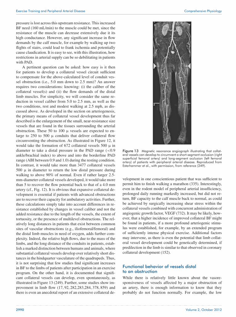
Exercise Training and Peripheral Arterial Disease Comprehensive Physiology
pressure is lost across this upstream resistance. This increasedBF need (160 mL/min) to the muscle could be met, since theresistance of the muscle can decrease extensively due it itshigh conductance. However, any significant increase in flowdemands by the calf muscle, for example by walking up twoflights of stairs, could lead to frank ischemia and potentiallycause claudication. It is easy to see, with this illustration, howrestrictions in arterial supply can be so debilitating in patientswith PAD.
A pertinent question can be asked: how easy is it thenfor patients to develop a collateral vessel circuit sufficientto compensate for the above-calculated level of conduit ves-sel obstruction (i.e., 5.0 mm down to 2.5 mm)? An answerrequires two considerations: knowing: (i) the caliber of thecollateral vessel(s) and (ii) the flow demands of the distallimb muscles. For simplicity, we will consider the same re-duction in vessel caliber from 5.0 to 2.5 mm, as well as thetwo conditions, rest and modest walking at 2.5 mph, as dis-cussed above. As developed in the section on arteriogenesis,the primary means of collateral vessel development thus fardescribed is the enlargement of the small, near-resistance sizevessels that are found in the tissues surrounding the arterialobstruction. These 50 to 100 μ vessels are expected to en-large to 250 to 500 μ conduits that deliver collateral flowcircumventing the obstruction. As illustrated in Figure 12, itwould take the formation of 672 collateral vessels 500 μ indiameter to take a distal pressure in the PAD range (<0.9ankle/brachial index) to above and into the borderline PADrange (ABI between 0.9 and 1.0) during the resting condition.In contrast, it would take more than 3477 collateral vessels500 μ in diameter to return the low distal pressure duringwalking to above 90% of normal. Even if rather larger 2.5-mm-diameter collateral vessels developed, it would take morethan 5 to recover the flow potential back to that of a 4.0 mmartery (cf., Fig. 12). It is obvious that expansive collateral de-velopment is essential if patients with advanced obstructionsare to recover their capacity for ambulatory activities. Further,these calculations simply take into account differences in re-sistance established by changes in vessel caliber and not theadded resistance due to the length of the vessels, the extent oftortuosity, or the presence of multilevel obstructions. The rel-atively long distances in patients that exist between commonsites of vascular obstructions (e.g., iliofemoral/femoral) andthe distal limb muscles in need of oxygen, adds further com-plexity. Indeed, the relative high flows, due to the mass of thelimbs, and the long distance of the conduits in patients, estab-lish a marked distinction between humans and animals, wheresubstantial collateral vessels develop over relatively short dis-tances in the hindquarter vasculature of the quadrupeds. Thus,it is not surprising that few studies find significant increasesin BF to the limbs of patients after participation in an exerciseprogram. On the other hand, it is documented that signifi-cant collateral vessels can develop, even spontaneously, asillustrated in Figure 13 (249). Further, some studies show im-provement in limb flow (17, 92, 282,283,284, 378, 859) andthere is even an anecdotal report of an extensive collateral de-
Figure 13 Magnetic resonance angiograph illustrating that collat-eral vessels can develop to circumvent a short-segment occlusion (rightsuperficial femoral artery) and long-segment occlusion (left femoralartery) of patients with peripheral arterial disease. Reproduced fromEsterhammer et al., with permission, from reference (249).
velopment in one conscientious patient that was sufficient topermit him to finish walking a marathon (335). Interestingly,even in the rodent model of peripheral arterial insufficiency,prolonged daily running markedly increased, but did not re-turn, BF capacity to the calf muscle back to normal, as couldbe achieved by surgically increasing shear stress within thecollateral vessels combined with concurrent administration ofangiogenic growth factor, VEGF (732). It may be likely, how-ever, that a higher incidence of improved collateral BF mightbe found in patients, if a more profound arteriogenic stimu-lus were established, for example, by an extended programof sufficiently intense physical exercise. Additional factorsmay intervene, as there is even the potential that limb collat-eral vessel development could be genetically determined, ifpredilection in the limb is similar to that observed in coronarycollateral development (152).
Functional behavior of vessels distalto an obstructionWhile there is relatively little known about the vasore-sponsiveness of vessels affected by a major obstruction ofan artery, there is enough information to know that theyprobably do not function normally. For example, the low
2990 Volume 2, October 2012

Comprehensive Physiology Exercise Training and Peripheral Arterial Disease
luminal pressure fostered on the downstream vasculature byan upstream obstruction would reduce radial wall tension ofeven otherwise normal vessels. A structural increase in the di-ameter of conduit vessels (saphenous a, soleus feed a, and calfmuscle feed a) ensues over time (915), possibly attempting torecover some of the lost radial wall tension that comes witha reduction in luminal pressure. This reduction in radial walltension has the potential to diminish the vasodilator responseof the vessel (914). A dulled FMD of the popliteal arterywas observed after ischemic exercise (leg occlusion), but notfollowing the same exercise without leg occlusion, even innormal healthy 17-year-old volunteers (73). Further, a dulledvasodilator response was observed in the small arteriolesof a distal limb muscle following upstream occlusion ofthe iliac artery (480). This could have contributed to thereduced conductance of the calf muscles following occlusionof the femoral artery, as compared to that of the normal flow,contralateral calf muscle (914). Whether an up regulation ofalpha-sympathetic receptors (802) in the distal vessels con-tributed to this response is not known. Similarly, exaggeratedresponses to norepinephrine (165, 459) have been observedin vessels obtained from patients with critical limb ischemia.However, this potential for an exaggerated vasoconstrictionof the limb vasculature seems inconsistent with the reducedsympathetic orthostatic response observed in patients withPAD (202). Interestingly, the dulled vasodilator response ofthe vasculature was improved by daily contractions of theischemic muscle, either by muscle stimulation (480) or bytreadmill walking (915). The evidence to date suggests thatthere is the potential to exacerbate the BF deficits caused bythe upstream arterial obstruction, if these aberrant responsesoccur in patients with PAD. On the other hand, the recoveryfrom vasomotor dysfunction with muscle contractions(480, 915) appears to offer the potential for improvement byparticipation in an exercise program.
Functional behavior of collateral vessels thatcircumvent an obstructionThe hemodynamic function of collateral vessels becomesunique among the traditional roles of vessels within thevasculature. For example, large conduit vessels are respon-sible to deliver flow downstream at a high rate, withoutsignificant resistance, and be responsive to luminal flowevents. Smaller resistance vessels, which are close to thetissue being perfused, are highly innervated to regulatecaliber, thereby exerting a high resistance to limit flow untillocal tissue demands initiate dilatation. Finally, capillariesfunction to “bathe the tissue in blood” to optimize diffusion ofmetabolites and gases between blood and cells. On the otherhand, to be effective, collateral vessels must function as “low”resistance conduits, albeit relatively small ones, but arisefrom even smaller arteriolar size vessels (113,118,813,1018).This raises the potential that these small vessels that are thesubstrate for the collateral circuit, retain their vasoconstrictorproperties as the vessels enlarge. Even the potential that local
tissue conditions exist to foster dilatation of the collateralvessels may not occur, since the site of collateral circuitis often within tissue that is not collateral dependent. Thisappears to be the case with proximal obstructions of largevessels (e.g., femoral artery), where the collateral circuitdevelops in the thigh region which enjoys a much higherBF than the collateral-dependent muscles of the lower limb(455, 1018, 1019). Thus, inordinate vasoconstriction of thecollateral circuit could exacerbate the difficulty in deliveringBF downstream, well beyond that established by the limitedphysical caliber of the collateral vessels themselves.
While very little is known about the vasomotor function ofthe collateral circuit, there are animal studies that have eval-uated the functional behavior of the collateral circuit in vivo,and the vasoresponsiveness of individual collateral vessels invitro. Opening of a nascent collateral circuit, following acuteocclusion of the femoral artery, can occur within minutes (776,777) and requires NO to be present (947,948,949,950,1026),likely related to FMD that must occur as these vessels beginto function. Further, NO continues to be important during theprocess of collateral vessel remodeling (947, 950). Collateralflow increases in response to the dilatory effect of adenosine(577). On the other hand, serotonin has been shown to causecollateral vasoconstriction (550). Further, a dominant sym-pathetic influence remains that tempers flow delivery down-stream. Blocking either alpha1-, alpha2-adrenergic receptors,or in combination, increases the conductance of the collat-eral circuit during exercise (914, 915). In addition, inhibitionof neuropeptide Y (NPY)-1 receptors, that would be respon-sive to the NPY released concurrent with release of nore-pinephrine (1054), increases the conductance of the collateralcircuit (915). Thus, sympathetic output, which is expected tobe increased in PAD (44,47) due to the muscle pressor reflex,likely serves to limit collateral BF during activity, by morethan that caused by the small structural diameter of the col-lateral vessels (e.g., the collateral plumbing). Interestingly, itmay be that exercise training has the potential of amelioratingthis detrimental condition in patients, since the muscle pres-sor reflex is less robust after training, even in normal healthysubjects (640,742) and collateral circuit conductance was im-proved by exercise training in experimental peripheral arterialinsufficiency (915).
Evaluating the vasoresponsiveness of individual collat-eral vessels in vitro provided consistent evidence with the invivo assessment discussed above, in demonstrating that anexaggerated vasoconstriction to increasing luminal flow wasreversed by exercise training (170). This assessment was per-formed on the reentry portion of a collateral vessel, whichdelivers collateral BF into the distal vasculature to completethe circuit bypassing the obstruction. Thus, it may not rep-resent the intermediate collateral vessel segments that con-nect the entry vessels to these reentry vessels. It is appar-ent that this preexisting vessel, that was coopted to serveas a collateral vessel, remodeled following occlusion of thefemoral artery. As illustrated in Figure 14, inhibition of thecyclooxygenase pathway with indomethacin reversed the in-
Volume 2, October 2012 2991

Exercise Training and Peripheral Arterial Disease Comprehensive Physiology
Collateral vessel responses to flow
80
60
40
20
0
–20
–40
–600 5 10 15
Shear stress (dynes/cm2)
Cha
nge
in d
iam
eter
(µm
)
Occludedtrained
Occludedsedentary
45 cmH2O
+Indo+L-Name
+Indo+L-Name
20 25 30
Figure 14 Influence of exercise training on the vasoresponsivenessof a collateral vessel as a function of shear stress. An initial modestdilatation to low shear stress in control animals (open circles) revertedto a dominant vasoconstriction at high shear stress. This responsewas eliminated in the presence of indomethacin, NG-nitro-L-argininemethyl ester (L-NAME), and in combination, as illustrated (filled cir-cles), to a modest vasodilatation at very high shear stress. In contrast,collateral vessels from trained animals exhibited a marked vasodila-tion in the presence of indomethacin, L-NAME, and in combination,as illustrated (filled squares). This implies that exercise training inducesa cyclooxygenase- and nitric oxide species-independent stimulus forvasodilatation. Data taken from Colleran et al.(170), with permission.
herent flow-mediated vasoconstriction to a modest vasodilata-tion, compared to normal vessels perfused at the low luminalpressure common to this vessel (170). Further, removing theinfluence of this cyclooxygenase product, putatively throm-boxane A2, in the vessels from trained animals resulted in arobust vasodilatation. Indeed, this marked flow-mediated va-sodilatation was impervious to NO removal with L-NAMEor by loss of both NOS and cyclooxygenase products. Thisindicates that exercise training causes a remodeling of thecollateral vessel to favor vasodilatation via enhanced pro-duction of the putative endothelium-derived hyperpolarizingfactor(s) (170). If these findings were applicable to patientswith PAD, there could be an improved collateral vessel func-tion and enhanced exercise tolerance following participationin a program of routine physical activity.
Cardiovascular Control in Patientswith Peripheral ArterialPAD is thought to be caused by a general atheroscle-rotic condition exacerbated by a chronic inflammatory state(55, 483, 835). There are a number of expected health conse-quences associated with this condition that appear as comor-bidities, including ischemic heart and cerebral vascular dis-
ease (213, 661, 829), hypertension (55), hyperlipidemia (55),diabetes (745,837), renal disease (55), and even obesity (724),and often with attendant elevated biomarkers (e.g., PAI-1 andCRP) (483). Thus, it is common that patients with PAD willexhibit cardiac, vascular, and metabolic dysfunction, resultingin a reduced activity level (55, 596). Many of these comor-bidities may be directly related to cardiovascular dysfunctionand thereby contribute to the increased morbidity and shorterlife span of PAD patients. However, PAD patients have anincreased risk for cardiovascular morbidity and mortality thatis independent of the typical risk factors for atherosclero-sis (596, 862), implying that features other than generalizedatherosclerotic disease are consequential. Further, there is asubset of PAD patients that do not present with ischemic heartdisease or cerebral vascular disease. Estimates place this pop-ulation at approximately 35% to 50% of all patients with PAD(596,656). Yet, the risk of premature death and increased mor-bidity is just the same as those patients with attendant cardiacand cerebral vascular disease (180, 862). Thus, the presenceof ostensibly “pure” PAD is not without its ominous risk ofpremature death. It is presently unclear what factor or factorscontribute to this unfortunate outcome, but some insight canbe gained by understanding cardiovascular dysregulation thatis likely promulgated by ischemia to the legs.
There is a reflex neural input from the periphery that con-tributes to cardiovascular regulation during exercise, termedthe exercise pressor reflex (476, 482, 669, 858, 865). Duringaerobic-type rhythmical exercise, afferent nerve traffic, aris-ing from within active muscles, stimulates the sympatheticcenters in the medulla. This neural input serves to activatesympathetic output, contributing to heightened cardiac func-tion and vascular control to support exercise. This, along withhigher center activation, increases cardiac output and the drivefor vasoconstriction throughout the body, especially the skin,kidney, and gastrointestinal tract. While this vasoconstrictoroutflow also extends to all muscles, there is lysis of this effectin the contracting muscles, where local vasodilatation greatlyenhances BF, thereby decreasing peripheral resistance andtempering any significant increase in blood pressure. Thus, theimportance of the muscle pressor response is to contribute tosympathetic control during exercise and help redistribute car-diac output to the active muscles (780). Interestingly, if exer-cise is static (isometric), with only high force development bythe muscle, there is little to no decrease in vascular resistancenor elevation in BF through the muscle. As a result, bloodpressure increases inordinately, ergo, the term–ischemic pres-sor response–is sometime used. Under this condition, the af-ferent input from the active muscle is greatly increased, result-ing in even greater elevation in blood pressure. An extremein elevated blood pressure is realized during exceptionallyhigh-load weight lifting of large muscle groups (564).
Components of the exercise pressor reflexThere are at least two features established within active mus-cle that prompt afferent traffic centrally: mechanoreceptors,
2992 Volume 2, October 2012

Comprehensive Physiology Exercise Training and Peripheral Arterial Disease
driven by tension within the muscle caused by either activeforce development or passive stretch, and metaboreceptors,acting as “chemical” sensors that are responsive to a widevariety of stimuli created within the environment of ischemicmuscle (476,482,669,858,865). These two sources were orig-inally distinguished in experiments where a large fraction(≥50%) of the hypertension developed during prolonged is-chemic isometric contractions, and continued as long as thetourniquet remained in place, even though the contractionswere stopped (15). Mechanosensors within the muscle areresponsive to muscle force, initiating afferent nerve traffic tothe brain, predominantly via group III nerves (477), in a man-ner proportional to the force developed (864). On the otherhand, metaboreceptors within the muscle initiate afferent in-put primarily via group IV nerves and are responsive to a widevariety of factors (e.g., lactate, H+, ATP, ROS, bradykinin, andcapsaicin) via a variety of receptors and sensitive membranechannels (494, 496, 858, 980). Interestingly, the intensity ofneural activation depends upon the type of muscle fiber con-tracting. Force development in the high-oxidative slow-twitch(type I) motor units prompts a lesser degree of afferent ac-tivation than when the low-oxidative fast-twitch (type IIb)motor units are contracting (38,1015). Thus, the muscle pres-sor reflex should become more exaggerated as the intensityof contraction increases in severity, as the low-oxidative fast-twitch motor units become more highly recruited. This likelycontributed to the exceptionally high blood pressures, in ex-cess of 300/200 mmHg, observed in experienced weight lifterswho performed near-maximally intense contractions of largemuscle groups (564, 693).
Exercise pressor reflex in heart failureGermane to the present consideration are elements of theexercise pressor reflex that change in conditions typical ofchronic disease, such as heart disease (858, 865) and hy-pertension (201, 633). For example, an exaggerated exercisepressor reflex, measured as hypertension and/or an elevatedsympathetic nerve output, is observed in patients with heartfailure (716, 848, 853). Studies evaluating the physiologicalbases for this exaggerated response, in animal models ofheart failure (279, 280, 482, 495, 496, 978, 979, 997), havefound that the mechanically sensitive fibers are overactive(531, 616, 617, 863, 866), whereas the metabolically sensitiveafferents are less responsive to activation (531,868,885). Thereasons for the increased responsiveness of the mechanore-flex remains unclear, whereas the loss in the metaboreflexis due, in part, to less active receptors on group IV afferentneurons, the transient receptor potential vanilloid 1 (TRPV1)(531, 868) and cannabinoid 1 receptors (997). It is possi-ble that this exaggerated exercise pressor reflex contributesto the hypersympathetic state that typifies heart failure pa-tients (260). Interestingly, animals with heart failure that areexercise trained reverse their low metaboreflex and elevatedmechanoreflex back toward normal (979).
Exercise pressor reflex in hypertensionThe exercise pressor reflex is exaggerated in hypertensivepatients and animals models of hypertension. Hypertensivepatients exhibit an enhanced elevation in blood pressure (28,479,805) and sympathetic nerve output (201) during ischemichandgrip exercise. It is likely that the metaboreflex contributedto this response in patients, since the blood pressure andsympathetic nerve activity remain elevated following contrac-tions, when the tourniquet cuff was left intact (201). Smithand co-workers also found that the exercise pressor reflex wasexaggerated in spontaneously hypertensive rats (867), due tochanges in both the mechano- and metaboreflexes (520,633).The exaggerated exercise pressor reflex in the condition ofhypertension seems rather predictable, since there is alreadydysregulation in blood pressure control. On the other hand, aswith heart failure, differences arising from the active muscleare intriguing. While there are many contributors to hyperten-sion, alterations in the central neural control centers can beimportant (260,614). Interestingly, even with long-term bloodpressure correction of hypertension in patients, the exercisepressor reflex remained somewhat elevated as compared tonormal healthy subjects (479). This raises the probability thatthe exaggerated exercise pressor reflex observed in the hyper-tensive condition involves more than just an enhanced afferentneural traffic from the contracting muscles. Future researchwill, no doubt, reveal some of the complex interactions amongthe mechano- and metaboreflex inputs and alterations amongthe central cardiovascular control elements.
Exercise pressor reflex in peripheralarterial insufficiencyIt is likely that the exercise pressor response contributes to hy-pertension in patients with PAD. However, it is not possible tomake a definitive assignment of the hypertension, commonlyobserved in these patients, to muscle afferent traffic, since hy-pertension is its own pathology and a common comorbiditywith PAD (55). On the other hand, as illustrated in Figure 15,the exercise pressor reflex could contribute to the frank hy-pertension that was induced during exercise in patients withPAD, as compared to age-matched controls (44, 47). The el-evated blood pressure was evident early during the treadmillwalk, well before the time of pain onset or when the patientshad to stop walking (47). As expected, the hypertension inPAD patients is a function of exercise intensity (44). Thiscould invoke another possible contributor to the elevated ex-ercise pressor reflex. PAD patients often exhibit a loss ofmuscle mass, which could foster earlier recruitment of themore reflex-responsive fast-twitch motor units. Thus, prob-able activation of these fast-twitch motor units, to achieveeven a modest intensity of locomotion, would exaggerate theafferent nerve traffic from the muscle (38, 1015). It is ap-parent, however, that differences in the muscle reflex occur,since even young healthy subjects will demonstrate an ex-ercise hypertension while walking when cuffs occlude limb
Volume 2, October 2012 2993

Exercise Training and Peripheral Arterial Disease Comprehensive Physiology
Figure 15 Example of hypertension during exercise in a group ofpatients with peripheral arterial disease who exhibit claudication. Notethat the elevation in blood pressure in the claudicant group is greaterthan that of aged-matched control group well prior to the cessation ofwalking. Figure reproduced from Bakke et al.(47), with permission.
BF (73). Interestingly, the absence of an exaggerated exercisepressor reflex between PAD patients and healthy control sub-jects, when induced by handgrip exercise (48), implies thatthe changes in the exercise pressor response with PAD are notgeneralized, but dependent upon afferent nerve traffic derivedfrom a diseased ischemic limb. Indeed, animal experimentshave shown this specificity, since the afferent neurons in thedorsal root ganglion remodel only at the cord level innervat-ing the muscles that are ischemic (542), the elevated exercisepressor reflex was observed only in the ischemic limb and notthe free-flow perfused contralateral limb of the same animal(936), and the dilatory capacity of ischemic muscle duringtreadmill exercise was markedly less than that of the normalflow, nonischemic muscle (914, 915). Further, the presence ofthe exaggerated exercise pressor reflex was dependent uponmuscle contractions, as neuromuscular blockade and dener-vation of the afferent loop of the reflex completely eliminatedthe pressor response.
Recent evidence has provided significant insights intohow the exercise pressor response is exaggerated in exper-imental animals with peripheral arterial insufficiency causedby femoral artery occlusion (542, 936,937,938, 1013, 1014).Femoral artery occlusion induces an increase in the exer-cise pressor response prompted by both muscle contractionsand passive stretch of the muscle, implicating both mechano-and metaboreceptors in the response. The time-course pos-tocclusion needed to observe this increase is surprisingly
brief, within 24 h, leading to a near-maximal response in72 h (542); yet, the exaggerated exercise pressor reflex isnot observed with acute occlusion (3 min) of the femoralartery (936). Further, the enhanced exercise pressor reflex isobserved without frank ischemia within the limb muscles.Occlusion of the femoral artery, as done in these studies, re-moves approximately 75% to 85% of the flow reserve to thecalf muscle (1026); however, nascent collateral blood capac-ity to the limb is sufficient to supply the calf muscle withBF (1021, 1026) that is approximately threefold greater than“resting” BF observed in muscle of quiescent anesthetizedanimals (565, 566). Thus, while the exercise capacity of theseanimals is greatly limited, there should be little stress on themuscle during limited cage activity. Nonetheless, factors mustbe promulgated within the muscle by femoral artery occlu-sion that initiate fairly immediate remodeling of the neuronalsensing and/or propagation processes responsive to tissuetension and/or metabolites, including: an increased responseto acid-sensing ion channels (e.g., responsive to lactic acid)(542,939); transient receptor potential vanilloid type I recep-tor (nociception) up regulation in the afferent dorsal root neu-rons (1013), possibly driven by up regulation of nerve growthfactor (543, 1014) although inhibition of TRPV1 receptorsdid not lessen the elevated exercise pressor response (936);inhibition of the exaggerated pressor response by activatingmicro-opioid receptors, likely implicating metaboreceptors(937); and sensitivity of the exaggerated exercise pressor re-flex to tetrodotoxin inhibition of sodium channels, but notTTX-resistant sodium channels in dorsal root ganglia (938);and the action of tempol, but not likely due to its ROS buffer-ing capacity (594). These important papers demonstrate thatcritical changes occur in the afferent arm of the exercise pres-sor reflex in the condition of peripheral arterial insufficiency,leading to an enhanced afferent traffic to the central cardiovas-cular control centers. Whether there are also adaptive changesinduced within the central cardiovascular control centers thatcould ameliorate or exacerbate conditions in PAD patients isunknown. Thus, it may be hypothesized that PAD patientsare at risk of hypertension during activity and that this el-evated afferent nerve traffic could cause neural adaptationsthat “bias” central neural cardiovascular control and lead to ahypersympathetic state even at rest, which has been observed(419). The added risks of premature death and morbidity, dueto a hypersympathetic condition, have been well recognized(260). Such a causative link could help explain the equivalentrisks of morbidity and mortality in PAD patients, even in theabsence of cardiac or cerebral vascular disease (180, 862).
Central cardiovascular control centers influencesympathetic outputRecent research has established that critical areas within thebrain contribute to the hypersympathetic output that appearscommon to cardiovascular diseases (641). While the com-plex interactions among the numerous important loci withinthe brain are beyond the scope of this review, a couple of
2994 Volume 2, October 2012

Comprehensive Physiology Exercise Training and Peripheral Arterial Disease
brain areas have received the most attention and merit com-ment. The paraventricular nucleus (PVN) in the hypothala-mus and the rostral ventrolateral medulla (RVLM) have be-come a major focus for evaluating experimental heart failure(1051,1053) and hypertension (256). In each case, substantialderangements of these centers, related to inherent alterationsof excitatory/inhibitor input, have been observed. For exam-ple, in experimental heart failure there is a modified behaviorof the PVN related to a reduced bioavailability of NO (703)and enhanced angiotensin II activation (1042), possible dueto a down regulation of local angiotensin converting enzyme(474) that contributes to the hypersympathetic state. Simi-larly, a deficit in NO bioavailability in the RVLM contributesto the hypertension characteristic of spontaneous hyperten-sive rats (142, 489). Interestingly, exercise training reversesthe hypersympathetic state (1052) in heart failure models, ap-parently by improving the antioxidant condition (280), NObioavailability (1043), and by reducing the excitatory influ-ence of glutamate in the PVN (493). Even in the absence ofimputed disease conditions, normal animals that are exercisetrained exhibit a smaller response to excitatory stimuli (glu-tamate) in the RVLM (641). Thus, recent work has developedcompelling evidence that central cardiovascular centers aresubject to dysregulation, found to be plastic, and potentiallyresponsive to afferent nerve traffic to modify their behavior.This raises the potential that significant changes could be pro-mulgated onto these control centers by a barrage of afferentnerve traffic arising from the exaggerated exercise pressorreflex observed in the condition of peripheral arterial insuf-ficiency. Further, the above-mentioned training responses inexperimental heart failure and hypertension raise the potentialthat exercise training in patients with PAD could induce ben-eficial central neural adaptations. However, these hypothesesremain to be tested.
ConclusionIt is apparent that involvement in recommended exercise pre-scription brings about a multitude of adaptations that are ben-eficial to the patient with PAD. While much is known aboutthese adaptations, the signals that bring them about, and therealized impact of these changes, there is much to be learnedof the impact of exercise in the condition of PAD. New ad-vances in technology are needed to assist in the managementof these patients. For example, there is difficulty in assessingthe extent of BF deficit to the diseased limb, in identifyingthe sites of vulnerability within the limb, and in evaluating theextent of therapeutic intervention. Further, while exercise pre-scription is one of the most effective means of managing PAD,in the absence of contraindications achieving compliance is asignificant clinical problem. Thus, there are many avenues foradvances in scientific inquiry, application of technology, clin-ical management of PAD patients, and in educating the publicof the need for life style changes to enhance the amount ofdaily physical activity.
AcknowledgementsCited work by the authors was supported by the followinggrants: TLH: Canadian Institutes of Health Research Grant#107537, Heart and Stroke Foundation of Canada Grant in Aid#NA 7059, and National Science and Engineering Council ofCanada Grant #222865; PGL: NHLBI F32-HL10406, NHLBIR01-HL084494; HTY and RLT: NIAMS R37-AR21617,NHLBI R01-HL37387, NHLBI P01-HL52490, NIAMS T32-AR048523, and NHLBI F32-HL10485.
References1. Abaci A, Oguzhan A, Kahraman S, Eryol NK, Unal S, Arinc H, Ergin
A. Effect of diabetes mellitus on formation of coronary collateralvessels. Circulation 99: 2239-2242, 1999.
2. Abou-Khalil R, Le Grand F., Pallafacchina G, Valable S, AuthierFJ, Rudnicki MA, Gherardi RK, Germain S, Chretien F, Sotiropou-los A, Lafuste P, Montarras D, Chazaud B. Autocrine and paracrineangiopoietin 1/Tie-2 signaling promotes muscle satellite cell self-renewal. Cell Stem Cell 5: 298-309, 2009.
3. Abounader R, Laterra J. Scatter factor/hepatocyte growth factor inbrain tumor growth and angiogenesis. Neuro Oncol 7: 436-451,2005.
4. Abumiya T, Sasaguri T, Taba Y, Miwa Y, Miyagi M. Shear stressinduces expression of vascular endothelial growth factor receptor Flk-1/KDR through the CT-rich Sp1 binding site. Arterioscler ThrombVasc Biol 22: 907-913, 2002.
5. Ackah E, Yu J, Zoellner S, Iwakiri Y, Skurk C, Shibata R,Ouchi N, Easton RM, Galasso G, Birnbaum MJ, Walsh K,Sessa WC. Akt1/protein kinase B alpha is critical for ischemicand VEGF-mediated angiogenesis. J Clin Invest 115: 2119-2127,2005.
6. Adair TH. Growth regulation of the vascular system: An emergingrole for adenosine. Am J Physiol 289: R283-R296, 2005.
7. Adams RH, Eichmann A. Axon guidance molecules in vascular pat-terning. Cold Spring Harb Perspect Biol 2: a001875, 2010.
8. Adolfsson J, Ljungqvist A, Tornling G, Unge G. Capillary increasein the skeletal muscle of trained young and adult rats. J Physiol 310:529-532, 1981.
9. Afaq A, Montgomery PS, Scott KJ, Blevins SM, Whitsett TL, Gard-ner AW. The effect of current cigarette smoking on calf musclehemoglobin oxygen saturation in patients with intermittent claudi-cation. Vasc Med 12: 167-173, 2007.
10. Agency for Healthcare Research and Quality. Treatment strategiesfor patients with peripheral areterial disease (PAD). EffectiveHealth Care Program www.effectivehealthcare.ahrq.gov/index.cfm/search-for-guides-reviews-and-reports/?productid=948&pageaction=displayproduct: 1-35, 2012.
11. Ahimastos AA, Pappas EP, Buttner PG, Walker PJ, Kingwell BA,Golledge J. A meta-analysis of the outcome of endovascular and non-invasive therapies in the treatment of intermittent claudication. J VascSurg 54: 1511-1521, 2011.
12. Akimoto T, Pohnert S, Li P, Zhang M, Gumbs C, Rosenberg P,Williams RS, Yan Z. Exercise stimuluates PGC-1alpha transcriptionin skeletal muscle through activation of the p38 MAPK pathway. JBiol Chem 280: 19587-19593, 2005.
13. Akiri G, Nahari D, Finkelstein Y, Le SY, Elroy-Stein O, Levi BZ.Regulation of vascular endothelial growth factor (VEGF) expression ismediated by internal initiation of translation and alternative initiationof transcription. Oncogene 17: 227-236, 1998.
14. Al H, Zen A., Oikawa A, Bazan-Peregrino M, Meloni M, EmanueliC, Madeddu P. Inhibition of delta-like-4-mediated signaling impairsreparative angiogenesis after ischemia. Circ Res 107: 283-293, 2010.
15. Alam M, Smirk FH. Observations in man upon a blood pressure raisingreflex arising from the voluntary muscles. J Physiol 146: 372-383,1937.
16. Allen JD, Stabler T, Kenjale A, Ham KL, Robbins JL, Duscha BD,Dobrosielski DA, Annex BH. Plasma nitrite flux predicts exerciseperformance in peripheral arterial disease after 3 months of exercisetrainng. Free Radic Biol Med 49: 1138-1144, 2010.
17. Alpert JS, Larsen OA, Lassen NA. Exercise and intermittent claudi-cation. Blood flow in the calf muscle during walking studied by thexenon-133 clearance method. Circulation 39: 353-359, 1969.
18. Alroy J, Goyal V, Skutelsky E. Lectin histochemistry of mammalianendothelium. Histochemistry 86: 603-607, 1987.
Volume 2, October 2012 2995

Exercise Training and Peripheral Arterial Disease Comprehensive Physiology
19. Amaral SL, Linderman RJ, Morse MM, Greene AS. Angiogenesisinduced by elecrical stimulation is mediated by angiotensis II andVEGF. Microcirculation 8: 57-67, 2001.
20. Ameln H, Gustafsson T, Sundberg CJ, Okamoto K, Jansson E,Poellinger L, Makino Y. Physiological activation of hypoxia induciblefactor-1 in human skeletal muscle. FASEB J 19: 1009-1011, 2005.
21. Andersen P, Henriksson J. Capillary supply of the quadriceps femorismuscle of man: Adaptive response to exercise. J Physiol 270: 677-690,1977.
22. Andersen P, Saltin B. Maximal perfusion of skeletal muscle in man.J Physiol 366: 233-249, 1985.
23. Andreozzi GM, Leone A, Laudani R, Deinite G, Martini R. Acuteimpairment of the endothelial function by maximal treadmill exercisein patients with intermittent claudication, and its improvement aftersupervised physical training. Int Angiol 26: 12-17, 2007.
24. Andreozzi GM, Martini R, Cordova R, D’Eri A, Salmistraro G, Mus-sap M, Plebani M. Circulating levels of cytokines (IL-6 and IL-1beta)in patients with intermittent claudication, at rest, after maximal exer-cise treadmill test and during restore phase. Could they be progressionmarkers of the disease? International Angiology 26: 245-252, 2007.
25. Angersbach D, Jukna JJ, Nicholson CD, Ochlich P, Wilke R. Theeffect of short-term and long-term femoral artery ligation on rat calfmuscle oxygen tension, blood flow, metabolism and function. Int JMicrocirc Clin Exp 7: 15-30, 1988.
26. Annex BH, Torgan CE, Lin P, Taylor DA, Thompson MA, Peters KG,Kraus WE. Induction and maintenance of increased VEGF protein bychronic motor nerve stimulation in skeletal muscle. Am J Physiol 274:H860-H867, 1998.
27. Aoi W, Naito Y, Mizushima K, Takanami Y, Kawai Y, IshikawaH, Yoshikawa T. The microRNA miR-696 regulates PGC-1alpha inmouse skeletal muscle in response to physical activity. Am J Physiol298: E799-E806, 2010.
28. Aoki K, Sato K, Kondo S, Pyon CB, Yamamoto M. Increased responseof blood pressure to rest and handgrip in subjects with essential hy-pertension. Jpn Circ J 47: 802-809, 1983.
29. Aoki M, Morishita R, Taniyama Y, Kida I, Moriguchi A, MatsumotoK, Nakamura T, Kaneda Y, Higaki J, Ogihara T. Angiogenesis in-duced by hepatocyte growth factor in non-infarcted myocardium andinfarcted myocardium: Up-regulation of essential transcription factorfor angiogenesis, ets. Gene Ther 7: 417-427, 2000.
30. Arany Z. PGC-1 coactivators and skeletal muscle adaptations in healthand disease. Curr Opin Genet Dev 18: 426-434, 2008.
31. Arany Z, Foo SY, Ma Y, Raus JL, Bommi-Reddy A, Gimun G, CooperM, Laznik D, Chinsomboon J, Rangwala SM, Baek KH, RosenzweigA, Spiegelman BM. HIF-independent regulation of VEGF and angio-genesis by the transcriptional coactivator PGC-1alpha. Nature 451:1008-1012, 2008.
32. Ardanaz N, Pagano PJ. Hydrogen peroxide as a paracrine vascularmediator: Regulation and signaling leading to dysfunction. Exp BiolMed 231: 237-251, 2006.
33. Armstrong RB, Laughlin MH. Rat muscle blood flows during highspeed locomotion. J Appl Physiol 59: 1322-1328, 1985.
34. Arras M, Ito WD, Scholz D, Winkler B, Schaper J, Schaper W. Mono-cyte activation in angiogenesis and collateral growth in the rabbithindlimb. J Clin Invest 101: 40-50, 1998.
35. Asahara T, Bauters C, Zheng LP, Takeshita S, Bunting S, Ferrara N,Symes JF, Isner JM. Synergistic effect of vascular endothelial growthfactor and basic fibroblast growth factor on angiogenesis in vivo.Circulation 92: II365-II371, 1995.
36. Ashton N. Neovascularization in ocular disease. Trans OphthalmolSoc UK 81: 145-161, 1961.
37. Ashworth NL, Chad KE, Harrison EL, Reeder BA, Marshall SC.Home versus center based physical activity programs in older adults.Cochrane Database Syst Rev 25: CD004017, 2005.
38. Askew CD, Green S, Walker PJ, Kerr GK, Green AA, Williams AD,Febbraio MA. Skeletal muscle phenotype is associated with exerciseintolerance in patients with peripheral arterial disease. J Vasc Surg 41:802-807, 2005.
39. Astrand PO, Rodahl K. Textbook of Work Physiology. New York:McGraw-Hill Book Company, 1970.
40. Augustin HG, Koh GY, Thurston G, Alitalo K. Control of vascularmorphogenesis and homeostasis through the angiopoietin-Tie system.Nat Rev Mol Cell Biol 10: 165-177, 2009.
41. Ausprunk DH, Folkman J. Migration and proliferation of endothelialcells in preformed and newly formed blood vessels during tumorangiogenesis. Microvasc Res 14: 53-65, 1977.
42. Azuma N, Duzgun SA, Ikeda M, Kito H, Akasaka N, Sasajima T,Sumpio BE. Endothelial cell response to different mechanical forces.J Vasc Surg 32: 789-794, 2000.
43. Baar K, Wende AR, Jones TE, Marison M, Nolte LA, Chen M, KellyDP, Holloszy JO. Adaptations of skeletal muscle to exercise: Rapidincrease in the transcriptional coactivator PGC-1. FASEB J 16: 1879-1886, 2002.
44. Baccelli G, Reggiani P, Mattioli A, Corbellini E, Garducci S, CatalanoM. The exercise pressor reflex and changes in radial arterial pressureand heart rate during walking in patients with arteriosclerosis obliter-ans. Angiology 50: 31-374, 1999.
45. Badr I, Brown MD, Egginton S, Hudlicka O, Milkiewicz M, VerhaegJ. Differences in local environment determine the site of physiologicalangiogenesis in rat skeletal muscle. Exp Physiol 88: 565-568, 2003.
46. Baffour R, Berman J, Garb JL, Rhee SW, Kaufman J, Friedmann P.Enhanced angiogenesis and growth of collaterals by in vivo adminis-tration of recombinant basic fibroblast growth factor in a rabbit modelof acute lower limb ischemia: Dose-response effect of basic fibroblastgrowth factor. J Vasc Surg 16: 181-191, 1992.
47. Bakke EF, Hisdal J, Jorgensen JJ, Kroese A, Stranden E. Blood pres-sure in patients with intermittent claudication increases continuouslyduring walking. Eur J Vasc Endovasc Surg 33: 20-25, 2007.
48. Bakke EF, Hisdal J, Kroese AJ, Jorgensen JJ, Stranden E. Bloodpressure response to isometric exercise in patients with peripheralatherosclerotic disease. Clin Physiol Funct Imaging 27: 109-115,2007.
49. Baldwin KM, Klinkerfuss GH, Terjung RL, Mole PA, Holloszy JO.Respiratory capacity of white, red, and intermediate muscle: Adaptiveresponse to exercise. Am J Physiol 222: 373-378, 1972.
50. Ballard HJ. The influence of lactic acid on adenosine release fromskeletal muscle in anaesthetized dogs. J Physiol 433: 95-108, 1991.
51. Ballard HJ. The role of intracellular pH in the control of adenosineoutput from red skeletal muscle. Biol Signals 4: 168-173, 1995.
52. Banai S, Jaklitsch MT, Shou M, Lazarous DF, Scheinowitz M, Biro S,Epstein SE, Unger EF. Angiogenic-induced enhancement of collateralblood flow to ischemic myocardium by vascular endothelial growthfactor in dogs. Circulation 89: 2183-2189, 1994.
53. Banai S, Shweiki D, Pinson A, Chandra M, Lazarovici G, KeshetE. Upregulation of vascular endothelial growth factor expression in-duced by myocardial ischaemia: Implications for coronary angiogen-esis. Cardiovasc Res 28: 1176-1179, 1994.
54. Barleon B, Sozzani S, Zhou D, Weich HA, Mantovani A, Marme D.Migration of human monocytes in response to vascular endothelialgrowth factor (VEGF) is mediated via the VEGF receptor flt-1. Blood87: 3336-3343, 1996.
55. Bartholomew JR, Olin JW. Pathophysiology of peripheral arterialdisease and risk factors for its development. Cleveland Clin J Med 73:S8-S14, 2006.
56. Bastide A, Karaa Z, Bornes S, Hieblot C, Lacazette E, Prats H, TouriolC. An upstream open reading frame within an IRES controls expres-sion of a specific VEGF-A isoform. Nucleic Acids Res 36: 2434-2445,2008.
57. Bates DO, MacMillan PP, Manjaly JG, Qiu Y, Hudson SJ, BevanHS, Hunter AJ, Soothill PW, Read M, Donaldson LF, Harper SJ.The endogenous anti-angiogenic family of splice variants of VEGF,VEGFxxxb, are down-regulated in pre-eclamptic placentae at term.Clin Sci (Lond) 110: 575-585, 2006.
58. Bauer TA, Brass EP, Barstow TJ, Hiatt WR. Skeletal muscle StO2kinetics are slowed during low work rate calf exercise in peripheralarterial disease. Eur J Appl Physiol 100: 143-151, 2007.
59. Baum O, Silva-Azevedo L, Willerding G, Wockel A, Planitzer G,Gossrau R, Pries AR, Zakrzewicz A. Endothelial NOS is mainmediator for shear stress-dependent angiogenesis in skeletal mus-cle after prazosin administration. Am J Physiol 287: H2300-H2308,2004.
60. Baumgartner I, Pieczek Manor O, Blair R, Kearney M, Walsh K,Isner JM. Constitutive expression of phVEGF165 after intramusculargene transfer promotes collateral vessel development in patients withcritical limb ischemia. Circulation 97: 1114-1123, 1998.
61. Bauters C, Asahara T, Zheng LP, Takeshita S, Bunting S, FerraraN, Symes JF, Isner JM. Site-specific therapeutic angiogenesis aftersystemic administration of vascular endothelial growth factor. J VascSurg 21: 314-324, 1995.
62. Bayless KJ, Davis GE. The Cdc42 and Rac1 GTPases are required forcapillary lumen formation in three-dimensional extracellular matrices.J Cell Sci 115: 1123-1136, 2002.
63. Beavers KM, Brinkley TE, Nicklas BJ. Effect of exercise training onchronic inflammation. Clin Chim Acta 411: 785-793, 2010.
64. Becker LC, Pitt B. Collateral blood flow in conscious dogs withchronic coronary artery occlusion. Am J Physiol 221: 1507-1510,1971.
65. Behnke BJ, Kindig CA, Musch TI, Koga S, Poole DC. Dynamics ofmicrovascluar oxygen pressuer across the rest-exercise transition inrat skeletal muscle. Respir Physiol 126: 53-63, 2001.
66. Behnke BJ, McDonough P, Padilla DJ, Musch TI, Poole DC. Oxygenexchange profile in rat muscles of ontrol of microvasc. J Physiol 549:597-605, 2003.
67. Belch JJF, McLaren M, Kahn F, Hickman P, Muir A, Stonebridge P.The inflammatory process in intermittent claudication. Eur Heart J 4:B31-B34, 2002.
2996 Volume 2, October 2012

Comprehensive Physiology Exercise Training and Peripheral Arterial Disease
68. Bellamy LM, Johnston AP, De Lisio M, Parise G. Skeletal muscle-endothelial cell cross talk through angiotensin II. Am J Physiol 299:C1402-C1408, 2010.
69. Bendermacher BL, Willigendael EM, Teijink JA, Prins MH. Super-vised exercise therapy versus non-supervised exercise therapy for in-termittent claudication. Cochrane Database Syst Rev 19: CD005263,2006.
70. Benedict KF, Coffin GS, Barrett EJ, Skalak TC. Hemodynamic sys-tems analysis of capillary network remodeling during the progressionof type 2 diabetes. Microcirculation 18: 63-73, 2011.
71. Benedito R, Roca C, Sorensen I, Adams S, Gossler A, Fruttiger M,Adams RH. The notch ligands Dll4 and Jagged1 have opposing effectson angiogenesis. Cell 137: 1124-1135, 2009.
72. Benoit H, Jordan M, Wagner H, Wagner PD. Effect of NO, vasodilatorprostaglandins, and adenosine on skeletal muscle angiogenic growthfactor gene expression. J Appl Physiol 86: 1513-1518, 1999.
73. Benzi CP, Tanaka H, Sugawara J. Effects of leg blood flow restrictionduring walking on cardiovascular function. Med Sci Sports Exer 42:726-732, 2010.
74. Bergers G, Brekken R, McMahon G, Vu TH, Itoh T, Tamaki K,Tanzawa K, Thorpe P, Itohara S, Werb Z, Hanahan D. Matrixmetalloproteinase-9 triggers the angiogenic switch during carcino-genesis. Nat Cell Biol 2: 737-744, 2000.
75. Bergmann CE, Hoefer IE, Meder B, Roth H, van Royen N, Breit SM,Jost MM, Aharinejad S, Hartmann S, Buschmann IR. Arteriogenesisdepends on circulating monocytes and macrophage accumulation andis severely depressed in op/op mice. J Leukoc Biol 80: 59-65, 2006.
76. Berra E, Benizri E, Ginouves A, Volmat V, Roux D, Pouyssegur J. HIFprolyl-hydroxylase 2 is the key oxygen sensor setting low steady-statelevels of HIF-1alpha in normoxia. EMBO J 22: 4082-4090, 2003.
77. Betsholtz C, Lindblom P, Gerhardt H. Role of pericytes in vascularmorphogenesis. EXS 115-125, 2005.
78. Bey L, Akunuri N, Zhao P, Hoffman EP, Hamilton DG, HamiltonMT. Patterns of global gene expression in rat skeletal muscle duringunloading and low-intensity ambulatory activity. Physiol Genomics13: 157-167, 2003.
79. Bigard AX, Brunet A, Guezennec CY, Monod H. Effects of chronichypoxia and endurance training on muscle capillarity in rats. PflugersArch 419: 225-229, 1991.
80. Birot OJ., Koulmann N, Peinnequin A, Bigard XA. Exercise-inducedexpression of vascular endothelial growth factor mRNA in rat skeletalmuscle is dependent on fibre type. J Physiol 552: 213-221, 2003.
81. Bjurstedt H, Eiken O. Graded restriction of blood flow in exercisingleg muscles: A human model. Adv Exp Med Biol 381: 147-156, 1995.
82. Blann AD, Belgore FM, McCollum CN, Silverman S, Lip PL, LipGY. Vascular endothelial growth factor and its receptor, Flt-1, in theplasma of patients with coronary or peripheral atherosclerosis, or TypeII diabetes. Clin Sci (Lond) 102: 187-194, 2002.
83. Bloor CM, White FC, Sanders TM. Effects of exercise on collateraldevelopment in myocardial ischemia in pigs. J Appl Physiol 56: 656-665, 1984.
84. Boger RH, Bode-Boger SM, Thiele W, Creutzig A, Alexander K,Frolich JC. Restoring vascular nitric oxide formation by L-arginineimproves the symptoms of intermittent claudication in patients withperipheral arterial occlusive disease. J Am Coll Cardiol 32: 1336-1344,1998.
85. Boodhwani M, Nakai Y, Mieno S, Voisine P, Bianchi C, Araujo EG,Feng J, Michael K, Li J, Sellke FW. Hypercholesterolemia impairs themyocardial angiogenic response in a swine model of chronic ischemia:Role of endostatin and oxidative stress. Ann Thorac Surg 81: 634-641,2006.
86. Booth FW, Roberts CK, Laye MJ. Lack of exercise is a major causeof chronic disease. Compr Physiol 2: 1143-1211, 2012.
87. Bornes S, Prado-Lourenco L, Bastide A, Zanibellato C, Iacovoni JS,Lacazette E, Prats AC, Touriol C, Prats H. Translational induction ofVEGF internal ribosome entry site elements during the early responseto ischemic stress. Circ Res 100: 305-308, 2007.
88. Brandsma JW, Robeer BG, van den Heuvel S, Smit B, Wittens CH,Oostendorp RA. The effect of exercises on walking distance of patientswith intermittent claudication: A study of randomized clinical trials.Phys Ther 78: 278-286, 1998.
89. Bratt A, Birot O, Sinha I, Veitonmaki N, Aase K, Ernkvist M, Holm-gren L. Angiomotin regulates endothelial cell-cell junctions and cellmotility. J Biol Chem 280: 34859-34869, 2005.
90. Breek JC, Hamming JF, DeVries J, Aquarius AE, Van Berge Hene-gouwen DP. Quality of life in patients with intermittent claudicationusing the World Health Organisation (WHO) questionnaire. Eur JVasc Endovasc Surg 21: 118-122, 2001.
91. Breen EC, Johnson EC, Wagner H, Tseng HM, Sung LA, Wagner PD.Angiogenic growth factor mRNA responses in muscle to a single boutof exercise. J Appl Physiol 81: 355-361, 1996.
92. Brendle DC, Joseph LJ, Corretti MC, Gardner AW, Katzel LI. Ef-fects of exercise rehabilitation on endothelial reqctivity in older pa-
tients with peripheral arterial disease. Am J Cardiol 87: 324-329,2001.
93. Brevetti G, De Caterina M, Martone VD, Ungaro B, Corrado F, Sil-vestro A, de Cristofaro T, Scopacasa F. Exercise increases solubleadhesion molecules ICAM-1 and VCAM-1 in patients with intermit-tent claudication. Clin Hemorheo Microcirc 24: 193-199, 2001.
94. Brevetti G, Giugliano G, Brevetti L, Hiatt WR. Inflammation in pe-ripheral artery disease. Circulation 122: 1862-1875, 2010.
95. Brevetti G, Schiano V, Chiariello M. Endothelial dysfunction: A key tothe pathophysiology and natural history of peripheral arterial disease?Atherosclerosis 197: 1-11, 2008.
96. Brevetti G, Silvestro A, Schiano V, Chiariello M. Endothelial dysfunc-tion and cardiovascular risk prediction in peripheral arterial disease.Additive value of flow-mediated dilation to ankle-brachial pressureindex. Circulation 108: 2093-2098, 2003.
97. Brevetti LS, Chang DS, Tang GL, Sarkar R, Messina LM. Overex-pression of endothelial nitric oxide synthase increases skeletal muscleblood flow and oxygenation in severe rat hind limb ischemia. Journalof Vascular Surgery 38: 820-826, 2003.
98. Brevetti LS, Paek R, Brady SE, Hoffman JI, Sarkar R, Messina LM.Exercise-induced hyperemia unmasks regional blood flow deficit inexperimental hindlimb ischemia. J Surg Res 98: 21-26, 2001.
99. Brixius K, Schoenberger S, Ladage D, Knigge H, Falkowski G,Hellmich M, Graf C, Latsch J, Montie GL, Prede GL, Bloch W.Long-term endurance exercise decreases antiangiogenic endostatinsignalling in overweight men aged 50-60 years. Br J Sports Med 42:126-129, 2008.
100. Brodal P, Ingjer F, Hermansen L. Capillary supply of skeletal musclefibers in untrained and endurance-trained men. Am J Physiol 232:H705-H712, 1977.
101. Brooks SV, Vasilaki A, Larkin LM, McArdle A. Repeated bouts ofaerobic exercise lead to reductions in skeletal muscle free radicalgeneration and nuclear factor kB activation. J Physiol 586: 3979-3990,2008.
102. Brosig M, Ferralli J, Gelman L, Chiquet M, Chiquet-Ehrismann R.Interfering with the connection between the nucleus and the cytoskele-ton affects nuclear rotation, mechanotransduction and myogenesis. IntJ Biochem Cell Biol 42: 1717-1728, 2010.
103. Brown MD, Kelsall CJ, Milkiewicz M, Anderson S, Hudlicka O.A new model of peripheral arterial disease: Sustained impairmentof nutritive microcirculation and its recovery by chronic electricalstimulation. Microcirc 12: 373-381, 2005.
104. Brown MD, Kent J, KELSALL CJ, Milkiewicz M, Hudlicka O. Re-modeling in the microcirculation of rat skeletal muscle during chronicischemia. Microcirc 10: 179-191, 2003.
105. Brown MD, Walter H, Hansen-Smith FM, Hudlicka O, Egginton S.Lack of involvement of basic fibroblast growth factor (FGF-2) incapillary growth in skeletal muscle exposed to long-term contractileactivity. Angiogenesis 2: 81-91, 1998.
106. Bruns RR, Palade GE. Studies on blood capillaries. I. General or-ganization of blood capillaries in muscle. J Cell Biol 37: 244-276,1968.
107. Bryan BA, Walshe TE, Mitchell DC, Havumaki JS, Saint-Geniez M,Maharaj AS, Maldonado AE, D’Amore PA. Coordinated vascularendothelial growth factor expression and signaling during skeletalmyogenic differentiation. Mol Biol Cell 19: 994-1006, 2008.
108. Bulmer AC, Coombes JS. Optimising exercise training in peripheralarterial disease. Sports Med 34: 983-1003, 2004.
109. Burri PH, Hlushchuk R, Djonov V. Intussusceptive angiogenesis: Itsemergence, its characteristics, and its significance. Dev Dyn 231: 474-488, 2004.
110. Burt JJ, Jackson R. The effects of physical exercise on the coronarycollateral circulation of dogs. J Sports Med Phys Fitness 5: 203-206,1965.
111. Burton HW, Barclay JK. Metabolic factors from exercising muscleand the proliferation of endothelial cells. Med Sci Sports Exerc 18:390-395, 1986.
112. Buschmann I, Heil M, Jost M, Schaper W. Influence of inflammatorycytokines on arteriogenesis. Microcirculation 10: 371-379, 2003.
113. Buschmann I, Schaper W. Arteriogenesis versus angiogenesis: Twomechanisms of vessel growth. News Physiol Sci 14: 121-125, 1999.
114. Buschmann IR, Hoefer IE, van Royen N, Katzer E, Braun-Dulleaus R,Heil M, Kostin S, Bode C, Schaper W. GM-CSF: A strong arteriogenicfactor acting by amplification of monocyte function. Atherosclerosis159: 343-356, 2001.
115. Busti C, Falcinelli E, Momi S, Gresele P. Matrix metalloproteinasesand peripheral arterial disease. Intern Emerg Med 5: 13-25, 2010.
116. Bylund AC, Hammarsten J, Holm J, Schersten T. Enzyme activities inskeletal muscles from patients with peripheral arterial insufficiency.Europ J Clin Invest 6: 425-429, 1976.
117. Caduff JH, Fischer LC, Burri PH. Scanning electron microscope studyof the developing microvasculature in the postnatal rat lung. Anat Rec216: 154-164, 1986.
Volume 2, October 2012 2997

Exercise Training and Peripheral Arterial Disease Comprehensive Physiology
118. Cai W, Schaper W. Mechanisms of arteriogenesis. Acta Biochim Bio-phys Sin 40: 681-692, 2008.
119. Cai WJ, Kocsis E, Luo X, Schaper W, Schaper J. Expression of en-dothelial nitric oxide synthase in the vascular wall during arteriogen-esis. Mol Cell Biochem 264: 193-200, 2004.
120. Cai WJ, Li MB, Wu X, Wu S, Zhu W, Chen D, Luo M, EitenmullerI, Kampmann A, Schaper J, Schaper W. Activation of the integrinsalpha 5beta 1 and alpha v beta 3 and focal adhesion kinase (FAK)during arteriogenesis. Mol Cell Biochem 322: 161-169, 2009.
121. Calvo JA, Daniels TG, Wang X, Paul A, Spiegelman BM, Steven-son SC, Rangwala SM. Muscle-specific expression of PPARgammacoactivator-1alpha improves exercise performance and increases peakoxygen uptake. J Appl Physiol 104: 1304-1312, 2008.
122. Canepari M, Pellegrino MA, D’Antona G, Bottinelli R. Single musclefiber progerties in aging and disuse. Scand J Med Sci Sports 20: 10-19,2010.
123. Cannon DT, White AC, Andriano MF, Kolkhorst FW, Rossiter HB.Skeletal muscle fatigue precedes the slow component of oxygen uptakekinetics during exercise in humans. J Physiol 589: 727-739, 2011.
124. Cao R, Brakenhielm E, Pawliuk R, Wariaro D, Post MJ, WahlbergE, Leboulch P, Cao Y. Angiogenic synergism, vascular stability andimprovement of hind-limb ischemia by a combination of PDGF-BBand FGF-2. Nat Med 9: 604-613, 2003.
125. Cao Y. Monotherapy versus combination therapy of angiogenic andarteriogenic factors for the treatment of ischemic disorders. Curr MolMed 9: 967-972, 2009.
126. Caporali A, Meloni M, Vollenkle C, Bonci D, Sala-Newby GB, Ad-dis R, Spinetti G, Losa S, Masson R, Baker AH, Agami R, le SageC, Condorelli G, Madeddu P, Martelli F, Emanueli C. Deregulationof microRNA-503 contributes to diabetes mellitus-induced impair-ment of endothelial function and reparative angiogenesis after limbischemia. Circulation 123: 282-291, 2011.
127. Carman TL, Fernandez BBJ. Contemporary management of peripheralarterial disease: II. Improving walking distance and quality of life.Cleveland Clin J Med 73: S38-S44, 2006.
128. Carmeliet P. Angiogenesis in health and disease. Nat Med 9: 653-660,2003.
129. Carmeliet P, Moons L, Luttun A, Vincenti V, Compernolle V, DeMol M, Wu Y, Bono F, Devy L, Beck H, Scholz D, Acker T, Di-Palma T, Dewerchin M, Noel A, Stalmans I, Barra A, Blacher S,Vandendriessche T, Ponten A, Eriksson U, Plate KH, Foidart JM,Schaper W, Charnock-Jones DS, Hicklin DJ, Herbert JM, Collen D,Persico MG. Synergism between vascular endothelial growth factorand placental growth factor contributes to angiogenesis and plasmaextravasation in pathological conditions. Nat Med 7: 575-583, 2001.
130. Carmeliet P, Ng YS, Nuyens D, Theilmeier G, Brusselmans K, Cor-nelissen I, Ehler E, Kakkar VV, Stalmans I, Mattot V, Perriard JC,Dewerchin M, Flameng W, Nagy A, Lupu F, Moons L, Collen D,D’Amore PA, Shima DT. Impaired myocardial angiogenesis andischemic cardiomyopathy in mice lacking the vascular endothelialgrowth factor isoforms VEGF164 and VEGF188. Nat Med 5: 495-502, 1999.
131. Carrow RE, Brown RE, Van Huss WD. Fiber sizes and capillary tofiber ratios in skeletal muscle of exercised rats. Anat Rec 159: 33-39,1967.
132. Carter SA, Hamel ER, Paterson JM, Snow CJ, Mymin D. Walkingability and ankle systolic pressures: Observations in patients withintermittent claudication in a short-term walking exercise program. JVasc Sur 10: 642-649, 1989.
133. Casillas JM, Gremeaux V, Damak S, Felki A, Perennou FD. Exercisetraining for patients with cardiovascular disease. Ann Readapt MedPhys 50: 403-418, 2007.
134. Cave AC, Brewer AC, Narayanapanicker A, Ray R, Grieve DJ, WalkerS, Shah AM. NADPH oxidases in cardiovascular health and disease.Antioxid Redox Signal 8: 691-728, 2006.
135. Celik T, Berdan ME, Iyisoy A, Kursaklioglu H, Turhan H, KilicS, Gulec M, Ozturk S, Isik E. Impaired coronary collateral vesseldevelopment in patients with proliferative diabetic retinopathy. ClinCardiol 28: 384-388, 2005.
136. Celis R, Pipinos II, Scott-Pandorf MM, Myers SA, Stergiou N, Johan-ning JM. Peripheral arterial disease affects kinematics during walking.J Vasc Surg 49: 127-132, 2009.
137. Ceradini DJ, Kulkarni AR, Callaghan MJ, Tepper OM, Bastidas N,Kleinman ME, Capla JM, Galiano RD, Levine JP, Gurtner GC. Pro-genitor cell trafficking is regulated by hypoxic gradients through HIF-1induction of SDF-1. Nat Med 10: 858-864, 2004.
138. Chae JK, Kim I, Lim ST, Chung MJ, Kim WH, Kim HG, Ko JK,Koh GY. Coadministration of angiopoietin-1 and vascular endothe-lial growth factor enhances collateral vascularization. ArteriosclerThromb Vasc Biol 20: 2573-2578, 2000.
139. Chalothorn D, Clayton JA, Zhang H, Pomp D, Faber JE. Collateraldensity, remodeling, and VEGF-A expression differ widely betweenmouse strains. Physiol Genomics 30: 179-191, 2007.
140. Chalothorn D, Faber JE. Strain-dependent variation in collateral cir-culatory function in mouse hindlimb. Physiol Genomics 42: 469-479,2010.
141. Chambers AF, Matrisian LM. Changing views of the role of matrixmetalloproteinases in metastasis. J Natl Cancer Inst 89: 1260-1270,1997.
142. Chan JY, Wang LL, Wu KL, Chan SH. Reduced functional expressionand molecular synthesis of inducible nitric oxide synthase in rostralventrolateral medulla of spontaneously hypertensive rats. Circulation104: 1676-1681, 2001.
143. Chansky M, Levy MN. Collateral circulation to myocardial regionssupplied by anterior descending and right coronary arteries in the dog.Circ Res 11: 414-417, 1962.
144. Chen Z, Rubin J, Tzima E. Role of PECAM-1 in arteriogenesisand specification of preexisting collaterals. Circ Res 107: 1355-1363,2010.
145. Cheng B, Essackjee HC, Ballard HJ. Evidence for control of adenosinemetabolism in rat oxidative skeletal muscle by changes in pH. J Physiol522 (Pt 3): 467-477, 2000.
146. Cheng XW, Kuzuya M, Kim W, Song H, Hu L, Inoue A, NakamuraK, Di Q, Sasaki T, Tsuzuki M, Shi GP, Okumura K, Murohara T.Exercise training stimulates ischemia-induced neovascularization viaphosphatidylinositol 3-kinase/Akt-dependent hypoxia-induced factor-1 alpha reactivation in mice of advanced age. Circulation 122: 707-716, 2010.
147. Cheng XW, Kuzuya M, Nakamura K, Maeda K, Tsuzuki M, Kim W,Sasaki T, Liu Z, Inoue N, Kondo T, Jin H, Numaguchi Y, OkumuraK, Yokota M, Iguchi A, Murohara T. Mechanisms underlying theimpairment of ischemia-induced neovascularization in matrix metal-loproteinase 2-deficient mice. Circ Res 100: 904-913, 2007.
148. Cherr GS, Wang J, Zimmerman PM, Dosluoglu HH. Depression isassociated with worse patency and recurrent leg symptoms after lowerextremity revascularization. J Vasc Surg 45: 744-750, 2007.
149. Cherwek DH, Hopkins MB, Thompson MJ, Annex BH, Taylor DA.Fiber type-specific differential expression of angiogenic factors inresponse to chronic hindlimb ischemia. Am J Physiol 279: H932-H938, 2000.
150. Chien S. Mechanotransduction and endothelial cell homeostasis: Thewisdom of the cell. Am J Physiol 292: H1209-H1224, 2007.
151. Chinsomboon J, Raus J, Gupta RK, Thom R, Shoag J, Rowe GC,Sawada N, Raghuram S, Arany Z. The transcriptional coactivatorPGC-1 alpha mediates exercise-induced angiogenesis. Proc Natl AcadSci U S A 106: 21401-21406, 2009.
152. Chittenden TW, Sherman JA, Xiong F, Hall AE, Lanahan AA, TaylorJM, Duan H, Pearlman JD, Moore JH, Schwartz SM, Simons M.Transcriptional profiling in coronary artery disease. Indications fornovel markers of coronary collateralizatioin. Circulation 114: 1811-1820, 2006.
153. Chleboun JO, Martins RN, Mitchell CA, Chirila TV. bFGF enhancesthe development of the collateral circulation after acute arterial occlu-sion. Biochem Biophys Res Commun 185: 510-516, 1992.
154. Chong PF, Golledge J, Greenhalgh RM, Davies AH. Exercise therapyor angioplasty? A summation analysis. Eur J Vasc Endovasc Surg 20:4-12, 2000.
155. Chou E, Suzuma I, Way KJ, Opland D, Clermont AC, Naruse K,Suzuma K, Bowling NL, Vlahos CJ, Aiello LP, King GL. Decreasedcardiac expression of vascular endothelial growth factor and its re-ceptors in insulin-resistant and diabetic states: A possible explanationfor impaired collateral formation in cardiac tissue. Circulation 105:373-379, 2002.
156. Christov C, Chretien F, Abou-Khalil R, Bassez G, Vallet G, Authier FJ,Bassaglia Y, Shinin V, Tajbakhsh S, Chazaud B, Gherardi RK. Musclesatellite cells and endothelial cells: Close neighbors and privilegedpartners. Mol Biol Cell 18: 1397-1409, 2007.
157. Chung AW, Hsiang YN, Matzke LA, McManus BM, van B,C., OkonEB. Reduced expression of vascular endothelial growth factor par-alleled with the increased angiostatin expression resulting from theupregulated activities of matrix metalloproteinase-2 and -9 in humantype 2 diabetic arterial vasculature. Circ Res 99: 140-148, 2006.
158. Claesson-Welsh L, Welsh M, Ito N, Anand-Apte B, Soker S, Zetter B,O’Reilly M, Folkman J. Angiostatin induces endothelial cell apoptosisand activation of focal adhesion kinase independently of the integrin-binding motif RGD. Proc Natl Acad Sci U S A 95: 5579-5583, 1998.
159. Clark ER, Clark EL. Microscopic observations on the growth of bloodcapillaries in the living mammal. Am J Anat 64 : 251-301, 1939.
160. Clark IM, Swingler TE, Sampieri CL, Edwards DR. The regulationof matrix metalloproteinases and their inhibitors. Int J Biochem CellBiol 40: 1362-1378, 2008.
161. Clauss M, Weich H, Breier G, Knies U, Rockl W, Waltenberger J,Risau W. The vascular endothelial growth factor receptor Flt-1 medi-ates biological activities. Implications for a functional role of placentagrowth factor in monocyte activation and chemotaxis. J Biol Chem271: 17629-17634, 1996.
2998 Volume 2, October 2012

Comprehensive Physiology Exercise Training and Peripheral Arterial Disease
162. Clayton JA, Chalothorn D, Faber JE. Vascular endothelial growthfactor-A specifies formation of native collaterals and regulates collat-eral growth in ischemia. Circ Res 103: 1027-1036, 2008.
163. Clyne CA, Mears H, Weller RO, O’Donnell TF. Calf muscle adapta-tion to peripheral arterial disease. Cardiovasc Res 19: 507-512, 1985.
164. Clyne CAC, Weller RO, Bradley WG, Silber DI, O’Donnell TF, Cal-low AD. Ultrastructural and capillary adaptation of gastrocnemiusmuscle to occlusive peripheral vascular disease. Surgery 92: 434-440,1982.
165. Coats P, Hillier C. Differential responses in human subcutaneous andskeletal muscle vascular beds to critical limb ischaemia. Eur J VascEndovasc Surg 19: 387-395, 2000.
166. Coffman JD. Peripheral collateral blood flow and vascular reactivityin the dog. J Clin Invest 45: 923-931, 1966.
167. Cohen M, Sherman W, Rentrop KP, Gorlin R. Determinants of col-lateral filling observed during sudden controlled coronary artery oc-clusion in human subjects. J Am Coll Cardiol 13: 297-303, 1989.
168. Cohen MV, Yipintsoi T, Malhotra A, Penpargkul S, Scheuer J. Effectof exercise on collateral development in dogs with normal coronaryarteries. J Appl Physiol 45: 797-805, 1978.
169. Cohen MV, Yipintsoi T, Scheuer J. Coronary collateral stimulationby exercise in dogs with stenotic coronary arteries. J Appl Physiol 52:664-671, 1982.
170. Colleran PN, Li Z, Yang HT, Laughlin MH, Terjung RL. Vasorespon-siveness of collateral vessels in the rat hindlimb: Influence of training.J Physiol 588: 1293-1307, 2010.
171. Compernolle V, Brusselmans K, Acker T, Hoet P, Tjwa M, Beck H,Plaisance S, Dor Y, Keshet E, Lupu F, Nemery B, Dewerchin M, VanVeldhoven P, Plate K, Moons L, Collen D, Carmeliet P. Loss of HIF-2alpha and inhibition of VEGF impair fetal lung maturation, whereastreatment with VEGF prevents fatal respiratory distress in prematuremice. Nat Med 8: 702-710, 2002.
172. Conley KE, Jubrias SA, Amara CE, Marcinek DJ. Mitochondrial dys-function: Impact on exercise performance and cellular aging. ExercSport Sci Rev 35: 43-49, 2007.
173. Conrad MC, Anderson JL, Garrett JB. Chronic collateral growth afterfemoral artery occlusion in the dog. J Appl Physiol 31: 550-555, 1971.
174. Copp SW, Hiral DM, Ferguson SK, Musch TI, Poole DC. Roleof neuronal nitric oxide synthase in modulating microvascular andcontractile function in rat skeletal muscle. Microcirc 18: 501-511,2011.
175. Corada M, Nyqvist D, Orsenigo F, Caprini A, Giampietro C, TaketoMM, Iruela-Arispe ML, Adams RH, Dejana E. The Wnt/beta-cateninpathway modulates vascular remodeling and specification by upregu-lating Dll4/Notch signaling. Dev Cell 18: 938-949, 2010.
176. Couffinhal T, Silver M, Kearney M, Sullivan A, Witzenbichler B,Magner M, Annex B, Peters K, Isner JM. Impaired collateral vesseldevelopment associated with reduced expression of vascular endothe-lial growth factor in ApoE-/- mice. Circulation 99: 3188-3198, 1999.
177. Couffinhal T, Silver M, Zheng LP, Kearney M, Witzenbichler B, IsnerJM. Mouse model of angiogenesis. Am J Pathol 152: 1667-1679, 1998.
178. Creasy TS, McMillan PJ, Fletcher EW, Collin J, Morris PJ. Is percuta-neous transluminal angioplasty better than exercies for claudication?Preliminary results from a prospective randomised trial. Eur J VascSurg 4: 135-140, 1990.
179. Cretu A, Roth JM, Caunt M, Akalu A, Policarpio D, Formenti S, GagneP, Liebes L, Brooks PC. Disruption of endothelial cell interactionswith the novel HU177 cryptic collagen epitope inhibits angiogenesis.Clin Cancer Res 13: 3068-3078, 2007.
180. Criqui MH. Peripheral arterial disease - epidemiological aspects. VascMed 6: 3-7, 2001.
181. Criqui MH, Fronek A, Barrett-Connor E, Klauber MR, Gabriel S,Goodman D. The prevalence of pheripheral arterial disease in a definedpopulation. Circulation 71: 510-515, 1985.
182. Criqui MH, Langer RD, Fronek A, Feigelson HS, Kaluber MR, Mc-Cann TJ, Browner D. Mortality over a periof of 10 years in pateintswith peripheral arterial disease. N Engl J Med 326: 381-386, 1992.
183. Crowther RG, Spinks WL, Leicht AS, Sangla K, Quigley F, GolledgeJ. Effects of a long-term exercise program on lower limb mobility,physiological responses, walking performance, and physical activitylevels in patients with peripheral arterial disease. J Vasc Surg 47:303-309, 2008.
184. Dahllof AG, Holm J, Schersten T, Sivertsson R. Peripheral arterialinsufficiency, effect of physical training on walking tolerance, calfblood flow, and blood flow resistance. Scand J Rehab Med 8: 19-26,1976.
185. Dai X, Faber JE. Endothelial nitric oxide synthase deficiency causescollateral vessel rarefaction and impairs activation of a cell cy-cle gene network during arteriogenesis. Circ Res 106: 1870-1881,2010.
186. Dapp C, Schmutz S, Hoppeler H, Fluck M. Transcriptional repro-gramming and ultrastructure during atrophy and recovery of mousesoleus muscle. Physiol Genomics 20: 97-107, 2004.
187. Darland DC, Massingham LJ, Smith SR, Piek E, Saint-GeniezM, D’Amore PA. Pericyte production of cell-associated VEGF isdifferentiation-dependent and is associated with endothelial survival.Dev Biol 264: 275-288, 2003.
188. Dashwood M, Tsui J. Further evidence for a role of endothelin-1(ET-1) in critical limb ischaemia. J Cell Comm Signaling 5: 45-49,2011.
189. Davies PF. Flow-mediated endothelial mechanotransduction. PhysiolRev 75: 519-560, 1995.
190. Davis GE, Bayless KJ, Mavila A. Molecular basis of endothelial cellmorphogenesis in three-dimensional extracellular matrices. Anat Rec268: 252-275, 2002.
191. Dawson DL, Cutler BS, Meissner MH, Strandness DE, Jr. Cilostazolhas beneficial effects in treatment of intermittent claudication: Re-sults from a multicenter, randomized, prospective, double-blind trial.Circulation 98: 678-686, 1998.
192. Dawson JM, Hudlicka O. The effects of long term administration ofprazosin on the microcirculation in skeletal muscles. Cardiovasc Res23: 913-920, 1989.
193. de Groot D, Pasterkamp G, Hoefer IE. Cardiovascular risk factorsand collateral artery formation. Eur J Clin Invest 39: 1036-1047,2009.
194. De Falco S, Gigante B, Persico MG. Structure and function of placentalgrowth factor. Trends Cardiovasc Med 12: 241-246, 2002.
195. De Muinck ED, Simons M. Re-evaluating therapeutic neovasculariza-tion. J Mol Cell Cardiol 36: 25-32, 2004
196. De Vivo, Palmer-Kazen U, Kalin B, Wahlberg E. Risk factors for poorcollateral development in claudication. Vasc Endovascular Surg 39:519-524, 2005.
197. De VC, Escobedo JA, Ueno H, Houck K, Ferrara N, Williams LT. Thefms-like tyrosine kinase, a receptor for vascular endothelial growthfactor. Science 255: 989-991, 1992.
198. Deindl E, Buschmann I, Hoefer IE, Podzuweit T, Boengler K, Vogel S,van R,N, Fernandez B, Schaper W. Role of ischemia and of hypoxia-inducible genes in arteriogenesis after femoral artery occlusion in therabbit. Circ Res 89: 779-786, 2001.
199. Dejana E, Orsenigo F, Lampugnani MG. The role of adherens junc-tions and VE-cadherin in the control of vascular permeability. J CellSci 121: 2115-2122, 2008.
200. Dejana E, Tournier-Lasserve E, Weinstein BM. The control of vascularintegrity by endothelial cell junctions: Molecular basis and patholog-ical implications. Dev Cell 16: 209-221, 2009.
201. Delaney EP, Greaney JL, Edwards DG, Rose WC, Fadel PH, FarquharWB. Exaggerated sympathetic and pressor responses to handgrip exer-cise in older hypertensive humans: Role of the muscle metabolreflex.Am J Physiol 299: H1318-H1327, 2010.
202. Delis KT, Lennox AF, Nicolaides AN, Wolfe JH. Sympathetic au-toregulation is peripheral vascular disease. Br J Surg 88: 523-528,2001.
203. Demicheva E, Hecker M, Korff T. Stretch-induced activation of thetranscription factor activator protein-1 controls monocyte chemoat-tractant protein-1 expression during arteriogenesis. Circ Res 103: 477-484, 2008.
204. Denis C, Chatard JC, Dormois D, Linossier MT, Geyssant A, LacourJR. Effects of endurance training on capillary supply of human skeletalmuscle on two age groups (20 and 60 years). J Physiol (Paris) 81: 379-383, 1986.
205. Deschenes MR, Ogilvie RW. Exercise stimulates neovascularizationin occluded muscle without affecting bFGF content. Med Sci SportsExerc 31: 1599-1604, 1999.
206. Deshpande N, Ferrucci L, Metter J, Faulkner KA, Strotmeyer E, Sat-terfield S, Schwartz A, Simonsick E. Association of lower limb cu-taneous sensitivity with gain speed in the elderly. Am J Phys MedRehabil 87: 921-928, 2008.
207. Deussen A, Bading B, Kelm M, Schrader J. Formation and salvageof adenosine by macrovascular endothelial cells. Am J Physiol 264:H692-H700, 1993.
208. Deussen A, Moser G, Schrader J. Contribution of coronary endothelialcells to cardiac adenosine production. Pflugers Arch 406: 608-614,1986.
209. Deveci D, Marshall JM, Egginton S. Chronic hypoxia induces pro-longed angiogenesis in skeletal muscles of rat. Exp Physiol 87: 287-291, 2002.
210. Dimmeler S, Fleming I, Fisslthaler B, Hermann C, Busse R, ZeiherAM. Activation of nitric oxide synthase in endothelial cells by Akt-dependent phosphorylation. Nature 399: 601-605, 1999.
211. Dinn RF, Yang HT, Terjung RL. The influence of pentoxifylline andtorbafylline on muscle blood flow in animals with peripheral arterialinsufficiency. J Clin Pharmacol 30: 704-710, 1990.
212. Dokun AO, Keum S, Hazarika S, Li Y, Lamonte GM, Wheeler F,Marchuk DA, Annex BH. A quantitative trait locus (LSq-1) on mousechromosome 7 is linked to the absence of tissue loss after surgicalhindlimb ischemia. Circulation 117: 1207-1215, 2008.
Volume 2, October 2012 2999

Exercise Training and Peripheral Arterial Disease Comprehensive Physiology
213. Dormandy JA, Rutherford RB. Management of periperal arterial dis-ease (PAD): TASC Working Group. J Vasc Surg 31: S1-S34, 2000.
214. Doyle JL, Haas TL. Differential role of beta-catenin in VEGF andhistamine-induced MMP-2 production in microvascular endothelialcells. J Cell Biochem 107: 272-283, 2009.
215. Droge W. Free radicals in the physiological control of cell function.Physiol Rev 82: 47-95, 2002.
216. Duan J, Murohara T, Ikeda H, Katoh A, Shintani S, Sasaki K, KawataH, Yamamoto N, Imaizumi T. Hypercholesterolemia inhibits angio-genesis in response to hindlimb ischemia: Nitric oxide-dependentmechanism. Circulation 102: 370-376, 2000.
217. Dudley GA, Abraham WM, Terjung RL. Influence of exercise inten-sity and duration on biochemical adaptations in skeletal muscle. JAppl Physiol 53: 844-850, 1982.
218. Dudley GA, Tullson PC, Terjung RL. Influence of mitochondrial con-tent on the sensitivity of respiratory control. J Biol Chem 262: 9109-9114, 1987.
219. Duh E, Aiello LP. Vascular endothelial growth factor and diabetes:The agonist versus antagonist paradox. Diabetes 48: 1899-1906,1999.
220. Duscha BD, Annex BH, Johnson JL, Huffman K, Houmard J, KrausWE. Exercise dose response in muscle. Int J Sports Med 33: 218-223,2012.
221. Duscha BD, Robbins JL, Jones WS, Kraus WE, Lye RJ, SandersJM, Allen JD, Regensteiner JG, Hiatt WR, Annex BH. Angiogenesisin skeletal muscle precede improvements in peak oxygen uptake inperipheral artery disease patients. Arterioscler Thromb Vasc Biol 31:2742-2748, 2011.
222. Dusseau JW, Hutchins PM. Hypoxia-induced angiogenesis in chickchorioallantoic membranes: A role for adenosine. Respir Physiol 71:33-44, 1988.
223. Ebrahimian TG, Heymes C, You D, Blanc-Brude O, Mees B, WaeckelL, Duriez M, Vilar J, Brandes RP, Levy BI, Shah AM, Silvestre JS.NADPH oxidase-derived overproduction of reactive oxygen speciesimpairs postischemic neovascularization in mice with type 1 diabetes.Am J Pathol 169: 719-728, 2006.
224. Eckstein RW. Effect of exercise and coronary artery narrowing oncoronary collateral circulation. Circ Res 5: 230-235, 1957.
225. Edwards AT, Biann AD, Suarez-Mendez VJ, Lardi AM, McCollumCN. Systemic responses in patients with intermittent claudication aftertreadmill exercise. Br J Surg 81: 1738-1741, 1994.
226. Egginton S, Gaffney E. Tissue capillary supply–it’s quality not quan-tity that counts! Exp Physiol 95: 971-979, 2010.
227. Egginton S, Gerritsen M. Lumen formation - in vivo versus in vitroobservations. Microcirc 10: 45-61, 2003.
228. Egginton S, Hudlicka O. Early changes in performance, blood flowand capillary fine structure in rat fast muscles induced by electricalstimulation. J Physiol 515: 265-275, 1999.
229. Egginton S, Hudlicka O, Brown MD, Graciotti L, Granata AL. Invivo pericyte-endothelial cell interaction during angiogenesis in adultcardiac and skeletal muscle. Microvasc Res 51: 213-228, 1996.
230. Egginton S, Hudlicka O, Brown MD, Walter H, Weiss JB, Bate A.Capillary growth in relation to blood flow and performance in over-loaded rat skeletal muscle. J Appl Physiol 85: 2025-2032, 1998.
231. Egginton S, Zhou AL, Brown MD, Hudlicka O. The role of pericytesin controlling angiogenesis in vivo. Adv Exp Med Biol 476: 81-99,2000.
232. Egginton S, Zhou AL, Brown MD, Hudlicka O. Unorthodox angio-genesis in skeletal muscle. Cardiovasc Res 49: 634-646, 2001.
233. Eitenmuller I, Volger O, Kluge A, Troidl K, BarancikM Cai WJ,Heil M, Pipp F, Fischer S, Horrevoets AJ, Schmitz-Rixen T, SchaperW. The range of adaptation by collateral vessels after femoral arteryocclusion. Circ Res 99: 656-662, 2006.
234. Ekroth R, Dahllof AG, Gundevall B, Holm J, Schersten T. Physicaltraining of patients with intermittent claudication: Indications, meth-ods, and results. Surgery 84: 640-643, 1978.
235. Elander A, Idstrom JP, Schersten T, Bylund-Fellenius AC. Metabolicadaptation to reduced muscle blood flow. I. Enzyme and metabolitealterations. Am J Physiol 249: E63-E69, 1985.
236. Elias AP, Dias S. Microenvironment changes (in pH) affect VEGFalternative splicing. Cancer Microenviron 1: 131-139, 2008.
237. Ellis CG, Mathieucostello O, Potter RF, Macdonald IC, Groom AC.Effect of sarcomere-length on total capillary length in skeletal-muscle- in vivo evidence for longitudinal stretching of capillaries. MicrovascRes 40: 63-72, 1990.
238. Ellsworth ML, Ellis CG, Goldman D, Stephenson AH, Dietrich HH,Sprague RS. Erythrocytes: Oxygen sensors and modulators of vasculartone. Physiol (Bethesda) 24: 107-116, 2009.
239. Eltzschig HK, Abdulla P, Hoffman E, Hamilton KE, Daniels D, Schon-feld C, Loffler M, Reyes G, Duszenko M, Karhausen J, Robinson A,Westerman KA, Coe IR, Colgan SP. HIF-1-dependent repression ofequilibrative nucleoside transporter (ENT) in hypoxia. J Exp Med 202:1493-1505, 2005.
240. Eltzschig HK, Faigle M, Knapp S, Karhausen J, Ibla J, RosenbergerP, Odegard KC, Laussen PC, Thompson LF, Colgan SP. Endothelialcatabolism of extracellular adenosine during hypoxia: The role ofsurface adenosine deaminase and CD26. Blood 108: 1602-1610, 2006.
241. Elvert G, Kappel A, Heidenreich R, Englmeier U, Lanz S, AckerT, Rauter M, Plate K, Sieweke M, Breier G, Flamme I. Cooperativeinteraction of hypoxia-inducible factor-2alpha (HIF-2alpha) and Ets-1in the transcriptional activation of vascular endothelial growth factorreceptor-2 (Flk-1). J Biol Chem 278: 7520-7530, 2003.
242. Ema M, Taya S, Yokotani N, Sogawa K, Matsuda Y, Fujii-Kuriyama Y.A novel bHLH-PAS factor with close sequence similarity to hypoxia-inducible factor 1alpha regulates the VEGF expression and is poten-tially involved in lung and vascular development. Proc Natl Acad SciU S A 94: 4273-4278, 1997.
243. Emanueli C, Caporali A, Krankel N, Cristofaro B, Van L,S., MadedduP. Type-2 diabetic Lepr(db/db) mice show a defective microvascularphenotype under basal conditions and an impaired response to angio-genesis gene therapy in the setting of limb ischemia. Front Biosci 12:2003-2012, 2007.
244. Engerman RL, Pfaffenbach D, Davis MD. Cell turnover of capillaries.Lab Invest 17: 738-743, 1967.
245. Erney TP, Mathien GM, Terjung RL. Muscle adaptations in trainedrats with peripheral arterial insufficiency. Am J Physiol 260: H445-H452, 1991.
246. Ernkvist M, Birot O, Sinha I, Veitonmaki N, Nystrom S, AaseK, Holmgren L. Differential roles of p80- and p130-angiomotin inthe switch between migration and stabilization of endothelial cells.Biochim Biophys Acta 1783: 429-437, 2008.
247. Ernkvist M, Luna Persson N, Audebert S, Lecine P, Sinha I, Liu M,Schlueter M, Horowitz A, Aase K, Weide T, Borg JP, Majumdar A,Holmgren L. The Amot/Patj/Syx signaling complex spatially controlsRhoA GTPase activity in migrating endothelial cells. Blood 113: 244-253, 2009.
248. Essen B, Jansson E, Henriksson J, Taylor AW, Saltin B. Metaboliccharacteristics of fibre types in human skeletal muscle. Acta PhysiolScand 95: 153-165, 1975.
249. Esterhammer R, Schocke MF, Gorny O, Posch L, Messner H, JaschkeW, Fraedrich G, Greiner A. Phosphocreatine kinetics in the calf mus-cle of patients with bilateral symptomatic peripheral arterial diseaseduring exhaustive incremental exercise. Mol Imaging Biol 10: 30-39,2008.
250. Etienne-Manneville S, Hall A. Rho GTPases in cell biology. Nature420: 629-635, 2002.
251. Evans MJ, Scarpulla RC. Interaction of nuclear factors with multi-ple sites in the somatic cytochrome c promoter. Characterization ofupstream NRF-1, ATF, and intron Sp1 recognition sequences. J BiolChem 264: 14361-14368, 1989.
252. Fabre JE, Rivard A, Magner M, Silver M, Isner JM. Tissue inhibitionof angiotensin-converting enzyme activity stimulates angiogenesis invivo. Circulation 99: 3043-3049, 1999.
253. Favier J, Germain S, Emmerich J, Corvol P, Gasc JM. Critical overex-pression of thrombospondin 1 in chronic leg ischaemia. J Pathol 207:358-366, 2005.
254. Felbor U, Dreier L, Bryant RA, Ploegh HL, Olsen BR, Mothes W. Se-creted cathepsin L generates endostatin from collagen XVIII. EMBOJ 19: 1187-1194, 2000.
255. Feoktistov I, Ryzhov S, Zhong H, Goldstein AE, Matafonov A, ZengD, Biaggioni I. Hypoxia modulates adenosine receptors in humanendothelial and smooth muscle cells toward an A2B angiogenic phe-notype. Hypertension 44: 649-654, 2004.
256. Ferguson AV, Latchford KJ, Samson WK. The paraventricular nucleusof the hypothalamus - a potential target for integrative treatment ofautonomic dysfuction. Expert Opin Ther Targets 12: 7117-7727, 2008.
257. Feringa HH, Bax JJ, van Waning VH, Boersma E, Elhendy A,Schouten O, Tangelder MJ, van Sambeek MH, van den MeirackerAH, Poldermans D. The long-term prognostic value of the resting andpostexercise ankle-brachial index. Arch Internal Med 166: 529-535,2006.
258. Findley CM, Mitchell RG, Duscha BD, Annex BH, Kontos CD.Plasma levels of soluble Tie2 and vascular endothelial growth fac-tor distinguish critical limb ischemia from intermittent claudicationin patients with peripheral arterial disease. J Am Coll Cardiol 52:387-393, 2008.
259. Fischer S, Knoll R, Renz D, Karliczek GF, Schaper W. Role of adeno-sine in the hypoxic induction of vascular endothelial growth factor inporcine brain derived microvascular endothelial cells. Endothelium 5:155-165, 1997.
260. Fisher JP, Young CN, Fadel PJ. Central sympathetic overactivity:Maladies and mechanisms. Auton Neurosci 148: 8-15, 2009.
261. Fisher KE, Sacharidou A, Stratman AN, Mayo AM, Fisher SB, MahanRD, Davis MJ, Davis GE. MT1-MMP- and Cdc42-dependent signal-ing co-regulate cell invasion and tunnel formation in 3D collagenmatrices. J Cell Sci 122: 4558-4569, 2009.
3000 Volume 2, October 2012

Comprehensive Physiology Exercise Training and Peripheral Arterial Disease
262. Flamme I, Frohlich T, von R,M., Kappel A, Damert A, Risau W. HRF,a putative basic helix-loop-helix-PAS-domain transcription factor isclosely related to hypoxia-inducible factor-1 alpha and developmen-tally expressed in blood vessels. Mech Dev 63: 51-60, 1997.
263. Fleming I, Busse R. Molecular mechanisms involved in the regulationof the endothelial nitric oxide synthase. Am J Physiol 284: R1-R12,2003.
264. Flu HC, Tamsma JT, Lindeman JH, Hamming JF, Lardenoye JH.A systematic review of implementation of established recommendedsecondary prevention measures in patients with PAOD. Eur J VascEndovasc Surg 39: 7-86, 2010.
265. Folkman J. Angiogenesis and apoptosis. Sem Cancer Biol 13: 159-167, 2003.
266. Fowkes FGR, Murray GD, Butcher I, Heald CL, Lee RJ, ChamblessLE, Folsom AR, Hirsch AT, Dramaix M, deBacker G, Wautrecht JC,Kornitzer M, Newman AB, Cushman M, Sutton-Tyrrell K, Lee AJ,Price JF, d’Agostino RB, Murabito J, Norman PE, Jamrozik K, CurbJD, Masaki KH, Rodriguez BL, Dekker JM, Bouter LM, Heine RJ,Nijpels G, Stehauwer CDA, Ferucci L, McDermott MM, Stoffers HE,Hooi JD, Knottnerus JA, Ogren M, Hedblad B, Witteman JC, BretelerMMB, Hunink MGM, Hofman A, Criqui MH, Langer RD, FronekA, Hiatt WR, Hmman R, Resnick HE, Guralnik J. Ankle brachialindex combined with Framingham risk score to predict cardiovascularevents and mortality. A meta-analysis. JAMA 300: 197-208, 2008.
267. Frans FA, Bipat S, Reekers JA, Legemate DA, Koelemay MJW. Sys-tematic review of exercises training or percutaneous transluminal an-gioplasty for intermittent claudication. Brit J Surg 99: 16-28, 2012.
268. Frey RS, Ushio-Fukai M, Malik AB. NADPH oxidase-dependent sig-naling in endothelial cells: Role in physiology and pathophysiology.Antioxid Redox Signal 11: 791-810, 2009.
269. Frisbee C. Obesity, insulin resistance, and microvessel density. Mi-crocirc 14: 289-298, 2007.
270. Fujii T, Yonemitsu Y, Onimaru M, Tanii M, Nakano T, Egashira K,Takehara T, Inoue M, Hasegawa M, Kuwano H, Sueishi K. Nonen-dothelial mesenchymal cell-derived MCP-1 is required for FGF-2-mediated therapeutic neovascularization: Critical role of the inflam-matory/arteriogenic pathway. Arterioscler Thromb Vasc Biol 26: 2483-2489, 2006.
271. Fujino H, Kohzuki H, Takeda I, Kiyooka T, Miyasaka T, Mohri S,Shimizu J, Kajiya F. Regression of capillary network in atrophiedsoleus muscle induced by hindlimb unweighting. J Appl Physiol 98:1407-1413, 2005.
272. Fujita M, Nakae I, Kihara Y, Hasegawa K, Nohara R, Ueda K, TamakiS, Otsuka K, Sasayama S. Determinants of collateral development inpatients with acute myocardial infarction. Clin Cardiol 22: 595-599,1999.
273. Fukino K, Sata M, Seko Y, Hirata Y, Nagai R. Genetic backgroundinfluences therapeutic effectiveness of VEGF. Biochem Biophys ResCommun 310: 143-147, 2003.
274. Fukumura D, Duda DG, Munn LL, Jain RK. Tumor microvasculatureand microenvironment: Novel insights through intravital imaging inpre-clinical models. Microcirc 17: 206-225, 2010.
275. Fulton D, Gratton JP, McCabe TJ, Fontana J, Fujio Y, Walsh K, FrankeTF, Papapetropoulos A, Sessa WC. Regulation of endothelium-derived nitric oxide production by the protein kinase Akt. Nature399: 597-601, 1999.
276. Gagne PJ, Tihonov N, Li X, Glaser J, Qiao J, Silberstein M, Yee H,Gagne E, Brooks P. Temporal exposure of cryptic collagen epitopeswithin ischemic muscle during hindlimb reperfusion. Am J Pathol167: 1349-1359, 2005.
277. Galea MN, Bray SR. Determinants of walking exercise among individ-uals with intermittent claudication: Does pain play a role? J CardiopulRehab Prevent 27: 107-113, 2007.
278. Galea MN, Bray SR, Ginis KA. Barriers and facilitators for walkingin individuals with intermittent claudication. J Aging Physical Activity16: 69-83, 2008.
279. Gao L, Wang W, Li YL, Schultz HD, Liu D, Cornish KG, ZuckerIH. Superoxide mediates sympathoexcitation in heart failure: Roles ofangiotensin II and NAD(P)H oxidase. Circ Res 95: 937-944, 2004.
280. Gao L, Wang W, Liu D, Zucker IH. Exercise training normalizessympathetic outflow by central antioxidant mechanisms in rabbitswith pacing-induced chronic heart failure. Circulation 115: 3095-3102, 2007.
281. Gardner AW, Clancy RJ. The relationship between ankle-brachialindex and leisure-time physical activity in patients with intermittentclaudication. Angiology 57: 539-545, 2006.
282. Gardner AW, Katzel LI, Sorkin JD, Bradham DD, Hochberg MC,Flinn WR, Goldberg AP. Exercise rehabilitation improves functionaloutcomes and peripheral circulation in patients with intermittent clau-dication: A randomized controlled trial. J Am Geriatrics Soc 49: 755-762, 2001.
283. Gardner AW, Katzel LI, Sorkin JD, Goldberg AP. Effects of long-termexercise rehabilitation on claudication distances in patients with pe-
ripheral arterial disease: A randomized controlled trial.[see comment].J Cardiopul Rehab 22: 192-198, 2002.
284. Gardner AW, Katzel LI, Sorkin JD, Lillewich LA, Ryan A, FlinnWR, Goldberg AP. Improved functional outcomes following exerciserehabilitation in patients with intermittent claudication. J Gerontol ABiol Sci Med Sci 55: M70-M577, 2000.
285. Gardner AW, Montgomery PS. The effect of metabolic syndromecomponents on exercise performance in patients with intermittentclaudication. J Vasc Surg 47: 1251-1258, 2008.
286. Gardner AW, Montgomery PS, Parker DE. Physical activity is a pre-dictor of all-cause mortality in patients with intermittent claudication.J Vasc Surg 47: 117-122, 2008.
287. Gardner AW, Montgomery PS, Ritti-Dias RM, Forrester L. The effectof claudication pain on temporal and spatial gait measures duringself-paced ambulation. Vasc Med 15: 21-26, 2010.
288. Gardner AW, Montgomery PS, Scott KJ, Afaq A, Blevins SM. Pat-terns of ambulatory activity in subjects with and without intermittentclaudication. J Vasc Surg 46: 1208-1214, 2007.
289. Gardner AW, Parker DE, Webb N, Montgomery PS, Scott KJ, BlevinsSM. Calf muscle hemoglobin oxygen saturation characteristics andexercise performance in patients with intermittent claudication. J VascSurg 48: 644-649, 2008.
290. Gardner AW, Poehlman ET. Exercise rehabilitation programs for thetreatment of claudication pain. A meta-analysis. JAMA 274: 975-980,1995.
291. Gardner AW, Skinner JS, Cantwell BW, Smith LK. Progressive vssingle-stage treadmill tests for evaluation of claudication. Med SciSports Exer 23: 402-408, 1991.
292. Gavin TP, Drew JL, Kubik CJ, Pofahl WE, Hickner RC. Acute re-sistance exercise increases skeletal muscle angiogenic growth factorexpression. Acta Physiol 191: 139-146, 2007.
293. Gavin TP, Robinson CB, Yeager RC, England JA, Nifong LW, HicknerRC. Angiogenic growth factor response to acute systemic exercise inhuman skeletal muscle. J Appl Physiol 96: 19-24, 2004.
294. Gavin TP, Ruster RS, Carrithers JA, Zwetsloot KA, Kraus RM, EvansCA, Knapp DJ, Drew JL, McCartney JS, Garry JP, Hickner RC.No difference in the skeletal muscle angiogenic response to aerobicexercise training between young and aged men. J Physiol 585: 231-239, 2007.
295. Gavin TP, Spector DA, Wagner H, Breen EC, Wagner PD. Nitricoxide synthase inhibition attenuates the skeletal muscle VEGF mRNAresponse to exercise. J Appl Physiol 88: 1192-1198, 2000.
296. Gavin TP, Wagner PD. Effect of short-term exercise training on an-giogenic growth factor gene responses in rats. J Appl Physiol 90:1219-1226, 2001.
297. Gee E, Milkiewicz M, Haas TL. p38 MAPK activity is stimulated byvascular endothelial growth factor receptor 2 activation and is essentialfor shear stress-induced angiogenesis . J Cell Physiol 222: 120-126,2010.
298. Gelin J, Jivegard L, Taft C, Karlsson J, Sullivan M, Dahllof AG,Sandstrom R, Arfvidsson B, Lundholm K. Treatment efficacy of in-termittent claudication by surgical intervention, supervised physicalexercise training compared to no treatment in unselected randomisedpatients I: One year results of functional and physiological improve-ments. Eur J Vasc Endovasc Surg 22: 107-113, 2001.
299. Geng T, Li P, Okutsu M, Yin X, Kwek J, Zhang M, Yan Z. PGC-1alphaplays a functional role in exercise-induced mitochondrial biogenesisand angiogenesis but not fiber-type transformation in mouse skeletalmuscle. Am J Physiol 298: C572-C579, 2010.
300. Gerhardt H, Golding M, Fruttiger M, Ruhrberg C, Lundkvist A,Abramsson A, Jeltsch M, Mitchell C, Alitalo K, Shima D, BetsholtzC. VEGF guides angiogenic sprouting utilizing endothelial tip cellfilopodia. J Cell Biol 161: 1163-1177, 2003.
301. Gerhardt H, Ruhrberg C, Abramsson A, Fujisawa H, Shima D, Bet-sholtz C. Neuropilin-1 is required for endothelial tip cell guidance inthe developing central nervous system. Dev Dyn 231: 503-509, 2004.
302. Gigante B, Morlino G, Gentile MT, Persico MG, De F,S. Plgf-/-eNos-/- mice show defective angiogenesis associated with increasedoxidative stress in response to tissue ischemia. FASEB J 20: 970-972,2006.
303. Gigante B, Tarsitano M, Cimini V, De F,S., Persico MG. Placentagrowth factor is not required for exercise-induced angiogenesis. An-giogenesis 7: 277-284, 2004.
304. Girolami B, Bernardi E, Prins MH, Ten Cate JW, Hettiarachchi R,Prandoni P, Girolami A, Buller HR. Treatment of intermittent clau-dication with physical training, smoking cessation, pentoxifylline, ornafronyl: A meta-analysis. Arch Int Med 159: 337-345, 1999.
305. Goerges AL, Nugent MA. Regulation of vascular endothelial growthfactor binding and activity by extracellular pH. J Biol Chem 278:19518-19525, 2003.
306. Goerges AL, Nugent MA. pH regulates vascular endothelial growthfactor binding to fibronectin: A mechanism for control of extracellularmatrix storage and release. J Biol Chem 279: 2307-2315, 2004.
Volume 2, October 2012 3001

Exercise Training and Peripheral Arterial Disease Comprehensive Physiology
307. Gokce N, Keaney JF, Hunter LM, Watkins MT, Nedeljkovic ZS,Menzoian JO, Vita JA. Predictive value of noninvasively determinedendothelial dysfunction for long-term cardiovascular events in patientswith peripheral vascular disease. J Am Coll Cardiol 41: 1769-1775,2003.
308. Goldstein RE, Michaelis LL, Morrow AG, Epstein SE. Coronary col-lateral function in patients without occlusive coronary artery disease.Circulation 51: 118-125, 1975.
309. Golomb BA, Dang TT, Criqui MH. Peripheral arterial disease. Mor-bidity and mortality implications. Circulation 114: 688-699, 2006.
310. Goodwin AM, Sullivan KM, D’Amore PA. Cultured endothelial cellsdisplay endogenous activation of the canonical Wnt signaling pathwayand express multiple ligands, receptors, and secreted modulators ofWnt signaling. Dev Dyn 235: 3110-3120, 2006.
311. Gorospe JR, Nishikawa BK, Hoffman EP. Recruitment of mast cellsto muscle after mild damage. J Neurol Sci 135: 10-17, 1996.
312. Goto M, Terada S, Kato M, Katoh M, Yokozeki T, Tabata I,Shimokawa T. cDNA cloning and mRNA analysis of PGC-1 inepitrochlearis muscle in swimming-exercised rats. Biochem BiophysRes Commun 274: 350-354, 2000.
313. Goukassian DA, Qin G, Dolan C, Murayama T, Silver M, Curry C,Eaton E, Luedemann C, Ma H, Asahara T, Zak V, Mehta S, BurgA, Thorne T, Kishore R, Losordo DW. Tumor necrosis factor-alphareceptor p75 is required in ischemia-induced neovascularization. Cir-culation 115: 752-762, 2007.
314. Gowdak LH, Poliakova L, Wang X, Kovesdi I, Fishbein KW, ZacheoA, Palumbo R, Straino S, Emanueli C, Marrocco-Trischitta M, LakattaEG, Anversa P, Spencer RG, Talan M, Capogrossi MC. Adenovirus-mediated VEGF(121) gene transfer stimulates angiogenesis in nor-moperfused skeletal muscle and preserves tissue perfusion after in-duction of ischemia. Circulation 102: 565-571, 2000.
315. Green S, Askew CD, Walker PJ. Effect of type 2 diabetes mellituson exercise intolerance and the physiological responses to exercise inperipheral arterial disease. Diabetologia 50: 859-866, 2007.
316. Greenhalgh RM, Belch JJ, Brown LC, Gaines PA, Gao L, Reise JA,Thompson SG, Participants aMT. The adjuvant benefit of angioplastyin patients with mild to moderate intermittent claudication (MIMIC)managed by supervised exercise, smoking cessation advice and bestmedical therapy: Results from two randomised trials for stenoticfemoropopliteal and aortoiliac arterial disease. Eur J Vasc EndovasSurg 36: 680-688, 2008.
317. Greiner A, Esterhammer R, Messner H, Biebl M, Muhlthaler H,Fraedrich G, Jaschke WR, Schocke MF. High-energy phosphatemetabolism during incremental calf exercise in patients with unilat-erally symptomatic peripheral arterial disease measured by phosphor31 magnetic resonance spectroscopy. J Vasc Surg 43: 978-986, 2006.
318. Groom AC, Ellis CG, Potter RF. Microvascular geometry in relationto modeling oxygen transport in contracted skeletal muscle. Am RevRespir Dis 129: S6-S9, 1984.
319. Grounds MD. Age-associated changes in the response of skeletal mus-cle cells to exercise and regeneration. Ann NY Acad Sci 854: 78-91,1998.
320. Grundmann S, Hoefer I, Ulusans S, Bode C, Oesterle S, Tijssen JG,Piek JJ, Buschmann I, van Royen N. Granulocyte-macrophage colony-stimulating factor stimulates arteriogenesis in a pig model of periph-eral artery disease using clinically applicable infusion pumps. J VascSurg 43: 1263-1269, 2006.
321. Gu JW, Brady AL, Anand V, Moore MC, Kelly WC, Adair TH.Adenosine upregulates VEGF expression in cultured myocardial vas-cular smooth muscle cells. Am J Physiol 277: H595-H602, 1999.
322. Gu JW, Gadonski G, Wang J, Makey I, Adair TH. Exercise increasesendostatin in circulation of healthy volunteers. BMC Physiol 4: 2,2004.
323. Gu JW, Shparago M, Tan W, Bailey AP. Tissue endostatin correlatesinversely with capillary network in rat heart and skeletal muscles.Angiogenesis 9: 93-99, 2006.
324. Gu W, Weihrauch D, Tanaka K, Tessmer JP, Pagel PS, Kersten JR,Chilian WM, Warltier DC. Reactive oxygen species are critical me-diators of coronary collateral development in a canine model. Am JPhysiol 285: H1582-H1589, 2003.
325. Gudmundsson G, Matthiasson SE, Arason H, Johannsson H, Runars-son F, Bjarnason H, Helgadottir K, Thorisdottir S, Ingadottir G, Lind-paintner K, Sainz J, Gudnason V, Frigge ML, Kong A, Gulcher JR,Stefansson K. Localization of a gene for peripheral arterial occlusivedisease to chromosome 1p31. Am J Hum Genet 70: 586-592, 2002.
326. Guidon M, McGee H. Exercise-based interventions and health-relatedquality of life in intermittentclaudication: A 20-year (1989-2008) re-view. Eur J Cardiovasc Prev Rehabil 17: 140-154, 2010.
327. Guo N, Krutzsch HC, Inman JK, Roberts DD. Thrombospondin 1and type I repeat peptides of thrombospondin 1 specifically induceapoptosis of endothelial cells. Cancer Res 57: 1735-1742, 1997.
328. Gustafsson T, Knutsson A, Puntschart A, Kaijser L, Nordqvist AC,Sundberg CJ, Jansson E. Increased expression of vascular endothelial
growth factor in human skeletal muscle in response to short-termone-legged exercise training. Pflugers Arch 444: 752-759, 2002.
329. Gustafsson T, Ameln H, Fischer H, Sundberg CJ, Timmons JA, Jans-son E. VEGF-A splice variants and related receptor expression inhuman skeletal muscle following submaximal exercise. J Appl Phys-iol 98: 2137-2146, 2005.
330. Gustafsson T, Kraus WE. Exercise-induced angiogenesis-relatedgrowth and transcription factors in skeletal muscle, and their mod-ification in muscle pathology. Front Biosci 6: D75-D89, 2001.
331. Gustafsson T, Puntschart A, Kaijser L, Jansson E, Sundberg CJ.Exercise-induced expression of angiogenesis-related transcription andgrowth factors in human skeletal muscle. Am J Physiol 276: H679-H685, 1999.
332. Gustafsson T, Rundqvist H, Norrbom J, Rullman E, Jansson E, Sund-berg CJ. The influence of physical training on the angiopoietin andVEGF-A systems in human skeletal muscle. J Appl Physiol 103: 1012-1020, 2007.
333. Haas TL. Endothelial cell regulation of matrix metalloproteinases.Can J Physiol Pharmacol 83: 1-7, 2005.
334. Haas TL, Milkiewicz M, Davis SJ, Zhou AL, Egginton S, Brown MD,Madri JA, Hudlicka O. Matrix metalloproteinase activity is requiredfor activity-induced angiogenesis in rat skeletal muscle. Am J Physiol279: H1540-H1547, 2000.
335. Hall JA, Dixon GH, Barnard RJ, Pritikin N. Effect of diet and exerciseon peripheral vascular disease. Phys Sports Med 10: 90-101, 1982.
336. Hamburg NM, Balady GJ. Exercise rehabilitation in peripheral arterydisease. Functional impact and mechanisms of benefits. Circulation123: 87-97, 2011.
337. Hammarsten J, Bylund-Fellenius AC, Holm J, Schersten T,Krotkiewski M. Capillary supply and muscle fibre types in patientswith intermittent claudication: Relationships between morphology andmetabolism. Eur J Clin Invest 10: 301-305, 1980.
338. Handschin C, Spiegelman BM. Peroxisome proliferator-activatedreceptor γ coactivator 1 coactivators, energy homeostasis, andmetabolism. Endocr Rev 27: 728-735, 2006.
339. Handschin C, Spiegelman BM. The role of exercise and PGC1alphain inflammation and chronic disease. Nature 454: 463-469, 2008.
340. Hang J, Kong L, Gu JW, Adair TH. VEGF gene expression is upregu-lated in electrically stimulated rat skeletal muscle. Am J Physiol 269:H1827-H1831, 1995.
341. Hansen-Smith FM, Hudlicka O, Egginton S. In vivo angiogenesis inadult rat skeletal muscle: Early changes in capillary network architec-ture and ultrastructure. Cell Tissue Res 286: 123-136, 1996.
342. Hansen-Smith FM, Watson L, Lu DY, Goldstein I. Griffonia simpli-cifolia I: Fluorescent tracer for microcirculatory vessels in nonper-fused thin muscles and sectioned muscle. Microvasc Res 36: 199-215,1988.
343. Hariawala MD, Horowitz JR, Esakof D, Sheriff DD, Walter DH, KeytB, Isner JM, Symes JF. VEGF improves myocardial blood flow butproduces EDRF-mediated hypotension in porcine hearts. J Surg Res63: 77-82, 1996.
344. Harken AH, Simson MB, Haselgrove., Wetstein L, Harden WR, Bar-low CH. Early ischemia after complete coronary ligation in the rabbit,dog, pig, and monkey. Am J Physiol 241: H202-H210, 1981.
345. Harrington LS, Sainson RC, Williams CK, Taylor JM, Shi W, Li JL,Harris AL. Regulation of multiple angiogenic pathways by Dll4 andNotch in human umbilical vein endothelial cells. Microvasc Res 75:144-154, 2008.
346. Hashimoto A, Miyakoda G, Hirose Y, Mori T. Activation of en-dothelial nitric oxide synthase by cilostazol via a cAMP/protein ki-nase A- and phosphatidylinositol 3-kinase/Akt-dependent mechanism.Atherosclerosis 189: 350-357, 2006.
347. Hashiya N, Jo N, Aoki M, Matsumoto K, Nakamura T, Sato Y,Ogata N, Ogihara T, Kaneda Y, Morishita R. In vivo evidenceof angiogenesis induced by transcription factor ets-1 - Ets-1 is lo-cated upstream of angiogenesis cascade. Circulation 109: 3035-3041,2004.
348. Haugen S, Casserly IP, Regensteiner JG, Hiatt WR. Risk assessmentin the patient with established periperal arterial disease. Vasc Med 12:343-350, 2007.
349. Hawke TJ, Garry DJ. Myogenic satellite cells: Physiology to molec-ular biology. J Appl Physiol 91: 534-551, 2001.
350. Hayashi S, Morishita R, Nakamura S, Yamamoto K, Moriguchi A,Nagano T, Taiji M, Noguchi H, Matsumoto K, Nakamura T, HigakiJ, Ogihara T. Potential role of hepatocyte growth factor, a novel an-giogenic growth factor, in peripheral arterial disease: Downregulationof HGF in response to hypoxia in vascular cells. Circulation 100:II301-II308, 1999.
351. Hazarika S, Dokun AO, Li Y, Popel AS, Kontos CD, Annex BH.Impaired angiogenesis after hindlimb ischemia in type 2 diabetesmellitus: Differential regulation of vascular endothelial growth factorreceptor 1 and soluble vascular endothelial growth factor receptor 1.Circ Res 101: 948-956, 2007.
3002 Volume 2, October 2012

Comprehensive Physiology Exercise Training and Peripheral Arterial Disease
352. Heil M, Eitenmueller I, Schmitz-Rixen T, Schaper W. Arteriogenesisversus angiogenesis: Similarities and differences. J Cell Mol Med 10:45-55, 2006.
353. Heil M, Schaper W. Influence of mechanical, cellular, and molecularfactors on collateral artery growth (arteriogenesis). Circ Res 95: 449-458, 2004.
354. Heil M, Schaper W. Insights into pathways of arteriogenesis. CurrPharm Biotechnol 8: 35-42, 2007.
355. Heil M, Ziegelhoeffer T, Pipp F, Kostin S, Martin S, Clauss M, SchaperW. Blood monocyte concentration is critical for enhancement of col-lateral artery growth. Am J Physiol 283: 2411-2419, 2002.
356. Heil M, Ziegelhoeffer T, Wagner S, Fernandez B, Helisch A, Mar-tin S, Tribulova S, Kuziel WA, Bachmann G, Schaper W. Collateralartery growth (arteriogenesis) after experimental arterial occlusionis impaired in mice lacking CC-chemokine receptor-2. Circ Res 94:671-677, 2004.
357. Helisch A, Schaper S. Arteriogenesis: The development and growthof collateral arteries. Microcirculation 10: 83-97, 2003.
358. Helisch A, Wagner S, Khan N, Drinane M, Wolfram S, Heil M, Ziegel-hoeffer T, Brandt U, Pearlman JD, Swartz HM, Schaper W. Impactof mouse strain differences in innate hindlimb collateral vasculature.Arterioscler Thromb Vasc Biol 26: 520-526, 2006.
359. Heljasvaara R, Nyberg P, Luostarinen J, Parikka M, Heikkila P, RehnM, Sorsa T, Salo T, Pihlajaniemi T. Generation of biologically activeendostatin fragments from human collagen XVIII by distinct matrixmetalloproteases. Exp Cell Res 307: 292-304, 2005.
360. Hellberg C, Ostman A, Heldin CH. PDGF and vessel maturation.Recent Results Cancer Res 180: 103-114, 2010.
361. Hellsten Y, Frandsen U. Adenosine formation in contracting primaryrat skeletal muscle cells and endothelial cells in culture. J Physiol 504:695-704, 1997.
362. Hellsten Y, Maclean D, Radegran G, Saltin B, Bangsbo J. Adenosineconcentrations in the interstitium of resting and contracting humanskeletal muscle. Circulation 98: 6-8, 1998.
363. Hellsten Y, Rufener N, Nielsen JJ, Hoier B, Krustrup P, Bangsbo J.Passive leg movement enhances interstitial VEGF protein, endothelialcell proliferation, and eNOS mRNA content in human skeletal muscle.Am J Physiol 294: R975-R982, 2008.
364. Hellsten Y, Skadhauge L, Bangsbo J. Effect of ribose supplementationon resynthesis of adenine nucleotides after intense intermittent trainingin humans. Am J Physiol 286: R182-R188, 2004.
365. Hellstrom M, Gerhardt H, Kalen M, Li X, Eriksson U, Wolburg H,Betsholtz C. Lack of pericytes leads to endothelial hyperplasia andabnormal vascular morphogenesis. J Cell Biol 153: 543-553, 2001.
366. Hellstrom M, Phng LK, Hofmann JJ, Wallgard E, Coultas L, LindblomP, Alva J, Nilsson AK, Karlsson L, Gaiano N, Yoon K, Rossant J,Iruela-Arispe ML, Kalen M, Gerhardt H, Betsholtz C. Dll4 signallingthrough Notch1 regulates formation of tip cells during angiogenesis.Nature 445: 776-780, 2007.
367. Hendricks DL, Pevec WC, Shestak KC, Rosenthal MC, Webster MW,Steed DL. A model of persistent partial hindlimb ischemia in therabbit. J Surg Res 49: 453-457, 1990.
368. Hengy B, Watanabe N, Williams AG, Downey HF. Does peripheralcollateralization also cause collateralization in the canine heart? ClinExp Pharmacol Physiol 16: 429-432, 1989.
369. Henry TD, Annex BH, McKendall GR, Azrin MA, Lopez JJ, Gior-dano FJ, Shah PK, Willerson JT, Benza RL, Berman DS, Gibson CM,Bajamonde A, Rundle AC, Fine J, McCluskey ER. The VIVA trial:Vascular endothelial growth factor in ischemia for vascular angiogen-esis. Circulation 107: 1359-1365, 2003.
370. Henry TD, Rocha-Singh K, Isner JM, Kereiakes DJ, Giordano FJ, Si-mons M, Losordo DW, Hendel RC, Bonow RO, Eppler SM, ZioncheckTF, Holmgren EB, McCluskey ER. Intracoronary administration of re-combinant human vascular endothelial growth factor to patients withcoronary artery disease. Am Heart J 142: 872-880, 2001.
371. Hepple RT, Hogan MC, Stary C, Bebout DE, Mathieu-Costello O,Wagner PD. Structural basis of muscle O2 diffusing capacity: Ev-idence from muscle function in situ. J Appl Physiol 88: 560-566,2000.
372. Hepple RT, Mackinnon SL, Thomas SG, Goodman JM, Plyley MJ.Quantitating the capillary supply and the response to resistance train-ing in older men. Pflugers Arch 433: 238-244, 1997.
373. Hermansen L, Wachtlova M. Capillary density of skeletal muscle inwell-trained and untrained men. J Appl Physiol 30: 860-863, 1971.
374. Hershey JC, Baskin EP, Glass JD, Hartman HA, Gilberto DB, RogersIT, Cook JJ. Revascularization in the rabbit hindlimb: Dissociationbetween capillary sprouting and arteriogenesis. Cardiovasc Res 49:618-625, 2001.
375. Hiatt WR. Medical treatment of peripheral arterial disease and clau-dication. N Eng J Med 344: 1608-1621, 2001.
376. Hiatt WR, Nawaz D, Brass EP. Carnitine metabolism during exercisein patients with peripheral vascular disease. J Appl Physiol 62: 2383-2387, 1987.
377. Hiatt WR, Nawaz D, Regensteiner JG, Hossack KF. The evaluationof exercise performance in patients with peripheral vascular disease.J Cardiopul Rehabil 12: 525-532, 1988.
378. Hiatt WR, Regensteiner JG, Hargarten ME, Wolfel EE, Brass EP.Benefit of exercise conditioning for patients with peripheral arterialdisease. Circulation 81: 602-609, 1990.
379. Hiatt WR, Regensteiner JG, Wolfel EE, Carry MR, Brass EP. Effectof exercise training on skeletal muscle histology and metabolism inperipheral arterial disease. J Appl Physiol 81: 780-788, 1996.
380. Hiatt WR, Wolfel EE, Meier RH, Regensteiner JG. Superiority oftreadmill walking exercise versus strength training for patients withperipheral arterial disease. Implications for the mechanism of thetraining response. Circulation 90: 1866-1874, 1994.
381. Hickey NC, Hudlicka O, Gosling P, Shearman CP, Simms MH. In-termittent claudication incites systemic neutrophil activation and in-creased vascular permeability. Br J Surg 80: 181-184, 1993.
382. Hickey NC, Hudlicka O, Simms MH. Claudication induces systemiccapillary endothelial swelling. Eur J Vasc Surg 6: 36-40, 1992.
383. Hijmering ML, Stoes ESG, Olijhoek J, Hutten BA, Blankestijn PJ,Rabelink TJ. Sympathetic activation markedly reduces endothelium-dependent, flow-mediated vasodilation. J Am Coll Cardiol 39: 683-688, 2002.
384. Hikida R, Gollnick PD, Dudley GA, Convertino V, Buchanan P. Struc-tural and metabolic characteristics of human skeletal muscle following30 days of simulated microgravity. Aviat Space Environ Med 60: 664-670, 1989.
385. Hiral DM, Copp SW, Ferreira LF, Musch TI, Poole DC. Nitric oxidebioavailability modulates the dynamics of microvascular oxygen ex-change during recovery from contractions. Acta Physiol 200: 159-169,2010.
386. Hirayama Y, Sumpio BE. Role of ligand-specific integrins in endothe-lial cell alignment and elongation induced by cyclic strain. Endothe-lium 14: 275-283, 2007.
387. Hirsch AT, Criqui MH, Treat-Jacobson D, Regensteiner JG, CreagerMA, Olin JW, Krook SH, Hunninghake DB, Comerota AJ, WalshME, McDermott MM, Hiatt WR. Peripheral arterial disease detection,awareness, and treatment in primary care. JAMA 286: 1317-1324,2001.
388. Hirsch AT, Haskal ZJ, Hertzer NR, Bakal CW, Creager MA, HalperinJ, Hiratzka LF, Murphy WR, Olin JW, Puschett JB, RosenfieldKA, Sacks D, Stanley JC, Taylor L, White CJ, White J, WhiteRA, Antman EM, Smith SC, Adams CD, Anderson JL, Faxon DP,Fuster V, Gibbons RJ, Halperin J, Hiratzka LF, Hunt SA, JacobsAK, Nishimura R, Ornato JP, Page RL, Riegel B. ACC/AHA 2005guidelines for the management of patients with peripheral arterialdisease: Executive summary. J Am Coll Cardiol 47: 1239-1312,2006.
389. Hirschi KK, D’Amore PA. Pericytes in the microvasculature. Cardio-vasc Res 32: 687-698, 1996.
390. Hiscock N, Fischer CP, Pilegaard H, Pedersen BK. Vascular endothe-lial growth factor mRNA expression and arteriovenous balance inresponse to prolonged, submaximal exercise in humans. Am J Physiol285: H1759-H1763, 2003.
391. Hlubek F, Spaderna S, Jung A, Kirchner T, Brabletz T. beta-cateninactivates a coordinated expression of the proinvasive factors laminin-5gamma 2 chain and MT1-MMP in colorectal carcinomas. Int J Cancer108: 321-326, 2004.
392. Ho TK, Rajkumar V, Black CM, Abraham DJ, Baker DM. Increasedangiogenic response but deficient arteriolization and abnormal mi-crovessel ultrastructure in critical leg ischaemia. Br J Surg 93: 1368-1376, 2006.
393. Hobbs SD, Marshall T, Fegan C, Adam DJ, Bradbury AW. The consti-tutive procoagulant and hypofibrinolytic state in patients with intermit-tent claudication due to infrainguinal disease significantly improveswith percutaneous transluminal balloon angioplasty. J Vasc Surg 43:40-46, 2006.
394. Hoefer IE, Grundmann S, van Royen N, Voskuil M, Schirmer SH,Ulusans S, Bode C, Buschmann IR, Piek JJ. Leukocyte subpopula-tions and arteriogenesis: Specific role of monocytes, lymphocytes andgranulocytes. Atherosclerosis 181: 285-293, 2005.
395. Hoefer IE, van R,N, Buschmann IR, Piek JJ, Schaper W. Time courseof arteriogenesis following femoral artery occlusion in the rabbit.Cardiovasc Res 49: 609-617, 2001.
396. Hoefer IE, van Royen N, Rectenwald JE, Deindl E, Hua J, Jost M,Grundmann S, Voskuil M, Ozaki CK, Piek JJ, Buschmann IR. Arte-riogenesis proceeds via ICAM-1/Mac-1- mediated mechanisms. CircRes 94: 1179-1185, 2004.
397. Hoffner L, Nielsen JJ, Langberg H, Hellsten Y. Exercise but notprostanoids enhance levels of vascular endothelial growth factor andother proliferative agents in humans skeletal muscle interstitium. JPhysiol 550: 217-225, 2003.
398. Hoier B, Nordsborg N, Andersen S, Jensen L, Nybo L, Bangsbo J,Hellsten Y. Pro- and anti-angiogenic factors in human skeletal muscle
Volume 2, October 2012 3003

Exercise Training and Peripheral Arterial Disease Comprehensive Physiology
in response to acute exercise and training. J Physiol 590: 595-606,2012.
399. Hoier B, Olsen K, Nyberg M, Bangsbo J, Hellsten Y. Contraction-induced secretion of VEGF from skeletal muscle cells is mediated byadenosine. Am J Physiol 299: H857-H862, 2010.
400. Holderfield MT, Hughes CCW. Crosstalk between vascular endothe-lial growth factor, notch, and transforming growth factor-beta in vas-cular morphogenesis. Circ Res 102: 637-652, 2008.
401. Holloszy JO. Biochemical adaptations in muscle. Effects of exerciseon itochondrial oxygen uptake and respiratory exzyme activity inskeletal muscle. J Biol Chem 242: 2278-2282, 1967.
402. Holloszy JO. Regulation by exercise of skeletal muscle content ofmitochondria nad GLUT4. J Physiol Pharmacol 59: 5-18, 2008.
403. Holloszy JO. Regulatioin of mitochondrial biogenesis and GLUT4expression by exercise. Compr Physiol 1: 921-940, 2011.
404. Holloszy JO, Coyle EF. Adaptations of skeletal muscle to enduranceexercise and their metabolic consequences. J Appl Physiol 56: 831-838, 1984.
405. Holm J, Bjorntorp P, Schersten T. Metabolic activity in human skeletalmuscle. Effect of peripheral arterial insufficiency. Eur J Clin Invest 2:321-325, 1972.
406. Holm J, Dahllof AG, Bjorntorp P, Schersten T. Enzyme studies inmuscles of patients with intermittent claudication. Effect of training.Scand J Clin Lab Invest 128: 201-205, 1973.
407. Holm J, Dahllof AG, Schersten T. Metabolic activity of skeletal musclein patients with peripheral arterial insufficiency. Scand J Clin Invest35: 81-86, 1975.
408. Holmqvist K, Cross MJ, Rolny C, Hagerkvist R, Rahimi N, Mat-sumoto T, Claesson-Welsh L, Welsh M. The adaptor protein shbbinds to tyrosine 1175 in vascular endothelial growth factor (VEGF)receptor-2 and regulates VEGF-dependent cellular migration. J BiolChem 279: 22267-22275, 2004.
409. Holt KG, Hamill J, Andres RO. Predicting the minimal energy costsof human walking. Med Sci Sports Exer 23: 491-498, 1991.
410. Holthofer H, Virtanen I, Kariniemi AL, Hormia M, Linder E, Mietti-nen A. Ulex europaeus I lectin as a marker for vascular endotheliumin human tissues. Lab Invest 47: 60-66, 1982.
411. Honig CR, Connett RJ, Gayeski TE. O2 transport and its interaactioinwith metabolism: A systems view of aerobic capacity. Med Sci SportsExer 24: 47-53, 1992.
412. Hood DA. Contractile activity-induced mitochondrial biogenesis inskeletal muscle. J Appl Physiol 90: 1137-1157, 2001.
413. Hood DA. Mechanisms of exercise-induced mitochondrial biogenesisin skeletal muscle. Appl Physiol Nutr Metab 34: 465-472, 2009.
414. Hori A, Shibata R, Morisaki K, Murohara T, Komori K. Cilostazolstimulates revascularisation in response to ischaemia via an eNOS-dependent mechanism. Eur j Vasc Endovasc Surg 43: 62-65, 2012.
415. Hou XY, Green S, Askew CD, Barker G, Green A, Walker PJ. Skeletalmuscle mitochondrial ATP production rate and walking performancein peripheral arterial disease. Clin Physiol Funct Imaging 22: 226-232,2002.
416. Houck KA, Leung DW, Rowland AM, Winer J, Ferrara N. Dualregulation of vascular endothelial growth factor bioavailability bygenetic and proteolytic mechanisms. J Biol Chem 267: 26031-26037,1992.
417. Hu CJ, Wang LY, Chodosh LA, Keith B, Simon MC. Differential rolesof hypoxia-inducible factor 1alpha (HIF-1alpha) and HIF-2alpha inhypoxic gene regulation. Mol Cell Biol 23: 9361-9374, 2003.
418. Hua Z, Lv Q, Ye W, Wong CK, Cai G, Gu D, Ji Y, Zhao C, Wang J,Yang BB, Zhang Y. MiRNA-directed regulation of VEGF and otherangiogenic factors under hypoxia. PLoS ONE 1: e116, 2006.
419. Huber KH, Rexroth W, Werle E, Koeth T, Weicker H, Hild R. Sym-pathetic neuronal activity in diabetic and non-diabetic subjects withperipheral arterial occlusive disease. Klinische Wochenschrift 69: 233-238, 1991.
420. Hudlicka O. Growth of capillaries in skeletal and cardiac muscle. CircRes 50: 451-461, 1982.
421. Hudlicka O. What makes blood vessels grow? J Physiol 444: 1-24,1991.
422. Hudlicka O, Brown MD, Egginton S, Dawson JM. Effect of long-termelectrical stimulation on vascular supply and fatigue in chronicallyischemic muscles. J Appl Physiol 77: 1317-1324, 1994.
423. Hudlicka O, Brown MD, May S, Zakrzewicz A, Pries AR. Changes incapillary shear stress in skeletal muscles exposed to long-term activity:Role of nitric oxide. Microcirc 13: 249-259, 2006.
424. Hudlicka O, Brown MD, Egginton S. Angiogenesis in skeletal andcardiac muscle. Physiol Rev 72: 369-417, 1992.
425. Hudlicka O, Brown MD, Silgram H. Inhibition of capillary growthin chronically stimulated rat muscles by N(G)-nitro-1-arginine, nitricoxide synthase inhibitor. Microvasc Res 59: 45-51, 2000.
426. Hudlicka O, Dodd L, Renkin EM, Gray SD. Early changes in fiberprofile and capillary density in long-term stimulated muscles. Am JPhysiol 243: H528-H535, 1982.
427. Hudlicka O, Garnham A, Shiner R, Egginton S. Attenuation of changesin capillary fine structure and leukocyte adhesion improves muscleperformance following chronic ischaemia in rats. J Physiol 586: 4961-4975, 2008.
428. Hudlicka O, West D, Kumar S, el K,F., Wright AJ. Can growth ofcapillaries in the heart and skeletal muscle be explained by the presenceof an angiogenic factor? Br J Exp Pathol 70: 237-246, 1989.
429. Huisinga JM, Pipinos II, Johanning JM, Stergiou N. The effect ofpharmacological treatment on gait biomechanics in peripheral arterialdisease patients. J Neuroeng Rehabil 7: 7-25, 2010.
430. Huisinga JM, Pipinos II, Stergiou N, Johanning JM. Treatment withpharmacologial agents in peripheral arterial disease patients does notresult in biomechanical gait changes. J Appl Biomech 26: 341-348,2010.
431. Hurst D, Taylor EB, Cline TD, Greenwood LJ, Compton CL, Lamb JD,Winder WW. AMP-activated protein kinase kinase activity and phos-phorylation of AMP-activated protein kinase in contracting muscle ofsedentary and endurance-trained rats. Am J Physiol 289: E710-E715,2005.
432. Hutchins GM, Miner MM, Bulkley BH. Tortuosity as an index ofthe age and diameter increase of coronary collateral vessels in pa-tients after acute myocardial infarction. Am J Cardiol 41: 210-215,1978.
433. Ianuzzo D, Patel P, Chen V, O’brien P, Williams C. Thyroidal trophicinfluence on skeletal muscle myosin. Nature 270: 74-76, 1977.
434. Ichikawa Y, Ishikawa T, Momiyama N, Kamiyama M, Sakurada H,Matsuyama R, Hasegawa S, Chishima T, Hamaguchi Y, Fujii S, SaitoS, Kubota K, Hasegawa S, Ike H, Oki S, Shimada H. Matrilysin (MMP-7) degrades VE-cadherin and accelerates accumulation of beta-cateninin the nucleus of human umbilical vein endothelial cells. Oncol Rep15: 311-315, 2006.
435. Ichioka S, Shibata M, Kosaki K, Sato Y, Harii K, Kamiya A. In vivomeasurement of morphometric and hemodynamic changes in the mi-crocirculation during angiogenesis under chronic [alpha]1-adrenergicblocker treatment. Microvasc Res 55: 165-174, 1998.
436. Iglarz M, Silvestre JS, Duriez M, Henrion D, Levy BI. Chronic block-ade of endothelin receptors improves ischemia-induced angiogenesisin rat hindlimbs through activation of vascular endothelial growthfactor-no pathway. Arterioscler Thromb Vasc Biol 21: 1598-1603,2001.
437. Ikeda S, Kawamoto H, Kasaoka K, Hitomi Y, Kizaki T, Sankai Y,Ohno H, Haga S, Takemasa T. Muscle tpe-specific responsse of PGC-1alpha and oxidative exzymes during voluntary wheel running inmouse skeletal muscle. Acta Physiol 188: 217-223, 2006.
438. Ikeda U, Ikeda M, Kano S, Kanbe T, Shimada K. Effect of cilostazol,a cAMP phosphodiesterase inhibitor, on nitric oxide production byvascular smooth muscle cells. Eur J Pharmacol 314: 197-202, 1996.
439. Inagaki T, Sonobe T, Poole DC, Kano Y. Arteriolar vasomotor controland contractile performance during fatiguing tetanic contractions inrat skeletal muscle: Role of sympathetic system. Adv Exp Med Biol662: 309-315, 2010.
440. Ingber DE. Cellular mechanotransduction: Putting all the pieces to-gether again. FASEB J 20: 811-827, 2006.
441. Ingjer F. Maximal aerobic power related to the capillary supply of thequadriceps femoris muscle in man. Acta Physiol Scand 104: 238-240,1978.
442. Ingjer F. Effects of endurance training on muscle fibre ATP-ase activ-ity, capillary supply and mitochondrial content in man. J Physiol 294:419-432, 1979.
443. Irrcher I, Adhihetty PJ, Sheehan T, Joseph AM, Hood DA. PPARgamacoactivator-1alpha expression during thyroid hormone- and contractileactivity-induced mitochondrial adaptations. Am J Physiol 284: C1669-C1677, 2003.
444. Iruela-Arispe ML, Davis GE. Cellular and molecular mechanisms ofvascular lumen formation. Dev Cell 16: 222-231, 2009.
445. Iruela-Arispe ML, Lombardo M, Krutzsch HC, Lawler J, RobertsDD. Inhibition of angiogenesis by thrombospondin-1 is mediated by2 independent regions within the type 1 repeats. Circulation 100:1423-1431, 1999.
446. Isbell DC, Berr SS, Toledano AY, Epstein FH, Meyer CH, Rogers WJ,Harthun NL, Hagspiel KD, Weltman A, Kramer CM. Delayed calfmuscle phosphocreatine recovery after exercise identifies peripheralarterial disease. J Am Coll Cardiol 47: 2289-2295, 2006.
447. Isbell DC, Epstein FH, Zhong X, DiMaria JM, Berr SS, Meyer CH,Rogers WJ, Harthun NL, Hagspiel KD, Weltman A, Kramer CM. Calfmuscle perfusion at peak exercise in peripheral arterial disease: Mea-surement by first-pass contrast-enhanced magnetic resonance imag-ing. J Mag Res Imaging 25: 1013-1020, 2007.
448. Isenberg JS, Hyodo F, Matsumoto K, Romeo MJ, Abu-Asab M,Tsokos M, Kuppusamy P, Wink DA, Krishna MC, Roberts DD.Thrombospondin-1 limits ischemic tissue survival by inhibiting ni-tric oxide-mediated vascular smooth muscle relaxation. Blood 109:1945-1952, 2007.
3004 Volume 2, October 2012

Comprehensive Physiology Exercise Training and Peripheral Arterial Disease
449. Isenberg JS, Jia Y, Fukuyama J, Switzer CH, Wink DA, RobertsDD. Thrombospondin-1 inhibits nitric oxide signaling via CD36 byinhibiting myristic acid uptake. J Biol Chem 282: 15404-15415, 2007.
450. Isenberg JS, Romeo MJ, Abu-Asab M, Tsokos M, Oldenborg A, Pap-pan L, Wink DA, Frazier WA, Roberts DD. Increasing survival ofischemic tissue by targeting CD47. Circ Res 100: 712-720, 2007.
451. Ishikawa H. Fine structure of skeletal muscle. Cell Muscle Motil 4:1-84, 1983.
452. Isner JM, Pieczek A, Schainfeld R, Blair R, Haley L, Asahara T,Rosenfield K, Razvi S, Walsh K, Symes JF. Clinical evidence ofangiogenesis after arterial gene transfer of phVEGF165 in patientwith ischaemic limb. Lancet 348: 370-374, 1996.
453. Ispanovic E, Haas TL. JNK and PI3K differentially regulate MMP-2and MT1-MMP mRNA and protein in response to actin cytoskeletonreorganization in endothelial cells. Am J Physiol 291: C579-C588,2006.
454. Ispanovic E, Serio D, Haas TL. Cdc42 and RhoA have opposing rolesin regulating membrane type 1-matrix metalloproteinase localizationand matrix metalloproteinase-2 activation. Am J Physiol 295: C600-C610, 2008.
455. Ito WD, Arras M, Scholz D, Winkler B, Htun P, Schaper W. An-giogenesis but not collateral growth is associated with ischemia afterfemoral artery occlusion. Am J Physiol 273: H1255-H1265, 1997.
456. Ito WD, Arras M, Winkler B, Scholz D, Schaper J, Schaper W. Mono-cyte chemotactic protein-1 increases collateral and peripheral conduc-tance after femoral artery occlusion. Circ Res 80: 829-837, 1997.
457. Iyer NV, Kotch LE, Agani F, Leung SW, Laughner E, Wenger RH,Gassmann M, Gearhart JD, Lawler AM, Yu AY, Semenza GL. Cellularand developmental control of O2 homeostasis by hypoxia-induciblefactor 1 alpha. Genes and Development Genes Dev 12: 149-162, 1998.
458. Jager S, Handschin C, St.Pierre J, Spiegelman BM. AMP-activatedprotein kinase (AMPK) action in skeletal muscle via direct phospho-rylation of PGC-1α. PNAS 104: 12017-12022, 2007.
459. Jarajapu YP, Coats P, McGrath JC, MacDonald A, Hillier C. Increasedalpha(1)- and alpha(2)-adrenoceptor-mediated contractile responsesof human skeletal muscle resistance arteries in chronic limb ischemia.Cardiovasc Res 49: 218-225, 2001.
460. Jasperse JL, Woodman CR, Price EM, Hasser EM, LaughlinMH. Hindlimb unweighting decreases ecNOS gene expression andendothelium-dependent dilation in rat soleus feed arteries. J ApplPhysiol 87: 1476-1482, 1999.
461. Jennische E, Ekberg S, Matejka GL. Expression of hepatocyte growthfactor in growing and regenerating rat skeletal muscle. Am J Physiol265: C122-C128, 1993.
462. Jensen L, Schjerling P, Hellsten Y. Regulation of VEGF and bFGFmRNA expression and other proliferative compounds in skeletal mus-cle cells. Angiogenesis 7: 255-267, 2004.
463. Jin ZG, Ueba H, Tanimoto T, Lungu AO, Frame MD, Berk BC.Ligand-independent activation of vascular endothelial growth factorreceptor 2 by fluid shear stress regulates activation of endothelial nitricoxide synthase. Circ Res 93: 354-363, 2003.
464. Jin ZG, Wong C, Wu J, Berk BC. Flow shear stress stimulates Gab1tyrosine phosphorylation to mediate protein kinase B and endothelialnitric-oxide synthase activation in endothelial cells. J Biol Chem 280:12305-12309, 2005.
465. Johnston AP, Baker J, Bellamy LM, McKay BR, De Lisio M, PariseG. Regulation of muscle satellite cell activation and chemotaxis byangiotensin II. PLoS ONE 5: e15212, 2010.
466. Jomayvaz FR, Shulman GI. Regulation of mitochondrial biogenesis.Essays Biochem 47: 69-84, 2010.
467. Jones EA, Yuan L, Breant C, Watts RJ, Eichmann A. Separatinggenetic and hemodynamic defects in neuropilin 1 knockout embryos.Development 135: 2479-2488, 2008.
468. Jones WS, Duscha BD, Robbins JL, Duggan NN, Regensteiner JG,Kraus WE, Hiatt WR, Dokun AO, Annex BH. Alteration in angiogenicand anti-angiogenic forms of vascular endothelial growth factor-A inskeletal muscle of patients with intermittent claudication followingexercise training. Vasc Med 17: 94-100, 2012.
469. Joras M, Poredos P. The association of acute exercise-induced is-chaemia with systemic vasodilator function in patients with peripheralarterial disease. Vasc Med 13: 255-262, 2008.
470. Jude EB, Eleftheriadou I, Tentolouris N. Peripheral arterial disease indiabetes–a review. Diabet Med 27: 4-14, 2010.
471. Kaelin WG, Jr., Ratcliffe PJ. Oxygen sensing by metazoans: Thecentral role of the HIF hydroxylase pathway. Mol Cell 30: 393-402,2008.
472. Kalebic T, Garbisa S, Glaser B, Liotta LA. Basement-membrane col-lagen - degradation by migrating endothelial-cells. Science 221: 281-283, 1983.
473. Kalka C, Tehrani H, Laudenberg B, Vale PR, Isner JM, Asahara T,Symes JF. VEGF gene transfer mobilizes endothelial progenitor cellsin patients with inoperable coronary disease. Ann Thorac Surg 70:829-834, 2000.
474. Kar S, Gao L, Zucker IH. Exercise training normalizes ACE and ACE2in the brain of rabbits with pacing-induced heart failure. J Appl Physiol108: 923-932, 2010.
475. Kasapis C, Thompson PD. The effects of physical activity on serumc-reactive protein and inflammatory markers. J Am Coll Cardiol 45:1563-1569, 2005.
476. Kaufman MP, Hayes SG. The exercise pressor reflex. Clin Auton Res12: 429-439, 2002.
477. Kaufman MP, Waldrop TG, Rybicki KJ, Ordway GA, Mitchell JH.Effects of static and rhythmic twitch contractions on the discharge ofgroup II an IV muscle afferents. Cardiovasc Res 18: 663-668, 1984.
478. Kaur S, Martin-Manso G, Pendrak ML, Garfield SH, Isenberg JS,Roberts DD. Thrombospondin-1 inhibits VEGF receptor-2 signalingby disrupting its association with CD47. J Biol Chem 285: 38923-38932, 2010.
479. Kazatani Y, Hamada M, Shigematsu Y, Hiwanda K, Kokubu T. Bene-ficial effect of a long-term antihypertensive therapy on blood pressuerresponse to isometric handgrip exercise in patients with essential hy-pertension. Am J Ther 2: 165-169, 1995.
480. Kelsall CJ, Brown MD, Hudlicka O. Alterations of small arterioles inrat skeletal muscle as a result of chronic ischaemia. J Vasc Res 38:212-218, 2001.
481. Keo H, Grob E, Guggisberg F, Widmer J, Baumgartner I, Schmid JP,Kalka C, Saner H. Long-term effects of supervised exercise trainingon walking capacity and quality of life in patients with intermittentclaudication. Vasa 37: 250-256, 2008.
482. Khan MH, Sinoway LI. Muscle reflex control of sympathetic nerveactivity in heart failure: The role of exercise conditioning. Heart FailRev 5: 87-100, 2000.
483. Khawaja FJ, Kullo IJ. Novel markers of peripheral arterial disease.Vasc Med 14: 381-392, 2009.
484. Kim HW, Lin A, Guldberg RE, Ushio-Fukai M, Fukai T. Essentialrole of extracellular SOD in reparative neovascularization induced byhindlimb ischemia. Circ Res 101: 409-419, 2007.
485. Kimura H, Miyashita H, Suzuki Y, Kobayashi M, Watanabe K, Son-oda H, Ohta H, Fujiware T, Shimosegawa T, Sato Y. Distinctivelocalization and opposed roles of vasohibin-1 and vasohibin-2 in theregulation of angiogenesis. Blood 113: 4810-4118, 2009.
486. Kindig CA, Poole DC. Effects of skeletal muscle sarcomere lengthon in vivo capillary distensibility. Microvasc Res 57: 144-152,1999.
487. Kindig CA, Richardson TE, Poole DC. Skeletal muscle capillaryhemodynamics from rest to contractions: Implications for oxygentransfer. J Appl Physiol 92: 2513-2520, 2002.
488. Kirk G, Kickman P, McLaren M, Stonebridge PA, Belch JJ.Interleukin-8 (IL-8) may contribute to the activation of neutrophilsin patients with peripheral occlusive disease (PAOD). Eur J Vasc En-dovasc Surg 18: 434-438, 1999.
489. Kishi T, Hirooka Y, Ito K, Sakai K, Shhimokawa H, Takeshita A.Cardiovascular effects of overexpresion of endothelial nitric oxidesynthase in the rostral ventrolateral medulla in stroke-prone sypontea-neously hypertensive rats. Hypertension 39: 264-268, 2002.
490. Kishlyansky M, Vojnovic J, Roudier E, Gineste C, Decary S, Forn P,Bergeron R, Desplanches D, Birot O. Striated muscle angio-adaptationrequires changes in Vasohibin-1 expression pattern. Biochem BiophysRes Commun 399: 359-364, 2010.
491. Kivela R, Silvennoinen M, Lehti M, Jalava S, Vihko V, Kainulainen H.Exercise-induced expression of angiogenic growth factors in skeletalmuscle and in capillaries of healthy and diabetic mice. CardiovascDiabetol 7: 13, 2008.
492. Kivela R, Silvennoinen M, Touvra AM, Lehti TM, Kainulainen H,Vihko V. Effects of experimental type 1 diabetes and exercise trainingon angiogenic gene expression and capillarization in skeletal muscle.FASEB J 20: 1570-1572, 2006.
493. Kleiber AC, Zheng H, Schultz D, Peuler JD, Patel KP. Exercise train-ing normalizes enhanced glutamate-mediated sympathetic activationfrom the PVN in heart failure. Am J Physiol 294: R1863-R1872, 2008.
494. Koba S, Hayes SG, Sinoway LI. Transient receptor potential A1 chan-nel contributes to activation of the muscle reflex. Am J Physiol 300:H201-H213, 2011.
495. Koba S, Xing J, Sinoway LI, Li J. Sympathetic nerve responses tomuscle contraction and stretch in ischemic heart failure. Am J Physiol294: H311-H321, 2008.
496. Koba S, Xing J, Sinoway LI, Li J. Bradykinin receptor blockadereduces symmpathetic nerve response to muscle contraction in ratswith ischemic heart failure. Am J Physiol 298: H1438-H144, 2010.
497. Koerner JE, Terjung RL. Effect of physical training on coronary col-lateral circulation of the rat. J Appl Physiol 52: 376-387, 1982.
498. Koh W, Mahan RD, Davis GE. Cdc42- and Rac1-mediated endotheliallumen formation requires Pak2, Pak4 and Par3, and PKC-dependentsignaling. J Cell Sci 121: 989-1001, 2008.
499. Korhonen P, Aarnio P. Borderline pheripheral arterial disease. Int JAngiol 17: 175-177, 2008.
Volume 2, October 2012 3005

Exercise Training and Peripheral Arterial Disease Comprehensive Physiology
500. Kou B, Zhang J, Singer DR. Effects of cyclic strain on endothelialcell apoptosis and tubulogenesis are dependent on ROS productionvia NAD(P)H subunit p22phox. Microvasc Res 77: 125-133, 2009.
501. Koutakis P, Johanning JM, Haynatzki GR, Myers SA, Stergiou N,Longo GM, Pipinos II. Abnormal joint powers before and after theonset of claudication symptoms. J Vasc Surg 52: 340-347, 2010.
502. Koutakis P, Pipinos II, Myers SA, Stergiou N, Lynch TG, JohanningJM. Jiont torques and powers are reduced during ambulation for bothlimbs in patients with unilateral claudication. J Vasc Med 51: 80-88,2010.
503. Kraus RM, Stallings HW, Yeager RC, Gavin TP. Circulating plasmaVEGF response to exercise in sedentary and endurance-trained men.J Appl Physiol 96: 1445-1450, 2004.
504. Krogh A. The number and distribution of capillaries in muscles withcalculations of the oxygen pressure head necessary for supplying thetissue. J Physiol 52: 409-415, 1919.
505. Kruidenier LM, Nicolai SP, Rouwet EV, Peters RJ, Prins MH, TeijinkJA. Additional supervised exercise therapy after a percutaneous vascu-lar intervention for peripheral arterial disease: A randomized clinicaltrial. J Vasc Interv Radiol 22: 961-968, 2011.
506. Ku DD, Zaleski JK, Liu S, Brock TA. Vascular endothelial growthfactor induces EDRF-dependent relaxation in coronary arteries. Am JPhysiol 265: H586-H592, 1993.
507. Kumar D, Branch BG, Pattillo CB, Hood J, Thoma S, Simpson S,Illum S, Arora N, Chidlow JH, Langston W, Teng X, Lefer DJ, PatelRP, Kevil CG. Chronic sodium nitrite therapy augments ischemia-induced angiogenesis and arteriogenesis. Proc Natl Acad Sci U S A105: 7540-7545, 2008.
508. Kupatt C, Hinkel R, Pfosser A, El-Aouni C, Wuchrer A, Fritz A,Globisch F, Thormann M, Horstkotte J, Lebherz C, Thein E, BanfiA, Boekstegers P. Cotransfection of vascular endothelial growthfactor-A and platelet-derived growth factor-B via recombinant adeno-associated virus resolves chronic ischemic malperfusion role of vesselmaturation. J Am Coll Cardiol 56: 414-422, 2010.
509. Laitinen L. Griffonia simplicifolia lectins bind specifically to endothe-lial cells and some epithelial cells in mouse tissues. Histochem J 19:225-234, 1987.
510. Lamalice L, Houle F, Jourdan G, Huot J. Phosphorylation of tyrosine1214 on VEGFR2 is required for VEGF-induced activation of Cdc42upstream of SAPK2/p38. Oncogene 23: 434-445, 2004.
511. LaMonte MJ, Blair SN, Church TS. Physical activity and diabetesprevention. J Appl Physiol 99: 1205-1213, 2005.
512. Lando D, Peet DJ, Gorman JJ, Whelan DA, Whitelaw ML, BruickRK. FIH-1 is an asparaginyl hydroxylase enzyme that regulates thetranscriptional activity of hypoxia-inducible factor. Genes and Devel-opment Genes Dev 16: 1466-1471, 2002.
513. Lanza IR, Nair SK. Regulation of skeletal muscle mitochondrial func-tion: Genes to proteins. Acta Physiol 199: 529-547, 2010.
514. Larsen OA, Lassen NA. Effect of daily muscular exercise in patientswith intermittent claudication. Lancet 2: 1093-1096, 1966.
515. Laufs U, Urhausen A, Werner N, Scharhag J, Heitz A, Kissner G,Bohm M, Kindermann W, Nickenig G. Running exercise of dif-ferent duration and intensity: Effect on endothelial progenitor cellsin healthy subjects. Eur J Cardiovasc Prev Rehabil 12: 407-414,2005.
516. Laufs U, Werner N, Link A, Endres M, Wassmann S, Jurgens K, MicheE, Bohm M, Nickenig G. Physical training increases endothelial pro-genitor cells, inhibits neointima formation, and enhances angiogene-sis. Circulation 109: 220-226, 2004.
517. Laughlin MH, Armstrong RB. Muscular blood flow distribution pat-terns as a function of running speed in rats. Am J Physiol 243: H296-H306, 1982.
518. Laughlin MH, Davis MJ, Secher NH, van Lieshout JJ, Arce-EsquivelAA, Simmons GH, Bender SB, Padilla J, Bache RJ, Merkus D,Duncker DJ. Peripheral circulation. Compr Physiol 2: 321-447, 2012.
519. Lazarous DF, Shou M, Scheinowitz M, Hodge E, Thirumurti V, Kit-siou AN, Stiber JA, Lobo AD, Hunsberger S, Guetta E, Epstein SE,Unger EF. Comparative effects of basic fibroblast growth factor andvasecular endothelial growth factor on coronary collateral develop-ment and the arterial response to injury. Circ Res 94: 1074-1082,1996.
520. Leal AK, Williams MA, Garry MG, Mitchell JH, Smith SA. Evidencefor functional alterations in the skeletal muscle mechanoreflex andmetaboreflex in hypertensive rats. Am J Physiol 295: H1429-H1438,2008.
521. Lee JS, Kim JM, Kim KL, Jang HS, Shin IS, Jeon ES, Suh W, ByunJ, Kim DK. Combined administration of naked DNA vectors encod-ing VEGF and bFGF enhances tissue perfusion and arteriogenesisin ischemic hindlimb. Biochem Biophys Res Commun 360: 752-758,2007.
522. Lee S, Chen TT, Barber CL, Jordan MC, Murdock J, Desai S, FerraraN, Nagy A, Roos KP, Iruela-Arispe ML. Autocrine VEGF signalingis required for vascular homeostasis 1. Cell 130: 691-703, 2007.
523. Lee S, Jilani SM, Nikolova GV, Carpizo D, Iruela-Arispe ML. Pro-cessing of VEGF-A by matrix metalloproteinases regulates bioavail-ability and vascular patterning in tumors. J Cell Biol 169: 681-691,2005.
524. Lee TM, Su SF, Tsai CH, Lee YT, Wang SS. Differential effects ofcilostazol and pentoxifylline on vascular endothelial growth factor inpatients with intermittent claudication. Clin Sci (Lond) 101: 305-311,2001.
525. Lefaucheur JP, Gjata B, Sebille A. Factors inducing mast cell accumu-lation in skeletal muscle. Neuropathol Appl Neurobiol 22: 248-255,1996.
526. Leick L, Hellsten Y, Fentz J, Lyngby SS, Wojtaszewski JFP, HIdalgoJ, Pilegaard H. PGC-1α mediates exercise-induced skeletal muscleVEGF expression in mice. Am J Physiol 297: E92-E103, 2009.
527. Leon AS, Bloor CM. Effects of exercise and its cessation on the heartand its blood supply. J ApplPhysiol 24: 485-490, 1968.
528. Leong-Poi H, Kuliszewski MA, Lekas M, Sibbald M, Teichert-Kuliszewska K, Klibanov AL, Stewart DJ, Lindner JR. Therapeuticarteriogenesis by ultrasound-mediated VEGF165 plasmid gene deliv-ery to chronically ischemic skeletal muscle. Circ Res 101: 295-303,2007.
529. Leung W, Cachianes G, Kuang WJ, Goeddel DV, Ferrara N. Vascularendothelial growth factor is a secreted angiogenic mitogen. Science246: 1306-1309, 1989.
530. Levy AP, Levy NS, Loscalzo J, Calderone A, Takahashi N, Yeo KT,Koren G, Colucci WS, Goldberg MA. Regulation of vascular endothe-lial growth factor in cardiac myocytes. Circ Res 76: 758-766, 1995.
531. Li J, Sinoway AN, Gao Z, Maile MD, Pu M, Sinoway LI. Musclemechoreflex and metaboreflex responses after myocardial infarctionin rats. Circulation 110: 3049-3054, 2004.
532. Li JM, Shah AM. Endothelial cell superoxide generation: Regulationand relevance for cardiovascular pathophysiology. Am J Physiol 287:R1014-R1030, 2004.
533. Li L, Okada H, Takemura G, Esaki M, Kobayashi H, KanamoriH, Kawamura I, Maruyama R, Fujiwara T, Fujiwara H, Tabata Y,Minatoguchi S. Sustained release of erythropoietin using biodegrad-able gelatin hydrogel microspheres persistently improves lower legischemia. J Am Coll Cardiol 53: 2378-2388, 2009.
534. Li P, Akimoto T, Zhang M, Williams RS, Yan Z. Resident stemcells are not required for exercise-induced fiber-type switching andangiogenesis but are necessary for activity-dependent muscle growth.Am J Physiol 290: C1461-C1468, 2006.
535. Li W, Shen W, Gill R, Corbly A, Jones B, Belagaje R, Zhang Y,Tang S, Chen Y, Zhai Y, Wang G, Wagle A, Hui K, Westmore M,Hanson J, Chen YF, Simons M, Singh J. High-resolution quantita-tive computed tomography demonstrating selective enhancement ofmedium-size collaterals by placental growth factor-1 in the mouseischemic hindlimb. Circulation 113: 2445-2453, 2006.
536. Lin J, Handschin C, Spiegelman BM. Metabolic control through thePGC-1 family of transcription coactivators. Cell Metab 1: 361-370,2005.
537. Lin J, Wu H, Tarr PT, Zhang CY, Wu Z, Boss O, Michael LF, Pul-gserver P, Isotani E, Olson EN, Lowell BB, Bassel-Duby R, Spiegel-man BM. Transcriptional co-activator PGC-1 alpha drives the forma-tion of slow-twitch muscle fibers. Nature 418: 797-801, 2002.
538. Lin JW, Sheu WH, Lee WJ, Chen YT, Liu TJ, Ting CT, Lee WL.Circulating hepatocyte growth factor level but not basic fibroblastgrowth factor level is elevated in angiography-proven symptomaticperipheral artery disease. Angiology 58: 420-428, 2007.
539. Lira VA, Benton CR, Yan Z, Bonen A. PGC-1alpha regulation byexercise training and its influences on muscle function and insulinsensitivity. Am J Physiol 299: E145-E161, 2010.
540. Lira VA, Brown DL, Lira AK, Kavazis AN, Soltow QA, Zeanah EH,Criswell DS. Nitric oxide and AMPK cooperatively regulate PGC-1in skeletal muscle cells. J Physiol 588: 3551-3566, 2010.
541. Liu H, Rigamonti D, Badr A, Zhang J. Ccm1 regulates microvascularmorphogenesis during angiogenesis. J Vasc Res 48: 130-140, 2011.
542. Liu J, Gao Z, Li J. Femoral artery occlusion increases expression ofASIC3 in dorsal root ganglion neurons. Am J Physiol 299: H1357-H1364, 2010.
543. Liu J, Li D, Lu J, Xing J, Li J. Contribution of nerve growth factor toupregulation of P2×3 expression in DRG neurons of rats with femoralartery occlusion. Am J Physiol 301: H1070-H1079, 2011.
544. Lloyd PG, Prior BM, Li H, Yang HT, Terjung RL. VEGF receptorantagonism blocks arteriogenesis, but only partially inhibits angio-genesis, in skeletal muscle of exercise-trained rats. Am J Physiol 288:H759-H768, 2005.
545. Lloyd PG, Prior BM, Yang HT, Terjung RL. Angiogenic growth factorexpression in rat skeletal muscle in response to exercise training. AmJ Physiol 284: H1668-H1678, 2003.
546. Lloyd PG, Yang HT, Terjung RL. Arteriogenesis and angiogenesisin rat ischemic hindlimb: Role of nitric oxide. Am J Physiol 281:H2528-H2538, 2001.
3006 Volume 2, October 2012

Comprehensive Physiology Exercise Training and Peripheral Arterial Disease
547. Lobov IB, Brooks PC, Lang RA. Angiopoietin-2 displays VEGF-dependent modulation of capillary structure and endothelial cell sur-vival in vivo. Proc Natl Acad Sci U S A 99: 11205-11210, 2002.
548. Lobov IB, Renard RA, Papadopoulos N, Gale NW, Thurston G, Yan-copoulos GD, Wiegand SJ. Delta-like ligand 4 (Dll4) is induced byVEGF as a negative regulator of angiogenic sprouting. Proc Natl AcadSci U S A 104: 3219-3224, 2007.
549. Loffler M, Morote-Garcia JC, Eltzschig SA, Coe IR, Eltzschig HK.Physiological roles of vascular nucleoside transporters. ArteriosclerThromb Vasc Biol 27: 1004-1013, 2007.
550. Lopez A, Armstrong ML, Harrison DG, Piegors DJ, Heistad DD.Responsiveness of iliac collateral vessels to constrictor stimuli inatherosclerotic primates. Circ Res 63: 1020-1028, 1988.
551. Lowry CV, Kimmey JS, Felder S, Chi MY, Kaiser KK, PassonneauPN, Kirk KA, Lowry OH. Enzyme paterns in single human musclefibers. J Biol Chem 253: 8260-8277, 1978.
552. Lu H, Huang D, Saederup N, Charo IF, Ransohoff RM, Zhou L.Macrophages recruited via CCR2 produce insulin-like growth factor-1 to repair acute skeletal muscle injury. FASEB J 25: 358-369, 2011.
553. Lumb G, Hardy LB. Collaterals and coronary artery narrowing. I. Theeffect of coronary artery narrowing on collateral channels in swine.Lab Invest 13: 1530-1540, 1964.
554. Lumb G, Singletary HP, Hardy LB. Collateral circulation followingexperimental gradual narrowing of the coronary arteries. Angiology13: 463-465, 1962.
555. Lundby C, Gassmann M, Pilegaard H. Regular endurance trainingreduces the exercise induced HIF-1alpha and HIF-2alpha mRNA ex-pression in human skeletal muscle in normoxic conditions. Eur J ApplPhysiol 96: 363-369, 2006.
556. Lundby C, Hellsten Y, Jensen MB, Munch AS, Pilegaard H. Erythro-poietin receptor in human skeletal muscle and the effects of acute andlong-term injections with recombinant human erythropoietin on theskeletal muscle. J Appl Physiol 104: 1154-1160, 2008.
557. Lundgren F, Dahllof AG, Lundholm K, Schersten T, Volkmann R.Intermittent claudication–surgical reconstruction or physical training?A prospective randomized trial of treatment efficiency. Annal Surg209: 346-355, 1989.
558. Lundgren F, Dahllof AG, Schersten T, Bylund-Fellenius AC. Mus-cle enzyme adaptation in patients with peripheral arterial insuffi-ciency: Spontaneous adaptation, effect of different treatments andconsequences on walking performance. Clin Sci 77: 485-493, 1989.
559. Luo F, Wariaro D, Lundberg G, Blegen H, Wahlberg E. Vasculargrowth factor expression in a rat model of severe limb ischemia. JSurg Res 108: 258-267, 2002.
560. Luo Y, Xu Z, Wan T, He Y, Jones D, Zhang H, Min W. Endothelial-specific transgenesis of TNFR2 promotes adaptive arteriogenesis andAangiogenesis. Arterioscler Thromb Vasc Biol 30: 1307-1314, 2010.
561. Luttun A, Tjwa M, Moons L, Wu Y, Angelillo-Scherrer A, Liao F,Nagy JA, Hooper A, Priller J, De Klerck B, Compernolle V, Daci E,Bohlen P, Dewerchin M, Herbert JM, Fava R, Matthys P, CarmelietG, Collen D, Dvorak HF, Hicklin DJ, Carmeliet P. Revascularizationof ischemic tissues by PlGF treatment, and inhibition of tumor angio-genesis, arthritis and atherosclerosis by anti-Flt1. Nat Med 8: 831-840,2002.
562. Lynge J, Juel C, Hellsten Y. Extracellular formation and uptake ofadenosine during skeletal muscle contraction in the rat: Role of adeno-sine transporters. J Physiol 537: 597-605, 2001.
563. Mac GF, Popel AS. Systems biology of vascular endothelial growthfactors. Microcirc 15: 715-738, 2008.
564. MacDougall JD, Tuxen D, Sale DG, Moroz JR, Sutton JR. Arterialblood pressure response to heavy resistance exercise. J Appl Physiol58: 785-790, 1985.
565. Mackie BG, Terjung RL. Blood flow to different skeletal muscle fibertypes during contraction. Am J Physiol 245: H265-H275, 1983.
566. Mackie BG, Terjung RL. Influence of training on blood flow to differ-ent skeletal muscle fiber types. J Appl Physiol 55: 1072-1078, 1983.
567. MacLean DA, Sinoway LI, Leuenberger U. Systemic hypoxia elevatesskeletal muscle interstitial adenosine levels in humans. Circulation 98:1990-1992, 1998.
568. Maglione D, Guerriero V, Viglietto G, li-Bovi P, Persico MG. Isolationof a human placenta cDNA coding for a protein related to the vascularpermeability factor. Proc Nat Acad Sci U S A 88: 9267-9271, 1991.
569. Mai JV, Edgerton VR, Barnard RJ. Capillarity of red, white and inter-mediate muscle fibers in trained and untrained guinea pigs. Experientia26: 1222-1223, 1970.
570. Maisonpierre PC, Suri C, Jones PF, Bartunkova S, Wiegand SJ,Radziejewski C, Compton D, McClain J, Aldrich TH, Papadopou-los N, Daly TJ, Davis S, Sato TN, Yancopoulos GD. Angiopoietin-2,a natural antagonist for Tie2 that disrupts in vivo angiogenesis. Science277: 55-60, 1997.
571. Makanya AN, Hlushchuk R, Djonov VG. Intussusceptive angiogene-sis and its role in vascular morphogenesis, patterning, and remodeling.Angiogenesis 12: 113-123, 2009.
572. Makin AJ, Chung NA, Silverman SH, Lip GY. Vascular endothelialgrowth factor and tissue factor in patients with established peripheralartery disease: A link between angiogenesis and thrombogenesis? ClinSci (Lond) 104: 397-404, 2003.
573. Makitie J. Skeletal muscle capillaries in intermittent claudication. ArchPathol Lab Med 101: 500-503, 1977.
574. Malek MH, Olfert IM. Global deletion of thrombospondin-1 increasescardiac and skeletal muscle capillarity and exercise capacity in mice.Exp Physiol 94: 749-760, 2009.
575. Mall G, Schikora I, Mattfeldt T, Bodle R. Dipyridamole-induced ne-oformation of capillaries in the rat heart. Quantitative stereologicalstudy on papillary muscles. Lab Invest 57: 86-93, 1987.
576. Mangiafico RA, Malatino LS, Spada RS, Santonocito M, MessinaR, Dell’Srte S, Attina T. Treadmill exercise-induced release ofendothelin-1 in patients with peripheral arterial occlusive disease atFontaine stage IIb. Int Angiol 19: 14-17, 2000.
577. Manthey J, Gaehtgens P, Hirche H, Hombach V, Steinhagen C. Col-lateral flow in canine skeletal muscle. Pflugers Arch 352: 303-313,1974.
578. Marcus BH, Williams DM, Dubbert PM, Sallis JF, King AC, YanceyAK, Franklin BA, Buchner D, Daniels SR, Claytor RP. Physical ac-tivity intervention studies. Circulation 114: 2739-2752, 2006.
579. Mason SD, Howlett RA, Kim MJ, Olfert IM, Hogan MC, McNultyW, Hickey RP, Wagner PD, Kahn CR, Giordano FJ, Johnson RS. Lossof skeletal muscle HIF-1alpha results in altered exercise endurance.PLoS Biol 2: e288, 2004.
580. Mason SD, Rundqvist H, Papandreou I, Duh R, McNulty WJ, HowlettRA, Olfert IM, Sundberg CJ, Denko NC, Poellinger L, JohnsonRS. HIF-1alpha in endurance training: Suppression of oxidativemetabolism. Am J Physiol 293: R2059-R2069, 2007.
581. Massague J. How cells read TGF-beta signals. Nat Rev Mol Cell Biol1: 169-178, 2000.
582. Masuda K, Abdelmohsen K, Gorospe M. RNA-binding proteins im-plicated in the hypoxic response. J Cell Mol Med 13: 2759-2769,2009.
583. Matas R. Testing the efficiency of the collateral circulation as a pre-liminary to the occlusion of the great surgical arteries. AnnSurg 53:1-43, 1911.
584. Mathien GM, Terjung RL. Influence of training following bilateralstenosis of the femoral artery in rats. Am J Physiol 250: H1050-H1059,1986.
585. Mathien GM, Terjung RL. Muscle blood flow in trained rats withperipheral arterial insufficiency. Am J Physiol 258: H759-H765, 1990.
586. Mathieu-Costello O, Hepple RT. Muscle structural capacity for oxy-gen flux from capillary to fiber mitochondria. Exerc Sport Sci Rev 30:80-84, 2002.
587. Mathieu-Costello O, Potter RF, Ellis CG, Groom AC. Capillary con-figuration and fiber shortening in muscles of the rat hindlimb: Corre-lation between corrosion casts and stereological measurements. Mi-crovasc Res 36: 40-55, 1988.
588. Matolo NM, Cohen SE, Wolfman EF. Use of an arteriovenous fis-tula for treatment of the severely ischemic extremity: Experimentalevaluation. Ann Surg 184: 622-625, 1976.
589. Matsumoto T, Bohman S, Dixelius J, Berge T, Dimberg A, Magnus-son P, Wang L, Wikner C, Qi JH, Wernstedt C, Wu J, Bruheim S,Mugishima H, Mukhopadhyay D, Spurkland A, Claesson-Welsh L.VEGF receptor-2 Y951 signaling and a role for the adapter moleculeTSAd in tumor angiogenesis. EMBO J 24: 2342-2353, 2005.
590. Matsunaga T, Warltier DC, Weihrauch DW, Moniz M, Tessmer J,Chilian WM. Ischemia-induced coronary collateral growth is depen-dent on vascular endothelial growth factor and nitric oxide. Circulation102: 3098-3103, 2000.
591. Mattfeldt T, Mall G. Dipyridamole-induced capillary endothelial cellproliferation in the rat heart–a morphometric investigation. Cardio-vasc Res 17: 229-237, 1983.
592. Maxwell MP, Hearse DJ, Yellon DM. Species variation in the coro-nary collateral circulation during regional myocardial ischaemia: Acritical determinant of the rate of evolution and extent of myocardialinfarction. Cardiovasc Res 21: 737-746, 1987.
593. Mazari FAK, Gulati S, Rahman MNA, Lee HDL, Mehta TA, McCol-lum PT, Chetter IC. Early outcomes from a randomized, controlledtrial of supervised exercise, angioplasty, and combined therapy inintermittent claudication. Ann Vasc Surg 24: 69-79, 2010.
594. McCord JL, Tsuchimochi H, Yamauchi K, Leal AK, Kaufman MP.Tempol attenuates the exercise presor reflex independently of neutral-izing reactive oxygen species in femoral arterial ligated rats. J ApplPhysiol 111: 971-979, 2011.
595. McCue S, Dajnowiec D, Xu F, Zhang M, Jackson MR, Langille BL.Shear stress regulates forward and reverse planar cell polarity of vas-cular endothelium in vivo and in vitro. Circ Res 98: 939-946, 2006.
596. McDermott MM. The magnitude of the problem of peripheral arterialdisease: Epidemiology and clinical significance. Cleveland Clin J Med73: S2-S7, 2006.
Volume 2, October 2012 3007

Exercise Training and Peripheral Arterial Disease Comprehensive Physiology
597. McDermott MM, Ades P, Guralnik JM, Dyer A, Ferrucci L, LiuK, Nelson M, Lloyd-Jones D, Van Horn L, Garside D, Kibbe M, Do-manchuk K, Stein JH, Liao Y, Tao H, Green D, Pearce WH, SchneiderJR, McPherson D, Laing ST, McCarthy WJ, Shroff A, Criqui MH.Treadmill exercise and resistance training in patients with peripheralarterial disease with and without intermittent claudication: A random-ized controlled trial. JAMA 301: 165-174, 2009.
598. McDermott MM, Ferrucci L, Guralnik JM, Dyer AR, Liu K, PearceWH, Clark E, Liao Y, Criqui MH. The ankle-brachial index is associ-ated with the magnitude of impaired walking endurance among menand women with peripheral arterial disease. Vasc Med 15: 251-257,2010.
599. McDermott MM, Greenland P, Green D, Guralnik JM, Criqui MH,Liu K, Chan C, Pearce WH, Taylor L, Ridker PM, Schneider JR,Martin GJ, Rifai N, Quann M, Fornage M. D-Dimer, inflammatorymarkers, and lower extremity functioning in patients with and withoutperipheral arterial disease. Circulation 107: 3191-3198, 2003.
600. McDermott MM, Greenland P, Liu K, Guralnik JM, Celic L, CriquiMH, Chan C, Martin GJ, Schneider J, Pearce WH, Taylor LM, Clark E.The ankle brachial index is associated with leg function and physicalactivity: The Walking and Leg Circulation Study. Ann Intern Med136: 873-883, 2002.
601. McDermott MM, Liu K, Criqui MH, Ruth K, Goff D, Saad MF, Wu C,Homma S, Sharrett AR. Ankle-brachial index and subclinical cardiacand carotid disease: The multi-ethnic study of artherosclerosis. Am JEpidemiol 162: 33-41, 2005.
602. McDermott MM, Liu K, Ferrucci L, Tian L, Guralnik JM, Green D,Tan J, Liao Y, Pearce WH, Schneider JR, McCue K, Ridker PM, RifaiN, Criqui MH. Circulating blood markers and functional impairmentin peripheral arterial disease. J Am Geriatric Soc 56: 1504-1510, 2008.
603. McDermott MM, Lloyd-Jones DM. The role of biomarkers and ge-netics in peripheral arterial disease. J Am Coll Cardiol 54: 1228-1237,2009.
604. McDohnough P, Behnke BJ, Pazdilla DJ, Musch TI, Poole DC. Con-trol of microvascular oxygen pressuers in rat muscles comprised ofdifferent fibre types. J Physiol 563: 903-913, 2005.
605. McGee SL, Hargreaves M. AMPK-mediated regulation of transcrip-tion in skeletal muscle. Clin Sci 118: 507-518, 2010.
606. McGuigan MR, Bronks R, Newton RU, Sharman MJ, Graham JC,Cody DV, Kraemer WJ. Muscle fiber characteristics in patients withperipheral arterial disease. Med Sci Sports Exerc 33: 2016-2021, 2001.
607. McKenna M, Wolfson SK, Kuller L. The ratio of ankle and arem arte-rial pressure as an independent predictor of mortality. Atherosclerosis87: 119-128, 1991.
608. Mees B, Wagner S, Ninci E, Tribulova S, Martin S, van Haperen R,Kostin S, Heil M, de Crom R, Schaper W. Endothelial nitric oxidesynthase activity is essential for vasodilation during blood flow re-covery but not for arteriogenesis. Arterioscler Thromb Vasc Biol 27:1926-1933, 2007.
609. Meininger CJ, Granger HJ. Mechanisms leading to adenosine-stimulated proliferation of microvascular endothelial cells. Am J Phys-iol 258: H198-H206, 1990.
610. Melidonis A, Tournis S, Kouvaras G, Baltaretsou E, Hadanis S, Ha-jissavas I, Tsatsoulis A, Foussas S. Comparison of coronary collateralcirculation in diabetic and nondiabetic patients suffering from coro-nary artery disease. Clin Cardiol 22: 465-471, 1999.
611. Meyer RA. A linear model of muscle respiration explains monoex-ponential phosphorcreative changes. Am J Physiol 254: C548-C553,1988.
612. Meyer RA, Foley JM. Cellular processes integrating the metabolicresponse to exercise. Compr Physiol (Suppl 29): 841-869, 2011.
613. Meyer RD, Latz C, Rahimi N. Recruitment and activation of phos-pholipase Cgamma1 by vascular endothelial growth factor receptor-2are required for tubulogenesis and differentiation of endothelial cells.J Biol Chem 278: 16347-16355, 2003.
614. Michelini LC, Stern JE. Exercise-induced neuronal plasticity in centralautonomic networks: Role in cardiovascular control. Exp Physiol 94:947-960, 2009.
615. Michell BJ, Griffiths JE, Mitchelhill KI, Rodriguez-Crespo I, TiganisT, Bozinovski S, de M,P.R., Kemp BE, Pearson RB. The Akt kinasesignals directly to endothelial nitric oxide synthase. Curr Biol 9: 845-848, 1999.
616. Middlekauff HR, Chiu J, Hamilton MA, Fonarow GC, Maclellan WR,Hage A, Moriguchi JD, Patel J. Muscle mechanoreceptor sensitivityin heart failure. Am J Physiol 287: H1937-H1943, 2004.
617. Middlekauff HR, Nitzsche EU, Hoh CK, Hamilton MA, Fonarow GC,Hage A, Moriguchi JD. Exaggerated muscle mechanoreflex controlof reflex renal vasoconstriction in heart failure. J Appl Physiol 90:1714-1719, 2001.
618. Milani RV, Lavie CJ. The role of exercise training in peripheral arterialdisease. Vasc Med 12: 351-358, 2007.
619. Milanini-Mongiat J, Pouyssegur J, Pages G. Identification of two Sp1phosphorylation sites for p42/p44 mitogen-activated protein kinases:
Their implication in vascular endothelial growth factor gene transcrip-tion. J Biol Chem 277: 20631-20639, 2002.
620. Milkiewicz M, Brown MD, Egginton S, Hudlicka O. Associationbetween shear stress, angiogenesis, and VEGF in skeletal muscles invivo. Microcirc 8: 229-241, 2001.
621. Milkiewicz M, Doyle JL, Fudalewski T, Ispanovic E, Aghasi M, HaasTL. HIF-1{alpha} and HIF-2{alpha} play a central role in stretch-induced but not shear stress-induced angiogenesis in rat skeletal mus-cle. J Physiol 583: 753-766, 2007.
622. Milkiewicz M, Haas TL. Effect of mechanical stretch on HIF-1{alpha}and MMP-2 expression in capillaries isolated from overloaded skele-tal muscles: Laser capture microdissection study. Am J Physiol 289:H1315-H1320, 2005.
623. Milkiewicz M, Hudlicka O, Brown MD, Silgram H. Nitric oxide,VEGF, and VEGFR-2: Interactions in activity-induced angiogenesisin rat skeletal muscle. Am J Physiol 289: H336-H343, 2005.
624. Milkiewicz M, Hudlicka O, Shiner R, Egginton S, Brown M. Vascularendothelial growth factor mRNA and protein do not change in parallelduring non-inflammatory skeletal muscle ischaemia in rat. J Physiol577: 671-678, 2006.
625. Milkiewicz M, Hudlicka O, Verhaeg J, Egginton S, Brown MD. Differ-ential expression of Flk-1 and Flt-1 in rat skeletal muscle in responseto chronic ischaemia: Favourable effect of muscle activity. Clin Sci105: 473-482, 2003.
626. Milkiewicz M, Kelland C, Colgan S, Haas TL. Nitric oxide and p38MAP Kinase mediate shear stress-dependent inhibition of MMP-2production in microvascular endothelial cells. J Cell Physiol 208:229-237, 2006.
627. Milkiewicz M, Mohammedzadeh F, Ispanovic E, Gee E, Haas TL.Static strain stimulates expression of matrix metalloproteinase-2 andVEGF in microvascular endothelium via JNK and ERK dependentpathways. J Cell Biochem 100: 750-761, 2007.
628. Milkiewicz M, Pugh CW, Egginton S. Inhibition of endogenous HIFinactivation induces angiogenesis in ischaemic skeletal muscles ofmice. J Physiol 560: 21-26, 2004.
629. Milkiewicz M, Roudier E, Doyle JL, Trifonova A, Birot O, HaasTL. Identification of a mechanism underlying regulation of theanti-angiogenic forkhead transcription factor FoxO1 in cultured en-dothelial cells and ischemic muscle. Am J Pathol 178: 935-944,2011.
630. Milkiewicz M, Uchida C, Gee E, Fudalewski T, Haas TL. Shearstress-induced Ets-1 modulates protease inhibitor expression in mi-crovascular endothelial cells. J Cell Physiol 217: 502-510, 2008.
631. Minchenko A, Bauer T, Salceda S, Caro J. Hypoxic stimulation ofvascular endothelial growth factor expression in vitro and in vivo. LabInvest 71: 374-379, 1994.
632. Minchenko A, Salceda S, Bauer T, Caro J. Hypoxia regulatory ele-ments of the human vascular endothelial growth factor gene. Cell MolBiol Res 40: 35-39, 1994.
633. Mizuno M, Murphy MN, Mitchell JH, Smith SA. Skeletal musclereflex-mediated changes in sympathetic nerve activity are abnormalin spontaneously hypertensive rats. Am J Physiol Heart Circ Physiol300: H968-H977, 2011. .
634. Mochizuki Y, Nakamura T, Kanetake H, Kanda S. Angiopoietin 2stimulates migration and tube-like structure formation of murine braincapillary endothelial cells through c-Fes and c-Fyn. J Cell Sci 115:175-183, 2002.
635. Mole PA, Chung Y, Tran TK, Sailasuta N, Hurd R, Jue T. Myoglobindesaturation with exercise intensity in human gastrocnemius muscle.Am J Physiol 277: R173-R180, 1999.
636. Monaghan-Benson E, Burridge K. The regulation of vascular en-dothelial growth factor-induced microvascular permeability requiresRac and reactive oxygen species. J Biol Chem 284: 25602-25611,2009.
637. Mora S, Cook N, Buring JE, Ridker PM, Lee IM. Physical activity andreduced risk of cardiovascular events. Circulation 116: 2110-2118,2007.
638. Morishita R, Nakamura S, Hayashi S, Taniyama Y, Moriguchi A,Nagano T, Taiji M, Noguchi H, Takeshita S, Matsumoto K, NakamuraT, Higaki J, Ogihara T. Therapeutic angiogenesis induced by humanrecombinant hepatocyte growth factor in rabbit hind limb ischemiamodel as cytokine supplement therapy. Hypertension 33: 1379-1384,1999.
639. Morrow NG, Kraus WE, Moore JW, Williams RS, Swain JL. In-creased expression of fibroblast growth factors in a rabbit skeletalmuscle model of exercise conditioning. J Clin Invest 85: 1816-1820,1990.
640. Mostoufi-Moab S, Widmaier EJ, Cornett JA, Gray KS, Sinoway LI.Forearm training reduces the exercise pressor reflex during ischemicrhythmic handgrip. J Appl Physiol 84: 277-283, 1998.
641. Mueller H. Physical (in)activity-dependent alterations at the rostralventrolateral medulla: Influence on sympathetic nervous system reg-ulation. Am J Physiol 298: R1468-R1474, 2010.
3008 Volume 2, October 2012

Comprehensive Physiology Exercise Training and Peripheral Arterial Disease
642. Muhs BE, Gagne P, Plitas G, Shaw JP, Shamamian P. Experimentalhindlimb ischemia leads to neutrophil-mediated increases in gastroc-nemius MMP-2 and -9 activity: A potential mechanism for ischemiainduced MMP activation. J Surg Res 117: 249-254, 2004.
643. Muhs BE, Plitas G, Delgado Y, Ianus I, Shaw JP, Adelman MA, Lam-parello P, Shamamian P, Gagne P. Temporal expression and activationof matrix metalloproteinases-2, -9, and membrane type 1-matrix met-alloproteinase following acute hindlimb ischemia. J Surg Res 111:8-15, 2003.
644. Muller-Delp JM. Aging-induced adaptations of microvascular reac-tivity. Microcirc 13: 301-314, 2006.
645. Murohara T, Asahara T, Silver M, Bauters C, Masuda H, Kalka C,Kearney M, Chen D, Symes JF, Fishman MC, Huang PL, Isner JM.Nitric oxide synthase modulates angiogenesis in response to tissueischemia. J Clin Invest 101: 2567-2578, 1998.
646. Murphy TP, Cutlip DE, Regensteiner JG, Mohler ER, Cohen DJ,Reynolds MR, Massaro JM, Lewis BA, Cerezo J, Oldenburg NC,Thum CC, Goldberg S, Jaff MR, Steffes MW, Comerota AJ, EhrmanJK, Treat-Jacobson D, Walsh ME, Collins T, Badenhop DT, BronasUG, Hirsch AT. Supervised exercise versus primary stenting for clau-dication resulting from aortoiliac peripheral artery disease: Six-monthoutcomes from the claudication: Exercise Versus Endoluminal Revas-cularization (CLEVER) Study. Circulation 125: 130-139, 2012.
647. Myers SA, Stergiou N, Pipinos II, Johanning JM. Gait variabilitypatterns are altered in healthy young individuals during the acutereperfusion phase of ischemia-reperfusion. J Surg Res 164: 6-12, 2010.
648. Myrhage R, Hudlicka O. Capillary growth in chronically stimulatedadult skeletal muscle as studied by intravital microscopy and histo-logical methods in rabbits and rats. Microvasc Res 16: 73-90, 1978.
649. Nagase H, Woessner JF. Matrix metalloproteinases. J Biol Chem 274:21491-21494, 1999.
650. Nakano M, Satoh K, Fulkumoto Y, Ito Y, Kagaya Y, Ishii N, Sug-amura K, Shimokawa H. Important role of erythropoietin recep-tor in promoting vascular endothelial growth factor expression andangiogenesis in peripheral ischemia in mice. Arterioscler ThrombVasc Biol 27: E101-E101, 2007.
651. Namiki A, Brogi E, Kearney M, Kim EA, Wu T, Couffinhal T, Var-ticovski L, Isner JM. Hypoxia induces vascular endothelial growthfactor in cultured human endothelial cells. J Biol Chem 270: 31189-31195, 1995.
652. Nawaz S, Walker RD, Wilkinson CH, Saxton JM, Pockley AG, WoodRF. The inflammatory response to upper and lower limb exercise andthe effects of exercise training in patients with claudication. J VascSurg 33: 392-399, 2001.
653. Nemeth ZH, Lutz CS, Csoka B, Deitch EA, Leibovich SJ, GauseWC, Tone M, Pacher P, Vizi ES, Hasko G. Adenosine augments IL-10 production by macrophages through an A2B receptor-mediatedposttranscriptional mechanism. J Immunol 175: 8260-8270, 2005.
654. Nestico PF, Hakki AH, Meissner MD, Bemis CE, Kimbiris D, MintzGS, Segal BL, Iskandrian AS. Effect of collateral vessels on prognosisin patients with one vessel coronary artery disease. J Am Coll Cardiol6: 1257-1263, 1985.
655. Neumann FJ, Waas W, Diehm C, Weiss T, Haupt HM, ZimmermannR, Tillmanns H, Kubler W. Activation and decreased deformability ofneutrophils after intermittent claudication. Circulation 82: 922-929,1990.
656. Newman AB, Siscovick DS, Manolio TA, Polak J, Fried LP, BorhaniNO, Wolfson SK. Ankle-arm index as a marker of atherosclerosis inthe cardiovascular health study. Circulation 88: 837-845, 1993.
657. Nicklas BJ, You T, Pahor M. Behavioural treatments for chronic sys-temic inflammation: Effects of dietary weight loss and exercise train-ing. Can Med Assoc J 172: 1199-1209, 2005.
658. Nicolai SP, Viechtbauer W, Kriudenier LM, Candel MJ, Prins MH,Teijink JA. Reliability of treadmill testing in peripheral arterial dis-ease: A meta-regression analysis. J Vasc Surg 50: 322-329, 2009.
659. Nishimura K, Li W, Hoshino Y, Kadohama T, Asada H, Ohgi S,Sumpio BE. Role of AKT in cyclic strain-induced endothelial cellproliferation and survival. Am J Physiol 290: C812-C821, 2006.
660. Noguera-Troise I, Daly C, Papadopoulos NJ, Coetzee S, Boland P,Gale NW, Lin HC, Yancopoulos GD, Thurston G. Blockade of Dll4inhibits tumour growth by promoting non-productive angiogenesis.Nature 444: 1032-1037, 2006.
661. Norgren L, Hiatt WR, Dormandy JA, Nehler MR, Harris KA, FowkesFGR. Inter-society consensus for the management of peripheral arte-rial disease (TASC II). J Vasc Surg 45: S5A-S67A, 2007.
662. Norrbom J, Sallstedt EK, Fischer H, Sundberg CJ, Rundqvist H,Gustafsson T. Alternative splice variant PGC-1α-b is strongly inducedby exercise in human skeletal muscle. Am J Physiol 301: E1092-E1098, 2011.
663. Norrbom J, Sundberg CJ, Ameln H, Kraus WE, Jansson E, GustafssonT. PGC-1alpha mRNA expression is influenced by metabolic perturba-tion in exercising human skeletal muscle. J Appl Physiol 96: 189-194,2004.
664. Novak A, Dedhar S. Signaling through beta-catenin and Lef/Tcf. CellMol Life Sci 56: 523-537, 1999.
665. Nugent MA, Iozzo RV. Fibroblast growth factor-2. Int J Biochem CellBiol 32: 115-120, 2000.
666. Nylaende M, Abdelnoor M, Stranden E, Morken BW, Whyman MR,Fowkes FGR, Kerracher EMG, Gillespie IN, Lee AJ, Housley E,Ruckley C. The Oslo balloon angioplasty versus conservative treat-ment study (OBACY)–the 2-years results of a single centre, prospec-tive, randomised study in patients with intermittent claudication. EurJ Vasc Endovas Surg 33: 3-12, 2007.
667. Nylaende M, Kroese A, Stranden E, Morken B, Sandbaek G, LindahlAK, Arnesen H, Seljeflot I. Markers of vascular inflammation re as-sociated with the extent of atherosclerosis assessed as angiographicscore and treadmill walking distances in patients with peripheral arte-rial occlusive disease. Vasc Med 11: 21-28, 2006.
668. Nylaende M, Kroese AJ, Morken B, Stranden E, Sandbaek G, LindahlAK, Armesen H, Seljeflot I. Beneficial effect of 1-year optimal medicaltreatment with and without aditional PTA on inflammatory markers ofatherosclerosis in patients with PAD. Results from the Oslo BalloonAngioplasty versus Conservative Treatment (OBACT) study. VascMed 12: 275-283, 2007.
669. O’Leary DS. Altered reflex cardiovascular control during exercise inheart failure: Animal studies. Exp Physiol 91: 73-77, 2006.
670. O’Reilly MS. Angiostatin: An endogenous inhibitor of angiogenesisand of tumor growth. EXS 79: 273-294, 1997.
671. O’Reilly MS, Boehm T, Shing Y, Fukai N, Vasios G, Lane WS, FlynnE, Birkhead JR, Olsen BR, Folkman J. Endostatin: An endogenousinhibitor of angiogenesis and tumor growth. Cell 88: 277-285, 1997.
672. O’Reilly MS, Holmgren L, Shing Y, Chen C, Rosenthal RA, MosesM, Lane WS, Cao Y, Sage EH, Folkman J. Angiostatin: A novelangiogenesis inhibitor that mediates the suppression of metastases bya Lewis lung carcinoma. Cell 79: 315-328, 1994.
673. Ochoa O, Sun D, Reyes-Reyna SM, Waite LL, Michalek JE, McManusLM, Shireman PK. Delayed angiogenesis and VEGF production inCCR2-/- mice during impaired skeletal muscle regeneration. Am JPhysiol 293: R651-R661, 2007.
674. Odorisio T, Schietroma C, Zaccaria ML, Cianfarani F, Tiveron C,Tatangelo L, Failla CM, Zambruno G. Mice overexpressing placentagrowth factor exhibit increased vascularization and vessel permeabil-ity. J Cell Sci 115: 2559-2567, 2002.
675. Oh IY, Yoon CH, Hur J, Kim JH, Kim TY, Lee CS, Park KW, Chae IH,Oh BH, Park YB, Kim HS. Involvement of E-selectin in recruitmentof endothelial progenitor cells and angiogenesis in ischemic muscle.Blood 110: 3891-3899, 2007.
676. Olesen J, Killerich K, Pilegaard H. PGC-1alpha-mediated adaptationsin skeletal muscle. Pflugers Arch 460: 153-162, 2010.
677. Olfert IM, Breen EC, Gavin TP, Wagner PD. Temporalthrombospondin-1 mRNA response in skeletal muscle exposed toacute and chronic exercise. Growth Factors 24: 253-259, 2006.
678. Olfert IM, Breen EC, Mathieu-Costello O, Wagner PD. Skeletal mus-cle capillarity and angiogenic mRNA levels after exercise training innormoxia and chronic hypoxia. J Appl Physiol 91: 1176-1184, 2001.
679. Olfert IM, Howlett RA, Wagner RD, Breen EC. Myocyte vascularendothelial growth factor is required for exercise-induced skeletalmuscle angiogenesis. Am J Physiol 299: R1059-R1067, 2010.
680. Olfert IM, Breen EC, Mathieu-Costello O, Wagner PD. Chronic hy-poxia attenuates resting and exercise-induced VEGF, flt-1, and flk-1mRNA levels in skeletal muscle. J Appl Physiol 90: 1532-1538, 2001.
681. Olfert IM, Birot O. Importance of anti-angiogenic factors in the regu-lation of skeletal muscle angiogenesis. Microcirc 18: 316-330, 2011.
682. Olfert IM, Howlett RA, Tang K, Dalton ND, Gu Y, Peterson KL, Wag-ner PD, Breen EC. Muscle-specific VEGF deficiency greatly reducesexercise endurance in mice. J Physiol 587: 1755-1767, 2009.
683. Olsson AK, Dimberg A, Kreuger J, Claesson-Welsh L. VEGF receptorsignalling - in control of vascular function. Nat Rev Mol Cell Biol 7:359-371, 2006.
684. Onimaru M, Yonemitsu Y, Suzuki H, Fujii T, Sueishi K. An au-tocrine linkage between matrix metalloproteinase-14 and Tie-2 viaectodomain shedding modulates angiopoietin-1-dependent functionin endothelial cells. Arterioscler Thromb Vasc Biol 30: 818-826,2010.
685. Oosthuyse B, Moons L, Storkebaum E, Beck H, Nuyens D, Brus-selmans K, Van Dorpe J, Hellings P, Gorselink M, Heymans S,Theilmeier G, Dewerchin M, Laudenbach V, Vermylen P, Raat H,Acker T, Vleminckx V, Van D, Bosch L, Cashman N, Fujisawa H,Drost MR, Sciot R, Bruyninckx F, Hicklin DJ, Ince C, Gressens P,Lupu F, Plate KH, Robberecht W, Herbert JM, Collen D, Carmeliet P.Deletion of the hypoxia-response element in the vascular endothelialgrowth factor promoter causes motor neuron degeneration. Nat Genet28: 131-138, 2001.
686. Orlidge A, D’Amore PA. Inhibition of capillary endothelial cell growthby pericytes and smooth muscle cells. J Cell Biol 105: 1455-1462,1987.
Volume 2, October 2012 3009

Exercise Training and Peripheral Arterial Disease Comprehensive Physiology
687. Oshima Y, Oshima S, Nambu H, Kachi S, Takahashi K, Umeda N,Shen J, Dong A, Apte RS, Duh E, Hackett SF, Okoye G, Ishibashi K,Handa J, Melia M, Wiegand S, Yancopoulos G, Zack DJ, CampochiaroPA. Different effects of angiopoietin-2 in different vascular beds: Newvessels are most sensitive. FASEB J 19: 963-965, 2005.
688. Ota H, Eto M, Kano MR, Ogawa S, Iijima K, Akishita M, Ouchi Y.Cilostazol inhibits oxidative stress-induced premature senescence viaupregulation of Sirt1 in human endothelial cells. Arterioscler ThrombVasc Biol 28: 1634-1639, 2008.
689. Owens CD, Ho KJ, Conte MS. Lower extremity vein graft failure: Atranslational approach. Vasc Med 13: 63-74, 2008.
690. Padilla DJ, McDonough P, Behnke BJ, Kano Y, Hageman KS, MuschTI, Poole DC. Effects of Type II diabetes on capillary hemodynamicsin skeletal muscle. Am J Physiol 291: H2439-H2444, 2006.
691. Pages G, Milanini J, Richard DE, Berra E, Gothie E, Vinals F, Pouysse-gur J. Signaling angiogenesis via p42/p44 MAP kinase cascade. AnnNY Acad Sci U S A 902: 187-200, 2000.
692. Pages G, Pouyssegur J. Transcriptional regulation of the vascular en-dothelial growth factor gene–a concert of activating factors. Cardio-vasc Res 65: 564-573, 2005.
693. Palatini P, Mos L, Munari L, Valle F, Del Torre M, Rossi A, VarottoL, Macor F, Martina S, Pessina AC. Blood pressure changes duringheavy-resistance exercise. J Hypertens 7: S72-S73, 1989.
694. Palumbo R, Gaetano C, Antonini A, Pompilio G, Bracco E,Ronnstrand L, Heldin CH, Capogrossi MC. Different effects of highand low shear stress on platelet-derived growth factor isoform releaseby endothelial cells: Consequences for smooth muscle cell migration.Arterioscler Thromb Vasc Biol 22: 405-411, 2002.
695. Paoni NF, Peale F, Wang F, Errett-Baroncini C, Steinmetz H, Toy K,Bai W, Williams PM, Bunting S, Gerritsen ME, Powell-Braxton L.Time course of skeletal muscle repair and gene expression followingacute hind limb ischemia in mice. Physiol Genomics 11: 263-272,2002.
696. Papapetropoulos A, Garcia-Cardena G, Madri JA, Sessa WC. Nitricoxide production contributes to the angiogenic properties of vascularendothelial growth factor in human endothelial cells. J Clin Invest100: 3131-3139, 1997.
697. Parenti A, Morbidelli L, Cui XL, Douglas JG, Hood JD, GrangerHJ, Ledda F, Ziche M. Nitric oxide is an upstream signal of vas-cular endothelial growth factor-induced extracellular signal-regulatedkinase1/2 activation in postcapillary endothelium. J Biol Chem 273:4220-4226, 1998.
698. Park B, Hoffman A, Yang Y, Yan J, Tie G, Bagshahi H, NowickiPT, Messina LM. Endothelial nitric oxide synthase affects both earlyand late collateral arterial adaptation and blood flow recovery afterinduction of hind limb ischemia in mice. J Vasc Surg 51: 165-173,2010.
699. Parmenter BJ, Raymond J, Fiatarone Singh MA. The effect of ex-ercise on haemodynamics in intermittent claudication: A systemicreview of randomized controlled trials. Sports Med 40: 433-447,2010.
700. Paskins-Hurlburt AJ, Hollenberg NK. “Tissue need” and limb collat-eral arterial growth. Skeletal contractile power and perfusion duringcollateral development in the rat. Circ Res 70: 546-553, 1992.
701. Pasyk S, Schaper W, Schaper J, Pasyk K, Miskiewicz G, Steinseifer B.DNA synthesis in coronary collaterals after coronary artery occlusionin conscious dog. Am J Physiol 242: H1031-H1037, 1982.
702. Patan S, Alvarez MJ, Schittny JC, Burri PH. Intussusceptive microvas-cular growth: A common alternative to capillary sprouting. Arch HistolCytol 55 (Suppl): 65-75, 1992.
703. Patel KP. Role of paraventricular nucleus in mediating sympatheticoutflow in heart failure. Heart Fail Rev 5: 73-86, 2000.
704. Payvandi L, Dyer A, McPherson D, Ades P, Stein JH, Liu K, FerrucciL, Criqui MH, Guralnik JM, Lloyd-Jones D, Kibbe MR, Liang ST,Kane B, Pearce WH, Verta M, McCarthy WJ, Schneider JR, ShroffA, McDermott MM. Physical activity during daily life and brachialartery flow-mediated dilation in peripheral arterial disease. Vasc Med14: 193-201, 2009.
705. Pedersen BK. Health benefits related to exercise in patients withchronic low-grade systemic inflammation. Am J Lifestyle Med 1: 289-298, 2007.
706. Pedersen BK. The diseaseome of physical inactivity–and the role ofmyokines in muscle–fat cross talk. J Physiol 587: 5559-5568, 2009.
707. Peng J, Zhang L, Drysdale L, Fong GH. The transcription factor EPAS-1/hypoxia-inducible factor 2alpha plays an important role in vascularremodeling. Proc Natl Acad Sci U S A 97: 8386-8391, 2000.
708. Pepper MS. Manipulating angiogenesis. Arterioscler Thromb VascBiol 17: 605-619, 1997.
709. Pepper MS. Role of the matrix metalloproteinase and plasminogenactivator-plasmin systems in angiogenesis. Arterioscler Thromb VascBiol 21: 1104-1117, 2001.
710. Perkins JM, Collin J, Creasy TS, Fletcher EW, Morris PJ. Exercisetraining versus angioplasty for stable claudication. Long and medium
term results of a prospective, randomised trial. Eur J Vasc EndovascSurg 11: 409-413, 1996.
711. Perkins JMT, Collin J, Creasy TS, Fletcher EW, Morris PJ. Exercisetraining versus angioplaty for stable claudication. Long and mediumterm results of a prospective, randomised trial. Eur J Vasc EndovasSurg 42: S41-S45, 2011.
712. Perrone-Filardi P, Cuocolo A, Brevetti G, Silvestro A, Storto G, Del-legrottaglie S, Corrado L, Cafiero M, Camerino R, Polimeno M,Zarrilli A, Caiazzo G, Maglione A, Petretta A, Chiariello M. Relationof brachial artery flow-mediated vasodilation to significant coronaryartery disease in patients with peripheral arterial disease. Am J Cardiol96: 1337-1341, 2005.
713. Peters BP, Goldstein IJ. The use of fluorescein-conjugated Bandeiraeasimplicifolia B4-isolectin as a histochemical reagent for the detectionof alpha-D-galactopyranosyl groups. Their occurrence in basementmembranes. Exp Cell Res 120: 321-334, 1979.
714. Petersen AM, Pedersen BK. The anti-inflammatory effect of exercise.J Appl Physiol 98: 1154-1162, 2005.
715. Pette D. Historical perspectives: Plasticity of mammalian skeletal mus-cle. J Appl Physiol 90: 1119-1124, 2001.
716. Piepoli M, Clark AL, Volterrani M, Adamopoulos S, Sleight P, CoatsAJ. Contribution of muscle afferents to the hemodynamic, autonomic,and ventilatory responses to exercise in patients with chronic heartfailure. Circulation 93: 940-952, 1996.
717. Pilegaard H, Saltin B, Neufer PD. Exercise induces transient transcrip-tional activation of the PGC-1alpha gene in human skeletal muscle. JPhysiol 546: 851-858, 2003.
718. Pipinos II, Judge AR, Selsby JT, Zhu Z, Swanson SA, Nella AA,Dodd SL. The myopathy of peripheral arterial occlusive disease: Part1. Functional and histomorphological changes and evidence for mito-chondrial dysfunction. Vasc Endovasc Surg 41: 481-489, 2008.
719. Pipinos II, Judge AR, Selsby JT, Zhu Z, Swanson SA, Nella AA, DoddSL. The myopathy of peripheral occlusive disease: Part 2. Oxidativestress, neuropathy, and shift in muscle fiber type. Vasc Endovasc Surg42: 101-112, 2008.
720. Pipinos II, Judge AR, Zhu Z, Selsby JT, Swanson SA, Johanning JM,Bascter BT, Lynch TG, Dodd SL. Mitochondrial defects and oxidativedamage in patients with peripheral arterial disease. Free Rad Biol Med41: 262-269, 2006.
721. Pipinos II, Sharow VG, Shepard AD, Anagnostopoulos PV, Kat-samouris A, Takor A, Filis KA, Sabbah HN. Abnormal mitochon-drial respiration in skeletal muscle in patients with peripheral arterialdisease. J Vasc Sur 38: 827-832, 2003.
722. Pipp F, Boehm S, Cai WJ, Adili F, Ziegler B, Karanovic G, RitterR, Balzer J, Scheler C, Schaper W, Schmitz-Rixen T. Elevated fluidshear stress enhances postocclusive collateral artery growth and geneexpression in the pig hind limb. Arterioscler Thromb Vasc Biol 24:1664-1668, 2004.
723. Pipp F, Heil M, Issbrucker K, Ziegelhoeffer T, Martin S, van denHeuvel J, Weich H, Fernandez B, Golomb G, Carmeliet P, SchaperW, Clauss M. VEGFR-1-selective VEGF homologue PlGF is arteri-ogenic: Evidence for a monocyte-mediated mechanism. Circ Res 92:378-385, 2003.
724. Planas A, Clara A, Pou JM, Vidal-Barraquer F, Gasol A, deMonerA, Contreras C, Marrugat J. Relationship of obesity distribution andpreipheral arterial occlusive disease in elderly men. Int J Obes RelatMetab Disord 25: 1068-1070, 2001.
725. Plate KH, Breier G, Weich HA, Risau W. Vascular endothelial growthfactor is a potential tumour angiogenesis factor in human gliomas invivo. Nature 359: 845-848, 1992.
726. Pogozelski AR, Geng T, Li P, Yin X, Lira VA, Zhang M, Chi JT, YanZ. p38gamma mitogen-activated protein kinase is a key regulator inskeletal muscle metabolic adaptation in mice. PLoS ONE 4: e7934,2009.
727. Pozzi A, LeVine WF, Gardner HA. Low plasma levels of matrixmetalloproteinase 9 permit increased tumor angiogenesis. Oncogene21: 272-281, 2002.
728. Price JF, Leng GC, Fowkes FG. Should claudicants receive angio-plasty or exercise training? Cardiovasc Surg 5: 463-470, 1997.
729. Prinzmetal M, Simkin B. Studies on the coronary circulation; thecollateral circulation of the normal human heart by coronary perfusionwith radioactive erythrocytes and glass spheres. AmHeart J 33: 420-442, 1947.
730. Prior BM, Lloyd PG, Ren J, Li H, Yang HT, Laughlin MH, TerjungRL. Time course of changes in collateral blood flow and isolated vesselsize and gene expression after femoral artery occlusion in rats. Am JPhysiol 287: H2434-H2447, 2004.
731. Prior BM, Lloyd PG, Ren J, Li Z, Yang HT, Laughlin MH, TerjungRL. Arteriogenesis: Role of nitric oxide. Endothelium 10: 207-216,2003.
732. Prior BM, Ren J, Terjung RL, Yang HT. Significant, but limited col-lateral blood flow increases occur with prolonged training in rats withfemoral artery occlusion. J Physiol Pharmacol 62: 197-205, 2011.
3010 Volume 2, October 2012

Comprehensive Physiology Exercise Training and Peripheral Arterial Disease
733. Prior BM, Yang HT, Terjung RL. What makes vessels grow withexercise training? J Appl Physiol 97: 1119-1128, 2004.
734. Pu LQ, Jackson S, Lachapelle KJ, Arekat Z, Graham AM, Lisbona R,Brassard R, Carpenter S, Symes JF. A persistent hindlimb ischemiamodel in the rabbit. J Invest Surg 7: 49-60, 1994.
735. Pu LQ, Sniderman AD, Brassard R, Lachapelle KJ, Graham AM,Lisbona R, Symes JF. Enhanced revascularization of the ischemiclimb by angiogenic therapy. Circulation 88: 208-215, 1993.
736. Pulgserver P, Wu Z, Park CW, Graves R, Wright M, Spiegeelman BM.A cold-inducible coactivator of nuclear receptors linked to adaptivethermogenesis. Cell 92: 829-839, 1998.
737. Quinn TP, Peters KG, De Vries C, Ferrara N, Williams LT. Fetal liverkinase 1 is a receptor for vascular endothelial growth factor and isselectively expressed in vascular endothelium. Proc Natl Acad Sci US A 90: 7533-7537, 1993.
738. Radack K, Wyderski RJ. Conservative management of intermittentclaudication. Ann Intern Med 113: 135-146, 1990.
739. Rajagopalan S, Mohler ER, Lederman RJ, Mendelsohn FO, SaucedoJF, Goldman CK, Blebea J, Macko J, Kessler PD, Rasmussen HS,Annex BH. Regional angiogenesis with vascular endothelial growthfactor in peripheral arterial disease: A phase II randomized, double-blind, controlled study of adenoviral delivery of vascular endothelialgrowth factor 121 in patients with disabling intermittent claudication.Circulation 108: 1933-1938, 2003.
740. Rakue H, Nakajima H, Katoh T, Usui M, Amemiya T, Miyagi M, HaraT, Tamura K, Sasame A, Naito Y, Nagai Y, Ibukiyama C. Low-dosebasic fibroblast growth factor and vascular endothelial growth factorfor angiogenesis in canine acute hindlimb insufficiency. Jpn Circ J62: 933-939, 1998.
741. Ramanathan M, Giladi A, Leibovich SJ. Regulation of vascular en-dothelial growth factor gene expression in murine macrophages bynitric oxide and hypoxia. Exp Biol Med (Maywood) 228: 697-705,2003.
742. Ray CA. Sympathetic adaptations to one-legged training. J Appl Phys-iol 86: 1583-1587, 1999.
743. Regensteiner JG. Exercise in the treatment of claudication: Assess-ment and treatment of functional impairment. Vasc Med 2: 238-242,1997.
744. Regensteiner JG. Exercise rehabilitation for the patient with in-termittent claudication: A highly effective yet underutilized treat-ment. Cur Drug Targets Cardiovasc Haematol Disord 4: 233-239,2004.
745. Regensteiner JG, Hiatt WR. Current medical therapies for patientswith peripheral arterial disease: A critical review. Am J Med 112:49-57, 2002.
746. Rehn M, Veikkola T, Kukk-Valdre E, Nakamura H, Ilmonen M, Lom-bardo C, Pihlajaniemi T, Alitalo K, Vuori K. Interaction of endostatinwith integrins implicated in angiogenesis. Proc Natl Acad Sci U S A98: 1024-1029, 2001.
747. Reiser PJ, Kline WO, Vaghy PL. Induction of neuronal type nitricoxide synthase in skeletal muscle by chronic electrical stimulation invivo. J Appl Physiol 82: 1250-1255, 1997.
748. Remijnse-Tamerius HC, Duprez D, De Buyzere M, Oeseburg B,Clement DL. Why is training effective in the treatment of patientswith intermittent claudication? Int Angiol 18: 103-112, 1999.
749. Resnick HE, Lindsay RS, McDermott MM, Devereax RB, Jones KL,Fabsitz RR, Howard BV. Relationship of high and low ankle brachialindex to all-causse and cardiovascular disease mortality. The StrongHeart Study. Circulation 109: 733-739, 2004.
750. Rey S, Semenza GL. Hypoxia-inducible factor-1-dependent mecha-nisms of vascular ization and vascular remodelling. Cardiovasc Res86: 236-242, 2010.
751. Rhoads RP, Johnson RM, Rathbone CR, Liu X, Temm-Grove C, Shee-han SM, Hoying JB, Allen RE. Satellite cell-mediated angiogenesisin vitro coincides with a functional hypoxia-inducible factor pathway.Am J Physiol 296: C1321-C1328, 2009.
752. Rice TW, Lumsden AB. Optimal medical management of peripheralarterial disease. Vasc Endovasc Surg 40: 312-327, 2006.
753. Richardson RS, Duteil S, Wary C, Wray DW, Hoff J, Carlier PG. Hu-man skeletal msucle intracellular oxygenation: The impact of ambientoxygen availability. J Physiol 571: 415-424, 2006.
754. Richardson RS, Noyszewski EA, Kendrick KF, Leigh JS, Wagner PD.Myoglobin O2 desaturation during exercise. Evidence of limited O2transport. J Clin Invest 96: 1916-1926, 1995.
755. Richardson RS, Wagner H, Mudaliar SR, Henry R, Noyszewski EA,Wagner PD. Human VEGF gene expression in skeletal muscle: Effectof acute normoxic and hypoxic exercise. Am J Physiol 277: H2247-H2252, 1999.
756. Richardson RS, Wagner H, Mudaliar SRD, Saucedo E, Henry R,Wagner PD. Exercise adaptation attenuates VEGF gene expression inhuman skeletal muscle. Am J Physiol 279: H772-H778, 2000.
757. Richter EA. Glucose utilization. Compr Physiol (Suppl 29): 912-951,2011.
758. Ridgway J, Zhang G, Wu Y, Stawicki S, Liang WC, Chanthery Y,Kowalski J, Watts RJ, Callahan C, Kasman I, Singh M, Chien M,Tan C, Hongo JA, de Sauvage F, Plowman G, Yan M. Inhibition ofDll4 signalling inhibits tumour growth by deregulating angiogenesis.Nature 444: 1083-1087, 2006.
759. Rissanen TT, Vajanto I, Hiltunen MO, Rutanen J, Kettunen MI, NiemiM, Leppanen P, Turunen MP, Markkanen JE, Arve K, Alhava E, Kaup-pinen RA, Yla-Herttuala S. Expression of vascular endothelial growthfactor and vascular endothelial growth factor receptor-2 (KDR/Flk-1)in ischemic skeletal muscle and its regeneration. Am J Pathol 160:1393-1403, 2002.
760. Rivard A, Fabre JE, Silver M, Chen D, Murohara T, Kearney M,Magner M, Asahara T, Isner JM. Age-dependent impairment of an-giogenesis. Circulation 99: 111-120, 1999.
761. Rivard A, Silver M, Chen D, Kearney M, Magner M, Annex B, PetersK, Isner JM. Rescue of diabetes-related impairment of angiogenesisby intramuscular gene therapy with adeno-VEGF. Am J Path 154:355-363, 1999.
762. Rivilis I, Milkiewicz M, Boyd P, Goldstein J, Brown MD, EggintonS, Hansen FM, Hudlicka O, Haas TL. Differential involvement ofMMP-2 and VEGF during muscle stretch- versus shear stress-inducedangiogenesis. Am J Physiol 283: H1430-H1438, 2002.
763. Robbins JL, Jones WS, Duscha BD, Allen JD, Kraus WE, Regen-steiner JG, Hiatt WR, Annex BH. Relationship between leg musclecapillary density and peak hyperemic blood flow with endurance ca-pacity in peripheral artery disease. J Appl Physiol 111: 81-86, 2011.
764. Robeer GG, Brandsma JW, van den Heuvel SP, Smit B, OostendorpRA, Wittens CH. Exercise therapy for intermittent claudication: Areview of the quality of randomised clinical trials and evaluation ofpredictive factors. Eur J Vasc Endovasc Surg 15: 36-43, 1998.
765. Roberts CK, Barnard RJ, Jasman A, Balon TW. Acute exercise in-creases nitric oxide synthase activity in skeletal muscle. Am J Physiol277: E390-E394, 1999.
766. Roberts KC, Nixon C, Unthank JL, Lash JM. Femoral artery ligationstimulates capillary growth and limits training-induced increases inoxidative capacity in rats. Microcirc 4: 253-260, 1997.
767. Robinson DM, Ogilvie RW, Tullson PC, Terjung RL. Increased peakoxygen consumption of trained muscle requires increased electronflux capacity. J Appl Physiol 77: 1941-1952, 1994.
768. Robinson WP, Nguyen LL, Bafford R, Belkin M. Results of second-time angioplasty and stenting for femoropopliteal occlusive diseaseand factors affecting outcomes. J Vasc Surg 53: 651-657, 2011.
769. Roca J, Agusti AG, Alonso A, Poole DC, Viegas C, Barbera JA,Rodriguez-Roisin R, Ferrer A, Wagner PD. Effects of training onmuscle O2 transport at VO2max. J Appl Physiol 73: 1067-1076, 1992.
770. Roca J, Gavin TP, Jordan M, Siafakas N, Wagner H, Benoit H, BreenE, Wagner PD. Angiogenic growth factor mRNA responses to passiveand contraction-induced hyperperfusion in skeletal muscle. J ApplPhysiol 85: 1142-1149, 1998.
771. Rocic P, Kolz C, Reed R, Potter B, Chilian WM. Optimal reac-tive oxygen species concentration and p38 MAP kinase are requiredfor coronary collateral growth. Am J Physiol 292: H2729-H2736,2007.
772. Rockl KS, Witczak CA, Goodyear LJ. Signaling mechanisms in skele-tal muscle: Acute responses and chronic adaptations to exercise.IUBMB Life 60: 145-153, 2008.
773. Romanul FC. Distribution of capillaries in relation to oxidativemetabolism of skeletal muscle fibers. Nature 201: 307-308, 1964.
774. Rose AJ, Hargreaves M. Exercise increases Ca2+-calmodulin-dependent protein kinase II activity in human skeletal muscle. J Phys-iol 553: 303-309, 2003.
775. Rosengart TK, Budenbender KT, Duenas M, Mack CA, Zhang QX,Isom OW. Therapeutic angiogenesis: A comparative study of the an-giogenic potential of acidic fibroblast growth factor and heparin. JVasc Surg 26: 302-312, 1997.
776. Rosenthal SL. Effects of collateral flow on muscle gas exchange afterfemoral artery occlusion. Am J Physiol 223: 461-465, 1972.
777. Rosenthal SL, Guyton AC. Hemodynamics of collateral vasodilatationfollowing femoral artery occlusion in anesthetized dogs. Circ Res 23:239-248, 1968.
778. Roudier E, Chapados N, Decary S, Gineste C, Le Bel C, LavoieJ, Marc, Bergeron R, Birot O. Angiomotin p80/p130 ratio: A newindicator of exercise-induced angiogenic activity in skeletal musclesfrom obese and non-obese rats? J Physiol 587: 4105-4119, 2009.
779. Roudier E, Gineste C, Wazna A, Dehghan K, Desplanches D, BirotO. Angio-adaptation in unloaded skeletal muscle: New insights intoan early and muscle type-specific dynamic process. J Physiol 588:4579-4591, 2010.
780. Rowell LB. Human Circulation Regulation During Physical Stress.New York: Oxford University Press, 1986.
781. Rowell LB, Saltin B, Kiens B, Christensen NJ. Is peak quadricepsblood flow in humans even higher during exercise with hypoxemia?Am J Physiol 251: H1038-H1044, 1986.
Volume 2, October 2012 3011

Exercise Training and Peripheral Arterial Disease Comprehensive Physiology
782. Rowlands TE, Donnelly R. Medical therapy for intermittent claudica-tion. Eur J Vasc Endovasc Surg 34: 314-321, 2007.
783. Roy S, Khanna S, Sen CK. Redox regulation of the VEGF signalingpath and tissue vascularization: Hydrogen peroxide, the common linkbetween physical exercise and cutaneous wound healing. Free RadicBiol Med 44: 180-192, 2008.
784. Ruhrberg C, Gerhardt H, Golding M, Watson R, Ioannidou S, Fuji-sawa H, Betsholtz C, Shima DT. Spatially restricted patterning cuesprovided by heparin-binding VEGF-A control blood vessel branchingmorphogenesis. Genes Dev 16: 2684-2698, 2002.
785. Ruiters MS, van Golde JM, Schaper NC, Stehouwer CD, HuijbertsMS. Diabetes imparis arteriogenesis in the periphare circulation: Re-view of molecular mechanisms. Clin Sci 119: 225-238, 2010.
786. Rullman E, Norrbom J, Stromberg A, Wagsater D, Rundqvist H,Haas T, Gustafsson T. Endurance exercise activates matrix metal-loproteinases in human skeletal muscle. J Appl Physiol 106: 804-812,2009.
787. Rullman E, Rundqvist H, Wagsater D, Fischer H, Eriksson P, SundbergCJ, Jansson E, Gustafsson T. A single bout of exercise activates matrixmetalloproteinase in human skeletal muscle. J Appl Physiol 102: 2346-2351, 2007.
788. Rundqvist H, Rullman E, Sundberg CJ, Fischer H, Eisleitner K,Stahlberg M, Sundblad P, Jansson E, Gustafsson T. Activation of theerythropoietin receptor in human skeletal muscle. Eur J Endocrinol161: 427-434, 2009.
789. Russell AP, Fellchenfeldt J, Schreiber S, Praz M, Crettenand A, Go-belet C, Meier CA, Bell DR, Kralli A, Giacobino JP, Deriaz O. En-durance training in humans leads to fiber type specific increases in lev-els of perixisome proliferator-activated receptor-gamma coactivator-1and perioxisome proliferator-activate receptor-alha in skeletal muscle.Diabetes 52: 2874-2881, 2003.
790. Ryan AS, Katzel LI, Gardner AW. Determinants of peak V(O2) inperipheral arterial occlusive disease patients. J Gerontol A Biol SciMed Sci 55: B302-B306, 2000.
791. Ryan NA, Zwetsloot KA, Westerkamp LM, Hickner RC, Pofahl WE,Gavin TP. Lower skeletal muscle capillarization and VEGF expressionin aged vs. young men. J Appl Physiol 100: 178-185, 2006.
792. Ryzhov S, McCaleb JL, Goldstein AE, Biaggioni I, Feoktistov I. Roleof adenosine receptors in the regulation of angiogenic factors andneovascularization in hypoxia. J Pharmacol Exp Ther 320: 565-572,2007.
793. Safdar A, Abadi A, Akhtar M, Hettinga BP, Tarnopolsky MA. miRNAin the regulation of skeletal muscle adaptation to acute enduranceexercise in C57Bl/6J male mice. PLoS ONE 4: e5610, 2009.
794. Safdar A, Little JP, Stokl AJ, Hettinga BP, Akhtar M, TarnopolskyMA. Exercise increases mitochondrial PGC-1alpha content and pro-motes nuclear-mitochondrial cross-talk to coordinate mitochondrialbiogenesis. J Biol Chem 286: 10605-10617, 2011.
795. Sager HB, Middendorff R, Rauche K, Weil J, Lieb W, Schunkert H,Ito WD. Temporal patterns of blood flow and nitric oxide synthaseexpression affect macrophage accumulation and proliferation duringcollateral growth. JAngiogenesRes 2: 18, 2010.
796. Sainson RC, Johnston DA, Chu HC, Holderfield MT, Nakatsu MN,Crampton SP, Davis J, Conn E, Hughes CC. TNF primes endothelialcells for angiogenic sprouting by inducing a tip cell phenotype. Blood111: 4997-5007, 2008.
797. Saltin B, Gollnick PD. Skeletal msucle adaptability: Significance formetabolism and performance. Compr Physiol (Suppl 27): 555-631,2011.
798. Sanders M, White FC, Peterson TM, Bloor CM. Effects of enduranceexercise on coronary collateral blood flow in miniature swine. Am JPhysiol 234: H614-H619, 1978.
799. Sanderson B, Askew C, Stewart I, Walker P, Gibbs H, Green S.Short-term effects of cycle and treadmill training on exercise tol-erance in peripheral arterial disease. J Vasc Surg 44: 119-127,2006.
800. Sandri M, Adams V, Gielen S, Linke A, Lenk K, Krankel N, LenzD, Erbs S, Scheinert D, Mohr FW, Schuler G, Hambrecht R. Effectsof exercise and ischemia on mobilization and functional activation ofblood-derived progenitor cells in patients with ischemic syndromes:Results of 3 randomized studies. Circulation 111: 3391-3399, 2005.
801. Sanne H, Sivertsson R. The effect of exercise on the developmentof collateral circulation after experimental occlusion of the femoralartery in the cat. Acta Physiol Scand 73: 257-263, 1968.
802. Sapienza P, Edwards JD, Mingoli A, McGregor PE, Cavallari N,Agrawal DK. Ischemia-induced peripheral arterial vasospasm role ofalpha1- and alpha2-adrenoceptors. J Surg Res 62: 192-196, 1996.
803. Sasso FC, Torella D, Carbonara O, Ellison GM, Torella M, ScardoneM, Marra C, Nasti R, Marfella R, Cozzolino D, Indolfi C, Cotrufo M,Torella R, Salvatore T. Increased vascular endothelial growth factorexpression but impaired vascular endothelial growth factor receptorsignaling in the myocardium of type 2 diabetic patients with chroniccoronary heart disease. J Am Coll Cardiol 46: 827-834, 2005.
804. Sato Y, Kanno S, Oda N, Abe M, Ito M, Shitara K, Shibuya M. Prop-erties of two VEGF receptors, Flt-1 and KDR, in signal transduction.Ann NY Acad Sci U S A 902: 201-205, 2000.
805. Sausen MT, Delaney EP, Stillabower ME, Farquhar WB. Exhancedmetabolreflex sensitivity in hypertensive humans. Eur J Appl Physiol105: 351-356, 2008.
806. Scarpulla RC. Nuclear control of respiratory chain expression by nu-clear respiratory factors and PGC-1-related coactivator. Ann NY AcadSci U S A 1147: 321-334, 2008.
807. Scarpulla RC. Transcriptional paradigms in mammalian mitochondrialbiogenesis and function. Physiol Rev 88: 611-638, 2008.
808. Schaper J, Borgers M, Schaper W. Ultrastructure of ischemia-inducedchanges in the precapillary anastomotic network of the heart. Am JCardiol 29: 851-859, 1972.
809. Schaper W. New paradigms for collateral vessel growth. Basic ResCardiol 88: 193-198, 1993.
810. Schaper W. Collateral circulation: Past and present. Basic Res Cardiol104: 5-21, 2009.
811. Schaper W, De Brabander M, Lewi P. DNA synthesis and mitoses incoronary collateral vessesls of the dog. Circ Res 28: 671-679, 1971.
812. Schaper W, Schaper J. Arteriogenesis. Boston: Kluwer AcademicPublishers, 2004,
813. Schaper W, Scholz D. Factors regulating arteriogenesis. ArteriosclThrom Vasc Biol 23: 1143-1151, 2003.
814. Schaper W, Flameng W, Winkler B, Wusten B, Turschmann W,Neugebauer G, Carl M, Pasyk S. Quantification of collateral resis-tance in acute and chronic experimental coronary occlusion in thedog. Circ Res 39: 371-377, 1976.
815. Schaper W, Eitenmueller I, Volger O, Troidl K, Schmitz-Rixen T.Factors influencing arteriogenesis. J Mol Cell Cardiol 38: 1064-1064,2005.
816. Scheel KW, Ingram LA, Wilson JL. Effects of exercise on the coro-nary and collateral vasculature of beagles with and without coronaryocclusion. Circ Res 48: 523-530, 1981.
817. Schierling W, Troidl K, Mueller C, Troidl C, Wustrack H, BachmannG, Kasprzak PM, Schaper W, Schmitz-Rixen T. Increased intravas-cular flow rate triggers cerebral arteriogenesis. J Cereb Blood FlowMetab 29: 726-737, 2009.
818. Schierling W, Troidl K, Troidl C, Schmitz-Rixen T, Schaper W, Eit-enmuller IK. The role of angiogenic growth factors in arteriogenesis.J Vasc Res 46: 365-374, 2009.
819. Schirmer SH, Fledderus JO, Bot PT, Moerland PD, Hoefer IE, BaanJ, Henriques JP, van dS,RJ, Vis M, Horrevoets AJ, Piek JJ, van RoyenN. Interferon-beta signaling is enhanced in patients with insufficientcoronary collateral artery development and inhibits arteriogenesis inmice. Circ Res 102: 1286-1294, 2008.
820. Schirmer SH, van N,FC, Piek JJ, van Royen R. Stimulation of col-lateral artery growth: Travelling further down the road to clinicalapplication. Heart 95: 191-197, 2009.
821. Schlager O, Giurgea A, Schuhfried O, Seidinger D, Hammer A, GrogerM, Fialka-Moser V, Gschwandtner M, Koppensteiner R, Steiner S.Exercise training increases endothelial progenitor cells and decreasesasymmetric dimethylarginine in peripheral arterial disease: A random-ized controlled trial. Atherosclerosis 217: 240-248, 2011.
822. Scholz D, Cai WJ, Schaper W. Arteriogenesis, a new concept ofvascular adaptation in occlusive disease. Angiogenesis 4: 247-257,2001.
823. Scholz D, Elsaesser H, Sauer A, Friedrich C, Luttun A, CarmelietP, Schaper W. Bone marrow transplantation abolishes inhibition ofarteriogenesis in placenta growth factor (PlGF)-/- mice. J Mol CellCardiol 35: 177-184, 2003.
824. Scholz D, Ito W, Fleming I, Deindl E, Sauer A, Wiesnet M, BusseR, Schaper J, Schaper W. Ultrastructure and molecular histology ofrabbit hind-limb collateral artery growth (arteriogenesis). VirchowsArch 436: 257-270, 2000.
825. Scholz D, Ziegelhoeffer T, Helisch A, Wagner S, Friedrich C,Podzuweit T, Schaper W. Contribution of arteriogenesis and angiogen-esis to postocclusive hindlimb perfusion in mice. J Mol Cell Cardiol34: 775-787, 2002.
826. Schrage WG, Woodman CR, Laughlin MH. Hindlimb unweightingalters endothelium-dependent vasodilation and ecNOS expression insoleus arterioles. J Appl Physiol 89: 1483-1490, 2000.
827. Seebach J, Dieterich P, Luo F, Schillers H, Vestweber D, OberleithnerH, Galla HJ, Schnittler HJ. Endothelial barrier function under laminarfluid shear stress. Lab Invest 80: 1819-1831, 2000.
828. Sellke FW, Wang SY, Stamler A, Lopez JJ, Li J, Li J, Simons M.Enhanced microvascular relaxations to VEGF and bFGF in chronicallyischemic porcine myocardium. Am J Physiol 271: H713-H720, 1996.
829. Selvin E, Hirsch AT. Contemporary risk factor control and walkingdysfunction in individuals with peripheral arterial disease: NHANES1999-2004. Atherosclerosis 201: 425-433, 2008.
830. Semenza GL. HIF-1 and mechanisms of hypoxia sensing. Curr OpinCell Biol 13: 167-171, 2001.
3012 Volume 2, October 2012

Comprehensive Physiology Exercise Training and Peripheral Arterial Disease
831. Semenza GL. Vasculogenesis, angiogenesis, and arteriogenesis:Mechanisms of blood vessel formation and remodeling. J CellBiochem 102: 840-847, 2007.
832. Semenza GL. Vascular responses to hypoxia and ischemia. Arte-rioscler Thromb Vasc Biol 30: 648-652, 2010.
833. Semenza GL, Wang GL. A nuclear factor induced by hypoxia viade novo protein synthesis binds to the human erythropoietin geneenhancer at a site required for transcriptional activation. Mol Cell Biol12: 5447-5454, 1992.
834. Senger DR, Galli SJ, Dvorak AM, Perruzzi CA, Harvey VS, DvorakHF. Tumor cells secrete a vascular permeability factor that promotesaccumulation of ascites fluid. Science 219: 983-985, 1983.
835. Sethi A, Arora RR. Medical management and cardiovascular riskreduction in peripheral arterial disease. Exp Clin Cardiol 13: 113-119,2008.
836. Sewell WH. Coronary cinearteriography for recognition of ‘demand’for collateral arteries. JAMA 186: 224-228, 1963.
837. Shammas NW. Epidemiology, classification, and modifiable risk fac-tors of peripheral arterial disease. Vasc Health Risk Manag 3: 229-234,2007.
838. Shaw JH, Xiang L, Shah A, Yin W, Lloyd PG. Placenta growth fac-tor expression is regulated by hydrogen peroxide in vascular smoothmuscle cells. Am J Physiol 300: C349-C355, 2011.
839. Shay-Salit A, Shushy M, Wolfovitz E, Yahav H, Breviario F, Dejana E,Resnick N. VEGF receptor 2 and the adherens junction as a mechanicaltransducer in vascular endothelial cells. Proc Natl Acad Sci U S A 99:9462-9467, 2002.
840. Shen M, Gao J, Li J, Su J. Effect of stimulation frequency on an-giogenesis and gene expression in ischemic skeletal muscle of rabbit.Can J Physiol Pharmacol 87: 396-401, 2009.
841. Shen M, Gao J, Li J, Su J. Effect of ischaemic exercise training ofa normal limb on angiogenesis of a pathological ischaemic limb inrabbits. Clin Sci (Lond) 117: 201-208, 2009.
842. Sheridan KM, Ferguson MJ, Distasi MR, Witzmann FA, Dalsing MC,Miller SJ, Unthank JL. Impact of genetic background and aging onmesenteric collateral growth capacity in Fischer 344, Brown Norway,and Fischer 344 x Brown Norway hybrid rats. Am J Physiol 293:H3498-H3505, 2007.
843. Sherman JA, Hall A, Malenka DJ, De M,ED, Simons M. Humoraland cellular factors responsible for coronary collateral formation. AmJ Cardiol 98: 1194-1197, 2006.
844. Shibuya M, Claesson-Welsh L. Signal transduction by VEGF recep-tors in regulation of angiogenesis and lymphangiogenesis. Exp CellRes 312: 549-560, 2006.
845. Shimada T, Takeshita Y, Murohara T, Sasaki K, Egami K, ShintaniS, Katsuda Y, Ikeda H, Nabeshima Y, Imaizumi T. Angiogenesis andvasculogenesis are impaired in the precocious-aging klotho mouse.Circulation 110: 1148-1155, 2004.
846. Shireman PK. The chemokine system in arteriogenesis and hind limbischemia. J Vasc Surg 45 (Suppl A): A48-A56, 2007.
847. Shoag J, Arany Z. Regulation of hyposia-inducible genes by PGC-1alpha. ATVB 30: 662-666, 2010.
848. Shoemaker JK, Kunselman AR, Siler DH, Sinoway LI. Maintainedexercise pressor response in heart failure. J Appl Physiol 85: 1793-1799, 1998.
849. Short RHD. Alveolar epithelium in relation to growth of the lung. PhilTrans R Soc Lond B 235: 35-86, 1950.
850. Shweiki D, Itin A, Soffer D, Keshet E. Vascular endothelial growthfactor induced by hypoxia may mediate hypoxia-initiated angiogene-sis. Nature 359: 843-845, 1992.
851. Shyy JY, Chien S. Role of integrins in endothelial mechanosensing ofshear stress. Circ Res 91: 769-775, 2002.
852. Shyy YJ, Hsieh HJ, Usami S, Chien S. Fluid shear stress inducesa biphasic response of human monocyte chemotactic protein 1 geneexpression in vascular endothelium. Proc Natl Acad Sci U S A 91:4678-4682, 1994.
853. Silber DH, Sutliff G, Yang QX, Smith MB, Sinoway LI, LeuenbergerUA. Altered mechanism of sympathetic activation during rhythmicforarm exercise in heart failure. J Appl Physiol 84: 1551-1559, 1998.
854. Sillau AH, Aquin L, Bui MV, Banchero N. Chronic hypoxia does notaffect guinea pig skeletal muscle capillarity. Pflugers Arch 386: 39-45,1980.
855. Silvestre JS, Mallat Z, Duriez M, Tamarat R, Bureau MF, Scherman D,Duverger N, Branellec D, Tedgui A, Levy BI. Antiangiogenic effectof interleukin-10 in ischemia-induced angiogenesis in mice hindlimb.Circ Res 87: 448-452, 2000.
856. Silvestro A, Scopacasa F, Oliva G, de Cristofaro T, Iuliano L, BrevettiG. Vitamin C prevents endothelial dysfunction induced by acute ex-ercise in patients with intermittent claudication. Atherosclerosis 165:277-283, 2002.
857. Simpson RJ, Guy K. Coupling aging immunity with a sedentarylifestyle: Has the damage already beeen done? Gerontology 56: 449-458, 2010.
858. Sinoway LI, Li J. A perspective on the muscle reflex: Implication forcongestive heart failure. J Appl Physiol 99: 5-22, 2005.
859. Skinner JS, Strandness DE. Exercise and intermittent claudication.II.Effect of physical training. Circulation 36: 23-29, 1967.
860. Skuli N, Liu L, Runge A, Wang T, Yuan L, Patel S, Iruela-Arispe L,Simon MC, Keith B. Endothelial deletion of hypoxia-inducible factor-2alpha (HIF-2alpha) alters vascular function and tumor angiogenesis.Blood 114: 469-477, 2009.
861. Slordahl SA, Wang E, Hoff J, Kemi OJ, Amundsen BH, Helgerud J.Effective training for patients with intermittent claudication. ScandCardiovasc J 39: 244-249, 2005.
862. Smith GD, Shipley MJ, Rose G. Intermittent claudication, heart dis-ease risk factors, and mortality. The Whitehall Study. Circulation 82:1925-1931, 1990.
863. Smith SA, Mammen PPA, Mitchell JH, Garry MG. Role of the exercisepressor reflex in rats with dilated cardiomyopathy. Circulation 108:1126-1132, 2003.
864. Smith SA, Mitchell JH, Garry MG. Electrically induced static exerciseelicits a pressor response in the decerebrate rat. J Physiol 537: 961-979, 2001.
865. Smith SA, Mitchell JH, Garry MG. The mammalian exercise pressorreflex in health and disease. Exp Physiol 91: 89-102, 2006.
866. Smith SA, Mitchell JH, Naseem RH, Garry MG. Mechanoreflex medi-ates the exaggerated exercise pressor reflex in heart failure. Circulation112: 2293-2300, 2005.
867. Smith SA, Williams MA, Leal AK, Mitchell JH, Garry MG. Exercisepressor reflex function is altered in spontaneously hypertensive rats. JPhysiol 577: 1009-1020, 2006.
868. Smith SA, Williams MA, Mitchell JH, Mammen PPA, Garry MG. Thecapsaicin-sensitive afferent neuron in skeletal muscle is abnormal inheart failure. Circulation 111: 2056-2065, 2005.
869. Smith T, Dhunnoo G, Mohan I, Charlton-Menys V. A pilot studyshowing an association between platelet hyperactivity and the severityof peripheral arterial disease. Platelets 18: 245-248, 2007.
870. Snyder GK, Byers RL, Kayar SR. Effects of hypoxia on tissue capil-larity in geese. Respir Physiol 58: 151-160, 1984.
871. Snyder GK, Farrelly C, Coelho JR. Adaptations in skeletal musclecapillarity following changes in oxygen supply and changes in oxygendemands. Eur J Appl Physiol 65: 158-163, 1992.
872. Snyder GK, Wilcox EE, Burnham EW. Effects of hypoxia on musclecapillarity in rats. Respir Physiol 62: 135-140, 1985.
873. Sodha NR, Clements RT, Boodhwani M, Xu SH, Laham RJ, BianchiC, Sellke FW. Endostatin and angiostatin are increased in diabeticpatients with coronary artery disease and associated with impairedcoronary collateral formation. Am J Physiol 296: H428-H434, 2009.
874. Soker S, Takashima S, Miao HQ, Neufeld G, Klagsbrun M.Neuropilin-1 is expressed by endothelial and tumor cells as an isoform-specific receptor for vascular endothelial growth factor 2. Cell 92:735-745, 1998.
875. Somlyo AV, Phelps C, Dipierro C, Eto M, Read P, Barrett M, GibsonJJ, Burnitz MC, Myers C, Somlyo AP. Rho kinase and matrix metallo-proteinase inhibitors cooperate to inhibit angiogenesis and growth ofhuman prostate cancer xenotransplants. FASEB J 17: 223-234, 2003.
876. Sonveaux P, Martinive P, DeWever J, Batova Z, Daneau G, Pelat M,Ghisdal P, Gregoire V, Dessy C, Balligand JL, Feron O. Caveolin-1expression is critical for vascular endothelial growth factor-inducedischemic hindlimb collateralization and nitric oxide-mediated angio-genesis. Circ Res 95: 154-161, 2004.
877. Sorlie D, Myhre K. Effects of physical training in intermittent claudi-cation. Scand J Clin Lab Invest 38: 217-222, 1978.
878. Spindler V, Schlegel N, Waschke J. Role of GTPases in control ofmicrovascular permeability. Cardiovasc Res 87: 243-253, 2010.
879. Spronk S, Bosch JL, den Hoed PT, Veen HF, Pattynama PM, HuninkMG. Cost-effectiveness of endovascular revascularization comparedto supervised hospital-based exercise training in patients with inter-mittent claudication: A randomized controlled trial. J Vasc Surg 48:1472-1480, 2008.
880. Spronk S, Bosch JL, den Hoed PT, Veen HF, Pattynama PM,Hunink MG. Intermittent claudication: Clinical effectiveness of en-dovascular revascularization versus supervised hospital-based exer-cise training–randomized controlled trial. Radiology 250: 586-595,2009.
881. Spronk S, White JV, Bosch JL, Hunink MG. Impact of claudicationand its treatment on quality of life. Semin Vasc Surg 20: 3-9, 2007.
882. Stabile E, Kinnaird T, La S,A, Hanson SK, Watkins C, Campia U,Shou M, Zbinden S, Fuchs S, Kornfeld H, Epstein SE, Burnett MS.CD8+ T lymphocytes regulate the arteriogenic response to ischemiaby infiltrating the site of collateral vessel development and recruitingCD4+ mononuclear cells through the expression of interleukin-16.Circulation 113: 118-124, 2006.
883. Stalmans I, Ng YS, Rohan R, Fruttiger M, Bouche A, Yuce A, FujisawaH, Hermans B, Shani M, Jansen S, Hicklin D, Anderson DJ, GardinerT, Hammes HP, Moons L, Dewerchin M, Collen D, Carmeliet P,
Volume 2, October 2012 3013

Exercise Training and Peripheral Arterial Disease Comprehensive Physiology
D’Amore PA. Arteriolar and venular patterning in retinas of miceselectively expressing VEGF isoforms. J Clin Invest 109: 327-336,2002.
884. Stavri GT, Zachary IC, Baskerville PA, Martin JF, Erusalimsky JD.Basic fibroblast growth facor upregulates the expression of vascularendothelial growth factor in vascular smooth muscle cells: Synergisticinteraction with hypoxia. Circulation 92: 11-14, 1995.
885. Sterns DA, Ettinger SM, Gray KS, Whisler SK, Mosher TJ, SmithMB, Sinoway LI. Skeletal muscle metaboreceptor exercise responsesare attenuated in heart failure. Circulation 84: 2034-2-39, 1991.
886. Stewart AH, Lamont PM. Exercise training for claudication. The Sur-geon 5: 291-299, 2007.
887. Stewart GN. Studies on the circulation in man : XVI. A study ofthe development of the collateral circulation in the right hand afterligation of the innominate artery for subclavian aneurysm. J Exp Med22: 694-700, 1915.
888. Stewart AHR, Smith FC, Baird RN, Lamont PM. Local versus sys-temic mechanisms underlying supervised exercise training for inter-mittent claudication. Vasc Endovasc Surg 42: 314-320, 2008.
889. Stewart KJ, Hiatt WR, Regensteiner JG, Hirsch AT. Exercise trainingfor claudication. N Engl J Med 347: 1941-1951, 2002.
890. Strick DM, Waycaster RL, Montani JP, Gay WJ, Adair TH. Mor-phometric measurements of chorioallantoic membrane vascularity:Effects of hypoxia and hyperoxia. Am J Physiol 260: H1385-H1389,1991.
891. Styp-Rekowska B, Hlushchuk R, Pries AR, Djonov V. Intussusceptiveangiogenesis: Pillars against the blood flow. Acta Physiol 202: 213-223, 2011.
892. Suchting S, Freitas C, le Noble F, Benedito R, Breant C, DuarteA, Eichmann A. The Notch ligand Delta-like 4 negatively regulatesendothelial tip cell formation and vessel branching. Proc Natl AcadSci U S A 104: 3225-3230, 2007.
893. Suhr F, Brixius K, de Marees M, Bolck B, Kleinoder H, Achtzehn S,Bloch W, Mester J. Effects of short-term vibration and hypoxia duringhigh-intensity cycling exercise on circulating levels of angiogenicregulators in humans. J Appl Physiol 103: 474-483, 2007.
894. Sullivan CJ, Doetschman T, Hoying JB. Targeted disruption of theFgf2 gene does not affect vascular growth in the mouse ischemichindlimb. J Appl Physiol 93: 2009-2017, 2002.
895. Sun D, Huang A, Koller A, Kaley G. Short-term daily exercise activityenhances endothelial NO synthesis in skeletal muscle arterioles. J ApplPhysiol 76: 2241-2247, 1994.
896. Sundberg CJ. Exercise and training during graded leg ischaemia inhealthy man with special reference to effects on skeletal muscle. ActaPhysiol Scand Suppl 615: 1-50, 1994.
897. Suzuki J, Gao M, Batra S, Koyama T. Effects of treadmill trainingon the arteriolar and venular portions of capillary in soleus muscle ofyoung and middle-aged rats. Acta Physiol Scand Suppl 159: 113-121,1997.
898. Symes JF, Losordo DW, Vale PR, Lathi KG, Esakof DD, MayskiyM, Isner JM. Gene therapy with vascular endothelial growth factorfor inoperable coronary artery disease. Ann Thorac Surg 68: 830-836,1999.
899. Tadaishi M, Miura S, Kai Y, Kawasaki E, Koshinaka K, KawanakaK, Nagata J, Oishih Y, Ezaki O. Effect of exercise intensity andAICAR on isoform-specific expressions of murine skeletal msuclePGC-1alpha mRNA: A role of beta2-adrenergic receptor activation.Am J Physiol Endocrinol Metab 300: E341-E349, 2010,
900. Taft C, Karlsson J, Gelin J, Jivegard L, Sandstrom R, Arfvidsson B,Dahllof AG, Lundholm K, Sullivan M. Treatment efficacy of inter-mittent claudication by invasive therapy, supervised physical exercisetraining compared to no treatment in unselected randomised patientsII: One-year results of health-related quality of life. Eur J Vasc En-dovasc Surg 22: 114-123, 2001.
901. Takahashi A, Kureishi Y, Yang J, Luo Z, Guo K, MukhopadhyayD, Ivashchenko Y, Branellec D, Walsh K. Myogenic Akt signalingregulates blood vessel recruitment during myofiber growth. Mol CellBiol 22: 4803-4814, 2002.
902. Takahashi T, Yamaguchi S, Chida K, Shibuya M. A single autophos-phorylation site on KDR/Flk-1 is essential for VEGF-A-dependentactivation of PLC-{{gamma}} and DNA synthesis in vascular en-dothelial cells. EMBO J 20: 2768-2778, 2001.
903. Takashima S, Kitakaze M, Asakura M, Asanuma H, Sanada S, TashiroF, Niwa H, Miyazaki J, J., Hirota S, Kitamura Y, Kitsukawa T, Fuji-sawa H, Klagsbrun M, Hori M. Targeting of both mouse neuropilin-1and neuropilin-2 genes severely impairs developmental yolk sac andembryonic angiogenesis. Proc Natl Acad Sci U S A 99: 3657-3662,2002.
904. Takeshita S, Weir L, Chen D, Zheng LP, Riessen R, Bauters C, SymesJF, Ferrara N, Isner JM. Therapeutic angiogenesis following arterialgene transfer of vascular endothelial growth factor in a rabbit modelof hindlimb ischemia. Biochem Biophys Res Commun 227: 628-635,1996.
905. Takeshita S, Zheng LP, Brogi E, Kearney M, Pu LQ, Bunting S, FerraraN, Symes JF, Isner JM. Therapeutic angiogenesis. A single intraarte-rial bolus of vascular endothelial growth factor augments revascular-ization in a rabbit ischemic hind limb model. J Clin Invest 93: 662-670,1994.
906. Tamarat R, Silvestre JS, Huijberts M, Benessiano J, Ebrahimian TG,Duriez M, Wautier MP, Wautier JL, Levy BI. Blockade of advancedglycation end-product formation restores ischemia-induced angiogen-esis in diabetic mice. Proc Natl Acad Sci U S A 100: 8555-8560, 2003.
907. Tang G, Charo DN, Wang R, Charo IF, Messina L. CCR2-/- knockoutmice revascularize normally in response to severe hindlimb ischemia.J Vasc Surg 40: 786-795, 2004.
908. Tang GL, Chang DS, Sarkar R, Wang R, Messina LM. The effectof gradual or acute arterial occlusion on skeletal muscle blood flow,arteriogenesis, and inflammation in rat hindlimb ischemia. J Vasc Surg41: 312-320, 2005.
909. Tang K, Breen EC, Gerber HP, Ferrara NM, Wagner PD. Capillaryregression in vascular endothelial growth factor-deficient skeletal mus-cle. Physiol Genomics 18: 63-69, 2004.
910. Tang K, Breen EC, Wagner H, Brutsaert TD, Gassmann M, WagnerPD. HIF and VEGF relationships in response to hypoxia and sciaticnerve stimulation in rat gastrocnemius. Respir Physiol Neurobiol 144:71-80, 2004.
911. Tang N, Wang L, Esko J, Giordano FJ, Huang Y, Gerber HP, FerraraN, Johnson RS. Loss of HIF-1alpha in endothelial cells disrupts ahypoxia-driven VEGF autocrine loop necessary for tumorigenesis.Cancer Cell 6: 485-495, 2004.
912. Taniyama Y, Morishita R, Aoki M, Nakagami H, Yamamoto K, Ya-mazaki K, Matsumoto K, Nakamura T, Kaneda Y, Ogihara T. Ther-apeutic angiogenesis induced by human hepatocyte growth factorgene in rat and rabbit hindlimb ischemia models: Preclinical studyfor treatment of peripheral arterial disease. Gene Ther 8: 181-189,2001.
913. Taniyama Y, Morishita R, Hiraoka K, Aoki M, Nakagami H, YamasakiK, Matsumoto K, Nakamura T, Kaneda Y, Ogihara T. Therapeu-tic angiogenesis induced by human hepatocyte growth factor genein rat diabetic hind limb ischemia model: Molecular mechanismsof delayed angiogenesis in diabetes. Circulation 104: 2344-2350,2001.
914. Taylor JC, Li Z, Yang HT, Laughlin MH, Terjung RL. Alpha-adrenergic inhibition increases collateral circuit conductance in ratsfollowing acute occlusion of the femoral artery. J Physiol 586: 1649-1667, 2008.
915. Taylor JC, Yang HT, Laughlin MH, Terjung RL. Alpha-adrenergic andneuropeptide Y Y1 receptor control of collateral circuit conductance:Influence of exercise training.[see comment]. J Physiol 586: 5984-5998, 2008.
916. Taylor KL, Henderson AM, Hughes CC. Notch activation during en-dothelial cell network formation in vitro targets the basic HLH tran-scription factor HESR-1 and downregulates VEGFR-2/KDR expres-sion. Microvasc Res 64: 372-383, 2002.
917. Tchaikovski V, Fellbrich G, Waltenberger J. The molecular basis ofVEGFR-1 signal transduction pathways in primary human monocytes.Arterioscler Thromb Vasc Biol 28: 322-328, 2008.
918. Tchaikovski V, Olieslagers S, Bohmer FD, Waltenberger J. Diabetesmellitus activates signal transduction pathways resulting in vascularendothelial growth factor resistance of human monocytes. Circulation120: 150-159, 2009.
919. Teichert-Kuliszewska K, Maisonpierre PC, Jones N, Campbell AI,Master Z, Bendeck MP, Alitalo K, Dumont DJ, Yancopoulos GD,Stewart DJ. Biological action of angiopoietin-2 in a fibrin matrixmodel of angiogenesis is associated with activation of Tie2. Cardio-vasc Res 49: 659-670, 2001.
920. Terada S, Goto M, Kato M, Kawanaka K, Shimokawa T, Tabata I.Effects of low-intensity prolonged exercise on PGC-1 mRNA expres-sion in rat epitrochlearis muscle. Biochem Biophys Res Commun 296:350-354, 2002.
921. Teravainen H, Makitie J. Striated muscle ultrastructure in intermittentclaudication. Arch Pathol Lab Med 101: 230-235, 1977.
922. Terjung RL, Zarzeczny R, Yang HT. Muscle blood flow and mito-chondrial function: Influence of aging. Int J Sport Nutr Exer Metabol12: 368-378, 2002.
923. Testa U, Pannitteri G, Condorelli GL. Vascular endothelial growthfactors in cardiovascular medicine. J Cardiovasc Med 9: 1190-1221,2008.
924. Thomason DB, Booth FW. Atrophy of the soleus muscle by hindlimbunweighting. J Appl Physiol 68: 1-12, 1990.
925. Thompson LV. Skeletal muscle adapttions with age, inactivity, andtherapeutic exercise. J Orthop Sports Phys Ther 32: 44-57, 2002.
926. Thyboll J, Kortesmaa J, Cao R, Soininen R, Wang L, Iivanainen A,Sorokin L, Risling M, Cao Y, Tryggvason K. Deletion of the lamininalpha4 chain leads to impaired microvessel maturation. Mol Cell Biol22: 1194-1202, 2002.
3014 Volume 2, October 2012

Comprehensive Physiology Exercise Training and Peripheral Arterial Disease
927. Tian H, McKnight SL, Russell DW. Endothelial PAS domain protein1 (EPAS1), a transcription factor selectively expressed in endothelialcells. Genes Dev 11: 72-82, 1997.
928. Tischer E, Mitchell R, Hartman T, Silva M, Gospodarowicz D, FiddesJC, Abraham JA. The human gene for vascular endothelial growthfactor. Multiple protein forms are encoded through alternative exonsplicing. J Biol Chem 266: 11947-11954, 1991.
929. Tisi PV, Hulse M, Chulakadabba A, Gosling P, Shearman CP. Ex-ercise training for intermittent claudication: Does it adversely affectbiochemical markers of the exercise-induced inflammatory response?Eur J Vasc Endovasc Surg 14: 344-350, 1997.
930. Tisi PV, Shearman CP. The evidence for exercise-induced inflamma-tion in intermittent claudication: Should we encourage patients to stopwalking? Eur J Vasc Endovasc Surg 15: 7-17, 1998.
931. Tojo T, Ushio-Fukai M, Yamaoka-Tojo M, Ikeda S, Patrushev N,Alexander RW. Role of gp91phox (Nox2)-containing NAD(P)H ox-idase in angiogenesis in response to hindlimb ischemia. Circulation111: 2347-2355, 2005.
932. Tornling G, Unge G, Skoog L, Ljungqvist A, Carlsson S, Adolf-sson J. Proliferative activity of myocardial capillary wall cells indipyridamole-treated rats. Cardiovasc Res 12: 692-695, 1978.
933. Troidl K, Tribulova S, Cai WJ, Ruding I, Apfelbeck H, SchierlingW, Troidl C, Schmitz-Rixen T, Schaper W. Effects of endogenousnitric oxide and of DETA NONOate in arteriogenesis. J CardiovascPharmacol 55: 153-160, 2010.
934. Troyanovsky B, Levchenko T, Mansson G, Matvijenko O, HolmgrenL. Angiomotin: An angiostatin binding protein that regulates endothe-lial cell migration and tube formation. J Cell Biol 152: 1247-1254,2001.
935. Tsang GM, Green MA, Crow AJ, Smith FC, Beck S, Hudlicka O,Shearman CP. Chronic muscle stimulation improves ischaemic muscleperformance in patients with peripheral vascular disease. Eur J VascSurg 8: 419-422, 1994.
936. Tsuchimochi H, McCord JL, Hayes sG, Koba S, Kaufman MP.Chronic femoral artery occlusion augments exercise pressor reflexin decerbrated rats. Am J Physiol 299: H106-H113, 2010.
937. Tsuchimochi H, McCord JL, Kaufman MP. Peripheral μ-opioid re-ceptors attenuate the augmented exercise pressor reflex in rats withchronic femoral artery occlusion. Am J Physiol 299: H557-H565,2010.
938. Tsuchimochi H, McCord JL, Leal AK, Kaufman MP. Dorsal roottetrodotoxin-resistant sodium channels do not contribute to the aug-mented exercise pressor reflex in rats with chronic femoral arteryocclusion. Am J Physiol 300: H652-H663, 2011.
939. Tsuchimochi H, Yamauchi K, McCord JL, Kaufman MP. Blockade ofacid sensing ion channels attenuates the augmented exercise pressorreflex in rats with chronic femoral artery occlusion. J Physiol 589:6173-6189, 2011.
940. Tuner SL, Easton C, Wilson J, Byrne DS, Rogers P, Kilduff LP,Kingsmore DB, Pitsiladis YP. Cardioulmonary responses to tread-mill and cycle ergometry exercise in patients with peripheral arterialdisease. J Vasc Surg 47: 123-130, 2008.
941. Tuomisto TT, Rissanen TT, Vajanto I, Korkeela A, Rutanen J, Yla-Herttuala S. HIF-VEGF-VEGFR-2, TNF-alpha and IGF pathways areupregulated in critical human skeletal muscle ischemia as studied withDNA array. Atherosclerosis 174: 111-120, 2004.
942. Turhan H, Yasar AS, Erbay AR, Yetkin E, Sasmaz H, Sabah I. Impairedcoronary collateral vessel development in patients with metabolic syn-drome. Coron Artery Dis 16: 281-285, 2005.
943. Turton EP, Coughlin PA, Kester RC, Scott DJ. Exercise training re-duces the acute inflammatory response associated with claudication.Eur J Vasc Endovasc Surg 23: 309-316, 2002.
944. Tuttle JL, Hahn TL, Sanders BM, Witzmann FA, Miller SJ, DalsingMC, Unthank JL. Impaired collateral development in mature rats. AmJ Physiol 283: H146-H155, 2002.
945. Uguccioni G, Hood DA. The importance of PGC-1α in contractileactivity-induced mitochondrial adaptations. Am J Physiol 300: E361-E371, 2011.
946. Unthank JL, Nixon JC, Burkhart HM, Fath SW, Dalsing MC. Earlycollateral and microvascular adaptations to intestinal artery occlusionin rat. Am J Physiol 271: H914-H923, 1996.
947. Unthank JL, Nixon JC, Daising MC. Nitric oxide maintains dilationof immature and mature collaterals in rat hindlimb. J Vasc Surg 33:471-479, 1996.
948. Unthank JL, Nixon JC, Dalsin MC. Acute compensation to abruptocclusion of rat femoral artery is prevented by NO synthase inhibitors.Am J Physiol 267: H2523-H2530, 1994.
949. Unthank JL, Fath SW, Burkhart HM, Miller SC, Dalsing MC. WallRemodeling during luminal expansion of mesenteric arterial collater-als in the rat. Circ Res 79: 1015-1023, 1996
950. Unthank JL, Nixon JC, Dalsing MC. Inhibition of NO sythase preventsacute collateral artery dilation in the rat hindlimb. J Surg Res 61: 463-468, 1996.
951. Urbich C, Reissner A, Chavakis E, Dernbach E, Haendeler J, Flem-ing I, Zeiher AM, Kaszkin M, Dimmeler S. Dephosphorylation ofendothelial nitric oxide synthase contributes to the anti-angiogeniceffects of endostatin. FASEB J 16: 706-708, 2002.
952. Ushio-Fukai M, Alexander RW. Reactive oxygen species as media-tors of angiogenesis signaling: Role of NAD(P)H oxidase. Mol CellBiochem 264: 85-97, 2004.
953. Ushio-Fukai M, Urao N. Novel role of NADPH oxidase in angio-genesis and stem/progenitor cell function. Antioxid Redox Signal 11:2517-2533, 2009.
954. Vajanto I, Rissanen TT, Rutanen J, Hiltunen MO, Tuomisto TT, ArveK, Narvanen O, Manninen H, Rasanen H, Hippelainen M, AlhavaE, Yla-Herttuala S. Evaluation of angiogenesis and side effects inischemic rabbit hindlimbs after intramuscular injection of adenoviralvectors encoding VEGF and LacZ. J Gene Med 4: 371-380, 2002.
955. Vamjatalo A, Poole DC, DiMenna FJ, Bailey SJ, Jones AM. Musclefiber recruitment and the slow component of O2 uptake: Constantwork rate vs. all-out sprint exercise. Am J Physiol 300: R700-R707,2011.
956. Van Belle E, Rivard A, Chen D, Silver M, Bunting S, Ferrara N, SymesJF, Bauters C, Isner JM. Hypercholesterolemia attenuates angiogen-esis but does not preclude augmentation by angiogenic cytokines.Circulation 96: 2667-2674, 1997.
957. Van Belle E, Witzenbichler B, Chen D, Silver M, Chang L, SchwallR, Isner JM. Potentiated angiogenic effect of scatter factor/hepatocytegrowth factor via induction of vascular endothelial growth factor: Thecase for paracrine amplification of angiogenesis. Circulation 97: 381-390, 1998.
958. van der Zee R, Murohara T, Luo Z, Zollmann F, Passeri J, LekutatC, Isner JM. Vascular endothelial growth factor/vascular permeabilityfactor augments nitric oxide release from quiescent rabbit and humanvascular endothelium. Circulation 95: 1030-1037, 1997.
959. van Golde JM, Ruiter MS, Schaper NC, Voo S, Waltenberger J, BackesWH, Post MJ, Huijberts MS. Impaired collateral recruitment and out-ward remodeling in experimental diabetes. Diabetes 57: 2818-2823,2008.
960. van Groningen JP, Wenink AC, Testers LH. Myocardial capillaries:Increase in number by splitting of existing vessels. Anat Embryol(Berl) 184: 65-70, 1991.
961. van Oostrom MC, van Oostrom O, Quax PH, Verhaar MC, HoeferIE. Insights into mechanisms behing arteriogeneisis: What does thefuture hold? J Leukoc Biol 84: 1379-1391, 2008.
962. van Weel V, de Vries M, Voshol PJ, Verloop RE, Eilers PH, van Hins-bergh VW, van Bockel JH, Quax PH. Hypercholesterolemia reducescollateral artery growth more dominantly than hyperglycemia or in-sulin resistance in mice. Arterioscler Thromb Vasc Biol 26: 1383-1390,2006.
963. van Weel V, Seghers L, de Vries MR, Kuiper EJ, Schlingemann RO,Bajema IM, Lindeman JH, Delis-van Diemen PM, van HinsberghVW, van Bockel JH, Quax PH. Expression of vascular endothelialgrowth factor, stromal cell-derived factor-1, and CXCR4 in humanlimb muscle with acute and chronic ischemia. Arterioscler ThrombVasc Biol 27: 1426-1432, 2007.
964. van Weel V, Toes RE, Seghers L, Deckers MM, de Vries MR, Eil-ers PH, Sipkens J, Schepers A, Eefting D, van Hinsbergh VW, vanBockel JH, Quax PH. Natural killer cells and CD4+ T-cells modu-late collateral artery development. Arterioscler Thromb Vasc Biol 27:2310-2318, 2007.
965. van Weel V, van Tongeren RB, van Hinsbergh VW, van Bockel JH,Quax PH. Vascular growth in ischemic limbs: A review of mechanismsand possible therapeutic stimulation. Ann Vasc Surg 22: 582-597,2008.
966. Vigano A, Ripamonti M, De Palma S, Capitanio D, Vasso M, WaitR, Lundby C, Cerretelli P, Gelfi C. Proteins modulation in humanskeletal muscle in the early phase of adaptation to hypobaric hypoxia.Proteomics 8: 4668-4679, 2008.
967. Vikkula M, Boon LM, Carraway KL, III, Calvert JT, Diamonti AJ,Goumnerov B, Pasyk KA, Marchuk DA, Warman ML, Cantley LC,Mulliken JB, Olsen BR. Vascular dysmorphogenesis caused by anactivating mutation in the receptor tyrosine kinase TIE2. Cell 87:1181-1190, 1996.
968. Vineberg AM, Chari RS, Pifarre R, Mercier C. The effect of persantinon intercoronary collateral circulation and survival during gradual ex-perimental coronary occlusion. A preliminary report. Can Med AssocJ 87: 336-345, 1962.
969. Virbasius JV, Scarpulla RC. Activation of the human mitochondrialtranscription factor A gene by nuclear respiratory factors: A potentialregulatory link between nuclear and mitochondrial gene expression inorgnaelle biogenesis. Proc Natl Acad Sci U S A 91: 1309-1313, 1994.
970. Voskuil M, Hoefer IE, van Royen N, Hua J, de Graaf S, Bode C,Buschmann IR, Piek JJ. Abnormal monocyte recruitment and collat-eral artery formation in monocyte chemoattractant protein-1 deficientmice. Vasc Med 9: 287-292, 2004.
Volume 2, October 2012 3015

Exercise Training and Peripheral Arterial Disease Comprehensive Physiology
971. Wagner PD. Skeletal muscle angiogenesis. A possible role for hypoxia.Adv Exp Med Biol 502: 21-38, 2001.
972. Wahl ML, Kenan DJ, Gonzalez-Gronow M, Pizzo SV. Angiostatin’smolecular mechanism: Aspects of specificity and regulation eluci-dated. J Cell Biochem 96: 242-261, 2005.
973. Wajih N, Sane DC. Angiostatin selectively inhibits signaling by hep-atocyte growth factor in endothelial and smooth muscle cells. Blood101: 1857-1863, 2003.
974. Walshe TE, Saint-Geniez M, Maharaj AS, Sekiyama E, MaldonadoAE, D’Amore PA. TGF-beta is required for vascular barrier function,endothelial survival and homeostasis of the adult microvasculature.PLoS ONE 4: e5149, 2009.
975. Waltenberger J, Claessonwelsh L, Siegbahn A, Shibuya M, HeldinCH. Different signal-transduction properties of Kdr and Flt1, 2 recep-tors for vascular endothelial growth-factor. J Biol Chem 269: 26988-26995, 1994.
976. Waltenberger J, Lange J, Kranz A. Vascular endothelial growth factor-A-induced chemotaxis of monocytes is attenuated in patients withdiabetes mellitus: A potential predictor for the individual capacity todevelop collaterals. Circulation 102: 185-190, 2000.
977. Walther C, Mobius-Winkler S, Linke A, Bruegel M, Thiery J, SchulerG, Halbrecht R. Regular exercise training compared with percuta-neous intervention leads to a reduction of inflammatory markers andcardiovascular events in patients with coronary artery disease. Eur JCardiovasc Prev Rehabil 15: 107-112, 2008.
978. Wang HJ, Li YL, Gao L, Zucker IH, Wang W. Alteration in skeletalmuscle afferents in rats with chronic heart failure. J Physiol 588:5033-5047, 2010.
979. Wang HJ, Pan YX, Wang WZ, Gao L, Zimmerman MC, Zucker IH,Wang W. Exercise training prevents the exaggerated exercise pressorreflex in rats with chronic heart failure. J Appl Physiol 108: 1365-1375,2010.
980. Wang HJ, Pan YX, Wang WZ, Zucker IH, Wang W. NADPH oxidaase-derived reactive oxygen species in skeletal muscle modulates the ex-ercise pressor reflex. J Appl Physiol 107: 450-459, 2009.
981. Wang L, Zeng H, Wang P, Soker S, Mukhopadhyay D. Neuropilin-1-mediated vascular permeability factor/vascular endothelial growthfactor-dependent endothelial cell migration. J Biol Chem 278: 48848-48860, 2003.
982. Wang SH, Zhang HT, Dai XZ, Sealock R, Faber JE. Genetic archi-tecture underlying variation in extent and remodeling of the collateralcirculation. Circ Res 300: 558-568, 2010.
983. Watanabe K, Hasegawa Y, Yamashita H, Shimizu K, Ding Y, Abe M,Ohta H, Imagawa K, Hojo K, Maki H, Sonoda H, Sato Y. Vasohibin asan endothelium-derived negative feedback regulator of angiogenesis.J Clin Invest 114: 898-907, 2004.
984. Waters RE, Terjung RL, Peters KG, Annex BH. Preclinical models ofhuman peripheral arterial occlusive disease: Implications for investi-gation of therapeutic agents. [Review] [64 refs]. J Appl Physiol 97:773-780, 2004.
985. Watson L, Ellis B, Leng GC. Exercise for intermittent claudication.Cochrane Database Syst Rev 8: CD000990, 2008.
986. Watson NL, Sutton-Tyrrell K, Youk AO, Boudreau RM, Mackey RH,Simonsick EM, Rosano C, Hardy SE, Windham BG, Harris TB, NijjarSS, Lakatta EG, Atkinson HH, Johnson KC, Bauer DC, Nemwan AB.Arterial stiffness and gait speed in older adults with and withoutperipheral arterial disease. Am J Hypertens 24: 90-95, 2011.
987. Weihrauch D, Lohr NL, Mraovic B, Ludwig LM, Chilian WM, PagelS, Warltier DC, Kersten JR. Chronic hyperglycemia attenuates coro-nary collateral development and impairs proliferative properties ofmyocardial interstitial fluid by production of angiostatin. Circulation109: 2343-2348, 2004.
988. Westenbrink BD, Lip+!’ic E, van der Meer P, van der Hast P, Oese-burg H, Du Marchie Sarvaas GJ, Koster J, Voors AA, van VeldhuisenDJ, van Gilst WH, Schoemaker RG. Erythropoietin improves cardiacfunction through endothelial progenitor cell and vascular endothelialgrowth factor mediated neovascularization. Eur Heart J 28: 2018-2027, 2007.
989. Westvik TS, Fitzgerald TN, Muto A, Maloney SP, Pimiento JM,Fancher TT, Magri D, Westvik HH, Nishibe T, Velazquez OC, DardikA. Limb ischemia after iliac ligation in aged mice stimulates angio-genesis without arteriogenesis. J Vasc Surg 49: 464-473, 2009.
990. White FC, Carroll SM, Magnet A, Bloor CM. Coronary collateraldevelopment in swine after coronary artery occlusion. Circ Res 71:1490-1500, 1992.
991. Whyman MR, Fowkes FG, Kerracher EM, Gillespie IN, Lee AJ, Hous-ley E, Ruckley CV. Randomised controlled trial of percutaneous trans-luminal angioplasty for intermittent claudication. Eur J Vasc EndovasSurg 12: 167-172, 1996.
992. Whyman MR, Fowkes FG, Kerracher EM, Gillespie IN, Lee AJ, Hous-ley E, Ruckley CV. Is intermittent claudication improved by percuta-neous transluminal angioplasty? A randomized controlled trial. J VascSurg 26: 551-557, 1997.
993. Williams CK, Li JL, Murga M, Harris AL, Tosato G. Up-regulation ofthe Notch ligand Delta-like 4 inhibits VEGF-induced endothelial cellfunction. Blood 107: 931-939, 2006.
994. Williams J, Cartland D, Rudge J, Egginton S. VEGF trap abolishesshear stress- and overload-dependent angiogenesis in skeletal muscle.Microcirc 13: 499-509, 2006.
995. Williams JL, Cartland D, Hussain A, Egginton S. A differential rolefor nitric oxide in two forms of physiological angiogenesis in mouse.J Physiol 570: 445-454, 2006.
996. Williams JL, Weichert A, Zakrzewicz A, Silva-Azevedo L, Pries AR,Baum O, Egginton S. Differential gene and protein expression inabluminal sprouting and intraluminal splitting forms of angiogenesis.Clin Sci 110: 587-595, 2006.
997. Williams MA, Smith SA, DE OB, Mitchell JH, Garry MG. The groupIV afferent neuron expresses multiple receptor alterations in cardiomy-opathic rats: Evidence at the cannabinoid CB1 receptor. J Physiol 586:835-845, 2008.
998. Wilson SE. Trials of endovascular treatment for superficial femoralartery occlusive lesions: A call for medically managed control patients.Ann Vasc Surg 24: 498-502, 2010.
999. Winder WW, Holloszy JO. Response of mitochondria of differenttypes of skeletal muscle to thyrotoxicosis. Am J Physiol 232: C180-C184, 1977.
1000. Winder WW, Holmes BF, Rubink DS, Jensen EB, Chen M, Hol-loszy JO. Activation of AMP-activated protein kinase increases mito-chondrial enzymes in skeletal muscle. J Appl Physiol 88: 2219-2226,2000.
1001. Woessner JF. The family of matrix metalloproteinases. Ann NY AcadSci 732: 11-21, 1994.
1002. Wolf C, Cai WJ, Vosschulte R, Koltai S, Mousavipour D, Scholz D,Afsah-Hedjri A, Schaper W, Schaper J. Vascular remodeling and al-tered protein expression during growth of coronary collateral arteries.J Mol Cell Cardiol 30: 2291-2305, 1998.
1003. Womack CJ, Ivey FM, Gardner AW, Macko RF. Fibrinolytic responseto acute exercise in patients with peripheral arterial disease. Med SciSports Exer 33: 214-219, 2001.
1004. Womack CJ, Nagelkirk PR, Coughlin AM. Exercise-induced chanegsin coagulation and fibrinolysis in healthy populations and patients withcardiovascular disease. Sports Med 33: 795-807, 2003.
1005. Womack CJ, Sieminski DJ, Katzel LI, Yataco A, Gardner AW. Im-proved walking economy in patients with peripheral arterial occlusivedisease. Med Sci Sports Exer 29: 1286-1290, 1997.
1006. Woolard J, Bevan HS, Harper SJ, Bates DO. Molecular diversity ofVEGF-A as a regulator of its biological activity. Microcirc 16: 572-592, 2009.
1007. Woolard J, Wang WY, Bevan HS, Qiu Y, Morbidelli L, Pritchard-Jones RO, Cui TG, Sugiono M, Waine E, Perrin R, Foster R, Digby-Bell J, Shields JD, Whittles CE, Mushens RE, Gillatt DA, Ziche M,Harper SJ, Bates DO. VEGF165b, an inhibitory vascular endothe-lial growth factor splice variant: Mechanism of action, in vivo effecton angiogenesis and endogenous protein expression. Cancer Res 64:7822-7835, 2004.
1008. Wright DC, Geiger PC, Han DH, Jones TE, Holloszy JO. Calcium in-duces increases in peroxisome proliferator-activated receptor gammacoactivator-1alpha and mitochondrial biogenesis by a pathway lead-ing to p38 mitogen-activated protein kinase activation. J Biol Chem282: 18793-18799, 2007.
1009. Wright DC, Han DH, Garcia-Roves PM, Geiger PC, Jones TE, Hol-loszy JO. Exercise-induced mitochondrial biogenesis begins beforethe increase in muscle PGC-1alpha expression. J Biol Chem 282:194-199, 2007.
1010. Wu FT, Stefanini MO, Mac Gabhann F, Kontos CD, Annex BH,Popel AS. VEGF and soluble VEGF receptor-1 (sFlt-1) distributionsin peripheral arterial disease: An in silico model. Am J Physiol 298:H2174-H2191, 2010.
1011. Wurdeman SR, Myers SA, Johanning JM, Pipinos II, Stergiou N.External work is deficient in both limgs of patients with unilateralPAD. Med Eng Phys 34: 000-000, 2012.
1012. Wustehube J, Bartol A, Liebler SS, Brutsch R, Zhu Y, Felbor U, SureU, Augustin HG, Fischer A. Cerebral cavernous malformation proteinCCM1 inhibits sprouting angiogenesis by activating DELTA-NOTCHsignaling. Proc Natl Acad Sci U S A 107: 12640-12645, 2010.
1013. Xing J, Gao Z, Lu J, Sinoway LI, Li J. Femoral artery occlusionaugments TRPV1-mediated sympathetic responsiveness. Am J Physiol295: H1262-H1269, 2008.
1014. Xing J, Lu J, Li J. Contribution of nerve growth factor to augmentedTRPV1 responses of muscle sensory neurons by femoral artery occlu-sion. Am J Physiol 296: H1380-H1387, 2009.
1015. Xing J, Sinoway LI, Li J. Differential responses of sensory neuronesinnervating glycolytic and oxidative muscle to protons and capsaicin.J Physiol 586: 3245-3252, 2008.
1016. Xu J, Rodriguez D, Petitclerc E, Kim JJ, Hangai M, Moon YS, DavisGE, Brooks PC, Yuen SM. Proteolytic exposure of a cryptic site within
3016 Volume 2, October 2012

Comprehensive Physiology Exercise Training and Peripheral Arterial Disease
collagen type IV is required for angiogenesis and tumor growth in vivo.J Cell Biol 154: 1069-1079, 2001.
1017. Yan Z, Okutsu M, Akhtar YN, Lira VA. Regulation of exercise-inducedfiber type transformation, mitochondrial biogenesis, and angiogenesisin skeletal muscle. J Appl Physiol 110: 264-274, 2011.
1018. Yang HT, Deschenes MR, Ogilvie RW, Terjung RL. Basic fibroblastgrowth factor increases collateral blood flow in rats with femoralarterial ligation. Circ Res 79: 62-69, 1996.
1019. Yang HT, Feng Y, Allen LA, Protter A, Terjung RL. Efficacy andspecificity of bFGF increased collateral flow in experimental pe-ripheral arterial insufficiency. Am J Physiol 278: H1966-H1973,2000.
1020. Yang HT, Feng Y. bFGF increases collateral blood flow in agedrats with femoral artery ligation. Am J Physiol 278: H85-H93,2000.
1021. Yang HT, Laughlin MH, Terjung RL. Prior exercise training increasescollateral-dependent blood flow in rats after acute femoral artery oc-clusion. Am J Physiol 279: H1890-H1897, 2000.
1022. Yang HT, Ogilvie RW, Terjung RL. Low-intensity training producesmuscle adaptations in rats with femoral artery stenosis. J Appl Physiol71: 1822-1829, 1991.
1023. Yang HT, Ogilvie RW, Terjung RL. Peripheral adaptations in trainedaged rats with femoral artery stenosis. Circ Res 74: 235-243, 1994.
1024. Yang HT, Ogilvie RW, Terjung RL. Training increases collateral-dependent muscle blood flow in aged rats. Am J Physiol 268: H1174-H1180, 1995.
1025. Yang HT, Ogilvie RW, Terjung RL. Heparin increases exercise-induced collateral blood flow in rats with femoral artery ligation.Circ Res 76: 448-456, 1995.
1026. Yang HT, Ren J, Laughlin MH, Terjung RL. Prior exercise trainingproduces NO-dependent increases in collateral blood flow after acutearterial occlusion. Am J Physiol 282: H301-H310, 2002.
1027. Yang HT, Yan Z, Abraham JA, Terjung RL. VEGF(121)- and bFGF-induced increase in collateral blood flow requires normal nitric oxideproduction. Am J Physiol 280: H1097-H1104, 2001.
1028. Yilmaz MB, Caldir V, Guray Y, Guray U, Altay H, Demirkan B,Cay S, Kisacik HL, Korkmaz S. Relation of coronary collateralvessel development in patients with a totally occluded right coro-nary artery to the metabolic syndrome. Am J Cardiol 97: 636-639,2006.
1029. Yu J, de Muinck ED, Zhuang Z, Drinane M, Kauser K, Rubanyi GM,Qian HS, Murata T, Escalante B, Sessa WC. Endothelial nitric oxidesynthase is critical for ischemic remodeling, mural cell recruitment,and blood flow reserve. Proc Natl Acad Sci U S A 102: 10999-11004,2005.
1030. Yu Q, Stamenkovic I. Cell surface-localized matrix metalloproteinase-9 proteolytically activates TGF-beta and promotes tumor invasion andangiogenesis. Genes Dev 14: 163-176, 2000.
1031. Yuan HT, Khankin EV, Karumanchi SA, Parikh SM. Angiopoietin 2is a partial agonist/antagonist of Tie2 signaling in the endothelium.Mol Cell Biol 29: 2011-2022, 2009.
1032. Yue X, Tomanek RJ. Stimulation of coronary vasculogene-sis/angiogenesis by hypoxia in cultured embryonic hearts. Dev Dyn216: 28-36, 1999.
1033. Yun S, Dardik A, Haga M, Yamashita A, Yamaguchi S, Koh Y, MadriJA, Sumpio BE. Transcription factor Sp1 phosphorylation induced byshear stress inhibits membrane type 1-matrix metalloprotinase expres-sion in endothelium. J Biol Chem 277: 34808-34814, 2002.
1034. Zachary I, Gliki G. Signaling transduction mechanisms mediatingbiological actions of the vascular endothelial growth factor family.Cardiovasc Res 49: 568-581, 2001.
1035. Zbinden R, Zbinden S, Billinger M, Windecker S, Meier B, Seiler C.Influence of diabetes mellitus on coronary collateral flow: An answerto an old controversy. Heart 91: 1289-1293, 2005.
1036. Zetterquist S. The effect of active training on the nutritive blood flowin exercising ischemic legs. Scand J Clin Lab Invest 25: 101-111,1970.
1037. Zhang J, Cao R, Zhang Y, Jia T, Cao Y, Wahlberg E. Differentialroles of PDGFR-alpha and PDGFR-beta in angiogenesis and vesselstability. FASEB J 23: 153-163, 2009.
1038. Zhang K, Lu J, Mori T, Smith-Powell L, Synold TW, Chen S, Wen W.Baicalin increases VEGF expression and angiogenesis by activatingthe ERR(alpha)/PGC-1(alpha) pathway. Cardiovasc Res 89: 426-435,2011.
1039. Zhang HT, Scott PA, Morbidelli L, Peak S, Moore J, Turley H, HarrisAL, Ziche M, Bicknell R. The 121 amino acid isoform of vascularendothelial growth factor is more strongly tumorigenic than othersplice variants in vivo. Br J Cancer 83: 63-68, 2000.
1040. Zhang Y, Furumura M, Morita E. Distinct signaling pathways conferdifferent vascular responses to VEGF 121 and VEGF 165. GrowthFactors 26: 125-131, 2008.
1041. Zhao M, New L, Kravchenko VV, Kato Y, Gram H, di Padova F,Olson EN, Ulevitch RJ, Han J. Regulation of the MEF2 family oftranscription factors by p38. Mol Cell Biol 19: 21-30, 1999.
1042. Zheng H, Li YF, Wang W, Patel KP. Enhanced angiotensin-mediatedexcitation of renal sympathetic nerve actvity within the paraventricularnucleus of anesthetized rats with heart failure. Am J Physiol 297:R1364-R1374, 2009.
1043. Zheng H, Yi YF, Cornish KG, Zucker IH, Patel KP. Exercise trainingimproves endogenous nitric oxide mechanisms within the paraven-tricular nucleus in rats with heart failure. Am J Physiol 288: H2332-H2341, 2005.
1044. Zheng W, Christensen LP, Tomanek RJ. Differential effects of cyclicand static stretch on coronary microvascular endothelial cell receptorsand vasculogenic/angiogenic responses. Am J Physiol 295: H794-H800, 2008.
1045. Zhou AL, Egginton S, Brown MD, Hudlicka O. Capillary growth inoverloaded, hypertrophic adult rat skeletal muscle: An ultrastructuralstudy. The Anatomical Record 252: 49-63, 1998.
1046. Zhou AL, Egginton S, Hudlicka O, Brown MD. Internal division ofcapillaries in rat skeletal muscle in response to chronic vasodilatortreatment with a1-antagonist prazosin. Cell Tissue Res 293: 293-303,1998.
1047. Ziada AM, Hudlicka O, Tyler KR, Wright AJ. The effect of long-termvasodilatation on capillary growth and performance in rabbit heart andskeletal muscle. Cardiovasc Res 18: 724-732, 1984.
1048. Ziche M, Morbidelli L, Choudhuri R, Zhang HT, Donnini S, GrangerHJ, Bicknell R. Niric oxide synthase lies downstream from vascu-lar endothelial growth factor-induced but not basic fibroblast growthfactor-induced angiogenesis. J Clin Invest 99: 2625-2634, 1997.
1049. Ziegler MA, Distasi MR, Bills RG, Miller SJ, Alloosh M, MurphyMP, George A, A., Sturek M, Dalsing MC, Unthank JL. Marvels,mysteries, and misconceptions of vascular compensation to peripheralartery occlusion. Microcirc 17: 3-20, 2010.
1050. Zoladz JA, Korzeniewski B, Grassi B. Training-induced accelerationof oxygen uptake kinetics in skeletal muscle: The underlying mecha-nisms. J Physiol Pharmacol 57: 67-84, 2006.
1051. Zucker IH. Novel mechanisms of sympathetic regulation on chronicheart failure. Hypertension 48: 1005-1011, 2006.
1052. Zucker IH, Patel KP, Schultz HD, Li YF, Wang W, Pliquett RU.Exercise training and sympathetic regulation in experimental heartfailure. Exerc Sport Sci Rev 32: 107-111, 2004.
1053. Zucker IH, Wang W, Pliquett R, Liu JL, Patel KP. The regulation ofsympathetic outflow in heart failure. The roles of angiotensin II, nitricoxide, and exercise training. Ann NY Acad Sci 940: 431-443, 2001.
1054. Zukowska Z, Pons J, Lee EW, Li L. Neuropeptide Y: A new mediatorlinking sympathetic nerves, blood vessels and immune system? CanJ Physiol Pharmacol 81: 89-94, 2003.
1055. Zwetsloot KA, Westerkamp LM, Holmes BF, Gavin TP. AMPK reg-ulates basal skeletal muscle capillarization and VEGF expression, butis not necessary for the angiogenic response to exercise. J Physiol586: 6021-6035, 2008.
Further Reading1056. Schaper W, Schaper J. Arteriogenesis. Boston: Kluwer Academic
Publishers, 2004, pp. 1-377.1057. Charkravarthy MV, Booth FW. Exercise. Philadelphia: Hanley & Bel-
fus, 2003, pp. 1-326.
Volume 2, October 2012 3017