Use of Lipid-Lowering Drugs and Blood Pressure Control in Patients With Arterial Hypertension
Upload
independentCategory
view
1download
0
Respiratory Medicine (2008) 102, 1791e1796
ava i lab le at www.sc ienced i rec t . com
j ourna l homepage : www.e lsev ier . com/ loca te / rmed
Peripheral endothelial dysfunction in patientswith pulmonary arterial hypertension
Nir Peled, Daniele Bendayan, David Shitrit, Ben Fox, Liora Yehoshua,Mordechai R. Kramer*
Pulmonary Institute, Rabin Medical Center, Beilinson Campus, Petah Tiqwa, and Sackler Faculty of Medicine,Tel Aviv University, Tel Aviv, Israel
Received 8 February 2008; accepted 25 June 2008Available online 3 August 2008
KEYWORDSPulmonaryhypertension;Endothelial dysfunction;Systemic
* Corresponding author at: Pulmona7221; fax: þ972 3 937 7142.
E-mail address: [email protected]
0954-6111/$ - see front matter ª 200doi:10.1016/j.rmed.2008.06.014
Summary
Background: Pulmonary endothelium plays an important role in the mechanism of pulmonaryarterial hypertension (PAH). However, there is only a few data regarding the systemic endothe-lium in this syndrome. This study focused on the systemic endothelial involvement in PAH.Methods: Endothelial function was evaluated in 54 patients with idiopathic (n Z 28), sclero-derma-associated (n Z 10), chronic thromboembolic (n Z 7), or Eisenmenger (n Z 9) PAHand 21 controls (13 healthy; eight scleroderma and normal pulmonary pressure). All underwentclinical evaluation, pulmonary assessment, echocardiography, and pulmonary cardiac stresstest. Endothelial function was evaluated by measuring the forearm blood flow dilatationresponse to brachial arterial occlusion by a non-invasive plethysmograph, yielding a peripheralarterial tone (PAT) ratio.Results: The PAT ratio was significantly lower (p < 0.05) than healthy controls in all patientsexcept the Eisenmenger group (control: 2.20 � 0.25; idiopathic 1.84 � 0.51; scleroderma1.66 � 0.66; thromboembolic 1.89 � 0.32; Eisenmenger 2.17 � 0.62). The impaired hyperemicresponse significantly correlated with disease severity, as measured by NYHA classification(r Z � 0.210, p Z 0.035), pulmonary pressure (r Z � 0.228, p Z 0.035), 6 min walkingdistance (r Z 0.215, p Z 0.047), and oxygen desaturation on effort (r Z 0.207, p Z 0.038).Mean systolic pulmonary pressure among patients was 54e99 mmHg.Conclusion: A systemic component of endothelial dysfunction might be involved in idiopathic,scleroderma-associated and chronic thromboembolic PAH that is correlated with diseaseseverity.ª 2008 Elsevier Ltd. All rights reserved.
ry Institute, Rabin Medical Center, Beilinson Campus, Petah Tiqwa 49100, Israel. Tel.: þ972 3 937
l (M.R. Kramer).
8 Elsevier Ltd. All rights reserved.
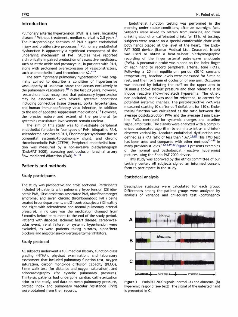
Figure 1 EndoPAT 2000 signals: normal (A) and abnormal (B)hyperemic respond (see text). The signal of the untested handis presented in C.
1792 N. Peled et al.
Introduction
Pulmonary arterial hypertension (PAH) is a rare, incurabledisease.1 Without treatment, median survival is 2.8 years.2
The histopathologic features of PAH suggest endothelialinjury and proliferative processes.3 Pulmonary endothelialdysfunction is apparently a significant component of theunderlying mechanism of PAH. Studies have reporteda chronically impaired production of vasoactive mediators,such as nitric oxide and prostacyclin, in patients with PAH,along with prolonged overexpression of vasoconstrictors,such as endothelin 1 and thromboxane A2.4e9
The term ‘‘primary pulmonary hypertension’’ was orig-inally coined to describe a condition of hypertensivevasculopathy of unknown cause that occurs exclusively inthe pulmonary vasculature.10 In the last 20 years, however,researchers have recognized that pulmonary hypertensionmay be associated with several systemic conditionsincluding connective tissue diseases, portal hypertension,and human immunodeficiency virus infection, in additionto the use of appetite suppressant medications.11 However,the precise nature and extent of the peripheral (orsystemic) vasculature involvement remain unclear.
The aim of this study was to investigate peripheralendothelial function in four types of PAH: idiopathic PAH,scleroderma-associated PAH, Eisenmenger syndrome due tocongenital systemic-to-pulmonary shunt, and chronicthromboembolic PAH (CTEPH). Peripheral endothelial func-tion was measured by a non-invasive plethysmograph(EndoPAT 2000), assessing post occlusion brachial arteryflow-mediated dilatation (FMD).12e18
Patients and methods
Study participants
The study was prospective and cross sectional. Participantsincluded 54 patients with pulmonary hypertension (28 idio-pathic PAH, 10 scleroderma-associated PAH, nine Eisenmengersyndrome, and seven chronic thromboembolic PAH) beingtreated in our department, and 21 control subjects (13 healthyand eight with scleroderma and normal pulmonary arterialpressure). In no case was the medication changed from3 months before enrollment to the end of the study period.Patients with diabetes, ischemic heart disease, cerebrovas-cular event, renal failure, or systemic hypertension wereexcluded, as were patients taking nitrates, alpha/betablockers and angiotensin-converting enzyme inhibitors.
Study protocol
All subjects underwent a full medical history, function classgrading (NYHA), physical examination, and laboratoryassessment that included pulmonary function test, oxygensaturation, carbon monoxide diffusion capacity (DLCO),6 min walk test (for distance and oxygen saturation), andechocardiography (for systolic pulmonary pressure).Thirty-six patients had undergone cardiac catheterizationprior to the study, and data on mean pulmonary pressure,cardiac index and pulmonary vascular resistance (PVR)were obtained from their records.
Endothelial function testing was performed in themorning under stable conditions, after an overnight fast.Subjects were asked to refrain from smoking and fromdrinking alcohol or caffeinated drinks for 12 h. At testing,subjects were seated on a special comfortable chair withboth hands placed at the level of the heart. The Endo-PAT 2000 device (Itamar Medical Ltd, Ceasarea, Israel)was used to obtain a beat-to-beat plethysmographicrecording of the finger arterial pulse-wave amplitude(PWA). A pneumatic probe was placed on the index fingerof each hand to record peripheral arterial tone (PAT).Following a 20 min equilibrium period (20 �C constanttemperature), baseline levels were measured for 5 min atrest, and then for 5 min of occlusion of one arm. Occlusionwas induced by inflating the cuff on the upper arm to50 mmHg above systolic pressure and then releasing it toinduce reactive (flow-mediated) hyperemia. The other,non-occluded, hand was used for reference, to correct forpotential systemic changes. The postobstructive PWA wasmeasured starting 90 s after cuff deflation, for 210 s. Endo-thelial function was calculated as the ratio between theaverage postobstruction PWA and the average 3 min base-line PWA, corrected for systemic changes and baselinesignal amplitude. The signals were analyzed with a comput-erized automated algorithm to eliminate intra- and inter-observer variability. Absolute endothelial dysfunction wasdefined as a PAT ratio of less than 1.67.19,20 This FMD testhas been used and compared with other methods12e18 inmany previous studies.13,14,19,20 Figure 1 presents examplesof the normal and pathological (reactive hyperemia)pictures using the Endo-PAT 2000 device.
This study was approved by the ethics committee of ourtertiary center. All subjects signed an informed consentform to participate in the study.
Statistical analysis
Descriptive statistics were calculated for each group.Differences among the patient groups were analyzed byanalysis of variance and chi-square test (contingency
Endothelial dysfunction in PAH 1793
tables), and differences between patients and controlswere analyzed by Student’s t-test. For post hoc analysis,the Scheffe test was used. A p value of <0.05 was consid-ered significant. The Statistical Package for Social Sciences(SPSS Corp., version 11) was used for data handling andanalysis.
Statement of responsibility
The authors had full access to the data and take respon-sibility for its integrity. All authors have read and agree tothe manuscript as written.
Results
Patient characteristics
The patient characteristics are shown in Table 1. The distri-butions of age and body mass index were similar in allgroups.
Comparison of the patients with the control subjectsyielded a statistically significant difference in NYHA class,
Table 1 Clinical and epidemiologic characteristics of patients w
HealthyControls
IdiopathicPAH
ThromboembolicPAH
Es
Background factorsN 13 28 7 9M:F 2:11 20:8 3:4 2Age (y) 46.0 � 14.1 51.9 � 13.6 50.0 � 12.4 4BMI (k/gm2) 24.3 � 3.6 26.3 � 6.7 28.7 � 6.0 2NYHA 1.0 1.9 � 0.7$ 2.0 � 0.6$
I, n (%) 13 7 (24%) 1 (14%) 1II, n (%) 17(61%) 5 (72%) 7IIIeIV, (%) 4 (14%) 1 (14%) 1
Treatment, n (%)Epoprostenol 4 (14%)Ilomedin 1Remodulin 6 (21%) 1Bosentan 3 (11%)Select. EB 9 (32%) 1 (14%) 7Viagra 8 (29%) 1 (14%)CCB 5 (18%) 2 (33%) 1
Clinical findingsPulse (/min) 73.5 � 10.6 78.2 � 12.0 75.4 � 13.3 7BL_sat (%) 97.5 � 1.4 94.3 � 5.1* 97.0 � 1.5 86MWT (m) 512 � 35 399 � 118 388 � 1436MWT sat (%) 98.3 � 0.8 89.8 � 9.1$ 93.7 � 3.8$ 7DLCO (%) 98.5 � 3.5 58.1 � 20.3$ 67.0 � 8.8$ 6PPR (mmHg) 23.0 � 4.2 70.5 � 21.6$ 75.9 � 19.0$ 9CO (l/min) 4.3 � 1.4 2.7 � 1.1PVR (MPa s/m3) 11.4 � 5.6 22.9 � 11.2 1
PAT ratio 2.20 � 0.25 1.84 � 0.51* 1.89 � 0.32* 2
* Z p < 0.05 compare to control; $ Z p < 0.01 compare to control.BMI Z body mass index. CCB Z calcium channel blocker. BL_sat Z baSelect EB Z selective endothelin blocker. 6MWT Z 6 min walking test.CO Z cardiac output. PVR<hsp spZ0.25"/>Z peripheral vascular res
baseline oxygen saturation, and DLCO. Among the variouspatient groups, the patients with Eisenmenger syndromehad a significantly lower oxygen saturation than thepatients with idiopathic or thromboembolic disease(p Z 0.030 and p Z 0.022, respectively; Scheffe post hoctest); no significant differences were found for the otherparameters. Mean distance on the 6 min walk test wassimilar in all groups; however oxygen desaturation on effortwas significantly worse in the patients with Eisenmengersyndrome and scleroderma.
Cardiac catheterization had been performed in 36 ofthe 54. No statistically significant difference in pulmo-nary pressure, measured by right heart catheterization,was noted among the patient groups. Cardiac outputand PVR were also similar among the patient groups,although there was a trend toward a higher PVR in thepatients with thromboembolic disease. Echocardio-graphic assessment showed a significantly higherpressure in the Eisenmenger syndrome group than inthe idiopathic PAH and scleroderma-PAH groups(p Z 0.009 and p Z 0.031, respectively). The differencefrom the thromboembolic groups was not significant(p Z 0.194).
ith PAH and controls
isenmengeryndrome
Scleroderma-PAH Sclerodermaonly control
p (pt. groups)
10 8:7 2:8 1:7 0.7432.0 � 20.1 54.3 � 14.2 58.5 � 12.1 0.2873.8 � 3.7 23.7 � 4.5 25.9 � 4.1 0.2432.0 � 0.5$ 2.2 � 0.6$ 1.0(11%) 1 (10%) 8(78%) 6 (61%) 0.550(11%) 3 (30%)
1 (14%)(11%)(11%) 1 (10%)
4 (40%)(78%) 3 (30%)
3 (30%)(11%) 2 (20%) 2 (25%)
4.9 � 12.6 81.2 � 12.6 83.2 � 7.8* 0.6757.9 � 10.9$ 94.2 � 5.5* 98.3 � 2.7 0.008361 � 66$ 315 � 77$ 451 � 53 0.2072.3 � 21.7$ 85.0 � 12.8$ 97.8 � 2.1 0.0032.4 � 21.1$ 43.0 � 28.1$ 74.0 � 28.8 0.0989.6 � 22.9$ 69.3 � 20.1$ 31.2 � 11.2 0.0064.1 � 1.4 4.4 � 1.5 5.9 � 2.3 0.6718.7 � 9.7 14.9 � 5.7 2.8 � 0.7 0.054
.17 � 0.62 1.66 � 0.66* 2.03 � 0.38 0.250
seline oxygen saturation. DLCO Z monoxide diffusion in the lung.NYHA Z New York Heart Association. PPR Z pulmonary pressure.
istance in pascal seconds per cubic meter (MPa s/m3).
PAT Ratio
NormalAbnormal
Dis
ta
nc
e 6
MW
T (m
)
700
600
500
400
300
200
100
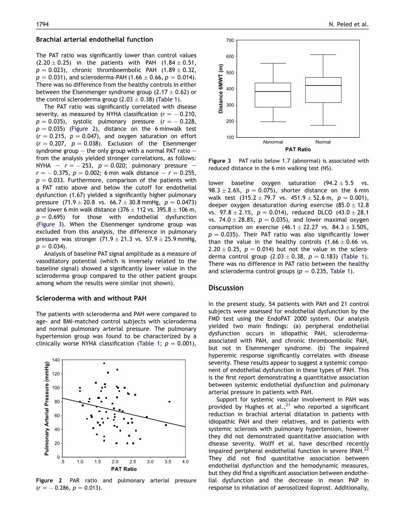
Figure 3 PAT ratio below 1.7 (abnormal) is associated withreduced distance in the 6 min walking test (NS).
1794 N. Peled et al.
Brachial arterial endothelial function
The PAT ratio was significantly lower than control values(2.20 � 0.25) in the patients with PAH (1.84 � 0.51,p Z 0.023), chronic thromboembolic PAH (1.89 � 0.32,p Z 0.031), and scleroderma-PAH (1.66 � 0.66, p Z 0.014).There was no difference from the healthy controls in eitherbetween the Eisenmenger syndrome group (2.17 � 0.62) orthe control scleroderma group (2.03 � 0.38) (Table 1).
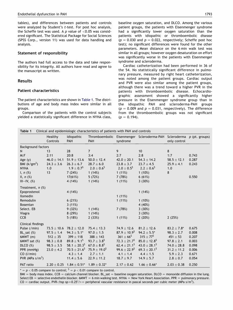
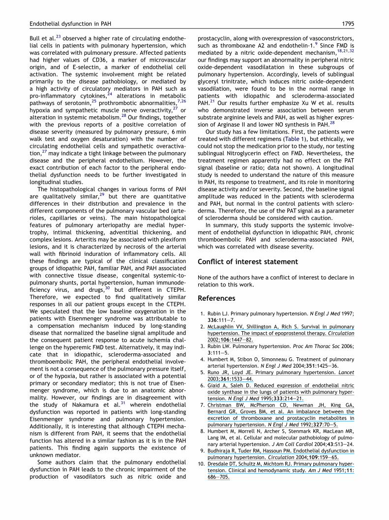
The PAT ratio was significantly correlated with diseaseseverity, as measured by NYHA classification (r Z � 0.210,p Z 0.035), systolic pulmonary pressure (r Z � 0.228,p Z 0.035) (Figure 2), distance on the 6 minwalk test(r Z 0.215, p Z 0.047), and oxygen saturation on effort(r Z 0.207, p Z 0.038). Exclusion of the Eisenmengersyndrome group e the only group with a normal PAT ratio efrom the analysis yielded stronger correlations, as follows:NYHA e r Z � 253, p Z 0.020; pulmonary pressure er Z � 0.375, p Z 0.002; 6 min walk distance e r Z 0.255,p Z 0.033. Furthermore, comparison of the patients witha PAT ratio above and below the cutoff for endothelialdysfunction (1.67) yielded a significantly higher pulmonarypressure (71.9 � 20.8 vs. 66.7� 30.8 mmHg, p Z 0.0473)and lower 6 min walk distance (376 � 112 vs. 395.8 � 106 m,p Z 0.695) for those with endothelial dysfunction(Figure 3). When the Eisenmenger syndrome group wasexcluded from this analysis, the difference in pulmonarypressure was stronger (71.9 � 21.3 vs. 57.9� 25.9 mmHg,p Z 0.034).
Analysis of baseline PAT signal amplitude as a measure ofvasodilatory potential (which is inversely related to thebaseline signal) showed a significantly lower value in thescleroderma group compared to the other patient groupsamong whom the results were similar (not shown).
Scleroderma with and without PAH
The patients with scleroderma and PAH were compared toage- and BMI-matched control subjects with sclerodermaand normal pulmonary arterial pressure. The pulmonaryhypertension group was found to be characterized by aclinically worse NYHA classification (Table 1; p Z 0.001),
PAT Ratio
4.03.53.02.52.01.51.0.5
Pu
lm
on
ary
A
rte
ria
l P
re
ss
ure
(m
mH
g)
140
120
100
80
60
40
20
0
Figure 2 PAR ratio and pulmonary arterial pressure(r Z � 0.286, p Z 0.013).
lower baseline oxygen saturation (94.2 � 5.5 vs.98.3 � 2.6%, p Z 0.075), shorter distance on the 6 minwalk test (315.2 � 79.7 vs. 451.9 � 52.6 m, p Z 0.001),deeper oxygen desaturation during exercise (85.0 � 12.8vs. 97.8 � 2.1%, p Z 0.014), reduced DLCO (43.0 � 28.1vs. 74.0 � 28.8%, p Z 0.035), and lower maximal oxygenconsumption on exercise (46.1 � 22.27 vs. 84.3 � 3.50%,p Z 0.035). Their PAT ratio was also significantly lowerthan the value in the healthy controls (1.66 � 0.66 vs.2.20 � 0.25, p Z 0.014) but not the value in the sclero-derma control group (2.03 � 0.38, p Z 0.183) (Table 1).There was no difference in PAT ratio between the healthyand scleroderma control groups (p Z 0.235, Table 1).
Discussion
In the present study, 54 patients with PAH and 21 controlsubjects were assessed for endothelial dysfunction by theFMD test using the EndoPAT 2000 system. Our analysisyielded two main findings: (a) peripheral endothelialdysfunction occurs in idiopathic PAH, scleroderma-associated with PAH, and chronic thromboembolic PAH,but not in Eisenmenger syndrome. (b) The impairedhyperemic response significantly correlates with diseaseseverity. These results appear to suggest a systemic compo-nent of endothelial dysfunction in these types of PAH. Thisis the first report demonstrating a quantitative associationbetween systemic endothelial dysfunction and pulmonaryarterial pressure in patients with PAH.
Support for systemic vascular involvement in PAH wasprovided by Hughes et al.,21 who reported a significantreduction in brachial arterial dilatation in patients withidiopathic PAH and their relatives, and in patients withsystemic sclerosis with pulmonary hypertension, howeverthey did not demonstrated quantitative association withdisease severity. Wolff et al. have described recentlyimpaired peripheral endothelial function in severe IPAH.22
They did not find quantitative association betweenendothelial dysfunction and the hemodynamic measures,but they did find a significant association between endothe-lial dysfunction and the decrease in mean PAP inresponse to inhalation of aerosolized iloprost. Additionally,
Endothelial dysfunction in PAH 1795
Bull et al.23 observed a higher rate of circulating endothe-lial cells in patients with pulmonary hypertension, whichwas correlated with pulmonary pressure. Affected patientshad higher values of CD36, a marker of microvascularorigin, and of E-selectin, a marker of endothelial cellactivation. The systemic involvement might be relatedprimarily to the disease pathobiology, or mediated bya high activity of circulatory mediators in PAH such aspro-inflammatory cytokines,24 alterations in metabolicpathways of serotonin,25 prothrombotic abnormalities,7,26
hypoxia and sympathetic muscle nerve overactivity,27 oralteration in systemic metabolism.28 Our findings, togetherwith the previous reports of a positive correlation ofdisease severity (measured by pulmonary pressure, 6 minwalk test and oxygen desaturation) with the number ofcirculating endothelial cells and sympathetic overactiva-tion,27 may indicate a tight linkage between the pulmonarydisease and the peripheral endothelium. However, theexact contribution of each factor to the peripheral endo-thelial dysfunction needs to be further investigated inlongitudinal studies.
The histopathological changes in various forms of PAHare qualitatively similar,29 but there are quantitativedifferences in their distribution and prevalence in thedifferent components of the pulmonary vascular bed (arte-rioles, capillaries or veins). The main histopathologicalfeatures of pulmonary arteriopathy are medial hyper-trophy, intimal thickening, adventitial thickening, andcomplex lesions. Arteritis may be associated with plexiformlesions, and it is characterized by necrosis of the arterialwall with fibrinoid induration of inflammatory cells. Allthese findings are typical of the clinical classificationgroups of idiopathic PAH, familiar PAH, and PAH associatedwith connective tissue disease, congenital systemic-to-pulmonary shunts, portal hypertension, human immunode-ficiency virus, and drugs,30 but different in CTEPH.Therefore, we expected to find qualitatively similarresponses in all our patient groups except in the CTEPH.We speculated that the low baseline oxygenation in thepatients with Eisenmenger syndrome was attributable toa compensation mechanism induced by long-standingdisease that normalized the baseline signal amplitude andthe consequent patient response to acute ischemia chal-lenge on the hyperemic FMD test. Alternatively, it may indi-cate that in idiopathic, scleroderma-associated andthromboembolic PAH, the peripheral endothelial involve-ment is not a consequence of the pulmonary pressure itself,or of the hypoxia, but rather is associated with a potentialprimary or secondary mediator; this is not true of Eisen-menger syndrome, which is due to an anatomic abnor-mality. However, our findings are in disagreement withthe study of Nakamura et al.31 wherein endothelialdysfunction was reported in patients with long-standingEisenmenger syndrome and pulmonary hypertension.Additionally, it is interesting that although CTEPH mecha-nism is different from PAH, it seems that the endothelialfunction has altered in a similar fashion as it is in the PAHpatients. This finding again supports the existence ofunknown mediator.
Some authors claim that the pulmonary endothelialdysfunction in PAH leads to the chronic impairment of theproduction of vasodilators such as nitric oxide and
prostacyclin, along with overexpression of vasoconstrictors,such as thromboxane A2 and endothelin-1.9 Since FMD ismediated by a nitric oxide-dependent mechanism,18,21,32
our findings may support an abnormality in peripheral nitricoxide-dependent vasodilatation in these subgroups ofpulmonary hypertension. Accordingly, levels of sublingualglyceryl trinitrate, which induces nitric oxide-dependentvasodilation, were found to be in the normal range inpatients with idiopathic and scleroderma-associatedPAH.21 Our results further emphasize Xu W et al. resultswho demonstrated inverse association between serumsubstrate arginine levels and PAH, as well as higher expres-sion of Arginase II and lower NO synthesis in PAH.28
Our study has a few limitations. First, the patients weretreated with different regimens (Table 1), but ethically, wecould not stop the medication prior to the study, nor testingsublingual Nitroglycerin effect on FMD. Nevertheless, thetreatment regimen apparently had no effect on the PATsignal (baseline or ratio; data not shown). A longitudinalstudy is needed to understand the nature of this measurein PAH, its response to treatment, and its role in monitoringdisease activity and/or severity. Second, the baseline signalamplitude was reduced in the patients with sclerodermaand PAH, but normal in the control patients with sclero-derma. Therefore, the use of the PAT signal as a parameterof scleroderma should be considered with caution.
In summary, this study supports the systemic involve-ment of endothelial dysfunction in idiopathic PAH, chronicthromboembolic PAH and scleroderma-associated PAH,which was correlated with disease severity.
Conflict of interest statement
None of the authors have a conflict of interest to declare inrelation to this work.
References
1. Rubin LJ. Primary pulmonary hypertension. N Engl J Med 1997;336:111e7.
2. McLaughlin VV, Shillington A, Rich S. Survival in pulmonaryhypertension. The impact of epoprostenol therapy. Circulation2002;106:1447e82.
3. Rubin LW. Pulmonary hypertension. Proc Am Thorac Soc 2006;3:111e5.
4. Humbert M, Stibon O, Simonneau G. Treatment of pulmonaryarterial hypertension. N Engl J Med 2004;351:1425e36.
5. Runo JR, Loyd JE. Primary pulmonary hypertension. Lancet2003;361:1533e44.
6. Giaid A, Saleh D. Reduced expression of endothelial nitricoxide synthase in the lungs of patients with pulmonary hyper-tension. N Engl J Med 1995;333:214e21.
7. Christman BW, McPherson CD, Newman JH, King GA,Bernard GR, Groves BM, et al. An imbalance between theexcretion of thromboxane and prostacyclin metabolites inpulmonary hypertension. N Engl J Med 1992;327:70e5.
8. Humbert M, Morrell N, Archer S, Stenmark KR, MacLean MR,Lang IM, et al. Cellular and molecular pathobiology of pulmo-nary arterial hypertension. J Am Coll Cardiol 2004;43:S13e24.
9. Budhiraja R, Tuder RM, Hassoun PM. Endothelial dysfunction inpulmonary hypertension. Circulation 2004;109:159e65.
10. Dresdale DT, Schultz M, Michtom RJ. Primary pulmonary hyper-tension. Clinical and hemodynamic study. Am J Med 1951;11:686e705.
1796 N. Peled et al.
11. Simonneau G, Galie N, Rubin LJ, Langleben D, Seeger W,Domenighetti G, et al. Clinical classification of pulmonaryhypertension. J Am Coll Cardiol 2004;43:5Se12S.
12. Bonetti PO, Pumper GM, Higano ST, Holmes DR, Kuvin JT,Lerman A. Noninvasive identification of patients with earlycoronary atherosclerosis by assessment of digital reactivehyperemia. J Am Coll Cardiol 2004;44:2137e41.
13. Kuvin JT, Patel AR, Sliney KA, Pandian NG, Sheffy J, Schnall RP,et al. Assessment of peripheral vascular endothelial functionwith finger arterial pulse wave amplitude. Am Heart J 2003;146:168e74.
14. Bonetti PO, Barsness GW, Keelen PC, Schnell TI, Pumper GM,Kuvin JT, et al. Enhanced external counterpulsation improvedendothelial function in patients with symptomatic coronaryartery disease. J Am Coll Cardiol 2003;41:176e8.
15. Kuvin J, Patel A, Karas R. Need for standardization of non-invasive assessment of vascular endothelial function. Am HeartJ 2001;1441:327e8.
16. Kuvin J, Patel A, Sliney K, Pandian NG, Rand WM, Udelson JE,et al. Peripheral vascular endothelial function testing asa non-invasive indicator of coronary artery disease. Am J CollCardiol 2001;38:1843e9.
17. Schroeter H, Heiss C, Balzer J, Kleibongard P, Keen CL,Hollenberg NK, et al. Epicatechin mediates beneficial effectsof flavanol-rich cocoa on vascular function in humans. ProcNatl Acad Sci USA 2006;103(4):1024e9.
18. Meredith IT, Currie KE, Anderson TJ, Roddy MA, Ganz P,Vreager MA. Postischemic vasodilation in human forearm isdependent on endothelium-derived nitric oxide. Am J Physiol1996;270:H1435e40.
19. Yinon D, Lowenstein L, Suraya S, Beloosesky R, Zmora O,Malhotra A, et al. Pre-eclampsia is associated with sleep-disordered breathing and endothelial dysfunction. Eur RespirJ 2006;27:328e33.
20. Itzhaki S, Lave L, Pillar G, Tal G, Lavie P. Endothelial dysfunc-tion in obstructive sleep apnea measured by peripheral arterialtone response in the finger to reactive hyperemia. Sleep 2005;28(5):594e600.
21. Hughes R, Tong J, Oats C, Lordan J, Corris PA. Evidence forsystemic endothelial dysfunction in patients and first-orderrelatives with pulmonary hypertension. Chest 2005;128:617S.
22. Wolff B, Lodziewski S, Bollmann T, Opitz CF, Ewert R. Impairedperipheral endothelial function in severe idiopathic pulmonary
hypertension correlates with the pulmonary vascular responseto inhaled iloprost. Am Heart J 2007;153. 1088e10e1-e8.
23. Bull TM, Golpon H, Hebbel RP, Solovey A, Cool CD, Tuder RM,et al. Circulating endothelial cells in pulmonary hypertension.Thromb Haemost 2003;90(4):698e703.
24. Dorfmuller P, Perros F, Balabanian K, Humbert M. Inflammation inpulmonary arterial hypertension. Eur Respir J 2003;22:358e63.
25. Eddahibi S, Humbert M, Fadel E, Raffestin B, Darmon M,Capron F, et al. Serotonin transporter overexpression is respon-sible for pulmonary arterial smooth muscle hyperplasia inprimary pulmonary hypertension. J Clin Invest 2001;108:1141e50.
26. Friedman R, Mears JG, Barst RJ. Continuous infusion of prosta-cyclin normalizes plasma markers of endothelial cell injury andplatelet aggregation in primary pulmonary hypertension.Circulation 1997;96:2782e4.
27. Velez-Roa S, Ciarka A, Najem B, Vachiery JL, Naeije R, Van deBorne P. Increased sympathetic nerve activity in pulmonaryhypertension. Circulation 2004;110:1308e12.
28. Xu W, Kaneko FT, Zheng S, Comhair SA, Janocha AJ, Goggans T,et al. Increased arginase II and decreased NO synthesis in endo-thelial cells of patients with pulmonary arterial hypertension.FASEB J 2004 Nov;18(14):1746e8.
29. Pietra GG, Edwards WD, Kay JM, Rich S, Kernis J, Schloo B,et al. Histopathology of primary pulmonary hypertension. Aqualitative and quantitative study of pulmonary blood vesselsfrom 58 patients in the National Heart, Lung, and Blood Insti-tute, Primary Pulmonary Hypertension Registry. Circulation1989;80:1198e206.
30. Galie N, Torbicki A, Barst R, Dartevelle P, Haworth S,Higenbottam T, et al. Guidelines on diagnosis and treatmentof pulmonary arterial hypertension. Eur Heart J 2004;25:2243e78.
31. Nakamura M, Yoshida H, Naganuma Y, Kon H, Sugawara S,Hiramori K. Peripheral vasodilatory dysfunction in adultpatients with congenital heart disease and severely elevatedpulmonary vascular resistance. Angiology 2002;53(6):715e20.
32. Corretti MC, Anderson TJ, Benjamin EJ, Celermajer D,Charbonneau F, Creager MA, et al. International BrachialArtery Reactivity Task Force. Guidelines for the ultrasoundassessment of endothelial-dependent flow-mediated vasodila-tion of the brachial artery: a report of the InternationalBrachial Artery Reactivity Task Force. J Am Coll Cardiol2002;39:257e65.
Copyright © 2022 FDOKUMEN