Technology developments in endovascular treatment of intracranial aneurysms
Upload
independentCategory
view
1download
0
A program of operative angioplasty: Endovascular intervention and the vascular surgeon Michael B. Silva, Jr. , M D , R o b e r t W. H o b s o n I I , M D , Zafar Jamil , M D , Clifford T. Araki , P h D , M a r k C. Go ldbe rg , M D , Pau l B. Hase r , M D , Bing C. Lee, M D , F r a n k T. Padbe rg , Jr. , M D , Pe te r J. Pappas , M D , and E d w i n P. Teehan , M D , Newark, N.J.
Purpose: Vascular surgeons are ideally suited to select and perform endovascular interven- tions either as primary therapy or as an adjunct to bypass surgery. Attaining proficiency in endovascular techniques is an important goal in the training of vascular surgeons. We report our initial experience with a program of endovascular intervention performed in the operating room by vascular surgeons. Methods: During the previous three years, we performed 109 angioplasty procedures, 60 aortoiliac (55%), 32 femoropopliteal (29%), and 17 popliteal/tibial (16%), using guide- wires and angioplasty balloons directed by intraoperative digital subtraction C-arm arteriography with road-mapping capabilities. Indications for angioplasty included dis- abling clandication in 59 patients (54%), rest pain in 18 (17%), and tissue loss in 32 (29%). Angioplasty was accompanied by stent placement in 39 of 60 aortoiliac procedures (65%) and in two of 32 femoral procedures (6%). In 16 cases (15%), the endovascular procedure was performed in conjunction with a bypass procedure. In selected cases (15, 14% ), duplex scanning was the sole diagnostic method used before surgery to identify the lesion, eliminating the need for preoperative arteriographic scans. Segmental pressure measure- ments, duplex ultrasound scans, and treadmill exercise testing as indicated were per- formed before and after surgery. The efficacy of the endovascular intervention was assessed at 3-month intervals during the first year and at &month intervals thereafter. Results: A successfifl result was defined using criteria recommended by the Ad Hoc Subcom- mittee on Reporting Standards for Endovascular Procedures from the Society for Vascular Surgery/International Society for Cardiovascular Surgery. This included the combination of symptomatic hnprovement, obtaining an anatolnically successful result with <30% residual lumen stenosis, and elimination of the translesion gradient with an improvement in high thigh-brachial index or ankle-brachial index greater than 0.15. Initial success was achieved in 55 of 60 aortoiliac (92%), 28 of 32 femoropopliteal (88%), and 16 of 17 popliteal/tibial (94%) angioplasty procedures. Clinical follow-up has been achieved in all cases, with contin- ued clinical success rates of 80%, 75%, and 82% for aortoiliac, femoropopliteal~ and popliteal/ tibia/angioplasty procedures, respectively, with a mean follow-up of 15.7 months. Conclusion: These results confirm the value of a program in which C-arm technology was used by vascular surgeons in the performance of angioplasty and stenting procedures in the operating room. This experience in therapeutic endovascular intervention will facili- tate the credentialing process for future vascular surgeons. (J Vase Surg 1996;24:963-73.)
From the Department of Surgery, Section of Vascular Surgery, Division of Endovascular Surgery, University of Medicine and Dentistry of New Jersey-New Jersey Medical School.
Presented at the Tentll Annual Meeting of the Eastern Vascular Society, Washington, D.C., May 3-5, 1996.
Reprint requests: Michael B. Silva, Jr., MD, Director of Endovas- cular Surgery, UMDNJ-New Jersey Medical School, MSB-H- 588, 185 S. Orange Ave., Newark, NJ 07103.
Copyright © 1996 by The Society for Vascular Surgery and Inter- national Society for Cardiovascular Surgery, North American Chapter.
0741-5214/96/$5.00 + 0 24/6/77582
Vascular surgeons traditionally have managed pa- tients who have a wide range o f vascular disease and they thereby offer superior diagnostic and therapeutic options. This encompasses the selection and perfor- mance ofendovascular procedures. Our approach has been to establish an intervenfionai program in the operating room, whereas othcrs have addressed the issue by establishing a referral relationship with an interventional radiologist or cardiologist. This collab- orative approach, however, has some theoret ic lirni-
963
JOURNAL OF VASCULAR SURGERY 9 6 4 Silva et aL December 1996
tations. Responsibility to the patient for complica- tions that result from procedures that are performed by others and the issue ofpreprocedural surgical evalu- ation and clinical diagnostic follow-up on a procedure performed by a colleague resulted in our decision to pursue a program of operative endovascular interven- tion for iliac and femoropopliteal occlusive disease.
The task of determining how a vascular surgeon can deliver these services, however, has frustrated the advancement of this portion of our practice and com- plicated our attempts to add an endovascular compo- nent to our training program. Four alternative loca- tions and collaborative approaches exist: using the angiography suite, using the cardiology suite, estab- lishing an independent facility, or using an existing facility such as the operating room. The first and second alternative approaches involve the interdisci- plinary use of space, which in many instances is not feasible. The development of the freestanding unit may be the ideal approach, but it probably would be a complex and expensive undertaking in most insti- tutions. The operating suite then represents the pre- ferred option for the development of an endovascular program that is managed by vascular surgeons.
Our objective was to demonstrate the utility of using conventional C-arm imaging techniques, which are available in virtually all operating suites, to establish a surgically based program in endovascular therapeutics. Our goal was to use these techniques as alternatives to standard vascular surgical procedures or as adjuncts to more complex arterial revasculariza- tion procedures.
The first author of the report on this series under- went a formal 1-year fellowship in diagnostic and therapeutic angiographic endovascular intervention in addition to a standard 2-year vascular surgical fellowship. The second and third authors had had substantial experience in the development and the evaluation of laser-assisted angioplasty procedures. They have desseminated this experience within the division to our vascular surgical trainees and to our vascular attending surgeons by a policy of planned proctoring. All o f the surgeons who perform these procedures have done so only after having met the credentialing criteria that are outlined by the Ad Hoc Committee on Credentialing of the Joint Councils of the vascular surgical societies)
P A T I E N T S A N D M E T H O D S
Patients were candidates for endovascular inter- vention if they reported lifestyle-limiting claudication refractory to standard exercise regimens and smoking cessation, or alternatively demonstrated rest pain or
ischemic tissue loss. Either angiographic or duplex ul- trasound scans were performed before surgery to iden- tify lesions that were potentially amenable to treatment.
Pa t ient assessment
Clinical evaluation. Stratification of patients on the basis of their symptoms was performed using rec- ommendations outlined by the Ad Hoc Subcommittee on Reporting Standards for Endovascular Procedures. 2 The clinical grades I (clandication), II (rest pain), and III (tissue loss) were determined for all patients.
Hemodynamic assessment. Doppler-derived seg- mental pressure measurements and computed ankle- brachialindexes (ABI) and thigh-brachial indexes (TBI) were obtained in all cases. Postexercise index measure- ments, pulse-volume recordings, and Doppler scans were performed in selected cases.
Risk factors. Patients were classified according to their history of smoking, hypertension, diabetes mellitus, hypercholesterolemia, renal insufficiency with a creatinine level greater than 2.0 mg/d l , chronic obstructive pulmonary disease, or coronary artery disease.
Descr ipt ion o f lesion
Locat ion. We classified all lesions by anatomic site, using three groups: aortoiliac (including aorta and common iliac and external iliac arteries), femo- ropopliteal (including common femoral, deep femo- ral, superficial femoral, and above-lmee popliteal ar- teries), and popliteal/tibial (including below-lmee popliteal, anterior fibial, tibioperoneal trunk, poste- rior tibial, and peroneal arteries).
Type. Using information obtained from a review of preoperative or intraoperafive angiographic scans or duplex scans, we characterized lesions according to occlusion versus stenosis, combined with measure- ments of length and assessment ofrunoffvessels. The length of the treated lesion was separated into the following categories: < 2 cm, > 2 cm to 5 cm, >5 cm to 10 cm, and >10 cm. The status of the runoff arteries was graded as either poor (0 to 1) or good (2 to 3). Runoff was defined as an adequately patent artery distal to the treated site (<50% stenosis by angiographic appearance or its equivalent on Duplex scan). The superficial and deep femoral arteries are the runoff vessels for iliac artery lesions, and the tibial arteries are the runoff vessels for procedures per- formed on femoral or popliteal arteries.
Criteria for early success
The treatment outcome was based on an intent- to-treat protocol and included all patients who con-

JOURNAL OF VASCULAR SURGEKY Volume 24, Number 6 Sil'p~ e~ al. 9 6 5
sented to undergo the procedure in the operating room. When more than one procedure was per- formed on the same arterial segment (i.e., common iliac angioplasty followed by deployment of a stent, and repeat post-stent deployment angioplasty), this was reported as a single endovascular procedure, with efficacy being determined by final outcome of all component procedures.
When the endovascular procedure was performed in conjunction with a surgical reconstruction or with another endovascular procedure of an adjacent inflow or outflow vascular bed, the patency rates o f each vascular bed were reported separately. Clinical or hemodynamic failures for either component o f ther- apy in these patients, however, were reported as being failures for both procedures.
The therapeutic results were based on the clinical, hemodynamic, and imaging or anatomic factors. All three criteria must have shown improvement in order for the result to be counted as a success. The defini- tions for success for each factor are outlined below.
Clinical success. Clinical success required symp- tomatic improvement of the patient as compared with their preprocedural assessment. Claudication was improved if resolved or improved greater than 50%. Rest pain must have been resolved, and tissue loss required healing with limb salvage.
H e m o d y n a m i c improvement . Hemodynamic improvement was defined as an improvement in the high thigh-brachial index greater than 0.15 for pa- tients undergoing aortoiliac procedures, or improve- ment in the anlde-brachial index greater than 0.15 for patients undergoing femoropopliteal or popli teal/ tibial procedures.
Ana tomic success. Initial anatomic success was defined as a residual luminal stenosis < 30% of normal diameter as measured by an intraoperative digital- subtraction angiographic scan. I t should be noted that the successful passage of a wire or device across a lesion alone without angiographic evidence of ade- quate restoration of lumen size and elimination of the translesion pressure gradient was not considered an initial technical success, but rather was recorded as being a failed procedure.
Cri ter ia fo r con t inued success
The, above criteria for immediate success applied to immediate postprocedural results. Continued suc- cess was determined once the patient was discharged and is defined as maintenance of the same criteria listed above for clinical, hemodynamic, and anatomic success. Clinical patency was based on sustained im- provement of at least one objective hemodynamic or
imaging test in addition to sustained clinical improve- ment. A deterioration of one or more category level clinically or hemodynamically constituted failure. An- atomic failure is defined as restenosis to >-50% of normal diameter as seen on angiographic assessment or doubling of velocities as evidenced by duplex scan. Patients were evaluated at 3-month intervals for the first year and 6-month intervals thereafter.
Per ioperat ive factors
Endovascular procedures were performed either by percutaneous approach or by arterial cutdown. Typically, a 0.03S-inch hydrophilic Glidewire (Med- itech, Boston Scientific. Watertown, Mass.) was ad- vanced across the lesion and a 5F introducer sheath was placed. One of a variety o f angiocatheters (Med- itech) would be selected on the basis of anatomic considerations and advanced over the wire for injec- tion o f contrast material and subsequent pressure measurements. Operative Lmaging was accomplished using C-arm digital subtraction arteriography ~OEC Medical Systems, Inc., Salt Lake City). Standard an~ teroposterior views were routinely augmented with oblique views, and minimal-volume (510 mB hand injections of contrast material were used to confirm location and severity of occlusive lesions. The pres- sure measurements above and below the lesion were obtained before intervention. Papaverine injections were used when necessary to evaluate gradients; a 10% reduction in pressure across the lesion was con- sidered significant Ultrathin angloplasty balloons (Meditcch) were selected on the basis of arterial measurements; commonly for an iliac angioplasty a 4 cm × 8 mm balloon would be positioned and inflated to 12 atmospheres o f pressure using an inflation device and the pressure would be sustained for a period o f 30 seconds. Patients received a SO00-unit bolus ofheparin sulfate intravenously before inflation of the angioplasty balloons.
Premounted Palmaz stents (Johnson & Johnson, Princeton, N.J. ~ were selected for deployment when angioplasty failed to relieve the translesion pressure gradient or when a technically unsatisfactory result with angioplasty (dissection or residual intraluminal stenosis >30%) was noted. Stent sxzes were selected on the basis o f arterial measurements and introduced through the SF sheath when possible. I f a 7F intro- ducer was required (necessary for stents larger than 9 mm in diameter), sequential dilators were used to facilitate the exchange. Stent deployment after dila- tion angioplasty with the next-higher-size balloon catheter was then performed for maximal stent to arterial wall apposition. Completion arteriographic

IOURNAL OF VASCULAR SURGERY 966 Silva et aL December 1996
Table I. Patient comorbidities and risk factors
Characteristic Percent
Hypertension 56 Diabetes mellitus 47 Coronary artery disease 44 Smoking 59 Chronic pulmonary disease 16 Hypercholester01emia 9 Stroke 8 Creatinine >2.0 2
scans and pressure measurements were obtained in all patients before removal of the introducer sheath. Once removed, if the procedure had been performed from a percutaneous approach, pressure was held for a minimum period of 20 minutes or as necessary to achieve hemostasis.
All patients were discharged on antiplatelct ther- apy (acetylsalicylic acid, 325 mg daily). Preprocedure and postprocedure thigh-brachial indexes and ankle- brachial indexes were expressed as mean + SD. Ap- parent differences were assessed for their statistical significance by performing X 2 analysis.
RESULTS
Between October 1993 and March 1996, we performed 109 angioplasty procedures and 41 in- traarterial stent deployment procedures in 68 patients who had been referred for evaluation of peripheral vascular disease. The male-to-female distribution was 56% male and 44% female, and the mean age of patients treated was 66.7 years, with a range of 43 to 88 years. Table I summarizes the frequencies of co- morbid illnesses and selected risk factors.
Indications for endovascular intervention were disabling claudication in 49%, rest pain in 18%, and tissue loss in 33% of the procedures performed. Table II describes the distribution of lesions and their char- acteristics.
Of the 60 aortoiliac angioplasty procedures per- formed (one aortic, 32 common iliac, and 27 external iliac), stents were used in 39 (65%). Of the 32 femo- ropopliteal artery angioplasty procedures performed, stents were used in two (6%). In no instance was a stent deployed below the level of the popliteal artery to accompany tibial angioplasty. Of the 41 stents deployed, 16 (39%) were used when there was a persistent translesion pressure gradient, and 25 (61%) were used for a poor anatomic result after angioplasty with either residual stenosis >30% (21 procedures) or with angiographic evidence of arterial wall dissection (four procedures). One of the femoral stents was
Table II; Description of lesion
Characteristic No. of lesions Percent of total
Location Aortoiliac 60 55 Femoropopliteal 32 29 Popliteal/tibial 17 16
Type Occlusion 6 6 Stenosis 103 94
Length -<2 cm 10 9 2 cm to 5 cm 60 55 5 cm to 10 cm 20 18 >10 cm 19 17
Runoff Good (0 to 1 vessel) 47 43 Poor (2 to 3 vessels) 62 57
placed in a patient who had documented blue toe syndrome and a heavily calcified superficial femoral artery lesion refractory to angioplasty; 13 months after the procedure was performed, this patient had resolution of her symptoms and normal distal circu- lation.
Of the 60 procedures that were performed in the aortoiliac location, five performed in four patients were considered immediate failures. One patient who had a known hypercoagulable condition and severe cardiac and pulmonary comorbidity was being treated for lower extremity ischemia manifested by tissue loss. He underwent percutaneous bilateral common iliac artery angioplasty and stent deploy- ment procedures with a resultant acute aortoiliac thrombotic occlusion. This single case accounted for two of the immediate failures. Application of lyric therapy in an attempt to reestablish patency was ultimately unsuccessful, and the procedure was con- verted to an axillofemoral and femorofemoral bypass. This patient died on the third day after surgery from disseminated intravascular coagulopathy and acute renal failure and represented the only procedure- related death in the series. The third failed procedure was in a patient who had bilateral lower extremity ischemia manifested by rest pain, who had previously undergone retroperitoneal dissection for creation of an ileal conduit with subsequent pelvic radiation for carcinoma of the bladder. Attempted balloon dilata- tion and stent deployment resulted in a common iliac artery injury with extravasation of the contrast me- diurn. Balloon control ofprograde flow could not be established, and an urgent retroperitoneal dissection with direct repair of the iliac artery injury and creation of a femorofemoral crossover bypass was performed. A fourth common iliac artery dilatation and stent placement occluded during surgery and was con-

JOURNAL OF VASCULAR SURGERY Volume 24, Number 6 Silva et al. 9 6 7
Table I I I . Preprocedure and postprocedure pressure ratios
Preintervention Postintervention
Aortoiliac procedures High thigh-brachial 0.60 _+ 0.13
index Femoropopliteal
procedures Ankte-brachial index 0.54 _+ 0.14
Popliteal/fibial procedures Ankle-brachial index 0.46 -+ 0.18
All procedures High thigh-brachial or 0.57 _+ 0.15
Ankle-brachial index
0.96 _+ 0.12
0.89 + 0.20
0.87 + 0.22
0.93 + 0.13
Values are mean _+ SD; p < 0.05.
verted to an aortobifemoral bypass procedure. The fifth immediate failure in the aortoiliac position rep- resented the single instance o f failure as a result o f an inability to cross the lesion with a wire. This patient underwent a subsequent common iliac-profunda femoris bypass procedure. The resultant initial suc- cess rate, defined previously to include the combina- tion of dimination of translesion pressure gradient, improvement in symptomatic status, and successful anatomic result was 55 o f 60 cases for the aortoiliac procedures (92%).
There was a significant increase in the high th igh- brachial artery indexes after completion o f these pro- cedures. Table III outlines mean systolic pressure ratios measured before and after the procedure for each location separately and for the combined series.
Of the 32 femoropopliteal angioplasty procedures that were attempted, all were for stenotic lesions. There were four immediate failures. In each of these instances the lesions were crossed and angioplasty was performed, but there was no improvement in the pressure gradient. All of these failures were in patients who had critical limb ischemia; two had tissue loss and poor outflow. Neithe~ had a satisfactory bypass alternative and both underwent below-knee amputa- tions, which healed successfully. The other two pa- tients had rest pain and were converted to open surgical procedures at the time o f their failed endo- vascular procedure. T h e initial success rate for this group was 28 o f 32, or 88%, Postprocedure anlde- brachial indexes were signi~canfly improved as com- pared with preprocedure measurements (Table Il l) .
There were 17 below~knee popliteal and tibial angioplasty procedures performed, all for stenotic lesions. Tibial angioplasty procedures were per- formed over 0 .018 inch smali~vessel Glidewires (Meditech) using lower-profile SUB-4 smallwessel balloon dilatation catheters (Meditech). The initial
Table IV. Minor and major complications
Complicario~ N~mber Perce~t
Dissection 4 3.6 Hematoma 2 1.8 Pseudoaneuwsm 1 0.9 Cardiac arrhythrnia 1 0.9 Arterial wall rupture 1 0.9 Acute arterial occlusion 3 2.7 Acute renal failure 1 0.9
success rate was 16 of 17, or 94%, The one Failure was an inability to significantly improve the distal ankle pressure after anterior tibial artery angioplasty. This patient with tissue loss and poor runoff went on to have a below-kaaee amputation. Overall, the mean postprocedure anlde-brachial index was significantly higher in this group when compared with the mean preprocedure index (Table III p.
In the series of t09 endovascular procedures, 16 (15%) were performed as an adjunct m a standard vascular surgical procedure. Eight iliac and Lwvo su- perficial femoral angioplasty procedures were per- formed as complementary inflow procedures to aug- ment lower extremity revascularizafions. Two of these procedures failed when their tibial grafts oc- cluded, and the patients went on to undergo below- knee amputations. This group has a continued suc- cess rate of 80% ~8 of 10), with a mean follow-up of 10.7 months. An additional external iliac-profunda bypass performed distal to a common iliac angio- plasty and stent placement for ischemic rest pare occluded at 6 months. This patient, however, re- mained symptomatically improved~ with an improve- ment of 0.30 in anlde-brachiai index and a parent stent by duplex scan and is therefore considered a continued success.
Six tibial angioplasty procedures were performed in conjunction with femoral-to-above-knee popliteal bypass procedures to augment outflow. One patient had early graft thrombosis with critical limb ischemia~ which was treated with successful graft revision. At~ inrraoperative angiographic scan demonstrated con- tinued patency of the previous tibial angioplasty. With this failure the success rate for this group was five of six (83%) with a mean follow-up of 8.6 months. Minor and major complications are summarized in Ta- ble IV. The overall complication rate was 11%.
Follow-up has been achieved in all patients, with a range o f 2 to 31 months and a mean of 15,7 months. Of the 60 aortoiliac procedures that were performed, five failed immediately, and an additional seven pro- cedures demonstrated deterioration in the follow-up period, for a continued success rate of 80% (48 of 60).
JOURNAL OF VASCULAR SURGERY 9 6 8 Si lva ct aL December 1996
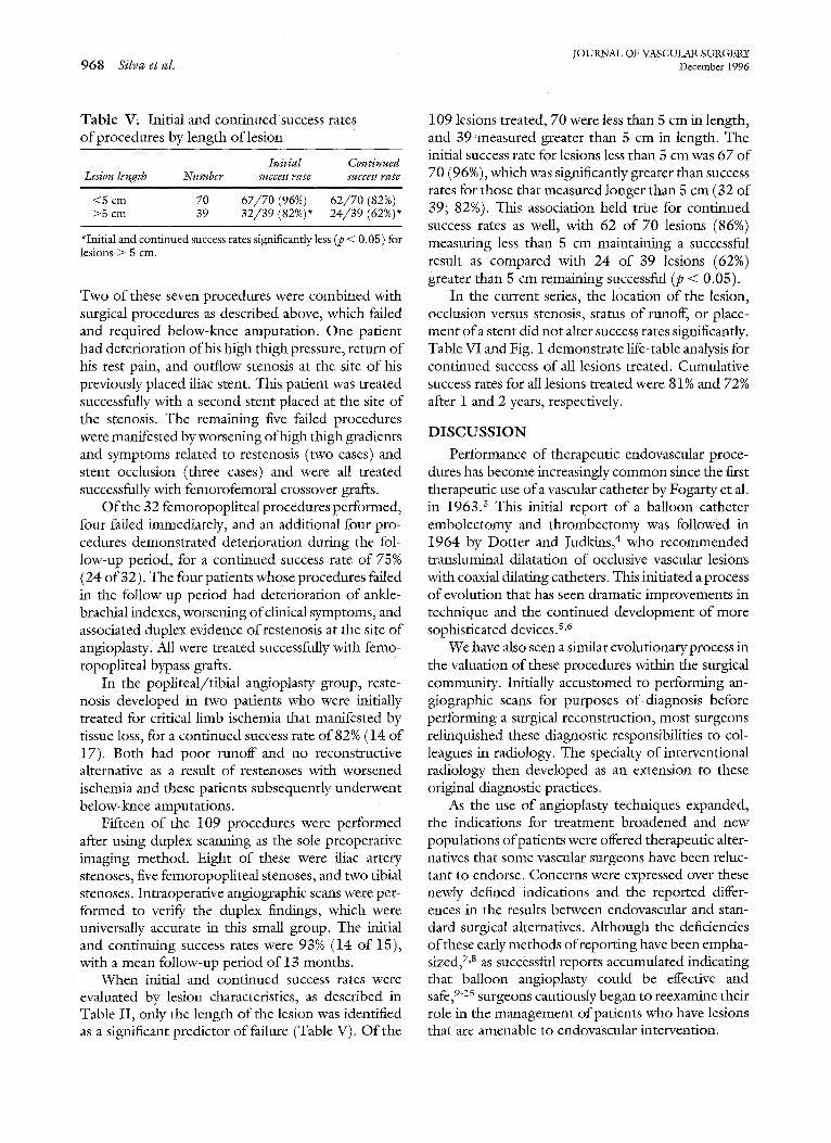
Table V. Initial and continuedsuccess rates of procedures by length of lesion
Ini t ial Continued Lesion length Number success rate success rate
<5 cm 70 6 7 / 7 0 (96%) 62 /70 (82%) >5 cm 39 32 /39 (82%)* 2 4 / 3 9 (62%)*
*Initial and continued success rates significantly less (p < 0.05) for lesions > 5 cm.
Two of these seven procedures were combined with surgical procedures as described above, which failed and required below-lmee amputation. One patient had deterioration of his high thigh pressure, return of his rest pain, and outflow stenosis at the site of his previously placed iliac stent. This patient was treated successfully with a second stent placed at the site of the stenosis. The remaining five failed procedures were manifested by worsening of high thigh gradients and symptoms related to restenosis (two cases) and stent occlusion (three cases) and were all treated successfully with femorofemoral crossover grafts.
Of the 32 femoropopliteal procedures performed, four failed immediately, and an additional four pro- cedures demonstrated deterioration during the fol- low-up period, for a continued success rate of 75% (24 of 32). The four patients whose procedures failed in the follow-up period had deterioration of ankle- brachial indexes, worsening of clinical symptoms, and associated duplex evidence ofrestenosis at the site of angioplasty. All were treated successfully with femo- ropopliteal bypass grafts.
In the popliteal/tibial angioplasty group, reste- nosis developed in two patients who were initially treated for critical limb ischemia that manifested by tissue loss, for a continued success rate of 82% (14 of 17). Both had poor runoff and no reconstructive alternative as a result of restenoses with worsened ischemia and these patients subsequently underwent below-knee amputations.
Fifteen of the 109 procedures were performed after using duplex scanning as the sole preoperative imaging method. Eight of these were iliac artery stenoses, five femoropopliteal stenoses, and two tibia] stenoses. Intraoperative angiographic scans were per- formed to verify the duplex findings, which were universally accurate in this small group. The initial and continuing success rates were 93% (14 of 15), with a mean follow-up period of 13 months.
When initial and continued success rates were evaluated by lesion characteristics, as described in Table II, only the length of the lesion was identified as a significant predictor of failure (Table V). Of the
109 lesions treated, 70 were less than 5 cm in length, and 39 measured greater than 5 cm in length. The initial success rate for lesions less than 5 cm was 67 of 70 (96%), which was significantly greater than success rates for those that measured longer than 5 cm (32 of 39; 82%). This association held true for continued success rates as well, with 62 of 70 lesions (86%) measuring less than 5 cm maintaining a successful result as compared with 24 of 39 lesions (62%) greater than 5 cm remaining successful (p < 0.05).
In the current series, the location of the lesion, occlusion versus stenosis, status of runoff, or place- ment ofa stent did not alter success rates significantly. Table VI and Fig. 1 demonstrate life-table analysis for continued success of all lesions treated. Cumulative success rates for all lesions treated were 81% and 72% after I and 2 years, respectively.
D I S C U S S I O N
Performance of therapeutic endovascular proce- dures has become increasingly common since the first therapeutic use of a vascular catheter by Fogarty et al. in 1963. 3 This initial report of a balloon catheter embolectomy and thrombectomy was followed in 1964 by Dotter and ~ud ldns , 4 who recommended transhiminal dilatation of occlusive vascular lesions with coaxial dilating catheters. This initiated a process of evolution that has seen dramatic improvements in technique and the continued development of more sophisticated devices, s,6
We have also seen a similar evolutionary process in the valuation of these procedures within the surgical community. Initially accustomed to performing an- giographic scans for purposes of diagnosis before performing a surgical reconstruction, most surgeons relinquished these diagnostic responsibilities to col- leagues in radiology. The specialty of interventional radiology then developed as an extension to these original diagnostic practices.
As the use of angioplasty techniques expanded, the indications for treatment broadened and new populations of patients were offered therapeutic alter- natives that some vascular surgeons have been reluc- tant to endorse. Concerns were expressed over these newly defined indications and the reported differ- ences in the results between endovascular and stan- dard surgical alternatives. Although the deficiencies of these early methods of reporting have been empha- sized, 7,8 as successful reports accumulated indicating that balloon angioplasty could be effective and safe, 9-15 surgeons cautiously began to reexamine their role in the management of patients who have lesions that are amenable to endovascular intervention.

JOURNAL OF VASCULAR SURGERY Volume 24, Number 6 Silva et al. 9 6 9
1 0 9
9 0
A 8 0 -
w ® 6 0 - o o
"~ 4 O - o
, m
, B
i O -
0 i t 3
| ....
8 2 6 4
3 7 I,, 3 0 2 5 t__. 18 10 i
t t
i I I I I i i i _ - -
6 9 12 15 18 21 24 27 30 33 36
Time (months)
Fig. 1. Life-table plot of cumulative clinical success for all procedures performed.
Table VI. Life-table analysis for all procedures performed
Interval No. at risk at No. withdrawn Interval Cumulative (too) start of interval No. of failures patent pa~ency rate patency (%) SE (%)
0-1 109 10 0 ,91 100 0.0 1-3 99 3 6 .97 91 2,74 3-6 90 2 6 .98 88 3.21 6-9 82 0 18 1.0 86 3.55 9-12 64 3 17 .95 86 3.55
12-15 44 2 5 .95 81 5.32 15-18 37 1 6 .97 77 6.07 18-21 30 0 5 1.0 75 6.85 21-24 25 1 6 .95 75 7.5 24-27 18 0 8 1.0 72 8.98 27-30 10 1 8 .83 72 12.04 30-33 1 0 1 1.0 60 37.9
Some recommended that the surgeon's role should be one of a participant in a team approach directed toward delivering endovascular therapy) 6 The vascular surgeon in this role brings to the group his knowledge of preoperative evaluation and the pathophysiologic mechanisms of vascular disease, the relative risks and benefits of varying therapeutic alter- natives, and an expertise with noninvasive means of postprocedure follow-up and assessment of efficacy. In this scenario, typically the actual endovascular treatment is provided by another member of the team, the interventional radiologist or cardiologist, and the vascular surgeon manages the preoperative evaluation and postoperative care and is available for surgical intervention for any complications of the endovascular procedure.
Although initially accepted by many, the limita- tions of this role have become evident, and vascular surgeons are now participating directly in the perfor- mance of endovascular procedures) 7-2° Realiziiig that evolving endovascular technology may comple- ment or supplant standard surgical alternatives and reluctant to accept a limited role in the management of our patients, many have advocated an expanded role for surgeons in the performance of endovascular procedures and training of vascular surgical fel- lows.l,18, 2o
Alternative and successful approaches to the de- velopment of endovascular training programs have included using cardiac catheterization laborato- Lies 18,20 and the development of freestanding facilt - ties. 21 Neither of these alternatives, however, was

JOURNAL OF VASCULAR SURGERY 970 Silva et al. December 1996
immediately available to us, which resulted in our choice o f using the operating suite as the site for the development o f a program o f operative angioplasty and endovascular intervention.
In our experience, we learned that the C-arm imaging that is available in the operating suite was satisfactory for the performance o f aortoiliac, femo- ropopliteal, and tibial angioplasty procedures. Our success and complication rates are comparable with those reported in other series for procedures that are performed by interventional radiologists who were worldng alone or with vascular surgeons. 9-~s Simi- larly, our initial success rate (108 of 109 procedures) in crossing the occlusive lesions with a guidewire has confirmed our bias that surgeons can readily apply their technical knowledge o f catheter and wire tech- niques to a practice o f operative angioplasty and stent placement.
I t is lmown that the short- term and long- term results are influenced by the characteristics o f the lesion treated. 22,2a In our series, the length o f the lesion was a predictor o f the clinical success; treated lesions that were shorter than 5 cm had improved initial and continued success rates compared with lesions that were longer than 5 cm. Other factors such as lesion location, status of runoff, and concomitant placement of a stent did not have a significant impact on our early results. We anticipate, however, that long-term follow-up may confirm the significant im- pact o f these variables among the treatment group. I t is our goal to continue a program o f surveillance for this group, which should contribute some under- standing to the significance o f these factors in deter- mining the efficacy of endovascular procedures.
The establishment o f this program has enabled us to provide our vascular surgical trainees with experi- ence that is sufficient to surpass the recommended minimal guidelines established by the SVS/ISCVS 1 for each of the last 3 years. As new technology be- comes available, the ongoing presence of this pro- gram should enable us to introduce technical innova- tions.
Using the experience demonstrated in this series, we recently have attained privileges to use the arte- riography suite in one of our affiliate institutions. This is an essential step and has been stimulated by our desire to eliminate the separate preprocedural acterio- graphic scan. In many of our patients, it is advanta- geous to proceed with the interventional therapy at the time of the diagnostic arteriogralShic scan. An- other approach, as reflected in 15 of our cases, has been the identification o f the occlusive lesion with a preoperative duplex ultrasound scan, thereby elimi-
nating preoperative diagnostic arteriographic scans. This suggests that we can significantly reduce the overall cost o f t reatment in two ways: by expanding the role o f preoperative duplex ultrasound scans, and by performing angiographic scans and therapeutic intervention during the same procedure.
We recommend that vascular surgeons become involved in the selection and performance of endo- vascular procedures in the operating room as well as in the angiography suite. By establishing an indepen- dent interventional program we plan to maintain an active role in the future development of endovascular surgical procedures.
REFERENCES
1. White RA, Fogarty TJ, Baker WH, Ahn SS, String ST. Endo- vascular surgery credentialing and training for vascular sur- geons. J Vase Surg 1993;17:1095-102.
2. Ahn SS, Rutherford RB, Becket GJ, Comerota AJ, Johnston KW, McClean GK~ et al. Reporting standards for lower ex- tremity arterial endovascular procedures. J Vasc Surg 1993;17: 1103-7.
3. Fogarty TJ, Crauley JJ, Kranse RJ. A method of extraction of arterial emboli and thrombi. Surg Gynecol Obstet 1963;116: 241-4.
4. Dotter CT, Judldns MP. Transluminal treatment of arterio- sclemtic obstruction: description of a new technique and a preliminary report of its application. Circulation 1964;30: 654-70.
5. Gruntzig A, Kumpe D. Technique ofpercutaneous translumi- hal angioplasty with the Gruntzig balloon catheter. AJRAm J Roentgenol 1979;132:547-52.
6. Veith FJ, Abbott WM, Yao JST, Goldstone J, White RA, Abel D, et al. Guidelines for development and use oftransluminally placed endovascular prosthetic grafts in the arterial system. J Vasc Surg 1995;21:670-85.
7. Porter JM. Interventional therapy: an alternative view. Ann Vasc Surg 1991;5:101-2.
8. Porter JM. Endovascular arterial intervention: expression of concern. J Vasc Surg 1995;21:995-7.
9. Johnston K-VV, Rae M, Hogg-Johnston SA, et al. Five-year results of a prospective study of percutaneous transluminal angioplasty. Ann Surg 1987;206:403-13.
10. Gallino A, Mahler F, Probst P, et al. Percutaneous translumi- nal angioplasty of the arteries of the lower limbs: a 5-year follow-up. Circulation 1984;70:619-23.
11. Veith FJ, Gupta SK, Samson RH, et al. Progress in limb salvage by reconstructive arterial surgery combined with new or improved adjunctive procedures. Ann Surg 1981;194:386- 99.
12. Bakal CW, Sprayregen S, Scheinbaum K, Cynamon J, Veith FJ. Percutaneous transluminal angioplasty of the infrapopli- teal arteries: results in 53 patients. Am J Radiol 1990;154: 171-4.
13. Palmaz JC, Laborde JC, Rivera FJ, et al. Stenting of the iliac arteries with the Palmaz stent: experience from a multicenter trial. Cardiovasc Intervent Radiol 1992;5:291-7.
14. Mnrphy KD, Encamacion CE, Ix VA, Palmaz JC. Iliac artery stent placement with the Palmaz stent: follow-up study. J Vasc Interv Radiol i995;6:321-9.
JOURNAL OF VASCULAR SUKGERY Volume 24, Number 6 Silv~ et aL 971
15. lohnston KW. Femoral and popliteal arteries: reanalysis of results of balloon angioplasty. Radiology 1992;183:767-71.
16. Moore WS. Organization of the multidisciptinary team. In: Ahn SS, Moore WS, editors. Endovascular surgery. Philadel- phia: W.B. Saunders 1992:12-5.
I7. Harris RW, Dulawa LB, Andros G, et al. Percutaneous trans- luminal angioplasty of the lower extremities by the vascular surgeon, Ann Vase Surg 1991;5:34- 53.
18. Hodgson KJ, Mattos M.A, Sumner DS. Canine or chameleon: the vascular surgeon's role in percutaneous endovascular ther- apy. Vase Forum 1993;1:237-47.
19. Schneider PA, Andros G, Harris RW. Balloon angioplasty in the operating room. Vase Forum 1993;4:254-9.
20. Hodgson KJ, Mattos MA, Monsour A, et al. Incorporation of
endovascular training into a vascular fellowship program. Am J Surg 1995;170:168-73.
21. Diethrich EB. Deficiencies of the modern vascular surgeon: a dilemma, a solution. Ann Vase Surg 1991;5:99-100.
22. Capek P, McLean GK, Berkowitz HD. Femoropopliteal an- gioplasqr: factors influencing long-term success. Circulation 1991;83(suppl I):170-80.
23. Johnston KW. Factors that influence the outcome ofaormiliac and femoropopliteal percutaneous transluminal an~oplasty. Surg Clin North Am 1992;72:843-50.
Submitted May 7, 1996; accepted Ang. 28, 1996.
DISCUSSION Dr. F rank J. Vei th (Bronx, N.Y.). I certainly applaud
Dr. Silva's presentation and generally agree with his con- clusions, although I am not sure that duplex scanning can regularly replace pretreatment arteriographic scans. I have one comment and two questions.
The main comment I would like to make is that vascular surgeons must become proficient with C-arm digital fluo- roscopy and catheter-guidewire techniques to simplify and improve many of their standard operations. In this regard, at this meeting last year, Drs. Parsons, Marin, and Veith and their colleagues reported on our technique of fluoroscopi- cally assisted thromboembolectomy (Ann Vase Surg 1996; 10:201). Using this technique, the vascular surgeon can navigate tortuous, diseased, clot-filled iliac arteries. Then using a doublequmen balloon catheter passed over a wire, the clot can be removed, stenotic lesions can be corrected by angioplasty and stents, and unsuspected retained clot can be removed: By visually controlling the inflation of the contrast-filled balloon, overinflation and damage to normal or diseased arteries, which is common with standard bal- loon catheters used without fluoroscopic guidance, can be avoided.
Another example of the use of digital C-arm fluoros- copy and catheter and guidewire techniques to improve or simplify standard operative procedures is the use of double- lumen balloon catheters to obtain proximal and distal con- trol when surgical access is difficult o r impossible. This technique is particularly valuable with arteriovenous fistulas and false aneurysms, especially in heavily infected fields. The same techniques can also be used to perform superior intraoperative angiographic scans, Cine techniques with digital fluoroscope provide a dynamic measure of flow through a bypass graft. Moreover, the entire bypass graft plus its inflow and outflow tracts can be visualized mad unexpected lesions can be treated without doing extensive open operations.
Thus there are many reasons why vascular surgeons should master the catheter-guidewire techniques and digi-
tal C-arm fluoroscopy mentioned here. The ability to per- form balloon angioplasty and stenting is only one such reason.
My first question to the authors relates to their appar- ently higher-than-usual proportion of patients with claudi- cation in their report today.-Do the authors have a lower threshold for performing endovascular interventions than standard operations? In this regard, I thiN< we must be carefial to be sure we do not fail into the same trap as some of our interventional colleagues do and treat lesions simply because they are there.
My second question regards how the authors first learned to perform these procedures. Who taught them? We must be sure to get appropriate instruction, help, and consultation in this regard, lest we be subject to the criti~ cism of learning on our patients. I enjoyed this interesting and important paper and appreciate the chance to discuss it.
Dr. Michael B. Silva, Jr. Thank you, Dr. Veith. Let me speak first to your suggestion that duplex ultrasono- graphic scans alone may not be effective in the preoperative diagnosis of all lesions that are potentially treatable by endovascular means. We agree. In this series only 15 of the 109 procedures were performed solely on the basis of duplex criteria. Although we are expanding our use of preoperative duplex scanning to reduce our reliance on traditional anglographic scans, we realize that there are a number of patients with multilevel disease who will require a more detailed preprocedure diagnostic evaluation for which the combination of duplex ultrasound and C-arm fluoroscopy may not be appropriate. To this end, we have obtained credentials to perform angiographic scans in the radiographic suite in one of our atfiliate institutions. This may allow us to offer our patients who have more complex disease the opportunity to have both their diagnostic eval- uation and therapeutic intervention during a single procedure.
Regarding the number o f patients with clandicarion in our series, a little over one half of these patients were treated for disabling clandication refractory to smoking

JOURNAL OF VASCULAR SURGERY 972 Si lva et al. December 1996
cessation, exercise regimens, and lifestyle modification. Al- though this may represent an expanded role for treatment on the basis of this indication when compared with surgical series of standard surgical therapeutics, we believe that it is appropriate on the basis of the risk-benefit ratio for endo- vascular procedures. We agree that lesions should not be treated simply because they are there. In this series, all lesions had demonstrable gradients and associated symptoms.
The question of how one learns the techniques in- volved in the performance of endovascular therapeutics is an important one. The first author of this report underwent a formal 1-year fellowship in diagnostic and therapeutic angiographic endovascular intervention. The second and third authors had substantial experience in the develop- ment and evaluation of laser-assisted ang!oplasty proce- dures. This experience has been disseminated through the division by a policy of planned proctoring. We believe that the vascular surgeon is ideally suited to perform catheter- based techniques. A number of the procedures that are performed routinely in the conduct of a vascular surgical practice, such as placement of indwelling central venous catheters, placement o f Swan-Ganz catheters, placement of arterial lines, placement of vena cavai interruption filters, catheter embolectomy, and thrombectomy, lend them- selves to th e development of skillful catheter-based tech- niques. Addressing that issue, it should be noted that of the 109 procedures performed, in only one instance was the procedure described as a failure on the basis of an inability to cross the lesion with the wire and the therapeutic device.
Dr. John J. Ricotta (Buffalo, N.Y~). One of the big advantages that you can offer to us has to do with what Dr. Veith said before, which is to start to work on algorithms for diagnosis of when to use duplex scans and when to combine duplex with angiographic scans. From a hospital administrator's point of view, this is going to be particularly attractive if you can show that it is cost-effective and avoids two trips to the hospital or two trips to the angiography suite. I think that that's a very important thing for us as a Society, and you and people who are doing this, to concen- trate on.
I am a little bit concerned about that common femoral lesion. I am a little concerned that we may be getting so far in bed with the devil that we're going to end up looking like the devil. It 's going to be important for us, as we go ahead with this technology, to also define some real indications of when patients are best treated with surgery and when patients are best treated with angioplasty. Again, that's a challenge I think perhaps our Issues Committee, or some- body in this Society, can start to look at. I f we start doing everything the radiologists do, we may live to regret it.
Dr. Silva. Let me respond first to the issue of how to reduce costs. In our institution we looked at the cost o f these procedures on the basis of the site within the hospital where they were performed, and the results were interest- ing, Operating suite charges are based on the time required for the procedure. Charges in the radiology suite and cardiology suite are based on the procedure itself. As an
example, for an aortogram with lower extremity runoff and iliac artery angioplasty performed in approximately 11/2 hours, the charges were actually 5% less when performed in the operating room than those generated by use of the cardiology suite or angiography suite. It is evident that the cost-effectiveness in the operating room can be further improved by reducing the overall time of the procedure itself. To that end we have made such modifications as holding pressure in the recovery room rather than in the operating suite.
To reduce the overall costs to the health care system, we are focusing on two areas. First, by expanding our role of preprocedure duplex ultrasound scanning to evaluate patients who have isolated superficial femoral or external iliac artery stenoses, we can eliminate the cost of the diag- nostic arteriogram. Second, by using the angiographic suite for patients who have more complex multilevel disease, we should be able to identify patients who would benefit from therapeutic endovascular intervention during the same pro- cedure.
As to the issue of sleeping with the devil, I agree with you that the techniques we have described do mimic those used in interventional radiology. Of course, surgeons be- gan this process in the 1960s, and many older surgeons recall doing all of their own diagnostic arteriographic scans. I think that to participate in the future of vascular surgery, which will encompass more and more endovascular tech- niques, we will need to have both procedural sldlls as well as diagnostic capabilities, and we will have to revisit some of those skills that we readily relinquished to our colleagues in interventional radiology in the past decades.
As for the 20-minute surgical repair of the common femoral lesion, I can assure you that that angioplasty took only 15 minutes and that the patient was doing well at her 1-year follow-up examination.
Dr. Thomas F. Panetta (Brooklyn, N.Y.). Clearly, vascular surgeons are both eager and anxious to gain expe- rience in endovascular techniques, and I think you and your group have provided an excellent means with careful fore- thought, which probably relates to your excellent results. I have three questions, though, relating to this.
First, what is your relationship with your interventional radiologists? Has this been a part of your approach to this? It seems that you're trying to avoid the preoperative anglo- gram and substitute the duplex evaluation. Along that line, if you're performing the angiographic scan in the operating room, are you getting as complete an angiogram? This may be difficult to do with the C-arm and requires a fair amount of images. Are you doing as complete an angiogram as you should and therefore avoiding proximal and distal injuries that could affect your primary repair?
Again, I also echo Dr. Veith's remarks about not cor- rupting our principles with regard to treating patients with claudication and liberalizing our indications for these pro- cedt~res.
Dr. Silva. As to the issue of being able to perform a complete aortogram with bilateral lower extremity artery

JOURNAL OF VASCULAR SURGERY Volume 24, Number 6 Silva et ~l. 973
runoff using the C-arm, we agree that the facilities in our angiography suite are superior to those of our operating room; and as mentioned previously, we are expanding our practice to include the diagnostic arteriographic scans for all of our vascular surgical procedures, which we perform ourselves in the radiology suite.
As to the relationship with our interventional radiolo- gists, as you might imagine, it has become a bit more competitive, but it remains a collegial one nonetheless. We have stressed that this is an important part of our surgical practice as vascular surgeons and that it is a necessary part of our training for our vascular fellows. To learn from one another and foster our mutual development, we have es- tablished a combined conference wherein we discuss endo-
vascular procedures performed by the Division of Vascular Surgery and the Department of Radiology.
As to the issue of liberalizing indications, I think that indications will probably be expanded to include pa- tients who have a level of clandication for which we would not offer a more complicated surgical solution, and this has to be based on the risk-benefit ratio of the proce- dure. If an isolated angioplasty procedure can be per- formed percutaneously with a local anesthetic on an outpa- tient basis with acceptable levels of risk and we can demon- strate I-year to 2-year efficacy rates in the 70% to 80% range, isn't this an acceptable solution for moderate to severe claudication? Our patients thiN< so, and we are inclined to agree.
L I F E L I N E F O U N D A T I O N R E S E A R C H A W A R D
The Lifeline Foundation of the Society for Vascular Surgery and the International Society for Cardiovascular Surgery, North American Chapter, invites grant applications for funding of merirorious research by young surgical investigators, The awards are intended for surgeons who have completed their formal surgical education in general surgery and who have completed or are in an advanced training program in vascular surgery.
To be considered for selection a candidate: 1. Should be certified by the American Board of Surgery or have completed the
requirements for certification 2. Should submit an application within 5 yearsofcompletionofanapprovedvascular
surgery residency traimng program 3. Must have either a faculty appointment in an approved medical school in the
United States or Canada or have received an academic appointment within the guidelines of the applicant's mstitution
Grant awards are not intended to supplement salary, which will remain the respon- sibility of the institution in which the awardee holds an appointment. The awardee is expected to devote a significant amount of time to the funded project. A progress report must be presented to the Executive Committee of the Foundation by the following April 1, and, on completion of the project, a brief oral report is to be presented to the memberships of the two societies during a plenary session at the Joint Annual Meeting.
A grant awards committee will review competitive applications. It is anticipated that two grants will be awarded annually totaling $50,000 each to include indirect costs. The $50.000 grant includes funding to enable the awardee to attend the loint Annual Meeting of the Vascular Societies to receive his or her award in the year of selection. Each award will be for 1 year with the option to extend for an additional year,
Holders of substantial research awards, such as an NIH ROI, FIRST Award, or similar support, are ineligible. The applicant must append to the application the abstracts of any funded or pending grants.
Grant applications may be obtained from: Chairman The Lifeline Foundation Thirteen Elm St. Research and Education Committee Manchester, MA 01944 (508)526-8330
The deadline for receiving applications in the Founda t ion office is January 15, 1997. Funds will be awarded by July l , I997.
Copyright © 2022 FDOKUMEN