Indolocarbazole antitumour compounds by combinatorial biosynthesis
Upload
khangminh22Category
view
2download
0
Fibronectin biosynthesis in the rat aorta in vitro. Changes due toexperimental hypertension.
R Saouaf, … , A V Chobanian, P Brecher
J Clin Invest. 1991;88(4):1182-1189. https://doi.org/10.1172/JCI115420.
This study was undertaken to determine if changes in fibronectin biosynthesis accompany the phenotypic changes thatoccur in aortic tissue following experimental hypertension. An in vitro procedure was developed to measure fibronectinsynthesis in aortic rings obtained from normotensive or hypertensive rats. There was a three to sixfold increase infibronectin biosynthesis by aortic rings taken from rats treated with deoxycorticosterone/salt for 7 and 21 d, the changebeing more pronounced at 21 d. In contrast, there was no major change at either time point in net incorporation into totalprotein. Studies comparing fibronectin biosynthesis in aortic rings from Wistar rats and spontaneously hypertensive rats atages between 10 and 40 wk showed increased fibronectin biosynthesis in older animals of both strains, but only slightdifferences between strains. Studies using rats infused with angiotensin II showed a correlation between blood pressureelevation and increased aortic fibronectin biosynthesis. Western blot analysis of aortic extracts showed that thefibronectin content was increased in the hypertensive models. The in vitro procedure for measuring fibronectinbiosynthesis appears to provide a reliable reflection of in vivo changes in fibronectin expression, and the methodologycould prove useful for studying the factors influencing protein expression in vascular tissue.
Research Article
Find the latest version:
https://jci.me/115420/pdf

Fibronectin Biosynthesis in the Rat Aorta In VitroChanges Due to Experimental Hypertension
Rola Saouaf, Izumi Takasaki, Elizabeth Eastman, Aram V. Chobanian, and Peter BrecherDepartments of Biochemistry and Medicine, Boston University School ofMedicine, Boston, Massachusetts 02118
Abstract
This study was undertaken to determine if changes in fibronec-tin biosynthesis accompany the phenotypic changes that occur
in aortic tissue following experimental hypertension. An in vi-tro procedure was developed to measure fibronectin synthesisin aortic rings obtained from normotensive or hypertensive rats.There was a three to sixfold increase in fibronectin biosynthesisby aortic rings taken from rats treated with deoxycorticoste-rone/salt for 7 and 21 d, the change being more pronounced at21 d. In contrast, there was no major change at either time pointin net incorporation into total protein. Studies comparing fibro-nectin biosynthesis in aortic rings from Wistar rats and sponta-neously hypertensive rats at ages between 10 and 40 wk showedincreased fibronectin biosynthesis in older animals of bothstrains, but only slight differences between strains. Studies us-
ing rats infused with angiotensin II showed a correlation be-tween blood pressure elevation and increased aortic fibronectinbiosynthesis. Western blot analysis of aortic extracts showedthat the fibronectin content was increased in the hypertensivemodels. The in vitro procedure for measuring fibronectin bio-synthesis appears to provide a reliable reflection of in vivochanges in fibronectin expression, and the methodology couldprove useful for studying the factors influencing protein expres-
sion in vascular tissue. (J. Clin. Invest. 1991. 88:1182-1189.)Key words: angiotensin * vascular * extracellular matrix * im-munoprecipitation
Introduction
Vascular complications of several diseases are associated withchanges in the extracellular matrix and accompanying intracel-lular changes in vascular cells (1). Since interactions betweenproteins of the extracellular matrix and cell receptors calledintegrins are known to influence cell structure, it is plausiblethat changes in the expression of components of the extracellu-lar matrix could have a causative role in the development of theresulting vascular lesions. Several studies have indicated thatvascular smooth muscle cells can undergo a change from a
contractile to synthetic phenotype during atherosclerosis (2, 3),and models for such changes have been established using cul-
This work was presented in part at the American Heart AssociationScientific Sessions, Dallas, TX, November 1990.
Address correspondence to Peter Brecher, Ph. D., Professor of Bio-chemistry, Boston University School of Medicine, 80 East ConcordStreet, Boston, MA02118.
Received for publication 7 February 1991 and in revised form 16May 1991.
tured cells (4). Fibronectin (FN)', an important component ofthe extracellular matrix, was shown to interact with vascularsmooth muscle cells in a manner influencing the interconver-sion between contractile and synthetic phenotypes (5). Interac-tions between fibronectin and the endothelial cell also inducefunctional changes in motility, morphology, and replication(6-8). Based on such studies performed with cultured cells, it ispossible that fibronectin could modulate the function of vascu-lar cells and perhaps have a direct influence on the develop-ment of vascular lesions.
Histochemical studies have documented the presence offibronectin in aortic tissue, and changes in fibronectin contenthave been reported in different disease states, such as diabetesand atherosclerosis (9-13), although quantitation has been dif-ficult due to the insoluble nature of the cellular fibronectin(14). Recently, a study by Glukhova et al. (15) showed thatdifferent alternately spliced forms of fibronectin are selectivelylocalized in the intima and media, and following either ballooninjury to the rat aorta or in human aortic atherosclerotic le-sions, a selective accumulation of an alternatively spliced formof fibronectin was found within the intimal lesion.
Hypertension leads to morphological and functionalchanges in the vessel wall, and accompanying changes in theextracellular matrix have been documented (16-18). However,there is little information available on changes in vascular fibro-nectin expression with hypertension. In a recent study ( 19), weshowed that steady-state mRNAlevels for rat aortic fibronectinincreased several-fold in deoxycorticosterone acetate (DOG)/salt-treated and angiotensin II-infused rats and in the spontane-ously hypertensive rat (SHR). An age-dependent increase inaortic fibronectin mRNAalso was shown in those studies (19).In this study, we have used an in vitro procedure for measuringprotein biosynthesis in aortic rings to determine if the biosyn-thesis of aortic fibronectin is increased in experimental hyper-tension concomitant with the previously documented changesin steady state mRNA. In addition, Western blot analysis wasused to measure the amount of fibronectin contained in aortictissue from normotensive and hypertensive rats.
Methods
Animals. Male Wistar rats, 250-300 g, were obtained from CharlesRiver Breeding Laboratories, Wilmington, MA. DOC/salt hyperten-sion was induced as previously described (20). Briefly, deoxycorticoste-rone pellets (100 mg) were implanted subcutaneously into uninephrec-tomized rats, and saline was substituted for drinking water. Controlanimals were uninephrectomized but not given DOC/salt treatment.Angiotensin II-induced hypertension was achieved by subcutaneousinfusion of angiotensin II (125 ng/min) using an Alzet Mini-Osmoticpump. (Alza Corp., Palo Alto, CA). SHRwere purchased from theCharles River Breeding Laboratories. Systolic blood pressure was mea-
1. Abbreviations used in this paper: DOC, deoxycorticosterone acetate;FN, fibronectin; SHR, spontaneously hypertensive rat; TGF-fl, trans-forming growth factor-#.
1182 R. Saouaf I. Takasaki, E. Eastman, A. V. Chobanian, and P. Brecher
J. Clin. Invest.© The American Society for Clinical Investigation, Inc.0021-9738/91/10/1182/08 $2.00Volume 88, October 1991, 1182-1189

sured by tail cuff plethysmography and a photoelectric cell detector asdescribed previously (21).
Tissue. Animals were killed with an overdose of sodium pentobar-bital. Aortae were removed, taking care to avoid stretching or compres-sion of the tissue, and placed into ice-cold buffer containing 137 mMNaCl, 2.7 mMKCl, 4.3 mMNa2HPO4* H20, 1.4 mMKH2PO4(pH7.4), and 11 mMglucose. The region of the aorta extending from thearch to the diaphragm was used in all studies. Aortae were carefullycleaned of periadventitial tissue and seven to eight rings of - 5 mmlength were obtained from the arch and thoracic region of each aorta.The cleaning and preparation of rings were performed with care toavoid unnecessary damage to the vessel. The procedure routinely took- 10 min per aorta. The rings prepared from a single rat aorta wereplaced into 2 ml of DMEMwithout L-methionine (Gibco Laboratories,Grand Island, NY) that was supplemented with 8 MML-methionineand 200 MACi "5S-methionine (New England Nuclear, Boston, MA). In-cubations were at 370C for 5 h with constant shaking in an atmosphereof 95% air and 5%CO2. The aortic rings were removed from incuba-tion media at the end of the incubation period, washed several times inPBS, and then weighed and frozen in liquid nitrogen. The weights ofthe aortic rings after incubation were considered as the aortic wetweight in the analysis of data. The incubation medium was stored at-700C.
Aortae were homogenized using a motor driven glass-glass appara-tus in PBS containing aprotinin (50 trypsin inhibitory units/ml), leu-peptin (10 mM), pepstatin A (1 mM), and PMSF(1 mM), at a 20:1ratio of buffer volume to tissue wet weight. The homogenate was centri-fuged at 25,000 g for 20 min, and the supernatant was removed andsaved. The pellet was resuspended in 4%SDS, the resuspension heatedat 100°C for 4 min, and then centrifuged at 12,000 g for 5 min. Thissupernatant, designated as the SDSextract, was removed and saved. Allsamples were stored at -700C. Protein concentrations were obtainedby using the BCAprotein assay reagent kit by Pierce Chemical Co.,Rockford, IL. In designated experiments, the initial pellet was ex-tracted with 2%deoxycholate before extraction with 4%SDS.
Immunoprecipitation. An aliquot (100-200 Ml) of tissue extractcontaining 300 Mgprotein was added to 800 Ml of a solution containing2.5% Triton X-100, 190 mMNaCl, 6 mMEDTA, 50 mMTris-HCI(pH 7.4), and 0.25 mMPMSF. A goat, anti-human polyclonal anti-body to fibronectin (F-1509; Sigma Chemical Co., St. Louis, MO)wasadded (20 ,l, 660 Mg protein), and the mixture incubated overnight at4°C. Samples were centrifuged at 12,000 g for 2 min at 4°C and thesupernatant was transferred to a clean microcentrifuge tube. 300 Ml of a1:1 suspension of protein A-Sepharose CL-4B (Pharmacia Fine Chemi-cals, Piscataway, NJ) was added and incubated using end-over-endmixing at room temperature for 2 h. Samples were then centrifuged for10 s and the supernatant aspirated. The precipitated beads were washedfour times with a solution of 0.1% Triton X-100, 0.02% SDS, 150 mMNaCl, 5 mMEDTA, 50 mMTris-HCI, pH 7.4,0.2 mMPMSF, and twoadditional times with a similar solution lacking detergents. The finalpellet was resuspended in 100 Ml of an SDSreducing buffer and boiledfor 4 min. The beads were then centrifuged and the supernatant wascollected and saved for subsequent analysis.
For analysis of the incubation medium, immunoprecipitation wasdone following concentration of the protein by TCAprecipitation. Forthose studies, 80 Mg BSAwas added to 1 ml of sample before mixingwith 100 Mil of TCA (100%). After incubation on ice for 30 min, thesample was centrifuged at 12,000 g for 15 min, washed with 5%TCAand recentrifuged, and finally resuspended in 100 Ml of 4% SDS. Analiquot (50 Ml) of this resuspension was added to 950 ,ul of the solutionused for immunoprecipitation.
Gel electrophoresis and immunoblotting. Proteins were analyzed bySDS-PAGEas described by Laemmli (22) using the Mini-Protean IIdual slab cell apparatus (Bio-Rad Laboratories, Richmond, CA). Foranalysis of methionine incorporation into protein, the separating gelswere of 7.5% acrylamide. Gels were vacuum/heat dried and exposed toKodak X-Omat AR x-ray film. Exposure times varied from 1 to 5 d.For Western blot analysis, a Trans Blot apparatus (Bio-Rad) was used.
Transfer onto nitrocellulose was done at 40C for 14 h in 20 mMTris,200 mMglycine, and 20% methanol. Immunological detection wasperformed using goat anti-human polyclonal antibody to fibronectinafter pretreatment of the nitrocellulose support with 10% Carnationevaporated milk for 1 h. Subsequent analysis used an anti-goat IgGhorseradish peroxidase conjugate (Sigma Chemical Co., St. Louis, MO)as a second antibody, and chemiluminescence emitted from luminoloxidized by peroxidase was used as a detection method (ECL Westernblotting detection system; Amersham International, Amersham, UK).The first antibody was used at a dilution of 1:3,000 and the secondantibody at a 1:75,000 dilution. The general protocol was similar tothat described in the instructions provided by the supplier. Whenthemonoclonal antibody directed towards the EIIIA insert provided to usby Dr. Zardi, Cell Biology Laboratory, Instituto Nazionale per la Ri-cersa sul Cancro, Genova, Italy, (protein concentration, 2.1 mg/ml)was used as the first antibody (23), the antibody was diluted 1:300 andan anti-mouse IgG horseradish peroxidase conjugate (Sigma) was usedas the second antibody.
Quantitation of radioactivity. Quantitation of radioactive fibronec-tin was performed by cutting out the fibronectin region on the dried gelas identified by radioautography, adding 5 M1 water and 10 ml of 5%Protosol/Econofluor solution (DuPont/NEN, Boston, MA), and incu-bating the samples overnight at 370C. Radioactivity was then deter-mined by scintillation counting.
Incorporation of labeled methionine into total protein was deter-mined by a filtration procedure. 10-20 Ml of the tissue fraction wasadded to 5 ml of 10%TCA, and incubated on ice for 30 min. Solutionswere then vacuum filtered onto GF/C glass filters (Whatman Inc., Clif-ton, NJ). The filters were washed four times with 1 ml of 5%TCAand afinal wash with 95%ethanol. The filters were then dried and radioactiv-ity determined by scintillation counting.
Statistical analysis. Values are listed as mean±SEM. Statisticallysignificant differences were determined using the one way analysis ofvariance, followed by the Dunnett t test.
Results
Preliminary experiments using rat aortic rings showed that rataortic rings were metabolically active with respect to proteinbiosynthesis. Fig. 1 shows the SDS-PAGEprofiles of 35S-me-thionine incorporation into proteins derived from the incuba-tion medium and aortic tissue extracts after a 5-h incubationperiod, and of the corresponding immunoprecipitates using a
205 kO P
Total protein
.-_
ml'1 2 3 4
Immunoprecipitate
-FN
1 2 3 4
Figure 1. Distribution of radiolabeled protein and fibronectin in aor-tic fractions. Aliquots of incubation media (IM); 25,000 g superna-tant, (SUP); deoxycholate extract, (DOC); and SDSextract (SDS)were analyzed by SDS-PAGEusing 7 ½/2% gels. Incubation time was5 h. Comparable aliquots were immunoprecipitated using a goatanti-human polyclonal antibody as described in Methods. The ali-quots used represent 5% of the total incubation media, and 0.9-1.0%of the tissue extracts. (1) IM; (2) SUP; (3) DOC; (4) SDS.
Aortic Fibronectin Biosynthesis in Hypertensive Rats 1183

Table L Effects of DOC/Salt Treatment on the Rat
Aortic protein contentTreatment Blood Heart wt Aortic Aorta wt
group n pressure Body wt wet wt Body wt Supernate SDSextract
mmHg x103 mg X1O mg/aorta mg/aorta
Control (7 d) 4 120±3.5 3.2±0.1 59.7±2.9 2.0±0.1 0.88±0.16 1.39±0.09DOC/salt (7 d) 5 150±6.4* 3.5±0.1 68.6±3.0 2.4±0.1$ 1.42±0.10 1.76±0.07*Control (21 d) 4 128±2.8 3.0±0.2 72.7±1.9 2.0±0.1 1.33±0.05 1.56±0.05DOC/salt (21 d) 4 174±6.9* 4.3±0.1I 99.0±5.8* 3.2±0.26 2.68±0.29* 2.79±0.196
Values are means±SEM; * P < 0.05; * P < 0.01; I P < 0.001.
polyclonal antibody directed towards fibronectin. The distri-bution of newly formed fibronectin among the different frac-tions was consistently greatest for the fraction obtained using4% SDS, which comprised - 60% of total radiolabeled fibro-nectin. About 15% was released into the medium, and the re-mainder was primarily in the deoxycholate extract.
Table I summarizes the comparative data on groups of ani-mals treated with DOC/salt for 7 or 21 d and the appropriateage-matched controls. Systolic blood pressure was significantlyhigher in the treated animals even after 7 d (150 vs. 120mmHg)and by 21 d the DOC/salt-treated group had systolicpressures that averaged 174 mmHg.Both heart weight and aor-tic weight increased progressively with treatment, and the ra-tios with respect to body weight were consistently higher in thetreated groups. The amount of soluble protein in both the su-pernatant (from the original aortic homogenate) and SDSex-tract was consistently greater in hypertensive vessels. This in-crease was proportionately greater than the increased wetweight and could reflect increased intracellular or extracellularprotein.
Comparative studies between normal rat aorta and aortictissue from hypertensive animals were performed using a pro-tocol where two animals from each group were killed on thesame day. The SDSextract obtained immediately after centrifu-gation of the homogenized tissue, which effectively combinedthe extracts using deoxycholate and SDSshown in Fig. 1, wasused for the biochemical comparisons shown in Fig. 2 and thesubsequent tables.
Fig. 2, A, B, and C, shows representative data on "S-me-thionine incorporation into total protein and immunoprecipit-able fibronectin in the aortic SDSextracts of control and 2 1-dtreated rats. Each lane contains protein from incubated ringsderived from a separate animal. The profile of total radioactiveprotein in the SDS fraction suggests that both high and lowmolecular weight proteins were made at about equivalent lev-els in control and DOC-treated animals. However, after im-munoprecipitation of fibronectin in those samples, there wasan obvious increase in the amount of labeled fibronectin im-munoprecipitated from the samples of DOC-treated animals(Fig. 2 B). Quantitative immunoprecipitation was confirmedby experiments showing that comparable amounts of labeledfibronectin were immunoprecipitated using levels of antibodyranging from 10 to 200% of the amount used under standardconditions (660 jig). This was true using extracts from controlor treated animals. If the gels containing the immunoprecipi-tated samples were stained directly for protein using Coomas-sie brilliant blue (Fig. 2 C), it was possible to visualize protein
in the region where fibronectin should be localized, particu-larly in the extracts from treated animals. This further indi-cated the efficacy of the immunoprecipitation procedure forquantitative analysis. The other bands on the gel are the IgGheavy chain (50 kD) and two other Coomassie-positive bandsthat were present even if the SDSextract was omitted. Fig. 2 Dshows representative immunoprecipitates from the incubationmedium of aortic rings taken from control or 2 1-d DOC/salt-treated rats. Increased secretion of radiolabeled fibronectin wasclearly observed in the fractions from treated animals, consis-tent with the data obtained using the SDSextracts.
Table II summarizes the quantitative data on protein andfibronectin biosynthesis in the aortic rings. All data are ex-pressed per 5-h incubation period. Two soluble extracts from
SDS extract SDS extract
A tN B !
C, e C, C.C, 0 c
C) C) C- 0Z1 I T 1
.. P"{
SfS extract Medium
4 F11fl- 1
-
Coormassle Blue
mmunopreci pit ate
Figure 2. Comparison of "5S-methionine incorporation into totalprotein and immunoprecipitable fibronectin in control and 21-dtreated DOC/salt rats. (A) Representative samples of the SDSextractfrom tissue of control and treated rats. Each lane contains the equiv-alent of 1% of the total SDSextract from a separate incubation con-taining aortic rings from a single rat. (B) Autoradiogram of immuno-precipitable radiolabeled fibronectin from the SDSextracts shown inA. Each lane contains the material derived from 60,ug of protein inthe original extract. (C) Coomassie stained gel of the immunopreci-pitable fraction of SDSextracts from control and DOC/salt treatedanimals. Each lane contains the material derived from 60 Mg of pro-tein in the original extract. (D) Representative samples of immuno-precipitates obtained from the incubation medium of aortae fromcontrol and DOC/salt-treated animals. Aliquots of the incubationmedium were precipitated with TCA, and the resuspended samplesimmunoprecipitated with polyclonal antibody to fibronectin as de-scribed in Methods. Each lane contains the equivalent of 5% of thetotal incubation media. Arrows refer to the position of the molecularweight standards myosin and ovalbumin, having molecular massesof 205 and 46.5 kD, respectively.
1184 R. Saouaf I. Takasaki, E. Eastman, A. V. Chobanian, and P. Brecher

Table II. Effect of DOC/Salt Treatment on Aortic Fibronectin Biosynthesis
"5-Methionine incorporation into total protein "5-Methionine incorporation into fibronectinTreatment
group n Supernate SDSextract SDSextract
pmol/fraction pmol/mg protein pmol FN/nmolpmol/fraction pmol/mg protein pmol/fraction pmolmg protein x 102 X 102 protein X 102
Control (7 d) 4 322±70 357±34 782±23 565±35 4.53±0.3 3.31±0.4 5.81±0.45DOC/salt (7 d) 5 480±33* 338±12 907±29* 517±25 16.7±2.8* 9.7±1.9* 18.6±3.4*Control (21 d) 4 413±34 310±18 889±34 571±20 9.84±0.4 6.31±0.1 11.1±0.31DOC/salt (21 d) 4 707±77* 264±9 1070±24* 387±20* 61.3±5.8§ 22.4±3.1* 57.6±6.1§
Values are means±SEM; * P < 0.05; t P < 0.01; § P < 0.001.
aortic tissue were analyzed, the supernatant and the SDSex-tract. The amount of total protein in those fractions was greaterin the treated animals than in controls, presumably reflectingthe increased tissue wet weight. Incorporation of methionineinto total protein did not change in either fraction after 7 d oftreatment when the data were expressed as nanomoles of methi-onine incorporated into TCA-precipitable protein per milli-gram total protein. After 21 d of treatment, there was a de-creased incorporation of methionine into protein for both thesupernatant and SDSextract when expressed relative to tissueprotein. However, in marked contrast to the comparisonsbased on total protein incorporation, incorporation into im-munoprecipitable fibronectin was significantly increased afterboth 7 and 21 d of treatment. Whenthe data were expressed asnanomoles of methionine incorporated into fibronectin rela-tive to incorporation into total protein, there was threefold andfivefold increase at 7 and 21 d of treatment, respectively. Thesedifferences were even greater if the data were expressed as theamount of newly synthesized fibronectin per total fraction.This latter comparison is of some relevance since the cellularresponse in this animal model is primarily one of hypertrophy,not hyperplasia. Thus, the total number of vascular cells isprobably similar in treated and control tissue, despite the in-crease in wet weight and tissue protein. Data are shown only for
BA 7 Days
0I 0o
--
5
Polyclonal
21 Days
"I" 1oC0f 0C
Polyclonal
C 21 Days
M
monoclonal (EIIIA)
Figure 3. Western blot analysis of fibronectin in aortic SDSextractsfrom control and DOC/salt-treated animals. Each lane contains 20zg protein from the SDSextract obtained from an individual rataorta. (A) Comparison of immunoreactive fibronectin in aortas of ratstreated with DOC/salt for 7 d with age-matched uninephrectomizedcontrols using the goat anti-human polyclonal antibody, which rec-ognizes all forms of fibronectin. (B) Comparison of immunoreactivefibronectin in aortas of rats treated with DOC/salt for 21 d with age-matched uninephrectomized controls using the goat anti-humanpolyclonal antibody. (C) Comparable data using a mouse anti-ratmonoclonal antibody directed specifically towards the sequence con-tained within the EII1A insert. The lanes labeled FN standard contain100 ng of purified rat plasma fibronectin.
the SDSextract, since almost no immunoprecipitable fibronec-tin was found in the supernatant fraction from control ortreated animals.
Fig. 3, A and B, depicts representative data using Westernblot analysis to compare aortic fibronectin in control and hy-pertensive rats. Each lane represents the SDSextract from aor-tic rings of a single rat. There was a clearly visible increase inaortic fibronectin in the DOC/salt-treated animals after either7 or 21 d of treatment. Densitometric analysis of data obtainedfrom six animals in each age matched group after only 7 d ofDOC/salt treatment showed a three to sixfold increase in im-munodetectable fibronectin even though blood pressure levelsdid not exceed 160 mmHgin the treated rats. After treatmentfor 21 d, immunodetectable fibronectin remained almost six-fold greater than in nonhypertensive age-matched controls.Fig. 3 Cshows a comparison between control and 21 -d treatedanimals using a monoclonal antibody directed towards theEIIIA region of fibronectin, which is contained only in thatalternatively spliced form of fibronectin thought to be made bynonhepatic cells and not found circulating in plasma. Again,there was an obvious increase in that form of fibronectin in theaortic fraction from DOC/salt-treated animals, but the sensitiv-ity of the method was not sufficient to obtain quantitative datain control samples. The antibody had a very weak cross-reac-tivity towards purified plasma fibronectin (FN standard), indi-cating that the increased amounts of fibronectin in the extractsfrom treated animals did not originate solely from circulatingprotein.
A series of experiments were performed to determine if dif-ferences in fibronectin biosynthesis could be found betweenthe SHRand age-matched control Wistar rats using 10, 20, and40-wk old animals. Table III summarizes the data obtained.Differences in blood pressure and heart weight/body weightratios were as expected, and aortic weight/body weight ratioswere consistently greater in the SHR. Total protein biosynthe-sis in the SDS extract was similar in both strains and whenexpressed relative to the protein concentration, there were onlyslight differences as a function of age. A significant differencebetween control and hypertensive animals with respect to fibro-nectin biosynthesis was found using 10-wk old animals, with atwo to threefold increase being found in the vessels from hyper-tensive animals, analogous to the findings in the DOC/saltmodel. There was no significant increase in fibronectin biosyn-thesis in the SHR of older animals when paired with age-matched normotensive controls nor did there appear to be ma-jor differences in overall protein biosynthesis. However, there
Aortic Fibronectin Biosynthesis in Hypertensive Rats 1185

sz
E ,
x
E
Q..
-E
P 16'Z x
E
0.
0
0 Z
0 -
+1 +1
8.o oR
- 00
+1 +1o) 0en --
+1 +1
to 8.0
Cl +lkn£
00 TC -+1 +1
_ei)Cl4 Cl4
006 6+1 +1.0* o~
.0
+l +lo oN
cl C-
9 oNN Cl+1 +1en 006 .i
+l +lo N
.
00
-o
ri -
+1 +l
Sr) '/-14 0C1 --4
8._O.. Cl_ N t en ena, 00 R -_ C
+1 +1 +1 +1 +1 +1E o ~ t St
Zb 0 en ONtnf8 tn' 't WI'. WI.
as +1 +1 +1 +100 81.0 00 all00 0) en O
E ON - C
LI' _ _-
- 0
+1 +100 Cl4Sfn enI'* t
o ONi -+ oQ., 1:7 1-: 8.0 -4 0) en.Q d Cl_'t WI
+1 +1 +1 +1 +1 +19 ON Cl o I, en S)8.0 00o 0ot ON
cn 4 d f V enrVC
.2 00 en 00t 'I0 - ON S)
3 +1 +1 +1 +1Cl_ .) It
ON WI. 00
N t
Q 6) 6+1 +1 +1 +1
-- C Cli
I +1 +1X 9~~
Cl Cl
6 6+1 +1- C
C1Cl
C _-
+1 +1 +1 +10"al en. 8") N-
WI) ON
en Cl4 00 ll'o o -
16 6 6 6o+1 +1 +1 +1x t 00 00 8.0
N
*
ONno 4: E +1 +1m E ^ 'IC
.
+1 +1r
_
- 8.
Cl +l+1 +1
W) -4
- N6 6+1 +1-6 6+1 +1
r- 00O
+1 +1
N 00
- C- 0
+1 +1
Cl N
- 8.
+°l +°loo N
rn en en en r
,,3 ., 3 3 3
;O 0 CT C
C)~~~~~~~~~~~~C.6~~~~~~~~~.
v~~~~~~~~~~~~~~~..
4+~~~~~~~~~~~~~.
rA~~~~~~~~.
0d
V 8.
>
r.c2
.0
r.
0
00
0
._
._.0
Cd.rA
xX
&n
0L
0 s
0
0C-._
00
C-.
V cE
._ CCA
o. C. X2 8.Q
r
0
Hb
R:0
0z
-..
N X
E
8.0
6+100In
9 o~ uCl 4 el+1 +1 +1
ON~ 00 N-
- o.' -
_tnSr.)_ 6+1 +1
Cl4 en_
_
00 _- ri
+1 +18.0 'I0rl 00
.0 £
"q0I-k ., +1 +1 +14 X en l- 00
Q. tOCe. N 00
00
+1
Cl
8..
Cl 00 00
+1 +1 +1 +1.0
m 0'8 o
Sr) 800
O ~ 0 0000 Cl1_ NC 00
g +1 +1 +1 +1>- l Cl4
en 00 -t Cl
s:
+1 +1 +1C 00 Cl'I8. 00
t Cl4
ON
8s .0 00 Cl4+1 +1 +1
0g +l +l. +lE tnCoo6
+1cq
8.0
+1
8.0
0 o - 0 0
+1 +1 +1 +1- 00 "1 8"^- - - Cl
4+ *
- 0 0 Cl
x +1 +1 +1 +1Xn 0 00 £o
- oas en ^- - - en
* *
k +l +l +l +lre Sr) 8. 00
Cl 8' -- 0 0 -
x~~+l +l +l +lx O - e., <8,
_ en 9 8.O
m.o*
all 81.0
+1 +1ON Cl4o t*_ 'I
Sr.)
+1en
m4
4F
r-+1
00
Sf)
- _ _u < U <
1186 R. Saouaf I. Takasaki, E. Eastman, A. V. Chobanian, and P. Brecher
0
r.
2JD
.00_I-0
0o M
c .
0
0
.0
C,,
0
._E
0o000 V
00
c0
.0 8
00 8.
0
-
v01
4
.)90V
*
.
+1CaeC
4)4)
4)
was a striking increase in aortic fibronectin biosynthesis withineach strain as the animals aged from 10 to 40 wk, an increasethat did not occur in overall protein biosynthesis. Thus, theratio of fibronectin biosynthesis to total protein biosynthesisfor 10-wk old Wistar rats (6.64 X 10⁻²) was increased fourfoldand sixfold in 20 and 40-wk old Wistar animals. These relativechanges were not as obvious for the SHRbecause of the higherrate of fibronectin biosynthesis in the 10-wk old animals.
Another hypertensive animal model used for these studiesinvolved infusion of angiotensin II at pressor levels using anosmotic pump. This was done for either 3 or 13 d, and thegeneral protocol outlined above for measuring protein and fi-bronectin biosynthesis was followed. The results are summa-rized in Table IV. Animals infused with angiotensin II for 13 dwere hypertensive and had increased heart and aortic wetweight. Animals infused for 3 d had only slightly elevated bloodpressures when measured before killing the animals, but theincreased aortic wet weight and a statistically insignificant in-crease in heart weight suggested that blood pressure levels werechronically elevated during the 3 d of treatment. Both proteinand fibronectin biosynthesis increased as a result of angioten-sin II infusion at both time periods studied, although interassayvariability did diminish the statistical significance of the in-crease in fibronectin biosynthesis.
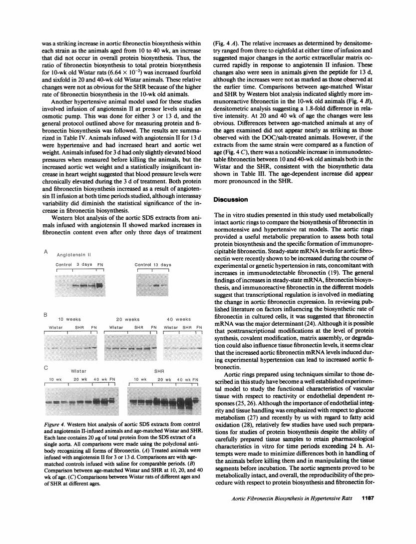
Western blot analysis of the aortic SDSextracts from ani-mals infused with angiotensin II showed marked increases infibronectin content even after only three days of treatment
A Angiotensin II
Control 3 days FNIr-
B10 weeks
Control 13 daysI
20 weeks 40 weeks
Wistar SHR FN Wistar SHR FN Wistar SHR FNI---I- I I I l I I1
C Wistar SHR
10 wk 20 wk 40 wk FN 10 wk 20 wk 40 wk FNI 11 1 1 I
__-__ - w"U
Figure 4. Western blot analysis of aortic SDSextracts from controland angiotensin II-infused animals and age-matched Wistar and SHR.Each lane contains 20 ug of total protein from the SDSextract of asingle aorta. All comparisons were made using the polyclonal anti-body recognizing all forms of fibronectin. (A) Treated animals wereinfused with angiotensin II for 3 or 13 d. Comparisons are with age-matched controls infused with saline for comparable periods. (B)Comparison between age-matched Wistar and SHRat 10, 20, and 40wk of age. (C) Comparisons between Wistar rats of different ages andof SHRat different ages.
(Fig. 4 A). The relative increases as determined by densitome-try ranged from three to eightfold at either time of infusion andsuggested major changes in the aortic extracellular matrix oc-curred rapidly in response to angiotensin II infusion. Thesechanges also were seen in animals given the peptide for 13 d,although the increases were not as marked as those observed atthe earlier time. Comparisons between age-matched Wistarand SHRby Western blot analysis indicated slightly more im-munoreactive fibronectin in the 10-wk old animals (Fig. 4 B),densitometric analysis suggesting a 1.8-fold difference in rela-tive intensity. At 20 and 40 wk of age the changes were lessobvious. Differences between age-matched animals at any ofthe ages examined did not appear nearly as striking as thoseobserved with the DOC/salt-treated animals. However, if theextracts from the same strain were compared as a function ofage (Fig. 4 C), there was a noticeable increase in immunodetec-table fibronectin between 10 and 40-wk old animals both in theWistar and the SHR, consistent with the biosynthetic datashown in Table III. The age-dependent increase did appearmore pronounced in the SHR.
Discussion
The in vitro studies presented in this study used metabolicallyintact aortic rings to compare the biosynthesis of fibronectin innormotensive and hypertensive rat models. The aortic ringsprovided a useful metabolic preparation to assess both totalprotein biosynthesis and the specific formation of immunopre-cipitable fibronectin. Steady-state mRNAlevels for aortic fibro-nectin were recently shown to be increased during the course ofexperimental or genetic hypertension in rats, concomitant withincreases in immunodetectable fibronectin (19). The generalfindings of increases in steady-state mRNA,fibronectin biosyn-thesis, and immunoreactive fibronectin in the different modelssuggest that transcriptional regulation is involved in mediatingthe change in aortic fibronectin expression. In reviewing pub-lished literature on factors influencing the biosynthetic rate offibronectin in cultured cells, it was suggested that fibronectinmRNAwas the major determinant (24). Although it is possiblethat posttranscriptional modifications at the level of proteinsynthesis, covalent modification, matrix assembly, or degrada-tion could also influence tissue fibronectin levels, it seems clearthat the increased aortic fibronectin mRNAlevels induced dur-ing experimental hypertension can lead to increased aortic fi-bronectin.
Aortic rings prepared using techniques similar to those de-scribed in this study have become a well established experimen-tal model to study the functional characteristics of vasculartissue with respect to reactivity or endothelial dependent re-sponses (25, 26). Although the importance of endothelial integ-rity and tissue handling was emphasized with respect to glucosemetabolism (27) and recently by us with regard to fatty acidoxidation (28), relatively few studies have used such prepara-tions for studies of protein biosynthesis despite the ability ofcarefully prepared tissue samples to retain pharmacologicalcharacteristics in vitro for time periods exceeding 24 h. At-tempts were made to minimize differences both in handling ofthe animals before killing them and in manipulating the tissuesegments before incubation. The aortic segments proved to bemetabolically intact, and overall, the reproducibility of the pro-cedure with respect to protein biosynthesis and fibronectin for-
Aortic Fibronectin Biosynthesis in Hypertensive Rats 1187

mation was sufficient to make quantitative comparisons be-tween groups.
Total protein biosynthesis in aortic rings from normoten-sive and hypertensive models was similar in these in vitro stud-ies when normalized to total protein content. This was some-what surprising since aortic hypertrophy is a consistent sequelato the hypertensive state (29), with hypertrophy often beingdefined as an increase in protein per cell. Using cultured vascu-lar smooth muscle cells, studies have shown that incubationwith angiotensin II leads to a hypertrophic response that in-cludes increased protein biosynthesis (30, 31). One explanationfor the similarity in protein biosynthesis between rings could bethat the rings are no longer subjected to mechanical forces thatexist in vivo, and increased wall tension could be a factor me-diating the biochemical differences observed in vivo. Concern-ing the effect of mechanical forces on protein metabolism, in-creased mechanical tension on isolated pulmonary artery seg-ments caused increased biosynthesis of collagen and elastin(32) and studies using cultured smooth muscle cells showedthat collagen biosynthesis was increased by cyclic stretch-ing (33).
The differences in aortic fibronectin biosynthesis betweencontrol and hypertensive groups were most obvious in theDOC/salt model, and the approximately sixfold increase in bio-synthetic rate after 21 d of treatment is quantitatively similar tothe previously reported difference in fibronectin steady-statemRNAlevels (19). Because of the protocol used, we cannotcompletely rule out the possibility that changes in amino aciduptake, precursor pools, or fibronectin degradation account forthe increase in net incorporation of labeled methionine intoimmunoprecipitable fibronectin. However, the absence of sig-nificant differences in net incorporation into total protein doesminimize the possibility that differences in uptake or precursorpools affected the results. With respect to fibronectin degrada-tion, we have not directly compared treated and control ani-mals, but in separate experiments using normal aortic tissue, apulse-chase protocol indicated that the amount of labeled me-thionine incorporated into fibronectin from the SDS extractduring a 4-h pulse was unchanged after a 2, 6, or 24-h chaseperiod. Thus the pool of newly synthesized fibronectin beingmeasured in these studies has a relatively slow turnover.
These data provide evidence that transcriptional events in-fluenced fibronectin biosynthesis in the aorta, and also rein-force the probability that the in vitro biosynthetic data reflectevents occurring in vivo. What was unexpected was the findingthat immunoreactive fibronectin was increased to such a signifi-cant extent, even after only 7 d of DOC/salt treatment, whenblood pressure changes were modest. The immunochemicalassays measure fibronectin that might originate from circulat-ing plasma as well as from aortic synthesis. Unless fibronectinturnover is rapid in aortic tissue, the large difference betweentreated and control groups could reflect increased insudation ofplasma fibronectin in addition to endogenously produced pro-tein. Using the monoclonal antibody to the alternativelyspliced form of fibronectin containing the EIIIA insert, whichis known not to circulate in plasma, we were able to showsignificant increases in endogenously produced material. How-ever, this does not rule out the possibility of infiltration ofplasma fibronectin secondary to DOC/salt treatment.
The changes observed with age also showed a reasonablecorrelation between fibronectin biosynthesis and the previ-ously reported data (19) on steady-state mRNAlevels, which
increased progressively between 5 and 40 wk of age. In general,the increased biosynthetic rate for fibronectin with age in bothWistar and SHRwas consistent with steady-state mRNAdata(19), and with the findings using Western blot analysis, whereincreases were seen in the 40-wk old animals compared tothose of 10 and 20 wk of age. The similar fibronectin biosyn-thetic rates between age-matched normotensive and hyperten-sive animals were in contrast to our previously reported steady-state mRNAdata, which suggested approximately two to three-fold greater levels in hypertensive animals at the ages studied.This could indicate that posttranscriptional regulatory mecha-nisms may influence fibronectin biosynthesis as the animalsage and the composition of the extracellular matrix changes, orit may reflect different regulatory mechanisms for fibronectinexpression in the SHRas compared to the Wistar. Interest-ingly, comparisons of immunodetectable fibronectin betweenthe age-matched strains were more consistent with the biosyn-thetic data than that for steady-state mRNA,further suggestingthe possibility of posttranscriptional control.
Fibronectin is generally considered to have an importantrole in the process of wound repair, and there are analogiesbetween the vascular response to hypertension and wound re-pair. Of potential relevance is the regulatory role of growthfactors in both processes. Transforming growth factor-(B (TGF-fl) was shown to influence fibronectin expression in culturedfibroblasts (34, 35) and is generally thought to influence thewound repair process in vivo, in part by its effects on the extra-cellular matrix. However, TGF-,3 did not influence collagen orfibronectin expression in cultured smooth muscle cells (36,37). Wehave shown that TGF-f# mRNAlevels were increasedin aortae of rats treated with DOC/salt (38) suggesting the possi-bility of autocrine or paracrine influences on aortic extracellu-lar matrix, but there is no definitive study to date showing thatTGF-f modulates aortic gene expression in vivo. Althoughgrowth factors clearly have a role in influencing fibronectinexpression, many other regulatory agents have been proposedand it is premature to state whether hemodynamic forces, para-crine and autocrine regulation, or intracellular regulatoryevents have a predominant role within the vessel wall. How-ever, the use of metabolically intact aortic rings that maintaincell to cell and cell to extracellular matrix interactions in amanner which is perhaps more physiological than that presentin cell culture, offers a potentially valuable system for deter-mining the factors regulating fibronectin biosynthesis.
Acknowledgments
The authors thank Dr. Luciani Zardi for providing the monoclonalantibody to fibronectin used in this study.
This work was supported by National Institutes of Health grantsHL-18318 and HL-31195. Dr. Takasaki was supported in part bygrants 63044118 and 01044120 from Joint Research, the InternationalScientific Research Program of the Ministry of Education, Science, andCulture, Japan.
References
1. Thyberg, J., U. Hedin, M. Sjolund, L. Palmberg, and B. A. Bottger. 1990.Regulation of differentiated properties and proliferation of arterial smooth mus-cle cells. Arteriosclerosis. 10:966-990.
2. Ross, R. 1986. The pathogenesis of atherosclerosis-an update. N. Engi. J.Med. 314:488-500.
3. Gabbiani, G., 0. Kocher, W. S. Bloom, J. Vandekerckhove, and K. Weber.1984. Actin expression in smooth muscle cells of rat aortic intimal thickening,
1188 R. Saouaf I. Takasaki, E. Eastman, A. V. Chobanian, and P. Brecher

human atheromatous plaque, and cultured rat aortic media. J. Clin. Invest.73:148-152.
4. Chamley-Cambell, J., G. R. Cambell, and R. Ross. 1979. The smoothmuscle cell in culture. Physiol. Rev. 59:1-61.
5. Hedin, U., B. A. Bottger, E. Forsberg, S. Johansson, and J. Thyberg. 1988.Diverse effects of fibronectin and laminin on phenotypic properties of culturedarterial smooth muscle cells. J. Cell Biol. 107:307-319.
6. Madri, J. A., B. M. Pratt, and A. M. Tucker. 1988. Phenotypic modulationof endothelial cells by transforming growth factor-# depends upon the composi-tion and organization of the extracellular matrix. J. Cell Biol. 106:1375-1384.
7. Madri, J. A., M. A. Reidy, 0. Kocher, and L. Bell. 1989. Endothelial cellbehavior after denudation injury is modulated by transforming growth factor-jlIand fibronectin. Lab. Invest. 60:755-765.
8. Ingber, D. E. 1990. Fibronectin controls capillary endothelial cell growth bymodulating cell shape. Proc. Natl. Acad. Sci. USA. 87:3579-3583.
9. Jensen, B. A., B. Holund, and I. Clemmensen. 1983. Demonstration offibronectin in normal and injured aorta by an indirect immunoperoxidase tech-nique. Histochemistry. 77:395-403.
10. Rasmussen, L. H., C. Garbarsch, J. Chemnitz, B. C. Christensen, and I.Lorenzen. 1989. Injury and repair of smaller muscularand elastic arteries, immu-nohistochemical demonstration of fibronectin and fibrinogen/fibrin and theirdegradation products in rabbit femoral and commoncarotid arteries following adilatation injury. Virchows Archiv. A Pathol. Anat. Histopathol. 415:579-585.
1 1. Orekhov, A. N., E. R. Andreeva, B. V. Shekhonin, V. V. Tertov, and V. N.Smirnov. 1984. Content and localization of fibronectin in normal intima, athero-sclerotic plaque, and underlying media of human aorta. Atherosclerosis. 53:213-219.
12. Smith, E. B., and C. Ashall. 1986. Fibronectin distribution in humanaortic intima and atherosclerotic lesions, concentration of soluble and collage-nase-releasable fractions. Biochim. Biophys. Acta. 880:10-15.
13. Phan-Thanh, L., J. Robert, J. C. Derouette, and J. Labat-Robert. 1987.Increased biosynthesis and processing of fibronectin in fibroblasts from diabeticmice. Proc. Natl. Acad. Sci. USA. 84:1911-1915.
14. Rasmussen, L. M., and L. Heickendorff. 1989. Quantification of fibronec-tin in extracts of human aortae by an ELISA. Scand. J. Clin. Lab. Invest. 49:205-210.
15. Glukhova, M. A., M. G. Frid, B. V. Shekhonin, T. D. Vasilevskaya, J.Grunwald, M. Saginati, and V. E. Koteliansky. 1989. Expression of extradomainA fibronectin sequence in vascular smooth muscle cells is phenotype dependent.J. Cell Biol. 109:357-366.
16. Wolinsky, H. 1970. Response of the rat aortic media to hypertension.Morphological and chemical studies. Circ. Res. 26:507-522.
17. Ooshima, A., G. C. Fuller, G. J. Cardinale, S. Spector, and S. Udenfriend.1974. Increased collagen synthesis in blood vessels of hypertensive rats and itsreversal by antihypertensive agents. Proc. Natl. Acad. Sci. USA. 71:3019-3023.
18. Brecher, P., C. T. Chan, C. Franzblau, B. Faris, and A. V. Chobanian.1978. Effects of hypertension and its reversal on aortic metabolism in the rat.Circ. Res. 43:561-569.
19. Takasaki, I., A. V. Chobanian, R. Sarzani, and P. Brecher. 1990. Effect ofhypertension on fibronectin expression in the rat aorta. J. Biol. Chem.265:21935-21939.
20. Sarzani, R., K. P. Claffey, A. V. Chobanian, and P. Brecher. 1988. Hyper-tension induces tissue-specific gene suppression of a fatty acid binding protein inrat aorta. Proc. Nall. Acad. Sci. USA. 85:7777-7781.
21. Haudenschild, C. C., M. F. Prescott, and A. V. Chobanian. 1980. Effectsof hypertension and its reversal on aortic intima lesions of the rat. Hypertension(Dallas). 2:33-44.
22. Laemmli, U. K. 1970. Cleavage ofthe structural proteinsduringthe assem-bly of the head of bacteriophage T4. Nature (Lond.). 227:680-685.
23. Borsi, L., B. Carnemolla, P. Castellani, C. Roselli, D. Vecchio, G. Alle-manni, S. E. Chang, J. Taylor-Papadimitriou, H. Pande, and L. Zardi. 1987.Monoclonal antibodies in the analysis of fibronectin isoforms generated by alter-native splicing of mRNAprecursors in normal and transformed human cells. J.Cell Biol. 104:595-600.
24. Hynes, R. 0. 1990. Fibronectins. Springer-Verlag NewYork Inc. p. 319.25. Gryglewski, R. J., R. M. Botting, and J. R. Vane. 1988. Mediators pro-
duced by the endothelial cell. Hypertension (Dallas). 12:530-548.26. Jones, A. W., B. B. Geisbuhler, S. D. Shukla, and J. M. Smith. 1988.
Altered biochemical and functional responses in aorta from hypertensive rats.Hypertension (Dallas). 11:727-634.
27. Morrison, A. D., L. Berwick, L. Orci, and A. I. Winegrad. 1976. Morphol-ogy and metabolism of an aortic intima-media preparation in which an intactendothelium is preserved. J. Clin. Invest. 57:650-660.
28. Takasaki, I., R. A. Cohen, A. V. Chobanian, and P. Brecher. 1990. Effectof endothelial cell denudation on fatty acid metabolism by rabbit aorta. Am. J.Physiol. 259 (Heart Circ. Physiol. 28):H442-H447.
29. Folkow, B. 1982. Physiological aspects of primary hypertension. Physiol.Rev. 62:347-504.
30. Geisterfer, A. A. T., M. J. Peach, and G. K. Owens. 1988. Angiotensin IIinduces hypertrophy, not hyperplasia, of cultured rat aortic smooth muscle cells.Circ. Res. 62:749-756.
31. Berk, B. C., V. Vekshtein, H. M. Gordon, and T. Tsuda. 1989. Angioten-sin II-stimulated protein synthesis in cultured vascular smooth muscle cells. Hppertension (Dallas). 13:305-314.
32. Tozzi, C. A., G. J. Poiani, A. M. Harangozo, C. D. Boyd, and D. J. Riley.1989. Pressure-induced connective tissue synthesis in pulmonary artery segmentsis dependent on intact endothelium. J. Clin. Invest. 84:1005-1012.
33. Leung, D. Y., S. Glagov, and M. B. Matthews. 1976. Cyclic stretchingstimulates synthesis of matrix components by arterial smooth muscle cells invitro. Science (Wash. DC). 191:475-477.
34. Ignotz, R. A., and J. Massague. 1986. Transforminggrowth factor-# stimu-lates the expression of fibronectin and collagen and their incorporation into theextra-cellular matrix. J. Biol. Chem. 261:4337-4345.
35. Roberts, C. J., T. M. Birkenmeier, J. J. McQuillan, S. K Akiyama, S. S.Yamada, W. T. Chen, K. M. Yamada, and J. A. McDonald. 1988. Transforminggrowth factor-fl stimulates the expression of fibronectin and of both subunits ofthe human fibronectin receptor by cultured human lung fibroblasts. J. Biol.Chem. 263:4586-4592.
36. Penttinen, R. P., S. Kobayashi, and P. Bornstein. 1988. Transforminggrowth factor Pi increases mRNAfor matrix proteins both in the presence and inthe absence of changes in mRNAstability. Proc. Natl. Acad. Sci. USA. 85:1105-1108.
37. Liau, G., and L. M. Chan. 1989. Regulation of extracellular matrix RNAlevels in cultured smooth muscle cells, relationship to cellular quiescence. J. Biol.Chem. 264:10315-10320.
38. Sarzani, R., P. Brecher, and A. V. Chobanian. 1989. Growth factorexpres-sion in aorta of normotensive and hypertensive rats. J. Clin. Invest. 83:1404-1408.
Aortic Fibronectin Biosynthesis in Hypertensive Rats 1189
Copyright © 2022 FDOKUMEN