Bladder cancer and reproductive factors among women in Spain
Upload
khangminh22Category
view
4download
0
Research ArticleEnhanced Efficacy of Bleomycin in Bladder Cancer Cellsby Photochemical Internalization
Yan Baglo,1 Lars Hagen,1 Anders Høgset,2 Finn Drabløs,1
Marit Otterlei,1,3 and Odrun A. Gederaas1
1 Department of Cancer Research and Molecular Medicine, Faculty of Medicine, Norwegian University of Scienceand Technology, P.O. Box 8905, 7491 Trondheim, Norway
2 PCI Biotech AS, Strandveien 55, 1366 Lysaker, Norway3 APIMTherapeutics AS, Sem Sælandsvei 14, 7084 Trondheim, Norway
Correspondence should be addressed to Yan Baglo; [email protected]
Received 8 March 2014; Revised 29 May 2014; Accepted 29 May 2014; Published 30 June 2014
Academic Editor: Swaran J. S. Flora
Copyright © 2014 Yan Baglo et al. This is an open access article distributed under the Creative Commons Attribution License,which permits unrestricted use, distribution, and reproduction in any medium, provided the original work is properly cited.
Bleomycin is a cytotoxic chemotherapeutic agent widely used in cancer treatment. However, its efficacy in different cancers islow, possibly due to limited cellular internalization. In this study, a novel approach known as photochemical internalization (PCI)was explored to enhance bleomycin delivery in bladder cancer cells (human T24 and rat AY-27), as bladder cancer is a potentialindication for use of PCI with bleomycin. The PCI technique was mediated by the amphiphilic photosensitizer disulfonatedtetraphenyl chlorin (TPCS
2a) and blue light (435 nm). Two additional strategies were explored to further enhance the cytotoxicity ofbleomycin; a novel peptide drug ATX-101 which is known to impair DNA damage responses, and the protease inhibitor E-64 whichmay reduce bleomycin degradation by inhibition of bleomycin hydrolase. Our results demonstrate that the PCI technique enhancesthe bleomycin effect under appropriate conditions, and importantly we show that PCI-bleomycin treatment leads to increased levelsof DNA damage supporting that the observed effect is due to increased bleomycin uptake. Impairing the DNA damage responsesby ATX-101 further enhances the efficacy of the PCI-bleomycin treatment, while inhibiting the bleomycin hydrolase does not.
1. Introduction
Bladder cancer is one of the most common cancers in theworld and causes more than 100 000 deaths every year [1, 2].In Norway bladder cancer has been one of the five mostcommon cancer types for men during the last ten years [3]and in United States it is estimated that 74 690 new casesand 15 580 deaths will be reported in 2014 [4]. Approxi-mately 70–80% of diagnosed bladder cancers worldwide arenonmuscular invasive bladder cancer (NMIBC) for whichintravesical chemotherapy is used as an adjuvant treatmentto the standard transurethral resection [1]. However, signif-icant improvements in preventing disease progression andrecurrence have not been obtained [1, 2]. Due to high intrin-sic cytotoxicity and low myelosuppression and immune-suppression, bleomycin is used in the treatment of cancerssuch as malignant lymphomas, testicular carcinomas, andsquamous cell carcinomas (see review by Ramotar andWang
[5] and references therein). However, good clinic efficacy hasnot been found in bladder cancer [6–8]. This could be dueto low uptake into the cells as bleomycin consists of ratherlarge water-soluble glycopeptidic molecules that are mostlikely unable to cross cell membranes by passive diffusion,and, thus, rely on endocytosis and/or transporters [9, 10].Studies have shown that cellular responses to bleomycinare cell type-dependent. In cell lines with low sensitivityto bleomycin, the drug resistance is considered mainly tobe due to membrane barrier, degradation by hydrolases inlysosomes or bleomycin hydrolase (BLMH) in the cytosol,elevated DNA repair capacity, and low activity of bleomycintransporters [9–13]. Severe side effects of bleomycin at highdose are therefore limiting its clinical applications [5]. Inan effort to overcome the membrane barrier for bleomycin,electropermeabilization was shown to enhance the efficacy[14, 15].
Hindawi Publishing CorporationBioMed Research InternationalVolume 2014, Article ID 921296, 10 pageshttp://dx.doi.org/10.1155/2014/921296
2 BioMed Research International
The PCI technology has been developed from photody-namic therapy (PDT) as an efficient drug delivery tool toenhance the effect of several types of therapeutic molecules[16]. In the PCI technology a membrane-embedded pho-tosensitizer is used together with a therapeutic agent byendocytotic delivery [17, 18]. The photosensitizers used inPCI, such as mesotetraphenyl chlorin disulfonate (TPCS
2a)used in this study, are designed as amphiphilic moleculesthat initially localize to the plasma membrane, but later areincorporated into the endosomal membranes by endocytosis[19–21]. When used together with bleomycin, the bleomycinmolecules are enclosed in the endocytotic vesicles, andexposure to light leads to endosomal rupture by phototoxicdamage and release of bleomycin molecules into the cytosol[22, 23]. Side effects often seen in the conventional systemictherapeutic strategies can also be reduced by PCI becausethe enhanced effect is localized to the area exposed to light[24, 25]. Enhanced efficacy of PCI with bleomycin has beendocumented in several preclinical studies and clinical trials[24–28]. A phase I clinical trial of TPCS
2a-mediated PCI ofbleomycin showed no severe side effects associated with thetreatment, and the efficacy and safety of the modality arecurrently being evaluated in a phase II interventional clinicaltrial [27, 29].
Once inside the nucleus, bleomycin-inducedDNA strandbreaks are leading to apoptosis, extended cell cycle arrest,mitotic cell death and increased risk of chromosome aberra-tions if not properly repaired [30–32]. A novel designed cell-penetrating peptide named ATX-101, containing the AlkBhomolog 2 PCNA-interactingmotif (APIM), has been shownto enhance cytotoxicity of several chemotherapeutic drugs[33].TheAPIM-motif mediates interaction with proliferatingcell nuclear antigen (PCNA) in many proteins involved inDNA repair, apoptosis, and restart of replication and cell cycleregulation after DNA damage. Impairing the interactionsbetween these proteins and PCNA by ATX-101 impairsthe cellular DNA damage responses and thus sensitizesthe cells to chemotherapeutic drugs [33–37]. In this study,cytotoxicity of TPCS
2a-mediated PCI of bleomycin (PCI-bleomycin) was studied in rat bladder cancer cells (AY-27)and human bladder cancer cells (T24) using the PCI strategyof illumination after bleomycin treatment [38, 39]. For allexperiments, a human epidermoid carcinoma cell line (A431)was used as reference due to its known sensitivity to PCI[40]. Furthermore, we examined the effects of inhibitingthe DNA damage response and bleomycin degradation incombination with PCI-bleomycin. The levels of inducedDNA damages were investigated using the comet assay. Ourresults demonstrate that PCI enhances bleomycin efficacy inhuman and rat bladder cancer cells under optimal conditions.Combination therapy using PCI-bleomycin and ATX-101further enhances the observed cytotoxicity.
2. Materials and Methods
2.1. Cell Culture. Rat bladder transitional carcinoma cells(AY-27) were maintained using the same RPMI culturemedium and conditions as described in our earlier study[41]. Human epidermoid carcinoma cells (A431) and human
bladder carcinoma cells (T24) were maintained in Dulbecco’sModified Eagle’s Medium (D6429) supplemented with 2mML-glutamine, 1% penicillin/streptomycin, 10mM HEPES,1mM sodium pyruvate (Lonza), and 10% (v/v) fetal bovineserum. All medium chemicals were purchased from Sigmaexcept noted.
2.2. Chemicals. TPCS2a (30mgmL−1, Amphinex) dissolved
in Tween 80 and 50mM Tris buffer was provided by PCIBiotech AS (Oslo, Norway) and stored at 4∘C in aliquots. Thestock was first diluted with 50mMTris phosphate buffer (pH8.5) to 0.06mgmL−1 and further diluted with fresh culturemedium immediately before use. All work with TPCS
2a wasperformed under subdued light or light protection.
Bleomycin powder (15000 IU, Baxter, Norway) was dis-solved with 0.9% salt water as stock (1000 IUmL−1) andstored at−20∘C in aliquots. Peptide drugATX-101 (1mM)wassupplied by APIM Therapeutics AS (Trondheim, Norway)and stored at 4∘C in aliquots. Protease inhibitor E-64 powder(Sigma), a known inhibitor of bleomycin hydrolase [42, 43],was diluted with deionized water as stock (1mM) and storedat −20∘C in aliquots. Resazurin sodium salt powder (Sigma)was dissolved with PBS to 2.5mM followed by filtering andsonication under subdued light. Resazurin stock was storedat −20∘C in aliquots. These chemical stocks were furtherdiluted to desired concentrations with fresh culture mediumimmediately before use.
DRAQ5 solution (5mM, BioStatus Limited, UK), a novelDNA-detecting far-red-fluorescing dye [44], was stored at4∘C under light protection and diluted with PBS (1 : 10000)under subdued light immediately before use.
2.3. Light Source. Blue light with 𝜆max of 435 nm used in thisstudywas available fromaLumiSource lamp (PCIBiotechAS,Norway). The lamp is designed specifically to provide stableand homogenous fluency with an irradiance of 12.9mWcm−2over a defined illumination area, allowing attached livingcells to be illuminated from the bottom of culture dishes orplates. With the light doses used in the PCI experiments, thephotosensitizer is not likely to be affected by photobleaching[45].
2.4. Cellular Uptake of 𝑇𝑃𝐶𝑆2𝑎. Cells were seeded out into
96-well plates (6000 cells/well, CytoOne, USA Scientific,Inc.). Attached cells were incubated with TPCS
2a at a seriesof concentrations (18 h). Subsequent to removal of TPCS
2a-medium, fluorescence intensity of cellular accumulatedTPCS
2a was measured using FLUOStar Omega microplatereader (410 nm/650 nm, BMG Labtech GmbH, Germany).The cells were immediately washed with cold PBS, fixedwith fresh 2% paraformaldehyde (150 𝜇L/well, 15min, on ice,no shaking), and stained with 0.5 𝜇M DRAQ5 (50 𝜇L/well,20min, RT, gentle shaking, in the dark).The plates were driedout after washing with cold PBS. Fluorescence intensity ofDNA binding DRAQ5 was measured using Odyssey Imagerat 700 nm channel according to the user manual (Li-CorInfrared imaging system, LI-COR Biosciences, Ltd., UK).Cellular accumulation of TPCS
2a was determined by dividing
BioMed Research International 3
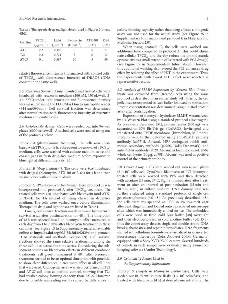
Table 1: Therapeutic drug and light doses (used in Figures 3(b) and4(b)).
Cell line TPCS2a Light Bleomycin ATX-101 E-64(𝜇gmL−1) (J cm−2) (IUmL−1) (𝜇M) (𝜇M)
A431 0.2 0.387 5 5 10T24 0.2 0.774 5 5 10AY-27 0.1 0.387 50 5 50
relative fluorescence intensity (normalized with control cells)of TPCS
2a with fluorescence intensity of DRAQ5 (DNAcontent in the same well).
2.5. Resazurin Survival Assay. Control and treated cells wereincubated with resazurin medium (200𝜇M, 130 𝜇L/well, 2-3 h, 37∘C) under light protection and fluorescence intensitywas measured using the FLUOStar Omega microplate reader(544 nm/590 nm). Cell survival fraction was determinedafter normalization with fluorescence intensity of resazurinmedium and control cells.
2.6. Cytotoxicity Assays. Cells were seeded out into 96-wellplates (6000 cells/well). Attached cells were treated using oneof the protocols below.
Protocol A (photodynamic treatment). The cells were incu-batedwithTPCS
2a for 18 h. Subsequent to removal of TPCS2a-
medium, cells were washed with culture medium twice andchased (4 h) in fresh drug-free medium before exposure toblue light at different intervals [26].
Protocol B (drug treatment). The cells were (co-)incubatedwith drug(s) (bleomycin, ATX-101 or E-64) for 4 h and thenwashed once with culture medium.
Protocol C (PCI-bleomycin treatment). Here protocol B wasincorporated into protocol A after TPCS
2a-treatment. Thetreated cells were (co-)incubated with bleomycin (and ATX-101/E-64) for 4 h instead of being chased in drug-freemedium. The cells were washed once before illumination.Therapeutic drug and light doses are listed in Table 1.
Finally, cell survival fractionwas determined by resazurinsurvival assay after postincubation for 48 h. The time pointof 48 h was selected based on bleomycin effect measured ateach day from 1 to 7 days after bleomycin treatment in thesecell lines (see Figure 1S in Supplementary material availableonline at http://dx.doi.org/10.1155/2014/921296 and protocolD in Materials and Methods, Section 2.9). Cell survivalfractions showed the same relative relationship among thethree cell lines across the time series. Considering the sub-sequent studies on bleomycin effects in different combinedtreatments, cell growth measured at 48 h after bleomycintreatment seemed to be an optimal time point with potentialto show clear differences in treatment effect in all cell linesthat were used. Clonogenic assay was also performed in T24and AY-27 cell lines as method control, showing that T24had weaker colony forming capacity than AY-27. However,due to possibly misleading results caused by differences in
colony forming capacity rather than drug effects, clonogenicassay was not used for the actual study (see Figure 2S inSupplementary Information and protocol E in Materials andMethods, Section 2.9).
When using protocol C, the cells were washed oneadditional time compared to protocol A. This could elimi-nate cellular TPCS
2a and thereby reduce the photodynamiccytotoxicity to a small extent in cells treated with PCI-drug(s)(see Figure 3S in Supplementary Information). However,this additional washing also favored the PCI-enhanced drugeffect by reducing the effect of PDT in the experiment. Thus,the experiments with lowest PDT effect were selected asrepresentative results.
2.7. Analysis of BLMH Expression by Western Blot. Proteinlysate was extracted from (treated) cells using the sameprotocol as described in an earlier study [41]. Briefly, the cellpellet was resuspended in lysis buffer followed by sonication.Protein concentrationwas determined using Bio-Rad proteinassay after centrifugation.
Expression of bleomycin hydrolase (BLMH)was analyzedby 1D Western blot using a standard protocol (Invitrogen).As previously described [34], protein lysates (100 𝜇g) wereseparated on 10% Bis-Tris gel (NuPAGE, Invitrogen) andtransferred onto PVDF membrane (Immobilon, Millipore).Proteins were further detected using anti-BLMH primaryantibody (ab77111, Abcam), HRP-conjugated rabbit anti-mouse secondary antibody (p0260, Dako Denamark), andanti-PCNA antibody (ab29, Abcam) as loading control. K562whole cell lysate (30 𝜇g, ab7911, Abcam) was used as positivecontrol of the primary antibody.
2.8. Comet Assay. Cells were seeded out into 6-well plates(5 × 10⁴ cells/well, CytoOne). Bleomycin or PCI-bleomycintreated cells were washed with PBS and then detachedwith accutase (15min, 37∘C, Sigma) immediately after treat-ment or after an interval of postincubation (15min and30min, resp.) in culture medium. DNA damage level wasfurther evaluated using a standard protocol of single cellgel electrophoresis [46–48]. As previously described [48],the cells were resuspended at 37∘C in 1% low-melt agarafter centrifugation and loaded onto a precoated microscopeslide which was immediately cooled on ice. The embeddedcells were lysed in fresh cold lysis buffer [48] overnightand then electrophoresed in cold alkaline buffer (pH 13.3),thus the comet assay detects single and double strand DNAbreaks, abasic sites, and repair intermediates. DNA fragmentsstained with ethidium bromide were visualized in an invertedfluorescence microscope (Zeiss Axiovert 200M, Germany)equipped with a Sony XCD-X700 camera. Several hundredsof comets in each sample were evaluated using Komet 5.5imaging software (Andor Technology).
2.9. Cytotoxicity Assays Used inthe Supplementary Information
Protocol D (long-term bleomycin cytotoxicity). Cells wereseeded out in 25 cm2 culture flasks (1 × 10⁶ cells/flask) andtreated with bleomycin (4 h) at desired concentrations. The
4 BioMed Research International
Fluo
resc
ence
inte
nsity
per
cell
0 10 20 30 40 500.0
0.3
0.6
0.9
1.2
1.5
0.1 0.3 0.6 1.2 2.4 4.8A431/AY-27
A431/AY-27
0.0952 0.1903 0.0002 0.0713 0.0004A431/T24 0.0280 0.1049 0.0086 0.0509
0.1473 0.3174 0.9727 0.3778 0.9958 0.02097.2 9.6 12 24 48
0.0012 0.0003 0.0001 0.0001A431/T24 0.0002 0.0002
0.1118 0.7095 0.0112 0.0041 0.0229
<0.0001
<0.0001<0.0001
<0.0001
<0.0001 <0.0001 <0.0001
Conc. (𝜇g mL−1)
Conc. (𝜇g mL−1)AY-27/T24
AY-27/T24
Original concentration of TPCS2a (𝜇g/mL)
Table of P values for Figure 1(a)
(a)
0.0 0.2 0.4 0.6 0.80
20
40
60
80
100
120
0.26 0.52 0.78
A431/T24
Cel
l sur
viva
l fra
ctio
n (%
)
A431/AY-27
AY-27/T24
<0.0001
<0.0001
<0.0001 <0.0001 <0.0001
<0.0001<0.0001
<0.0001 <0.0001
Light dose, 435nm (J cm−2)
Light dose (J cm−2)Table of P values for Figure 1(b)
(b)
0 50 100 150 2000
20
40
60
80
100
1 5 25
A431/T240.4916 0.8159 0.0624
50 100 200
A431/T24
Cel
l sur
viva
l fra
ctio
n (%
)
Bleomycin (IU mL−1)
A431/AY-27
A431/AY-27
<0.0001
<0.0001 <0.0001
<0.0001 <0.0001
<0.0001
<0.0001
<0.0001 <0.0001 <0.0001
<0.0001 <0.0001
<0.0001<0.0001<0.0001
A431T24AY27
AY-27/T24
AY-27/T24
Concentration (IU mL−1)
Concentration (IU mL−1)
Table of P values for Figure 1(c)
(c)
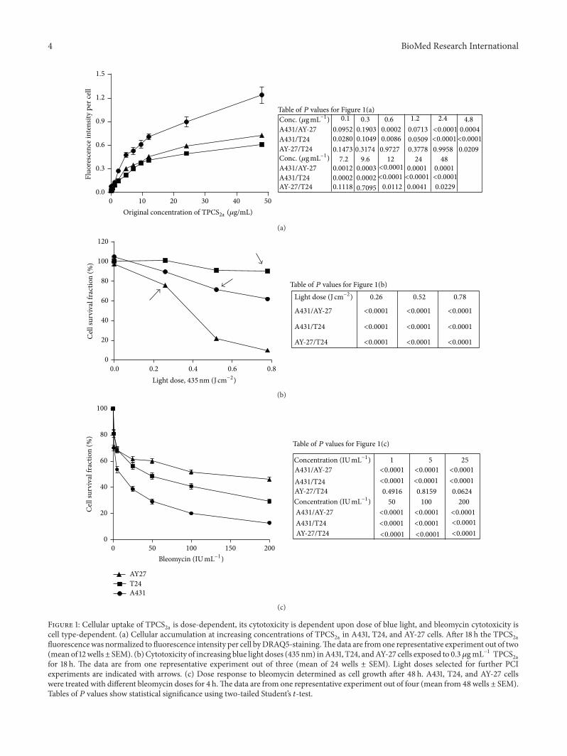
Figure 1: Cellular uptake of TPCS2a is dose-dependent, its cytotoxicity is dependent upon dose of blue light, and bleomycin cytotoxicity is
cell type-dependent. (a) Cellular accumulation at increasing concentrations of TPCS2a in A431, T24, and AY-27 cells. After 18 h the TPCS
2afluorescencewas normalized to fluorescence intensity per cell byDRAQ5-staining.Thedata are fromone representative experiment out of two(mean of 12wells± SEM). (b)Cytotoxicity of increasing blue light doses (435 nm) inA431, T24, andAY-27 cells exposed to 0.3 𝜇gmL−1 TPCS
2afor 18 h. The data are from one representative experiment out of three (mean of 24 wells ± SEM). Light doses selected for further PCIexperiments are indicated with arrows. (c) Dose response to bleomycin determined as cell growth after 48 h. A431, T24, and AY-27 cellswere treated with different bleomycin doses for 4 h.The data are from one representative experiment out of four (mean from 48 wells ± SEM).Tables of 𝑃 values show statistical significance using two-tailed Student’s 𝑡-test.

BioMed Research International 5
cells were washed with PBS and detached with trypsin beforereseeding into 96-well plates (1000 cells/well). Cell survivalfraction was determined by resazurin survival assay from thenext day, day 1 until day 7.
Protocol E (clonogenic assay). Cells were seeded out andtreated with bleomycin (4 h) as described in Protocol Dbefore reseeding into petri dishes (100 cells/dish). The cellswere incubated with culture medium for 7–10 days allowingcolony formation. After washing with PBS, the colonies werefixed with 6% glutaraldehyde (30min) and then stainedwith 0.5% crystal violet (30min). Plating efficiency (PE) andsurviving fraction (SF) were calculated after counting the air-dried colonies (PE = number of colonies counted/number ofcells plated × 100%; SF = PE of treated sample/PE of control× 100%).
3. Results and Discussion
3.1. Cellular Uptake of 𝑇𝑃𝐶𝑆2𝑎
and Dose Responses to BlueLight and Bleomycin. Theuptake of TPCS
2a to the endosomalmembrane is essential for drug delivery. To assess the uptakecapacity in the bladder cancer cell lines T24 and AY-27,compared to the reference skin cancer cell line A431 (earliershown to have good PCI efficacy [40]), fluorescence intensityof cellular TPCS
2a was measured and normalized againstcell count. The results showed that the uptake was dose-dependent in all three cell lines (Figure 1(a)). Importantly,a much higher uptake was seen in the skin cancer cell lineA431, compared to the bladder cancer cell lines T24 and AY-27 (Figure 1(a)). The endocytotic rate is cell type-dependent[49], and this may account for differences in cellular TPCS
2auptake as seen in these cell lines.
According to the principle of PCI, the applied doses ofboth photosensitizer and light (termed photodynamic dosein this paper) are supposed to be sublethal, leading mainlyto damage to the endosomal/lysosomalmembrane.Thereforewe set out to determine the sublethal photodynamic dosesbefore evaluating PCI-bleomycin efficacy. No dark toxicity(Figure 4S in Supplementary Information), TPCS
2a buffertoxicity (Tween 80 and 50mM Tris buffer), or light toxicitywas observed in any of the cell lines under the experimentalconditions (data not shown), thus the cytotoxicity is depen-dent upon light activation of the TPCS
2a molecules. Theresults showed that T24 cells were resistant to activation byblue light although they had similar TPCS
2a uptake as theAY-27 cells that were very light sensitive (Figure 1(b)). A431cells, which accumulated more TPCS
2a molecules than thetwo other cell lines, were medium sensitive to light activation(Figure 1(b)). Light doses that gave low (0–20%) reductionin cell survival in this experiment (Figure 1(b), arrows) wereselected for the PCI-bleomycin experiments, but in thefollowing experiments a further three-time reduction of theTPCS
2a concentration was applied in order to further reducethe background cytotoxicity. In summary, these results showthat the cell lines accumulate different levels of TPCS
2a, butthat this did not directly correlate with their light sensitivity.
Next, we tested the bleomycin sensitivity of the differentcell lines. We treated the cells with bleomycin for 4 hours and
0
20
40
60
80
100
Cel
l sur
viva
l fra
ctio
n (%
)
A431
PDT
A431
bleo
myc
in
A431
PCI-
bleo
myc
in
AY27
PDT
AY27
bleo
myc
in
AY27
PCI-
bleo
myc
in
T24
PDT
T24
bleo
myc
in
T24
PCI-
bleo
myc
in
P < 0.0001
P < 0.0001 P < 0.0001
Figure 2: PCI enhances cytotoxic effect of bleomycin in all three celllines. Cell survival fractions were determined 48 h after incubationwith 0.1𝜇gmL−1 TPCS
2a for 18 h, 50 IUmL−1 bleomycin or freshmedium for 4 h, and illumination (light doses as indicated inFigure 1(b)) in sequence. The data are from one representativeexperiment out of three (mean of 24 wells ± SD). 𝑃 values werecalculated using two-tailed Student’s 𝑡-test and the values (𝑃 <0.0001) indicated significant differences between bleomycin andPCI-bleomycin treatments.
measured the cell growth after 48 hours. The results showedthat the reference cell line A431 was the most and AY-27 theleast sensitive cell line to bleomycin (Figure 1(c)).
3.2. The Effect of Bleomycin Is Increased in Combination withPCI. To determine the efficacy of PCI-bleomycin in the threecell lines, sublethal doses of light (see arrows in Figure 1(b))andTPCS
2a (0.1 𝜇gmL−1) were used.𝑃 values were calculatedusing two-tailed Student’s 𝑡-test and the values (𝑃 < 0.0001)indicated that differences in cytotoxicity between bleomycinversus PCI-bleomycin treatment were highly significant. Inaddition, the PDT effect included in PCI-bleomycin treat-ment in each cell line was less than its PDT control dueto an additional washing (see Section 2.6 in Materials andMethods). The results showed that bleomycin cytotoxicitywas enhanced up to 20% by PCI, independent of cell type(Figure 2).This is likely due to increased uptake of bleomycinin the cells. At the selected conditions, A431 cells were moresensitive than the bladder cancer cells (44% surviving cellsversus 55%, respectively, Figure 2). This probably reflectsthat this reference cell line had highest uptake of TPCS
2ain addition to being the most sensitive towards bleomycin(Figures 1(a) and 1(c)). These results are in agreement withrecent results reported by Arentsen et al. [28]. Since PCI-bleomycin effect was still shown to be lower in the bladdercancer cell lines than in the reference skin cell line, twoadditional combination strategies to improve the effect wereexplored as shown in the next two sections.
6 BioMed Research International
K-562
posit
ivel
AY27
cont
rol
AY27
E-64
treat
ed
T24
cont
rol
T24
E-64
treat
ed
A431
cont
rol
A431
E-64
treat
ed
BLMH
PCNA
53kDa
29kDa
(a)C
ell s
urvi
val f
ract
ion
(%)
0
20
40
60
80
100
T24
bleo
myc
in (5
IU m
L−1)
T24
bleo
myc
in(5
IUm
L−1)+
E-64
A431
bleo
myc
in (5
IU m
L−1)
A431
bleo
myc
in(5
IUm
L−1)+
E-64
A431
bleo
myc
in (50
IU m
L−1)
A431
bleo
myc
in(5
0IU
mL−
1)+
E-64
T24
bleo
myc
in (50
IU m
L−1)
T24
bleo
myc
in(5
0IU
mL−
1)+
E-64
AY27
bleo
myc
in (50
IU m
L−1)
AY27
bleo
myc
in(5
0IU
mL−
1)+
E-64
AY27
bleo
myc
in (5
IU m
L−1)
AY27
bleo
myc
in(5
IUm
L−1)+
E-64
(b)
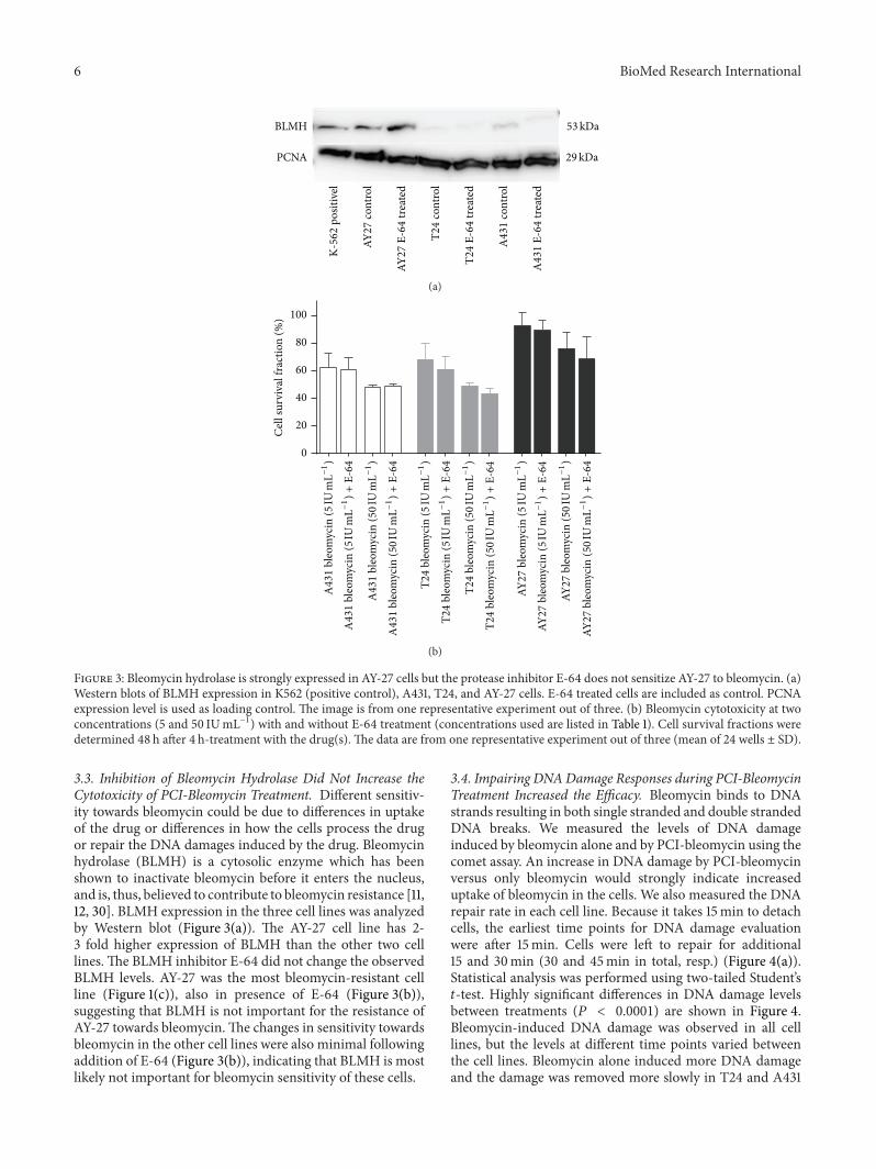
Figure 3: Bleomycin hydrolase is strongly expressed in AY-27 cells but the protease inhibitor E-64 does not sensitize AY-27 to bleomycin. (a)Western blots of BLMH expression in K562 (positive control), A431, T24, and AY-27 cells. E-64 treated cells are included as control. PCNAexpression level is used as loading control. The image is from one representative experiment out of three. (b) Bleomycin cytotoxicity at twoconcentrations (5 and 50 IUmL−1) with and without E-64 treatment (concentrations used are listed in Table 1). Cell survival fractions weredetermined 48 h after 4 h-treatment with the drug(s). The data are from one representative experiment out of three (mean of 24 wells ± SD).
3.3. Inhibition of Bleomycin Hydrolase Did Not Increase theCytotoxicity of PCI-Bleomycin Treatment. Different sensitiv-ity towards bleomycin could be due to differences in uptakeof the drug or differences in how the cells process the drugor repair the DNA damages induced by the drug. Bleomycinhydrolase (BLMH) is a cytosolic enzyme which has beenshown to inactivate bleomycin before it enters the nucleus,and is, thus, believed to contribute to bleomycin resistance [11,12, 30]. BLMH expression in the three cell lines was analyzedby Western blot (Figure 3(a)). The AY-27 cell line has 2-3 fold higher expression of BLMH than the other two celllines. The BLMH inhibitor E-64 did not change the observedBLMH levels. AY-27 was the most bleomycin-resistant cellline (Figure 1(c)), also in presence of E-64 (Figure 3(b)),suggesting that BLMH is not important for the resistance ofAY-27 towards bleomycin.The changes in sensitivity towardsbleomycin in the other cell lines were also minimal followingaddition of E-64 (Figure 3(b)), indicating that BLMH is mostlikely not important for bleomycin sensitivity of these cells.
3.4. Impairing DNADamage Responses during PCI-BleomycinTreatment Increased the Efficacy. Bleomycin binds to DNAstrands resulting in both single stranded and double strandedDNA breaks. We measured the levels of DNA damageinduced by bleomycin alone and by PCI-bleomycin using thecomet assay. An increase in DNA damage by PCI-bleomycinversus only bleomycin would strongly indicate increaseduptake of bleomycin in the cells. We also measured the DNArepair rate in each cell line. Because it takes 15min to detachcells, the earliest time points for DNA damage evaluationwere after 15min. Cells were left to repair for additional15 and 30min (30 and 45min in total, resp.) (Figure 4(a)).Statistical analysis was performed using two-tailed Student’s𝑡-test. Highly significant differences in DNA damage levelsbetween treatments (𝑃 < 0.0001) are shown in Figure 4.Bleomycin-induced DNA damage was observed in all celllines, but the levels at different time points varied betweenthe cell lines. Bleomycin alone induced more DNA damageand the damage was removed more slowly in T24 and A431
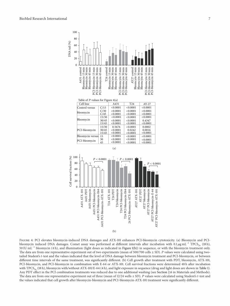
BioMed Research International 7
DN
A ta
il (%
)
A431
cont
rol
Bleo
myc
in15min
Bleo
myc
in30min
Bleo
myc
in45min
PCI-
bleo
myc
in15min
T24
cont
rol
Bleo
myc
in15min
Bleo
mcy
in30min
Bleo
mcy
in45min
PCI-
bleo
mcy
in15min
PCI-
bleo
mcy
in30min
PCI-
bleo
mcy
in45min
AY27
cont
rol
Bleo
mcy
in15min
Bleo
mcy
in30min
Bleo
mcy
in45min
PCI-
bleo
mcy
in15min
PCI-
bleo
mcy
in30min
PCI-
bleo
myc
in30min
PCI-
bleo
myc
in45min
PCI-
bleo
myc
in45min
0
20
40
60
80
100
Cell line A431 T24Control versus bleomycin
C/15C/30C/45
Bleomycin 15/3030/45 0.434715/4515/30 0.5676 0.000230/45 0.0242 0.001615/45
Bleomycin versus 153045
AY-27<0.0001
<0.0001<0.0001<0.0001
<0.0001<0.0001
<0.0001<0.0001<0.0001<0.0001
<0.0001
<0.0001
<0.0001<0.0001<0.0001
<0.0001<0.0001
<0.0001
<0.0001<0.0001<0.0001<0.0001
<0.0001
<0.0001<0.0001<0.0001
<0.0001
<0.0001
<0.0001<0.0001
<0.0001
PCI-bleomycin
PCI-bleomycin
Table of P-values for Figure 4(a)
(a)
0
20
40
60
80
100
Cel
l sur
viva
l fra
ctio
n (%
)
A431
ATX-101
A431
PDT
A431
bleo
myc
inA431
PCI-
bleo
myc
inA431
PCI-
bleo
myc
in-E64
A431
PCI-
bleo
myc
in-A
TX-101
T24
ATX-101
T24
PDT
T24
bleo
myc
inT24
PCI-
bleo
myc
inT24
PCI-
bleo
myc
in-E64
24
PCI-
bleo
myc
in-A
TX-101
AY27
ATX-101
AY27
PDT
AY27
bleo
myc
inAY
27
PCI-
bleo
myc
inAY
27
PCI-
bleo
myc
in-E
-64
AY27
PCI-
bleo
myc
in-A
TX-101
P < 0.0001 P < 0.0001
P < 0.0001
(b)
Figure 4: PCI elevates bleomycin-induced DNA damages and ATX-101 enhances PCI-bleomycin cytotoxicity. (a) Bleomycin and PCI-bleomycin induced DNA damages. Comet assay was performed at different intervals after incubation with 0.1𝜇gmL−1 TPCS
2a (18 h),50 IUmL−1 bleomycin (4 h), and illumination (light doses as indicated in Figure 1(b)) in sequence, or with the bleomycin treatment only.The data are from one representative experiment out of two experiments (mean of 500/700 cells ± SD). 𝑃 values were calculated using two-tailed Student’s 𝑡-test and the values indicated that the level of DNA damage between bleomycin treatment and PCI-bleomycin, or betweendifferent time intervals of the same treatment, was significantly different. (b) Cell growth after treatment with PDT, bleomycin, ATX-101,PCI-bleomycin, and PCI-bleomycin in combination with E-64 or ATX-101. Cell survival fractions were determined 48 h after incubationwith TPCS
2a (18 h), bleomycin with/without ATX-101/E-64 (4 h), and light exposure in sequence (drug and light doses are shown in Table 1).Any PDT effect in the PCI combination treatments was reduced due to one additional washing (see Section 2.6 in Materials and Methods).The data are from one representative experiment out of three (mean of 12/24 wells ± SD). 𝑃 values were calculated using Student’s 𝑡-test andthe values indicated that cell growth after bleomycin-bleomycin and PCI-bleomycin-ATX-101 treatment were significantly different.
8 BioMed Research International
than in AY-27 cells, consistent with the observation thatAY-27 was more resistant to bleomycin. Furthermore, theresults showed that PCI-bleomycin induced a higher levelof DNA damage than bleomycin alone in all three cell lines(Figure 4(a)), supporting increased import of bleomycin.Thehighest increase in levels of DNA damage was seen in AY-27 cells, and importantly no reduction in the level of DNAdamage could be detected 45 minutes after PCI-bleomycintreatment in this cell line, suggesting that the repair capacitywas saturated. The observation that cell survival after 48hours was 55% (see Figure 2) shows that a large fraction ofthe cells were eventually repaired, but at later time points.
The novel peptide drug ATX-101 has the potential toreduce several aspects of the cellular defense systems, includ-ing DNA repair, and is therefore enhancing the efficacy ofseveral chemotherapeutics [33–35]. We tested if ATX-101could increase the efficacy of PCI-bleomycin and found thatATX-101 enhanced the PCI-bleomycin efficacy with 14.7%,30.5%, and 20.7% in A431, T24, and AY-27 cells, respectively,showing statistically significant differences (Figure 4(b)).The cytotoxic effects of the double combinations ATX-101-bleomycin and ATX-101-TPCS
2a-PDT are similar or lowerthan PCI-bleomycin and lower than the triple combinationATX-101-PCI-bleomycin (data not shown). It should be takeninto account that any PDT effect in these PCI combinationtreatments was reduced due to one additional washing (seeSection 2). The additive effects of ATX-101 were stronger inthe bladder cancer cells than in the skin cancer cells (referencecell line) under these experimental conditions (Figure 4(b)).Because the bladder cancer cells were less sensitive towardsbleomycin both with regard to cell survival and inductionof DNA damage (Figures 1 and 4) these results suggestthat the cellular DNA repair capacity is a crucial factor forthe efficacy of bleomycin, and therefore also the efficacy ofPCI-bleomycin. As expected from the results in Figure 3(b),addition of E-64 did not affect PCI-bleomycin efficacy.
4. Conclusions
This is a fundamental study of specific aspects of the PCItechnique. Using sublethal photodynamic doses, our resultsshow that the cytotoxic effect of bleomycin is enhanced byTPCS
2a-mediated PCI in the tested bladder cancer cells,although these cell lines show clear differences with respectto sensitivity to photosensitizer uptake, light dose, and DNArepair capacity. We show that the PCI technique elevatesbleomycin-induced DNA damage levels in all three celllines, strongly suggesting that more bleomycin moleculesenter the nuclei compared to treatment with bleomycin asa single agent. Thus, the membrane barrier to bleomycinseems to be bypassed by the PCI technique. We have furtherdemonstrated that application of a cocktail of PCI-bleomycinand ATX-101, under condition where individual drug levelshave no or low toxicity, reaches a promising therapeutic effectin the human bladder cancer cells T24 which is two-foldstronger than in the reference cell line, the human skin cancercells A431. Combining PCI-chemotherapy treatments withinhibitors of DNA repair therefore seems to be a promisingtherapeutic strategy for increased efficacy “on site.”
Conflict of Interests
Professor Marit Otterlei is an inventor, minority shareholder,and CSO in APIM Therapeutics, a spin-off company of theNorwegianUniversity of Science and Technology. Dr. AndersHøgset is an inventor, minority shareholder, and CSO in PCIBiotech AS.
Acknowledgments
The authors would like to thank PCI Biotech AS (Oslo,Norway) for TPCS
2a and APIM Therapeutics AS (Trond-heim, Norway) for ATX-101 supply. The cell line AY-27 wasgenerously supplied by Professor Steven H. Selman (MedicalCollege of Ohio, Toledo, OH). This study was supported bygrants from Liaison Committee between the Central NorwayRegional Health Authority and the Norwegian Universityof Science and Technology (NTNU). They gratefully thankPh.D. candidate Linda Helander, department engineer SiriBackhe, and senior engineer Nina-Beate Liabakk at theirdepartment, and senior engineer Karin Solvang-Garten atDepartment of Circulation and Medical Imaging (NTNU)for their lab support and discussions. They gratefully thankScientist Qian Peng (Department of Pathology, the Norwe-gian Radium Hospital, Oslo) for his excellent professionaldiscussions and suggestions.
References
[1] M. C. Hall, S. S. Chang, G. Dalbagni et al., “Guideline for themanagement of nonmuscle invasive bladder cancer (stages Ta,T1, and Tis): 2007 update,” The Journal of Urology, vol. 178, no.6, pp. 2314–2330, 2007.
[2] T. R. L. Griffiths, “Current perspectives in bladder cancermanagement,” International Journal of Clinical Practice, vol. 67,no. 5, pp. 435–448, 2013.
[3] I. K. Larsen, B. Sæther, andB.Aagnes, “Cancer inNorway,” 2010,http://www.kreftregisteret.no/no/Generelt/Publikasjoner/Can-cer-in-Norway/Cancer-in-Norway-2010/.
[4] National Cancer Institute, Bladder Cancer, 2014, http://www.cancer.gov/cancertopics/types/bladder.
[5] D. Ramotar and H. Wang, “Protective mechanisms againstthe antitumor agent bleomycin: lessons from Saccharomycescerevisiae,” Current Genetics, vol. 43, no. 4, pp. 213–224, 2003.
[6] R. B. Bracken, D. E. Johnson, L. Rodriquez, M. L. Samuels, andA. Ayala, “Treatment of multiple superficial tumors of bladderwith intravesical bleomycin,” Urology, vol. 9, no. 2, pp. 161–163,1977.
[7] A. G. Turner, K. R. Durrant, and J. S. Malpas, “A trial ofbleomycin versus adriamycin in advanced carcinoma of thebladder,” British Journal of Urology, vol. 51, no. 2, pp. 121–124,1979.
[8] N. Gad-el-Mawla, R. Hamsa, E. Chevlen, and J. L. Ziegler,“Phase II trial of bleomycin in bilharzial bladder cancer,”CancerTreatment Reports, vol. 62, no. 7, pp. 1109–1110, 1978.
[9] G. Pron, N. Mahrour, S. Orlowski et al., “Internalisation ofthe bleomycin molecules responsible for bleomycin toxicity: areceptor-mediated endocytosis mechanism,” Biochemical Phar-macology, vol. 57, no. 1, pp. 45–56, 1999.
BioMed Research International 9
[10] M. Aouida, R. Poulin, and D. Ramotar, “The human carnitinetransporter SLC22A16mediates high affinity uptake of the anti-cancer polyamine analogue bleomycin-A5,” Journal of BiologicalChemistry, vol. 285, no. 9, pp. 6275–6284, 2010.
[11] S. M. Sebti, J. P. Jani, S. Mistry, E. Gorelik, and J. S. Lazo,“Metabolic inactivation: a mechanism of human tumor resis-tance to bleomycin,” Cancer Research, vol. 51, no. 1, pp. 227–232,1991.
[12] D. R. Schwartz, G. E. Homanics, D. G. Hoyt, E. Klein, J.Abernethy, and J. S. Lazo, “The neutral cysteine proteasebleomycin hydrolase is essential for epidermal integrity andbleomycin resistance,” Proceedings of the National Academy ofSciences of the United States of America, vol. 96, no. 8, pp. 4680–4685, 1999.
[13] L.H. Einhorn, “Curingmetastatic testicular cancer,”Proceedingsof the National Academy of Sciences of the United States ofAmerica, vol. 99, no. 7, pp. 4592–4595, 2002.
[14] Y. Kubota, T. Nakada, H. Yanai, K. Itoh, I. Sasagawa, and K.Kawai, “Histological evaluation of the effects of electroperme-abilization after administration of bleomycin on bladder cancerin the rat,” European Urology, vol. 34, no. 4, pp. 372–376, 1998.
[15] Y. Kubota, T. Nakada, H. Yanai, H. Kakizaki, I. Sasagawa,and M. Watanabe, “Electropermeabilization in bladder cancerchemotherapy,” Cancer Chemotherapy and Pharmacology, vol.39, no. 1-2, pp. 67–70, 1996.
[16] T. J. Dougherty, “Photodynamic therapy,” Photochemistry andPhotobiology, vol. 58, no. 6, pp. 895–900, 1993.
[17] K. Berg, M. Folini, L. Prasmickaite et al., “Photochemical inter-nalization: a new tool for drug delivery,”Current PharmaceuticalBiotechnology, vol. 8, no. 6, pp. 362–372, 2007.
[18] K. Berg, P. K. Selbo, L. Prasmickaite et al., “Photochemical inter-nalization: a novel technology for delivery of macromoleculesinto cytosol,”Cancer Research, vol. 59, no. 6, pp. 1180–1183, 1999.
[19] K. Berg, S. Nordstrand, P. K. Selbo, D. T. T. Tran, E. Angell-Petersen, and A. Hogset, “Disulfonated tetraphenyl chlorin(TPCS
2𝑎), a novel photosensitizer developed for clinical uti-
lization of photochemical internalization,” Photochemical &Photobiological Sciences, vol. 10, no. 10, pp. 1637–1651, 2011.
[20] P. K. Selbo, G. Sivam, O. Fodstad, K. Sandvig, and K. Berg, “Invivo documentation of photochemical internalization, a novelapproach to site specific cancer therapy,” International Journalof Cancer, vol. 92, pp. 761–766, 2001.
[21] M. Lilletvedt, H. H. Tønnesen, A. Høgset, L. Nardo, and S.Kristensen, “Physicochemical characterization of the photo-sensitizers TPCS
2𝑎and TPPS
2𝑎1. Spectroscopic evaluation of
drug—solvent interactions,” Die Pharmazie, vol. 65, no. 8, pp.588–595, 2010.
[22] K. Berg, A. Dietze, O. Kaalhus, and A. Høgset, “Site-specificdrug delivery by photochemical internalization enhances theantitumor effect of bleomycin,” Clinical Cancer Research, vol. 11,no. 23, pp. 8476–8485, 2005.
[23] K. Berg, P. K. Selbo, L. Prasmickaite, and A. Høgset, “Photo-chemical drug and gene delivery,”Current Opinion inMolecularTherapeutics, vol. 6, no. 3, pp. 279–287, 2004.
[24] O. Norum, K. Giercksky, and K. Berg, “Photochemical internal-ization as an adjunct to marginal surgery in a human sarcomamodel,” Photochemical and Photobiological Sciences, vol. 8, no.6, pp. 758–762, 2009.
[25] O. J. Norum, J. V. Gaustad, E. Angell-Petersen et al., “Pho-tochemical internalization of bleomycin is superior to photo-dynamic therapy due to the therapeutic effect in the tumor
periphery,” Photochemistry and Photobiology, vol. 85, no. 3, pp.740–749, 2009.
[26] K. Berg, A. Weyergang, L. Prasmickaite et al., “Photochemicalinternalization (PCI): a technology for drug delivery,”Methodsin Molecular Biology, vol. 635, pp. 133–145, 2010.
[27] “Study of Amphinex Based Photochemical Internalisation(PCI) of Bleomycin in Patients with Cutaneous Cancer,”http://clinicaltrials.gov/show/NCT00993512.
[28] H. C. Arentsen, J. Falke, A. Høgset, E. Oosterwijk, and J.A. Witjes, “The effect of photochemical internalization ofbleomycin in the treatment of urothelial carcinoma of thebladder: an in vitro study,” Urologic Oncology, vol. 32, no. 1, pp.49.e1–49.e6, 2014.
[29] ClinicalTrials.gov, “A Study to Evaluate the Safety andEfficacy of PC-A11 in Patients with Recurrent Headand Neck Squamous Cell Carcinoma,” August 2013,http://clinicaltrials.gov/ct2/show/study/NCT01606566.
[30] J. Chen and J. Stubbe, “Bleomycins: towards better therapeutics,”Nature Reviews Cancer, vol. 5, no. 2, pp. 102–112, 2005.
[31] O. Tounekti, G. Pron, J. Belehradek Jr., and L. M. Mir,“Bleomycin, an apoptosis-mimetic drug that induces two typesof cell death depending on the number of molecules internal-ized,” Cancer Research, vol. 53, no. 22, pp. 5462–5469, 1993.
[32] O. Tounekti, A. Kenani, N. Foray, S. Orlowski, and L. M. Mir,“The ratio of single-to double-strand DNA breaks and theirabsolute values determine cell death pathway,” British Journalof Cancer, vol. 84, no. 9, pp. 1272–1279, 2001.
[33] R. Muller, K. Misund, T. Holien et al., “Targeting proliferatingcell nuclear antigen and its protein interactions induces apop-tosis in multiple myeloma cells,” PLoS ONE, vol. 8, no. 7, ArticleID e70430, 2013.
[34] K.M. Gilljam, E. Feyzi, P. A. Aas et al., “Identification of a novel,widespread, and functionally important PCNA-binding motif,”Journal of Cell Biology, vol. 186, no. 5, pp. 645–654, 2009.
[35] K. M. Gilljam, R. Muller, N. B. Liabakk, and M. Otterlei,“Nucleotide excision repair is associated with the replisome andits efficiency depends on a direct interaction between XPA andPCNA,” PLoS ONE, vol. 7, no. 11, Article ID e49199, 2012.
[36] A. Ciccia, A. V. Nimonkar, Y. Hu et al., “PolyubiquitinatedPCNA recruits the ZRANB3 translocase to maintain genomicintegrity after replication stress,” Molecular Cell, vol. 47, no. 3,pp. 396–409, 2012.
[37] A. Bacquin, C. Pouvelle, N. Siaud et al., “The helicase FBH1 istightly regulated by PCNA via CRL4(Cdt2)-mediated proteol-ysis in human cells,” Nucleic Acids Research, vol. 41, no. 13, pp.6501–6513, 2013.
[38] L. Prasmickaite, A. Høgset, P. K. Selbo, B. Ø. Engesæter, M.Helium, and K. Berg, “Photochemical disruption of endocyticvesicles before delivery of drugs: a new strategy for cancertherapy,” British Journal of Cancer, vol. 86, no. 4, pp. 652–657,2002.
[39] M. B. Berstad, A. Weyergang, and K. Berg, “Photochemi-cal internalization (PCI) of HER2-targeted toxins: synergy isdependent on the treatment sequence,” Biochimica et BiophysicaActa, vol. 1820, no. 12, pp. 1849–1858, 2012.
[40] A.Weyergang, P. K. Selbo, and K. Berg, “Photochemically stim-ulated drug delivery increases the cytotoxicity and specificity ofEGF-saporin,” Journal of Controlled Release, vol. 111, no. 1-2, pp.165–173, 2006.
[41] Y. Baglo, M. M. L. Sousa, G. Slupphaug et al., “Photodynamictherapy with hexyl aminolevulinate induces carbonylation,
10 BioMed Research International
posttranslational modifications and changed expression of pro-teins in cell survival and cell death pathways,” Photochemicaland Photobiological Sciences, vol. 10, no. 7, pp. 1137–1145, 2011.
[42] S. K. Sreedharan, C. Verma, L. S. D. Caves et al.,“Demonstration that 1-trans-epoxysuccinyl-L-leucylamido-(4-guanidino)butane (E-64) is one of the most effective low Mrinhibitors of trypsin-catalysed hydrolysis. Characterization bykinetic analysis and by energy minimization and moleculardynamics simulation of the E-64-𝛽-trypsin complex,” TheBiochemical Journal, vol. 316, part 3, pp. 777–786, 1996.
[43] G. Morris, J. S. Mistry, J. P. Jani, S. M. Sebti, and J. S. Lazo,“Cysteine proteinase inhibitors and bleomycin-sensitive and -resistant cells,” Biochemical Pharmacology, vol. 41, no. 11, pp.1559–1566, 1991.
[44] R. Edward, “Red/far-red fluorescing dna-specificanthraquinones for nucl: cyto segmentation and viabilityreporting in cell-based assays,” Methods in Enzymology, vol.505, pp. 23–45, 2012.
[45] J. T.Wang, K. Berg, A. Høgset, S. G. Bown, andA. J.MacRobert,“Photophysical and photobiological properties of a sulfonatedchlorin photosensitiser TPCS
2𝑎for photochemical internalisa-
tion (PCI),” Photochemical & Photobiological Sciences, vol. 12,no. 3, pp. 519–526, 2013.
[46] N. P. Singh, M. T. McCoy, R. R. Tice, and E. L. Schneider, “Asimple technique for quantitation of low levels of DNA damagein individual cells,” Experimental Cell Research, vol. 175, no. 1,pp. 184–191, 1988.
[47] P. L. Olive and J. P. Banath, “The comet assay: a method tomeasure DNAdamage in individual cells,”Nature Protocols, vol.1, no. 1, pp. 23–29, 2006.
[48] A. Hanssen-Bauer, K. Solvang-Garten, K. M. Gilljam et al.,“The region of XRCC1 which harbours the three most commonnonsynonymous polymorphic variants, is essential for thescaffolding function of XRCC1,” DNA Repair, vol. 11, no. 4, pp.357–366, 2012.
[49] S. Mukherjee, R. N. Ghosh, and F. R. Maxfield, “Endocytosis,”Physiological Reviews, vol. 77, no. 3, pp. 759–803, 1997.

Submit your manuscripts athttp://www.hindawi.com
Stem CellsInternational
Hindawi Publishing Corporationhttp://www.hindawi.com Volume 2014
Hindawi Publishing Corporationhttp://www.hindawi.com Volume 2014
MEDIATORSINFLAMMATION
of
Hindawi Publishing Corporationhttp://www.hindawi.com Volume 2014
Behavioural Neurology
EndocrinologyInternational Journal of
Hindawi Publishing Corporationhttp://www.hindawi.com Volume 2014
Hindawi Publishing Corporationhttp://www.hindawi.com Volume 2014
Disease Markers
Hindawi Publishing Corporationhttp://www.hindawi.com Volume 2014
BioMed Research International
OncologyJournal of
Hindawi Publishing Corporationhttp://www.hindawi.com Volume 2014
Hindawi Publishing Corporationhttp://www.hindawi.com Volume 2014
Oxidative Medicine and Cellular Longevity
Hindawi Publishing Corporationhttp://www.hindawi.com Volume 2014
PPAR Research
The Scientific World JournalHindawi Publishing Corporation http://www.hindawi.com Volume 2014
Immunology ResearchHindawi Publishing Corporationhttp://www.hindawi.com Volume 2014
Journal of
ObesityJournal of
Hindawi Publishing Corporationhttp://www.hindawi.com Volume 2014
Hindawi Publishing Corporationhttp://www.hindawi.com Volume 2014
Computational and Mathematical Methods in Medicine
OphthalmologyJournal of
Hindawi Publishing Corporationhttp://www.hindawi.com Volume 2014
Diabetes ResearchJournal of
Hindawi Publishing Corporationhttp://www.hindawi.com Volume 2014
Hindawi Publishing Corporationhttp://www.hindawi.com Volume 2014
Research and TreatmentAIDS
Hindawi Publishing Corporationhttp://www.hindawi.com Volume 2014
Gastroenterology Research and Practice
Hindawi Publishing Corporationhttp://www.hindawi.com Volume 2014
Parkinson’s Disease
Evidence-Based Complementary and Alternative Medicine
Volume 2014Hindawi Publishing Corporationhttp://www.hindawi.com
Copyright © 2022 FDOKUMEN