Protein kinase C activation during progesterone-stimulated acrosomal exocytosis in human spermatozoa
Upload
independentCategory
view
3download
0
Molecular Biology of the CellVol. 13, 830–846, March 2002
Stretch-regulated Exocytosis/Endocytosis in BladderUmbrella CellsSteven T. Truschel,*† Edward Wang,*† Wily G. Ruiz,* Som-Ming Leung,* RaulRojas,* John Lavelle,* Mark Zeidel,* David Stoffer,‡ and Gerard Apodaca*§
*Renal-Electrolyte Division, Department of Medicine, Laboratory of Epithelial Cell Biology, andDepartment of Cell Biology and Physiology, University of Pittsburgh, Pittsburgh, Pennsylvania 15261;and ‡Department of Statistics, University of Pittsburgh, Pittsburgh, Pennsylvania 15260
Submitted September 4, 2001; Revised November 13, 2001; Accepted December 5, 2001Monitoring Editor: Howard Riezman
The epithelium of the urinary bladder must maintain a highly impermeable barrier despite largevariations in urine volume during bladder filling and voiding. To study how the epitheliumaccommodates these volume changes, we mounted bladder tissue in modified Ussing chambersand subjected the tissue to mechanical stretch. Stretching the tissue for 5 h resulted in a 50%increase in lumenal surface area (from �2900 to 4300 �m2), exocytosis of a population of discoidalvesicles located in the apical cytoplasm of the superficial umbrella cells, and release of secretoryproteins. Surprisingly, stretch also induced endocytosis of apical membrane and 100% of biotin-labeled membrane was internalized within 5 min after stretch. The endocytosed membrane wasdelivered to lysosomes and degraded by a leupeptin-sensitive pathway. Last, we show that theexocytic events were mediated, in part, by a cyclic adenosine monophosphate, protein kinaseA-dependent process. Our results indicate that stretch modulates mucosal surface area by coor-dinating both exocytosis and endocytosis at the apical membrane of umbrella cells and provideinsight into the mechanism of how mechanical forces regulate membrane traffic in nonexcitablecells.
INTRODUCTION
The plasma membrane forms the interface between the celland its extracellular environment and in response to externalstimuli coordinates changes in cellular function. The sizeand function of this organelle are regulated by the processesof endocytosis and exocytosis. Exocytosis is involved in thecell surface delivery and secretion of newly synthesized andrecycled molecules, whereas endocytosis is required for theinternalization and recovery of exocytosed membrane. Inspite of the growing body of literature describing the effectsof mechanical force on exocytosis (Lang et al., 1985; Dietz,1988; Wirtz and Dobbs, 1990; Sadoshima and Izumo, 1993;Sadoshima et al., 1993; Xu et al., 1996; Steers et al., 1998;Clemow et al., 2000), much less is known about how me-
chanical stimuli affect endocytosis, although membrane ten-sion may play an important role in this process (Fink andCooper, 1996; Dai et al., 1997; Homann, 1998; Raucher andSheetz, 1999; Apodaca, 2002; Morris and Homann, 2001).
One cell type that may provide additional insight intothese phenomena is the specialized umbrella cell that linesthe mucosal surface of the bladder and forms the barrierbetween the urine and the underlying tissue. This barriermust be maintained in spite of dramatic changes in lumenalurine volume during filling and voiding. The bladder ac-commodates these changes in large part by macroscopic andmicroscopic folding and unfolding of the mucosal surface.Furthermore, it is hypothesized that during filling the um-brella cells recruit a subapical pool of cytoplasmic vesicles(which are either fusiform or discoidal shaped) to fuse withthe apical membrane, thereby increasing the mucosal sur-face area (Hicks, 1966; Porter et al., 1967). The evidence forvesicle insertion is largely based on two observations. First,a morphometric analysis showed that umbrella cells fromfull bladders contained significantly fewer vesicles thanthose from contracted bladders (Minsky and Chlapowski,1978); and second, osmotic or mechanical stretch resulted inincreased tissue capacitance, a measure of membrane sur-face area (Lewis and de Moura, 1982). However, a study thatcorrelates stretch-activated changes in apical and/or baso-
Article published online ahead of print. Mol. Biol. Cell 10.1091/mbc.01–09–0435. Article and publication date are at www.molbiol-cell.org/cgi/doi/10.1091/mbc.01–09–0435.
† These authors contributed equally to this work and are listed inno particular order.
§ Corresponding author. E-mail address: [email protected] used: HRP, horseradish peroxidase; IBMX,3-isobutyl-1-methylxanthine; MESNA, 2-mercaptoethanesulfo-nic acid; PKA, protein kinase A; TEM, transmission electronmicroscopy; TER, transepithelial resistance.
830 © 2002 by The American Society for Cell Biology

lateral surface area with changes in vesicle dynamics has yetto be performed. In addition, the underlying mechanismthat regulates exocytosis in these cells is largely unexplored,although the cytoskeleton is likely to play a role in thisprocess (Lewis and de Moura, 1982).
On voiding, the extra surface membrane is believed to beendocytosed, which replenishes the population of cytoplas-mic vesicles, followed by a refolding of the umbrella cellmembrane and mucosal surface in preparation for the nextfilling cycle (Porter et al., 1967; Minsky and Chlapowski,1978). The actual role of endocytosis in these cells is unclear.The increases in capacitance observed after osmotic or hy-drostatic stretch were partially reversed by removal of thestretch (Lewis and de Moura, 1984). In addition, some stud-ies showed that when fluid phase markers were added to thebladder lumen, the markers were detected in vesicles aftervoiding (Hicks, 1966; Porter et al., 1967; Chang et al., 1994),consistent with reinternalization of membrane. However,the uptake of marker was modest, indicating that much ofthe vesicle membrane may not be derived from the apicalsurface. Other investigators failed to demonstrate uptake ofhorseradish peroxidase (HRP) or HRP-labeled lectin intovesicles during bladder contraction but instead observedmarker in multivesicular bodies and lysosomes (Amano etal., 1991). Thus, the role that endocytosis plays in apicalsurface modulation of umbrella cells is currently not under-stood. Moreover, the effects of mechanical stimuli on endo-cytosis remain poorly characterized in this or other cell types(Apodaca, 2002).
Based on our results, we propose an alternative model ofvesicle trafficking and surface area modulation in umbrellacells. We observed that initially upon stretch, there was littlechange in the apical or basolateral surface area. However,continued stretching induced large increases in apical sur-face area in combination with a decrease in vesicle surfacearea. The increase in apical surface area coincided with asignificant increase in surface expression of uroplakin III, amembrane/vesicle protein, and secretion of 35S-labeled pro-
teins. In contrast to the current model, we observed thatstretch-induced vesicle fusion was accompanied by a simul-taneous apical membrane endocytosis and that the internal-ized membrane was delivered to lysosomes. Last, we deter-mined that the exocytic, but not the endocytic events weremodulated, in part, by a cAMP, protein kinase A (PKA)-dependent mechanism. These data indicate that in additionto activating exocytosis, mechanical stimuli also induce en-docytosis and the balance of these events governs the size ofthe umbrella cell apical plasma membrane.
MATERIALS AND METHODS
MaterialsUnless specified otherwise, all chemicals were of reagent quality orbetter and were obtained from Sigma (St. Louis, MO).
Mechanical Stretch of UroepitheliumAnimal experiments were performed in accordance with the AnimalUse and Care Committee of the University of Pittsburgh, Pittsburgh,PA. Urinary bladders were obtained from female New ZealandWhite rabbits (3–4 kg). Rabbits were euthanized with 300 mg ofsodium pentobarbital, the bladder was exposed, the ends of thebladder were clamped with hemostats, an incision was madelengthwise along the bladder, and the opened bladder was surgi-cally excised and washed in Krebs’ solution (110 mM NaCl, 5.8 mMKCl, 25 mM NaHCO3, 1.2 mM KH2PO4, 2.0 mM CaCl2, 1.2 mMMgSO4, 11.1 mM glucose, pH 7.4). The bladder was trimmed ofexcess fat and placed on a rack (see Figure 1b in Lewis and Hanra-han, 1990), mucosal side down, in the same solution at 37°C bubbledwith 95% air, 5% CO2 gas. The smooth muscle layers were removedby dissection with scissors and forceps. A plastic ring (containing a2-cm2 opening) with sharp pins around the edges was coated withhigh vacuum grease (Dow Corning, Midland, MI), placed under-neath the tissue, lifted up, and the tissue was pressed onto the pinswith forceps to secure the piece onto the ring. Excess tissue sur-rounding the ring was trimmed with scissors and the ring (nowwith the tissue attached) was mounted between two halves of amodified Ussing chamber (Figure 1). Each hemichamber (apical and
Figure 1. Modified Ussing chambers used tostretch bladder tissue. Electrode ports (A), Luerports (B), plastic ring containing excised tissue (C),basal (serosal) chamber (D), apical (mucosal) cham-ber (E), and water jacket ports (F).
Stretch and Uroepithelial Traffic
Vol. 13, March 2002 831

basolateral) was filled with 12.5 ml of Krebs’ solution, the basolat-eral hemichamber was bubbled with gas, and the tissue was equil-ibrated for 30–60 min. Normally, each bladder yielded three ringsof mounted tissue. Tissue capacitance and transepithelial resistance(TER) were monitored throughout the equilibration and only thosepreparations that exhibited a starting capacitance of �1.8–2.1 �Fand a TER of �8000 cm2 were used.
After equilibration, to induce stretch, additional Krebs’ solutionwas added to the apical hemichamber to a volume of 14 ml(hemichamber capacity), and an additional 0.5 ml of Krebs’ solutionwas injected by syringe to increase the back pressure in the apicalhemichamber to 8 cm H2O as measured by a force transducer (ADInstruments, Mountain View, CA). In rabbit, 8 cm H2O is represen-tative of a pressure normally observed during the extended storagephase of a bladder filling (Levin and Wein, 1982). When we com-pared this rapid filling with slower filling (0.1 ml/min), we ob-served no discernible difference in capacitance changes and there-fore used the rapid filling technique to simplify our experiments.Capacitance and TER were measured throughout the experiment.
Capacitance MeasurementCapacitance was measured as a means of estimating surface area(where 1 �F � 1 cm2 of actual membrane area) as described previ-ously (Lewis and Diamond, 1976; Lewis and de Moura, 1982).Because the apical membrane of the umbrella cell is the major site ofresistance, changes in capacitance primarily reflect changes in apicalcell surface area (Lewis and Diamond, 1976; Lewis and de Moura,1982). To measure capacitance, a square current pulse of 1 �A,generated by a MacLab 8s A/D convertor (AD Instruments, Victo-ria, Australia) interfaced with a 400-MHz PowerPC G3 Macintoshcomputer (Apple, Cupertino, CA) and a VCC MC6 current/voltageclamp (Physiological Instruments, San Diego, CA), was appliedacross the tissue for 200 ms following a 25-ms delay. The voltageresponse of the tissue was digitized and then recorded every 100 �sby using the Scope program (AD Instruments). An average of eightsweeps was recorded for each current pulse. The time constant, �,was determined by calculating the length of time required to reach63% of the steady-state voltage by using a data transformationroutine that included curve fitting the voltage response to a singleexponential. The R values for curve fitting were �0.99 under allconditions. The capacitance was determined using the formula C ��/R, where C is the capacitance and R is the resistance. Resistancewas determined by dividing the amplitude of the steady-state volt-age response by the amplitude of the square current pulse. Becausethe area of the tissue mounted on the ring is 2 cm2, a capacitance of�2 �F ensured that the tissue was fairly smooth and free of largefolds. This was confirmed by transmission electron microscopy(TEM) and scanning electron microscopy.
TEMTissue was fixed by isovolumetric exchange of Krebs’ buffer with100 mM cacodylate buffer containing 1.5% (vol/vol) glutaralde-hyde, 2% (wt/vol) paraformaldehyde, 1 mM CaCl2, 0.5 mM MgCl2for 1–2 h at room temperature. When relevant, 0.01% (wt/vol)ruthenium red was included with the fixative. The samples werethen osmicated 1–2 h with 1.5% (wt/vol) OsO4 in 100 mM cacody-late, washed several times with distilled water, and then blockstained overnight in 0.5% (wt/vol) aqueous uranyl acetate. Cellswere dehydrated in a graded series of ethanol, embedded in theepoxy resin LX-112 (Ladd, Burlington, VT), and sectioned with adiamond knife (Diatome, Fort Washington, PA). Sections, silver incolor, were mounted on butvar-coated nickel grids (slotted grids forserial sectioning), contrasted with uranyl acetate and lead citrate,and viewed at 80 kV in a JEOL (Tokyo, Japan) 100 CX electronmicroscope.
Stereological AnalysisEpon blocks were sectioned perpendicular to the length of theepithelium to obtain vertical sections necessary to determine meancell volume, apical and basolateral surface areas, and discoidalvesicle volume and surface area.
Mean Cell Volume. The mean umbrella cell volume was estimatedin either control or stretched cells by taking a minimum of 50photographs at 1400� of randomly chosen umbrella cells, printingan 8- � 10-inch image of each cell, and overlaying a cycloid latticegrid on the image (Griffiths, 1993). When necessary, more than onephotograph was taken to include the entire cell and the photographswere overlapped as a montage. The average volume fraction of thenucleus [V(nuc)] was determined by point counting and calculatingthe ratio of counts on the nucleus [P(nuc)] relative to counts on thecell [P(cell)] as described by the following formula (Griffiths, 1993):
V�nuc� �
�i�1
n
p�nuc�
�i�1
n
p�cell�
In a separate experiment, optical sections through the umbrella cellnuclei were taken by confocal microscopy, and the images wereprojected on to one another to determine the average diameter ofthe nucleus. The average nuclear volume was calculated using theformula 4/3�r3. Rabbit umbrella cells usually contain one nucleusper cell and the nuclei are generally spherical. The mean cell volumewas calculated by dividing the volume of the nucleus by V(nuc)(Griffiths, 1993).
Membrane Surface Areas. The apical and basolateral membranesurface areas of the umbrella cells were determined by overlaying acycloid lattice grid as described above on the 8- � 10-inch images.Intersections of the cycloids with the apical or basolateral surfacedomains along with the number of points on the cell were counted.The surface density (Sv) was calculated using the following formula(Baddeley et al., 1986):
Sv � 2�p/l�
�i�1
n
Ii
�i�1
n
Pi
where (p/l) is the ratio of test points to test curve length and is afunction of the cycloid grid, which in this case was 1/4.59 �m. Fordiscoidal vesicle surface area, photographs were taken at 3600� andoverlapping 8- � 10-inch images of the umbrella cells were used todetermine Sv, which was calculated by counting the intersections ofthe cycloids with the vesicle membrane domains relative to thepoints on the cell. The cycloid grid used for this purpose had a p/lratio of 1/0.87 �m. Surface area was determined by multiplying themembrane surface density by the volume of the cell (Baddeley et al.,1986).
Error Analysis. The SE of the estimated values was determined bythe bootstrap method (Efron and Tibshirani, 1993). Briefly, 50 indi-vidual geometric parameters (volume or surface area) were ob-tained for umbrella cells analyzed after point/intersection countingwith the cycloid grid as described above. The values were recordedand copied into the Microsoft Excel computer software program(Microsoft, Redmond, WA) as a workbook. By using the Resam-pling Stats Excel add-in (available at www.resample.com), the data
S.T. Truschel et al.
Molecular Biology of the Cell832

were resampled, with replacement, to obtain a pseudosample ofdata. The parameter was estimated from the pseudodata and thevalue was recorded in a separate column. This process was repeated50 times to generate a column of 50 bootstrapped parameter esti-mates. A new mean (Xboot) and variance (s2
boot) were determined forthe bootstrapped values and the SE of the original estimated mean(Xorig) was determined by the following formula:
Standard error � ��Xorig � Xboot�2 � sboot
2
Statistical significance was determined by Student’s t test, p � 0.05
Immunogold Labeling of Ultrathin CryosectionsUrinary bladders were isolated and stripped of muscle tissue asdescribed above. After the experiment cells were fixed 45 min atroom temperature with 0.1% (vol/vol) glutaraldehyde and 2% (wt/vol) paraformaldehyde in 100 mM cacodylate buffer containing 0.5mM MgCl2 � 1 mM CaCl2, and cut into 0.5–1.0-mm2 cubes. Thecubes were incubated 30 min at 37°C in 3% (wt/vol) gelatin andthen overnight at 4°C in 1.3 M sucrose and 20% (wt/vol) polyvi-nylpyrrolidone (Mr 10,000). The cubes were mounted on cryo-stubsand frozen in liquid nitrogen. Cryosectioning was performed at110°C in a Leica (Deerfield, IL) Ultracut E ultramicrotome with anFCS cryochamber attachment. The cryosections were labeled withculture supernatant from the mouse hybridoma cell line K8B12,which produces anti-uroplakin III antibody (Truschel et al., 1999)and subsequently with goat anti-mouse IgG Fc 5-nm gold (Amer-sham Biosciences, Piscataway, NJ) as described previously (Alts-chuler et al., 2000). Sections were viewed at 80–100 kV in a Jeol100CX electron microscope (Peabody, MA).
Biotinylation of Apical Membrane ProteinsThe tissue was mounted and equilibrated as described and thenstretched or incubated in the chamber for the indicated amount oftime. The tissue was placed on ice and the apical medium wasreplaced with 10 ml of ice-cold Krebs’ solution containing 1 mg/mlsulfo-N-hydroxysuccinimide-disulfo-biotin (Pierce Chemical, Rock-ford, IL) and incubated for 15 min to biotinylate apical membraneproteins. After incubation, the biotin/Krebs solution was aspiratedand fresh biotin in Krebs’ was added for another 15 min on ice. Thereaction was quenched by exchanging the apical solution withice-cold minimal essential medium (MEM) (Invitrogen, Carlsbad,CA) � 10% fetal bovine serum (Hyclone Laboratories, Logan, UT)and washing 3 � 15 min.
Measurement of Uroplakin III at Cell SurfaceAfter biotinylation, the chamber was disassembled and the epithe-lial cells were gently scraped and placed in 0.15 ml of 0.5% SDS-lysisbuffer (100 mM NaCl, 50 mM tetraethylammonium, 5 mM EDTA,and 0.5% SDS) containing protease inhibitor cocktail (5 �g/ml leu-peptin, 5 �g/ml antipain, 5 �g/ml pepstatin, and 1 mM phenyl-methylsulfonyl fluoride). The samples were boiled for 5 min andthen shaken in a vortex mixer (Eppendorf - 543� mixer, Boulder,CO) for 15 min at 4°C. Protein lysate (25 �g) in 500 �l of mixedmicelle buffer (150 mM NaCl, 20 mM triethanolamine, 5 mM EDTA,110 mM sucrose, 1% Triton X-100, and 0.2% SDS) was incubatedovernight at 4°C on a rotator with 80 �l of a 50% slurry of strepta-vidin beads (Pierce Chemical) washed before use three times withmixed micelle buffer and one time with final wash buffer (150 mMNaCl, 20 mM triethanolamine pH 8.1, and 5 mM EDTA). Afterovernight incubation, the beads were isolated by centrifugation at14,000 rpm for 10 s, washed three times with mixed micelle bufferand once with final wash buffer. Biotinylated proteins bound to thestreptavidin beads were separated by boiling for 5 min in Laemmli’ssample buffer containing 100 mM dithiothreitol. An aliquot of thestarting lysate (2.5 or 1.25 �g) was similarly boiled in sample buffer.The proteins were resolved on a 15% SDS-polyacrylamide gel and
were transferred to Immobilon-P membranes (Millipore, Bedford,MA) for 75 min at 375-mA constant current. The membrane wasblocked overnight in 5% (wt/vol) bovine serum albumin dissolvedin phosphate-buffered saline (PBS). The blot was incubated for 2 h atroom temperature with rotation with K8B12 supernatant diluted1:1000 in PBS containing 1% dehydrated nonfat milk. The mem-brane was then washed with Tris-buffered saline-Tween (2.68 mMKCl, 0.5 M NaCl, 25 mM Tris-HCl pH 8.0, and 0.05% [vol/vol]Tween 20) 2 � 15 min and then 1 � 60 min and incubated in goatanti-mouse antibodies conjugated to HRP (Jackson ImmunoresearchLaboratories, West Grove, PA) diluted 1:25,000 in PBS-milk for 1 hwith rotation. The membrane was washed 2 � 15 min and 1 � 60min in Tris-buffered saline-Tween, incubated for 1 min in SuperSignal ECL reagent (Pierce Chemical), and exposed to XAR-5 film(Eastman Kodak, Rochester, NY). The bands were quantified bydensitometry using the Molecular Analyst Program (Bio-Rad, Her-cules, CA).
Apical SecretionThe tissue was mounted and equilibrated as described and thenwashed once with PBS (containing 2.0 mM Ca2� and 1.0 mM Mg2�)and incubated for 30 min in cysteine/methionine-deficient MEM tostarve the cells. The basolateral media were removed, the chamberwas placed on its side (apical hemichamber down), and [35S]cys-teine/methionine express labeling mix (taken from a 10-mCi/mlstock; PerkinElmer Life Sciences, Boston, MA) diluted in cysteine/methiomide deficient MEM to a final concentration of 0.25 mCi/mlin a volume of 267 �l was added to the basal side of the tissue for30 min to metabolically label the cells. The tissue was washed threetimes with Krebs’ solution and then chased for 60 min in Krebs’solution. After the chase, 500-�l aliquots were taken from the baso-lateral and apical media to determine background radioactivity; 500�l of Krebs’ solution was added back to each hemichamber; andthen the tissue was stretched for 5, 15, 120, or 300 min or incubatedwithout stretch as a control. At the indicated times, 500-�l aliquotswere taken from the apical and basolateral hemichambers andspotted on 1.5-in2 pieces of filter paper (Whatman, Maidstone, En-gland) and incubated at room temperature until dry. The pieceswere placed in ice-cold 10% (vol/vol) trichloroacetic acid to precip-itate secretory proteins, incubated with shaking at 4°C for 30 min,washed 3 � 5 min with ice-cold 70% (vol/vol) ethanol at 4°C, andthen incubated at room temperature until dry. The pieces wereplaced in 6-ml scintillation vials, the vials were filled with 5 ml ofScintisafe scintillation fluid (Fisher Scientific, Pittsburgh, PA), andthe radioactivity was determined by counting in a scintillationcounter (Wallac, Turku, Finland).
Diffusion of Urea across UroepitheliumThe tissue was mounted and equilibrated as described and thebasolateral or apical chamber was incubated with 167 �Ci of [14C]u-rea (taken from a 5-mCi/ml stock diluted in H2O; American Radio-labeled Chemicals, St. Louis, MO). The tissue was then stretched orincubated without stretch as a control. Duplicate 30-�l aliquots weretaken from the basolateral and apical hemichambers at 0, 15, 120, or300 min and 60 �l of Krebs’ solution was replaced to maintain aconstant buffer volume. The aliquots were mixed with 10 ml ofscintillation fluid and the samples were counted in a liquid scintil-lation counter. The amount of urea that crossed the uroepitheliumwas expressed as a percentage of the number of counts in the apicalmedium (or basolateral medium when added apically) divided bythe total number of counts in the apical and basolateral media ateach time point.
Measurement of EndocytosisTissue was mounted, equilibrated, and then biotinylated as de-scribed above. After biotinylation, the tissue was warmed to 37°C in
Stretch and Uroepithelial Traffic
Vol. 13, March 2002 833

Krebs’ solution and stretched or incubated without stretch for theindicated amount of time to allow for internalization of the biotin-ylated proteins. After incubation, the chambers were placed on iceto stop all membrane trafficking. Cell-surface biotin was removedby incubating the tissue for 30 min at 4°C with 17 mg/ml 2-mer-captoethanesulfonic acid (MESNA) in 50 mM Tris pH 8.6, 100 mMNaCl, 0.2% bovine serum albumin, and 1 mM EDTA. The buffer wasaspirated and the reaction was quenched for 10 min at 4°C with 25mg/ml iodoacetic acid diluted in PBS. The iodoacetic acid wasremoved and the tissue was washed two times with ice-cold Krebs’buffer. The chamber was disassembled and the apical surface wasgently scraped to remove the uroepithelium. The scraped cells wereplaced in 150 �l of 0.5% SDS-lysis buffer containing a proteaseinhibitor cocktail (described above). The samples were boiled for 5min and then vortex shaken for 15 min at 4°C. Protein lysate (5 �g)was mixed with sample buffer lacking reducing agent and theproteins were resolved on 15% SDS-polyacrylamide gels and trans-ferred to Immobilon-P as described above. To measure endocytosis,the biotinylated proteins were detected by incubating the blots with0.1 �g/ml streptavidin-HRP (Jackson Immunoresearch Laborato-ries) in PBS containing 1% (wt/vol) dehydrated nonfat milk for 2 hat room temperature with rotation. The blots were washed and thebands were visualized and quantified by densitometry as describedabove. The amount of endocytosis was calculated as the quotient ofthe signal intensity of the MESNA-treated lane and the untreatedlane for each time point.
Wheat Germ Agglutinin-Fluorescein Isothiocyanate(WGA-FITC) InternalizationThe tissue was mounted and equilibrated as described and thencooled to 4°C. The apical compartment was incubated with 25�g/ml WGA-FITC (Vector Laboratories, Burlingame, CA) in Krebs’solution, and the tissue was warmed and incubated for 15 min withor without stretch. The tissue was then placed on ice and the cellsurface WGA-FITC was removed by washing 3 � 30 min with 50mM N-acetyl glucosamine in Krebs’ buffer on a shaker at 4°C. Thetissue was then fixed, stained with 4,6-diamidino-2-phenylindole(Molecular Probes, Eugene, OR), and mounted as described (Apo-daca et al., 1994). Samples were viewed in a TE300 Nikon (Melville,NY) microscope equipped for epifluorescence and outfitted with aLudl (Electronic Products, Hawthorne, NY) Z-focus controller. Theimages were collected and processed by digital deconvolution withOpen Lab software (Improvision, Coventry, England). Alterna-tively, the tissue was stretched for 2 or 5 h, the pressure wasreleased, and WGA-FITC was added to the apical chamber. Thetissue was then restretched for 15 min, cooled, and washed withN-acetyl glucosamine at 4°C followed by fixation and mounting asdescribed above. To observe endocytosis in whole bladders, excisedbladders were washed with Krebs’ buffer as described above butotherwise left intact. The contracted bladders were incubated inKrebs’ buffer (gassed with 95% air, 5% CO2) for 60 min and thenattached to a 60-ml syringe by using ligatures. The bladders werefilled with 30 ml of Krebs’ buffer containing WGA-FITC and thenincubated for 15 min at 37°C. The bladders were rapidly chilled to4°C, surface-bound WGA-FITC was removed by treatment withN-acetyl glucosamine at 4°C, and the tissue was fixed as describedabove. Imaging for the latter two experiments was performed on aTCS confocal microscope equipped with krypton, argon, and heli-um-neon lasers (Leica). Images were acquired using a 40� plan-apochromat objective (zoom of 2) and the appropriate filter combi-nation. The images (1024 � 1024 pixels) were saved in a taginformation file format and the contrast levels of the images ad-justed in the Photoshop program (Adobe Photoshop, MountainView, CA) on a Power PC G-4 Macintosh (Apple). The contrast-corrected images were imported into Freehand (Macromedia, SanFrancisco, CA) and printed from a Kodak 8650PS dye sublimationprinter.
Measurement of Intracellular Levels of cAMPBladder tissue was mounted and was incubated for 15–300 min inthe presence or absence of stretch. After the designated time point,the tissue was removed from the chamber and the mucosal surfacewas gently scraped with a cell scraper (number 83.1830; Sarstedt,Newton, NC) to selectively remove the epithelial cells. Intracellularconcentrations of cAMP in the epithelial cells were determinedusing a Biotrak kit (RPA 538; Amersham Biosciences) according tothe manufacturer’s instructions.
RESULTS
Stretch-regulated Changes in Umbrella Cell ApicalSurface AreaIt has been argued that umbrella cells accommodate in-creased urine volume by simply unfolding their surfacemembranes (Koss, 1969; Staehelin et al., 1972). Therefore, ourfirst goal was to determine whether stretch resulted inchanges in the apical surface area of umbrella cells or simplycaused unfolding of the membrane. To mimic the extendedstorage phase of bladder filling we chose a method of stretchthat exposed the uroepithelium to a constant physiologicalpressure over relatively long periods of time. In rabbits thisphase lasts �5 h (Levin et al., 1993) and the lumenal pressure(�8 cm H2O) remains relatively constant (Levin and Wein,1982).
Rabbit uroepithelium, which is comprised of umbrella,intermediate, and basal cell layers was stripped of underly-ing muscle tissue and mounted in modified Ussing cham-bers. The muscle tissue was removed as previously de-scribed to prevent unwanted contraction and ease of tissuemanipulation (Lewis and Hanrahan, 1990). After a period ofequilibration, hydrostatic force was applied to the mucosalchamber until a pressure of 8 cm H2O was generated andthen maintained throughout the duration of the experiment(see MATERIALS AND METHODS). To confirm that thetissue remained intact and retained its barrier function, TERwas monitored throughout the experiment. The initial TER(11,396 760 �cm2) dipped after stretch, but rapidly recov-ered and at the end of the 5-h stretch TER was 14,222 1,062�cm2. The initial fall in TER likely reflects increased ionconductance because the short-circuit current significantlyincreased upon stretch and then subsided (our unpublisheddata).
Changes in surface area were measured in real time bymonitoring tissue capacitance. This electrophysiological pa-rameter is directly proportional to surface area (1 �F � 1cm2) and in bladder tissue is thought to primarily measurechanges in the surface area of the apical plasma membraneof the umbrella cells (Lewis and Diamond, 1976). If surfaceunfolding was sufficient to accommodate the stretch re-sponse, no changes in capacitance were expected. However,when tissue was stretched the capacitance rapidly rose�15% within the first 20 min followed by a slower increaseto 50% after 5 h of stretch (Figure 2). This biphasic responsein capacitance was similar to earlier studies that used os-motic stretch (Lewis and de Moura, 1982; Lewis and deMoura, 1984). Stretching the tissue for longer than 5 h didnot significantly increase the capacitance above 50% (ourunpublished data).
As a control, the tissue was incubated in the chamberwithout the addition of hydrostatic pressure. Throughout
S.T. Truschel et al.
Molecular Biology of the Cell834

the 5-h period of incubation the capacitance of the controltissue was unchanged, confirming that incubation in thechamber was not sufficient to elicit changes in surface area(Figure 2). Tissue pretreated with cycloheximide exhibitedan initial rise in capacitance of �25% that remained un-changed for the 5-h period of stretch (our unpublished data),indicating that protein synthesis was important in the laterstages of stretch. In summary, the data indicated that ourmethod of stretch was effective for increasing apical surfacearea of umbrella cells and were inconsistent with the hy-pothesis that surface unfolding is the only mechanism re-sponsible for accommodating increased urine volume (Koss,1969; Staehelin et al., 1972).
Morphological Changes in Stretched UroepitheliumThe morphological changes that accompanied stretch werealso examined. Tissue was mounted in the chambers andstretched for either 20 min or 5 h, or was incubated withoutstretch as a control. The tissue was subsequently fixed andexamined by TEM. In control tissue (unstretched), the um-brella cells were roughly cuboidal and often exhibited por-tions of cytoplasm that extended downward into the under-lying intermediate cell layer (Figure 3A). This morphology istypical of umbrella cells observed in contracted bladders(Hicks, 1975). Both the apical and the basolateral surfacesexhibited some degree of folding, although there was con-siderable variation among the cells. When viewed at highermagnification, numerous discoidal vesicles were commonlyobserved throughout the cytoplasm but appeared to be con-centrated in the apical region of the cell just beneath theplasma membrane (Figure 3B). There were no observabledifferences between cells incubated without stretch for 20min or 5 h.
When the cells were subjected to stretch for 20 min, weobserved very few morphological changes relative to con-trols. The majority of the cells retained a cuboidal-like shapeand exhibited a similar degree of apical and basolateralsurface folding (Figure 3C). Similar to controls, higher mag-nification images of these cells revealed an abundance ofdiscoidal vesicles present in the cytoplasm just below theapical membrane (Figure 3D), although the relative abun-dance of vesicles localized in the center of the cell appeared
to be diminished. This change in distribution may representa shuttling of vesicles to the apical membrane during theinitial period of stretch to prime the vesicles for fusion.
In contrast to control cells, or to those stretched for 20 min,umbrella cells subjected to 5 h of stretch underwent dra-matic changes in morphology (Figure 3E). The depth of thecell was drastically reduced with a concomitant increase incell length. In addition to these changes, the apical and basalsurfaces were flattened and retained only a small degree ofmembrane folding. Some cells exhibited an exaggeratedfolding of the lateral membrane (Figure 3F), which is alsoobserved in expanded bladders (Minsky and Chlapowski,1978). Another striking feature of the 5-h stretch sampleswas the paucity of discoidal vesicles in the cytoplasm (Fig-ure 3F). This morphological change in the uroepitheliumafter 5 h of stretch was consistent with an earlier study thatexamined umbrella cells in expanded bladders (Minsky andChlapowski, 1978). These data not only confirmed the phys-iological nature of our system but also demonstrated that thestretch-induced morphological transition in umbrella cellsrequired a considerable amount of time to occur.
Quantification of Changes in Apical, Basolateral,and Vesicle Surface Area in Response to StretchChanges in apical surface area as determined by capacitanceare only estimates and do not provide any informationabout the source of added membrane. Furthermore, some ofthese changes in capacitance may reflect alterations in baso-lateral surface area. Therefore, we sought an alternativeexperimental method to quantify changes in apical, basolat-eral, and vesicle surface area. To do this, we analyzed thetissue by stereology, a statistical analysis that calculatesthree-dimensional geometric parameters (volume and sur-face area) based on multiple two-dimensional images (tissuesections) (Griffiths, 1993).
First, it was necessary to demonstrate that a stereologicalanalysis could accurately distinguish between the apicalmembrane and the vesicular membrane pools. Indeed, it hasbeen argued that many of the vesicular structures observedby TEM are actually continuations of the plasma membranethat only appear to be separate structures based on the planeof the cross section (Koss, 1969). Obviously, this visual arti-fact would render our stereological analysis ineffective formeasuring changes in surface area of either membrane pool.Therefore, to confirm that the vesicular structures observedby TEM are distinct from the plasma membrane, bladdertissue was incubated for 20 min without stretch and thenfixed in the presence of ruthenium red, a membrane-imper-meant stain used to distinguish between the plasma mem-brane (including its invaginations) and structures that aredistinct from the plasma membrane (Hayat, 1989). In tissuethat was incubated with ruthenium red, none of the vesiclesacquired the stain (Figure 4A), including those vesicles thatclosely abutted the plasma membrane (arrows). These dataclearly demonstrated that the vesicular structures representa distinct pool of membrane that is not in continuity with theapical surface.
It has also been proposed that the vesicles are not distinctfrom one another but instead form a large interconnectednetwork of tubular canals (Koss, 1969). To confirm that thevesicles are individual and distinct intracellular structures,bladder tissue was mounted in the chambers for 20 min
Figure 2. Capacitance changes in response to stretch. Bladder tis-sue was mounted and equilibrated as described in MATERIALSAND METHODS. The change in capacitance of stretched cells (f) orunstretched control cells (�) is shown. Data is mean SEM (n � 5).The error bars are smaller than the symbols.
Stretch and Uroepithelial Traffic
Vol. 13, March 2002 835

Figure 3. Morphological analysis of control and stretched umbrella cells. Cells were mounted in chambers and incubated for 5 h withoutstretch (A and B), or stretched for 20 min (C and D) or 5 h (E and F), fixed, and processed for TEM. (A, C, and E) Low-magnification viewsof representative umbrella cells from each experimental condition. The arrows delineate the borders of the cells. (B, D, and F) Highermagnification views of the apical portion of each umbrella cell. Representative discoidal vesicles are marked by arrowheads.
S.T. Truschel et al.
Molecular Biology of the Cell836
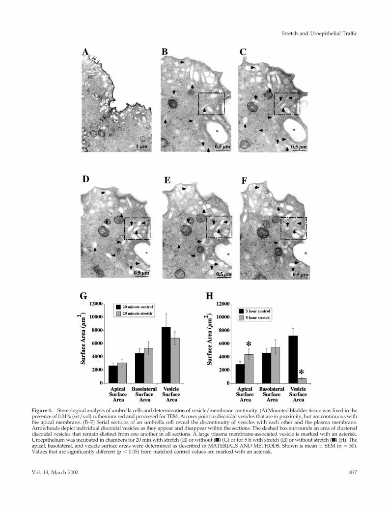
Figure 4. Stereological analysis of umbrella cells and determination of vesicle/membrane continuity. (A) Mounted bladder tissue was fixed in thepresence of 0.01% (wt/vol) ruthenium red and processed for TEM. Arrows point to discoidal vesicles that are in proximity, but not continuous withthe apical membrane. (B–F) Serial sections of an umbrella cell reveal the discontinuity of vesicles with each other and the plasma membrane.Arrowheads depict individual discoidal vesicles as they appear and disappear within the sections. The dashed box surrounds an area of clustereddiscoidal vesicles that remain distinct from one another in all sections. A large plasma membrane-associated vesicle is marked with an asterisk.Uroepithelium was incubated in chambers for 20 min with stretch (u) or without (f) (G) or for 5 h with stretch (u) or without stretch (f) (H). Theapical, basolateral, and vesicle surface areas were determined as described in MATERIALS AND METHODS. Shown is mean SEM (n � 50).Values that are significantly different (p � 0.05) from matched control values are marked with an asterisk.
Stretch and Uroepithelial Traffic
Vol. 13, March 2002 837
without stretch, fixed, and processed for TEM. The tissuewas serial sectioned and the sections were ordered succes-sively to enable us to track individual vesicles as they ap-peared and disappeared within the cytoplasm of a singlecell. Figure 4, B–F, displays an example of five TEM micro-graphs taken from the apical portion of an umbrella cell.Each arrowhead tracks an individual vesicle as it appearsand then fades from view. Analysis of many areas wherevesicles were clustered together (dashed box) showed nofusion of individual vesicles. In addition, all vesicular struc-tures remained separate from the plasma membrane in allsections examined with the exception of occasional infold-ings of the plasma membrane that appeared as much largervacuolar structures. An example of the latter is marked bythe asterisks in Figure 4, B and C. Taken together, theseresults confirm that the vesicles are individual, intracellularstructures that are distinct from the apical surface.
Next, we quantified the apical, basolateral, and vesiclesurface areas of the umbrella cells in 20-min and 5-h controlsamples, or tissue that had been stretched for 20 min or 5 h.In 20-min control samples, the average apical cell surfacearea was �2700 �m2, the basolateral surface area was mea-sured at �4600 �m2, and the vesicles occupied �8500 �m2
of surface area (Figure 4G). Thus, the surface area of thevesicles in control conditions was �3 times the surface areaof the apical membrane. These results clearly indicated thatthe cells possess a tremendous reserve capacity to increaseapical surface area via the fusion of vesicles with the apicalmembrane. Relative to controls, the samples stretched for 20min exhibited no significant changes in any of the threeparameters. These data were consistent with the morpho-logical observations described above but were inconsistentwith the capacitance data (see DISCUSSION).
After 5 h of stretch, the apical surface area was increased�50% over the matched control (from �2900 to 4300 �m2)with no significant change in basolateral surface area (Figure4H). These results were consistent with the capacitance dataand indicated that a considerable amount of membrane wasadded to the apical surface in response to stretch. However,when the changes in vesicle surface area were quantifiedafter 5 h of stretch, it was found that the surface area of thevesicles decreased from �7200 �m2 to �1000 �m2 (Figure4H). This revealed that the increase in apical surface area(�1400 �m2) accounted for only a small fraction of thedecrease in vesicle surface area (�6200 �m2), indicating thatan additional cellular event was occurring during the stretchresponse.
Stretch Increases Cell Surface Expression ofUroplakin III and Induces Apical Secretion ofMetabolically Labeled ProteinsOne prediction of the vesicle fusion model is that stretch willresult in the delivery and incorporation of vesicle cargo intothe apical membrane. A cargo molecule that can be used forthis purpose is uroplakin III, a 47-kDa transmembrane com-ponent of both the apical membrane plaques and discoidalvesicles (Figure 5A) (Wu and Sun, 1993; Liang et al., 2001).We demonstrated that uroplakin III was associated with theplasma membrane and discoidal vesicles of rabbit tissue byimmunogold labeling with a uroplakin III antibody (Figure5A). Although uroplakin III was present on both the apical
surface and the discoidal vesicles of umbrella cells, it wasnot found in significant amounts elsewhere in the epithe-lium (Wu and Sun, 1993; our unpublished data).
To test the hypothesis that the discoidal vesicles werecontributing to the increase in apical surface area, bladdertissue was left unstretched or was stretched for either 20 minor 5 h. The cell surface was biotinylated, the cells were lysed,and the biotinylated fraction was recovered with streptavi-din beads. The amount of uroplakin III at the cell surfacerelative to the total cellular uroplakin III was determined byWestern blot (Figure 5B). In control conditions, �5% of thetotal uroplakin III pool was present on the apical surface(Figure 5C). This is consistent with the large amount ofsurface area present in the vesicles relative to the apicalmembrane. This value remained unchanged after 20 min ofstretch. In contrast, after 5 h of stretch, the fraction of uro-plakin III at the cell surface increased to �32%, and theabsolute amount at the surface increased �67 18% abovethat observed in controls. This latter value is consistent withvesicle fusion and delivery of uroplakin III to the apicalsurface. In support of this, immunogold labeling demon-strated that uroplakin III was found at the plasma mem-brane in stretched tissue (Figure 5D). As noted in Figure 3F,there was a significant decrease in the number of discoidalvesicles.
Also evident from the Western blot analysis was that thetotal cell expression of uroplakin III was considerably lowerafter 5 h of stretch than in control. This effect was not simplydue to a conformational change in the protein, becauseblotting for total cell surface proteins with streptavidin-HRPyielded a similar decrease in signal (Figure 6E). This resultindicated that a significant portion of the apical membrane,including uroplakin III, was being degraded during stretchand is consistent with the decrease in vesicles observed bothin this study and in that of Minsky and Chlapowski (1978).
To confirm that exocytosis was occurring throughout the5-h stretch period, we measured the secretion of metaboli-cally labeled proteins over time. Unlike proteins anchored tothe membrane, secreted proteins are not efficiently reinter-nalized after reaching the apical surface and thus provide analternative way to detect exocytosis. Growth hormone, asecretory protein, has previously been localized to discoi-dal/fusiform vesicles (Kerr et al., 1997). Tissue was radiola-beled with [35S]cysteine/methionine for 30 min and chasedfor 1 h to label secretory proteins. The tissue was thenstretched for 5, 15, 120, or 300 min or incubated withoutstretch as a control and the amount of radioactivity in theapical medium was determined. In control tissue, the radio-activity detected in the apical medium was �100 counts/minute (cpm) regardless of the duration of incubation, indi-cating that little secretion occurred under these conditions(Figure 5E). In contrast, within 5 min of stretch the apicalcpm measured 7,980 2,100 and this amount increased to214,687 40,252 cpm after 5 h. This secretion was com-pletely blocked by adding 10 �g/ml brefeldin A (our un-published data), demonstrating a bona fide membrane traf-ficking event and not nonspecific release.
Stretch also stimulated significant release of secretory pro-teins into the basolateral chamber (our unpublished data).To further ensure that the apical secretion we observed wasnot the result of paracellular diffusion of secretory proteinsfrom the basolateral-to-apical chamber, [14C]urea was added
S.T. Truschel et al.
Molecular Biology of the Cell838

Figure 5. Uroplakin III localization and stretch-induced increases in cell surface uroplakin III expression and metabolically labeled proteinsecretion. (A) Ultrathin cryosections of 5-h control tissue labeled with anti-uroplakin III antibody and secondary antibody conjugated to 5-nmgold. Asterisks denote examples of uroplakin III-positive vesicles. (B) Representative Western blots of total and cell surface uroplakin IIIexpression in control or stretched samples. The bottom panel is exposed longer to allow the total cell signal in stretched samples to be visible.(C) Quantification of relative expression of cell-associated uroplakin III at the apical surface for each condition. Shown is mean SEM (n �5). (D) Samples were stretched for 5 h and labeled with anti-uroplakin III antibody and secondary antibody conjugated to 5-nm gold. (E) Cellswere pulsed with [35S]cysteine/methionine, chased for 1 h, and stretched or incubated without stretch for 5, 15, 120, or 300 min. Shown isaverage cpm present in apical medium at each time point SEM (n � 3). (F) Percentage of basally added [14C]urea that appeared in the apicalmedium after 0, 15, 120, or 300 min of stretch or incubation without stretch. Shown is mean SEM (n � 3).
Stretch and Uroepithelial Traffic
Vol. 13, March 2002 839

Figure 6. Effect of stretch on endocytosis. Umbrella cells were surface biotinylated and incubated for 0, 5, 15, 30, 60, or 120 min withoutstretch (A) or with stretch (B). MESNA-protected biotinylated proteins were isolated and Western blots were probed with streptavidin-HRP.Representative blot of control tissue (A) shows no visible endocytosis by the lack of signal in MESNA-treated lanes. Stretched tissue (B)exhibits large MESNA-protected signal, indicating endocytosis. (C) Quantification of endocytosis in control and stretched tissue. Shown ismean SEM (n � 5). Values that are significantly different (p � 0.05) from matched control values are marked with an asterisk. (D)WGA-FITC was internalized for 15 min in control unstretched (a) or stretched (b) tissue. Surface-bound WGA-FITC was removed byincubation at 4°C with N-acetyl glucosamine, and the samples were fixed, stained with 4,6-diamidino-2-phenylindole, and then examined by
S.T. Truschel et al.
Molecular Biology of the Cell840

to the basolateral chamber and the appearance of radioac-tivity in the apical chamber was determined in control orstretched tissue. In either case the amount of [14C]urea thatcrossed the cells was �0.4% after 5 h of stretch, demonstrat-ing that the integrity of the umbrella cell layer was notcompromised (Figure 5F). Apical-to-basolateral diffusion of[14C]urea was also determined and was not significantlydifferent from basolateral-to-apical diffusion (our unpub-lished data). Taken together, these results confirmed thatstretch induces a rapid and continual secretion of proteinand that this secretion represents a genuine exocytic event.
Stretch Induces Endocytosis at the Apical Surface ofUmbrella CellsThe stereological analysis revealed that after 5 h of stretchthe increase in apical surface area accounted for only a smallfraction of the loss in vesicle surface area. Furthermore, thebiochemical data indicated that uroplakin III was degradedduring the 5 h of stretch. Therefore, we hypothesized thatmembrane added to the apical surface during stretch wasbeing endocytosed and degraded.
To test whether stretch induced endocytosis, the tissuewas mounted in the modified Ussing chambers and theapical cell surface was biotinylated at 4°C with sulfo-N-hydroxysuccinimide-disulfo-biotin to label surface proteins.Several protein species were biotinylated (Figure 6A). Themajor protein species was a doublet that migrated with amolecular mass in the 67–70 kDa range. The identity of thedoublet is not known. Several smaller molecular weightspecies were also detected, one of which was confirmed tobe uroplakin III by probing blots of biotinylated proteinswith anti-uroplakin III antibodies (our unpublished data).Exposing the blot for longer periods of time also revealedthat other protein species were biotinylated, including pro-teins that migrated �15, 27, and 28 kDa (our unpublisheddata), which are the molecular masses of uroplakins II, Ia,and Ib, respectively (Wu et al., 1994). After biotinylation, thetissue was warmed and stretched for 0–120 min or incu-bated for the same amount of time in the absence of stretchas a control. The internalized biotin-labeled proteins weredetected by treating cells with the membrane-impermeant
reducing agent MESNA (see MATERIALS AND METH-ODS).
In control cells, the MESNA treatment removed essen-tially all of the biotin regardless of the duration of incubation(Figure 6A). This showed that little or no endocytosis oc-curred. In contrast, stretched cells exhibited a large MESNA-protected signal after 5 min, demonstrating that a significantamount of the surface membrane was rapidly internalized(Figure 6B). The quantification of this and other blots indi-cated that nearly 100% of the labeled membrane was endo-cytosed within 5 min of stretch (Figure 6C) and this valueremained constant over a 2-h period of incubation. The latterdata indicated that in the time frame examined, little recy-cling of biotinylated apical proteins occurred after endocy-tosis.
WGA-FITC, which binds to N-acetyl glucosamine-modi-fied proteins and lipids on the apical surface of the umbrellacells (Fujiyama et al., 1995), was used as alternative markerof endocytosis. The lectin was included in the apical half ofthe Ussing chamber and the tissue was then stretched for 15min or incubated without stretch for the same amount oftime. After the incubation, surface-bound WGA-FITC wasremoved with N-acetyl glucosamine treatment at 4°C andthe internalization of WGA-FITC was determined by fluo-rescence microscopy. No uptake was observed in control(unstretched) samples either after 15 min (Figure 6Da) orafter 5 h (our unpublished data). Cells stretched for 15 minexhibited a significant amount of fluorescence in punctatestructures, consistent with uptake into vesicular structures(Figure 6Db). Confocal microscopy verified that the endo-cytic structures were localized to the apical cytoplasm of theumbrella cells and that no staining was observed in theintermediate cell layer. To assess whether endocytosis con-tinued throughout the stretch period, WGA-FITC wasadded to the apical chamber of the preparations 2 or 5 h afterthe initiation of stretch. Uptake of WGA-FITC was observedunder both conditions (data for uptake after 2 h is shown inFigure 6Dc).
As final confirmation that stretch stimulates endocytosisin bladder umbrella cells we filled excised but otherwiseintact bladders with buffer containing WGA-FITC for 15 minat 37°C. The mucosal surface was treated to remove cellsurface lectin, fixed, and imaged. Again, significant uptakeof lectin was observed in the umbrella cell population (Fig-ure 6Dd). The latter observation is consistent with previousexperiments that showed uptake of fluid-phase markers inbladders that were emptied and then filled with fluid-phasemarker (Hicks, 1966; Porter et al., 1967). Although theseoriginal observations were taken as evidence of uptake aftervoiding, filling the bladder stretches the tissue, and theinternalization previously observed is likely to be thestretch-regulated endocytosis described in this study.
Endocytosed Membrane Is Delivered to LysosomesHaving established that stretch induced apical membraneendocytosis, we next examined the hypothesis that internal-ized membrane was being degraded. Accordingly, the ki-netics of degradation was determined by biotinylating thecell surface at 4°C and subsequently stretching for 5–300min. After each time point, the tissue was lysed; the cohortof biotinylated proteins was isolated; and the proteins wereseparated by SDS-PAGE, transferred to membranes, and
Figure 6 (cont). epifluorescence microscopy. A projection of digi-tally deconvoluted sections is shown. (Dc) Bladder cells werestretched for 2 h, WGA-FITC was added to the chamber and thesamples stretched for an additional 15 min. Surface WGA-FITC wasremoved, the samples were fixed, and then examined in a confocalmicroscope. A projection of multiple XY sections is shown. (Dd)WGA-FITC was added to an excised but otherwise intact bladderfor 15 min. Surface WGA-FITC was removed, the samples werefixed, and then examined in a confocal microscope. A projection ofmultiple XY sections is shown. (E) Apical surface proteins werebiotinylated and the tissue was stretched for 0–300 min. The cellswere lysed and biotinylated proteins detected by probing Westernblots with streptavidin-HRP. Treatment with 40 �M leupeptin (Leup)prevented the degradation, demonstrating a lysosomal-mediated path-way, whereas treatment with lactacystin (Lact) had no effect. (F) Quan-tification of apical membrane protein degradation upon stretch. Valuesare relative to unstretched tissue biotinylated at t � 0. Shown ismean SEM (n � 4). Values that are significantly different (p � 0.5)from unstretched tissue at t � 0 are marked with an asterisk.
Stretch and Uroepithelial Traffic
Vol. 13, March 2002 841

Figure 7. Role of cAMP and PKA in the stretch response. (A) Intracellular levels of cAMP were measured in control cells (f) and stretchedcells (u) after 15, 30, 120, or 300 min. Shown is mean SEM (n � 4). Values that are significantly different (p � 0.05) from matched controlvalues are marked with an asterisk. (B) Effects of forskolin and H89 on capacitance. Cells were treated with 10 �M forskolin/500 �M IBMXwithout stretch (f), treated with forskolin/IBMX and 10 �M H89 (Œ), or stretched in the presence of 10 �M H89 alone (F). For comparison,the capacitance readings from Figure 1 of stretched (�) and control (�) cells are shown. Shown is mean SEM (n � 6). (C) Effects offorskolin/IBMX on umbrella cell morphology. Tissue was treated with forskolin/IBMX without stretch for 5 h and processed for TEM. Noticethe highly convoluted apical membrane. (D) Changes in surface area resulting from Forskolin/IBMX treatment. Tissue was treated with
S.T. Truschel et al.
Molecular Biology of the Cell842

probed with streptavidin-HRP (Figure 6E). The fraction ofprotein remaining after each time point relative to the start-ing amount was quantified (Figure 6F). The total amount ofbiotinylated proteins remained constant for up to 120 min ofstretch. However, by 210 min there was a significant de-crease in protein to �20% of the starting value and by 300min the protein levels were reduced to �9% (Figure 6F).This result was consistent with our previous experimentwith uroplakin III and indicated that the apical membraneproteins were being degraded.
Two mechanisms can account for degradation withincells; delivery to lysosomes or proteasomes. To discriminatebetween the two pathways, the apical cell surface was bio-tinylated at 4°C, and the tissue was stretched for 5 h in thepresence of 40 �M leupeptin to block lysosomal degradation(Ihrke et al., 1998). Consistent with lysosomal delivery, in-cubation with leupeptin inhibited the degradation that wasobserved in cells stretched without the drug treatment (Fig-ure 6, E and F). To rule out the possibility of proteasomal-mediated degradation, we treated the cells with 10 �Mlactacystin to inhibit proteasome function (Fenteany et al.,1995). Lactacystin treatment had no effect on the degradationof apical membrane proteins (Figure 6F), confirming thatdegradation was via a lysosomal-mediated pathway. Thesedata supported our hypothesis that the internalized mem-brane was delivered to lysosomes where it was degraded.
The Stretch Response Is a cAMP-mediated EventIn other mechanically sensitive tissues, mechanical stimula-tion is associated with elevated levels of the second messen-ger cAMP (Russo et al., 1989; Sandy et al., 1989; Watson et al.,1989). To ascertain the role of cAMP in the uroepithelium,we measured intracellular levels of cAMP in control andstretched tissue over time. Stretching the tissue caused theintracellular levels of cAMP to rise more than twofoldwithin 15 min (Figure 7A). These levels remained elevatedthroughout the 5-h period of stretch, implicating cAMP inthe stretch response. To test the effects of elevating cAMP inthe umbrella cells, we incubated the tissue with 10 �Mforskolin and 500 �M 3-isobutyl-1-methylxanthine (IBMX)to raise intracellular cAMP levels. Unexpectedly, we ob-served an unusually large increase of 120% in capacitanceover a 5-h period, versus the 50% change observed withstretch alone (Figure 7B). Incubating the tissue with 8-bro-mo-cAMP, a membrane-permeant analog of cAMP, induceda similar effect as forskolin/IBMX (our unpublished data)and treatment with di-deoxy forskolin, a nonfunctional for-skolin analog, had no effect on capacitance (our unpublisheddata). We also explored the effect on capacitance when for-skolin was added to stretched tissue. The response wassimilar to control tissue treated with forskolin but the netrise in capacitance was slightly greater (�140 vs. 120%). Theabove-mentioned data demonstrated that cAMP dramati-
cally altered membrane trafficking at the apical surface ofumbrella cells.
A likely effector of cAMP is PKA, the target of the H89kinase inhibitor (Fujihara et al., 1993). In stretched tissuetreated with H89, the capacitance initially rose �20% butfailed to increase any further, implicating PKA as a possibleeffector of the stretch response (Figure 7B). Moreover, therise in capacitance observed after incubating the tissue inforskolin/IBMX was also significantly inhibited when thetissue was pretreated with H89. These findings underscoredthe importance of cAMP and PKA in the regulation ofmucosal surface area and indicated that cAMP modulated,in part, the response of the umbrella cell to mechanicalstretch.
The significant changes observed in capacitance when thetissue was incubated with forskolin/IBMX prompted us toexamine the morphology of the umbrella cells after forskolintreatment and to assess the effect of these drugs on endocy-tosis. Accordingly, we treated unstretched umbrella cellswith forskolin/IBMX for 5 h and processed the tissue forTEM. Treatment with forskolin/IBMX (Figure 7C) elicitedexaggerated convolutions in the apical cell membranes, sup-porting the large increases observed in capacitance. Stereo-logical measurements confirmed the capacitance results(Figure 7D). The apical surface area of forskolin-treated cellsincreased to �6500 �m2, more than double the apical surfacearea of an unstretched umbrella cell. No significant changesin basolateral surface area were detected. There was a sig-nificant decrease in vesicle area that accompanied the fors-kolin/IBMX-induced fusion. However, unlike stretched tis-sue a significant amount of vesicle area remained. Last, wemeasured the effects of forskolin/IBMX on endocytosis incontrol unstretched tissue. The amount of MESNA-pro-tected biotin remained constant over a 2-h time period,demonstrating that no endocytosis occurred (Figure 7E).This result indicated that forskolin may be modulating api-cal surface area by means of discoidal vesicle exocytosiswithout affecting apical membrane endocytosis.
DISCUSSION
Stretch-induced Changes in Apical Surface Area ofUmbrella CellsThe ability of the bladder to retain urine requires a barrierepithelium that can accommodate large variations in urinevolume. At the cellular level this is likely accomplished bychanges in umbrella cell architecture and surface area. Thelatter is thought to be modulated by discoidal vesicle exo-cytosis (Minsky and Chlapowski, 1978; Lewis and de Moura,1982, 1984). The underlying intermediate and basal cell lay-ers also undergo morphological transitions, but much less isknown about their cellular biology (Hicks, 1975).
Our data confirm that stretch increases the apical surfacearea of umbrella cells, and these changes are likely mediatedby discoidal vesicles. Mechanical distention of the uroepi-thelium resulted in an �50% increase in capacitance after a5-h period of stretch. This large change in capacitance isconsistent with, but significantly greater than the previousobservation that short-term mechanical bowing of the uro-epithelium increased capacitance by �18% (Lewis and deMoura, 1982). Furthermore, a stereological analysis con-firmed that the apical surface area of the umbrella cells
Figure 7 (cont). forskolin/IBMX (u) and stereological analysis wasperformed as described. Control surface areas are shown for com-parison (f), n � 50. Values that are significantly different (p � 0.05)from matched control values are marked with an asterisk. (E) En-docytosis was determined in forskolin/IBMX-treated cells. Shownis representative Western blot after 0, 5, 15, 30, 60, or 120 min withor without MESNA treatment.
Stretch and Uroepithelial Traffic
Vol. 13, March 2002 843

increased by 50%, whereas there was no statistically signif-icant change in basolateral surface area. The stereologicalanalysis also revealed that stretch was accompanied by afourfold decrease in vesicle surface area. These data concurwith previous observations that the numerical density ofdiscoidal vesicles was decreased in expanded versus con-tracted bladders (Minsky and Chlapowski, 1978). Biotinyla-tion experiments demonstrated that both the relative andabsolute amount of uroplakin III (a “cargo” molecule foundin discoidal vesicles) at the cell surface significantly in-creased after 5 h of stretch, consistent with discoidal vesiclefusion. Last, secretion of metabolically labeled proteins con-firmed a rapid and significant apical secretion in stretchedtissue that increased over time. Although exocytosis wasobserved shortly after stretch, the majority of exocytosisoccurred between the 2- and 5-h period. Although theabove-mentioned results are consistent with the fusion ofdiscoidal vesicles during stretch, we cannot rule out thatsome of the observed changes in surface area were the resultof other membranous organelles fusing with the apical sur-face of the umbrella cells.
Release of metabolically labeled secretory proteins was ob-served within 5 min of stretch, indicating that stretch rapidlystimulated exocytosis in umbrella cells. Whether these earlyexocytic events result in a change in the apical surface area ofthese cells is unclear. We observed a small increase (�15%) incapacitance within the first 20 min of stretch. However, astereological analysis showed no significant change in apical,basolateral, or vesicle surface area. Furthermore, we observedthat the amount of uroplakin III at the cell surface was un-changed after 20 min of stretch. Because stretch stimulates iontransport (our unpublished data), the small capacitance changewe observe may be imaginary (Gillis, 1995). Alternatively, thearea change may be too small to be detected by either stereol-ogy or biotinylation.
Mechanical Stretch Stimulates Apical Endocytosisin Umbrella CellsIn the classical model, vesicle endocytosis is proposed tooccur only upon voiding (i.e., after release of stretch) (Hicks,966; Porter et al., 1967). Our data are inconsistent with thisaspect of the model. Most notably, we observed that essen-tially all biotinylated apical membrane proteins, generalmarkers of the apical plasma membrane, were endocytosedwithin 5 min of mechanical stretch. However, we cannotrule out that only a subpopulation of apical membrane isbiotinylated and that we are overestimating the amount ofendocytosis. Moreover, we observed uptake of WGA-FITCin stretched preparations of isolated uroepithelium or uponfilling excised but otherwise intact bladders.
Despite significant amounts of endocytosis, there was littleapparent change in apical surface area during the first 20 minof stretch, thus indicating that the amount of endocytosis musthave been balanced by roughly equivalent amounts of exocy-tosis. This rapid exocytosis could be a result of either recycledmembrane or delivery of newly synthesized vesicle membrane.Because apical surface area increased over the longer timescale, the rate of exocytosis must ultimately outpace the rate ofendocytosis. This is supported by the large increase in secretionbetween 2 and 5 h of stretch. Nonetheless, endocytosis contin-ues throughout the entire period of stretch as evidenced byuptake of WGA-FITC at 2 and 5 h of stretch.
Although is may seem counterintuitive that exocytosisand endocytosis are occurring simultaneously in stretchedumbrella cells, these two processes occur constitutively andsimultaneously in all cells, even under resting conditions(Harter and Wieland, 1996; Mukherjee et al., 1997). Coupledexocytosis/endocytosis likely plays a key role in fine-tuningthe changes in umbrella cell surface area as a result of stretchand/or other external stimuli (see discussion below). Fur-thermore, rapid endocytosis/exocytosis would provide theumbrella cells with a means of quickly replacing old ordamaged surface plaques with new ones in preparation forimminent lumen expansion. In this regard, it has recentlybeen shown that expression of uroplakin III, a major plaqueconstituent, is required for the maintenance of bladder bar-rier function (Hu et al., 2000). Finally, exocytosis decreasesmembrane tension (Morris and Homann, 2001), which iscompensated for by endocytosis. Regardless of its function,endocytosis clearly represents a major pathway of mem-brane trafficking during stretch and its specific functions andregulations will be addressed in future studies.
In the present analysis, we have not directly examined theevents that occur when stretch is released. Because stretchincreases apical surface area, this added membrane must beendocytosed at some point after voiding. Minsky andChlapowski (1978) observed that within 10 min after void-ing, the number of discoidal vesicles in the umbrella cellsincreased by approximately twofold compared with thenumber of vesicles in distended bladders (Minsky andChlapowski, 1978). This implies that some vesicles are re-plenished within a short period of time after stretch. Theincreased number of discoidal vesicles could reflect apicalmembrane internalization and/or rapid delivery of newlysynthesized vesicles from the Golgi (Hicks, 1966). This as-sembly of vesicles in the Golgi is likely to be significant.Only a small fraction of discoidal/fusiform vesicles is la-beled by apically endocytosed fluid or membrane markers,indicating that they are primarily involved in exocytic traffic(Hicks, 1966; Porter et al., 1967; Amano et al., 1991). More-over, from our data it is evident that the amount of inter-nalized membrane would be insufficient to replenish thesurface area of the discoidal vesicles lost during stretch.
Endocytosed Membrane Is Delivered to LysosomesDiscoidal vesicles or discoidal vesicle-derived membranewas previously observed in multivesicular bodies as well aslysosomes (Porter et al., 1967; Amano et al., 1991). However,the extent of apical membrane entry into this pathway andthe role that stretch played in this process were previouslyunknown. We observed that upon internalization biotin-labeled apical surface proteins were eventually degraded.This proteolytic event was prevented by the lysosomal en-zyme inhibitor leupeptin, but not by the proteasome inhib-itor lactacystin. This finding is consistent with the decreasein vesicles observed in this study and that described previ-ously (Minsky and Chlapowski, 1978). However, degrada-tion was not apparent until some time after 2 h of stretch. Inother cell types degradation can proceed as quickly as 15–20min after internalization and delivery to lysosomes usuallyoccurs within 30–45 min of endocytosis (Griffiths et al.,1989). It is possible that either the delivery of endocytosedmembrane to late endosomes/lysosomes is slow in umbrellacells or that stretch up-regulates a pathway that results in
S.T. Truschel et al.
Molecular Biology of the Cell844

enhanced degradation of discoidal vesicles. The latter pos-sibility would explain the sudden change in protein signalafter 120 min of stretch. Alternatively, there may be somerecycling of vesicle membrane that escaped our detection.
Role of cAMP in Stretch-regulated ExocytosisIn addition to elucidating the dynamics of membrane traffickingin response to stretch, we have also gained insight into one of thesignal transduction pathways that may regulate these traffickingevents. Our data indicate that a cAMP-PKA pathway plays a rolein the stretch response. Increased amounts of cAMP were ob-served in stretched tissue and elevating the intracellular concen-tration of cAMP in unstretched umbrella cells with forskolin/IBMX resulted in a �100% increase in apical cell surface area.These events were blocked by treatment with the PKA antagonistH89. Importantly, stretch-regulated changes in membrane capac-itance were significantly decreased by H89, further confirmingthat the cAMP-PKA pathway is required for vesicle exocytosis.Our results are consistent with the recent observation that cul-tured uroepithelial cells or bladder explants secrete tissue-typeplasminogen activator and urokinase in a cAMP-dependent man-ner (Deng et al., 2001), although the study did not examine theeffects of stretch on secretion. The results of our experimentationhave additional significance because they indicate that the surfacearea of umbrella cells can be regulated by external stimuli inde-pendent of stretch. These stimuli could come in the form ofgrowth factors, hormones, or neurotransmitters present in theblood or released by the bladder-associated musculature andnerves. Interestingly, some afferent neuronal processes terminatewithin the uroepithelium and umbrella cells apparently expresssurface receptors for various neurotransmitters (Birder et al., 1998).As such, the apical surface area of umbrella cells may be regulatedby mechanical as well as exocrine/paracrine stimuli.
The dramatic increases in umbrella cell surface area ob-served in unstretched tissue treated with agents that raiseintracellular cAMP occurred in the apparent absence of endo-cytosis. This observation underscores the importance of endo-cytosis in regulating umbrella cell apical surface area. Al-though stretch stimulated endocytosis of the apical membrane,treatment with forskolin/IBMX without stretch had no effecton endocytosis but resulted in a greater increase in surfacearea. These observations indicate that the response to stretchmay involve at least two levels of regulation, one that operatesby modulating intracellular levels of cAMP and the other thatuses another uncharacterized mechanism. Other cellularevents rapidly triggered by mechanical stretch include stimu-lation of stretch-activated channels, activation of the integrin-associated focal adhesion kinase, phosphoinositide turnover,and autocrine release of growth factors (Vandenburgh, 1992).Some or all of these mechanosensory mechanisms could playan important role in signaling exocytosis/endocytosis in um-brella cells.
In summary, our findings further elucidate the poorlyunderstood process of mucosal surface area regulation in theuroepithelium during stretch. The data not only shed lighton the membrane trafficking that occurs in these cells butalso provide insight into the intracellular signal cascade thatis responsible for this trafficking. Moreover, we have uncov-ered an additional pathway for apical endocytosis that ismodulated by mechanical force (other examples of stretch-regulated endocytosis are given in Apodaca, 2002). Thisdiscovery broadens our understanding of the effects of me-
chanical stimuli on membrane traffic, especially in regard tothe pivotal function of endocytosis in balancing the exocyticresponse of the cell. The coordinated balance between thesetwo plasma membrane events in umbrella cells is likely toplay a similarly important role in other cell types that areresponsive to mechanical forces.
ACKNOWLEDGMENTS
We thank Drs. Rebecca Hughey, Nick Johnson, Linton Traub, andOra Weisz for insightful comments and critiques while preparingthis manuscript. This work was supported by R01 grants from theNational Institute of Diabetes and Digestive and Kidney Diseases ofthe National Institutes of Health to G.A. (R01DK51970) and M.Z(R01DK48217). The Laboratory of Epithelial Cell Biology is sup-ported in part by an equipment grant from Dialysis Clinic Inc.
REFERENCES
Altschuler, Y., Kinlough, C., Poland, P., Apodaca, G., Weisz, O., andHughey, R. (2000). Clathrin-mediated endocytosis of MUC1 is reg-ulated by glycosylation. Mol. Biol. Cell 11, 819–831.
Amano, O., Kataoka, S., and Yamamoto, T. (1991). Turnover of asym-metric unit membranes in the transitional epithelial superficial cells ofthe rat urinary bladder. Anat. Rec. 229, 9–15.
Apodaca, G., Katz, L., and Mostov, K. (1994). Receptor-mediated trans-cytosis of IgA in MDCK cells is via apical recycling endosomes. J. CellBiol. 125, 67–86.
Apodaca, G. (2002). Modulation of membrane traffic by mechanicalstimuli. Am. J. Physiol. 282, F179–F190.
Baddeley, A., Gunderson, H., and Cruz-Orive, L. (1986). Estimation ofsurface area from vertical sections. J. Microsc. 142, 259–276.
Birder, L., Apodaca, G., De Groat, W., and Kanai, A. (1998). Adrener-gic- and capsaicin-evoked nitric oxide release from urothelium andafferent nerves in urinary bladder. Am. J. Physiol. 275, F226–F229.
Chang, A., Hammond, T., Sun, T., and Zeidel, M. (1994). Permeabilityproperties of the mammalian bladder apical membrane. Am. J. Physiol.267, C1483–C1492.
Clemow, D., Steers, W., and Tuttle, J. (2000). Stretch-activated signalingof nerve growth factor secretion in bladder, and vascular smoothmuscle cells from hypertensive, and hyperactive rats. J. Cell. Physiol.183, 289–300.
Dai, J., Ting-Beall, P., and Sheetz, M. (1997). The Secretion-coupledendocytosis correlates with membrane tension changes in RBL 2H3cell. J. Gen. Physiol. 110, 1–10.
Deng, F., Ding, M., Lavker, R., and Sun, T.-T. (2001). Urothelial functionreconsidered: a role in urinary protein secretion. Proc. Natl. Acad. Sci.USA 98, 154–159.
Dietz, J. (1988). Release of atrial natriuretic factor from heart-lungpreparation by atrial distension. Am. J. Physiol. 247, R1093–R1096.
Efron, B., and Tibshirani, R.J. (1993). Introduction to the Bootstrap, BocaRaton, FL: CRC Press.
Fenteany, G., Standaert, R., Lane, W., Choi, S., Corey, E., and Schreiber,S. (1995). Inhibition of proteasome activities and subunit-specific ami-no-terminal threonine modification by lactacystin. Science 268, 726–731.
Fink, R., and Cooper, M. (1996). Apical membrane turnover is acceler-ated near cell-cell contacts in an embryonic epithelium. Dev. Biol. 174,180–189.
Stretch and Uroepithelial Traffic
Vol. 13, March 2002 845

Fujihara, M., Muroi, M., Muroi, Y., Ito, N., and Suzuki, T. (1993).Mechanism of lipopolysaccharide-triggered junB activation in a mousemacrophage-like cell line. J. Biol. Chem. 268, 14898–14905.
Fujiyama, C., Masake, Z., and Sugihara, H. (1995). Reconstruction ofthe urinary bladder mucosa in three-dimensional collagen gel culture:fibroblast-extracellular matrix interactions on the differentiation oftransitional epithelial cells. J. Urol. 153, 2060–2067.
Gillis, K. (1995). Techniques for membrane capacitance measurements.In: Single Channel Recording, ed. B. Sakman and E. Neher, New York:Plenum Press, 155–198.
Griffiths, G. (1993). Fine Structure Immuno-cytochemistry, Berlin:Springer-Verlag.
Griffiths, G., Back, R., and Marsh, M. (1989). A quantitative analysis ofthe endocytic pathway in baby hamster kidney cells. J. Cell Biol. 109,2703–2720.
Harter, C., and Wieland, F. (1996). The secretory pathway: mechanismsof protein sorting and transport. Biochim. Biophys. Acta 1286, 75–93.
Hayat, M. (1989). Principles and Techniques of Electron Microscopy,Boca Raton, FL: CRC Press.
Hicks, R. (1966). The function of the Golgi complex in transitionalepithelium. J. Cell Biol. 30, 623–644.
Hicks, M. (1975). The mammalian urinary bladder: an accommodatingorgan. Biol. Rev. 50, 215–246.
Homann, U. (1998). Fusion and fission of plasma-membrane materialaccommodates for osmotically induced changes in the surface area ofguard-cell protoplasts. Planta 206, 329–333.
Hu, P., Deng, F.-M., Liang, F.-X., Hu, C.-M., Auerbach, A., Shapiro, E.,Wu, X.-R., Kachlar, B., and Sun, T.-T. (2000). Ablation of uroplakin IIIgene results in small urothelial plaques, urothelial leakage, and vesi-coureteral reflux. J. Cell Biol. 151, 961–971.
Ihrke, G., Martin, G., Shanks, M., Schrader, M., Schroer, T., and Hub-bard, A. (1998). Apical plasma membrane proteins and endolyn-78travel through a subapical compartment in polarized WIF-B hepato-cytes. J. Cell Biol. 141, 115–133.
Kerr, D., Liang, F., Bondioli, K., Zhao, H., Kreibich, G., Wall, R., andSun, T.-T. (1997). The bladder as a bioreactor: urothelium productionand secretion of growth hormone into urine. Nat. Biotech. 16, 75–79.
Koss, L. (1969). The asymmetric unit membranes of the epithelium ofthe urinary bladder of the rat. Lab. Invest. 21, 154–168.
Lang, R., Tholken, H., Ganten, F., Luft, F., Ruskoaho, H., and Unger, T.(1985). Atrial natriuretic factor-a circulating hormone stimulated byvolume loading. Nature 314, 264–266.
Levin, R., Longhurst, P., Monson, F., Haugaard, N., and Wein, A.(1993). Experimental studies on bladder outlet obstruction. In: ProstateDiseases, ed. H. Lepor and R. K. Lawson, Philadelphia: WB Saunders,119–130.
Levin, R., and Wein, A. (1982). Response of the in vitro whole bladder(rabbit) preparation to autonomic agonists. J. Urol. 128, 1087–1090.
Lewis, S., and Diamond, J. (1976). Na� transport by rabbit urinarybladder, a tight epithelium. J. Membr. Biol. 28, 1–40.
Lewis, S., and Hanrahan, J. (1990). Physiological approaches for study-ing mammalian urinary bladder epithelium. Methods Enzymol. 192,632–650.
Lewis, S., and de Moura, J. (1982). Incorporation of cytoplasmic vesiclesinto apical membrane of mammalian urinary bladder epithelium. Na-ture 297, 685–688.
Lewis, S., and de Moura, J. (1984). Apical membrane area of rabbiturinary bladder increases by fusion of intracellular vesicle: an electro-physiological study. J. Membr. Biol. 82, 123–136.
Liang, F.-X., Riedel, I., Deng, F.-M., Zhou, G., Xu, C., Wu, X.-R., Kong,X.-P., Moll, R., and Sun, T.-T. (2001). Organization of uroplakin sub-units: transmembrane topology, pair formation and plaque composi-tion. Biochem. J. 355, 13–18.
Minsky, B., and Chlapowski, F. (1978). Morphometric analysis of thetranslocation of lumenal membrane between cytoplasm and cell sur-face of transitional epithelial cells during the expansion-contractioncycles of mammalian urinary bladder. J. Cell Biol. 77, 685–697.
Morris, C., and Homann, U. (2001). Cell surface area regulation, andmembrane tension. J. Membr. Biol. 179, 79–102.
Mukherjee, S., Ghosh, R., and Maxfield, F. (1997). Endocytosis Physiol.Rev. 77, 759–803.
Porter, K., Kenyon, K., and Badenhausen, S. (1967). Specializations ofthe unit membrane. Protoplasma 63, 262–274.
Raucher, D., and Sheetz, M. (1999). Membrane expansion increasesendocytosis rate during mitosis. J. Cell Biol. 144, 497–506.
Russo, L., Rannels, S., Laslow, K., and Rannels, D. (1989). Stretch-related changes in lung cAMP after partial pneumonectomy. Am. J.Physiol. 257, E261–E268.
Sadoshima, J., and Izumo, S. (1993). Mechanical stretch rapidly acti-vates multiple signal transduction pathways in cardiac myocytes: po-tential involvement of an autocrine/paracrine mechanism. EMBO J. 12,1681–1692.
Sandy, J., Meghji, S., Farndale, R., and Meikle, M. (1989). Dual elevationof cyclic AMP and inositol phosphates in response to mechanicaldeformation of murine osteoblasts. Biochim. Biophys. Acta 1010, 265–269.
Sadoshima, J., Xu, Y., Slayter, H., and Izumo, S. (1993). Autocrinerelease of angiotensin II mediates stretch-induced hypertrophy of car-diac myocytes in vitro. Cell 75, 977–984.
Staehelin, L., Chlapowski, F., and Bonneville, M. (1972). Lumenalplasma membrane of the urinary bladder I. Three-dimensional recon-struction from freeze-etch images. J. Cell Biol. 53, 73–91.
Steers, W., Broder, S., Persson, K., Bruns, D., Ferguson II, J., Bruns, M.,and Tuttle, J. (1998). Mechanical stretch increases secretion of parathy-roid hormone-related protein by cultured bladder smooth mucsle cells.J. Urol. 160, 908–912.
Truschel, S., Ruiz, W., Shulman, T., Pilewski, J., Sun, T.-T., Zeidel, M.,and Apodaca, G. (1999). Primary uroepithelial cultures: a model sys-tem to analyze umbrella cell barrier function. J. Biol. Chem. 274, 15020–15029.
Vandenburgh, H. (1992). Mechanical forces and their second messen-gers in stimulating cell growth in vitro. Am. J. Physiol. 262, R350–R355.
Watson, P., Haneda, T., and Morgan, H. (1989). Effect of higher aorticpressure on ribosome formation and cAMP content in rat heart. Am. J.Physiol. 256, C1257–C1261.
Wirtz, H., and Dobbs, L. (1990). Calcium mobilization and exocytosisafter one mechanical stretch of lung epithelial cells. Science 250, 1266–1269.
Wu, X.-R., Lin, J.-H., Walz, T., Haner, M., Yu, J., Aebi, U., and Sun, T.-T.(1994). Mammalian uroplakins. J. Biol. Chem. 269, 13716–13724.
Wu, X.-R., and Sun, T.-T. (1993). Molecular cloning of a 47 kDa tissue-specific and differentiation-dependent urothelial cell surface glycopro-tein. J. Cell Sci. 106, 31–43.
Xu, J., Liu, M., Liu, J., Caniggia, I., and Post, M. (1996). Mechanicalstrain induces constitutive and regulated secretion of glycosaminogly-cans and proteoglycans in fetal lung cells. J. Cell Sci. 109, 1605–1613.
S.T. Truschel et al.
Molecular Biology of the Cell846
Copyright © 2022 FDOKUMEN