Optical Coherence Tomography Characteristics of Choroidal Metastasis
MicroRNA-7 Inhibits Epithelial-to-MesenchymalTransition and Metastasis of Breast Cancer Cells viaTargeting FAK ExpressionXiangjun Kong1., Gaopeng Li1., Yan Yuan1, Yan He1, Xiaoli Wu1, Weijie Zhang1, Zhengsheng Wu2,
Tingting Chen2, Wenyong Wu3, Peter E. Lobie4*, Tao Zhu1*
1 Hefei National Laboratory for Physical Sciences at Microscale and School of Life Sciences, University of Science and Technology of China, Hefei, Anhui, P.R. China,
2 Department of Pathology, Anhui Medical University, Hefei, Anhui, P.R. China, 3 Department of General Surgery, The First Affiliated Hospital of Anhui Medical University,
Hefei, Anhui, P.R. China, 4 Cancer Science Institute of Singapore and Department of Pharmacology, National University of Singapore, Singapore, Singapore
Abstract
Focal adhesion kinase (FAK) is an important mediator of extracellular matrix integrin signaling, cell motility, cell proliferationand cell survival. Increased FAK expression is observed in a variety of solid human tumors and increased FAK expression andactivity frequently correlate with metastatic disease and poor prognosis. Herein we identify miR-7 as a direct regulator ofFAK expression. miR-7 expression is decreased in malignant versus normal breast tissue and its expression correlatesinversely with metastasis in human breast cancer patients. Forced expression of miR-7 produced increased E-CADHERIN anddecreased FIBRONECTIN and VIMENTIN expression in breast cancer cells. The levels of miR-7 expression was positivelycorrelated with E-CADHERIN mRNA and negatively correlated with VIMENTIN mRNA levels in breast cancer samples. Forcedexpression of miR-7 in aggressive breast cancer cell lines suppressed tumor cell monolayer proliferation, anchorageindependent growth, three-dimensional growth in Matrigel, migration and invasion. Conversely, inhibition of miR-7 in theHBL-100 mammary epithelial cell line promoted cell proliferation and anchorage independent growth. Rescue of FAKexpression reversed miR-7 suppression of migration and invasion. miR-7 also inhibited primary breast tumor development,local invasion and metastatic colonization of breast cancer xenografts. Thus, miR-7 expression is decreased in metastaticbreast cancer, correlates with the level of epithelial differentiation of the tumor and inhibits metastatic progression.
Citation: Kong X, Li G, Yuan Y, He Y, Wu X, et al. (2012) MicroRNA-7 Inhibits Epithelial-to-Mesenchymal Transition and Metastasis of Breast Cancer Cells viaTargeting FAK Expression. PLoS ONE 7(8): e41523. doi:10.1371/journal.pone.0041523
Editor: Rakesh K. Srivastava, The University of Kansas Medical Center, United States of America
Received February 2, 2012; Accepted June 22, 2012; Published August 2, 2012
Copyright: � 2012 Kong et al. This is an open-access article distributed under the terms of the Creative Commons Attribution License, which permitsunrestricted use, distribution, and reproduction in any medium, provided the original author and source are credited.
Funding: This work was supported by the National Key Scientific Program of China (2010CB912804,2012CB934002), National Natural Science Foundation ofChina (30971492), Cancer Science Institute of Singapore and the Chinese Academy of Sciences Visiting Professorship for Senior International Scientists(2010T2SO3). The funders had no role in study design, data collection and analysis, decision to publish, or preparation of the manuscript.
Competing Interests: The authors have declared that no competing interests exist.
* E-mail: [email protected] (TZ); [email protected] (PEL)
. These authors contributed equally to this work.
Introduction
miRNAs are a class of evolutionarily conserved, non-coding
single stranded RNAs (18–24 nucleotides) that inhibit gene
expression at the post-transcriptional level [1]. Mature miRNAs
operate via sequence specific interactions with the 39 untranslated
region (UTR) of cognate mRNA targets, causing degradation of
mRNAs and suppression of translation [2,3]. More than 60% of
human protein coding genes have been under selective pressure to
maintain pairing to miRNAs, suggesting that most mammalian
mRNAs are conserved targets of miRNAs [4]. In the past decade,
emerging evidences have demonstrated a central role for miRNAs
in the establishment and progression of human tumors. miRNAs
act as either oncogenes (e.g., miR-10b, miR-103/107 and miR-
30d) [5,6,7] or tumor suppressors (e.g., miR-31, miR-29 and miR-
200) [8,9,10].
Recently, miR-7 has been found to reduce EGFR (epidermal
growth factor receptor) expression in glioblastoma, breast and
prostate cancer cells [11,12]. miR-7 was also observed to reduce
the expression of several oncogenes including PAK1 (p21 activated
kinase 1) [13] and IGF-1R (insulin-like growth factor 1 receptor) in
breast cancer and tongue squamous cell carcinoma (TSCC) cell
lines respectively [14]. Furthermore, miR-7 was reported to be
down-regulated in glioblastoma and advanced TSCC [11,14].
However, only a rather limited amount of clinical specimens were
examined in these studies. Interestingly, Martens et al. (2008)
found that miR-7 and other three miRNAs were significantly
associated with aggressiveness of estrogen receptor positive (ER+)
primary breast tumors of patients with lymph node-negative
(LNN) disease [15], suggesting that miR-7 may be an oncomiR.
Therefore, there is a need to further delineate the expression and
function of miR-7 in breast cancer systematically.
Results
miR-7 is Down-regulated in Cancer Versus Normal Breastand Inversely Correlated with Metastasis
In an attempt to understand the role of miR-7 during breast
cancer progression, we first determined the expression of miR-7 in
27 fresh specimens of normal breast tissues and 42 cases of breast
PLoS ONE | www.plosone.org 1 August 2012 | Volume 7 | Issue 8 | e41523

cancer using quantitative reverse-transcription PCR. We observed
that miR-7 expression was significantly decreased in breast cancer
tissue compared with normal breast tissue (p,0.001, Fig. 1A). To
determine whether miR-7 is associated with breast cancer
metastasis, we further examined the miR-7 expression levels in
42 archived primary breast tumors. These tumors consisted of
primary tumors resected from 23 patients with lymph node
metastasis as well as tumors resected from 19 patients with no
detectable lymph node metastasis. qPCR analysis revealed that
patients who experienced metastasic relapse exhibited a markedly
lower miR-7 expression than in those without (p,0.001, Fig. 1B).
These results suggest that miR-7 may play an important role in
breast cancer progression and that decreased expression of miR-7
is associated with breast cancer metastasis.
FAK is a Direct Target of miR-7To identify downstream targets of miR-7, we performed
bioinformatics analysis by use of three algorithms that predict
the mRNA targets of a particular miRNA – PicTar [16],
TargetScan [17], and miRanda [18]. FAK (PTK2) was one of
the putative target genes that were predicted by all three
algorithms (Fig. S1). FAK was of particular interest as its
expression has been observed to be upregulated and associated
with metastasis in several cancers including breast cancer [19].
Using RNAhybrid [20], we located two potential binding sites for
miR-7 at the 39UTR of FAK mRNA. These two sites were highly
conserved in several species (Fig. 2A). In an effort to determine
whether FAK is regulated by miR-7 through direct binding to its
39UTR, a series of 39UTR fragments including the full-length wild
type 39UTR, binding site 1 mutant and binding site 2 mutant
(Fig. 2B) were constructed and inserted into the psiCHECK2
luciferase reporter plasmid. The wild type and mutant vectors
were co-transfected with mature miR-7 and control miRNA in
MDA-MB-435s cells. miR-7 significantly decreased the luciferase
activity of wild type and binding site 2 mutant FAK 39UTR (more
than 50%) but not binding site 1 mutant FAK 39UTR. This
suggested that miR-7 interacts with the FAK mRNA 39UTR
through binding with the site 1 position because the activity of the
luciferase reporter that carries a binding site 1 mutant FAK
39UTR with substitution of four nucleotides within the miR-7
binding site was not reduced by miR-7 (Fig. 2C).
To assess the relationship between the endogenous levels of
FAK and miR-7, we next determined miR-7 expression and FAK
protein expression in a variety of breast cancer cell lines. FAK
protein levels were low in nonmalignant human mammary
epithelial HBL-100 cells and moderately invasive breast cancer
MDA-MB-468, MDA-MB-453 and MCF-7 cells but relatively
higher in highly invasive breast cancer MDA-MB-435s, MDA-
MB-231 and BT-549 cells. In contrast, miR-7 levels were
relatively high in HBL-100, MDA-MB-468, MDA-MB-453 and
MCF-7 cells and much lower in MDA-MB-435s, MDA-MB-231
and BT-549 cells (Fig. 2D). The T47D cell line is a moderately
invasive cell line but has a relatively low miR-7 and high FAK
expression, suggestive of alternate pathways to regulate miR-7 and
FAK expression in this cell line. In addition, immunoblot analyses
indicated that forced expression of miR-7 significantly reduced
endogenous FAK protein expression in both MDA-MB-435s and
MDA-MB-231 cells (Fig. 2E, Fig. S2A and S2B). Levels of
phosphorylated FAK (Tyr397), which is a critical event in integrin
mediated FAK signaling [21] was also decreased by forced
expression of miR-7 (Fig. 2E, S2A and S2B). Furthermore, after
transfection of miR-7 inhibitor in HBL-100 cell, the expression of
FAK and the levels of phospho-FAK (Tyr397) were dramatically
increased (Fig. 2E and Fig. S2C). These results indicate that miR-7
expression is inversely correlated with FAK expression and
activation in breast cancer cell lines and that miR-7 directly
regulates FAK expression.
miR-7 Determines Epithelial Phenotype of Breast CancerCells
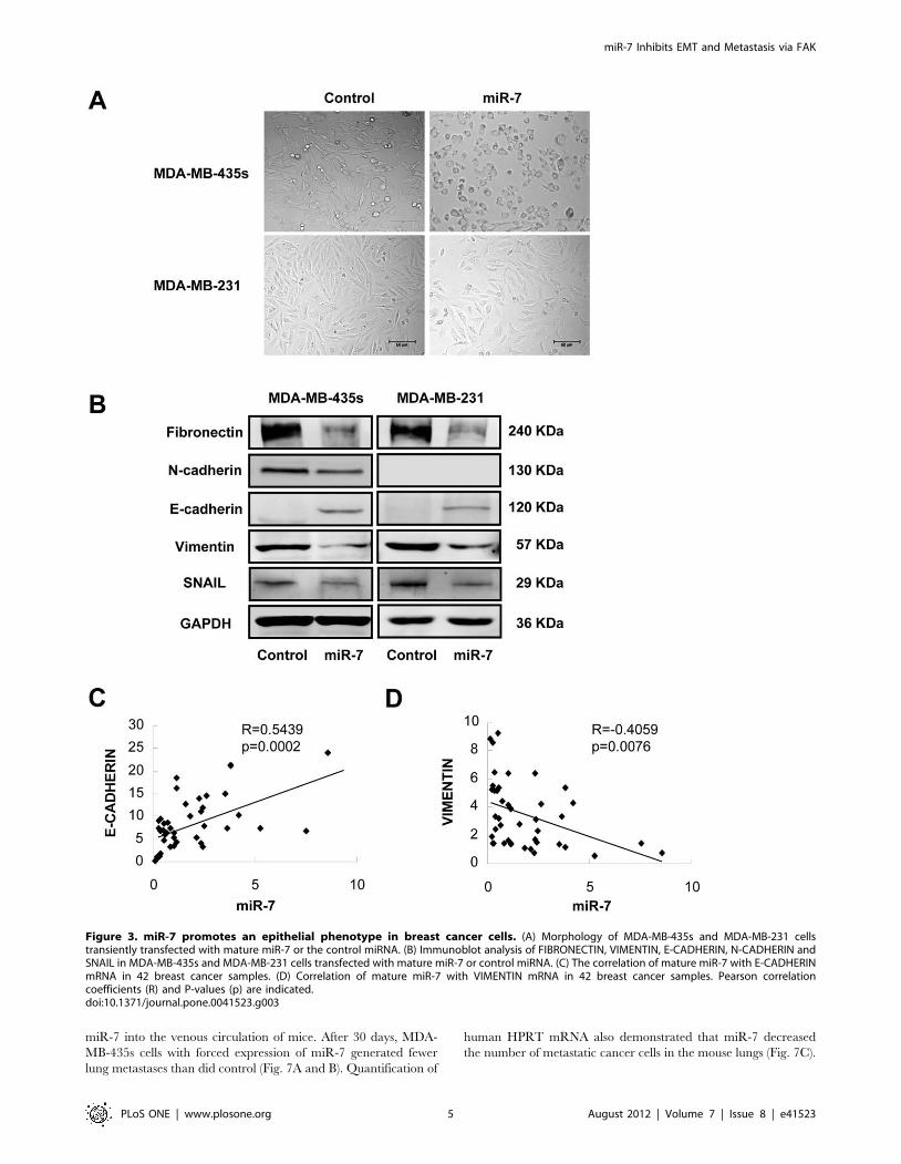
We noticed that forced expression of miR-7 promoted striking
change in the morphology of MDA-MB-435s and MDA-MB-231
cells, whereby the spindle-, fibroblast-like morphology switched to
the cobblestone-like appearance of epithelial cells (Fig. 3A). The
altered cell morphology produced by forced expression of miR-7
was also quantified by measuring dendricity/inverse shape factor
(Fig. S2D). These morphological changes are hallmarks of reduced
epithelial-to-mesenchymal transition (EMT). To determine if the
Figure 1. miR-7 expression is decreased in breast cancer and is associated with tumor metastasis. (A) The expression level of maturemiR-7 in breast cancer (n = 42) or normal breast tissues (n = 27) was determined using quantitative PCR. Both breast cancer and normal breast arefresh tissues. Box-plot lines represent medians and interquartile ranges of the normalized threshold values; whiskers and spots indicate 10–90percentiles and the remaining data points. (B) The relative expression of mature miR-7 in 19 non-metastatic breast cancer tissue and 23 metastaticbreast cancer tissue samples. The expression level of mature miR-7 is normalized to U6 small nuclear RNA.doi:10.1371/journal.pone.0041523.g001
miR-7 Inhibits EMT and Metastasis via FAK
PLoS ONE | www.plosone.org 2 August 2012 | Volume 7 | Issue 8 | e41523
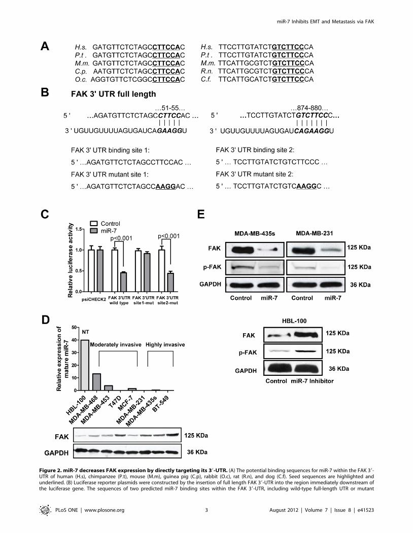
Figure 2. miR-7 decreases FAK expression by directly targeting its 39-UTR. (A) The potential binding sequences for miR-7 within the FAK 39-UTR of human (H.s), chimpanzee (P.t), mouse (M.m), guinea pig (C.p), rabbit (O.c), rat (R.n), and dog (C.f). Seed sequences are highlighted andunderlined. (B) Luciferase reporter plasmids were constructed by the insertion of full length FAK 39-UTR into the region immediately downstream ofthe luciferase gene. The sequences of two predicted miR-7 binding sites within the FAK 39-UTR, including wild-type full-length UTR or mutant
miR-7 Inhibits EMT and Metastasis via FAK
PLoS ONE | www.plosone.org 3 August 2012 | Volume 7 | Issue 8 | e41523

molecular changes typical of a reduced EMT occurred in miR-7
expressing cells, we examined the expression of mesenchymal
markers, such as FIBRONECTIN, VIMENTIN, N-CADHERIN,
SNAIL and the epithelial marker E-CADHERIN in MDA-MB-
435s and MDA-MB-231 cells. Immunoblot analysis showed that
expression of both FIBRONECTIN, VIMENTIN and SNAIL
were decreased in MDA-MB-435s and MDA-MB-231 cells with
forced expression of miR-7 (Fig. 3B). The protein level of N-
CADHERIN was also decreased in MDA-MB-435s cells with
forced expression of miR-7. N-CADHERIN protein expression
was not detectable by immunoblot analysis in MDA-MB-231 cells
(Fig. 3B). Furthermore, forced expression of miR-7 increased
expression of E-CADHERIN in both cell lines whereas the control
transfected cells remained E-CADHERIN negative (Fig. 3B).
To determine whether the expression of E-CADHERIN
correlated to miR-7 levels in breast cancer, we quantified miR-7
as well as E-CADHERIN mRNA expression in a cohort of breast
cancer samples. From the cohort of 42 primary breast cancer, we
observed a significant positive correlation between E-CADHERIN
mRNA and miR-7 miRNA expression (Fig. 3D). We also analyzed
the expression of miR-7 versus the expression of VIMENTIN
mRNA in this cohort. We observed a significant inverse
correlation between miR-7 miRNA and VIMENTIN mRNA
expression in patient tumors (Fig. 3E). This data suggested a strong
association between miR-7 expression and markers of epithelial
differentiation.
miR-7 Impairs Breast Cancer Cell Migration and Invasionin vitro
Given that the expression of miR-7 was inversely correlated
with metastasis of breast cancer, we considered whether miR-7
might possess an important role in breast cancer cell migration
and invasion. Transwell migration and Matrigel invasion assays
demonstrated that miR-7 significantly reduced the migration and
invasion capacity of MDA-MB-435s and MDA-MB-231 cells
(Fig. 4A). As FAK is frequently up-regulated in breast cancer and
promotes cell migration and invasion, and as miR-7 can directly
regulate the expression of FAK, we next ascertained whether
reduction of FAK expression might provide an explanation for the
reduction of cell migration and invasion observed following forced
expression of miR-7. We therefore forced the expression of miR-7
in MDA-MB-435s and MDA-MB-231 cells together with a
construct containing the FAK coding sequence but lacking the
39UTR of the FAK-encoding mRNA; and as such, this construct
yielded a FAK mRNA that is resistant to miR-7. The restoration
of FAK expression was confirmed through immunoblot analysis
(Fig. 4C). Transwell assays indicated that restoration of FAK
expression significantly abrogated miR-7 reduced cell migration
and invasiveness (Fig. 4B), indicative that FAK is both a direct and
functional target for miR-7.
miR-7 Inhibits Breast Cancer Cell Growth in vitroTo determine the function of miR-7 in the progression of breast
cancer, we sought to determine whether miR-7 may also affect the
proliferation of breast cancer cells. The proliferation rates of
MDA-MB-435s and MDA-MB-231 cells transfected with mature
miR-7 were significantly decreased when compared with those of
control miRNA transfected cells (Fig. 5A). We subsequently
utilized HBL-100 cells, which possess high relative expression of
miR-7 to further determine the functional effects of inhibition of
miR-7. Transfection of miR-7 inhibitor increased cell proliferation
in HBL-100 cells (Fig. 5A). Furthermore, transfection of miR-7
mimics in MDA-MB-435s cells produced a significant decrease in
colony formation in soft agar compared with control mimics
(Fig. 5B). Conversely, silencing of miR-7 in HBL-100 cells
increased colony formation in soft agar (Fig. 5C). These results
indicated that miR-7 inhibits breast cancer cells monolayer
proliferation and anchorage independent growth in vitro.
We also cultured the cells in three-dimensional Matrigel [22];
growing cells in or on gels which revealed cellular behavior that
are more relevant to EMT and metastasis. Less colony formation
was observed for miR-7 transfected MDA-MB-435s and MDA-
MB-231 cells compared to control transfected cells (Fig. 5D).
Moreover, the colonies formed by control transfected cells in 3D
Matrigel were larger and the cells in those colonies appeared more
motile and invasive compared with colonies formed by miR-7
transfected cells (Fig. 5D). miR-7 expressing cells produced
circumscribed colonies, whereas a large number of control
transfected cells spread from the main bulk of the colony
(Fig. 5D), suggesting that miR-7 inhibited invasive cellular
behavior.
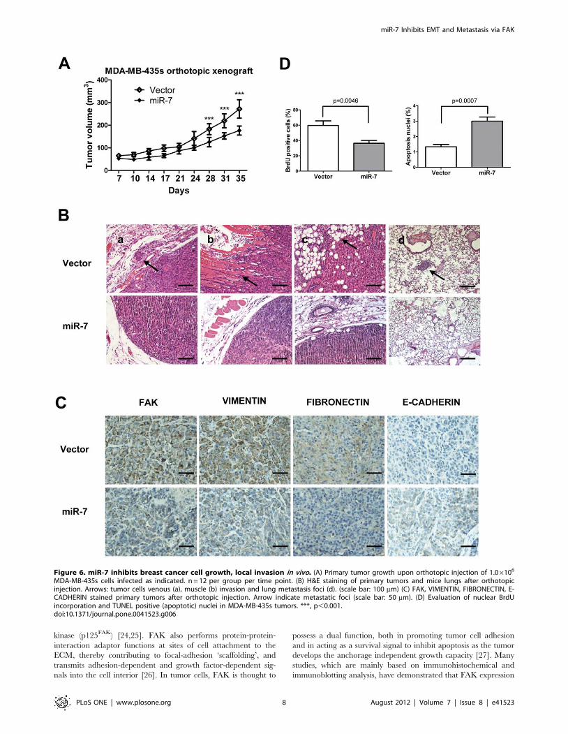
miR-7 Decreases Tumor Growth and SuppressesMetastasis in vivo
To determine whether miR-7 regulates tumor growth and
metastasis in vivo, we utilized xenograft models by injection of
MDA-MB-435s cells, with forced expression of miR-7, orthotop-
ically in the mammary fat pad of nude mice. Forced expression of
miR-7 decreased primary tumor growth by 1.5-fold and corre-
spondingly decreased cell proliferation as determined by immu-
nohistochemical analysis of nuclear incorporation of BrdU (Fig. 6A
and D) and increased cell apoptosis as determined by TUNEL
assay (Fig. 6 D). The tumor xenografts derived from cells with
forced expression of miR-7 exhibited lower expression of FAK,
FIBRONECTIN and VIMENTIN than control tumors (Fig. 6C).
Strikingly, a significant number of miR-7 expressing carcinoma
cells exhibited staining for human E-CADHERIN (Fig. 6C). The
control tumors were essentially E-CADHERIN negative (Fig. 6C).
The relative expression of FAK, FIBRONECTIN and VIMEN-
TIN between miR-7 expressing tumors and control tumors were
also analyzed by quantifying DAB staining intensity (Fig. S2E).
Control primary tumors displayed evidence of local invasion
(Fig. 6B); however, tumors with forced expression of miR-7 were
well encapsulated and non-invasive (Fig. 6B). Not only did cells
with forced expression of miR-7 generate smaller primary tumors,
but miR-7 also strikingly impaired in their capacity to seed lung
metastasis. Cells with forced expression of miR-7 produced no
metastatic lesions in contrast to control cells that formed lesions in
the lungs in 2 of 6 mice with tumors orthotopically implanted
(Fig. 6B).
We also determined the effect of miR-7 on metastasis was also
attributable to effects on later steps of the invasion-metastasis
cascade, independent of miR-7 influence on cellular invasion.
Thus, we injected MDA-MB-435s cells with forced expression of
(highlighted and underlined) binding site are shown. (C) Relative luciferase activity was analyzed after the above reporter plasmids or control reporterplasmid were cotransfected with miR-7 mimics or control mimics in MDA-MB-435s cells. (D) Relative expression of miR-7 by quantitative PCR (top)and immunoblot for FAK expression (bottom) in the indicated cell lines. NT, non-tumorigenic. (E) Immunoblot assays of endogenous FAK proteinlevels in MDA-MB-435s and MDA-MB-231 cells transfected with miR-7 mimics or control mimics and those in HBL-100 cells transfected with miR-7inhibitor or control inhibitor.doi:10.1371/journal.pone.0041523.g002
miR-7 Inhibits EMT and Metastasis via FAK
PLoS ONE | www.plosone.org 4 August 2012 | Volume 7 | Issue 8 | e41523
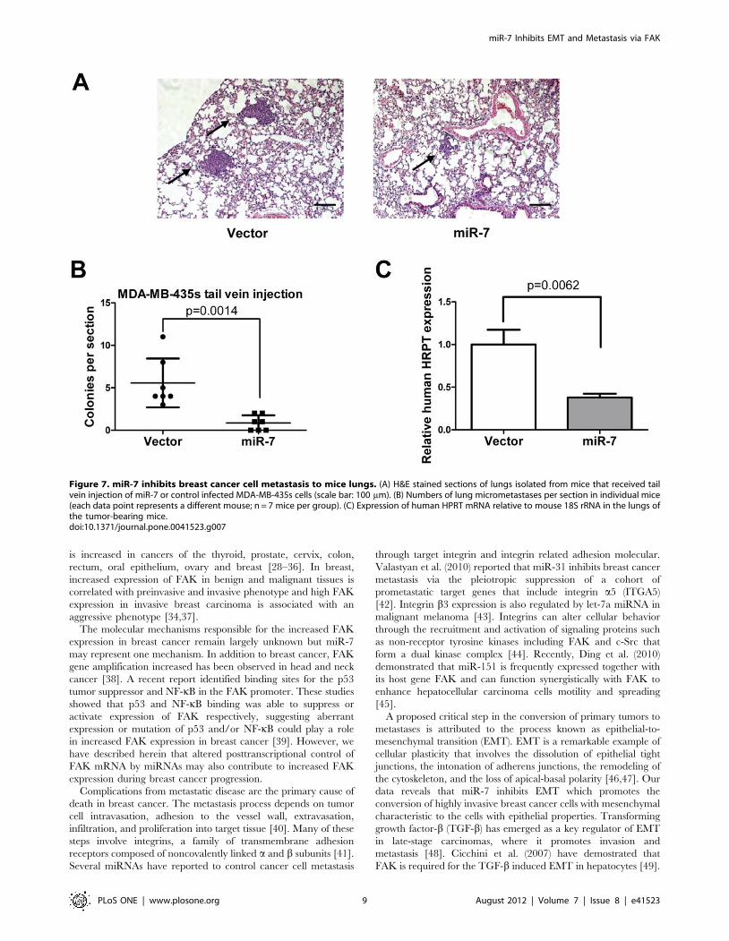
miR-7 into the venous circulation of mice. After 30 days, MDA-
MB-435s cells with forced expression of miR-7 generated fewer
lung metastases than did control (Fig. 7A and B). Quantification of
human HPRT mRNA also demonstrated that miR-7 decreased
the number of metastatic cancer cells in the mouse lungs (Fig. 7C).
Figure 3. miR-7 promotes an epithelial phenotype in breast cancer cells. (A) Morphology of MDA-MB-435s and MDA-MB-231 cellstransiently transfected with mature miR-7 or the control miRNA. (B) Immunoblot analysis of FIBRONECTIN, VIMENTIN, E-CADHERIN, N-CADHERIN andSNAIL in MDA-MB-435s and MDA-MB-231 cells transfected with mature miR-7 or control miRNA. (C) The correlation of mature miR-7 with E-CADHERINmRNA in 42 breast cancer samples. (D) Correlation of mature miR-7 with VIMENTIN mRNA in 42 breast cancer samples. Pearson correlationcoefficients (R) and P-values (p) are indicated.doi:10.1371/journal.pone.0041523.g003
miR-7 Inhibits EMT and Metastasis via FAK
PLoS ONE | www.plosone.org 5 August 2012 | Volume 7 | Issue 8 | e41523

Discussion
MicroRNAs are increasingly implicated in regulating the
malignant progression of cancer by directly targeting oncogenes
and tumor suppressor genes. Each miRNA can potentially interact
with several mRNA targets via perfect or imperfect base pairing,
primarily in the 39-UTR portion. A number of target prediction
algorithms, including TargetScan, PicTar and miRanda, relying
on seed sequence pairing rules and conservational analysis, have
been developed to score possible recognition sites and identify
putative gene targets. However, these predictions usually yield a
large number of false-positive candidates and experimental
validation is thus strictly required [23]. In the present study, we
located two potential binding sites (site 1 and site 2) of miR-7 in the
FAK 39UTR with RNAhybrid (Fig. 2A). However, only binding
site 2 was predicted by three algorithms (PicTar, TargetScan and
miRanda). Luciferase reporter assays with mutant binding site 1
and binding site 2 of FAK 39UTR revealed that miR-7 interacts
with FAK 39UTR was through binding with site 1 (Fig. 2C), thus
confirming the importance of experimental validation.
It is likely that some changes in cell adhesion occur at focal
adhesion plaques which are cell/extracellular matrix (ECM)
contact points containing integrin receptors, cytoskeletal compo-
nents and intracellular signalling proteins such as focal adhesion
Figure 4. Restoration of FAK attenuates miR-7 mediated cell migration and invasion inhibition. (A) Transwell migration and invasionassays of MDA-MB-435s and MDA-MB-231 cells transfected with miR-7 mimics and control mimics. (B) Transwell migration and invasion assays ofMDA-MB-435s and MDA-MB-231 cells transfected with miR-7 mimics and control mimics with or without FAK restoration. (C) Immunoblot analysis ofFAK expression in MDA-MB-435s and MDA-MB-231 cells transfected with miR-7 mimics or control mimics with or without FAK restoration. ** p,0.01,*** p,0.001.doi:10.1371/journal.pone.0041523.g004
miR-7 Inhibits EMT and Metastasis via FAK
PLoS ONE | www.plosone.org 6 August 2012 | Volume 7 | Issue 8 | e41523

Figure 5. miR-7 inhibits breast cancer cell tumorigenesis in vitro. (A) miR-7 decreases MDA-MB-435s and MDA-MB-231 cell proliferation invitro and inhibition of endogenous miR-7 expression promotes HBL-100 cell monolayer proliferation. (B) miR-7 inhibits MDA-MB-435s anchorageindependent growth in soft agar. (C) Inhibition of miR-7 promotes HBL-100 cell anchorage independent growth in soft agar. (D) Morphology of MDA-MB-435s and MDA-MB-231 cells cultured in three-dimensional Matrigel after transfection with mature miR-7 or the control miRNA.doi:10.1371/journal.pone.0041523.g005
miR-7 Inhibits EMT and Metastasis via FAK
PLoS ONE | www.plosone.org 7 August 2012 | Volume 7 | Issue 8 | e41523
kinase (p125FAK) [24,25]. FAK also performs protein-protein-
interaction adaptor functions at sites of cell attachment to the
ECM, thereby contributing to focal-adhesion ‘scaffolding’, and
transmits adhesion-dependent and growth factor-dependent sig-
nals into the cell interior [26]. In tumor cells, FAK is thought to
possess a dual function, both in promoting tumor cell adhesion
and in acting as a survival signal to inhibit apoptosis as the tumor
develops the anchorage independent growth capacity [27]. Many
studies, which are mainly based on immunohistochemical and
immunoblotting analysis, have demonstrated that FAK expression
Figure 6. miR-7 inhibits breast cancer cell growth, local invasion in vivo. (A) Primary tumor growth upon orthotopic injection of 1.06106
MDA-MB-435s cells infected as indicated. n = 12 per group per time point. (B) H&E staining of primary tumors and mice lungs after orthotopicinjection. Arrows: tumor cells venous (a), muscle (b) invasion and lung metastasis foci (d). (scale bar: 100 mm) (C) FAK, VIMENTIN, FIBRONECTIN, E-CADHERIN stained primary tumors after orthotopic injection. Arrow indicate metastatic foci (scale bar: 50 mm). (D) Evaluation of nuclear BrdUincorporation and TUNEL positive (apoptotic) nuclei in MDA-MB-435s tumors. ***, p,0.001.doi:10.1371/journal.pone.0041523.g006
miR-7 Inhibits EMT and Metastasis via FAK
PLoS ONE | www.plosone.org 8 August 2012 | Volume 7 | Issue 8 | e41523
is increased in cancers of the thyroid, prostate, cervix, colon,
rectum, oral epithelium, ovary and breast [28–36]. In breast,
increased expression of FAK in benign and malignant tissues is
correlated with preinvasive and invasive phenotype and high FAK
expression in invasive breast carcinoma is associated with an
aggressive phenotype [34,37].
The molecular mechanisms responsible for the increased FAK
expression in breast cancer remain largely unknown but miR-7
may represent one mechanism. In addition to breast cancer, FAK
gene amplification increased has been observed in head and neck
cancer [38]. A recent report identified binding sites for the p53
tumor suppressor and NF-kB in the FAK promoter. These studies
showed that p53 and NF-kB binding was able to suppress or
activate expression of FAK respectively, suggesting aberrant
expression or mutation of p53 and/or NF-kB could play a role
in increased FAK expression in breast cancer [39]. However, we
have described herein that altered posttranscriptional control of
FAK mRNA by miRNAs may also contribute to increased FAK
expression during breast cancer progression.
Complications from metastatic disease are the primary cause of
death in breast cancer. The metastasis process depends on tumor
cell intravasation, adhesion to the vessel wall, extravasation,
infiltration, and proliferation into target tissue [40]. Many of these
steps involve integrins, a family of transmembrane adhesion
receptors composed of noncovalently linked a and b subunits [41].
Several miRNAs have reported to control cancer cell metastasis
through target integrin and integrin related adhesion molecular.
Valastyan et al. (2010) reported that miR-31 inhibits breast cancer
metastasis via the pleiotropic suppression of a cohort of
prometastatic target genes that include integrin a5 (ITGA5)
[42]. Integrin b3 expression is also regulated by let-7a miRNA in
malignant melanoma [43]. Integrins can alter cellular behavior
through the recruitment and activation of signaling proteins such
as non-receptor tyrosine kinases including FAK and c-Src that
form a dual kinase complex [44]. Recently, Ding et al. (2010)
demonstrated that miR-151 is frequently expressed together with
its host gene FAK and can function synergistically with FAK to
enhance hepatocellular carcinoma cells motility and spreading
[45].
A proposed critical step in the conversion of primary tumors to
metastases is attributed to the process known as epithelial-to-
mesenchymal transition (EMT). EMT is a remarkable example of
cellular plasticity that involves the dissolution of epithelial tight
junctions, the intonation of adherens junctions, the remodeling of
the cytoskeleton, and the loss of apical-basal polarity [46,47]. Our
data reveals that miR-7 inhibits EMT which promotes the
conversion of highly invasive breast cancer cells with mesenchymal
characteristic to the cells with epithelial properties. Transforming
growth factor-b (TGF-b) has emerged as a key regulator of EMT
in late-stage carcinomas, where it promotes invasion and
metastasis [48]. Cicchini et al. (2007) have demostrated that
FAK is required for the TGF-b induced EMT in hepatocytes [49].
Figure 7. miR-7 inhibits breast cancer cell metastasis to mice lungs. (A) H&E stained sections of lungs isolated from mice that received tailvein injection of miR-7 or control infected MDA-MB-435s cells (scale bar: 100 mm). (B) Numbers of lung micrometastases per section in individual mice(each data point represents a different mouse; n = 7 mice per group). (C) Expression of human HPRT mRNA relative to mouse 18S rRNA in the lungs ofthe tumor-bearing mice.doi:10.1371/journal.pone.0041523.g007
miR-7 Inhibits EMT and Metastasis via FAK
PLoS ONE | www.plosone.org 9 August 2012 | Volume 7 | Issue 8 | e41523
Thus, FAK is an important regulatory element in the process of
EMT. miR-7 has also been reported to target PAK1 in breast
cancer cells [13]. PAK1 phosphorylates and modulates the
subcellular localization of snail in BC cells and subsequent EMT
[50]. miR-7 may therefore co-ordinately regulate breast cancer
cells EMT.
Analysis of miR-7 as well as E-CADHERIN and VIMENTIN
mRNA levels in breast cancer tissues reveals that miR-7 expression
is an indicator of epithelial differentiation in breast cancer. Recent
studies have also identified the miR-200 family as a powerful
marker and determining factor of the epithelial phenotype of
cancer cells by targeting the E-CADHERIN repressors ZEB1 and
ZEB2 [10,51].
While we were preparing the manuscript, it was reported that
miR-7 regulated cancer cell invasion by targeting FAK expression
in glioblastoma [52]. In that report, miR-7 levels in glioma tissues
are inverse correlated with FAK expression when combined with
work herein in breast cancer, it appears that miR-7 is a conserved
miRNA that inhibits the same target gene and plays similar
functions in several tumor types.
Materials and Methods
Tissue SamplesAll patients signed informed consent approving the use of their
tissues for research purposes and the study was approved by the
Institutional Review Board of the Anhui Medical University. Fresh
tissues including 42 breast cancer and 27 normal breast samples
derived from 69 patients that underwent surgery at the First
Affiliated Hospital of Anhui Medical University between 2009 and
2010 were used for the study. All tissue samples were H&E stained
and had been reviewed by two independent pathologists in Anhui
Medical University.
Cell LinesHBL-100, MCF-7, T47D, MDA-MB-468, MDA-MB-453,
MDA-MB-231, MDA-MB-435s, BT-549 and HEK293T cell lines
were from ATCC and cultured as recommended.
ConstructsThe human FAK cDNA expression plasmid pKH3-FAK was
kind gift from Dr. Junlin Guan (University of Michigan). The
ORF region of FAK cDNA was subcloned into pIRES neo3
plasmid. The 39 untranslated region (39-UTR) sequence of FAK
was amplified from the genomic DNA of normal breast tissues and
subcloned into the psiCHECK2 dual luciferase reporter plasmid
(Promega). The mutant construct of FAK 39UTR was generated
using a QuikChange Site-Directed Mutagenesis Kit (Stratagene).
RNA Isolation, miRNA and mRNA DetectionTotal RNA, inclusive of the small RNA fraction, was extracted
from cultured cells and clinical samples with a mirVana miRNA
Isolation Kit (Ambion). Mature miR-7 was reverse-transcribed
with specific RT primers, quantified with a TaqMan probe, and
normalized by U6 small nuclear RNA using TaqMan miRNA
assays (Applied Biosystems). mRNA expression analysis was
conducted by quantitative PCR using SYBR green dye, with
relative changes calculated by the DDCt method.
miRNA Gene Cloning and Ectopic ExpressionThe human miR-7 gene was PCR-amplified from normal
genomic DNA and cloned into the pBabe-puro retroviral vector.
The pBabe-amphotrophic viruses were generated by cotransfec-
tion of HEK293T cells with the pBabe constructs, pCMV-VSVG
and Gag/Pol using Attractene Transfection Reagent (Qiagen).
Virus was harvested at 48 and 72 h posttransfection and infections
were performed in the presence of 8 mg/mL of polybrene
(SigmaAldrich). Following transduction, cells were selected with
1 mg/mL puromycin (SigmaAldrich).
PrimersPrimers used were as follows: FAK ORF (GCGCGGCTAG-
CATGGCAGCTGCTTACCTTGACCCCA,
ATAGCGGCCGCTCAGTGTGGTCTCGTCTGCCCAAGC);
miR-7 ectopic expression (AGGATCCTACAGGAACACAG-
GACCAGA, CCGAATTCTGATAAACACGTCCATTACA),
FAK 39UTR cloning (ATCTCGAGGCCTCCCCTAGGAG-
CACGTCTT, GCGCGGCCGCTTTACTGGTAA-
CACCTTTTTAAT), E-CADHERIN, VIMENTIN and
GAPDH quantitative PCR: E-CADHERIN (CTGAGAAC-
GAGGCTAACGTC, TGTCCACCATCATCATTCAATA);
VIMENTIN (AGACAGGCTTTAGCGAGTTATT,
GGGCTCCTAGCGGTTTAG); GAPDH (TGCACCAC-
CAACTGCTTAGC, GGCATGGACTGTGGTCATGAG). For
the in vivo xenograft, the following primers were used:
hHPRT(TTCCTTGGTCAGGCAGTATAATCC,
AGTCTGGCTTATATCCAACACTTCG); mouse 18s rRNA(-
GAAACGGCTACCACATCC, ACCAGACTTGCCCTCCA).
Oligonucleotide and Plasmid TransfectionmiRNA mimics (Genepharma, Shanghai, China) and miScript
miRNA inhibitor (Qiagen) were transfected using HiPerFect
Transfection Reagent (Qiagen) following the manufacturer’s
instructions. miRNA mimics and plasmid co-transfection were
performed by using Lipofectamine 2000 (Invitrogen). Twenty-four
hours after transfection, cells were plated for proliferation, soft
agar, migration, invasion assays or harvested for the luciferase
reporter assay. Cells were harvested for RNA and protein analyses
at forty-eight hours after transfection.
In vitro Migration and Invasion AssayFor transwell migration assays, 106104 cells were plated in the
top chamber with the non-coated membrane (24-well insert; 8 mm
pore size; BD Biosciences). For invasion assays, 26105 cells were
plated in the top chamber with Matrigel-coated membrane (24-
well insert; 8 mm pore size; BD Biosciences). In both assays, cells
were plated in medium without serum, and medium supplemented
with 10% serum was used as a chemoattractant in the lower
chamber. The cells were incubated for 24 to 36 hours and cells
that did not migrate or invade through the pores were removed by
a cotton swab. Filters were fixed with 90% ethanol, stained by
0.1% crystal violet, photographed and cells numbers were
counted.
Cell Proliferation and Soft Agar Colony Formation AssaysCell proliferation was determined by Cell Counting Kit-8
(Dojindo, Shanghai, China) according to the manufacturer’s
instructions. For soft agar colony formation assay, cells (56103)
in 1.5 mL medium supplemented with 0.35% agarose were
layered on a 1.5 mL base medium with 0.5% agarose. Soft agar
assays were performed in six well plates and in triplicate. Cells
were cultured for 14 days and colonies were counted.
Three-dimensional Matrigel CultureEight hundred cells per well were plated in 10% FBS medium
supplemented with 4% Matrigel (BD Bioscience) in a 96-well plate
which coated with Matrigel before addition of the cells. Matrigel-
miR-7 Inhibits EMT and Metastasis via FAK
PLoS ONE | www.plosone.org 10 August 2012 | Volume 7 | Issue 8 | e41523
containing (4%) medium was renewed every 3 d until the
experiment was terminated after 10 d.
Orthotopic InjectionThe animals were maintained in a pathogen-free barrier facility
at the Aninal Center of the University of Science and Technology
of China (USTC), and closely monitored by animal facility staff.
All animal work procedures were approved by USTC Ethics
Committee for Animal Care and Use (Protocol number :UST-
CACUC1201040) and were performed in accordance with the
regulations of animal care of USTC and conformed to the legal
mandates and national guidelines for the care and maintenance of
laboratory animals.
MDA-MB-435s cells (26106 cells in 100 mL PBS) were injected
orthotopically into the each side mammary fat pad of female
BALB/c-nu/nu mice (Slaccas, Shanghai, China). Each group
consisted of six mice. Tumor growth rates were analyzed by
measuring tumor length (L) and width (W), and calculating volume
through the use of the formula LW2/2. Six hours before sacrifice,
mice were i.p injected BrdU solution at the 100 mg/g body weight.
Tail Vein InjectionMDA-MB-435s cells (16106 cells in 200 mL PBS) were injected
directly into the lateral tail vein of 6- to 8-week-old female BALB/
c-nu/nu mice. Each group consisted of eight mice. Mice were
sacrificed on day 35 (orthotopic injection) or day 30 (tail vein
injection) and mammary tumors, mice liver and lung were fixed in
10% neutral buffered formalin and embedded in paraffin for
histology examination or were frozen at 280uC in RNALater
(Ambion) for RNA extraction and qPCR analysis.
ImmunohistochemistryFormalin-fixed, paraffin-embedded tissue was cut into 5 mm
section, de-paraffinized in xylene, rehydrated through graded
ethanol, quenched for endogenous peroxidase activity in 3% (v/v)
hydrogen peroxide, and processed for antigen retrieval by heating
in 10 mM citrate buffer (pH 6.0) at 90–100uC (FAK, VIMEN-
TIN, FIBRONECTIN, E-CADHERIN) or digesting with 0.1%
trypsin at 37uC (BrdU). Sections were incubated at 4uC overnight
with FAK (3258, 1:200, Cell Signaling Technology), VIMENTIN
(550513, 1:200, BD Pharmingen), FIBRONECTIN (610077,
1:200, BD Transduction Laboratories), E-CADHERIN (610181,
1:200, BD Transduction Laboratories) or BrdU (MAB0188, 1:100,
Maixin, Fuzhou, China ) antibody. Immunostaining was per-
formed using UltraSensitive S-P Detection Kit (KIT-9720,
Maixin, Fuzhou, China), and then color was developed by using
a DAB kit (DAB-0031, Maixin, Fuzhou, China). Subsequently,
sections were counterstained with hematoxylin. TUNEL assay was
performed with an in situ cell death detection kit (Roche)
according to the manufacturer’s instructions.Quantification of
immunohistochemical stain intensity was performed as previously
described [53].
Luciferase Reporter AssayLuciferase reporter assays were performed using the psi-
CHECK2-FAK-39-UTR vector. Cells were grown to approxi-
mately 60% confluence in 24-well plates and cotransfected with
psiCHECK2-FAK-39-UTR (wild type or mutant) or psiCHECK2
empty vector plus miR-7 mimics or control mimics using
Lipofectamine 2000 (Invitrogen). After 24 hours of incubation,
Firefly and Renilla luciferase activities were evaluated using the
Dual-Luciferase Reporter Assay system (Promega).
ImmunoblotCells were extracted in modified RIPA lysis buffer
(150 mM NaCl, 50 mM Tris, pH 7.4, 1% NP-40, 0.25% Na-
deoxycholate, 1 mM EDTA, protease inhibitor cocktail (Roche)).
Proteins from total cell lysates were resolved by SDS-PAGE,
transferred to the Nitrocellulose membrane (GE healthcare),
blocked in 5% non-fat milk in PBS/Tween-20, and blotted with
the antibodies for FAK (3258, 1:1000, Cell Signaling Technology),
phospho-FAK(Tyr397) (3283, 1:1000, Cell Signaling Technology),
VIMENTIN (550513, 1:5000, BD Pharmingen), FIBRONECTIN
(610077, 1:5000, BD Transduction Laboratories), E-CADHERIN
(610181, 1:5000, BD Transduction Laboratories), N-CADHERIN
(610920, 1:2000, BD Transduction Laboratories), SNAIL (sc-
28199, 1:1000, SantaCruz Biotechnology) and GAPDH (M20028,
1:5000, Abmart, Shanghai, China).
Measuring DendricityMeasuring cell dendricity was performed as previously de-
scribed [54].
Statistical AnalysisData are presented as mean 6 SD (standard deviation).
Student’s t test (two tailed) was used to compare two groups,
p,0.05 was considered significant.
Supporting Information
Figure S1 miR-7 target genes that predicted by alltheree algorithms. (A) Schematic illustration of target genes by
three algorithms respectively. (B) 114 target genes of miR-7 that
can be predicted by all three algorithms.
(TIF)
Figure S2 Quantification of immunoblot bands intens-tity and IHC DAB staining intensity. (A) Quantification of
immunoblot FAK and p-FAK bands intensity relative to GAPDH
intensity in MDA-MB-435s cells are shown. (B) Quantification of
immunoblot FAK and p-FAK band intensity relative to GAPDH
intensity in MDA-MB-231 cells are shown. (C) Quantification of
immunoblot FAK and p-FAK band intensity relative to GAPDH
intensity in HBL-100 cells are shown. (D) Quantification of
dendricty/inverse shape factor in MDA-MB-435s and MDA-MB-
231 cells. (E) Quantification of relative DAB staining intensity in
MDA-MB-435s derived tumor sections are shown.
(TIF)
Acknowledgments
We greatly appreciate the gift of human FAK cDNA from Dr. Junlin Guan
(University of Michigan).
Author Contributions
Conceived and designed the experiments: TZ XK PEL. Performed the
experiments: XK GL YY YH XW WZ ZW TC WW. Analyzed the data:
TZ PEL ZW. Wrote the paper: XK PEL TZ.
References
1. Bartel DP (2004) MicroRNAs: genomics, biogenesis, mechanism, and function.
Cell 116: 281–297.
2. Ambros V (2004) The functions of animal microRNAs. Nature 431: 350–355.
miR-7 Inhibits EMT and Metastasis via FAK
PLoS ONE | www.plosone.org 11 August 2012 | Volume 7 | Issue 8 | e41523
3. Bartel DP (2009) MicroRNAs: target recognition and regulatory functions. Cell
136: 215–233.4. Friedman RC, Farh KK, Burge CB, Bartel DP (2009) Most mammalian
mRNAs are conserved targets of microRNAs. Genome Res 19: 92–105.
5. Ma L, Teruya-Feldstein J, Weinberg RA (2007) Tumour invasion and metastasisinitiated by microRNA-10b in breast cancer. Nature 449: 682–688.
6. Martello G, Rosato A, Ferrari F, Manfrin A, Cordenonsi M, et al. (2010) AMicroRNA targeting dicer for metastasis control. Cell 141: 1195–1207.
7. Yao J, Liang L, Huang S, Ding J, Tan N, et al. (2010) MicroRNA-30d promotes
tumor invasion and metastasis by targeting Galphai2 in hepatocellularcarcinoma. Hepatology 51: 846–856.
8. Valastyan S, Reinhardt F, Benaich N, Calogrias D, Szasz AM, et al. (2009) Apleiotropically acting microRNA, miR-31, inhibits breast cancer metastasis. Cell
137: 1032–1046.9. Xiong Y, Fang JH, Yun JP, Yang J, Zhang Y, et al. (2010) Effects of microRNA-
29 on apoptosis, tumorigenicity, and prognosis of hepatocellular carcinoma.
Hepatology 51: 836–845.10. Park SM, Gaur AB, Lengyel E, Peter ME (2008) The miR-200 family
determines the epithelial phenotype of cancer cells by targeting the E-cadherinrepressors ZEB1 and ZEB2. Genes Dev 22: 894–907.
11. Kefas B, Godlewski J, Comeau L, Li Y, Abounader R (2008) microRNA-7
inhibits the epidermal growth factor receptor and the Akt pathway and is down-regulated in glioblastoma. Cancer Res 68: 3566–3572.
12. Webster RJ, Giles KM, Price KJ, Zhang PM, Mattick JS, et al. (2009)Regulation of epidermal growth factor receptor signaling in human cancer cells
by microRNA-7. J Biol Chem 284: 5731–5741.13. Reddy SD, Ohshiro K, Rayala SK, Kumar R (2008) MicroRNA-7, a homeobox
D10 target, inhibits p21-activated kinase 1 and regulates its functions. Cancer
Res 68: 8195–8200.14. Jiang L, Liu X, Chen Z, Jin Y, Heidbreder CE, et al. (2010) MicroRNA-7 targets
IGF1R (insulin-like growth factor 1 receptor) in tongue squamous cell carcinomacells. Biochem J 432: 199–205.
15. Foekens JA, Sieuwerts AM, Smid M, Look MP, de Weerd V, et al. (2008) Four
miRNAs associated with aggressiveness of lymph node-negative, estrogenreceptor-positive human breast cancer. Proc Natl Acad Sci U S A 105:
13021–13026.16. Krek A, Grun D, Poy MN, Wolf R, Rosenberg L, et al. (2005) Combinatorial
microRNA target predictions. Nat Genet 37: 495–500.17. Lewis BP, Burge CB, Bartel DP (2005) Conserved seed pairing, often flanked by
adenosines, indicates that thousands of human genes are microRNA targets. Cell
120: 15–20.18. Enright AJ, John B, Gaul U, Tuschl T, Sander C, et al. (2003) MicroRNA
targets in Drosophila. Genome Biol 5: R1.19. Luo M, Guan JL (2010) Focal adhesion kinase: a prominent determinant in
breast cancer initiation, progression and metastasis. Cancer Lett 289: 127–139.
20. Rehmsmeier M, Steffen P, Hochsmann M, Giegerich R (2004) Fast and effectiveprediction of microRNA/target duplexes. RNA 10: 1507–1517.
21. Siesser PM, Hanks SK (2006) The signaling and biological implications of FAKoverexpression in cancer. Clin Cancer Res 12: 3233–3237.
22. Chu JH, Yu S, Hayward SW, Chan FL (2009) Development of a three-dimensional culture model of prostatic epithelial cells and its use for the study of
epithelial-mesenchymal transition and inhibition of PI3K pathway in prostate
cancer. Prostate 69: 428–442.23. Migliore C, Petrelli A, Ghiso E, Corso S, Capparuccia L, et al. (2008)
MicroRNAs impair MET-mediated invasive growth. Cancer Res 68: 10128–10136.
24. Clark EA, Brugge JS (1995) Integrins and signal transduction pathways: the road
taken. Science 268: 233–239.25. Schaller MD, Borgman CA, Cobb BS, Vines RR, Reynolds AB, et al. (1992)
pp125FAK a structurally distinctive protein-tyrosine kinase associated with focaladhesions. Proc Natl Acad Sci U S A 89: 5192–5196.
26. McLean GW, Carragher NO, Avizienyte E, Evans J, Brunton VG, et al. (2005)
The role of focal-adhesion kinase in cancer - a new therapeutic opportunity. NatRev Cancer 5: 505–515.
27. Schlaepfer DD, Hanks SK, Hunter T, van der Geer P (1994) Integrin-mediatedsignal transduction linked to Ras pathway by GRB2 binding to focal adhesion
kinase. Nature 372: 786–791.28. Owens LV, Xu L, Dent GA, Yang X, Sturge GC, et al. (1996) Focal adhesion
kinase as a marker of invasive potential in differentiated human thyroid cancer.
Ann Surg Oncol 3: 100–105.29. Tremblay L, Hauck W, Aprikian AG, Begin LR, Chapdelaine A, et al. (1996)
Focal adhesion kinase (pp125FAK) expression, activation and association withpaxillin and p50CSK in human metastatic prostate carcinoma. Int J Cancer 68:
164–171.
30. McCormack SJ, Brazinski SE, Moore JL Jr, Werness BA, Goldstein DJ (1997)
Activation of the focal adhesion kinase signal transduction pathway in cervicalcarcinoma cell lines and human genital epithelial cells immortalized with human
papillomavirus type 18. Oncogene 15: 265–274.
31. Kornberg LJ (1998) Focal adhesion kinase expression in oral cancers. Head
Neck 20: 634–639.
32. Kornberg LJ (1998) Focal adhesion kinase and its potential involvement in
tumor invasion and metastasis. Head Neck 20: 745–752.
33. Judson PL, He X, Cance WG, Van Le L (1999) Overexpression of focal
adhesion kinase, a protein tyrosine kinase, in ovarian carcinoma. Cancer 86:1551–1556.
34. Cance WG, Harris JE, Iacocca MV, Roche E, Yang X, et al. (2000)
Immunohistochemical analyses of focal adhesion kinase expression in benign
and malignant human breast and colon tissues: correlation with preinvasive andinvasive phenotypes. Clin Cancer Res 6: 2417–2423.
35. Lark AL, Livasy CA, Calvo B, Caskey L, Moore DT, et al. (2003)Overexpression of focal adhesion kinase in primary colorectal carcinomas and
colorectal liver metastases: immunohistochemistry and real-time PCR analyses.Clin Cancer Res 9: 215–222.
36. Gabriel B, Mildenberger S, Weisser CW, Metzger E, Gitsch G, et al. (2004)Focal adhesion kinase interacts with the transcriptional coactivator FHL2 and
both are overexpressed in epithelial ovarian cancer. Anticancer Res 24: 921–927.
37. Lark AL, Livasy CA, Dressler L, Moore DT, Millikan RC, et al. (2005) Highfocal adhesion kinase expression in invasive breast carcinomas is associated with
an aggressive phenotype. Mod Pathol 18: 1289–1294.
38. Agochiya M, Brunton VG, Owens DW, Parkinson EK, Paraskeva C, et al.
(1999) Increased dosage and amplification of the focal adhesion kinase gene inhuman cancer cells. Oncogene 18: 5646–5653.
39. Golubovskaya V, Kaur A, Cance W (2004) Cloning and characterization of thepromoter region of human focal adhesion kinase gene: nuclear factor kappa B
and p53 binding sites. Biochim Biophys Acta 1678: 111–125.
40. Felding-Habermann B, O’Toole TE, Smith JW, Fransvea E, Ruggeri ZM, et al.
(2001) Integrin activation controls metastasis in human breast cancer. Proc NatlAcad Sci U S A 98: 1853–1858.
41. Ruoslahti E (1999) Fibronectin and its integrin receptors in cancer. Adv CancerRes 76: 1–20.
42. Valastyan S, Chang A, Benaich N, Reinhardt F, Weinberg RA (2010)Concurrent suppression of integrin alpha5, radixin, and RhoA phenocopies
the effects of miR-31 on metastasis. Cancer Res 70: 5147–5154.
43. Muller DW, Bosserhoff AK (2008) Integrin beta 3 expression is regulated by let-
7a miRNA in malignant melanoma. Oncogene 27: 6698–6706.
44. Mitra SK, Schlaepfer DD (2006) Integrin-regulated FAK-Src signaling innormal and cancer cells. Curr Opin Cell Biol 18: 516–523.
45. Ding J, Huang S, Wu S, Zhao Y, Liang L, et al. (2010) Gain of miR-151 onchromosome 8q24.3 facilitates tumour cell migration and spreading through
downregulating RhoGDIA. Nat Cell Biol 12: 390–399.
46. Wang HR, Zhang Y, Ozdamar B, Ogunjimi AA, Alexandrova E, et al. (2003)
Regulation of cell polarity and protrusion formation by targeting RhoA fordegradation. Science 302: 1775–1779.
47. Zavadil J, Narasimhan M, Blumenberg M, Schneider RJ (2007) Transforminggrowth factor-beta and microRNA:mRNA regulatory networks in epithelial
plasticity. Cells Tissues Organs 185: 157–161.
48. Zavadil J, Bottinger EP (2005) TGF-beta and epithelial-to-mesenchymal
transitions. Oncogene 24: 5764–5774.
49. Cicchini C, Laudadio I, Citarella F, Corazzari M, Steindler C, et al. (2008)
TGFbeta-induced EMT requires focal adhesion kinase (FAK) signaling. ExpCell Res 314: 143–152.
50. Yang Z, Rayala S, Nguyen D, Vadlamudi RK, Chen S, et al. (2005) PAK1phosphorylation of snail, a master regulator of epithelial-to-mesenchyme
transition, modulates snail’s subcellular localization and functions. Cancer Res65: 3179–3184.
51. Gregory PA, Bert AG, Paterson EL, Barry SC, Tsykin A, et al. (2008) The miR-200 family and miR-205 regulate epithelial to mesenchymal transition by
targeting ZEB1 and SIP1. Nat Cell Biol 10: 593–601.
52. Wu DG, Wang YY, Fan LG, Luo H, Han B, et al. (2011) MicroRNA-7 regulates
glioblastoma cell invasion via targeting focal adhesion kinase expression. ChinMed J (Engl) 124: 2616–2621.
53. Park SI, Zhang J, Phillips KA, Araujo JC, Najjar AM, et al. (2008) TargetingSRC family kinases inhibits growth and lymph node metastases of prostate
cancer in an orthotopic nude mouse model. Cancer Res. 68: 3323–3333.
54. Heffron DS, Mandell JW (2005) Opposing roles of ERK and p38 MAP kinases
in FGF2-induced astroglial process extension. Mol Cell Neurosci 28: 779–790.
miR-7 Inhibits EMT and Metastasis via FAK
PLoS ONE | www.plosone.org 12 August 2012 | Volume 7 | Issue 8 | e41523
Copyright © 2022 FDOKUMEN