Multiplex Screening for Interacting Compounds in Paediatric ...
Upload
khangminh22Category
view
0download
0
Oncogene (2020) 39:7005–7018https://doi.org/10.1038/s41388-020-01512-8
ARTICLE
LncRNA SNHG11 facilitates tumor metastasis by interacting with andstabilizing HIF-1α
Linguo Xu1,2● Lin Huan 1,2
● Tianan Guo2,3● Yangjun Wu1,2
● Yanfang Liu1,2● Qifeng Wang2,4
● Shenglin Huang 1,2●
Ye Xu 2,3● Linhui Liang 1,2,5
● Xianghuo He 1,2,5
Received: 19 May 2020 / Revised: 26 September 2020 / Accepted: 5 October 2020 / Published online: 15 October 2020© The Author(s) 2020. This article is published with open access
AbstractEpigenetic alteration is one of the hallmarks of colorectal cancer (CRC). Many driver genes are regulated by DNAmethylation in CRC. However, the role of DNA methylation regulating lncRNAs remain elusive. Here, we identify thatSNHG11 (small nucleolar RNA host gene 11) is upregulated by promotor hypomethylation in CRC and is associated withpoor prognosis in CRC patients. SNHG11 can promote CRC cell migration and metastasis under hypoxia. Interestingly, theDNA-binding motif of SNHG11 is similar to that of HIF-1α. In addition, SNHG11-associated genes are enriched withmembers of the HIF-1 signaling pathway in CRC. Mechanistically, SNHG11 binds to the pVHLrecognition sites on HIF-1α,thus blocking the interaction of pVHL with HIF-1α and preventing its ubiquitination and degradation. Moreover, SNHG11upregulates the expression of HIF-1α target genes, i.e., AK4, ENO1, HK2, and Twist1. Notably, SNHG11 can bind to theHRE sites in the promoter of these genes and increase their transcription. In summary, these results identify a SNHG11/ HIF-1α axis that plays a pivotal role in tumor invasion and metastasis.
Introduction
Colorectal cancer (CRC) is one of the most common can-cers worldwide and the leading cause of cancer-relateddeaths [1]. The serious health problem caused by CRCworldwide increases the demand for new biomarkers andtherapeutic targets. Epigenetic changes are one of the hall-marks of CRC, particularly DNA methylation alteration. Inaddition to chromosomal instability and microsatelliteinstability, a subtype of CRC called CpG island methylatorphenotype is identified, which harbors high frequency ofDNA hypermethylation [2]. Expression of a group pf genescan be regulated by DNA methylation in the initiation andprogression of CRC [3]. Studies on long noncoding RNAs(lncRNAs) have been emerging in the last decade.LncRNAs can regulate the proliferation, apoptosis, inva-sion, metastasis, and multidrug resistance of CRC, high-lighting the potential of lncRNAs to act as new biomarkersand therapeutic targets for CRC patients [4–10]. However,whether lncRNAs regulated by DNA methylation play arole in CRC remain elusive. Recently, emerging studieshave indicated that RNAs are crucial for the function oftranscription factors and chromatin regulators [11–14]. Thetranscription factor Yin-Yang 1 (YY1) can be trapped byRNAs in gene regulatory elements to stabilize the gene
These authors contributed equally: Linguo Xu, Lin Huan
* Ye [email protected]
* Linhui [email protected]
* Xianghuo [email protected]
1 Fudan University Shanghai Cancer Center and Institutes ofBiomedical Sciences, Shanghai Medical College, FudanUniversity, Shanghai 200032, China
2 Department of Oncology, Shanghai Medical College, FudanUniversity, Shanghai 200032, China
3 Department of Colorectal Surgery, Fudan University ShanghaiCancer Center, Shanghai 200032, China
4 Department of Pathology, Fudan University Shanghai CancerCenter, Shanghai 200032, China
5 Key Laboratory of Breast Cancer in Shanghai, Fudan UniversityShanghai Cancer Center, Fudan University, Shanghai 200032,China
Supplementary information The online version of this article (https://doi.org/10.1038/s41388-020-01512-8) contains supplementarymaterial, which is available to authorized users.
1234
5678
90();,:
1234567890();,:

expression [15], which suggests that RNAs contribute to thestability of the gene transcription process. Although thebinding of RNAs with YY1 might not be strongly sequencespecific, YY1 binding sites are enriched in lncRNA-associated enhancer-interacting promoters [16]. In additionto YY1, the chromatin regulator CTCF also interacts withRNAs to regulate chromatin structure [17]. Deletion ofRNA binding sites in CTCF significantly dampens theability of CTCF to form chromatin loops [17]. These find-ings indicate that the crosstalk between lncRNAs and TF/chromatin regulators is crucial for the chromatin structureand gene transcription program.
To explore the role of DNA-methylation-regulatedlncRNAs in CRC, we analyzed DNA-methylation-regulated lncRNAs in CRC cells in previous study, andselected nuclear lncRNAs for further investigation. Wefound that SNHG11 could promote the invasion andmetastasis of CRC cells. Furthermore, we identified thatSNHG11 interacted with and stabilized HIF-1α. In addition,SNHG11 enhanced the transcriptional activity of HIF-1αand promoted CRC progression.
Results
SNHG11 is upregulated by promoterhypomethylation in CRC
Previous work identified 20 lncRNAs that might be medi-ated by DNA methylation in The Cancer Genome Atlas(TCGA)-COAD (Supplementary Fig. 1A) [18]. The func-tion of a lncRNA is dependent on its subcellular localiza-tion, so knowing the localization of lncRNAs enables theprediction of their biological function [19]. By analyzingRNA-seq data from the lncATLAS database (http://lncatlas.crg.eu/) [20], we determined the subcellular localization ofthe 18 candidate lncRNAs that were negatively correlatedwith DNA methylation in TCGA-COAD and found that 7of them localized in the nucleus (Supplementary Fig. 1B).Among these nuclear lncRNAs, SNHG11 had the basalexpression level in nucleus second to PVT1 and had thehighest DNA methylation CpG site in normal colon tissues(Supplementary Fig. 1A, B), so we selected SNHG11 forfurther investigation.
We analyzed the characteristics of the genomic locus ofSNHG11 and found the CpG island lying at the transcrip-tional activation site of SNHG11 (Fig. 1A). Interrogatingthe DNA methylation status from TCGA-COAD, eighteenprobes were found covering SNHG11 genomic locus inIllumina Human Methylation 450 platform (SupplementaryFig. 1C). Among these, five sites (cg05890898,cg07702509, cg13293885, cg17526424, and cg26306893)were significantly demethylated in CRC compared to
normal tissues (Fig. 1B). In addition, DNA methylation ofthese sites was negatively correlated with SNHG11expression in TCGA-COAD (Fig. 1C). These suggestedthat DNA methylation may modulate SNHG11 expression.Next, we treated CRC cells with 5-Aza, an inhibitor ofDNA methyltransferase, and found that the expression ofSNHG11 was significantly upregulated in 5-Aza-treatedCRC cells (Fig. 1D). Moreover, the induction of SNHG11in 5-Aza treated cells was consistent with the basal DNAmethylation of these cells (Supplementary Fig. 1D).
We performed rapid amplification of cDNA ends(RACE) assays and validated the sequence of SNHG11(Supplementary Fig. 2A, B), which was located at chro-mosomal 20q11.23 and consist of five exons (Supplemen-tary Fig. 2B). Next, we determined the subcellularlocalization of SNHG11 in CRC cells by cellular fractio-nation and RNA fluorescence in situ hybridization. Theresults suggested that SNHG11 predominantly localized tothe nucleus (Supplementary Fig. 2C, D). The predominantnuclear localization implies a non-protein-coding role forSNHG11, agreeing with the prediction made by the CodingPotential Assessment Tool and by the PhyloCSF score(Supplementary Fig. 2E).
SNHG11 promotes the migration, invasion, andmetastasis of CRC cells in vitro and in vivo
To study the potential role of SNHG11 in CRC progression,we performed transwell assays to evaluate the effect ofSNHG11 on the migration and invasion of CRC cells.Overexpression of SNHG11 hardly affect the migration andinvasion of cells (Fig. 2A and Supplementary Fig. 3A). Wehypothesized that SNHG11 might affect CRC cells underexternal stress, so we performed experiments under hypoxicconditions and found that SNHG11 overexpression sig-nificantly increased the migration and invasion abilities ofCRC cells treated with CoCl2 or 1% O2 (Fig. 2A andSupplementary Fig. 3A). Consistently, SNHG11 knock-down suppressed CRC cell migration and invasion underhypoxic conditions (Fig. 2B and Supplementary Fig. 3A).We also performed wound healing assays under normoxicand hypoxic conditions. Consistent with the transwellassays, SNHG11 remarkably increased the wound healingability of CRC cells treated with CoCl2 (Supplementary Fig.3B, C).
In addition, we further constructed a metastatic lungcolonization model by inoculating HCT-116 cells stablytransfected with SNHG11 or a control vector into the tailveins of NOD/SCID mice to validate the prometastaticeffects of SNHG11 in vivo. The mice were sacrificed6 weeks later, and the lung tissues were dissected andstained. The results showed that the number of metastaticlung nodules was significantly increased in the SNHG11-
7006 L. Xu et al.

Fig. 1 SNHG11 is regulated by DNA methylation. A ChIP-seq andDNase-seq data from ENCODE showed the chracteristics of SNHG11genomic locus. B DNA methylation with indicated probes fromTCGA-COAD Illumina Human Methylation 450 platform. n= 294CRC tissues and 39 adjacent normal tissues, two-tailed Student’s t test.
C Correlation between DNA methylation and SNHG11 from TCGA-COAD. n= 291 CRC tissues in COAD, pearson correlation.D SNHG11 expression after 5-Aza treatment was detected by qPCR. n= 3 independent experiments, two-tailed Student’s t test. *p < 0.05,**p < 0.01, and ***p < 0.001.
LncRNA SNHG11 facilitates tumor metastasis by interacting with and stabilizing HIF-1α 7007

7008 L. Xu et al.

overexpressing group compared with that of the controlgroup (Fig. 2C). Collectively, our data suggested thatSNHG11 could promote CRC cell invasion and metastasisin vitro and in vivo.
SNHG11 binds to HIF-1α at its N-terminal pVHLrecognition site
Since SNHG11 is located in the nucleus of CRC cells, weperformed chromatin isolation by RNA purificationsequencing (ChIRP-seq) to reveal the binding sites ofSNHG11 in chromatin. Intriguingly, motif analysis ofSNHG11 binding sites using ChIRP-seq data indicated thatthe SNHG11 binding motif was nearly identical to the HREsequence based on the HIF-1α motif (Fig. 3A and Supple-mentary Figs. 4, 5). In addition, SNHG11-correlated genesin CRC were enriched in the HIF-1 signaling pathway(Supplementary Fig. 6A). Gene Set Enrichment Analysis(GSEA) also indicated that SNHG11 is involved in HIF-1αdownstream targtes in TCGA-COAD (Supplementary Fig.6B). Considering that SNHG11 only dramatically promotesCRC cell migration and invasion under hypoxic conditions,we proposed that SNHG11 might interact with HIF-1α orproteins regulating HIF-1α stability. To verify this possi-bility, we performed an MS2 RNA pull-down assay toevaluate the binding between SNHG11 and HIF-1α, PHDsand pVHL. We found that HIF-1α could bind to SNHG11(Fig. 3B). Next, we performed RNA pull-down experimentsusing biotin-labeled SNHG11 and found that SNHG11interacted with HIF-1α (Fig. 3C). RNA immunoprecipita-tion (RIP) assay with a HIF-1α antibody indicated thatSNHG11 was enriched in HIF-1α immunoprecipitates (Fig.3D). Moreover, in situ experiments indicated that SNHG11colocalized with HIF-1α in the nucleus (Supplementary Fig.6C). These results suggested that SNHG11 bound to HIF-1α. Next, we sought to dissect the interaction betweenSNHG11 and HIF-1α. We generated a series of SNHG11truncation mutants and then utilized them in RNA pull-down experiments. Western blot analysis following theRNA pull-down experiments showed that the 601–1064 ntregion of SNHG11 was indispensable for the interactionbetween SNHG11 and HIF-1α (Fig. 3E). Moreover, no
increase of migration and invasion was observed in CRCcells overexpressing SNHG11 truncate without 601–1064nt region under hypoxia (Supplementary Fig. 6D, E).
HIF-1α belongs to the basic helix-loop-helix-PER-ARNT-SIM protein family, and it is regulated by cellularoxygen tension [21]. Under normoxic conditions, PHDenzymes hydroxylate HIF-1α in its oxygen-dependentdegradation (ODD) domain [22, 23]. Hydroxylated HIF-1α is recognized, polyubiquitinated, and targeted to theproteasome for degradation by pVHL [24]. To elucidate thebinding domain of SNHG11 on HIF-1α, we generated aseries of FLAG-tagged HIF-1α domain constructs andtransfected these constructs into HEK293T cells. RIP resultsshowed that the constructs including the N-terminal pVHLrecognition site could significantly enrich SNHG11 (Fig. 3Fand Supplementary Fig. 6F) [24, 25], indicating that thepVHL recognition site might be essential for the interactionwith SNHG11. To confirm this, we constructed N-terminalpVHL recognition site-deleted FLAG-tagged expressionvectors (Supplementary Fig. 6G). RIP results revealed thatdeleting the N-terminal pVHL recognition site could sig-nificantly decrease the binding of SNHG11 to HIF-1α (Fig.3G), demonstrating that the N-terminal pVHL recognitionsite in HIF-1α was the specific binding site for SNHG11.
SNHG11 stabilizes HIF-1α by blocking theinteraction between HIF-1α and pVHL
Considering that SNHG11 binds to the N-terminal pVHLrecognition site of HIF-1α, we next investigated whetherSNHG11 could affect the interaction between HIF-1α andpVHL. Our results revealed that SNHG11 overexpressioncould prevent binding between HIF-1α and pVHL in CRCcells (Fig. 4A and Supplementary Fig. 7A). Following thebinding of pVHL and HIF-1α, HIF-1α is ubiquitinated anddelivered for proteasomal degradation [22–25]. Therefore,we detected the ubiquitination of HIF-1α followingSNHG11 overexpression. The results showed that SNHG11could significantly decrease the ubiquitination level of HIF-1α (Fig. 4B). Next, we tested whether SNHG11 affectedHIF-1α expression. The results showed that SNHG11knockdown reduced HIF-1α protein levels, whereas MG132treatment diminished this reduction (Fig. 4C and Supple-mentary Fig. 7B, C). In contrast, SNHG11 overexpressioncaused an increase in HIF-1α protein levels (Fig. 4D).SNHG11 overexpression under hypoxia did not changepVHL protein levels or HIF-1α mRNA levels (Supple-mentary Fig. 7D, E). Notably, SNHG11 mainly affected theexpression of nuclear HIF-1α (Fig. 4E), which is consistentwith the subcellular localization of SNHG11. SinceSNHG11 affects the interaction between HIF-1α andpVHL, we studied whether SNHG11 affects HIF-1α stabi-lization. We treated HTC-116 and LoVo cells with
Fig. 2 SNHG11 promotes hypoxia-induced CRC cell migration,invasion, and metastasis. A Migration and invasion of SNHG11-overexpressing and control CRC cells under non-treated, CoCl2-trea-ted, and hypoxic conditions. n= 3 independent experiments, two-tailed Student’s t test. B Cell migration and invasion assays wereperformed in SNHG11-silenced cells and negative control cells underCoCl2-treated and hypoxic conditions. n= 3 independent experiments,two-tailed Student’s t test. C The prometastatic role of SNHG11 wasindicated by a mouse model. n= 8 mice, two-tailed Student’s t test.Representative photographs of pulmonary nodules are shown on theright. *p < 0.05, **p < 0.01, and ***p < 0.001.
LncRNA SNHG11 facilitates tumor metastasis by interacting with and stabilizing HIF-1α 7009
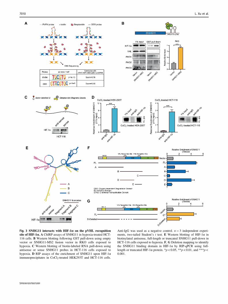
Fig. 3 SNHG11 interacts with HIF-1α on the pVHL recognitionsite of HIF-1α. A ChIRP assays of SNHG11 in hypoxia-treated HCT-116 cells. B Western blotting following GST pull-down using emptyvector or SNHG11-MS2 fusion vector in RKO cells exposed tohypoxia. C Western blotting of biotin-labeled RNA pull-down usingantisense or sense SNHG11 probes in HCT-116 cells exposed tohypoxia. D RIP assays of the enrichment of SNHG11 upon HIF-1αimmunoprecipitates in CoCl2-treated HEK293T and HCT-116 cells.
Anti-IgG was used as a negative control. n= 3 independent experi-ments, two-tailed Student’s t test. E Western blotting of HIF-1α inbiotinylated antisense, full-length or truncated SNHG11 pull-down inHCT-116 cells exposed to hypoxia. F, G Deletion mapping to identifythe SNHG11 binding domain in HIF-1α by RIP-qPCR using full-length or truncated HIF-1α protein. *p < 0.05, **p < 0.01, and ***p <0.001.
7010 L. Xu et al.

cycloheximide, an inhibitor of protein biosynthesis, afterculturing cells in hypoxia. Western blotting analysis showedthat SNHG11 overexpression increased the half-life of HIF-1α compared with that of the control cells (Fig. 4F andSupplementary Fig. 7F). Furthermore, the transcriptionalactivity of HIF-1α was also regulated by SNHG11 (Fig.4G), suggesting that SNHG11 regulated HIF-1α down-stream transcription events under hypoxia. These results
demonstrate that SNHG11 not only increases HIF-1α sta-bility by blocking pVHL recognition but also promotesHIF-1α transcriptional activity under hypoxic conditions.Taken together, SNHG11 inhibits the association of pVHLwith HIF-1α and alleviates pVHL-mediated ubiquitinationof HIF-1α, which causes HIF-1α accumulation.
Next, we selected several genes downstream of HIF-1αthat are related to metastasis [26–29] and determined their
LncRNA SNHG11 facilitates tumor metastasis by interacting with and stabilizing HIF-1α 7011

expression by quantitative polymerase chain reaction(qPCR). The results showed that SNHG11 could regulateAK4, ENO1, HK2, and Twist1 expression in CRC cells(Fig. 4H). Based on ChIRP data, we designed primers forHRE sites at these gene promoters and performed ChIRP-qPCR to validate the interaction between SNHG11 and thepromoters of these target genes. ChIRP-qPCR showed thatSNHG11 bound to HRE sites at the promoters of AK4,ENO1, HK2, and Twist1 (Fig. 4I). Konckdown of thesetarget genes by siRNAs could significantly inhibit themigration and invasion capability of CRC cells underhypoxia (Supplementary Fig. 8A, B). These findings pro-vide evidence that hypoxia-induced SNHG11 increasesHIF-1α stability and transcriptional activity by binding toHIF-1α and blocking the interaction between HIF-1αand pVHL.
The effect of SNHG11 on migration and invasion ismediated by HIF-1α in CRC cells
Next, we determined whether SNHG11 exerts its functionthough HIF-1α. We found that the promotion of SNHG11on migration and invasion was remarkably abolished whenwe silenced HIF-1α expression in SNHG11-overexpressingCRC cells (Fig. 5A, B). Consistent with these results,transfecting a HIF-1α plasmid into SNHG11 knockdowncells strikingly eliminated the suppressive influence causedby SNHG11 knockdown on CRC cell migration and inva-sion (Fig. 5C, D), suggesting that SNHG11-mediated HIF-1α upregulation contributes to the promotion of SNHG11
on CRC cell migration and invasion. Since hypoxia is acommon feature of many solid tumors, we explored whetherthe HIF-1α/SNHG11 axis functions in other cancers.Interestingly, TCGA data revealed that SNHG11 wasupregulated in cancers other than CRC (Supplementary Fig.9A). In addition, SNHG11-correlated genes in rectumadenocarcinoma, prostate adenocarcinoma, kidney renalpapillary cell carcinoma, kidney renal clear cell carcinoma,and breast invasive carcinoma were also enriched in theHIF-1 signaling pathway (Supplementary Fig. 9B). Thesedata suggest that the HIF-1α/SNHG11 axis might alsofunction in other cancers.
SNHG11 is upregulated in CRC and predicts anunfavorable prognosis for CRC patients
Next, we analyzed the expression and clinical significanceof SNHG11 in TCGA database. We found that SNHG11was frequently upregulated in COAD samples (Fig. 6A).CRC patients with lymphatic invasion had a higherSNHG11 than CRC patients without lymphatic invasion(Fig. 6A). Moreover, CRC patients with higher SNHG11levels had shorter survival times (Fig. 6B). Then, weexamined the expression level of SNHG11 in our inde-pendent cohorts 1 (n= 93) and 2 (n= 78) of CRC samples.Consistent with the results from TCGA data, SNHG11 wassignificantly upregulated in cohort 1 CRC tissues (Fig. 6C).Moreover, Kaplan–Meier analysis showed that patients withhigher SNHG11 expression were associated with reducedoverall survival time in cohort 1 CRC samples (Fig. 6C).SNHG11 was also upregulated in cohort 2 CRC samples(Fig. 6D). CRC with metastasis showed higher SNHG11expression than CRC samples without metastasis (Fig. 6Eand Supplementary Table 1). Intriguingly, patients withdistant recurrence also had higher SNHG11 levels thanpatients without distant recurrence (Fig. 6E and Supple-mentary Table 1). Collectively, these results revealed thatSNHG11 was upregulated in CRC and that high expressionlevels of SNHG11 were associated with poor outcomes inCRC patients.
Discussion
Most lncRNAs exert their function by binding to variousproteins. The cellular distribution of lncRNAs is informa-tive for lncRNA functions [30]. In this study, we analyzednuclear lncRNAs regulated by DNA methylation and foundthat SNHG11 regulated the stabilization and transcriptionalactivity of HIF-1α. Our results indicate that aberrant DNAmethylation also regulates the expression of lncRNAs,which can exert important function in CRC progression.Mechanistically, SNHG11 binds to HIF-1α and regulates
Fig. 4 SNHG11 stabilizes HIF-1α by blocking its interaction withpVHL and positively regulates HIF-1α target genes. A Immuno-precipitation of anti-HIF-1α in hypoxia- and MG132-treated HCT-116and RKO cells transfected with SNHG11. B The ubiquitination levelof HIF-1α in hypoxia- and MG132-treated HCT-116 and RKO cellstransfected with SNHG11. C The HIF-1α expression level in CRCcells transfected with siSNHG11 and exposed to hypoxia was analyzedby western blotting. D The HIF-1α expression level was analyzed bywestern blotting in CRC cells overexpressing SNHG11 and exposed tohypoxia. E Western blotting detected the expression levels of HIF-1αin the cytoplasm and nucleus of HCT-116 cells transfected withSNHG11 and exposed to hypoxia. F Control and SNHG11-overexpressing HCT-116 and LoVo cells were incubated underhypoxic conditions for 24 h and then were treated with CHX (10 μM)and were allowed to recover from hypoxia for the indicated times.Western blotting analysis was performed to determine the protein levelof HIF-1α. G Luciferase reporter assay analysis of HIF-1α transcrip-tion activity in CoCl2-treated CRC cells after overexpression orknockdown of SNHG11. n= 3 independent experiments, two-tailedStudent’s t test. H Expression levels of AK4, ENO1, HK2, and Twist1in hypoxia-treated CRC cells with SNHG11 overexpression orknockdown. n= 3 independent experiments, two-tailed Student’st test. I ChIRP-qPCR validated that SNHG11 interacted with HREsites at AK4, ENO1, HK2, and Twist1 locus in hypoxia-treated HCT-116 cells. n= 3 independent experiments, two-tailed Student’s t test.*p < 0.05, **p < 0.01, and ***p < 0.001.
7012 L. Xu et al.

HIF-1α stability. HIF-1α is upregulated in CRC [31] andexacerbates metastasis by regulating many downstreamtargets [32]. We discover that SNHG11 promotes theinvasion and metastasis of CRC cells. SNHG11 promotesCRC progression by regulating the transcription of HIF-1αdownstream targets.
The stabilization of HIF-1α is important for cells in theresponse to oxygen change. HIF-1α stabilization is tightlyregulated by PHD and pVHL. In addition to PHD andpVHL, HIF-1α stabilization can also be regulated by otherregulators, including noncoding RNAs. Reports of non-coding RNAs regulating HIF-1α have accumulated in recentyears. For example, miR-200b, miR-200c, and miR-429 areinduced by HIF-1α and bind to the 3′-UTR of PHD2mRNA [33]. Overexpression of miR-200 can upregulateHIF-1α protein by downregulating PHD2 protein. Recently,lncRNAs have also been found to be involved in the reg-ulation of HIF-1α stabilization. The cytoplasmic lncRNA
LINK-A facilitates the binding between BRK kinase andthe EGFR:GPNMB complex and enhances BRK kinaseactivation, which phosphorylates the HIF-1α protein andstabilizes HIF-1α under normoxic conditions [34].LncRNAs can also directly bind to HIF-1α protein to sta-bilize it. LincRNA-p21 disrupts the interaction of HIF-1αand pVHL and promotes hypoxia-induced glycolysis [35].In our study, we demonstrate that SNHG11 can stabilizeand accumulate HIF-1α. SNHG11 binds to the pVHLbinding site on HIF-1α and prevents the interaction of HIF-1α with pVHL in the nucleus of CRC cells. Groulx et al.reported that the degradation of HIF-1α can be initiated inthe nucleus by PHD prolyl hydroxylation and then can becaptured by pVHL [36]; thus, modified HIF-1α is exportedto the cytoplasm for proteasome-mediated degradation. Ourresults reveal that SNHG11 can promote nuclear HIF-1αaccumulation under hypoxic conditions. This is differentfrom lincRNA-p21, which stabilizes HIF-1α in the
Fig. 5 HIF-1α mediates SNHG11-induced cell migration and invasion. A–D Cell migration and invasion assays of hypoxia-treated HCT-116and LoVo cells with the indicated treatments. n= 3 independent experiments, two-tailed Student’s t test. *p < 0.05, **p < 0.01, and ***p < 0.001.
LncRNA SNHG11 facilitates tumor metastasis by interacting with and stabilizing HIF-1α 7013

cytoplasm [35]. Our results together with those from pre-vious studies indicate that the regulation of HIF-1α stabi-lization is complex on multiple layers through variousregulators, including noncoding RNAs.
In addition to stabilizing HIF-1α, SNHG11 alsoincreased the transcriptional activation of HIF-1α. SNHG11promotes the migration and invasion of CRC cells byinducing HIF-1α downstream targets. Shih et al. reportedthat lncRNA miR31HG can act as a HIF-1α coactivator to
drive oral cancer progression [37]. miR31HG is required forhypoxia-induced activation of HIF-1α downstream targets.However, the expression of miR31HG is downregulated inCRC samples [38]. Therefore, we propose that the regula-tion of HIF-1α by lncRNAs is tissue-dependent. HIF-1αactivation requires different lncRNAs in various tissues.
In conclusion, we demonstrate that lncRNA SNHG11promotes the stabilization and activation of HIF-1α in CRCcells. SNHG11 promotes CRC progression by reinforcing
Fig. 6 SNHG11 is upregulated in CRC tissues and predicts poorprognosis. A SNHG11 expression was increased in 41 paired COADpatients in TCGA (left), and the expression of SNHG11 is shown forCOAD patients with or without lymphatic invasion (right) two-tailedStudent’s t test. B Kaplan–Meier analysis of the overall survival curveof CRC patients in COAD-TCGA. C The expression and
Kaplan–Meier analysis of SNHG11 in 93 paired CRC tissue speci-mens (cohort 1). n= 93 CRC patients, two-tailed Student’s t test.D The expression of SNHG11 in CRC tissue specimens of cohort 2. n= 78 CRC patients and 71 matched adjacent normal tissues, two-tailedStudent’s t test. E The expression of SNHG11 in CRC patients with orwithout metastasis and distant recurrence, two-tailed Student’s t test.
7014 L. Xu et al.

the invasion and metastasis of CRC cells (Fig. 7). SNHG11could serve as a prognostic indicator and potential ther-apeutic target for CRC patients.
Materials and methods
Patients and specimens
CRC tissues and adjacent tissues (cohort 1 and cohort 2)were acquired from the surgical specimen archives of FudanUniversity Shanghai Cancer Center (Shanghai, China). Allstudies on human specimens were approved by the EthicsCommittee of Fudan University Shanghai Cancer Center.No statistical method was used to predetermine sample sizedue to the availability. Written informed consent wasobtained from each patient in accordance with institutionalguidelines. Cohort 1 included 93 CRC tissues with matchedadjacent normal tissues; Cohort 2 included 78 CRC tissueswith 71 matched adjacent normal tissues.
MS2-based GST pull-down experiments
First, we established RKO cells stably expressing MS2-GST as a fusion protein. SNHG11 was subcloned into alenti-sgRNA (MS2) vector and named SNHG11-MS2.SNHG11-MS2 or the empty vector was transfected intocells expressing the MS2-GST fusion protein. Cells werethen exposed to hypoxia. The experimental procedure wassimilar to that of the RIP protocol with minor modifications.Briefly, 50 μl of magneGST glutathione particles (Promega,WI, USA) was added to the cell lysate. Twenty percent ofthe beads were lysed in TRIzol, and 80% of the beads werelysed in SDS loading buffer for Western blotting.
RNA pull-down assays
Probes of sense, antisense, or truncated SNHG11 RNAswere transcribed and labeled with a Biotin RNA Labeling
Mix (Roche, IN, USA); then, they were treated with RNase-free DNase I (TaKaRa, Tokyo, Japan) and were purifiedwith a RNeasy Mini kit (Qiagen, Hilden, Germany). CRCcells treated with hypoxic conditions were lysed in RIPAbuffer supplemented with RNase and proteinase inhibitorsand then were incubated with equal moles of each bioti-nylated RNA at 4 °C for 4 h. The mixture was added to40 μl of prewashed streptavidin beads (Thermo Fisher Sci-entific, IL, USA) and was incubated on a rotator at roomtemperature for 1 h. The beads were washed briefly fivetimes with NT2 buffer and boiled in SDS loading buffer.The protein samples were separated by SDS-PAGE. Theprimers to generate sense, antisense, or truncatedSNHG11 sequences are listed in Supplementary Table 2.
RNA immunoprecipitation (RIP)
Cells were treated with CoCl2 for 24 h. RIP experimentswere performed with a Magna RIP RNA-Binding ProteinImmunoprecipitation kit (Millipore, MA, USA) accordingto the manufacturer’s instructions. Anti-IgG and the indi-cated antibodies were used to enrich RNAs. The copreci-pitated RNAs were detected by qPCR. For the RIP assays ofdeletion mutants of HIF-1α, equal plasmids with FLAG-tagged full-length or truncated HIF-1α were transientlytransfected into HEK293T cells grown in hypoxic condi-tions, and cell lysates were immunoprecipitated with a Flagantibody (Sigma-Aldrich, MO, USA).
In vivo assays
Six-week-old male NOD/SCID mice were housed in lami-nar flow cabinets under specific pathogen-free conditionswith food and water provided ad libitum. Sample size wasnot predetermined for these experiments. CRC cells (2 ×10⁶) were injected via lateral tail vein injections (n= 8 pergroup). After 8 weeks, the mice were sacrificed, and thelungs were immediately removed. The metastatic noduleswere counted after haematoxylin and eosin staining. Thisstudy was approved by the Animal Care and Use Com-mittee of Fudan University.
Chromatin isolation by RNA purification sequencing(ChIRP-seq)
ChIRP assays of SNHG11 in hypoxia-treated HCT-116cells were performed as previously described [39]. Briefly,we collected CoCl2-treated HCT-116 cells and hybridizedthe sonicated chromatin with biotinylated antisense probesets against SNHG11 or LacZ (negative control). Probeswere grouped into “ODD” and “EVEN” sets based on theirpositions along the RNA. Then, we purified DNA bound tothe probes and performed DNA sequencing. DNA
Fig. 7 Schematic model of SNHG11-HIF-1α signaling axis in CRC.Demethylated SNHG11 locus increases the expression of SNHG11,and in hypoxic CRC cells SNHG11 interacts with HIF-1α directly andblocks the interaction between HIF-1α and pVHL. As a result, theSNHG11/HIF-1α complex binds to the HRE sites of target genes topromote CRC cell invasion and metastasis.
LncRNA SNHG11 facilitates tumor metastasis by interacting with and stabilizing HIF-1α 7015

sequencing libraries were constructed from ChIRPed DNAusing an NEBNext Ultra II DNA Library Prep kit fromIllumina (New England Biolabs, MA, USA). The top 5000enriched ChIRP sites by EVEN probes or the top 5000enriched ChIRP sites by ODD probes were used to generatethe SNHG11 interacting motifs using the HOMER find-motifsgenome program. The motif length of the findmo-tifsgenome program was 6, 8, 10, and 12. ChIRP probes areshown in Supplementary Table 3. Peaks generated fromChIRP-seq are shown in Supplementary Table 4.
Quantitative real-time PCR (qPCR)
RNA was extracted using TRIzol (Invitrogen, CA, USA),and it was reverse transcribed using a PrimeScript RTReagent kit (TaKaRa, Tokyo, Japan). Real-time qPCRswere performed with SYBR Green Premix Ex Taq(TaKaRa, Tokyo, Japan). Relative RNA expression levelswere measured by an ABI system (Thermo Fisher Scien-tific, IL, USA). The sequences for the gene-specific primersused are listed in Supplementary Table 5. β-actin wasemployed as an internal control.
Subcellular fractionation
Cytoplasmic and nuclear fractions were separated by aPARIS cytoplasmic and nuclear extraction kit (Life Tech-nologies, MA, USA). β-actin and U2 were used as cyto-plasmic and nuclear positive controls, respectively.
5′ and 3′ RACE analysis
5′ and 3′ RACE was performed to determine the tran-scriptional initiation and termination sites of SNHG11 witha SMARTer RACE cDNA Amplification kit (Clontech, CA,USA) according to the manufacturer’s instructions. Thesequences for the gene-specific PCR primers used for 5′ and3′ RACE analysis are listed in Supplementary Table 6.
Oligonucleotide transfection
Negative control (siNC) and siRNAs were synthesized byBiotend (Shanghai, China). All siRNAs are shown in Sup-plementary Table 7. Cells were seeded in six-well plates. Thenext day, 5 μL of siRNAs and siNC (20 μM) were transfectedinto cells by Lipofectamine RNAiMAX (Invitrogen, CA,USA) according to the manufacturer’s instructions. Cells wereharvested for further experiments after 48 h.
Lentivirus production and infection
Full-length SNHG11 was generated by RACE experimentsand then was cloned into a pCDH-Puro lentiviral vector.
The ORF of HIF-1α was cloned into the pCDH-3×Flaglentiviral vector. The primers used to amplify the sequencesare listed in Supplementary Table 8. shRNA sequenceswere synthesized and subcloned into a LentiGuide-Purolentiviral vector. shRNA sequences are listed in Supple-mentary Table 8. To generate lentiviruses, pAX2 andpMD2.G were cotransfected into HEK293T cells usingLipofectamine 2000 (Invitrogen, CA, USA). After 48 h,lentiviruses were collected and used to infect CRC cells.
Western blotting
Proteins were separated by SDS-PAGE and then weretransferred to nitrocellulose membranes. The membraneswere blocked with 5% milk and probed with primary anti-bodies overnight at 4 °C. Immune complexes were detectedusing a LumiBest ECL Reagent Solution kit (Share-Bio,Shanghai, China) after being probed with HRP-conjugatedsecondary antibodies. The antibodies used in this study arelisted in Supplementary Table 9.
TCGA, ENCODE, and CCLE data
The expression and clinical data of SNHG11 in TCGA-COAD were obtained through the TCGA data portal. Illu-mina Human Methylation 450 platform-based beta value ofDNA methylation in TCGA-COAD was downloaded fromXena (https://xenabrowser.net/). To search the character-istics of the genomic locus, DNase-seq (ENCSR000ENM),H3K27Ac ChIP-seq (ENCSR661KMA), POLR2A ChIP-seq (ENCSR000EUU), POLR2AphosphoS5 ChIP-seq(ENCSR000BML) of HCT-116 cells and H3K27AcChIP-seq (ENCSR561YSH), POLR2A ChIP-seq(ENCSR724FCJ) of sigmoid colon were obtained fromENCODE (https://www.encodeproject.org/). DNA methy-lation status across CRC cell lines was obtained fromCancer Cell Line Encyclopedia (CCLE) (https://portals.broadinstitute.org/ccle/).
Correlations of each genes with SNHG11 in TCGA-COAD were generated from circlncRNAnet (app.cgu.edu.tw/circlnc/circlncRNAnet/) (Supplementary Table 10).GSEA was conducted by the preranked method using theclusterProfiler package in R (https://www.r-project.org/).C2 (curated genesets), C5 (GO genesets), C6 (oncogenicsignatures), and hallmark genesets from MSigDB (Mole-cular Signatures Database) were analyzed.
Statistical analysis
Data are presented as the mean ± standard error of the mean(SEM) and Student’s t test or χ2-test was analyzed usingSPSS 19.0, GraphPad Prism 5, and R project (https://www.r-project.org/). Manuscript investigators were not blinded
7016 L. Xu et al.

during the experiments. But there was no human bias givenall data performed at least three experiments. Student’s t testwas performed to evaluate the differences between twogroups, followed by F-test to estimate the variation. TheKaplan–Meier method was performed to analyze the cor-relation between SNHG11 levels and overall survival.Differences with p < 0.05 were considered statistically sig-nificant (*p < 0.05, **p < 0.01, and ***p < 0.001).
Data availability
The authors declare that all relevant data of this study areavailable within the article or from the corresponding authoron reasonable request.
Funding This work was supported by grants from National NaturalScience Foundation of China (81672366, 81930123, 81790252) andChina Postdoctoral Science Foundation (2020M671006).
Author contributions XH and LL designed the project. LX, LH, YW,QW, and YL performed the experiments. LH, LL, and SH processedthe data. LL, LH, and XH wrote the manuscript. TG and YX collectedthe CRC samples and clinical information.
Compliance with ethical standards
Conflict of interest The authors declare that they have no conflict ofinterest.
Publisher’s note Springer Nature remains neutral with regard tojurisdictional claims in published maps and institutional affiliations.
Open Access This article is licensed under a Creative CommonsAttribution 4.0 International License, which permits use, sharing,adaptation, distribution and reproduction in any medium or format, aslong as you give appropriate credit to the original author(s) and thesource, provide a link to the Creative Commons license, and indicate ifchanges were made. The images or other third party material in thisarticle are included in the article’s Creative Commons license, unlessindicated otherwise in a credit line to the material. If material is notincluded in the article’s Creative Commons license and your intendeduse is not permitted by statutory regulation or exceeds the permitteduse, you will need to obtain permission directly from the copyrightholder. To view a copy of this license, visit http://creativecommons.org/licenses/by/4.0/.
References
1. Athauda A, Segelov E, Ali Z, Chau I. Integrative molecularanalysis of colorectal cancer and gastric cancer: what have welearnt? Cancer Treat Rev. 2019;73:31–40.
2. Toyota M, Ahuja N, Ohe-Toyota M, Herman JG, Baylin SB, IssaJP. CpG island methylator phenotype in colorectal cancer. ProcNatl Acad Sci USA. 1999;96:8681–6.
3. Moinova HR, Chen WD, Shen L, Smiraglia D, Olechnowicz J,Ravi L, et al. HLTF gene silencing in human colon cancer. ProcNatl Acad Sci USA. 2002;99:4562–7.
4. Shi L, Hong X, Ba L, He X, Xiong Y, Ding Q, et al. Long non-coding RNA ZNFX1-AS1 promotes the tumor progression andmetastasis of colorectal cancer by acting as a competing
endogenous RNA of miR-144 to regulate EZH2 expression. CellDeath Dis. 2019;10:150.
5. Neve B, Jonckheere N, Vincent A, Van Seuningen I. Epigeneticregulation by lncRNAs: an overview focused on UCA1 in col-orectal cancer. Cancers. 2018;10:440.
6. Sun Y, Ma L. New insights into long non-coding RNA MALAT1in cancer and metastasis. Cancers. 2019;11:216.
7. Zhao W, Ma X, Liu L, Chen Q, Liu Z, Zhang Z, et al. SNHG20: avital lncRNA in multiple human cancers. J Cell Physiol.2019;234:14519–525.
8. Bhan A, Soleimani M, Mandal SS. Long noncoding RNA andcancer: a new paradigm. Cancer Res. 2017;77:3965–81.
9. Han P, Li JW, Zhang BM, Lv JC, Li YM, Gu XY, et al. ThelncRNA CRNDE promotes colorectal cancer cell proliferation andchemoresistance via miR-181a-5p-mediated regulation of Wnt/beta-catenin signaling. Mol Cancer. 2017;16:9.
10. Huan L, Guo T, Wu Y, Xu L, Huang S, Xu Y, et al. Hypoxiainduced LUCAT1/PTBP1 axis modulates cancer cell viability andchemotherapy response. Mol Cancer. 2020;19:11.
11. Kondo Y, Shinjo K, Katsushima K. Long non-coding RNAs as anepigenetic regulator in human cancers. Cancer Sci.2017;108:1927–33.
12. Han P, Chang CP. Long non-coding RNA and chromatin remo-deling. RNA Biol. 2015;12:1094–8.
13. Pefanis E, Wang J, Rothschild G, Lim J, Kazadi D, Sun J, et al.RNA exosome-regulated long non-coding RNA transcriptioncontrols super-enhancer activity. Cell. 2015;161:774–89.
14. Schmitt AM, Chang HY. Long noncoding RNAs in cancerpathways. Cancer Cell. 2016;29:452–63.
15. Sigova AA, Abraham BJ, Ji X, Molinie B, Hannett NM, Guo YE,et al. Transcription factor trapping by RNA in gene regulatoryelements. Science. 2015;350:978–81.
16. Hou Y, Zhang R, Sun X. Enhancer LncRNAs influence chromatininteractions in different ways. Front Genet. 2019;10:936.
17. Saldana-Meyer R, Rodriguez-Hernaez J, Escobar T, Nishana M,Jacome-Lopez K, Nora EP, et al. RNA interactions are essentialfor CTCF-mediated genome organization. Mol Cell.2019;76:412–22.e5.
18. Li Z, Tan H, Yu H, Deng Z, Zhou X, Wang M. DNA methylationand gene expression profiles characterize epigenetic regulation oflncRNAs in colon adenocarcinoma. J Cell Biochem.2020;121:2406–15.
19. Gudenas BL, Wang L. Prediction of LncRNA subcellular locali-zation with deep learning from sequence features. Sci Rep.2018;8:16385.
20. Mas-Ponte D, Carlevaro-Fita J, Palumbo E, Hermoso Pulido T,Guigo R, Johnson R. LncATLAS database for subcellular loca-lization of long noncoding RNAs. RNA. 2017;23:1080–7.
21. Wang GL, Jiang BH, Rue EA, Semenza GL. Hypoxia-induciblefactor 1 is a basic-helix-loop-helix-PAS heterodimer regulated bycellular O2 tension. Proc Natl Acad Sci USA. 1995;92:5510–4.
22. Gao S, Lu L, Bai Y, Zhang P, Song W, Duan C. Structural andfunctional analysis of amphioxus HIFalpha reveals ancient fea-tures of the HIFalpha family. FASEB J. 2014;28:1880–90.
23. Huang LE, Gu J, Schau M, Bunn HF. Regulation of hypoxia-inducible factor 1alpha is mediated by an O2-dependent degra-dation domain via the ubiquitin-proteasome pathway. Proc NatlAcad Sci USA. 1998;95:7987–92.
24. Masson N, Willam C, Maxwell PH, Pugh CW, Ratcliffe PJ.Independent function of two destruction domains in hypoxia-inducible factor-alpha chains activated by prolyl hydroxylation.EMBO J. 2001;20:5197–206.
25. Tanimoto K, Makino Y, Pereira T, Poellinger L. Mechanism ofregulation of the hypoxia-inducible factor-1 alpha by the vonHippel-Lindau tumor suppressor protein. EMBO J.2000;19:4298–309.
LncRNA SNHG11 facilitates tumor metastasis by interacting with and stabilizing HIF-1α 7017

26. Dengler VL, Galbraith M, Espinosa JM. Transcriptional regula-tion by hypoxia inducible factors. Crit Rev Biochem Mol Biol.2014;49:1–15.
27. Jan YH, Lai TC, Yang CJ, Lin YF, Huang MS, Hsiao M. Ade-nylate kinase 4 modulates oxidative stress and stabilizes HIF-1alpha to drive lung adenocarcinoma metastasis. J Hematol Oncol.2019;12:12.
28. Zhan P, Zhao S, Yan H, Yin C, Xiao Y, Wang Y, et al. alpha-enolase promotes tumorigenesis and metastasis via regulatingAMPK/mTOR pathway in colorectal cancer. Mol Carcinog.2017;56:1427–37.
29. Eckert MA, Lwin TM, Chang AT, Kim J, Danis E, Ohno-Machado L, et al. Twist1-induced invadopodia formation pro-motes tumor metastasis. Cancer Cell. 2011;19:372–86.
30. Chen LL. Linking long noncoding RNA localization and function.Trends Biochem Sci. 2016;41:761–72.
31. Kaidi A, Qualtrough D, Williams AC, Paraskeva C. Direct tran-scriptional up-regulation of cyclooxygenase-2 by hypoxia-inducible factor (HIF)-1 promotes colorectal tumor cell survivaland enhances HIF-1 transcriptional activity during hypoxia.Cancer Res. 2006;66:6683–91.
32. Krishnamachary B, Berg-Dixon S, Kelly B, Agani F, Feldser D,Ferreira G, et al. Regulation of colon carcinoma cell invasion byhypoxia-inducible factor 1. Cancer Res. 2003;63:1138–43.
33. Lee ST, Chu K, Jung KH, Yoon HJ, Jeon D, Kang KM, et al.MicroRNAs induced during ischemic preconditioning. Stroke.2010;41:1646–51.
34. Lin A, Li C, Xing Z, Hu Q, Liang K, Han L, et al. The LINK-AlncRNA activates normoxic HIF1alpha signalling in triple-negative breast cancer. Nat Cell Biol. 2016;18:213–24.
35. Yang F, Zhang H, Mei Y, Wu M. Reciprocal regulation of HIF-1alpha and lincRNA-p21 modulates the Warburg effect. Mol Cell.2014;53:88–100.
36. Groulx I, Lee S. Oxygen-dependent ubiquitination and degrada-tion of hypoxia-inducible factor requires nuclear-cytoplasmictrafficking of the von Hippel-Lindau tumor suppressor protein.Mol Cell Biol. 2002;22:5319–36.
37. Shih JW, Chiang WF, Wu ATH, Wu MH, Wang LY, Yu YL,et al. Long noncoding RNA LncHIFCAR/MIR31HG is a HIF-1alpha co-activator driving oral cancer progression. Nat Commun.2017;8:15874.
38. Yan S, Tang Z, Chen K, Liu Y, Yu G, Chen Q, et al. Longnoncoding RNA MIR31HG inhibits hepatocellular carcinomaproliferation and metastasis by sponging microRNA-575to modulate ST7L expression. J Exp Clin Cancer Res.2018;37:214.
39. Chu C, Quinn J, Chang HY. Chromatin isolation by RNA pur-ification (ChIRP). J Vis Exp. 2012;61:3912.
7018 L. Xu et al.
Copyright © 2022 FDOKUMEN