Institution facilitates building and sustenance of innate - Dr ...
© 2014 Gao et al. This work is published by Dove Medical Press Limited, and licensed under Creative Commons Attribution – Non Commercial (unported, v3.0) License. The full terms of the License are available at http://creativecommons.org/licenses/by-nc/3.0/. Non-commercial uses of the work are permitted without any further
permission from Dove Medical Press Limited, provided the work is properly attributed. Permissions beyond the scope of the License are administered by Dove Medical Press Limited. Information on how to request permission may be found at: http://www.dovepress.com/permissions.php
Neuropsychiatric Disease and Treatment 2014:10 1489–1495
Neuropsychiatric Disease and Treatment Dovepress
submit your manuscript | www.dovepress.com
Dovepress 1489
O r i g i N a l r e s e a r c h
open access to scientific and medical research
Open access Full Text article
http://dx.doi.org/10.2147/NDT.S65695
correspondence: Wen JiangDepartment of Neurology, Xijing hospital, Fourth Military Medical University, 17 changle West road, Xi'an 710032, People’s republic of chinaTel +86 29 8477 1319Fax +86 29 8255 1806email [email protected]
Wei rencollege of life sciences, shaanxi Normal University, 199 changan south road, Xi’an 710062, People’s republic of china Tel +86 29 8531 0268Fax +86 29 8531 0269email: [email protected]
acute lipopolysaccharide exposure facilitates epileptiform activity via enhanced excitatory synaptic transmission and neuronal excitability in vitro
Fei gao1,2
Zhiqiang liu3
Wei ren3
Wen Jiang1
1Department of Neurology, Xijing hospital, Fourth Military Medical University, Xi’an 710032, People’s republic of china; 2Department of Neurology, First affiliated hospital of Xi'an Medical University, Xi'an 710077, People’s republic of china; 3college of life sciences, shaanxi Normal University, Xi’an 710062, People’s republic of china
Journal name: Neuropsychiatric Disease and TreatmentJournal Designation: Original ResearchYear: 2014Volume: 10Running head verso: Gao et alRunning head recto: Lipopolysaccharide exposure facilitates epileptiform activityDOI: http://dx.doi.org/10.2147/NDT.S65695
Abstract: Growing evidence indicates brain inflammation has been involved in the genesis
of seizures. However, the direct effect of acute inflammation on neuronal circuits is not well
known. Lipopolysaccharide (LPS) has been used extensively to stimulate brain inflammatory
responses both in vivo and in vitro. Here, we observed the contribution of inflammation induced
by 10 μg/mL LPS to the excitability of neuronal circuits in acute hippocampal slices. When
slices were incubated with LPS for 30 minutes, significant increased concentration of tumor
necrosis factor α and interleukin 1β were detected by enzyme-linked immunosorbent assay.
In electrophysiological recordings, we found that frequency of epileptiform discharges and
spikes per burst increased 30 minutes after LPS application. LPS enhanced evoked excitatory
postsynaptic currents but did not modify evoked inhibitory postsynaptic currents. In addition,
exposure to LPS enhanced the excitability of CA1 pyramidal neurons, as demonstrated by a
decrease in rheobase and an increase in action potential frequency elicited by depolarizing
current injection. Our observations suggest that acute inflammation induced by LPS facilitates
epileptiform activity in vitro and that enhancement of excitatory synaptic transmission and
neuronal excitability may contribute to this facilitation. These results may provide new clues
for treating seizures associated with brain inflammatory disease.
Keywords: lipopolysaccharide, hippocampus, inflammation, epileptiform activity, synaptic
transmission, neuronal excitability
IntroductionBrain injuries such as trauma, stroke, and infection are often associated with acute
occurrence of seizures.1,2 Although the underlying mechanism remains unclear, accu-
mulating clinical and experimental evidence has suggested that inflammatory processes
involved in these injuries may contribute to the genesis of seizures.3 Traditionally, the
brain has been considered an immunoprivileged organ because of the presence of the
blood–brain barrier and the lack of a conventional lymphatic system. Nevertheless,
both the innate and adaptive immune responses are readily evoked within the brain after
varied injuries. Resident cells in brain parenchyma such as microglia, astrocytes, and
neurons can respond to these stimuli and create inflammatory molecules. In addition,
peripheral immune cells extravasating from cerebral vascular can also produce inflam-
matory media and aggravate inflammatory surroundings.4 These inflammatory media-
tors, including tumor necrosis factor α (TNF-α), interleukin 1β (IL-1β), nitric oxide
(NO), and reactive oxygen species, are reported to increase cellular excitability.5–7
In animal experiments, lipopolysaccharide (LPS), a major component of the outer
membrane of gram-negative bacteria, has been used extensively in investigating
mechanisms of brain inflammation both in vivo and in vitro. Mainly in microglia,8 by
Neuropsychiatric Disease and Treatment 2014:10submit your manuscript | www.dovepress.com
Dovepress
Dovepress
1490
gao et al
the stimulation of toll-like receptor 4 LPS results in the induc-
tion of transcriptional factors such as nuclear factor κB, which
trigger various proinflammatory genes such as those encoding
cytokines, chemokines, proteins of the complement system,
and inducible nitric oxide.9,10 Furthermore, LPS induces a
rapid glutamate (a major excitatory neurotransmitter in brain)
release in rat cortex slices, which may lead to an imbalance
between excitation and inhibition in the neuronal circuit.11
In vivo, previous studies showed that LPS intraperitoneal or
intracerebroventricular injection enhanced seizure susceptibil-
ity through increasing IL-1β, cyclooxygenase 2, NO, or pros-
taglandins in different epilepsy models.12–15 However, little is
known about the direct effects of the acute application of LPS
on epileptiform activity, synaptic strength, and neuronal excit-
ability in vitro and the possible underlying mechanisms.
In the electrophysiology of seizure, an imbalance between
excitation and inhibition is thought to mediate seizure activ-
ity. Increased excitation and/or decreased inhibition can
induce the initiation of a seizure. Neuronal excitability and
synaptic transmission are both involved in maintaining this
balance.16,17 Therefore, in the present study, we focused on
the effects of acute inflammation induced by LPS on the
epileptiform discharges, neuronal excitability, and synaptic
transmission in hippocampal slices.
Materials and methodsslices preparationAll procedures used were in accordance with the National
Institutes of Health Guide for the Care and Use of Laboratory
Animals and were approved by the Fourth Military Medical
University Animal Care Committee. Sprague-Dawley rats
(14–17 days old) were anesthetized with chloral hydrate
(400 mg/kg intraperitoneally) and decapitated. Brain was
removed rapidly, and transverse 400 μM hippocampal slices
were obtained with a Vibratome 1,000 plus (Vibratome
Company, St Louis, MO, USA) in ice-cold oxygenated
(95% O2/5% CO
2) artificial cerebrospinal fluid (ACSF)
containing (in mM) 125 NaCl, 3 KCl, 2.4 CaCl2, 1.2 MgCl
2,
26 NaHCO3, 1.25 NaH
2PO
4, and 10 glucose at pH 7.4 when
gassed with 95% O2 and 5% CO
2. In Mg2+-free ACSF, MgCl
2
was omitted, and the concentration of NaCl was adjusted to
126 mM for stable osmolarity. Slices were transferred to a
holding chamber and incubated in oxygenated ACSF for at
least 1 hour before electrophysiological recording.
All chemicals, unless otherwise stated, were obtained
from Sigma-Aldrich Co. (St Louis, MO, USA). LPS
(Escherichia coli serotype O55:B5) was prepared in
18 MΩ water and added to ACSF as needed. The final
concentration of LPS used was 10 μg/mL. This concentra-
tion was chosen because it was previously used in acute
brain hippocampal slices in vitro.18
enzyme-linked immunosorbent assayAfter being incubated in oxygenated ACSF for 1 hour,
slices were transferred to 12 multiwell plates and treated
with 10 μg/mL LPS for 30 minutes. The treated slices and
untreated control slices were homogenized and centrifuged
at 8,000 rpm for 15 minutes at 4°C. The supernatant was
collected for subsequent analysis of cytokine concentration.
Commercially available enzyme-linked immunosorbent
assay kits for IL-1β and TNF-α were used according to the
manufacturer’s instructions (Westang Bio-tech Co, Ltd,
Shanghai, People’s Republic of China).
electrophysiological recordingsSlices were transferred to an immersion recording chamber
and perfused with oxygenated ACSF continuously (flow rate,
1–2 mL/minute). Patch electrodes were pulled by a electrode
puller (model P-97; Sutter Instrument Company, Novato,
CA, USA) and had a resistance of 3–5 MΩ when filled with
the internal solution that contained (in mM) 120 K-gluconate,
20 KCl, 0.2 ethylene glycol tetraacetic acid, 10 HEPES (N-
2-hydroxyethylpiperazine-N-ethane-sulphonicacid), 2 MgCl2,
4 Na2-ATP (adenosine triphosphate), 0.3 Tris-GTP (guanos-
ine triphosphte), and 7 phosphocreatine at pH adjusted to
7.2–7.3 with KOH. Whole-cell recordings of hippocampal
CA1 pyramidal neurons in the current- or voltage-clamp
modes were performed with a Multi-Clamp 700B amplifier
(Axon Instruments, Union City, CA, USA) in all electro-
physiological experiments. Epileptiform discharges were
induced with 100 μM 4-aminopyridine (4-AP) added to
Mg2+-free ACSF. Synaptic responses were evoked by Schaf-
fer collaterals stimulation through a pair of Elgiloy electrodes
(Elgin Specialty Metals, Elgin, ILL, USA) placed in the
stratum radiatum near the border of the CA1 pyramidal layer.
Stimuli were single delivered at 0.033 Hz via an isolated pulse
stimulator (model 2100; AM System Inc, Billerica, MA,
USA), and stimulation intensity (0.1 ms, 50–200 μA) was
adjusted to produce evoked excitatory postsynaptic current
(eEPSC) or evoked inhibitory postsynaptic current (eIPSC)
amplitudes that were 60%–70% the maximal responses in
voltage-clamp experiments. Membrane potential was held
at −70 mV for recording eEPSCs and −40 mV for eIPSCs.
For eIPSC recording, glutamatergic α-amino-3-hydroxy-
5-methyl-4-isoxazolepropionic acid and N-methyl-D- aspartate
receptor antagonists (6,7-dinitroquinoxaline-2,3-dione,
Neuropsychiatric Disease and Treatment 2014:10 submit your manuscript | www.dovepress.com
Dovepress
Dovepress
1491
lipopolysaccharide exposure facilitates epileptiform activity
20 μM; aminophosphonovaleric acid, 50 μM) were added to
ACSF; for eEPSCs recording, γ-aminobutyric acid type A
receptor antagonist (Pierotoxin, 100 μM) was added. Action
potential frequency was calculated in response to a series of
current steps 1 second in duration (10 pA per step, 0–200 pA).
Neuron was rejected if the resting membrane potential was
more positive than −60 mV or if action potential amplitude
was less than 60 mV. Data were filtered at 2 KHz and trans-
ferred to the hard disk of a Dell computer (Round Rock,
TX, USA). The software pCLAMP 10.0 (Axon Instruments,
Union City, CA, USA) was used for data analysis offline.
statisticsSPSS 16.0 (IBM Inc, Chicago, IL, USA) for Windows was
used for statistical analysis. Data were expressed as mean ±
standard deviation. Statistical significance was assessed using
paired-sample Student’s t-test or two-factor (current and treat-
ment) analysis of variance for electrophysiological recordings
and independent-samples Student’s t-test for cytokine concen-
tration. P0.05 was considered statistically significant.
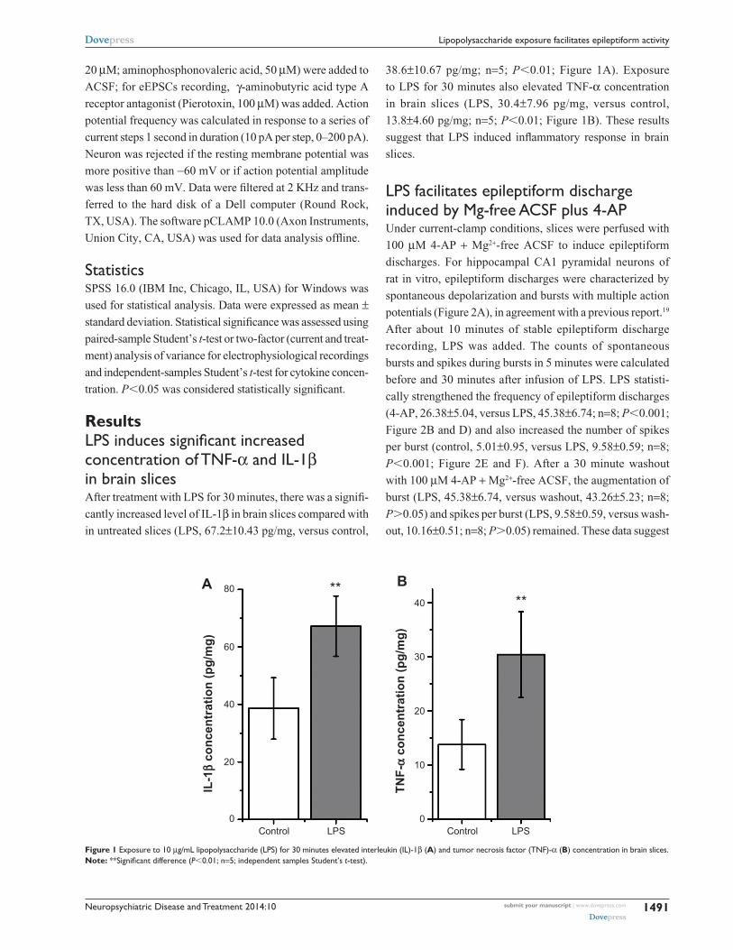
ResultsLPS induces significant increased concentration of TNF-α and il-1β in brain slicesAfter treatment with LPS for 30 minutes, there was a signifi-
cantly increased level of IL-1β in brain slices compared with
in untreated slices (LPS, 67.2±10.43 pg/mg, versus control,
38.6±10.67 pg/mg; n=5; P0.01; Figure 1A). Exposure
to LPS for 30 minutes also elevated TNF-α concentration
in brain slices (LPS, 30.4±7.96 pg/mg, versus control,
13.8±4.60 pg/mg; n=5; P0.01; Figure 1B). These results
suggest that LPS induced inflammatory response in brain
slices.
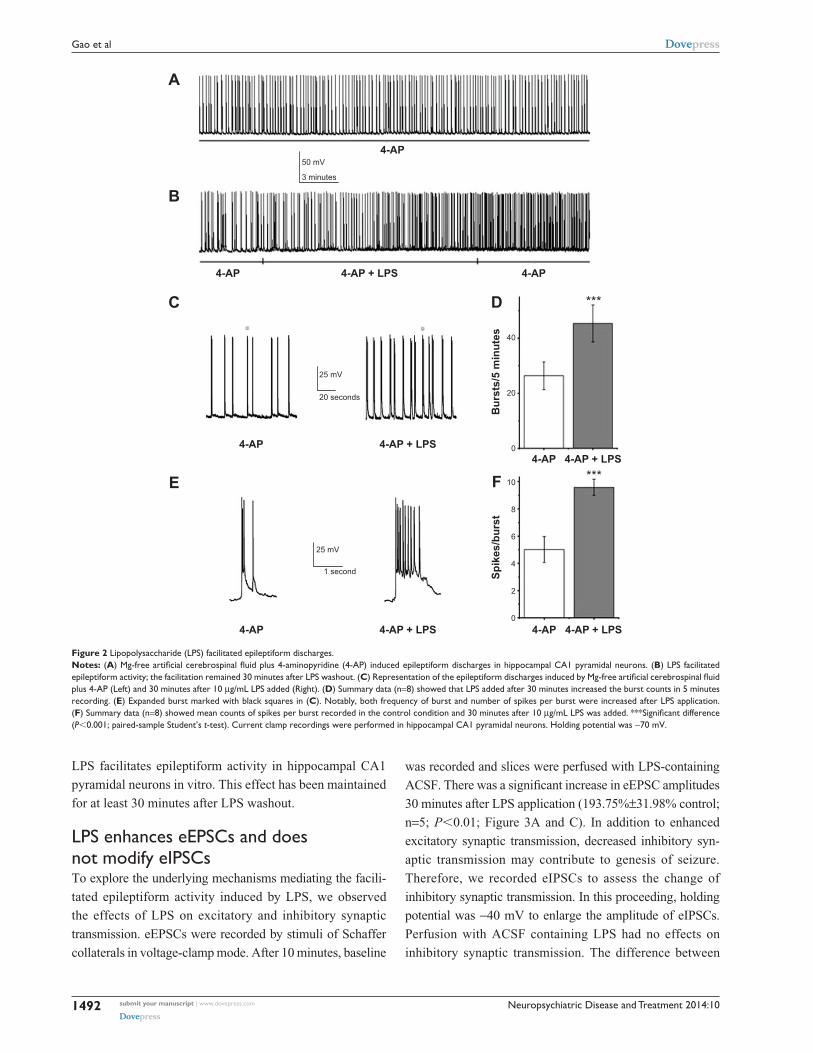
lPs facilitates epileptiform discharge induced by Mg-free acsF plus 4-aP Under current-clamp conditions, slices were perfused with
100 μM 4-AP + Mg2+-free ACSF to induce epileptiform
discharges. For hippocampal CA1 pyramidal neurons of
rat in vitro, epileptiform discharges were characterized by
spontaneous depolarization and bursts with multiple action
potentials (Figure 2A), in agreement with a previous report.19
After about 10 minutes of stable epileptiform discharge
recording, LPS was added. The counts of spontaneous
bursts and spikes during bursts in 5 minutes were calculated
before and 30 minutes after infusion of LPS. LPS statisti-
cally strengthened the frequency of epileptiform discharges
(4-AP, 26.38±5.04, versus LPS, 45.38±6.74; n=8; P0.001;
Figure 2B and D) and also increased the number of spikes
per burst (control, 5.01±0.95, versus LPS, 9.58±0.59; n=8;
P0.001; Figure 2E and F). After a 30 minute washout
with 100 μM 4-AP + Mg2+-free ACSF, the augmentation of
burst (LPS, 45.38±6.74, versus washout, 43.26±5.23; n=8;
P0.05) and spikes per burst (LPS, 9.58±0.59, versus wash-
out, 10.16±0.51; n=8; P0.05) remained. These data suggest
A
IL-1
β co
ncen
trat
ion
(pg/
mg)
Control0
20
40
60
80
LPS
** B
TNF-
α co
ncen
trat
ion
(pg/
mg)
Control
40
30
20
10
0LPS
**
Figure 1 exposure to 10 μg/ml lipopolysaccharide (lPs) for 30 minutes elevated interleukin (il)-1β (A) and tumor necrosis factor (TNF)-α (B) concentration in brain slices.Note: **Significant difference (P0.01; n=5; independent samples student’s t-test).
Neuropsychiatric Disease and Treatment 2014:10submit your manuscript | www.dovepress.com
Dovepress
Dovepress
1492
gao et al
LPS facilitates epileptiform activity in hippocampal CA1
pyramidal neurons in vitro. This effect has been maintained
for at least 30 minutes after LPS washout.
lPs enhances eePscs and does not modify eiPscsTo explore the underlying mechanisms mediating the facili-
tated epileptiform activity induced by LPS, we observed
the effects of LPS on excitatory and inhibitory synaptic
transmission. eEPSCs were recorded by stimuli of Schaffer
collaterals in voltage-clamp mode. After 10 minutes, baseline
was recorded and slices were perfused with LPS-containing
ACSF. There was a significant increase in eEPSC amplitudes
30 minutes after LPS application (193.75%±31.98% control;
n=5; P0.01; Figure 3A and C). In addition to enhanced
excitatory synaptic transmission, decreased inhibitory syn-
aptic transmission may contribute to genesis of seizure.
Therefore, we recorded eIPSCs to assess the change of
inhibitory synaptic transmission. In this proceeding, holding
potential was −40 mV to enlarge the amplitude of eIPSCs.
Perfusion with ACSF containing LPS had no effects on
inhibitory synaptic transmission. The difference between
A
B
C D
E F
4-AP
4-AP + LPS
4-AP + LPS
4-AP + LPS 4-AP + LPS4-AP
4-AP + LPS4-AP
4-AP4-AP
4-AP
4-AP
25 mV
25 mV
50 mV
Bur
sts/
5 m
inut
esSp
ikes
/bur
st
20 seconds
3 minutes
1 second
40
10
8
6
4
2
0
20
0
***
***
Figure 2 lipopolysaccharide (lPs) facilitated epileptiform discharges.Notes: (A) Mg-free artificial cerebrospinal fluid plus 4-aminopyridine (4-AP) induced epileptiform discharges in hippocampal CA1 pyramidal neurons. (B) lPs facilitated epileptiform activity; the facilitation remained 30 minutes after lPs washout. (C) Representation of the epileptiform discharges induced by Mg-free artificial cerebrospinal fluid plus 4-aP (left) and 30 minutes after 10 μg/ml lPs added (right). (D) summary data (n=8) showed that lPs added after 30 minutes increased the burst counts in 5 minutes recording. (E) expanded burst marked with black squares in (C). Notably, both frequency of burst and number of spikes per burst were increased after lPs application. (F) summary data (n=8) showed mean counts of spikes per burst recorded in the control condition and 30 minutes after 10 μg/mL LPS was added. ***Significant difference (P0.001; paired-sample student’s t-test). current clamp recordings were performed in hippocampal ca1 pyramidal neurons. holding potential was −70 mV.

Neuropsychiatric Disease and Treatment 2014:10 submit your manuscript | www.dovepress.com
Dovepress
Dovepress
1493
lipopolysaccharide exposure facilitates epileptiform activity
eIPSC amplitudes in control slices and slices bathed with
ACSF con taining LPS for 30 minutes was not significant
(103.38%±10.87% control; n=5; P0.05; Figure 3B and D).
lPs enhances the excitability of hippocampal ca1 pyramidal neurons In addition to synaptic mechanisms, the facilitated epileptiform
discharges by LPS may also be attributed to the increased
excitability of hippocampal CA1 pyramidal neurons. To
address this issue, we compared the responses evoked by
incremental strength of injected depolarizing currents in
ACSF with those elicited by identical currents in ACSF
containing LPS. After a 30 minute application of LPS, the
frequency of action potential was increased when injected
currents were 90–200 pA (Figure 4A and B). LPS significantly
increased action potential frequency compared with control
(F[1, 14]=54.35; P0.001; two-way analysis of variance;
Figure 4B). There was a significantly decreased rheobase
current amplitude (control, 100±17.73 pA, versus LPS,
78.75±25.88 pA; P0.05; n=8; paired-sample Student’s t-test)
after LPS was added (Figure 4C). LPS did not change the ampli-
tude (control, 81.09±9.21 mV, versus LPS, 82.45±5.12 mV;
P0.05; n=8; paired-sample Student’s t-test) or half-width of
the first action potential (control, 1.70±0.17 ms, versus LPS,
1.62±0.28 ms; P0.05; n=8; paired-sample Student’s t-test)
evoked by rheobase current (Figure 4D and E).
DiscussionOur data indicate that acute administration of LPS induces
increased concentration of TNF-α and IL-1β and facilitates
the epileptiform discharges in brain slices in vitro. Exposure
to LPS enhanced eEPSC and neuronal excitability but did
not modify eIPSC.
A B
DC
eEPSC eIPSCControl
Control0
100
200 100
50
0ControlLPS LPS
LPS
eEPS
C (%
)
eIPS
C (%
)**
100 pA
10 ms
Figure 3 lipopolysaccharide (lPs) enhanced evoked excitatory postsynaptic currents (eePscs) but did not modify evoked inhibitory postsynaptic currents (eiPscs) in hippocampal ca1 pyramidal neurons.Notes: representative traces of eePsc (A) and eiPsc (B) recorded in artificial cerebrospinal fluid (black lines) and 30 minutes after lipopolysaccharide was added (gray lines) under voltage clamp. summary data depicted mean eePsc (C) and eiPsc (D) peak amplitudes in control conditions and in the presence of lipopolysaccharide (n=5). **Significant difference (P0.01; paired-sample student’s t-test). holding potential was −70 mV for recording eePscs and −40 mV for eiPscs.Abbreviation: ms, millisecond.
Figure 4 lipopolysaccharide (lPs) enhanced excitability of hippocampal ca1 pyramidal neurons.Notes: (A) representative traces showed neuronal responses to a 190 pa depolar-izing current for 1 second in control artificial cerebrospinal fluid (Left) and 30 minutes after lPs was added (right). Note the enhanced neuronal excitability in the presence of lPs. (B) graph of action potential (aP) frequency shown in mean ± standard deviation for control condition and lPs exposure. The mean action potential frequency significantly increased after LPS exposure. *,#Significant differences (P0.05 and P0.01, respectively) compared lPs with control conditions. (C) reduction of rheobase was significant 30 minutes after LPS application (P0.05; n=8; paired-sample student’s t-test). (D) Representative traces showed the first action potential evoked by rheobase current in control artificial cerebrospinal fluid and 30 minutes after LPS was added. (E) Summary data showed there were not significant differences in amplitude and half-width of the first action potential under the rheobase current injection (both P0.05; n=8; paired-sample student’s t-test). holding potential was −70 mV in these processes.Abbreviation: ms, millisecond.
A
B C
D E
Control
Freq
uenc
y of
APs
(Hz)
500 ms
LPS
25 mV
Current (pA)Control
Control
LPS
LPS
2 ms20 mV
ControlControl
0 0
1
2
20
40
60
80
100
50
0
LPSLPS Control LPS
Am
plitu
de o
f 1st
AP
(mV)
Rhe
obas
e (p
A)
Hal
f-wid
th o
f 1st
AP
(ms)
0
50 100 150 200
5
10
15 *

Neuropsychiatric Disease and Treatment 2014:10submit your manuscript | www.dovepress.com
Dovepress
Dovepress
1494
gao et al
The important role of inflammatory processes in epilepsy
is increasingly recognized. In the previous studies in vivo, it
has been found that injection of LPS increases seizure suscep-
tibility in different epileptic models.13–15 In addition, cortex
neuronal excitability increases when LPS is directly applied to
rat cortex.20 Here, our data suggest that acute administration of
LPS induced brain slice inflammatory response, demonstrated
by an elevated level of TNF-α and IL-1β after 30 minutes of
incubation with LPS. Exposure to LPS exacerbated the epilep-
tiform discharges of CA1 pyramidal neurons in hippocampal
slices. These results are in accordance with those in vivo. In
our experiments, the effects of LPS on epileptiform activ-
ity had remained at least 30 minutes after LPS withdrawal.
Transient LPS application might alter epileptiform activity for
a long time. It is in agreement with the results in vivo that a
single LPS injection causes a long-lasting increase in seizure
susceptibility.21 The exact mechanism is still unknown and
should be clarified in the future.
A balance between excitatory and inhibitory synapse
plays an important role in the control of neuronal excitabil-
ity. Increase of excitatory synaptic strength and/or decrease
of inhibitory synaptic strength may contribute to hyperex-
citability of neuronal circuits, leading to onset of seizure.
We observed that LPS enhanced eEPSCs and did not modify
eIPSCs in hippocampal slices. This suggests that increasing
excitatory synaptic strength, rather than decreasing inhibitory
synaptic strength, is involved in facilitatory effects of LPS
on epileptiform activity in this condition. Wang and White
report that LPS induces glutamate release, and this excitatory
transmitter can directly increase excitatory synaptic transmis-
sion.11 It is possible that an increase of excitatory synaptic
strength is mediated by other factors associated with immune/
inflammatory responses induced by LPS. IL-1β, an impor-
tant cytokine involved in brain inflammation by stimulus of
LPS, inhibits glutamate reuptake by astrocytes and enhances
astrocytic glutamate release.22 In addition, IL-1β can directly
activate N-methyl-D-aspartate receptor and strengthen excit-
atory synaptic transmission.23 TNF-α, another important
factor released by LPS, is found to enhance the frequency of
spontaneous EPSCs in hippocampal and spinal neurons.24–27
An increased level of IL-1β and TNF-α may partly lead to
hyperexcitability of neuronal circuits after LPS application
in our experiment. The effects of acute exposure of LPS on
inhibitory synapse have not been reported previously. Acute
application of LPS did not alter inhibitory transmission in our
study, thereby excluding the participation of inhibitory synapse
in the facilitated effects of LPS on epileptiform discharges.
However, in organotypic slices, inhibitory synaptic strength
is potentiated after 7 days of exposure to 100 ng/mL LPS.28
In addition to concentration, this different result may stem from
the duration of time that brain slices are exposed to LPS.
Alterations of neuronal properties control cellular excit-
ability and also play important roles in seizure occurrence.
We found that acute LPS application enhanced neuronal
excitability but did not alter the amplitude and half-width
of action potential in hippocampal CA1 pyramidal neu-
rons. Ion fluxes across the cellular membrane control
neuronal excitability. Inflammatory mediators yielded
after LPS application are various and have complicated
effects on ion channels. For example, IL-1β can activate
voltage-gated Ca2+ channels29 and inhibit voltage-gated Na+
channels.30 TNF-α can up-regulate voltage-gated sodium
channel (Nav)1.3 and Nav1.8, which increase sodium ion
influx.31 Therefore, our observations might reveal the col-
lective effects of all these factors on neuronal excitability.
Brain responses to LPS challenge induce the production
of various inflammatory media such as IL-1β, NO, and
prostaglandins. Our present study in vitro did not indi-
cate which factor was involved in the change of synaptic
transmission and neuronal excitability, or whether these
changes were ascribed to LPS itself. Further study should
clarify these problems.
Our findings demonstrate that acute LPS application
induces inflammatory responses and facilitates the epilep-
tiform activity of CA1 pyramidal neurons in hippocampal
slices. Strengthened excitatory synaptic transmission and
neuronal excitability are most likely attributable to this
facilitation, whereas inhibitory synaptic transmission does
not alter in this process. It may provide new clues for treating
seizures associated with brain inflammatory disease.
AcknowledgmentThis work was supported by a grant from the Natural Science
Foundation of China (No. 30870840).
DisclosureThe authors report no conflicts of interest in this work.
References1. Bartfai T, Sanchez-Alavez M, Andell-Jonsson S, et al. Interleukin-1
system in CNS stress: seizures, fever, and neurotrauma. Ann N Y Acad Sci. 2007;1113(1):173–177.
2. Pitkänen A, Sutula TP. Is epilepsy a progressive disorder? Prospects for new therapeutic approaches in temporal-lobe epilepsy. Lancet Neurol. 2002;1(3):173–181.
3. Wirrell E, Farrell K, Whiting S. The epileptic encephalopathies of infancy and childhood. Can J Neurol Sci. 2005;32(4):409–418.
4. Banks WA, Erickson MA. The blood–brain barrier and immune function and dysfunction. Neurobiol Dis. 2010;37(1):26–32.

Neuropsychiatric Disease and Treatment
Publish your work in this journal
Submit your manuscript here: http://www.dovepress.com/neuropsychiatric-disease-and-treatment-journal
Neuropsychiatric Disease and Treatment is an international, peer-reviewed journal of clinical therapeutics and pharmacology focusing on concise rapid reporting of clinical or pre-clinical studies on a range of neuropsychiatric and neurological disorders. This journal is indexed on PubMed Central, the ‘PsycINFO’ database and CAS,
and is the official journal of The International Neuropsychiatric Association (INA). The manuscript management system is completely online and includes a very quick and fair peer-review system, which is all easy to use. Visit http://www.dovepress.com/testimonials.php to read real quotes from published authors.
Dovepress
Neuropsychiatric Disease and Treatment 2014:10 submit your manuscript | www.dovepress.com
Dovepress
Dovepress
1495
lipopolysaccharide exposure facilitates epileptiform activity
5. Galic MA, Riazi K, Pittman QJ. Cytokines and brain excitability. Front Neuroendocrinol. 2012;33(1):116–125.
6. Nasif FJ, Hu XT, Ramirez OA, Perez MF. Inhibition of neuronal nitric oxide synthase prevents alterations in medial prefrontal cortex excitabil-ity induced by repeated cocaine administration. Psychopharmacology (Berl). 2011;218(2):323–330.
7. Li Z, Ji G, Neugebauer V. Mitochondrial reactive oxygen species are activated by mGluR5 through IP3 and activate ERK and PKA to increase excitability of amygdala neurons and pain behavior. J Neurosci. 2011;31(3):1114–1127.
8. Lehnardt S, Massillon L, Follett P, et al. Activation of innate immunity in the CNS triggers neurodegeneration through a Toll-like recep-tor 4-dependent pathway. Proc Natl Acad Sci U S A. 2003;100(14): 8514–8519.
9. Rivest S. Molecular insights on the cerebral innate immune sys-tem. Brain Behav Immun. 2003;17(1):13–19.
10. Laflamme N, Echchannaoui H, Landmann R, Rivest S. Cooperation between toll-like receptor 2 and 4 in the brain of mice challenged with cell wall components derived from gram-negative and gram-positive bacteria. Eur J Immunol. 2003;33(4):1127–1138.
11. Wang YS, White TD. The bacterial endotoxin lipopolysaccharide causes rapid inappropriate excitation in rat cortex. J Neurochem. 1999;72(2):652–660.
12. Sayyah M, Najafabadi IT, Beheshti S, Majzoob S. Lipopolysaccharide retards development of amygdala kindling but does not affect fully-kindled seizures in rats. Epilepsy Res. 2003;57(2–3):175–180.
13. Magni DV, Souza MA, Oliveira AP, et al. Lipopolysaccharide enhances glutaric acid-induced seizure susceptibility in rat pups: behavioral and electroencephalographic approach. Epilepsy Res. 2011;93(2–3):138–148.
14. Sayyah M, Javad-Pour M, Ghazi-Khansari M. The bacterial endotoxin lipopolysaccharide enhances seizure susceptibility in mice: involvement of proinflammatory factors: nitric oxide and prostaglandins. Neurosci-ence. 2003;122(4):1073–1080.
15. Auvin S, Shin D, Mazarati A, Sankar R. Inflammation induced by LPS enhances epileptogenesis in immature rat and may be partially reversed by IL1RA. Epilepsia. 2010;51(suppl 3):34–38.
16. Scharfman HE. The neurobiology of epilepsy. Curr Neurol Neurosci Rep. 2007;7(4):348–354.
17. Jefferys JG. Advances in understanding basic mechanisms of epilepsy and seizures. Seizure. 2010;19(10):638–646.
18. Jo JH, Park EJ, Lee JK, Jung MW, Lee CJ. Lipopolysaccharide inhibits induction of long-term potentiation and depression in the rat hippocam-pal CA1 area. Eur J Pharmacol. 2001;422(1–3):69–76.
19. Fernández M, Lao-Peregrín C, Martín ED. Flufenamic acid suppresses epileptiform activity in hippocampus by reducing excitatory syn-aptic transmission and neuronal excitability. Epilepsia. 2010; 51(3):384–390.
20. Rodgers KM, Hutchinson MR, Northcutt A, Maier SF, Watkins LR, Barth DS. The cortical innate immune response increases local neuronal excitability leading to seizures. Brain. 2009;132(Pt 9):2478–2486.
21. Galic MA, Riazi K, Heida JG, et al. Postnatal inflammation increases seizure susceptibility in adult rats. J Neurosci. 2008;28(27): 6904–6913.
22. Ye ZC, Sontheimer H. Cytokine modulation of glial glutamate uptake: a possible involvement of nitric oxide. Neuroreport. 1996;7(13): 2181–2185.
23. Vezzani A, Conti M, De Luigi A, et al. Interleukin-1beta immunoreac-tivity and microglia are enhanced in the rat hippocampus by focal kain-ate application: functional evidence for enhancement of electrographic seizures. J Neurosci. 1999;19(12):5054–5065.
24. Beattie EC, Stellwagen D, Morishita W, et al. Control of synaptic strength by glial TNFalpha. Science. 2002;295(5563):2282–2285.
25. Zhang H, Nei H, Dougherty PM. A p38 mitogen-activated protein kinase-dependent mechanism of disinhibition in spinal synaptic transmission induced by tumor necrosis factor-alpha. J Neurosci. 2010;30(38):12844–12855.
26. Jakubs K, Bonde S, Iosif RE, et al. Inflammation regulates func-tional integration of neurons born in adult brain. J Neurosci. 2008; 28(47):12477–12488.
27. Kawasaki Y, Zhang L, Cheng JK, Ji RR. Cytokine mechanisms of central sensitization: distinct and overlapping role of interleukin-1beta, interleukin-6, and tumor necrosis factor-alpha in regulating synap-tic and neuronal activity in the superficial spinal cord. J Neurosci. 2008;28(20):5189–5194.
28. Hellstrom IC, Danik M, Luheshi GN, Williams S. Chronic LPS exposure produces changes in intrinsic membrane properties and a sustained IL-beta-dependent increase in GABAergic inhibition in hippocampal CA1 pyramidal neurons. Hippocampus. 2005;15(5):656–664.
29. Zhang R, Yamada J, Hayashi Y, Wu Z, Koyama S, Nakanishi H. Inhibition of NMDA-induced outward currents by interleukin-1beta in hippocampal neurons. Biochem Biophys Res Commun. 2008; 372(4):816–820.
30. Zhou C, Qi C, Zhao J, et al. Interleukin-1β inhibits voltage-gated sodium currents in a time- and dose-dependent manner in cortical neurons. Neurochem Res. 2011;36(6):1116–1123.
31. He XH, Zang Y, Chen X, et al. TNF-α contributes to up-regulation of Nav1.3 and Nav1.8 in DRG neurons following motor fiber injury. Pain. 2010;151(2):266–279.
Copyright © 2022 FDOKUMEN