Vinculin Facilitates Cell Invasion into Three-dimensional Collagen Matrices
13
Vinculin Facilitates Cell Invasion into Three-dimensional Collagen Matrices * □ S Received for publication, November 20, 2009, and in revised form, February 8, 2010 Published, JBC Papers in Press, February 24, 2010, DOI 10.1074/jbc.M109.087171 Claudia T. Mierke ‡1 , Philip Kollmannsberger ‡ , Daniel Paranhos Zitterbart ‡ , Gerold Diez ‡ , Thorsten M. Koch ‡ , Susanna Marg § , Wolfgang H. Ziegler § , Wolfgang H. Goldmann ‡ , and Ben Fabry ‡ From the ‡ Center for Medical Physics and Technology, Biophysics Group, Friedrich-Alexander-University of Erlangen-Nuremberg, 91052 Erlangen, Germany and § Interdisziplinäres Zentrum fu ¨r Klinische Forschung Leipzig, Faculty of Medicine, University of Leipzig, 404103 Leipzig, Germany The cytoskeletal protein vinculin contributes to the mechan- ical link of the contractile actomyosin cytoskeleton to the extra- cellular matrix (ECM) through integrin receptors. In addition, vinculin modulates the dynamics of cell adhesions and is asso- ciated with decreased cell motility on two-dimensional ECM substrates. The effect of vinculin on cell invasion through dense three-dimensional ECM gels is unknown. Here, we report how vinculin expression affects cell invasion into three-dimensional collagen matrices. Cell motility was investigated in vinculin knockout and vinculin expressing wild-type mouse embryonic fibroblasts. Vinculin knockout cells were 2-fold more motile on two-dimensional collagen-coated substrates compared with wild-type cells, but 3-fold less invasive in 2.4 mg/ml three-di- mensional collagen matrices. Vinculin knockout cells were softer and remodeled their cytoskeleton more dynamically, which is consistent with their enhanced two-dimensional motil- ity but does not explain their reduced three-dimensional inva- siveness. Importantly, vinculin-expressing cells adhered more strongly to collagen and generated 3-fold higher traction forces compared with vinculin knockout cells. Moreover, vinculin-ex- pressing cells were able to migrate into dense (5.8 mg/ml) three- dimensional collagen matrices that were impenetrable for vin- culin knockout cells. These findings suggest that vinculin facilitates three-dimensional matrix invasion through up-regu- lation or enhanced transmission of traction forces that are needed to overcome the steric hindrance of ECMs. Cell migration is an important and fundamental biome- chanical process that plays an essential role in inflammatory diseases, embryonic development, wound healing, and metas- tasis formation. Current concepts of cell migration have been established in two-dimensional models, but they can explain only partially the migratory behavior in three dimensions. For instance, the migratory capability of cells on two-dimensional substrates depends mainly on adhesion strength, adhesion dynamics, and the dynamics of cytoskeletal remodeling (1, 2), whereas the migratory capability of cells in three-dimensional connective tissue depends also on the steric hindrance of the matrix, matrix degradation by proteolytic enzyme secretion, and the generation of protrusive or contractile forces (1, 3–5). The balance of all these parameters—adhesion strength, cytoskeletal remodeling, matrix degradation, and the genera- tion and transmission of contractile forces—is important for the migration speed in three-dimensional extracellular matrix (ECM) 2 (6). Depending on this balance, a broad variety of inva- sion strategies between different cell types and even within the same cell type are possible (7). The connection between the ECM and the actomyosin cytoskeleton through integrin-type cell-matrix adhesion recep- tors is facilitated by the mechano-coupling protein vinculin (8, 9). The effect of vinculin on the migration of cells has previously been investigated using two-dimensional ECM substrates, where decreased vinculin expression caused increased cell migration (10). This finding has been explained by increased paxillin and focal adhesion kinase phosphorylation (11) and increased turnover of focal adhesions (12). In a recent study, we reported that vinculin acts as a mechano-regulating protein by increasing the generation of contractile forces (9). Because increased contractile forces are thought to facilitate three-di- mensional cell invasion, we hypothesize that vinculin-express- ing cells show higher invasiveness into three-dimensional ECMs. Hence, we suggest here that the inhibitory effect of vin- culin on two-dimensional ECM migration is not present during three-dimensional ECM invasion. The aim of this study is to test this prediction. We analyzed the migratory behavior of mouse embryonic fibroblasts wild- type (MEFvin wt/wt ) and vinculin-deficient (MEFvin / ) cells on two-dimensional ECM substrates and in three-dimensional ECM. We found that MEFvin wt/wt cells were 3-fold more inva- sive compared with MEFvin / cells. RNA interference-medi- ated vinculin knockdown in MEFvin wt/wt cells considerably reduced their invasiveness into three-dimensional ECMs. * This work was supported, in whole or in part, by National Institutes of Health Grant HL65960. This work was also supported by Deutsche Forschungsge- meinschaft Grants FA336/2-1 and GO598/13-1, the Cluster of Excellence “Engineering of Advanced Materials” Program, Deutsche Krebshilfe Grant 107384, Deutscher Akademischer Austausch Dienst, Bavaria California Technology Center, and Bayerisch-Franzö-Sisches Hochschulzentrum. □ S The on-line version of this article (available at http://www.jbc.org) contains supplemental Figs. S1 and S2. 1 To whom correspondence should be addressed: University of Erlangen-Nur- emberg, Center for Medical Physics and Technology, Biophysics Group, Henkestr. 91, 91052 Erlangen, Germany. Tel.: 49-9131-85-25607; Fax: 49-9131-85-25601; E-mail: [email protected]. 2 The abbreviations used are: ECM, extracellular matrix; MEF, mouse embry- onic fibroblast; DMEM, Dulbecco’s modified Eagle’s medium; PBS, phos- phate-buffered saline; FACS, fluorescence-activated cell sorter; MMP, matrix metalloproteinase; MT1, membrane type 1; FN, fibronectin; MSD, mean square displacement; siRNA, small interfering RNA; vin-siRNA, vincu- lin-specific siRNA. THE JOURNAL OF BIOLOGICAL CHEMISTRY VOL. 285, NO. 17, pp. 13121–13130, April 23, 2010 © 2010 by The American Society for Biochemistry and Molecular Biology, Inc. Printed in the U.S.A. APRIL 23, 2010 • VOLUME 285 • NUMBER 17 JOURNAL OF BIOLOGICAL CHEMISTRY 13121 by guest on August 4, 2016 http://www.jbc.org/ Downloaded from by guest on August 4, 2016 http://www.jbc.org/ Downloaded from by guest on August 4, 2016 http://www.jbc.org/ Downloaded from by guest on August 4, 2016 http://www.jbc.org/ Downloaded from
Transcript of Vinculin Facilitates Cell Invasion into Three-dimensional Collagen Matrices
Vinculin Facilitates Cell Invasion into Three-dimensionalCollagen Matrices*□S
Received for publication, November 20, 2009, and in revised form, February 8, 2010 Published, JBC Papers in Press, February 24, 2010, DOI 10.1074/jbc.M109.087171
Claudia T. Mierke‡1, Philip Kollmannsberger‡, Daniel Paranhos Zitterbart‡, Gerold Diez‡, Thorsten M. Koch‡,Susanna Marg§, Wolfgang H. Ziegler§, Wolfgang H. Goldmann‡, and Ben Fabry‡
From the ‡Center for Medical Physics and Technology, Biophysics Group, Friedrich-Alexander-University of Erlangen-Nuremberg,91052 Erlangen, Germany and §Interdisziplinäres Zentrum fur Klinische Forschung Leipzig, Faculty of Medicine, University ofLeipzig, 404103 Leipzig, Germany
The cytoskeletal protein vinculin contributes to the mechan-ical link of the contractile actomyosin cytoskeleton to the extra-cellular matrix (ECM) through integrin receptors. In addition,vinculin modulates the dynamics of cell adhesions and is asso-ciated with decreased cell motility on two-dimensional ECMsubstrates. The effect of vinculin on cell invasion through densethree-dimensional ECM gels is unknown. Here, we report howvinculin expression affects cell invasion into three-dimensionalcollagen matrices. Cell motility was investigated in vinculinknockout and vinculin expressing wild-type mouse embryonicfibroblasts. Vinculin knockout cells were 2-fold more motile ontwo-dimensional collagen-coated substrates compared withwild-type cells, but 3-fold less invasive in 2.4 mg/ml three-di-mensional collagen matrices. Vinculin knockout cells weresofter and remodeled their cytoskeleton more dynamically,which is consistent with their enhanced two-dimensionalmotil-ity but does not explain their reduced three-dimensional inva-siveness. Importantly, vinculin-expressing cells adhered morestrongly to collagen and generated 3-fold higher traction forcescompared with vinculin knockout cells. Moreover, vinculin-ex-pressing cells were able tomigrate into dense (5.8mg/ml) three-dimensional collagen matrices that were impenetrable for vin-culin knockout cells. These findings suggest that vinculinfacilitates three-dimensional matrix invasion through up-regu-lation or enhanced transmission of traction forces that areneeded to overcome the steric hindrance of ECMs.
Cell migration is an important and fundamental biome-chanical process that plays an essential role in inflammatorydiseases, embryonic development, wound healing, and metas-tasis formation. Current concepts of cell migration have beenestablished in two-dimensional models, but they can explainonly partially the migratory behavior in three dimensions. For
instance, the migratory capability of cells on two-dimensionalsubstrates depends mainly on adhesion strength, adhesiondynamics, and the dynamics of cytoskeletal remodeling (1, 2),whereas the migratory capability of cells in three-dimensionalconnective tissue depends also on the steric hindrance of thematrix, matrix degradation by proteolytic enzyme secretion,and the generation of protrusive or contractile forces (1, 3–5).The balance of all these parameters—adhesion strength,cytoskeletal remodeling, matrix degradation, and the genera-tion and transmission of contractile forces—is important forthe migration speed in three-dimensional extracellular matrix(ECM)2 (6). Depending on this balance, a broad variety of inva-sion strategies between different cell types and even within thesame cell type are possible (7).The connection between the ECM and the actomyosin
cytoskeleton through integrin-type cell-matrix adhesion recep-tors is facilitated by the mechano-coupling protein vinculin (8,9). The effect of vinculin on themigration of cells has previouslybeen investigated using two-dimensional ECM substrates,where decreased vinculin expression caused increased cellmigration (10). This finding has been explained by increasedpaxillin and focal adhesion kinase phosphorylation (11) andincreased turnover of focal adhesions (12). In a recent study, wereported that vinculin acts as a mechano-regulating protein byincreasing the generation of contractile forces (9). Becauseincreased contractile forces are thought to facilitate three-di-mensional cell invasion, we hypothesize that vinculin-express-ing cells show higher invasiveness into three-dimensionalECMs. Hence, we suggest here that the inhibitory effect of vin-culin on two-dimensional ECMmigration is not present duringthree-dimensional ECM invasion.The aim of this study is to test this prediction. We analyzed
the migratory behavior of mouse embryonic fibroblasts wild-type (MEFvinwt/wt) and vinculin-deficient (MEFvin�/�) cellson two-dimensional ECM substrates and in three-dimensionalECM.We found that MEFvinwt/wt cells were 3-fold more inva-sive compared with MEFvin�/� cells. RNA interference-medi-ated vinculin knockdown in MEFvinwt/wt cells considerablyreduced their invasiveness into three-dimensional ECMs.
* This work was supported, in whole or in part, by National Institutes of HealthGrant HL65960. This work was also supported by Deutsche Forschungsge-meinschaft Grants FA336/2-1 and GO598/13-1, the Cluster of Excellence“Engineering of Advanced Materials” Program, Deutsche Krebshilfe Grant107384, Deutscher Akademischer Austausch Dienst, Bavaria CaliforniaTechnology Center, and Bayerisch-Franzö-Sisches Hochschulzentrum.
□S The on-line version of this article (available at http://www.jbc.org) containssupplemental Figs. S1 and S2.
1 To whom correspondence should be addressed: University of Erlangen-Nur-emberg, Center for Medical Physics and Technology, Biophysics Group,Henkestr. 91, 91052 Erlangen, Germany. Tel.: 49-9131-85-25607; Fax:49-9131-85-25601; E-mail: [email protected].
2 The abbreviations used are: ECM, extracellular matrix; MEF, mouse embry-onic fibroblast; DMEM, Dulbecco’s modified Eagle’s medium; PBS, phos-phate-buffered saline; FACS, fluorescence-activated cell sorter; MMP,matrix metalloproteinase; MT1, membrane type 1; FN, fibronectin; MSD,mean square displacement; siRNA, small interfering RNA; vin-siRNA, vincu-lin-specific siRNA.
THE JOURNAL OF BIOLOGICAL CHEMISTRY VOL. 285, NO. 17, pp. 13121–13130, April 23, 2010© 2010 by The American Society for Biochemistry and Molecular Biology, Inc. Printed in the U.S.A.
APRIL 23, 2010 • VOLUME 285 • NUMBER 17 JOURNAL OF BIOLOGICAL CHEMISTRY 13121
by guest on August 4, 2016
http://ww
w.jbc.org/
Dow
nloaded from
by guest on August 4, 2016
http://ww
w.jbc.org/
Dow
nloaded from
by guest on August 4, 2016
http://ww
w.jbc.org/
Dow
nloaded from
by guest on August 4, 2016
http://ww
w.jbc.org/
Dow
nloaded from
Using Fourier transform traction microscopy, we found thatMEFvinwt/wt cells generated 3-fold higher contractile forcescompared with MEFvin�/� cells, suggesting that vinculin-me-diated enhanced contractility contributed to the ability ofinvading cells to overcome the steric hindrance of three-dimen-sional ECMs.
EXPERIMENTAL PROCEDURES
Cell Lines and Cell Culture—Mouse embryonic wild-typefibroblasts (MEFvinwt/wt) as well as vinculin-deficient fibro-blasts (MEFvin�/�) were derived from littermate embryos(embryonic day 9) of mice carrying a vinculin null allele thatlacks vinculin exon 3 (21). A reason for the new generation ofMEFs was that MEFs from E. D. Adamson might be altered byseveral passages in different laboratories and by environmentalinfluences.We therefore generated independent populations ofMEFvinwt/wt and MEFvin�/� cells derived from a recent mat-ing of the samemouse line. The latter populationswere immor-talized using largeT antigen transfection and were used as con-trol for vinculin-independent effects of immortalization.Invasion experiments were performed with both sets ofMEFvinwt/wt/MEFvin�/� populations, and no significant dif-ferences were observed for the experiments reported in thismanuscript (data not shown). NewlyMEFs were used in Figs. 1,2, and 6, and Adamson MEFs were used for the experimentspresented in Figs. 3–5.The cells weremaintained in low glucose (1 g/liter, Adamson
cells; cells grow too rapidly in high glucose medium) or highglucose (4.5 g/liter, newMEFpopulations)Dulbecco’smodifiedEagle’s medium supplemented with 10% fetal calf serum (lowendotoxin), 2 mM L-glutamine, and 100 units/ml penicillin-streptomycin (DMEM complete medium; all from Biochrom,Berlin, Germany). The cells were harvested with Accutase afterthey had reached 80% confluency. The percentage of dead cellsafter detachment with Accutase was less than 1%. Mycoplasmacontamination was excluded using a mycoplasma detection kit(Roche Applied Science). All of the chemicals were purchasedfrom Sigma unless otherwise indicated.Three-dimensional Collagen Invasion Assay—For the prepa-
ration of 5.8 mg/ml collagenmatrices, 0.775 ml of collagen R (2mg/ml rat collagen type I; Serva, Heidelberg, Germany), 1.95mlof collagen R (10.68 mg/ml rat collagen type I; Becton Dickin-son, Heidelberg, Germany), and 0.775 ml of collagen G (4mg/ml bovine collagen type I; Biochrom) weremixed. After theaddition of 0.4 ml of NaHCO3 buffer (26.5 mM) and 0.4 ml of10�DMEM, themixture was neutralized, pipetted into 3.5-cmdishes, andpolymerized at 37 °C, 5%CO2, and 95%humidity for2 h. Then 2ml ofDMEMcompletemediumwas pipetted on topof the gels and incubated at 37 °C, 5% CO2, and 95% humidityovernight (13). Other matrices with lower collagen concentra-tions (2.4 and 3.7 mg/ml) were obtained by dilution of the non-polymersized 5.8 mg/ml collagen mixture with PBS buffer.100,000 cells were seeded on top of the collagen matrix andcultured for 72 h. At this time period, the differences in theinvasiveness of cells were clearly visible. After fixation with2.5% glutaraldehyde solution in PBS, the number of invadedcells and their invasion depth were determined in 12 randomlyselected fields of view. To determine the percentage of invaded
cells, the adherent cells on top of the collagen fiber networkwere also counted.Vinculin Knockdown—200,000 MEFvinwt/wt cells were
seeded in Ø 3.5-cm dishes and cultured in 2ml of DMEM com-plete medium. 5 �l of a 20 �M vinculin RNA interference solu-tion (target sequence CAGAATTATGTGATGATCCTA), 12�l of HiPerFect reagent (Qiagen), and 100 �l of DMEM weremixed. Before addition to the cells, we let themixture rest for 10min at room temperature. RNA interference-mediated vincu-lin-knockdownwas confirmed after 1, 2, and 3 days usingWest-ern blotting. Transfection efficiency was 99% as confirmed byFACS analysis using 20 �M Alexafluor546-labeled controlsiRNA (Allstar; Qiagen).Inhibition of Enzymatic Degradation—To inhibit enzymatic
degradation of the ECM, we added 100 �M of the broad spec-trummatrix metalloproteinase (MMP) inhibitor GM6001 (14).To test whether theGM6001 inhibitor is functional in our inva-sion assay, we used invasive MDA-MB-231 breast cancer cellsand could show that their invasiveness decreased after inhibitoraddition (data not shown).Flow Cytometry—For flow cytometry measurements of inte-
grin andmembrane type 1 (MT1)-MMP cell surface expressionlevels, the cells were harvested, resuspended in HEPES buffer(20 mM HEPES, 125 mM NaCl, 45 mM glucose, 5 mM KCl, 0.1%albumin, pH 7.4), and incubated with primary rat antibodiesdirected against mouse �v integrin subunit (CD51, rat IgG1,clone RMV-7; Chemicon, Temecula, CA), �1 integrin subunit(CD29, rat IgG2, clone MB1.2; Chemicon), and �5�1 integrin(VLA-5, rat IgG, clone BMA5; Chemicon). For MT1-MMPdetection, we used a mouse monoclonal anti-human/mouseMT1-MMP antibody (mouse IgG1; R & D Systems). Isotype-matched IgG antibodies were used as control (all from Caltag,Burlingame, CA). After 30 min of incubation at 4 °C, the cellswere washed and stained with a secondary antibody (R-phy-coerythrin-labeled donkey anti-rat IgG (F(ab)2 fragment) orR-phycoerythrin-labeled goat anti-mouse IgG (F(ab)2 frag-ment); both from Dianova). Analysis by flow cytometry wasperformed using a FACSCalibur system (Becton Dickinson).Cell Spreading and Immunofluorescence—100,000 cells were
seeded on glass coverslips (Menzel, Braunschweig, Germany)that were coated with 50 �g/ml collagen type I for 1 h at 37 °C.After 4 and 24 h, adherent cells were fixed using 3% paraform-aldehyde solution for 15 min at room temperature. The cellswere washed with PBS buffer twice and stained for 30 min atroom temperature with 66 nM Alexafluor546 Phalloidin(Molecular Probes, Eugene, Oregon) in 3% paraformaldehydesolution containing 500 �g/ml L-�-lysophosphatidylcholine.Finally, the cell nuclei were stained using 1 mg/ml Hoechst dye33342 for 5min at room temperature. The cells were embeddedin 30 �l of Vectashield mounting medium (Vector Laborato-ries, Burlingame, CA). 10–20 fields of view (20� magnifica-tion) were recorded randomly for each cell line. The cells werecounted in each image, and the projected area (spreading area)was computed using a custom image processing program writ-ten in MATLAB.Immunofluorescence—After paraformaldehyde fixation, the
cells were permeabilized with PBS containing 0.1% TritonX-100 for 5 min at room temperature. After washing, the cells
Vinculin Facilitates Cell Invasion
13122 JOURNAL OF BIOLOGICAL CHEMISTRY VOLUME 285 • NUMBER 17 • APRIL 23, 2010
by guest on August 4, 2016
http://ww
w.jbc.org/
Dow
nloaded from
were blockedwithDMEMcompletemediumand stained for 30min at room temperature with a primary antibody directedagainst vinculin (Sigma) or paxillin (Chemicon), each diluted1:200 in DMEM. After washing, the cells were incubated with asecondary Cy2-labeled antibody directed against mouse IgG(1:200) and with 66 nM Alexa Fluor 546 Phalloidin in DMEMcomplete medium. The cell nuclei were stained with 1 mg/mlHoechst dye 33342 for 5 min, and finally the cells were embed-ded in 30 �l of Mowiol solution (Sigma). 10–20 randomlyselected fields of view were recorded at 40� magnification.Magnetic Tweezer—For creepmeasurements, a staircase-like
sequence of step forces ranging from 0.5 to 10 nNwas applied tosuperparamagnetic 4.5-�m epoxylated and fibronectin (FN)-coated beads (100 �g/ml FN in PBS at 4 °C for 24 h; RocheApplied Science) using a magnetic tweezer (9, 13, 15). After FNcoating, the beads were washed in PBS and stored at 4 °C. Priorto measurements, the beads were sonicated, added to the cells(2 � 105 beads/1 � 105 cells), and incubated at 5% CO2 and37 °C. After 30 min of bead incubation, measurements wereperformed at 37 °C on an inverted microscope (DMI Leica)with 40� magnification. When a force step with an amplitude�F was applied to a cell-bound bead, it moved with a displace-ment d(t) toward the tip of the tweezer needle. The ratiod(t)/�F defines a creep response J(t). J(t) of the cells followed apower law with time, J(t) � a(t/t0)b, where the prefactor, a, andthe power law exponent, b, were both force-dependent, and thereference time, t0, was set to 1 s. The coefficients a and b weredetermined by a least squares fit (9). The prefactor a (units of�m/nN) characterizes the elastic cell properties and corre-sponds to a compliance (i.e. inverse of stiffness) (9). b reflectsthe dynamics of the force-bearing structures of the cell that areconnected to the bead (16). A power law exponent of b � 0 isindicative of a purely elastic solid, and b � 1 is indicative of apurely viscous fluid (16). In cells, the power law exponent usu-ally falls in the range between 0.1 and 0.5, whereby higher valueshave been linked to a higher turnover rate of cytoskeletal struc-tures. Moreover, higher b values are often associated withreduced cell stiffness (16).Bead Detachment—50,000 cells were seeded into Ø 3.5-cm
dishes. After 2 days, the cells were incubated with FN-coatedbeads for 30 min at 37 °C, 5% CO2, and 95% humidity. Thedetachment of FN-coated beads from the cells was measuredduring force application ranging from 0.5 to 10 nN. The per-centage of detached beads in relation to the detachment forcewas used to quantify the bead binding strength to the cell. Totest whether the FN-coated beads bind specifically to integrinreceptors, we added 10 �g/ml of the adhesion blocking anti-CD29 (�1 integrin, clone 9EG7; BD) monoclonal antibody tothe cells 30 min prior to FN bead addition.Traction Microscopy—Gels for traction experiments were
cast on rectangular 75� 25-mmnonelectrostatic silane-coatedglass slides (17). Gels with 6.1% acrylamide, 0.24% bis-acrylam-ide were used. The Young’s modulus of the gels was 13 kPa.Yellow-green fluorescent 0.5-�m carboxylated beads (Invitro-gen) were suspended in the gels and centrifuged at 300 � gtoward the gel surface during polymerization at 4 °C (18). Thesebeads served as markers for gel deformation. The surface of thegel was activated with Sulfo-SANPAH (Pierce) and coated with
50 �g/ml collagen type I. The cell suspension added to the gelwas contained in a silicone ring (flexi-perm; In Vitro, Gottin-gen,Germany) attached to a glass slide.We seededMEFvinwt/wtandMEFvin�/� cells on top of collagen-coated polyacrylamidegels and cultured them overnight to let them completelyadhere. Cell tractions were computed from an unconstraineddeconvolution of the gel surface displacement field measuredbefore and after cell detachment using a drug mixture ofcytochalasin D (15 �M), ML-7 (15 �M), and trypsin/EDTA(10�) (19). Gel deformations were estimated using a Fourier-based difference-with-interpolation image analysis (18). Thestrain energy was calculated as the product of local tractionsand matrix deformations, integrated over the spreading area ofthe cell (19).Migration Assay—50,000 cells were seeded into 3.5-cm
dishes coated with 50 �g/ml collagen type I. After 30 min, thecells were placed in a microscope incubation chamber (37 °C,5% CO2), and phase contrast images were recorded everyminute for 2 h (10 � magnification). The cell movements werecomputed using a Fourier-based difference-with-interpolationimage analysis (18). These cells moved spontaneously with amean square displacement (MSD) that also followed a powerlaw with time, MSD � D�(t/t0)�, where t0 is the time interval ofthe image recordings (1 min), the prefactor D is the apparentdiffusivity, equivalent to the square of the distance traveled dur-ing 1-min intervals, and the power law exponent � is a measureof the persistence, with � � �1 for randomly migrating cellsand � � �2 for directed, ballistic motion along a straight path(18).Statistics—The data are expressed as the mean values � S.E.,
if not indicated otherwise. Statistical analysis was performedusing a two-tailed paired t test. p � 0.05 was considered to bestatistically significant.
RESULTS
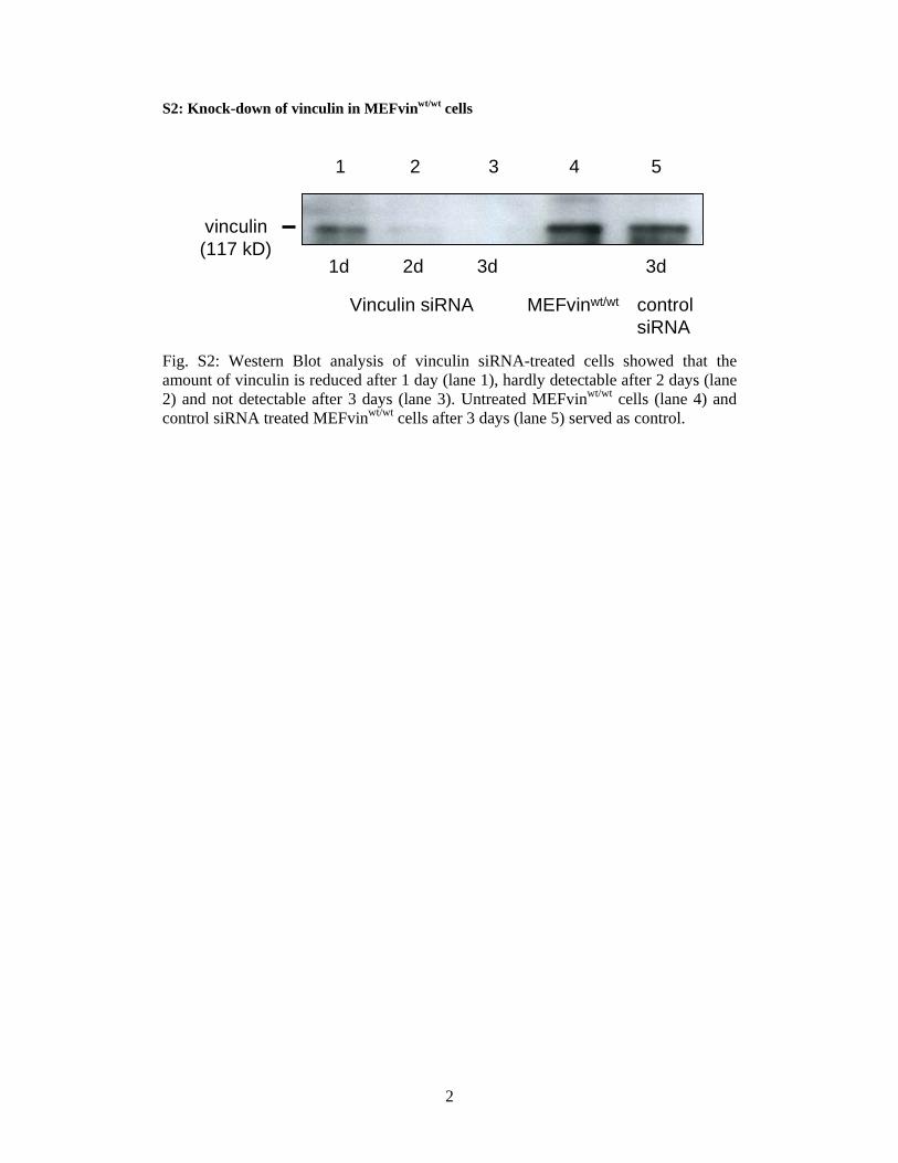
Function of Vinculin in Cell Motility—Here, we analyze theeffect of vinculin on cell invasion using MEFvin�/� andMEFvinwt/wt. Cell invasion into dense three-dimensional colla-gen matrices was analyzed after 3 days of culture. InvasiveMEFvinwt/wt and MEFvin�/� cells were spindle-shaped (Fig.1A). MEFvinwt/wt control cells showed a higher percentage ofinvasive cells compared with vinculin-deficient MEFs (32.9 �3.5% versus 0.5� 0.1%; Fig. 1B). The invasion profiles expressedas the cumulative probability of finding a cell at or below a giveninvasion depth reveal that MEFvinwt/wt cells invade muchdeeper into three-dimensional collagen matrices comparedwithMEFvin�/� cells (Fig. 1C). To confirm that the differencesbetweenMEFvinwt/wt andMEFvin�/� cell lines were caused bydifferent levels of vinculin expression and not by other con-founding effects, we analyzed MEFvinwt/wt cells that weretreated for 24 h prior to the start of the invasion assay withvinculin-specific siRNA (vin-siRNA) or nonspecific controlsiRNA. Immunoblot analysis in vin-siRNA-treated cellsrevealed that vinculin protein expression was reduced to lessthan 50% of wild-type levels after 1 day and was not detectableafter 2–3 days of treatment (supplemental Fig. S2). After 3 daysof culture on collagenmatrices, the fraction of invasive cells andthe invasion profile were determined. Invasive control siRNA
Vinculin Facilitates Cell Invasion
APRIL 23, 2010 • VOLUME 285 • NUMBER 17 JOURNAL OF BIOLOGICAL CHEMISTRY 13123
by guest on August 4, 2016
http://ww
w.jbc.org/
Dow
nloaded from
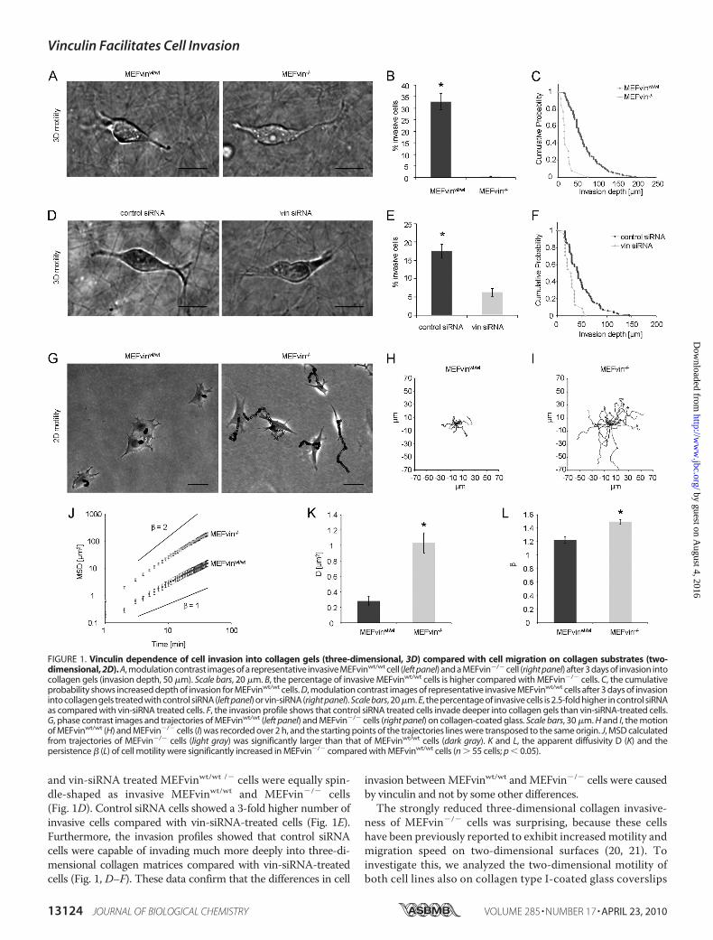
and vin-siRNA treated MEFvinwt/wt /� cells were equally spin-dle-shaped as invasive MEFvinwt/wt and MEFvin�/� cells(Fig. 1D). Control siRNA cells showed a 3-fold higher number ofinvasive cells compared with vin-siRNA-treated cells (Fig. 1E).Furthermore, the invasion profiles showed that control siRNAcells were capable of invading much more deeply into three-di-mensional collagen matrices compared with vin-siRNA-treatedcells (Fig. 1, D–F). These data confirm that the differences in cell
invasion betweenMEFvinwt/wt andMEFvin�/� cells were causedby vinculin and not by some other differences.The strongly reduced three-dimensional collagen invasive-
ness of MEFvin�/� cells was surprising, because these cellshave been previously reported to exhibit increasedmotility andmigration speed on two-dimensional surfaces (20, 21). Toinvestigate this, we analyzed the two-dimensional motility ofboth cell lines also on collagen type I-coated glass coverslips
FIGURE 1. Vinculin dependence of cell invasion into collagen gels (three-dimensional, 3D) compared with cell migration on collagen substrates (two-dimensional, 2D). A, modulation contrast images of a representative invasive MEFvinwt/wt cell (left panel) and a MEFvin�/� cell (right panel) after 3 days of invasion intocollagen gels (invasion depth, 50 �m). Scale bars, 20 �m. B, the percentage of invasive MEFvinwt/wt cells is higher compared with MEFvin�/� cells. C, the cumulativeprobability shows increased depth of invasion for MEFvinwt/wt cells. D, modulation contrast images of representative invasive MEFvinwt/wt cells after 3 days of invasioninto collagen gels treated with control siRNA (left panel) or vin-siRNA (right panel). Scale bars, 20 �m. E, the percentage of invasive cells is 2.5-fold higher in control siRNAas compared with vin-siRNA treated cells. F, the invasion profile shows that control siRNA treated cells invade deeper into collagen gels than vin-siRNA-treated cells.G, phase contrast images and trajectories of MEFvinwt/wt (left panel) and MEFvin�/� cells (right panel) on collagen-coated glass. Scale bars, 30 �m. H and I, the motionof MEFvinwt/wt (H) and MEFvin�/� cells (I) was recorded over 2 h, and the starting points of the trajectories lines were transposed to the same origin. J, MSD calculatedfrom trajectories of MEFvin�/� cells (light gray) was significantly larger than that of MEFvinwt/wt cells (dark gray). K and L, the apparent diffusivity D (K) and thepersistence � (L) of cell motility were significantly increased in MEFvin�/� compared with MEFvinwt/wt cells (n � 55 cells; p � 0.05).
Vinculin Facilitates Cell Invasion
13124 JOURNAL OF BIOLOGICAL CHEMISTRY VOLUME 285 • NUMBER 17 • APRIL 23, 2010
by guest on August 4, 2016
http://ww
w.jbc.org/
Dow
nloaded from
and confirmed that MEFvin�/� cells show an �3-fold highermigration speed in two-dimensional than MEFvinwt/wt cells(Fig. 1, G–L). The speed and persistence of cell migrationwas further investigated by analyzing the time evolution ofthe MSD of the cells (Fig. 1J). The MSD increased with timeaccording to a power law relationship (22): MSD � D�(t/t0)�,where t0 is the time interval, the prefactor D is the apparentdiffusivity, and the exponent � is a measure of the persis-tence. The apparent diffusivity D was 4-fold increased inMEFvin�/� cells compared with MEFvinwt/wt cells (Fig. 1K).In addition, MEFvin�/� cells migrated more persistently(� � 1.5) compared with MEFvinwt/wt cells (� � 1.2) (Fig. 1,J and L). Together, these results establish that vinculin hasdifferent effects on two-dimensional versus three-dimen-sional cell motility: MEFvin�/� cells show increased motilityon two-dimensional substrates but decreased invasion inthree-dimensional collagen matrices. In the following, we
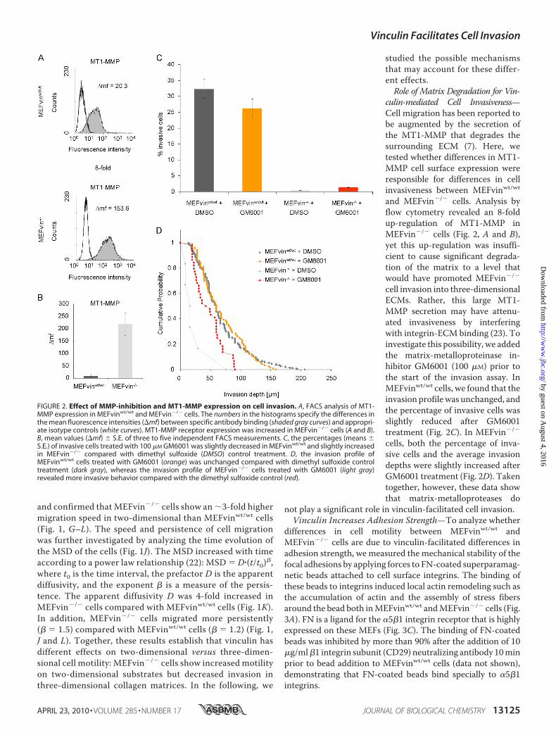
studied the possible mechanismsthat may account for these differ-ent effects.Role of Matrix Degradation for Vin-
culin-mediated Cell Invasiveness—Cell migration has been reported tobe augmented by the secretion ofthe MT1-MMP that degrades thesurrounding ECM (7). Here, wetested whether differences in MT1-MMP cell surface expression wereresponsible for differences in cellinvasiveness between MEFvinwt/wtand MEFvin�/� cells. Analysis byflow cytometry revealed an 8-foldup-regulation of MT1-MMP inMEFvin�/� cells (Fig. 2, A and B),yet this up-regulation was insuffi-cient to cause significant degrada-tion of the matrix to a level thatwould have promoted MEFvin�/�
cell invasion into three-dimensionalECMs. Rather, this large MT1-MMP secretion may have attenu-ated invasiveness by interferingwith integrin-ECM binding (23). Toinvestigate this possibility, we addedthe matrix-metalloproteinase in-hibitor GM6001 (100 �M) prior tothe start of the invasion assay. InMEFvinwt/wt cells, we found that theinvasion profilewas unchanged, andthe percentage of invasive cells wasslightly reduced after GM6001treatment (Fig. 2C). In MEFvin�/�
cells, both the percentage of inva-sive cells and the average invasiondepths were slightly increased afterGM6001 treatment (Fig. 2D). Takentogether, however, these data showthat matrix-metalloproteases do
not play a significant role in vinculin-facilitated cell invasion.Vinculin Increases Adhesion Strength—To analyze whether
differences in cell motility between MEFvinwt/wt andMEFvin�/� cells are due to vinculin-facilitated differences inadhesion strength, we measured the mechanical stability of thefocal adhesions by applying forces to FN-coated superparamag-netic beads attached to cell surface integrins. The binding ofthese beads to integrins induced local actin remodeling such asthe accumulation of actin and the assembly of stress fibersaround the bead both inMEFvinwt/wt andMEFvin�/� cells (Fig.3A). FN is a ligand for the �5�1 integrin receptor that is highlyexpressed on these MEFs (Fig. 3C). The binding of FN-coatedbeads was inhibited by more than 90% after the addition of 10�g/ml�1 integrin subunit (CD29) neutralizing antibody 10minprior to bead addition to MEFvinwt/wt cells (data not shown),demonstrating that FN-coated beads bind specially to �5�1integrins.
FIGURE 2. Effect of MMP-inhibition and MT1-MMP expression on cell invasion. A, FACS analysis of MT1-MMP expression in MEFvinwt/wt and MEFvin�/� cells. The numbers in the histograms specify the differences inthe mean fluorescence intensities (�mf) between specific antibody binding (shaded gray curves) and appropri-ate isotype controls (white curves). MT1-MMP receptor expression was increased in MEFvin�/� cells (A and B).B, mean values (�mf) � S.E. of three to five independent FACS measurements. C, the percentages (means �S.E.) of invasive cells treated with 100 �M GM6001 was slightly decreased in MEFvinwt/wt and slightly increasedin MEFvin�/� compared with dimethyl sulfoxide (DMSO) control treatment. D, the invasion profile ofMEFvinwt/wt cells treated with GM6001 (orange) was unchanged compared with dimethyl sulfoxide controltreatment (dark gray), whereas the invasion profile of MEFvin�/� cells treated with GM6001 (light gray)revealed more invasive behavior compared with the dimethyl sulfoxide control (red).
Vinculin Facilitates Cell Invasion
APRIL 23, 2010 • VOLUME 285 • NUMBER 17 JOURNAL OF BIOLOGICAL CHEMISTRY 13125
by guest on August 4, 2016
http://ww
w.jbc.org/
Dow
nloaded from
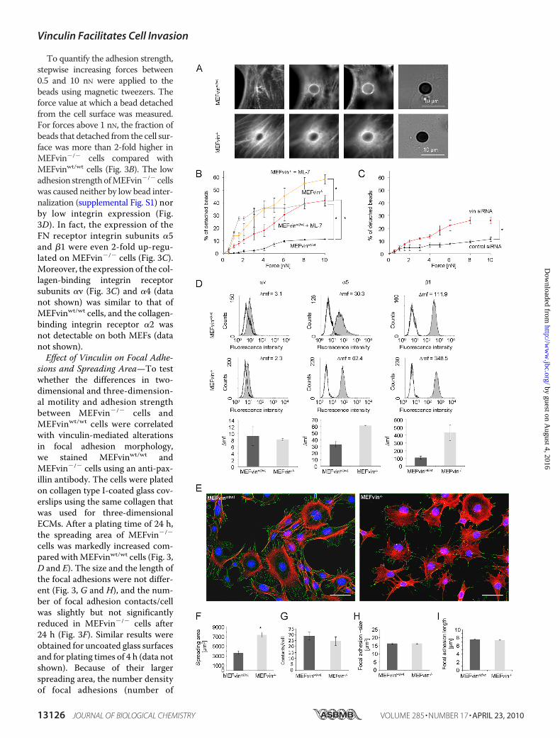
To quantify the adhesion strength,stepwise increasing forces between0.5 and 10 nN were applied to thebeads using magnetic tweezers. Theforce value at which a bead detachedfrom the cell surface was measured.For forces above 1 nN, the fraction ofbeads that detached from the cell sur-face was more than 2-fold higher inMEFvin�/� cells compared withMEFvinwt/wt cells (Fig. 3B). The lowadhesion strengthofMEFvin�/� cellswas caused neither by low bead inter-nalization (supplemental Fig. S1) norby low integrin expression (Fig.3D). In fact, the expression of theFN receptor integrin subunits �5and �1 were even 2-fold up-regu-lated on MEFvin�/� cells (Fig. 3C).Moreover, the expression of the col-lagen-binding integrin receptorsubunits �v (Fig. 3C) and �4 (datanot shown) was similar to that ofMEFvinwt/wt cells, and the collagen-binding integrin receptor �2 wasnot detectable on both MEFs (datanot shown).Effect of Vinculin on Focal Adhe-
sions and Spreading Area—To testwhether the differences in two-dimensional and three-dimension-al motility and adhesion strengthbetween MEFvin�/� cells andMEFvinwt/wt cells were correlatedwith vinculin-mediated alterationsin focal adhesion morphology,we stained MEFvinwt/wt andMEFvin�/� cells using an anti-pax-illin antibody. The cells were platedon collagen type I-coated glass cov-erslips using the same collagen thatwas used for three-dimensionalECMs. After a plating time of 24 h,the spreading area of MEFvin�/�
cells was markedly increased com-pared withMEFvinwt/wt cells (Fig. 3,D and E). The size and the length ofthe focal adhesions were not differ-ent (Fig. 3, G and H), and the num-ber of focal adhesion contacts/cellwas slightly but not significantlyreduced in MEFvin�/� cells after24 h (Fig. 3F). Similar results wereobtained for uncoated glass surfacesand for plating times of 4 h (data notshown). Because of their largerspreading area, the number densityof focal adhesions (number of
Vinculin Facilitates Cell Invasion
13126 JOURNAL OF BIOLOGICAL CHEMISTRY VOLUME 285 • NUMBER 17 • APRIL 23, 2010
by guest on August 4, 2016
http://ww
w.jbc.org/
Dow
nloaded from
focal adhesions/unit of spreading area) was markedlyreduced in MEFvin�/� cells (2-fold), which may have con-tributed to their weaker adhesion strength and hence theirhigher two-dimensional motility and impaired three-dimen-sional motility.Vinculin Influences Cytoskeletal Dynamics and Cell Stiffness—
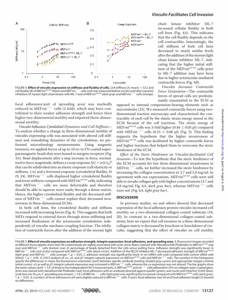
To analyze whether a change in three-dimensional motility ofvinculin-expressing cells was associated with altered cell stiff-ness and remodeling dynamics of the cytoskeleton, we per-formed microrheology measurements. Using magnetictweezers, we applied forces of up to 10 nN to FN-coated super-paramagnetic beads that were bound to integrin receptors (Fig.3A). Bead displacement after a step increase in force, normal-ized to forcemagnitude, defines a creep response J(t)� a�(t/t0)bthat can be subdivided into an elastic response (cell elasticity orstiffness, 1/a) and a frictional response (cytoskeletal fluidity, b)(9, 24). MEFvin�/� cells displayed higher cytoskeletal fluidityand lower stiffness comparedwithMEFvinwt/wt cells, indicatingthat MEFvin�/� cells are more deformable and thereforeshould be able to squeeze more easily through a dense matrix.Hence, the higher cytoskeletal fluidity and the decreased stiff-ness of MEFvin�/� cells cannot explain their decreased inva-siveness in three-dimensional ECMs.In both cell lines, the cytoskeletal fluidity and stiffness
increasedwith increasing forces (Fig. 4). This suggests that bothMEFs respond to external forces through stress stiffening andincreased fluidization of the actomyosin cytoskeleton inde-pendently of vinculin mechano-coupling function. The inhibi-tion of contractile forces after the addition of the myosin light
chain kinase inhibitor ML-7increased cellular fluidity in bothcell lines (Fig. 4A). This indicatesthat the cell fluidity depends on thecell contractility. Interestingly, thecell stiffness of both cell linesdecreased to nearly similar levelsafter the addition of themyosin lightchain kinase inhibitor ML-7, indi-cating that the higher initial stiff-ness of the MEFvinwt/wt cells priorto ML-7 addition may have beendue to higher actomyosin-mediatedcontractile forces (Fig. 4B).Vinculin Increases Contractile
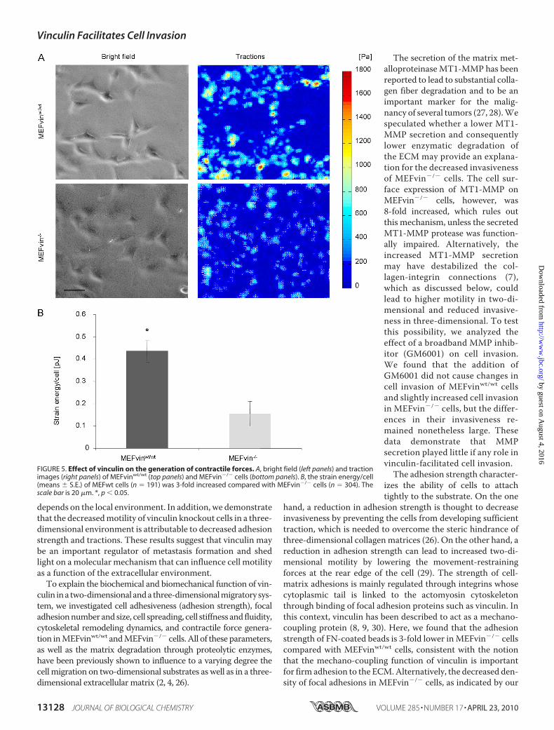
Force Generation—The contractileforces of spread cells are predomi-nantly transmitted to the ECM as
opposed to internal compression-bearing elements such asmicrotubules (25). We measured contractile forces using two-dimensional traction microscopy and characterized the con-tractility of each cell by the elastic strain energy stored in theECM because of the cell tractions. The strain energy ofMEFvinwt/wt cells was 3-fold higher (0.44 � 0.05 pJ) comparedwith MEFvin�/� cells (0.15 � 0.06 pJ) (Fig. 5). This findingsupports the hypothesis that the higher invasiveness ofMEFvinwt/wt cells was facilitated by higher contractile forcesand higher tractions that helped them to overcome the sterichindrance of the ECM.Effect of the Steric Hindrance on Vinculin-facilitated Cell
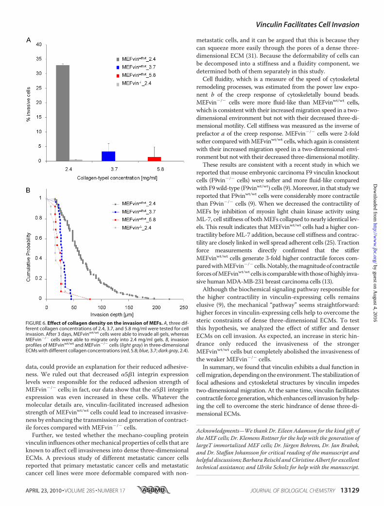
Invasion—To test the hypothesis that the steric hindrance ofthe ECM accounts for low three-dimensional invasiveness inMEFvin�/� cells, we further increased the steric hindrance byincreasing the collagen concentration to 3.7 and 5.8 mg/ml. Inagreement with our expectations, MEFvinwt/wt cells were stillable to invade collagen gels with higher concentrations (3.7 and5.8 mg/ml; Fig. 6A, dark gray bar), whereas MEFvin�/� cellswere not (Fig. 6A, light gray bar).
DISCUSSION
In previous studies, we and others showed that decreasedexpression of the focal adhesion protein vinculin increased cellmotility on a two-dimensional collagen-coated substrate (10,20). In contrast to a two-dimensional collagen-coated sub-strate, here we report that cell invasion in a three-dimensionalcollagenmatrix is decreased by knockout or knockdown of vin-culin, suggesting that the effect of vinculin on cell motility
FIGURE 3. Effect of vinculin expression on adhesion strength, integrin expression, focal adhesions, and spreading area. A, fluorescent images recordedat different focus depths show that FN-coated beads are tightly associated with actin stress fibers (stained with Alexafluor546 Phalloidin) in MEFvinwt/wt (toprow) and MEFvin�/� cells (bottom row). B, percentage of beads detached from the cells versus pulling force. Adhesion strength was significantly lower inMEFvin�/� cells (red circles) compared with MEFvinwt/wt cells (black circles). Addition of the MLCK inhibitor ML-7 decreased adhesion strength of MEFvinwt/wt
(light gray) and MEFvin�/� cells (orange). *, p � 0.05. C, adhesion strength was significantly lower in vin-siRNA cells (red) compared with control siRNA cells(black). *, p � 0.05. D, FACS analysis of �v, �5, and �1 integrin subunit expression on MEFvinwt/wt cells and MEFvin�/� cells. The numbers in the histogramsspecify the differences in mean the fluorescence intensities (�mf) between specific antibody binding (shaded gray curves) and appropriate isotype controls(white curves). �5 as well as �1 integrin subunit expression was increased in MEFvin�/� cells, whereas the �v expression was not altered. The bar graphs showthe mean values (�mf) � S.E. of three to five independent FACS measurements. E, MEFvinwt/wt and vin�/� cells adhered for 24 h on collagen type I-coated glass.Actin was stained with Alexafluor546 Phalloidin (red), focal adhesions with an antibody directed against paxillin (green), and nuclei with Hoechst 33342 (blue).Scale bars are 30 �m. F, spreading area (means � S.E.) of MEFvin�/� cells (light gray) was significantly increased compared with MEFvinwt/wt cells (dark gray).*, p � 0.05. G, numbers of focal adhesions/cell were slightly reduced in MEFvin�/� cells. H and I, focal adhesion size (H) and focal adhesion length (I) showedno differences.
FIGURE 4. Effect of vinculin expression on stiffness and fluidity of cells. Cell stiffness (A; means � S.E.) andcell fluidity (B) of MEFvinwt/wt (black) and MEFvin�/� cells (red) was measured before (circles) and after (squares)inhibition of myosin light chain kinase with ML-7 and of MEFvinwt/wt (dark gray) and MEFvin�/� cells (orange).
Vinculin Facilitates Cell Invasion
APRIL 23, 2010 • VOLUME 285 • NUMBER 17 JOURNAL OF BIOLOGICAL CHEMISTRY 13127
by guest on August 4, 2016
http://ww
w.jbc.org/
Dow
nloaded from
depends on the local environment. In addition, we demonstratethat the decreasedmotility of vinculin knockout cells in a three-dimensional environment is attributable to decreased adhesionstrength and tractions. These results suggest that vinculin maybe an important regulator of metastasis formation and shedlight on amolecular mechanism that can influence cell motilityas a function of the extracellular environment.To explain the biochemical and biomechanical function of vin-
culin ina two-dimensional anda three-dimensionalmigratory sys-tem, we investigated cell adhesiveness (adhesion strength), focaladhesionnumberandsize, cell spreading, cell stiffness and fluidity,cytoskeletal remodeling dynamics, and contractile force genera-tion inMEFvinwt/wt andMEFvin�/� cells. All of these parameters,as well as the matrix degradation through proteolytic enzymes,have been previously shown to influence to a varying degree thecellmigration on two-dimensional substrates as well as in a three-dimensional extracellular matrix (2, 4, 26).
The secretion of the matrix met-alloproteinase MT1-MMP has beenreported to lead to substantial colla-gen fiber degradation and to be animportant marker for the malig-nancy of several tumors (27, 28).Wespeculated whether a lower MT1-MMP secretion and consequentlylower enzymatic degradation ofthe ECM may provide an explana-tion for the decreased invasivenessof MEFvin�/� cells. The cell sur-face expression of MT1-MMP onMEFvin�/� cells, however, was8-fold increased, which rules outthis mechanism, unless the secretedMT1-MMP protease was function-ally impaired. Alternatively, theincreased MT1-MMP secretionmay have destabilized the col-lagen-integrin connections (7),which as discussed below, couldlead to higher motility in two-di-mensional and reduced invasive-ness in three-dimensional. To testthis possibility, we analyzed theeffect of a broadband MMP inhib-itor (GM6001) on cell invasion.We found that the addition ofGM6001 did not cause changes incell invasion of MEFvinwt/wt cellsand slightly increased cell invasionin MEFvin�/� cells, but the differ-ences in their invasiveness re-mained nonetheless large. Thesedata demonstrate that MMPsecretion played little if any role invinculin-facilitated cell invasion.The adhesion strength character-
izes the ability of cells to attachtightly to the substrate. On the one
hand, a reduction in adhesion strength is thought to decreaseinvasiveness by preventing the cells from developing sufficienttraction, which is needed to overcome the steric hindrance ofthree-dimensional collagenmatrices (26). On the other hand, areduction in adhesion strength can lead to increased two-di-mensional motility by lowering the movement-restrainingforces at the rear edge of the cell (29). The strength of cell-matrix adhesions is mainly regulated through integrins whosecytoplasmic tail is linked to the actomyosin cytoskeletonthrough binding of focal adhesion proteins such as vinculin. Inthis context, vinculin has been described to act as a mechano-coupling protein (8, 9, 30). Here, we found that the adhesionstrength of FN-coated beads is 3-fold lower inMEFvin�/� cellscompared with MEFvinwt/wt cells, consistent with the notionthat the mechano-coupling function of vinculin is importantfor firm adhesion to the ECM.Alternatively, the decreased den-sity of focal adhesions in MEFvin�/� cells, as indicated by our
FIGURE 5. Effect of vinculin on the generation of contractile forces. A, bright field (left panels) and tractionimages (right panels) of MEFvinwt/wt (top panels) and MEFvin�/� cells (bottom panels). B, the strain energy/cell(means � S.E.) of MEFwt cells (n � 191) was 3-fold increased compared with MEFvin�/� cells (n � 304). Thescale bar is 20 �m. *, p � 0.05.
Vinculin Facilitates Cell Invasion
13128 JOURNAL OF BIOLOGICAL CHEMISTRY VOLUME 285 • NUMBER 17 • APRIL 23, 2010
by guest on August 4, 2016
http://ww
w.jbc.org/
Dow
nloaded from
data, could provide an explanation for their reduced adhesive-ness. We ruled out that decreased �5�1 integrin expressionlevels were responsible for the reduced adhesion strength ofMEFvin�/� cells; in fact, our data show that the �5�1 integrinexpression was even increased in these cells. Whatever themolecular details are, vinculin-facilitated increased adhesionstrength of MEFvinwt/wt cells could lead to increased invasive-ness by enhancing the transmission and generation of contract-ile forces compared with MEFvin�/� cells.
Further, we tested whether the mechano-coupling proteinvinculin influences othermechanical properties of cells that areknown to affect cell invasiveness into dense three-dimensionalECMs. A previous study of different metastatic cancer cellsreported that primary metastatic cancer cells and metastaticcancer cell lines were more deformable compared with non-
metastatic cells, and it can be argued that this is because theycan squeeze more easily through the pores of a dense three-dimensional ECM (31). Because the deformability of cells canbe decomposed into a stiffness and a fluidity component, wedetermined both of them separately in this study.Cell fluidity, which is a measure of the speed of cytoskeletal
remodeling processes, was estimated from the power law expo-nent b of the creep response of cytoskeletally bound beads.MEFvin�/� cells were more fluid-like than MEFvinwt/wt cells,which is consistent with their increasedmigration speed in a two-dimensional environment but not with their decreased three-di-mensional motility. Cell stiffness was measured as the inverse ofprefactor a of the creep response. MEFvin�/� cells were 2-foldsofter compared withMEFvinwt/wt cells, which again is consistentwith their increased migration speed in a two-dimensional envi-ronment but not with their decreased three-dimensionalmotility.These results are consistent with a recent study in which we
reported that mouse embryonic carcinoma F9 vinculin knockoutcells (F9vin�/� cells) were softer and more fluid-like comparedwith F9wild-type (F9vinwt/wt) cells (9).Moreover, in that studywereported that F9vinwt/wt cells were considerably more contractilethan F9vin�/� cells (9). When we decreased the contractility ofMEFs by inhibition of myosin light chain kinase activity usingML-7, cell stiffness of bothMEFs collapsed to nearly identical lev-els. This result indicates that MEFvinwt/wt cells had a higher con-tractility beforeML-7 addition, because cell stiffness and contrac-tility are closely linked in well spread adherent cells (25). Tractionforce measurements directly confirmed that the stifferMEFvinwt/wt cells generate 3-fold higher contractile forces com-paredwithMEFvin�/�cells.Notably, themagnitudeofcontractileforcesofMEFvinwt/wt cells is comparablewith thoseofhighly inva-sive humanMDA-MB-231 breast carcinoma cells (13).Although the biochemical signaling pathway responsible for
the higher contractility in vinculin-expressing cells remainselusive (9), the mechanical “pathway” seems straightforward:higher forces in vinculin-expressing cells help to overcome thesteric constraints of dense three-dimensional ECMs. To testthis hypothesis, we analyzed the effect of stiffer and denserECMs on cell invasion. As expected, an increase in steric hin-drance only reduced the invasiveness of the strongerMEFvinwt/wt cells but completely abolished the invasiveness ofthe weaker MEFvin�/� cells.In summary, we found that vinculin exhibits a dual function in
cellmigration,dependingon theenvironment.Thestabilizationoffocal adhesions and cytoskeletal structures by vinculin impedestwo-dimensional migration. At the same time, vinculin facilitatescontractile forcegeneration,whichenhances cell invasionbyhelp-ing the cell to overcome the steric hindrance of dense three-di-mensional ECMs.
Acknowledgments—We thank Dr. Eileen Adamson for the kind gift ofthe MEF cells; Dr. Klemens Rottner for the help with the generation oflargeT immortalized MEF cells; Dr. Jurgen Behrens, Dr. Jan Brabek,and Dr. Staffan Johansson for critical reading of the manuscript andhelpful discussions; BarbaraReischl andChristineAlbert for excellenttechnical assistance; and Ulrike Scholz for help with the manuscript.
FIGURE 6. Effect of collagen density on the invasion of MEFs. A, three dif-ferent collagen concentrations of 2.4, 3.7, and 5.8 mg/ml were tested for cellinvasion. After 3 days, MEFvinwt/wt cells were able to invade all gels, whereasMEFvin�/� cells were able to migrate only into 2.4 mg/ml gels. B, invasionprofiles of MEFvinwt/wt and MEFvin�/� cells (light gray) in three-dimensionalECMs with different collagen concentrations (red, 5.8; blue, 3.7; dark gray, 2.4).
Vinculin Facilitates Cell Invasion
APRIL 23, 2010 • VOLUME 285 • NUMBER 17 JOURNAL OF BIOLOGICAL CHEMISTRY 13129
by guest on August 4, 2016
http://ww
w.jbc.org/
Dow
nloaded from
REFERENCES1. Horwitz, A. R., and Parsons, J. T. (1999) Science 286, 1102–11032. Mierke, C. T., Rosel, D., Fabry, B., and Brabek, J. (2008) Eur. J. Cell Biol. 87,
669–6763. Lauffenburger, D. A., and Horwitz, A. F. (1996) Cell 84, 359–3694. Friedl, P., and Brocker, E. B. (2000) Cell Mol. Life Sci. 57, 41–645. Webb, D. J., Brown, C.M., and Horwitz, A. F. (2003)Curr. Opin. Cell Biol.
15, 614–6206. Friedl, P., and Wolf, K. (2003) Nat. Rev. Cancer 3, 362–3747. Wolf, K., Mazo, I., Leung, H., Engelke, K., von Andrian, U. H., Deryugina,
E. I., Strongin, A. Y., Brocker, E. B., and Friedl, P. (2003) J. Cell Biol. 160,267–277
8. Ezzell, R. M., Goldmann, W. H., Wang, N., Parasharama, N., and Ingber,D. E. (1997) Exp. Cell Res. 231, 14–26
9. Mierke, C. T., Kollmannsberger, P., Zitterbart, D. P., Smith, J., Fabry, B.,and Goldmann, W. H. (2008) Biophys. J. 94, 661–670
10. Goldmann,W.H., Schindl,M., Cardozo, T. J., and Ezzell, R.M. (1995)Exp.Cell Res. 221, 311–319
11. Subauste, M. C., Pertz, O., Adamson, E. D., Turner, C. E., Junger, S., andHahn, K. M. (2004) J. Cell Biol. 165, 371–381
12. Saunders, R. M., Holt, M. R., Jennings, L., Sutton, D. H., Barsukov, I. L.,Bobkov, A., Liddington, R. C., Adamson, E. A., Dunn, G. A., and Critchley,D. R. (2006) Eur. J. Cell Biol. 85, 487–500
13. Mierke, C. T., Zitterbart, D. P., Kollmannsberger, P., Raupach, C.,Schlotzer-Schrehardt, U., Goecke, T. W., Behrens, J., and Fabry, B. (2008)Biophys. J. 94, 2832–2846
14. McNulty, A. L., Weinberg, J. B., and Guilak, F. (2009) Clin. Orthop. Relat.Res. 467, 1557–1567
15. Kollmannsberger, P., and Fabry, B. (2007) Rev. Sci. Instrum. 78,114301–114306
16. Fabry, B., Maksym, G. N., Butler, J. P., Glogauer, M., Navajas, D., andFredberg, J. J. (2001) Phys. Rev. Lett. 87, 148102–148106
17. Pelham, R. J., Jr., and Wang, Y. (1997) Proc. Natl. Acad. Sci. U.S.A. 94,13661–13665
18. Raupach, C., Zitterbart, D. P.,Mierke, C. T.,Metzner, C.,Muller, F. A., andFabry, B. (2007) Phys. Rev. E Stat. Nonlin Soft Matter. Phys. 76, 011918
19. Butler, J. P., Tolia-Nçrrelykke, I. M., Fabry, B., and Fredberg, J. J. (2002)Am. J. Physiol. Cell Physiol. 282, C595–C605
20. Coll, J. L., Ben-Ze’ev, A., Ezzell, R. M., Rodríguez Fernandez, J. L., Barib-ault, H., Oshima, R. G., and Adamson, E. D. (1995) Proc. Natl. Acad. Sci.U.S.A. 92, 9161–9165
21. Xu, W., Coll, J. L., and Adamson, E. D. (1998) J. Cell Sci. 111, 1535–154422. Dieterich, P., Klages, R., Preuss, R., and Schwab, A. (2008)Proc. Natl. Acad.
Sci. U.S.A. 105, 459–46323. Baciu, P. C., Suleiman, E. A., Deryugina, E. I., and Strongin, A. Y. (2003)
Exp. Cell Res. 291, 167–17524. Fabry, B., and Fredberg, J. J. (2003) Respir. Physiol. Neurobiol. 137,
109–12425. Wang, N., Tolia-Nçrrelykke, I. M., Chen, J., Mijailovich, S.M., Butler, J. P.,
Fredberg, J. J., and Stamenovia, D. (2002) Am. J. Physiol. Cell Physiol. 282,C606–C616
26. Zaman, M. H., Trapani, L. M., Siemeski, A., Mackellar, D., Gong, H.,Kamm, R. D., Wells, A., Lauffenburger, D. A., and Matsudaira, P. (2006)Proc. Natl. Acad. Sci. U.S.A. 103, 10889–10894
27. Okada, A., Bellocq, J. P., Rouyer, N., Chenard,M. P., Rio,M. C., Chambon,P., and Basset, P. (1995) Proc. Natl. Acad. Sci. U.S.A. 92, 2730–2734
28. Davidson, B., Goldberg, I., Gotlieb, W. H., Kopolovic, J., Ben-Baruch, G.,Nesland, J. M., Berner, A., Bryne, M., and Reich, R. (1999) Clin. Exp. Me-tastasis 17, 799–808
29. Gallant, N. D., Michael, K. E., and García, A. J. (2005) Mol. Biol. Cell 16,4329–4340
30. Mohl, C., Kirchgessner, N., Schafer, C., Kupper, K., Born, S., Diez, G.,Goldmann, W. H., Merkel, R., and Hoffmann, B. (2009) Cell Motil.Cytoskeleton 66, 350–364
31. Guck, J., Schinkinger, S., Lincoln, B.,Wottawah, F., Ebert, S., Romeyke,M.,Lenz, D., Erickson, H. M., Ananthakrishnan, R., Mitchell, D., Kas, J.,Ulvick, S., and Bilby, C. (2005) Biophys. J. 88, 3689–3698
Vinculin Facilitates Cell Invasion
13130 JOURNAL OF BIOLOGICAL CHEMISTRY VOLUME 285 • NUMBER 17 • APRIL 23, 2010
by guest on August 4, 2016
http://ww
w.jbc.org/
Dow
nloaded from
1
SUPPLEMENTAL DATA
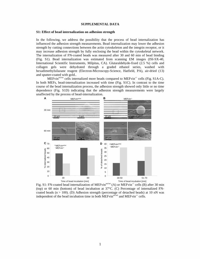
S1: Effect of bead internalization on adhesion strength In the following, we address the possibility that the process of bead internalization has influenced the adhesion strength measurements. Bead internalization may lower the adhesion strength by cutting connections between the actin cytoskeleton and the integrin receptor, or it may increase adhesion strength by fully enclosing the bead within the cytoskeletal network. The internalization of FN-coated beads was measured after 30 and 60 min of bead binding (Fig. S1). Bead internalization was estimated from scanning EM images (ISI-SX-40, International Scientific Instruments, Milpitas, CA). Glutaraldehyde-fixed (2.5 %) cells and collagen gels were dehydrated through a graded ethanol series, washed with hexadimethylsilazane reagent (Electron-Microscopy-Science, Hatfield, PA), air-dried (13) and sputter-coated with gold..
MEFvinwt/wt cells internalized more beads compared to MEFvin-/- cells (Fig. S1A-C). In both MEFs, bead-internalization increased with time (Fig. S1C). In contrast to the time course of the bead internalization process, the adhesion strength showed only little or no time dependence (Fig. S1D) indicating that the adhesion strength measurements were largely unaffected by the process of bead-internalization.
MEFvinwt/wt MEFvin-/-
30 min
60 min
A B
C
5 µm
5 µm
2 µm
2 µm
D
0
5
10
15
20
25
30
35
40
30-50 51-70Time of bead incubation [min]
% o
f det
ache
d be
ads
MEFvinwt/wt
MEFvin-/-
0
10
20
30
40
50
60
70
30 60Time of bead incubation [min]
% in
tern
aliz
ed b
eads
MEFvinwt/wt
MEFvin-/-
20 µm
20 µm
20 µm
20 µm
Fig. S1: FN-coated bead internalization of MEFvinwt/wt (A) or MEFvin-/- cells (B) after 30 min (top) or 60 min (bottom) of bead incubation at 37°C. (C) Percentage of internalized FN-coated beads (n > 100). (D) Adhesion strength (percentage of detached beads) at 10 nN was independent of the bead incubation time in both MEFvinwt/wt and MEFvin-/- cells.
2
S2: Knock-down of vinculin in MEFvinwt/wt cells
1d 2d 3d
MEFvinwt/wtVinculin siRNA controlsiRNA
vinculin(117 kD)
3d
1 2 3 4 5
Fig. S2: Western Blot analysis of vinculin siRNA-treated cells showed that the amount of vinculin is reduced after 1 day (lane 1), hardly detectable after 2 days (lane 2) and not detectable after 3 days (lane 3). Untreated MEFvinwt/wt cells (lane 4) and control siRNA treated MEFvinwt/wt cells after 3 days (lane 5) served as control.
Ben FabryThorsten M. Koch, Susanna Marg, Wolfgang H. Ziegler, Wolfgang H. Goldmann and Claudia T. Mierke, Philip Kollmannsberger, Daniel Paranhos Zitterbart, Gerold Diez,
Vinculin Facilitates Cell Invasion into Three-dimensional Collagen Matrices
doi: 10.1074/jbc.M109.087171 originally published online February 24, 20102010, 285:13121-13130.J. Biol. Chem.
10.1074/jbc.M109.087171Access the most updated version of this article at doi:
Alerts:
When a correction for this article is posted•
When this article is cited•
to choose from all of JBC's e-mail alertsClick here
Supplemental material:
http://www.jbc.org/content/suppl/2010/03/05/M109.087171.DC1.html
http://www.jbc.org/content/285/17/13121.full.html#ref-list-1
This article cites 30 references, 12 of which can be accessed free at
by guest on August 4, 2016
http://ww
w.jbc.org/
Dow
nloaded from