A systematic analysis of nucleosome core particle ... - Nature
Upload
khangminh22Category
view
6download
0
NDF, a nucleosome destabilizing factorthat facilitates transcription throughnucleosomesJia Fei,1 Haruhiko Ishii,2 Marten A. Hoeksema,3,4 Franz Meitinger,2 George A. Kassavetis,1
Christopher K. Glass,3,4 Bing Ren,2,3,5 and James T. Kadonaga1
1Section of Molecular Biology, University of California at San Diego, La Jolla, California 92093, USA; 2Ludwig Institute for CancerResearch, San Diego Branch, La Jolla, California 92093, USA; 3Department of Cellular and Molecular Medicine, University ofCalifornia at San Diego, La Jolla, California 92093, USA; 4Department of Medicine, University of California at San Diego, La Jolla,California 92093, USA; 5Center for Epigenomics, Institute of GenomeMedicine,Moores Cancer Center, University of California atSan Diego, La Jolla, California 92093, USA
Our understanding of transcription by RNA polymerase II (Pol II) is limited by our knowledge of the factors thatmediate this critically important process. Here we describe the identification of NDF, a nucleosome-destabilizingfactor that facilitates Pol II transcription in chromatin. NDFhas a PWWPmotif, interactswith nucleosomes near thedyad, destabilizes nucleosomes in an ATP-independent manner, and facilitates transcription by Pol II through nu-cleosomes in a purified and defined transcription system as well as in cell nuclei. Upon transcriptional induction,NDF is recruited to the transcribed regions of thousands of genes and colocalizes with a subset of H3K36me3-enriched regions. Notably, the recruitment of NDF to gene bodies is accompanied by an increase in the transcriptlevels of many of the NDF-enriched genes. In addition, the global loss of NDF results in a decrease in the RNA levelsof many genes. In humans, NDF is present at high levels in all tested tissue types, is essential in stem cells, and isfrequently overexpressed in breast cancer. These findings indicate that NDF is a nucleosome-destabilizing factorthat is recruited to gene bodies during transcriptional activation and facilitates Pol II transcription throughnucleosomes.
[Keywords: nucleosome; transcription; gene expression; RNA polymerase II; chromatin dynamics]
Supplemental material is available for this article.
Received March 7, 2018; revised version accepted April 11, 2018.
In the eukaryotic nucleus, transcription of protein-codinggenes is carried out by RNA polymerase II (Pol II). Our un-derstanding of Pol II transcription is limited, however, byour knowledge of the factors that mediate this essentialprocess. In spite of considerable progress, many importantelements of Pol II transcription remain unsolved.One particularly intriguing aspect of transcription by
Pol II is the ability of the enzyme to traverse the nucleoso-mal template. A key protein that promotes transcriptionand DNA replication in chromatin is FACT (facilitateschromatin transcription/transactions) (for example, seeReinberg and Sims 2006; Formosa 2008). FACT binds tonucleosomes and can remove an H2A–H2B dimer or reor-ganize the nucleosome into an altered structure. In hu-mans, however, the SSRP1 subunit of FACT is notdetectable in many tissue types (Supplemental Fig. S1A;Garcia et al. 2011; Uhlén et al. 2015). It thus appearsthat critical components of the human transcription elon-gation machinery have yet to be identified.
In our studies of chromatin dynamics, we becamedrawn to the study of Pol II elongation in chromatinthrough our analysis of the prenucleosome, a conforma-tional isomer of the nucleosome that associates with∼80-base-pair (bp) DNA rather than ∼147-bp DNA as ina canonical nucleosome (Torigoe et al. 2011, 2013; Feiet al. 2015; Khuong et al. 2015). Notably, H3K56, whichis located at the DNA entry and exit points of the nucleo-some, can be acetylated by p300 in prenucleosomes butnot in canonical nucleosomes (Fei et al. 2015). This obser-vation is consistent with the partial unwrapping of theDNA in a prenucleosome relative to a nucleosome.The properties of the prenucleosome led us to consider
whether there might be factors that could partiallydisassemble a nucleosome and facilitate the acetylationof nucleosomalH3K56. These studies resulted in the iden-tification of a protein that we term NDF (nucleosome-
Corresponding author: [email protected] published online ahead of print. Article and publication date areonline at http://www.genesdev.org/cgi/doi/10.1101/gad.313973.118.
© 2018 Fei et al. This article is distributed exclusively by Cold SpringHarbor Laboratory Press for the first six months after the full-issue publi-cation date (see http://genesdev.cshlp.org/site/misc/terms.xhtml). Aftersix months, it is available under a Creative Commons License (At-tribution-NonCommercial 4.0 International), as described at http://creati-vecommons.org/licenses/by-nc/4.0/.
GENES & DEVELOPMENT 32:1–13 Published by Cold Spring Harbor Laboratory Press; ISSN 0890-9369/18; www.genesdev.org 1
Cold Spring Harbor Laboratory Press on July 7, 2022 - Published by genesdev.cshlp.orgDownloaded from
destabilizing factor), which destabilizes nucleosomes,is enriched in the transcribed regions of many activegenes, facilitates transcription by Pol II through nucleo-somes, and is present in all tested human tissue types.The identification of NDF reveals a new Pol II elongationfactor that is structurally and functionally distinct fromFACT.
Results
Identification and purification of NDF, a novelnucleosome-destabilizing factor
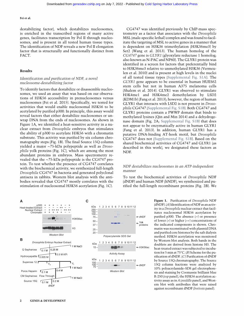
To identify factors that destabilize or disassemble nucleo-somes, we used an assay that was based on our observa-tions of H3K56 accessibility in prenucleosomes versusnucleosomes (Fei et al. 2015). Specifically, we tested foractivities that would enable nucleosomal H3K56 to beacetylated by purified p300. In principle, this assay wouldreveal factors that either destabilize nucleosomes or un-wrap DNA from the ends of nucleosomes. As shown inFigure 1A, we identified a heat-sensitive activity in a nu-clear extract from Drosophila embryos that stimulatesthe ability of p300 to acetylate H3K56 with a chromatinsubstrate. This activity was purified by six column chro-matography steps (Fig. 1B). The final Source 15Q columnyielded a major ∼75-kDa polypeptide as well as Droso-phila yolk proteins (Fig. 1C), which are among the mostabundant proteins in embryos. Mass spectrometry re-vealed that the ∼75-kDa polypeptide is the CG4747 pro-tein. To test whether the presence of CG4747 correlateswith the biochemical activity, we synthesized full-lengthDrosophila CG4747 in bacteria and generated polyclonalantisera in rabbits. Western blot analysis with the anti-bodies revealed that CG4747 mostly correlates with thestimulation of nucleosomal H3K56 acetylation (Fig. 1C).
CG4747 was identified previously by ChIP-mass spec-trometry as a factor that associates with the DrosophilaMSL (male-specific lethal) complex andwas found to facil-itate the targeting ofMSL to active genes in amanner thatis dependent on H3K36 trimethylation (H3K36me3) bySet2 (Wang et al. 2013). The human homolog of theCG4747 gene is GLYR1 (glyoxylate reductase 1 homolog;also known asN-PAC andNP60). TheGLYR1 proteinwasidentified in a screen for factors that preferentially bindto H3K36me3 relative to unmethylated H3K36 (Vermeu-len et al. 2010) and is present at high levels in the nucleiof all tested tissue types (Supplemental Fig. S1A). TheGLYR1 gene appears to be essential in human HUES62stem cells but not in human A375 melanoma cells(Shalem et al. 2014). GLYR1 was observed to stimulateH3K4me1 and H3K4me2 demethylation by LSD2(KDM1b) (Fang et al. 2013); however, the region of humanGLYR1 that interacts with LSD2 is not present in Droso-phila CG4747 (Supplemental Fig. S1B). Both CG4747 andGLYR1 proteins contain a PWWP domain that binds tomethylated lysines (Qin and Min 2014) and a dehydroge-nase domain (Fig. 2A; Supplemental Fig. S1B) that doesnot appear to be enzymatically active in human GLYR1(Fang et al. 2013). In addition, human GLYR1 has aputative DNA-binding AT-hook motif, but DrosophilaCG4747 does not (Supplemental Fig. S1B). Based on theshared biochemical activities of CG4747 and GLYR1 (asdescribed in this work), we designated these factors asNDF.
NDF destabilizes nucleosomes in an ATP-independentmanner
To test the biochemical activities of Drosophila NDF(dNDF) and human NDF (hNDF), we synthesized and pu-rified the full-length recombinant proteins (Fig. 2B). We
A
B
C Figure 1. Purification of Drosophila NDF(dNDF). (A) IdentificationofNDFasanactiv-ity in aDrosophila nuclear extract that facil-itates nucleosomal H3K56 acetylation bypurified p300. The absence (−) or presenceof lower (+) or higher (++) concentrations ofthe indicated components is shown. Chro-matinwas reconstituted with plasmidDNAand purified core histones by the salt dialysismethod. H3K56 acetylation was monitoredby Western blot analysis. Both bands in thedoublets are derived from histone H3. Theheat-treated extractwas subjected to incuba-tion for 5min at 75°C. (B) Scheme for the pu-rification of dNDF. (C ) Purification of dNDFby Source 15Q chromatography. The Source15Q column fractions were analyzed by10% polyacrylamide–SDS gel electrophore-sis and staining by Coomassie brilliant blueR-250 (top panel), the H3K56 acetylation ac-tivity assay as inA (middlepanel), andWest-ern blot with antibodies that were raisedagainst recombinant dNDF (bottom panel).
Fei et al.
2 GENES & DEVELOPMENT
Cold Spring Harbor Laboratory Press on July 7, 2022 - Published by genesdev.cshlp.orgDownloaded from

observed that dNDF and hNDF are able to stimulatethe acetylation of nucleosomal H3K56 by p300 in adose-dependent and ATP-independent manner (Fig. 2C;Supplemental Fig. S2A,B). Purified dNDF and hNDF didnot exhibit intrinsic histone acetyltransferase activity(Fig. 2C) and also did not stimulate the acetylation offree histones by p300 (Supplemental Fig. S2C). Thus, puri-fied recombinant dNDF and hNDF can mediate the stim-ulation of nucleosomal H3K56 acetylation, as seen in ouroriginal assay with the native Drosophila factor (Fig. 1).Wenext examinedwhetherNDFhas the ability to disas-
semblenucleosomes.To this end,we investigated the abil-ity of NDF to disrupt nucleosome-mediated supercoilingof covalently closed circular plasmid DNA. The wrappingof DNA around the core histone octamer in a nucleosomecauses achange in the linkingnumberof approximately−1(Germond et al. 1975). Therefore, nucleosome disassem-bly can be monitored by the loss of negative supercoilingin chromatin in circular plasmid DNA. We reconstitutednucleosomes onto plasmid DNA by using salt dialysismethodology and then treated the chromatin with NDFin thepresenceof topoisomerase I.The resultingDNAspe-cieswere analyzed by agarose gel electrophoresis in the ab-sence or presence of chloroquine, which introducespositive supercoils into theDNA and enables the differen-tiation between relaxed circular DNA and nicked circularDNA (Fig. 2D; Supplemental Fig. S2D,E). These experi-ments revealed that hNDF and dNDF are able to mediatethe partial disassembly of nucleosomes (as seen by theloss of negative DNA supercoiling) in a dose- and time-dependent manner. Hence, the results from the nucleoso-mal H3K56 acetylation and the chromatin disruptionexperiments indicate that hNDF and dNDF can partiallydisassemble nucleosomes. In addition, becauseNDF func-tions in an ATP-independent manner, it appears that thebinding of NDF to chromatin provides the energy forthe destabilization of nucleosomes.
The recruitment of NDF to gene bodies upontranscriptional induction is accompanied by an increasein transcript levels
To analyze the properties of hNDF, we generated fourhNDF knockout lines in HeLa cells as well as rabbit poly-clonal antisera against the full-length protein (Fig. 3A;Supplemental Fig. S2F). hNDF appears to be essential inhuman HUES62 stem cells but not in human A375 mela-noma cells (Shalem et al. 2014). The hNDF knockoutHeLa cells are viable but exhibit reduced growth rates rel-ative towild-type cells (Fig. 3B). The hNDFknockout cellswere found to have a higher percentage of cells in G1phase than wild-type cells (Fig. 3C). Single-cell live imag-ing experiments showed that the hNDF knockout cellshave a longer duration of the cell cycle than wild-typecells and no apparent mitotic alterations (SupplementalFig. S3).To determine the localization of NDF, we carried out
ChIP-seq (chromatin immunoprecipitation [ChIP] com-bined with high-throughput sequencing) analyses inwild-type and knockout (as background control) cells.We observed that endogenous hNDF is often enrichedover the transcribed regions of active genes and colocal-izes with H3K36me3 over those genes (Fig. 3E,F; Supple-mental Fig. S4A). This distribution of hNDF is similar tothat seen with epitope-tagged dNDF in transgenic Droso-phila larvae (Wang et al. 2013) and is likely to be due, atleast in part, to the binding of the PWWP domain toH3K36me3 (Wang et al. 2013; Qin and Min 2014). Con-sistent with the presence of NDF over transcribed genes,hNDF is found predominantly in introns and exons(Supplemental Fig. S4B). Strikingly, hNDF exhibits a dis-tinct preference for longer genes relative to shorter genes(Fig. 3F).To investigate the relationship between hNDF (ChIP),
H3K36me3 (ChIP), and gene activity (RNA sequencing
CA
B D
Figure 2. Destabilization of nucleosomes by purified re-combinant dNDF and humanNDF (hNDF). (A) Schemat-ic diagramof hNDFand dNDF. The drawing is roughly toscale. Specific features are described in the text. (B) Puri-fication of bacterially synthesized N-terminally His6-tagged dNDF and hNDF. (C ) Recombinant dNDF andhNDF facilitate the acetylation of nucleosomal H3K56by p300. This assay was performed as in Figure 1A. (D)Loss of DNA supercoiling in chromatin reveals destabili-zation of nucleosomes by hNDF. Chromatin was recon-stituted with plasmid DNA and purified core histonesby the salt dialysis method and then incubated (at 0.3µM nucleosome concentration) with purified recombi-nant hNDF and topoisomerase I (Topo I) as indicated.Samples were deproteinized and subjected to 0.8% aga-rose gel electrophoresis in the absence or presence of 10ng/mL chloroquine. DNA was visualized by stainingwith ethidium bromide. As a control, reactions were car-ried out in parallel with plasmid DNA.
Pol II transcription through nucleosomes
GENES & DEVELOPMENT 3
Cold Spring Harbor Laboratory Press on July 7, 2022 - Published by genesdev.cshlp.orgDownloaded from

[RNA-seq]), we subjected these three variables in 15,407genes to k-means clustering and generated four clustersof genes (Fig. 4A,B; Supplemental Fig. S4C). Cluster 1 con-tained active genes that are enriched in NDF andH3K36me3 along the gene body. Cluster 2 had activegenes with lower levels of NDF and intermediate to highlevels of H3K36me3. Clusters 3 and 4 contained less ac-tive or inactive genes with low levels of NDF andH3K36me3. This analysis shows that active genes canbe subdivided into a cluster that is enriched in NDF (clus-ter 1) and a cluster that is not enriched in NDF (cluster 2).It can also be seen that the length distribution of genes incluster 1 was distinct from that of genes in cluster 2 (Sup-plemental Fig. S4D).
To determine whether NDF affects gene activity, weperformed RNA-seq analyses with wild-type and hNDFknockout cells. These experiments revealed that theloss of hNDF results in a decrease in the steady-stateRNA levels of many genes (Fig. 4C; Supplemental Fig.S4E). This effect was observed in genes with higherhNDF levels (cluster 1) as well as genes with lower NDFlevels (clusters 2–4). It is possible, for instance, thatNDF is important for transcription of genes with low aswell as high NDF occupancy. In addition, some of theeffects could be indirect, such as through the down-regulation of transcriptional activators or repressors. Nev-ertheless, itappearsthatNDFcontributestotheexpressionof a broad range of genes.
We also carried out global run-on sequencing (GRO-seq)analysis of wild-type and hNDF knockout cells. These ex-periments revealed differences in the enrichment of tran-scriptionally engaged polymerases over gene bodies inwild-type relative to knockout cells (Supplemental Fig.S5). These findings further support a role of NDF in thetranscription process.
We then examined the dynamics of mouse NDF(mNDF) in primary bone marrow-derived macrophages(BMDMs). Because mNDF is nearly identical to hNDF(Supplemental Fig. S1B), our hNDF antibodies recognizemNDF. In these studies, we used Kdo2-lipid A (KLA), ahighly specific TLR4 agonist (Raetz et al. 2006), to activateTLR4-inducible genes in BMDMs (Kaikkonen et al. 2013;Oishi et al. 2017). The gene encodingmNDF isnot inducedby KLA (Supplemental Fig. S6A). In these experiments, weobserved that mNDF is recruited to the gene bodies of>2000 genes after 6 h of KLA activation (Fig. 5A,B; Supple-mental Fig. S6B). This increase in the occupancy of NDFcorrelates with the increase in H3K36me3 as well aswith transcript levels (Fig. 5C,D). These results thus reveala dynamic role of NDF during gene activation.
These experiments show that NDF is recruited to thetranscribed regions of >2000 genes upon transcriptionalinduction (Fig. 5A,B). Moreover, this recruitment ofNDF is accompanied by an increase in the transcript lev-els of manyNDF-enriched genes (Fig. 5D). In addition, theglobal loss of hNDF results in a decrease in theRNA levels
B CA
D E
F
Figure 3. NDF is enriched in transcribed re-gions of protein-coding genes, with a prefer-ence for longer genes relative to shortergenes. (A) Generation of human cell lineslacking NDF. By using CRISPR–Cas9 meth-odology, four knockout cell lines were gener-ated in HeLa cells by disruption of eitherexon 1 (KO-1.1 and KO-1.2) or exon 2 (KO-2.1 and KO-2.2) of the gene encoding NDF.The loss of NDF was shown byWestern blotanalysis. The largest subunit of RNA Pol IIwas used as a loading control. (B) The loss ofNDF reduces the growth rate of cells. On day0, 1 × 10⁴ cells were seeded in triplicate in 6-cm culture dishes, and attached cells werecounted on the indicated days. (C) The lossof NDF increases the fraction of cells in G1,as seen by FACS analysis. (D) NDF ChIP-seq (chromatin immunoprecipitation [ChIP]combined with high-throughput sequencing)data at the PCBP2 gene. (E) NDF colocalizeswith H3K36me3 over transcribed regions.Metagene analysis of NDF and H3K36me3occupancy at 20,232 protein-coding genes.(F) NDF preferentially localizes to longer pro-tein-coding genes relative to shorter protein-codinggenes.ByChIP-seqanalysis,∼4500pro-tein-coding genes were found to be enrichedin NDF in HeLa cells. Overlaid bar graphs ofthe size distributions of all protein-codinggenes (blue) versus NDF-enriched protein-coding genes (pink) are shown. Purple indi-cates regions of overlap of the two bar graphs.
Fei et al.
4 GENES & DEVELOPMENT
Cold Spring Harbor Laboratory Press on July 7, 2022 - Published by genesdev.cshlp.orgDownloaded from
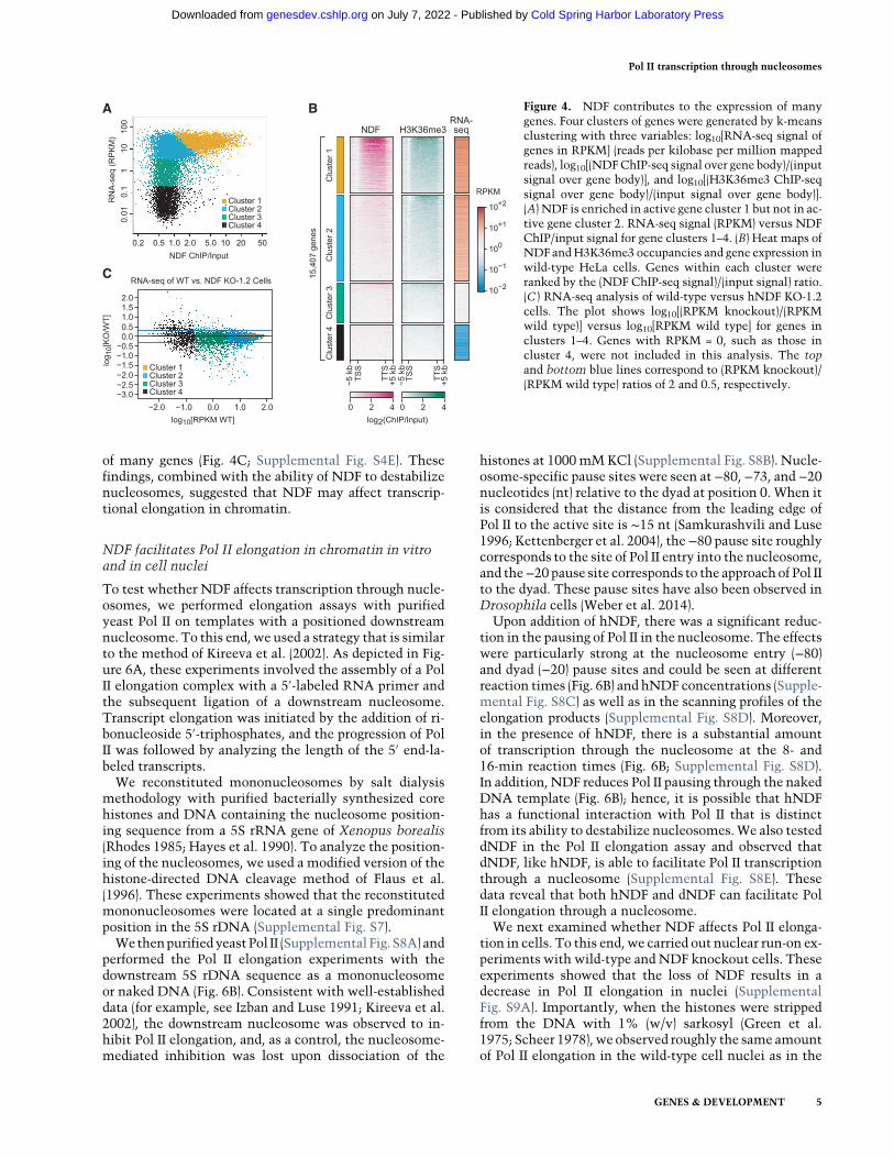
of many genes (Fig. 4C; Supplemental Fig. S4E). Thesefindings, combined with the ability of NDF to destabilizenucleosomes, suggested that NDF may affect transcrip-tional elongation in chromatin.
NDF facilitates Pol II elongation in chromatin in vitroand in cell nuclei
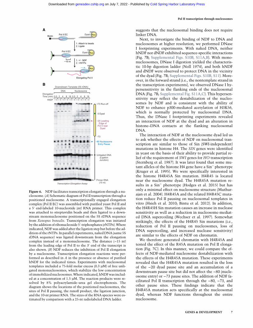
To test whether NDF affects transcription through nucle-osomes, we performed elongation assays with purifiedyeast Pol II on templates with a positioned downstreamnucleosome. To this end, we used a strategy that is similarto the method of Kireeva et al. (2002). As depicted in Fig-ure 6A, these experiments involved the assembly of a PolII elongation complex with a 5′-labeled RNA primer andthe subsequent ligation of a downstream nucleosome.Transcript elongation was initiated by the addition of ri-bonucleoside 5′-triphosphates, and the progression of PolII was followed by analyzing the length of the 5′ end-la-beled transcripts.We reconstituted mononucleosomes by salt dialysis
methodology with purified bacterially synthesized corehistones and DNA containing the nucleosome position-ing sequence from a 5S rRNA gene of Xenopus borealis(Rhodes 1985; Hayes et al. 1990). To analyze the position-ing of the nucleosomes, we used a modified version of thehistone-directed DNA cleavage method of Flaus et al.(1996). These experiments showed that the reconstitutedmononucleosomes were located at a single predominantposition in the 5S rDNA (Supplemental Fig. S7).WethenpurifiedyeastPol II (Supplemental Fig. S8A) and
performed the Pol II elongation experiments with thedownstream 5S rDNA sequence as a mononucleosomeor naked DNA (Fig. 6B). Consistent with well-establisheddata (for example, see Izban and Luse 1991; Kireeva et al.2002), the downstream nucleosome was observed to in-hibit Pol II elongation, and, as a control, the nucleosome-mediated inhibition was lost upon dissociation of the
histones at 1000mMKCl (Supplemental Fig. S8B). Nucle-osome-specific pause sites were seen at −80, −73, and −20nucleotides (nt) relative to the dyad at position 0. When itis considered that the distance from the leading edge ofPol II to the active site is ∼15 nt (Samkurashvili and Luse1996; Kettenberger et al. 2004), the −80 pause site roughlycorresponds to the site of Pol II entry into the nucleosome,and the−20 pause site corresponds to the approach of Pol IIto the dyad. These pause sites have also been observed inDrosophila cells (Weber et al. 2014).Upon addition of hNDF, there was a significant reduc-
tion in the pausing of Pol II in the nucleosome. The effectswere particularly strong at the nucleosome entry (−80)and dyad (−20) pause sites and could be seen at differentreaction times (Fig. 6B) and hNDF concentrations (Supple-mental Fig. S8C) as well as in the scanning profiles of theelongation products (Supplemental Fig. S8D). Moreover,in the presence of hNDF, there is a substantial amountof transcription through the nucleosome at the 8- and16-min reaction times (Fig. 6B; Supplemental Fig. S8D).In addition, NDF reduces Pol II pausing through the nakedDNA template (Fig. 6B); hence, it is possible that hNDFhas a functional interaction with Pol II that is distinctfrom its ability to destabilize nucleosomes.We also testeddNDF in the Pol II elongation assay and observed thatdNDF, like hNDF, is able to facilitate Pol II transcriptionthrough a nucleosome (Supplemental Fig. S8E). Thesedata reveal that both hNDF and dNDF can facilitate PolII elongation through a nucleosome.We next examined whether NDF affects Pol II elonga-
tion in cells. To this end,we carried out nuclear run-on ex-periments with wild-type andNDF knockout cells. Theseexperiments showed that the loss of NDF results in adecrease in Pol II elongation in nuclei (SupplementalFig. S9A). Importantly, when the histones were strippedfrom the DNA with 1% (w/v) sarkosyl (Green et al.1975; Scheer 1978), we observed roughly the same amountof Pol II elongation in the wild-type cell nuclei as in the
BA
C
Figure 4. NDF contributes to the expression of manygenes. Four clusters of genes were generated by k-meansclustering with three variables: log10[RNA-seq signal ofgenes in RPKM] (reads per kilobase per million mappedreads), log10[(NDFChIP-seq signal over gene body)/(inputsignal over gene body)], and log10[(H3K36me3 ChIP-seqsignal over gene body)/(input signal over gene body)].(A) NDF is enriched in active gene cluster 1 but not in ac-tive gene cluster 2. RNA-seq signal (RPKM) versus NDFChIP/input signal for gene clusters 1–4. (B) Heat maps ofNDF andH3K36me3 occupancies and gene expression inwild-type HeLa cells. Genes within each cluster wereranked by the (NDF ChIP-seq signal)/(input signal) ratio.(C ) RNA-seq analysis of wild-type versus hNDF KO-1.2cells. The plot shows log10[(RPKM knockout)/(RPKMwild type)] versus log10[RPKM wild type] for genes inclusters 1–4. Genes with RPKM = 0, such as those incluster 4, were not included in this analysis. The topand bottom blue lines correspond to (RPKM knockout)/(RPKM wild type) ratios of 2 and 0.5, respectively.
Pol II transcription through nucleosomes
GENES & DEVELOPMENT 5
Cold Spring Harbor Laboratory Press on July 7, 2022 - Published by genesdev.cshlp.orgDownloaded from

knockout cell nuclei (Supplemental Fig. S9B). Hence, thereduced levels of transcription in the NDF knockout cellnuclei appear to be due to blockage by nucleosomes. In ad-dition, the transcription was sensitive to 4 µg/mL α-ama-nitin and was thus mediated by Pol II (Supplemental Fig.S9). These findings therefore indicate that NDF facilitatesPol II transcription through nucleosomes in vitro as wellas in cell nuclei.
NDF interacts with nucleosomes in the vicinityof the dyad
To gain a better understanding of the function of NDF innucleosome destabilization and transcription elongation
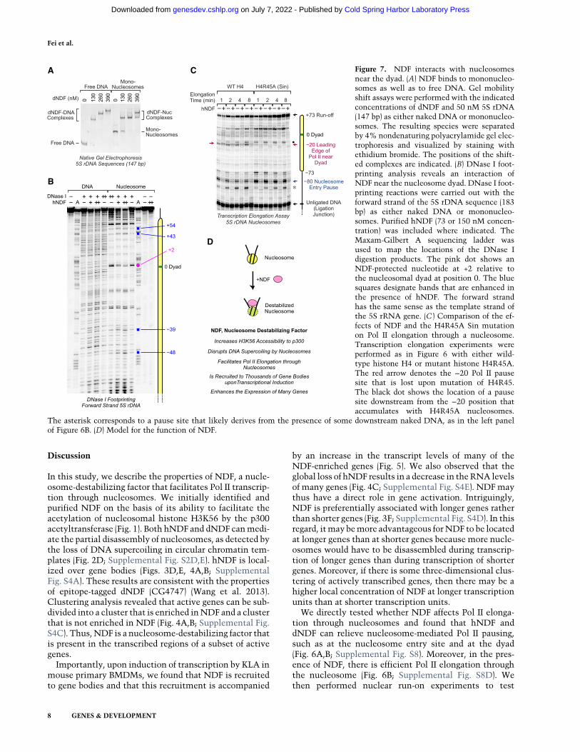
in chromatin, we examined the interaction of NDF withnaked DNA and nucleosomes. First, we performed gelmobility shift analyses. (In these studies, we used dNDFinstead of hNDF because the hNDF-DNA and hNDF-mononucleosome species were not stable under the gelelectrophoresis conditions.) These experiments revealedthat NDF binds to 147-bp 5S rDNA sequences as eithernaked DNA or mononucleosomes (Fig. 7A). To test forany potential sequence specificity in binding, we carriedout identical experiments with a sequence from a Droso-phila hsp70 promoter and obtained essentially the sameresults (Supplemental Fig. S10A). Thus, NDF binds to na-ked DNA as well as to nucleosomes. In addition, the bind-ing of NDF to 147-bpDNA-containingmononucleosomes
B
C D
A Figure 5. NDF is recruited to the transcribed regionsof thousands of genes in mouse primary BMDMsupon transcriptional induction by KLA. (A) Genomebrowser view of H3K36me3 ChIP-seq, mNDF ChIP-seq, and RNA-seq with or without induction by KLAfor 6 h. The region in the vicinity of theTmem67 geneis indicated by the box. (B) Recruitment of mNDF togene bodies upon activation of BMDMs by KLA.Metagene analysis of NDF and H3K36me3 ChIP-seqsignals at 2249 genes in which NDF occupancy in-creases by at least twofold upon 6 h of inductionwith KLA. (C ) The KLA-induced increase in NDF oc-cupancy correlates with an increase in H3K36me3.The scatter plot (35,933 total genes) shows 2249 genesin which NDF occupancy increases (up arrow) by atleast twofold upon KLA induction for 6 h and 4097genes in which NDF occupancy decreases (down ar-row) by at least twofold upon KLA induction for 6h. (D) The KLA-induced increase in NDF occupancygenerally correlates with an increase in transcript lev-els. The genes at whichNDF levels change upon KLAinduction are as in C.
Fei et al.
6 GENES & DEVELOPMENT
Cold Spring Harbor Laboratory Press on July 7, 2022 - Published by genesdev.cshlp.orgDownloaded from
suggests that the nucleosomal binding does not requirelinker DNA.Next, to investigate the binding of NDF to DNA and
nucleosomes at higher resolution, we performed DNaseI footprinting experiments. With naked DNA, neitherhNDF nor dNDF exhibited sequence-specific interactions(Fig. 7B; Supplemental Figs. S10B, S11A,B). With mono-nucleosomes, DNase I digestion yielded the characteris-tic 10-bp digestion ladder (Noll 1974), and both hNDFand dNDF were observed to protect DNA in the vicinityof the dyad (Fig. 7B; Supplemental Figs. S10B, S11). More-over, in the forward strand (i.e., the nontemplate strand inthe transcription experiments), we observed DNase I hy-persensitivity in the flanking ends of the nucleosomalDNA (Fig. 7B; Supplemental Fig. S11A,C). This hypersen-sitivity may reflect the destabilization of the nucleo-somes by NDF and is consistent with the ability ofNDF to enhance p300-mediated acetylation of H3K56,which is normally protected by nucleosomal DNA.Thus, the DNase I footprinting experiments revealedan interaction of NDF at the dyad and an alteration inhistone–DNA contacts at the flanking nucleosomalDNA.The interaction of NDF at the nucleosome dyad led us
to ask whether the effects of NDF on nucleosomal tran-scription are similar to those of Sin (SWI-independent)mutations in histone H4. The SIN genes were identifiedin yeast on the basis of their ability to provide partial re-lief of the requirement of SWI genes for HO transcription(Sternberg et al. 1987). It was later found that some mu-tant alleles of the histone H4 gene have a Sin− phenotype(Kruger et al. 1995). We were specifically interested inthe histone H4R45A Sin mutation. H4R45 is locatednear the nucleosome dyad. The H4R45A mutation re-sults in a Sin− phenotype (Hodges et al. 2015) but hasonly a minimal effect on nucleosome structure (Muthur-ajan et al. 2004). H4R45A and the related H4R45C muta-tion reduce Pol II pausing on nucleosomal templates invitro (Hsieh et al. 2010; Bintu et al. 2012). In addition,the H4R45H Sin mutation causes an increase in nucleasesensitivity as well as a reduction in nucleosome-mediat-ed DNA supercoiling (Wechser et al. 1997). Somewhatstrikingly, the effects of the H4R45 Sin mutations (i.e.,reduction of Pol II pausing on nucleosomes, loss ofDNA supercoiling, and increased nuclease sensitivity)are similar to the effects of NDF on chromatin.We therefore generated chromatin with H4R45A and
tested the effect of the R45A mutation on Pol II elonga-tion (Fig. 7C). In this manner, we could compare the ef-fects of NDF-mediated nucleosome destabilization withthe effects of the H4R45A mutation. These experimentsrevealed that the H4R45A mutation resulted in the lossof the −20 dyad pause site and an accumulation of adownstream pause site but did not affect the −80 (nucle-osome entry) or −73 pause sites. The addition of NDF fa-cilitated Pol II transcription through the −80, −73, andother pause sites. These findings indicate that theH4R45A mutation acts specifically at the nucleosomaldyad, whereas NDF functions throughout the entirenucleosome.
A
B
Figure 6. NDF facilitates transcription elongation through a nu-cleosome. (A) Schematic diagram of Pol II transcription through apositioned nucleosome. A transcriptionally engaged elongationcomplex (Pol II EC) was assembled with purified yeast Pol II anda 5′ end-labeled 10-nucleotide (nt) RNA primer. This complexwas attached to streptavidin beads and then ligated to a down-stream mononucleosome positioned on the 5S rDNA sequencefrom Xenopus borealis. Transcription elongation was initiatedby the addition of ribonucleoside 5′-triphosphates (rNTPs).Whereindicated,NDFwas added after the ligation step but before the ad-ditionof the rNTPs. In parallel experiments, nakedDNA (same5SrDNA sequence) was ligated downstream from the elongationcomplex instead of a mononucleosome. The distance (∼15 nt)from the leading edge of Pol II to the 3′ end of the transcript isalso shown. (B) NDF reduces the inhibition of Pol II elongationby a nucleosome. Transcription elongation reactions were per-formed as described in A in the presence or absence of purifiedhNDF for the indicated times. Experiments with nucleosomaltemplates included a 75-fold molar excess (0.3 µM) of free unli-gated mononucleosomes, which stabilize the low concentrationof immobilizednucleosomes.Where indicated, hNDFwas includ-ed at a concentration of 1.5 µM. The reaction products were re-solved by 8% polyacrylamide–urea gel electrophoresis. Thediagram shows the locations of the positioned nucleosomes, thesites of Pol II pausing, the runoff product, the ligation junction,and the 10-nt primer RNA. The sizes of the RNA species were es-timated by comparison with a 25-nt radiolabeled DNA ladder.
Pol II transcription through nucleosomes
GENES & DEVELOPMENT 7
Cold Spring Harbor Laboratory Press on July 7, 2022 - Published by genesdev.cshlp.orgDownloaded from
Discussion
In this study, we describe the properties of NDF, a nucle-osome-destabilizing factor that facilitates Pol II transcrip-tion through nucleosomes. We initially identified andpurified NDF on the basis of its ability to facilitate theacetylation of nucleosomal histone H3K56 by the p300acetyltransferase (Fig. 1). Both hNDF and dNDF canmedi-ate the partial disassembly of nucleosomes, as detected bythe loss of DNA supercoiling in circular chromatin tem-plates (Fig. 2D; Supplemental Fig. S2D,E). hNDF is local-ized over gene bodies (Figs. 3D,E, 4A,B; SupplementalFig. S4A). These results are consistent with the propertiesof epitope-tagged dNDF (CG4747) (Wang et al. 2013).Clustering analysis revealed that active genes can be sub-divided into a cluster that is enriched inNDF and a clusterthat is not enriched in NDF (Fig. 4A,B; Supplemental Fig.S4C). Thus,NDF is a nucleosome-destabilizing factor thatis present in the transcribed regions of a subset of activegenes.
Importantly, upon induction of transcription by KLA inmouse primary BMDMs, we found that NDF is recruitedto gene bodies and that this recruitment is accompanied
by an increase in the transcript levels of many of theNDF-enriched genes (Fig. 5). We also observed that theglobal loss of hNDF results in a decrease in theRNA levelsof many genes (Fig. 4C; Supplemental Fig. S4E). NDFmaythus have a direct role in gene activation. Intriguingly,NDF is preferentially associated with longer genes ratherthan shorter genes (Fig. 3F; Supplemental Fig. S4D). In thisregard, itmay bemore advantageous forNDF to be locatedat longer genes than at shorter genes because more nucle-osomes would have to be disassembled during transcrip-tion of longer genes than during transcription of shortergenes. Moreover, if there is some three-dimensional clus-tering of actively transcribed genes, then there may be ahigher local concentration of NDF at longer transcriptionunits than at shorter transcription units.
We directly tested whether NDF affects Pol II elonga-tion through nucleosomes and found that hNDF anddNDF can relieve nucleosome-mediated Pol II pausing,such as at the nucleosome entry site and at the dyad(Fig. 6A,B; Supplemental Fig. S8). Moreover, in the pres-ence of NDF, there is efficient Pol II elongation throughthe nucleosome (Fig. 6B; Supplemental Fig. S8D). Wethen performed nuclear run-on experiments to test
A
D
B
C Figure 7. NDF interacts with nucleosomesnear the dyad. (A) NDF binds to mononucleo-somes as well as to free DNA. Gel mobilityshift assays were performed with the indicatedconcentrations of dNDF and 50 nM 5S rDNA(147 bp) as either naked DNA or mononucleo-somes. The resulting species were separatedby 4% nondenaturing polyacrylamide gel elec-trophoresis and visualized by staining withethidium bromide. The positions of the shift-ed complexes are indicated. (B) DNase I foot-printing analysis reveals an interaction ofNDF near the nucleosome dyad. DNase I foot-printing reactions were carried out with theforward strand of the 5S rDNA sequence (183bp) as either naked DNA or mononucleo-somes. Purified hNDF (73 or 150 nM concen-tration) was included where indicated. TheMaxam-Gilbert A sequencing ladder wasused to map the locations of the DNase Idigestion products. The pink dot shows anNDF-protected nucleotide at +2 relative tothe nucleosomal dyad at position 0. The bluesquares designate bands that are enhanced inthe presence of hNDF. The forward strandhas the same sense as the template strand ofthe 5S rRNA gene. (C ) Comparison of the ef-fects of NDF and the H4R45A Sin mutationon Pol II elongation through a nucleosome.Transcription elongation experiments wereperformed as in Figure 6 with either wild-type histone H4 or mutant histone H4R45A.The red arrow denotes the −20 Pol II pausesite that is lost upon mutation of H4R45.The black dot shows the location of a pausesite downstream from the −20 position thataccumulates with H4R45A nucleosomes.
The asterisk corresponds to a pause site that likely derives from the presence of some downstream naked DNA, as in the left panelof Figure 6B. (D) Model for the function of NDF.
Fei et al.
8 GENES & DEVELOPMENT
Cold Spring Harbor Laboratory Press on July 7, 2022 - Published by genesdev.cshlp.orgDownloaded from
whether NDF contributes to Pol II elongation in cells andfound that NDF is important for Pol II elongation throughchromatin in cell nuclei (Supplemental Fig. S9).To gain further insight into this activity, we examined
the interaction of NDF with chromatin and found thatboth hNDF and dNDF interact with nucleosomes in thevicinity of the dyad (Fig. 7A,B; Supplemental Figs. S10,S11). Comparison of the effects of NDF with those ofthe H4R45A Sin mutation revealed that H4R45A affectstranscription at the dyad, whereas NDF affects transcrip-tion throughout the nucleosome (Fig. 7C). Altogether,these data indicate that NDF is a nucleosome-bindingand -destabilizing factor that is recruited to gene bodiesupon induction of transcription and facilitates Pol II elon-gation in chromatin (Fig. 7D).NDF is present in most animals and perhaps in plants
but does not appear to have a homolog in Saccharomycescerevisiae. NDF contains a conserved N-terminal PWWPmotif and a conserved C-terminal dehydrogenase domain(Fig. 2A; Supplemental Fig. S1B), but hNDF does not ex-hibit dehydrogenase activity (Vermeulen et al. 2010).The PWWP methyl lysine-binding domain (Qin and Min2014) is likely to be involved in the binding of NDF toH3K36me3. However, it appears that H3K36me3 is neces-sary but not sufficient for the recruitment of NDF to chro-matin. The proper localization of dNDF in chromatin isdependent on the Set2 histone H3K36 methyltransferase(Wang et al. 2013); however, many genes with moderateto high levels of H3K36me3 are not enriched in NDF(Fig. 4A,B; Supplemental Fig. S4C), and the sharp increasein NDF occupancy at gene bodies upon transcriptional in-duction is not mirrored by a similarly sharp increase inH3K36me3 (Fig. 5A,B). Thus, H3K36me3 does not appearto be sufficient for the recruitment of NDF. It is also nota-ble that the biochemical activities of NDF, such as thedestabilization of nucleosomes and relief of nucleo-some-mediated Pol II pausing, do not absolutely requireH3K36 methylation, as our experiments were performedwith nucleosomes that were reconstituted with unmodi-fied bacterially synthesized histones. These findings sug-gest that, once recruited to its site of action, NDF candestabilize nucleosomes and facilitate transcription by amechanism that is not strictly dependent on H3K36me3.Because NDF relieves nucleosome-mediated inhibition
of Pol II elongation, it is essential to compare NDF withFACT, the well-studied factor that facilitates transcrip-tion and DNA replication in chromatin (for example, seeReinberg and Sims 2006; Formosa 2008). FACT is presentin many eukaryotes, including S. cerevisiae, Drosophila,and humans. FACT and NDF are not related in terms oftheir primary amino acid sequences or protein domains.Moreover, FACT binds to the histones and exhibits corehistone chaperone activity, whereas NDF does not appearto be a core histone chaperone (data not shown). In yeast,the Spt16 subunit of FACT is enriched over gene bodiesand depleted over promoter regions of active genes (Trueet al. 2016). In contrast, both subunits of mouse FACTwere found to be strongly enriched in the vicinity of tran-scription start sites but also present over gene bodies(Mylonas and Tessarz 2018). These findings suggest that
there are substantial differences in the biological func-tions of FACT in yeast versus mice. The localization ofmNDF and hNDF (Figs. 3E, 5B) is more similar to thatof yeast FACT than mouse FACT. It is also notablethat hNDF was observed to be present at high levels inthe nuclei of 44 out of 44 tissue types tested, whereasthe levels of the SSRP1 subunit of FACT were found tobe low (16 out of 43) or not detectable (nine out of 43) inthe same tissue types (Supplemental Fig. S1A; Garciaet al. 2011; Uhlén et al. 2015). In addition, it appearsthat NDF is essential in human HUES62 stem cells,whereas subunits of FACT are not (Shalem et al. 2014).Thus, inmammals, bothNDF and FACT are able to desta-bilize nucleosomes and facilitate transcription in chroma-tin, but they function by distinct mechanisms and indifferent tissues.It is possible and perhaps likely that there is at least one
other factor that is related to NDF. The existence of anNDF-related factor is suggested by the observation thatNDF is not essential for the viability of HeLa cells orA375 cells. In addition, the absence ofNDFdoes not resultin a general loss of transcription of genes that are enrichedin NDF (Fig. 4). These findings could be explained by thepresence of an NDF-like factor that is recruited to mostlythe same genes asNDF (cluster 1 genes) (Fig. 4), such as viaa PWWP motif. This NDF-like factor would then largelycompensate for the absence of NDF, especially at genesthat are enriched for both factors. The identification andcharacterization of an NDF-related factor would lead tosignificant new insights into the transcription process.Based on the effects of NDF on transcription and gene
expression, it is also important to consider whether alter-ations in the protein sequence or levels of NDF might berelevant to human diseases. In this regard, the NDF/GLYR1 gene is overexpressed in ∼21% of breast cancers(Supplemental Fig. S12A,B; Forbes et al. 2017) andmutatedin ∼51% of microsatellite unstable colorectal cancers(Alhopuro et al. 2012). The up-regulation of NDF/GLYR1alsocorrelateswitha reduced survival probability in breastcancer patients (Supplemental Fig. S12C; Uhlen et al.2017). The knowledge of the biochemical activities ofNDF would be useful in assessing the potential roles thatthis factor may have in breast cancer or other diseases.In conclusion, the properties of NDF indicate that it has
a key role in the transcription of chromatin by Pol II in an-imals. The further analysis ofNDF should reveal addition-al factors andmechanisms that orchestrate the processionof Pol II through active genes.
Materials and methods
To ensure the reproducibility of the results, each experimentalcondition was performed independently at least twice.
Antibodies
Rabbit polyclonal antisera against dNDFand hNDFwere generat-ed with purified full-length Escherichia coli synthesized proteinsat PoconoRabbit FarmandLaboratory.The following commercialantibodies were used in this study: anti-H3K56ac (1:450 dilution;
Pol II transcription through nucleosomes
GENES & DEVELOPMENT 9
Cold Spring Harbor Laboratory Press on July 7, 2022 - Published by genesdev.cshlp.orgDownloaded from

Millipore Sigma, 07-677I), anti-H3K56ac (1:1000 dilution; ActiveMotif, 39281), anti-H3K56ac (1:1000 dilution; Cell SignalingTechnology, 4243), anti-H3 (1:4000 dilution; Cell Signaling Tech-nology, 4499), anti-H3K9ac (1:1000 dilution; Abcam, ab8898),anti-H3K36me3 (1:1000 dilution; Abcam, ab9050), anti-his(1:1000 dilution; Santa Cruz Biotechnology, sc-803), and anti-PolII (1:1000 dilution; Santa Cruz Biotechnology, sc-9001).
Purification of native NDF from Drosophila embryos
Native dNDFwas purified from the 0.1MKCl version of the solu-ble nuclear fraction (Kamakaka et al. 1991) fromDrosophila em-bryos by using the scheme shown in Figure 1B. Buffer N (20 mMHEPES K+ at pH 7.5, 10% [v/v] glycerol, 0.5 mM EDTA, 0.5 mMEGTA, 1.5 mMMgCl2, 0.1 M NaCl, 10 mM ß-glycerophosphate,1mMDTT, 0.2mMPMSF)was used throughout the purification.Proteins in the peak activity fractions of the final Source 15Qchromatography step were identified by mass spectroscopy (Bio-molecular/Proteomics Mass Spectrometry Facility, Universityof California at San Diego).
Purification of recombinant proteins
The cDNAs for dNDF (CG4747;DrosophilaGenomics ResourceCenter) and hNDF (GLYR1; Dharmacon) (note that three mis-matches in the cDNA were corrected) were subcloned intopET21b with N-terminal His6 tags to give pET21b-His6-dNDFand pET21b-His6-hNDF, and the integrity of the sequences wasconfirmed by DNA sequence analysis. The N-terminally His6-tagged dNDF and hNDF proteins were purified by the followingmethod. Freshly transformed E. coli BL21(DE3) was grown in2 L of LB medium containing 40 µg/mL ampicillin at 37°C toan A600 nm of ∼0.6, and the synthesis of the recombinant NDFwas induced by the addition of IPTG to 0.4 mM final concentra-tion. The culture was then incubated for an additional 16–18 h inan 18°C shaking water bath, and the bacteria were collected bycentrifugation at 6000 rpm for 10 min at 22°C (Fiberlite F9-4x1000y rotor). Unless stated otherwise, all subsequent opera-tions were performed at 4°C. The bacterial pellets were resus-pended in 30 mL of lysis buffer (10 mM Tris-HCl at pH 7.5,0.2% [v/v] nonidet P-40, 1 M NaCl, 10% [v/v] glycerol, 15 mMimidazole, 5 mM 2-mercaptoethanol, 0.2 mM PMSF, 1× proteaseinhibitor cocktail [Millipore Sigma, P9599]). The suspension wassubjected to sonication on ice four times for 30 sec (Branson Soni-fier 450 with a 0.25-in microtip at 20% output). Insoluble materi-al was removed by centrifugation at 13,000 rpm for 30 min(Fiberlite F21S-8x50y rotor), and the supernatant was incubatedwith 3 mL (pre-equilibrated in lysis buffer) of Ni-NTA Superflow(Qiagen) for 3 h on a nutator. The mixture was transferred into aBio-Rad Econo-Pac chromatography column, and the flow-through, which contained ∼80% of the His6-NDF proteins, wascollected and incubated with 2 mL (pre-equilibrated in lysis buff-er) of Ni-NTA agarose (Qiagen) for 2 h on a nutator. The mixturewas transferred into a Bio-Rad Econo-Pac chromatography col-umn, and the resin was washed twice with 10 mL of buffer A(10 mM Tris-HCl at pH 7.5, 0.2% [v/v] nonidet P-40, 10% [v/v]glycerol, 15mMimidazole, 5mM2-mercaptoethanol) containing1MNaCl followed by twicewith 10mLof buffer A containing 0.2MNaCl. TheHis6-NDFwas eluted four timeswith 0.5mL of elu-tion buffer (10 mM Tris-HCl at pH 7.5, 0.2% [v/v] nonidet P-40,0.2MNaCl, 10% [v/v] glycerol, 250mM imidazole, 5mM2-mer-captoethanol). With His6-dNDF, it was necessary to remove acontaminating nuclease by dialysis into buffer A containing0.1 M NaCl followed by chromatography on a 1-mL Source 15Q(GE Life Sciences) anion exchange column with linear gradient
elution from 0.1 M to 1.0 M NaCl. The His6-dNDF eluted at∼0.16 M NaCl. The purified NDF proteins were frozen in liquidnitrogen and stored at −80°C.Humanp300proteinwas synthesized inSf9 cells andpurified as
described inKraus andKadonaga (1998). RecombinantDrosophilamelanogaster core histones were synthesized in E. coli and puri-fied by the method of Khuong et al. (2017). The catalytic domainof Drosophila topoisomerase I was synthesized and purified asdescribed by Fyodorov and Kadonaga (2003). The S. cerevisiaestrain that expresses protein A-tagged Rbp3 subunit of Pol II wasthe generous gift of Dr. Craig Kaplan (Texas A&M University),and the Pol II was purified as described (Kaplan et al. 2008).
Nucleosome reconstitution
The X. borealis 5S rDNA and Drosophila hsp70 (87A) nucleo-some positioning sequences were amplified by PCR and purifiedby 1.5% agarose gel electrophoresis and gel extraction (QiaQuickgel extraction kit, Qiagen). The resulting fragments were then re-constituted into mononucleosomes by salt dialysis (Stein 1989).In the transcription elongation studies, the PCR-amplifiedDNAwas digested with TspRI restriction enzyme (New EnglandBiolabs) for 2 h at 65°C prior to agarose gel electrophoresis, gel ex-traction, and mononucleosome reconstitution. Nucleosomeswere reconstituted onto CsCl-purified pGIE-0 plasmid DNA(Pazin et al. 1994) by salt dialysis (Stein 1989; Fei et al. 2015) ata core histone to DNA mass ratio of 0.8:1.
H3K56 acetylation assay
The H3K56 acetylation assays were performed with either nucle-osomes reconstituted onto plasmid DNA by salt dialysis (1.4 µMconcentration of nucleosomes) or free core histones (1.4 µM con-centration of core histone octamers) along with 1 µg of purifiedhuman p300, 30 µM acetyl-CoA, and 1.4 µg of bovine serum albu-min in buffer K (25 mMHEPES K+ at pH 7.6, 100 mMKCl, 5 mMMgCl2, 0.1 mM EDTA, 10% [v/v] glycerol) in a total volume of50 µL. The mixture was incubated for 1 h at 30°C. The histoneswere precipitated with 25% (w/v) trichloroacetic acid, subjectedto 15% polyacrylamide–SDS gel electrophoresis, and detectedby Western blot analysis.
Transcription elongation assay
The transcriptionelongationassay is depicted inFigure6Aand is amodified version of the method of Kireeva et al. (2002). In our ex-periments, the 10-nt 5′ 32P-labeled primer RNA (1.6 pmol = 8 µLof a 0.2 µM stock) was annealed to an equimolar amount of tem-plate strand DNA. Purified Pol II (4 pmol) was then added to 1.6pmol of the RNA–template strand complex, and the componentswere incubated for 10min at 30°C.Next, 1.6 pmol of the 5′ biotin-labelednontemplate strandwas added and incubated for 15min at30°C to form the elongation complex, which was then immobi-lized onto 10 µL of Dynabeads MyOne Streptavidin C1 magneticbeads (Invitrogen). The beads were washed twice with 50 µL ofEB1000 buffer (20mMTris-HCl at pH 7.5, 1MKCl, 5mMMgCl2,10mMDTT) followed by twicewith 50 µL of EB40 buffer (20mMTris-HCl at pH 7.5, 40 mM KCl, 5 mMMgCl2, 10 mMDTT) andthen resuspended in 16 µL of EB40 buffer. The immobilization ef-ficiency of the elongation complex was estimated with a Geigercounter. Typically,∼60%of the elongation complexwas immobi-lized onto the Dynabeads. Next, 3 pmol of either mononucleo-somes or the corresponding naked DNA fragments (both withcomplementary TspRI overhangs) was ligated to the immobilizedelongation complex by using 2000 U of T4 DNA ligase (New
Fei et al.
10 GENES & DEVELOPMENT
Cold Spring Harbor Laboratory Press on July 7, 2022 - Published by genesdev.cshlp.orgDownloaded from
England Biolabs) in 0.2× (final concentration) T4DNA ligase reac-tion buffer (NewEngland Biolabs) in a total volume of 50 µL for 25min at 22°C. After ligation, the beads were collected and resus-pended in 50 µL of EB40 buffer containing 20 µg of bovine serumalbumin (ThermoFisher Pierce), 100Uof SUPERase•InRNase in-hibitor (Thermo Fisher Invitrogen), and 30 pmol of unlabeled freemononucleosomes on 147-bp DNA. The unlabeled free mononu-cleosomes were added only to the elongation complex templatesthat were ligated to a downstream nucleosome and served to sta-bilize the template-associated nucleosomes by increasing theoverall concentration of nucleosomes in the reaction medium(for example, see Godde and Wolffe 1995). The beads were thenaliquoted into tubes for the individual reactions. Each reactioncontained 5 µL of the bead-containing suspension. If the ligationefficiency were 100%, then each tube would contain ∼0.08 pmolof elongation complex template. Fornucleosome-containing tem-plates, each tube additionally contained 3 pmol of unlabeled freemononucleosomes to stabilize the template-associated nucleo-somes. Next, NDF or the corresponding buffer (as a negative con-trol) was added to each tube, EB40 buffer was added to a finalvolume of 9 µL, and the samples were incubated for 20 min at30°C. At this stage, the KCl concentration was 100 mM unlessstated otherwise. Next, transcription elongation was initiatedby the addition of 1 µL of 2.5 mM rNTPs, and the reactions wereallowed to proceed for the indicated times at 22°C. The reactionswere terminatedwith 10 µL of gel loading buffer (1mL of gel load-ing buffer = 0.9 mL of deionized formamide, 0.1 mL of 0.5 MEDTA, 2 µL of 4% [w/v] bromphenol blue). The samples were an-alyzed by denaturing 8% polyacrylamide–urea gel electrophore-sis, and the gel images were collected on a GE Typhoon imager(GE Healthcare).
Analysis of mNDF in primary BMDMs
Bone marrow was isolated from 8- to-10-wk-old C57Bl/6 (HarlanLaboratories) female mice. BMDMs were generated in DMEMsupplemented with M-CSF for 7 d and activated with KLA for6 h as described (Kaikkonen et al. 2013). RNA-seq libraries wereprepared from poly(A)-enriched mRNA as described previously(Kaikkonen et al. 2013). For ChIP-seq, 5 million BMDMs werecross-linked in 1% formaldehyde for 10 min. ChIP-seq was per-formed as described previously (Kaikkonen et al. 2013) with2 µL of hNDF antisera 7 or 2 µL of H3K36me3 antibodies (Abcam,ab9050). (We also performed ChIP-seq experiments with hNDFantisera 6 and obtained results essentially identical to thosewith hNDF antisera 7 [data not shown].) ChIP-seq libraries wereprepared from ChIP DNA and inputs by blunting, A-tailing, andadapter ligation as described previously (Heinz et al. 2010) by us-ing NextFlex barcode adapters. Libraries were amplified, size-se-lected by gel extraction, and subsequently sequenced on theIllumina Hi-Seq 4000 sequencer by using single-end 75-bp reads.Sequencing datawere aligned to themm10 genome by using Bow-tie2, and all data were subsequently analyzed with HOMER andvisualized in the University of California at Santa Cruz genomebrowser. Pathway analysis was performed by using Metascape(http://metascape.org).Additional Materials and Methods are included in the Supple-
mental Material.The genome-wide data have been deposited at theGene Expres-
sion Omnibus (accession no. GSE109690).
Acknowledgments
We thank Peter Geiduschek, Grisel Cruz, and Long Vo ngoc forcritical reading of themanuscript.We are grateful to Craig Kaplan
for the tagged yeast Pol II strain, Gaoyang Liang for CRISPR–Cas9vectors, andDongWang and JunXu for their invaluable advice onthe elongation assay and Pol II purification. We also thank Yun-Ching Chen for help in the analysis of the RNA-seq data. J.T.K.is the Amylin Chair in the Life Sciences. F.M. was supportedby a fellowship from the Deutsche Forschungsgemeinschaft(ME 4713/1-1) and a National Institutes of Health (NIH) grantto KarenOegema (GM074207). This workwas supported by fund-ing from the Ludwig Institute for Cancer Research to B.R., theNIH/National Institute of Diabetes and Digestive and KidneyDiseases (DK091183) and Foundation Le Ducq to C.K.G, andthe NIH/National Institute of General Medical Sciences (R35GM118060) to J.T.K.Author contributions: This work was carried out in an interac-
tive manner in which all authors made important intellectualcontributions to the development of the project. J.F. identifiedand purified NDF, performed essentially all of the biochemicalanalyses of the protein, and generated the knockout cell lines.H.I. and B.R. contributed the genome-wide analyses of NDF inhuman cells. M.A.H. and C.K.G. contributed the analyses ofNDF in mouse cells, including the KLA induction experiments.H.I. had a lead role in the analysis of the genome-wide data.F.M. performed the cell cycle studies. G.A.K. contributed to thedesign and analysis of the transcription and footprinting experi-ments. J.F., G.A.K., and J.T.K. oversaw the overall execution ofthis work. J.F. and J.T.K. were primarily responsible for writingthe manuscript and received substantial contributions from allother authors in the preparation of figures and the writing andediting of the text.
References
Alhopuro P, Sammalkorpi H, Niittymäki I, BiströmM, Raitila A,Saharinen J, Nousiainen K, Lehtonen HJ, Heliövaara E,Puhakka J, et al. 2012. Candidate driver genes in microsatel-lite-unstable colorectal cancer. Int J Cancer 130: 1558–1566.
Bintu L, Ishibashi T, Dangkulwanich M, Wu YY, Lubkowska L,Kashlev M, Bustamante C. 2012. Nucleosomal elementsthat control the topography of the barrier to transcription.Cell 151: 738–749.
Fang R, Chen F, Dong Z, Hu D, Barbera AJ, Clark EA, Fang J,Yang Y, Mei P, Rutenberg M, et al. 2013. LSD2/KDM1B andits cofactor NPAC/GLYR1 endow a structural and molecularmodel for regulation of H3K4 demethylation. Mol Cell 49:558–570.
Fei J, Torigoe SE, Brown CR, Khuong MT, Kassavetis GA, BoegerH, Kadonaga JT. 2015. The prenucleosome, a stable con-formational isomer of the nucleosome. Genes Dev 29:2563–2575.
Flaus A, Luger K, Tan S, Richmond TJ. 1996. Mapping nucleo-some position at single base-pair resolution by using site-di-rected hydroxyl radicals. Proc Natl Acad Sci 93: 1370–1375.
Forbes SA, Beare D, Boutselakis H, Bamford S, Bindal N, Tate J,Cole CG, Ward S, Dawson E, Ponting L, et al. 2017. COSMIC:somatic cancer genetics at high-resolution.Nucleic Acids Res45: D777–D783.
Formosa T. 2008. FACT and the reorganized nucleosome. MolBiosyst 4: 1085–1093.
Fyodorov DV, Kadonaga JT. 2003. Chromatin assembly in vitrowith purified recombinant ACF and NAP-1. Methods Enzy-mol 371: 499–515.
Garcia H, Fleyshman D, Kolesnikova K, Safina A, Commane M,Paszkiewicz G, Omelian A, Morrison C, Gurova K. 2011. Ex-pression of FACT in mammalian tissues suggests its role in
Pol II transcription through nucleosomes
GENES & DEVELOPMENT 11
Cold Spring Harbor Laboratory Press on July 7, 2022 - Published by genesdev.cshlp.orgDownloaded from
maintaining of undifferentiated state of cells. Oncotarget 2:783–796.
Germond JE, Hirt B, Oudet P, Gross-BellardM, Chambon P. 1975.Folding of the DNA double helix in chromatin-like structuresfrom simian virus 40. Proc Natl Acad Sci 72: 1843–1847.
Godde JS, Wolffe AP. 1995. Disruption of reconstituted nucleo-somes. The effect of particle concentration, MgCl2 and KClconcentration, the histone tails, and temperature. J BiolChem 270: 27399–27402.
Green MH, Buss J, Gariglio P. 1975. Activation of nuclear RNApolymerase by Sarkosyl. Eur J Biochem 53: 217–225.
Hayes JJ, Tullius TD,Wolffe AP. 1990. The structure of DNA in anucleosome. Proc Natl Acad Sci 87: 7405–7409.
Heinz S, Benner C, SpannN, Bertolino E, Lin YC, Lasko P, ChengJX,MurreC, SinghH,GlassCK. 2010. Simple combinations oflineage-determining transcription factors prime cis-regulato-ry elements required for macrophage and B cell identities.Mol Cell 38: 576–589.
Hodges AJ, Gallegos IJ, Laughery MF, Meas R, Tran L, Wyrick JJ.2015. Histone sprocket arginine residues are important forgene expression, DNA repair, and cell viability in Saccharo-myces cerevisiae. Genetics 200: 795–806.
Hsieh FK, Fisher M, Ujvari A, Studitsky VM, Luse DS. 2010. His-tone Sin mutations promote nucleosome traversal and his-tone displacement by RNA polymerase II. EMBO Rep 11:705–710.
Izban MG, Luse DS. 1991. Transcription on nucleosomal tem-plates by RNA polymerase II in vitro: inhibition of elongationwith enhancement of sequence-specific pausing. Genes Dev5: 683–696.
Kaikkonen MU, Spann NJ, Heinz S, Romanoski CE, Allison KA,Stender JD, Chun HB, Tough DF, Prinjha RK, Benner C, et al.2013. Remodeling of the enhancer landscape during macro-phage activation is coupled to enhancer transcription. MolCell 51: 310–325.
Kamakaka RT, Tyree CM, Kadonaga JT. 1991. Accurate and effi-cient RNA polymerase II transcription with a soluble nuclearfraction derived fromDrosophila embryos. ProcNatl Acad Sci88: 1024–1028.
Kaplan CD, Larsson KM, Kornberg RD. 2008. The RNA polymer-ase II trigger loop functions in substrate selection and isdirectly targeted by α-amanitin. Mol Cell 30: 547–556.
KettenbergerH, Armache KJ, Cramer P. 2004. Complete RNApo-lymerase II elongation complex structure and its interactionswith NTP and TFIIS. Mol Cell 16: 955–965.
Khuong MT, Fei J, Ishii H, Kadonaga JT. 2015. Prenucleosomesand active chromatin. Cold Spring Harbor Symp Quant Biol80: 65–72.
Khuong MT, Fei J, Cruz-Becerra G, Kadonaga JT. 2017. A simpleand versatile system for the ATP-dependent assembly of chro-matin. J Biol Chem 292: 19478–19490.
Kireeva ML, Walter W, Tchernajenko V, Bondarenko V, KashlevM, Studitsky VM. 2002. Nucleosome remodeling induced byRNA polymerase II: loss of the H2A/H2B dimer during tran-scription. Mol Cell 9: 541–552.
KrausWL, Kadonaga JT. 1998. p300 and estrogen receptor cooper-atively activate transcription via differential enhancement ofinitiation and reinitiation. Genes Dev 12: 331–342.
KrugerW, Peterson CL, Sil A, Coburn C, Arents G,MoudrianakisEN, Herskowitz I. 1995. Amino acid substitutions in thestructured domains of histones H3 and H4 partially relievethe requirement of the yeast SWI/SNF complex for transcrip-tion. Genes Dev 9: 2770–2779.
Muthurajan UM, Bao Y, Forsberg LJ, Edayathumangalam RS,Dyer PN, White CL, Luger K. 2004. Crystal structures of his-
tone Sinmutant nucleosomes reveal altered protein–DNA in-teractions. EMBO J 23: 260–271.
Mylonas C, Tessarz P. 2018. Transcriptional repression by FACTis linked to regulation of chromatin accessibility at the pro-moter of ES cells. bioRxiv doi: 10.1101/251611.
NollM. 1974. Internal structure of the chromatin subunit.Nucle-ic Acids Res 1: 1573–1578.
Oishi Y, Spann NJ, Link VM, Muse ED, Strid T, Edillor C, KolarMJ,Matsuzaka T, Hayakawa S, Tao J, et al. 2017. SREBP1 con-tributes to resolution of pro-inflammatory TLR4 signaling byreprogramming fatty acid metabolism. Cell Metab 25: 412–427.
Pazin MJ, Kamakaka RT, Kadonaga JT. 1994. ATP-dependentnucleosome reconfiguration and transcriptional activationfrom preassembled chromatin templates. Science 266:2007–2011.
Qin S, Min J. 2014. Structure and function of the nucleo-some-binding PWWP domain. Trends Biochem Sci 39:536–547.
Raetz CR,Garrett TA, ReynoldsCM, ShawWA,Moore JD, SmithDC Jr, Ribeiro AA, Murphy RC, Ulevitch RJ, Fearns C, et al.2006. Kdo2-lipid A of Escherichia coli, a defined endotoxinthat activates macrophages via TLR-4. J Lipid Res 47:1097–1111.
Reinberg D, Sims RJ III. 2006. deFACTo nucleosome dynamics. JBiol Chem 281: 23297–23301.
Rhodes D. 1985. Structural analysis of a triple complex betweenthe histone octamer, a Xenopus gene for 5S RNA and tran-scription factor IIIA. EMBO J 4: 3473–3482.
Samkurashvili I, Luse DS. 1996. Translocation and transcription-al arrest during transcript elongation by RNA polymerase II. JBiol Chem 271: 23495–23505.
Scheer U. 1978. Changes of nucleosome frequency in nucleolarand non-nucleolar chromatin as a function of transcription:an electron microscopic study. Cell 13: 535–549.
ShalemO, Sanjana NE, Hartenian E, Shi X, Scott DA, MikkelsonT, Heckl D, Ebert BL, Root DE, Doench JG, et al. 2014. Ge-nome-scale CRISPR–Cas9 knockout screening in humancells. Science 343: 84–87.
Stein A. 1989. Reconstitution of chromatin from purified compo-nents. Methods Enzymol 170: 585–603.
Sternberg PW, Stern MJ, Clark I, Herskowitz I. 1987. Activationof the yeast HO gene by release from multiple negative con-trols. Cell 48: 567–577.
Torigoe SE, Urwin DL, Ishii H, Smith DE, Kadonaga JT. 2011.Identification of a rapidly formed non-nucleosomal histone-DNA intermediate that is converted into chromatin by ACF.Mol Cell 43: 638–648.
Torigoe SE, Patel A, Khuong MT, Bowman GD, Kadonaga JT.2013. ATP-dependent chromatin assembly is functionally dis-tinct from chromatin remodeling. eLife 2: e00863.
True JD, Muldoon JJ, Carver MN, Poorey K, Shetty SJ, BekiranovS, Auble DT. 2016. The modifier of transcription 1 (Mot1)ATPase and Spt16 histone chaperone co-regulate transcrip-tion through preinitiation complex assembly and nucleosomeorganization. J Biol Chem 291: 15307–15319.
Uhlén M, Fagerberg L, Hallström BM, Lindskog C, Oksvold P,Mardinoglu A, Sivertsson Å, Kampf C, Sjöstedt E, AsplundA, et al. 2015. Tissue-based map of the human proteome. Sci-ence 347: 1260419.
Uhlen M, Zhang C, Lee S, Sjöstedt E, Fagerberg L, Bidkhori G,Benfeitas R, Arif M, Liu Z, Edfors F, et al. 2017. A pathologyatlas of the human cancer transcriptome. Science 357:eaan2507.
Fei et al.
12 GENES & DEVELOPMENT
Cold Spring Harbor Laboratory Press on July 7, 2022 - Published by genesdev.cshlp.orgDownloaded from
VermeulenM, EberlHC,Matarese F,MarksH,Denissov S, ButterF, Lee KK, Olsen JV, HymanAA, Stunnenberg HG, et al. 2010.Quantitative interaction proteomics and genome-wide profil-ing of epigenetic histone marks and their readers. Cell 142:967–980.
Wang CI, Alekseyenko AA, LeRoy G, Elia AE, Gorchakov AA,Britton LM, Elledge SJ, Kharchenko PV, Garcia BA, KurodaMI. 2013. Chromatin proteins captured by ChIP-mass spec-
trometry are linked to dosage compensation in Drosophila.Nat Struct Mol Biol 20: 202–209.
Weber CM, Ramachandran S, Henikoff S. 2014. Nucleosomes arecontext-specific, H2A.Z-modulated barriers to RNA polymer-ase. Mol Cell 53: 819–830.
WechserMA, KladdeMP, Alfieri JA, PetersonCL. 1997. Effects ofSin– versions of histone H4 on yeast chromatin structure andfunction. EMBO J 16: 2086–2095.
Pol II transcription through nucleosomes
GENES & DEVELOPMENT 13
Cold Spring Harbor Laboratory Press on July 7, 2022 - Published by genesdev.cshlp.orgDownloaded from
10.1101/gad.313973.118Access the most recent version at doi: published online May 14, 2018Genes Dev.
Jia Fei, Haruhiko Ishii, Marten A. Hoeksema, et al. through nucleosomesNDF, a nucleosome destabilizing factor that facilitates transcription
Material
Supplemental
http://genesdev.cshlp.org/content/suppl/2018/05/14/gad.313973.118.DC1
Published online May 14, 2018 in advance of the full issue.
License
Commons Creative
.http://creativecommons.org/licenses/by-nc/4.0/at Creative Commons License (Attribution-NonCommercial 4.0 International), as described
). After six months, it is available under ahttp://genesdev.cshlp.org/site/misc/terms.xhtmlsix months after the full-issue publication date (see This article is distributed exclusively by Cold Spring Harbor Laboratory Press for the first
ServiceEmail Alerting
click here.right corner of the article or
Receive free email alerts when new articles cite this article - sign up in the box at the top
Published by © 2018 Fei et al.; Published by Cold Spring Harbor Laboratory Press
Cold Spring Harbor Laboratory Press on July 7, 2022 - Published by genesdev.cshlp.orgDownloaded from
Copyright © 2022 FDOKUMEN