ERCC1-XPF endonuclease facilitates DNA double-strand break repair
12
Published Ahead of Print 9 June 2008. 10.1128/MCB.00293-08. 2008, 28(16):5082. DOI: Mol. Cell. Biol. Niedernhofer Paul Hasty, Jan H. J. Hoeijmakers and Laura J. Ellen van Drunen, H. Berna Beverloo, David B. Weisberg, Anwaar Ahmad, Andria Rasile Robinson, Anette Duensing, Double-Strand Break Repair ERCC1-XPF Endonuclease Facilitates DNA http://mcb.asm.org/content/28/16/5082 Updated information and services can be found at: These include: SUPPLEMENTAL MATERIAL Supplemental material REFERENCES http://mcb.asm.org/content/28/16/5082#ref-list-1 at: This article cites 87 articles, 44 of which can be accessed free CONTENT ALERTS more» articles cite this article), Receive: RSS Feeds, eTOCs, free email alerts (when new http://journals.asm.org/site/misc/reprints.xhtml Information about commercial reprint orders: http://journals.asm.org/site/subscriptions/ To subscribe to to another ASM Journal go to: on March 29, 2014 by guest http://mcb.asm.org/ Downloaded from on March 29, 2014 by guest http://mcb.asm.org/ Downloaded from
-
Upload
independent -
Category
Documents
-
view
0 -
download
0
Transcript of ERCC1-XPF endonuclease facilitates DNA double-strand break repair

Published Ahead of Print 9 June 2008. 10.1128/MCB.00293-08.
2008, 28(16):5082. DOI:Mol. Cell. Biol. NiedernhoferPaul Hasty, Jan H. J. Hoeijmakers and Laura J.Ellen van Drunen, H. Berna Beverloo, David B. Weisberg, Anwaar Ahmad, Andria Rasile Robinson, Anette Duensing, Double-Strand Break RepairERCC1-XPF Endonuclease Facilitates DNA
http://mcb.asm.org/content/28/16/5082Updated information and services can be found at:
These include:
SUPPLEMENTAL MATERIAL Supplemental material
REFERENCEShttp://mcb.asm.org/content/28/16/5082#ref-list-1at:
This article cites 87 articles, 44 of which can be accessed free
CONTENT ALERTS more»articles cite this article),
Receive: RSS Feeds, eTOCs, free email alerts (when new
http://journals.asm.org/site/misc/reprints.xhtmlInformation about commercial reprint orders: http://journals.asm.org/site/subscriptions/To subscribe to to another ASM Journal go to:
on March 29, 2014 by guest
http://mcb.asm
.org/D
ownloaded from
on M
arch 29, 2014 by guesthttp://m
cb.asm.org/
Dow
nloaded from
MOLECULAR AND CELLULAR BIOLOGY, Aug. 2008, p. 5082–5092 Vol. 28, No. 160270-7306/08/$08.00�0 doi:10.1128/MCB.00293-08Copyright © 2008, American Society for Microbiology. All Rights Reserved.
ERCC1-XPF Endonuclease Facilitates DNA Double-Strand Break Repair�†Anwaar Ahmad,1,2 Andria Rasile Robinson,1,3 Anette Duensing,1,4 Ellen van Drunen,5
H. Berna Beverloo,5 David B. Weisberg,1 Paul Hasty,6Jan H. J. Hoeijmakers,7 and Laura J. Niedernhofer1,2*
University of Pittsburgh Cancer Institute, Hillman Cancer Center, Research Pavilion 2.6, 5117 Centre Avenue, Pittsburgh,Pennsylvania 15213-18631; Department of Microbiology and Molecular Genetics, University of Pittsburgh School of Medicine,E1240 BSTWR, 200 Lothrop St., Pittsburgh, Pennsylvania 152612; Department of Human Genetics, University of Pittsburgh School ofPublic Health, A300 Crabtree Hall, GSPH, 130 Desoto Street, Pittsburgh, Pennsylvania 152613; Department of Pathology, University of
Pittsburgh School of Medicine, S417 BSTWR, 200 Lothrop St., Pittsburgh, Pennsylvania 152614; TumorCytogenetic Laboratory,Department of Clinical Genetics, Erasmus Medical Center, P.O. Box 1738, 3000 DR Rotterdam, The Netherlands5;Department of Molecular Medicine, University of Texas Health Science Center, San Antonio, Texas 78245-32076;
and Medical Genetics Center, Department of Cell Biology and Genetics, Center of Biomedical Genetics,Erasmus Medical Center, P.O. Box 1738, 3000 DR Rotterdam, The Netherlands7
Received 21 February 2008/Returned for modification 17 March 2008/Accepted 29 May 2008
ERCC1-XPF endonuclease is required for nucleotide excision repair (NER) of helix-distorting DNA lesions.However, mutations in ERCC1 or XPF in humans or mice cause a more severe phenotype than absence of NER,prompting a search for novel repair activities of the nuclease. In Saccharomyces cerevisiae, orthologs ofERCC1-XPF (Rad10-Rad1) participate in the repair of double-strand breaks (DSBs). Rad10-Rad1 contributesto two error-prone DSB repair pathways: microhomology-mediated end joining (a Ku86-independent mecha-nism) and single-strand annealing. To determine if ERCC1-XPF participates in DSB repair in mammals,mutant cells and mice were screened for sensitivity to gamma irradiation. ERCC1-XPF-deficient fibroblastswere hypersensitive to gamma irradiation, and �H2AX foci, a marker of DSBs, persisted in irradiated mutantcells, consistent with a defect in DSB repair. Mutant mice were also hypersensitive to irradiation, establishingan essential role for ERCC1-XPF in protecting against DSBs in vivo. Mice defective in both ERCC1-XPF andKu86 were not viable. However, Ercc1�/� Ku86�/� fibroblasts were hypersensitive to gamma irradiationcompared to single mutants and accumulated significantly greater chromosomal aberrations. Finally, in vitrorepair of DSBs with 3� overhangs led to large deletions in the absence of ERCC1-XPF. These data support theconclusion that, as in yeast, ERCC1-XPF facilitates DSB repair via an end-joining mechanism that is Ku86independent.
ERCC1-XPF is a highly conserved endonuclease identifiedfor its essential role in nucleotide excision repair (NER) ofhelix-distorting DNA lesions, in particular, UV-induced dam-age (4, 74). Defects in NER cause xeroderma pigmentosum(XP), a rare disorder characterized by photosensitivity, a dra-matically increased risk of skin cancer, and neurodegenerationin severe cases. In contrast, the only reported patient with amutation in ERCC1 had severe congenital anomalies (cerebro-oculo-facial-skeletal syndrome) (33). Patients with subtle mu-tations in XPF have a mild form of XP (46), consistent withonly a partial defect in NER. However, a mutation in XPF thatseverely compromises protein levels causes dramatically accel-erated aging (53). This observation implies additional func-tions for mammalian ERCC1-XPF distinct from NER. Con-sistent with that, ERCC1- and XPF-deficient mice have a muchmore severe phenotype than mice defective in NER. Xpa�/�
mice with undetectable NER are indistinguishable from wild-type (WT) mice until challenged with carcinogens (12). In
contrast, Ercc1�/� and Xpf�/� mice have a constellation ofprogeroid symptoms affecting the musculoskeletal, dermato-logic, hepatobiliary, renal, and hematopoietic systems (48, 53,79, 84) and die of liver failure before sexual maturation (72).
XPF contains the catalytic domain of the nuclease (18),whereas ERCC1 is required for DNA binding and stabilizationof XPF (51, 80). The endonuclease is structure specific, incis-ing double-stranded DNA 5� to a junction with single-strandedDNA. Thus, ERCC1-XPF can remove 3� single-stranded flapsfrom DNA ends (11) and cleaves the 5� side of a bubble inNER to excise the lesion (74). Incision by ERCC1-XPF createsa 3� OH group that is used to prime DNA synthesis to replaceexcised bases (74). Neither ERCC1 nor XPF has structuraldomains that suggest that the protein functions other than as anuclease (73). Thus, novel functions of ERCC1-XPF that pro-tect against rapid aging are likely contributions to other DNArepair mechanisms.
Indeed, ERCC1-XPF is required for the repair of DNAinterstrand cross-links (ICLs) via a mechanism distinct fromNER (47, 54) and ICLs are implicated in contributing to thedramatic premature aging phenotype caused by ERCC1-XPFdeficiency (53). However, orthologs of ERCC1-XPF, AtErcc1-AtRad1 in Arabidopsis thaliana (29), DmERCC1-MEI-9 inDrosophila melanogaster (3), and Rad10-Rad1 in Saccharomy-ces cerevisiae (22, 32) are also implicated in double-strand
* Corresponding author. Mailing address: University of PittsburghCancer Institute, Hillman Cancer Center, Research Pavilion 2.6, 5117Centre Avenue, Pittsburgh, PA 15213-1863. Phone: (412) 623-7763.Fax: (412) 623-7761. E-mail: [email protected].
† Supplemental material for this article may be found at http://mcb.asm.org/.
� Published ahead of print on 9 June 2008.
5082
on March 29, 2014 by guest
http://mcb.asm
.org/D
ownloaded from

break (DSB) repair. DNA DSBs are extremely cytotoxic le-sions because both strands of the double helix are affected.DSBs are caused by both environmental and endogenous pro-cesses, including ionizing radiation (IR), radiomimetic drugs,programmed cleavage by endonucleases during meiotic recom-bination and V(D)J recombination, and replication of DNAcontaining single-strand breaks, ICLs, or topoisomerase I-in-duced lesions (6, 30). Failure to repair DSBs can lead to theaccumulation of chromosomal aberrations or cell death (6, 30,37). Thus, humans with genetic defects in DSB recognition orrepair, or model organisms mimicking these human syn-dromes, are prone to cancer and segmental premature aging(21, 30).
There are two major mechanisms of DSB repair in eu-karyotes: homologous recombination (HR)-mediated repairand nonhomologous end joining (NHEJ) (6). HR is an error-free mechanism in which sequence information lost at a bro-ken end is recovered from the sister chromatid. Therefore, HRis primarily restricted to S and G2 phases of the cell cycle, whena sister chromatid is available. NHEJ, on the other hand,rejoins two broken ends via ligation. Thus, it is not restricted toproliferating cells and this repair mechanism is frequently usedin mammals (36). Since NHEJ does not require sequence ho-mology, inappropriate ends can potentially be joined, leadingto chromosomal translocations (6, 30). In addition, bases maybe lost at broken ends, resulting in deletions. However, NHEJappears to be primarily error free (27, 81).
There are several well-defined error-prone mechanisms ofDSB repair in yeast (28). Both single-strand annealing (SSA)and microhomology-mediated end-joining (MMEJ) pathwaysalign two broken DNA ends by pairing homologous sequencesat or near the DSB. In both mechanisms, if the homology is notimmediately at the broken end, Rad10-Rad1 endonuclease,the ortholog of ERCC1-XPF, is required to remove the 3� flapof nonhomologous sequence from the end, permitting DNAsynthesis and ligation and thereby creating a deletion. Despitetheir similarity, SSA and MMEJ appear to have distinct geneticrequirements. SSA requires Rad52 and Rad10-Rad1, while forshort patches of homology, Rad59, Msh2, and Msh3 are alsoimplicated (28). In contrast, MMEJ is not dependent on Rad52or Ku86 but requires Rad10-Rad1, Mre11-Rad50-Xrs2 (28),the flap endonuclease Sae2 (40), and mismatch repair proteinsMsh2 and Pms1 (10). DSB repair events that utilize shortpatches of sequence homology were recognized in mammaliancells for quite some time (65). Evidence exists for SSA inmammalian cells (34, 42), particularly when HR or NHEJ isdefective (76). NHEJ mutant mammalian cells can still supportend joining of DSBs (35) by utilizing short sequences of mi-crohomology at the broken ends (38), demonstrating thatMMEJ also occurs in mammalian cells. Recently, this alterna-tive end-joining mechanism of microhomology-mediated DSBrepair was shown to support class switch recombination inNHEJ-deficient B cells (87). The genetic requirements for SSAand MMEJ and the biological significance of these pathways inmammals remain unknown.
In this study, we investigated if the 3� flap endonucleaseERCC1-XPF is involved in DSB repair in mammalian cells.ERCC1-XPF-deficient Chinese hamster ovary cell lines werereported to be moderately hypersensitive to IR, particularlyunder hypoxic conditions (50, 86). In contrast, human XP-F
fibroblasts and murine Ercc1�/� embryonic stem (ES) cells arenot hypersensitive to IR (50, 52). To look more systematicallyfor a role of the mammalian nuclease in DSB repair, wescreened Ercc1�/� mouse embryonic fibroblasts (MEFs), XPF-deficient human fibroblasts, and ERCC1-deficient mice forsensitivity to IR. We also used a genetic approach to determineif ERCC1-XPF is epistatic with NHEJ proteins with respect tosensitivity to IR.
MATERIALS AND METHODS
Generation of cell lines and mice. Ercc1�/� mouse ES cells were generatedand cultured as previously described (52). Ercc1�/�, DNA-Pkcs
�/�, Ku86�/�, andCsb�/� primary MEFs were developed from embryonic day 12 to 15 embryosderived from crossing inbred C57BL/6 mice heterozygous for each null allele, aspreviously described (13, 41, 43, 84). Ercc1�/� Ku86�/� and Ercc1�/� DNA-Pkcs
�/� double-knockout primary MEFs were created from embryos derivedfrom crossing double-heterozygous mice in an inbred C57BL/6 background.Genomic DNA was isolated from a tissue sample of each embryo with theNucleoSpin DNA extraction system (Macherey-Nagel, Inc.). Genotyping ofthe Ercc1 allele was done by PCR coamplification of the 3� end of exon 7 fromthe WT allele and the neomycin resistance marker cloned into exon 7 of thetargeted allele with primers specific for exon 7, neor, and intron 7 (5�-AGCCGACCTCCTTATGGAAA, 5�-TCGCCTTCTTGACGAGTTCT, and 5�-ACAGATGCTGAGGGCAGACT, respectively). WT (0.25-kb) and mutant (0.4-kb)products were separated by electrophoresis on a 2% agarose gel. Genotyping ofthe Ku86 alleles by PCR was done as previously described (82). Genotyping ofDNA-Pkcs alleles was done by PCR with forward (5�-GCATCGCCTTCTATCGCCTT) and reverse (5�-GCTGAGACATCCTGGACTGAA) primers for thenull allele (0.4 kb) and the forward primer 5�-GGAATTGACTTTGGACATGCG with the same reverse primer as for the WT allele.
MEFs were cultured in a 1:1 mixture of Dulbecco’s modified Eagle’s mediumand Ham’s F10 with 10% fetal calf serum and antibiotics and incubated at 3%oxygen (59). Each experimental replica was done with a new MEF line, createdfrom a unique embryo, in its second or third passage. Cell lines derived from WTlittermate embryos were used as controls in all experiments. A spontaneouslytransformed Ercc1�/� MEF line was stably transfected with a plasmid expressinghuman ERCC1 cDNA fused in frame with enhanced yellow fluorescent protein(YFP) (52). Human fibroblasts immortalized with hTert derived from a normalindividual (C5RO) and a patient with severe progeria caused by a mutation inXPF (XFE) (53) were cultured in Ham’s F10 with 10% fetal calf serum andantibiotics and incubated at 3% oxygen.
Double-heterozygous (Ercc1�/� Ku86�/� and Ercc1�/� Ku86�/�) mice in amixed genetic background (50:50 C57BL/6 and FVB/n) were bred to recoverErcc1�/� Ku86�/� mice. DNA was extracted from an ear plug of 2-week-old pupsand genotyped by PCR as described above. The mutant allele of Ercc1 (�) wasamplified by adding a fourth primer (5�-CTAGGTGGCAGCAGGTCATC) toamplify the neo cassette in the � allele (0.5 kb).
Clonogenic survival assays. Early-passage primary MEFs were seeded in 6-cmdishes in triplicate at 103 to 104 per plate, depending on the dose of genotoxin.After 16 h, cells were irradiated with a 137Cs source. Seven to 10 days later,cultures were fixed and stained with 50% methanol, 7% acetic acid, and 0.1%Coomassie blue. Colonies (defined as �10 cells) were counted with a NikonSMZ 2B stereomicroscope with a 10� eyepiece. The data were plotted as thenumber of colonies that grew on the treated plates relative to untreated plates �the standard error of the mean for at least three independent experiments eachwith a unique cell line.
Immunofluorescence. Cells were trypsinized and seeded at 25% confluence onglass coverslips. Sixteen hours later, the cells were irradiated with 2 Gy of gammarays. The cells were incubated in fresh medium at 37°C for the indicated amountof time and then fixed with 2% paraformaldehyde in phosphate-buffered saline,pH 7.4, for 15 min. Cells were permeabilized with 0.1% Triton X-100 in phos-phate-buffered saline, and the phosphorylated form of H2AX (�H2AX) wasdetected with polyclonal anti-�H2AX (1:1,000; Upstate Biotechnology) and Al-exa 488-conjugated goat anti-rabbit immunoglobulin (1:500; Molecular Probes)in phosphate-buffered saline with 0.15% glycine and 0.5% bovine serum albumin.�H2AX foci were counted with an Olympus BX51 fluorescent microscope witha 60� to 100� objective.
Animal survival. Six-week-old WT and mutant animals (six per genotype) wereexposed to 6 Gy of whole-body IR from a 137Cs source, and their health wasmonitored daily. Animals were euthanized when deemed terminal in accordance
VOL. 28, 2008 ERCC1-XPF FACILITATES DOUBLE-STRAND BREAK REPAIR 5083
on March 29, 2014 by guest
http://mcb.asm
.org/D
ownloaded from

with the University of Pittsburgh IACUC standards. Survival data were recordedas a Kaplan-Meier curve.
Histological analysis and immunohistochemistry. Tissues (liver, femur, andbowel) were collected from terminal gamma-irradiated Ercc1�/� mice or 2 dayspostirradiation. Age-matched untreated and treated WT and untreated Ercc1�/�
mice were sacrificed simultaneously as controls. Tissues were fixed with 10%formalin and embedded in paraffin. Five-micrometer sections were cut andstained with hematoxylin and eosin. Proliferation of cells within the crypts of thesmall intestine was visualized by immunostaining with a rat anti-mouse Ki67monoclonal antibody (clone TEC-3; Dako North America, Inc., Carpinteria,CA) as recommended by the manufacturer.
Cytogenetics. MEFs were transformed by stable transfection with a plasmidexpressing the simian virus 40 large T antigen and exposed to 2 Gy (WT andErcc1�/�) or 0.4 Gy (Ku86�/� and Ercc1�/� Ku86�/�) of IR. Bromodeoxyuri-dine (10 �M) and colcemid (0.1 �g/10 ml) were added to the medium 48 and36 h, respectively, prior to harvesting of the cells. The cells were harvested bytrypsinization, and metaphase spreads were prepared as previously described(15). The numbers of fragments, breaks, fusions, radials, and marker chromo-somes per diploid metaphase spread were determined.
Calculation of population doubling number. WT and mutant MEFs wereplated at a density of 0.5 � 106 per 6-cm dish. Cells were trypsinized at conflu-ence, counted, and replated at the same density until double-knockout cellsstopped growing. The total number of cells at each passage was calculated asfollows: (no. of cells at previous passage/no. of cells plated) � no. of cells atcurrent passage. The total cell number was plotted as the log cell number.
In vitro DNA repair assay. WT and Ercc1�/� MEFs were grown on 6-cmdishes and transfected by using either a Nucleofector MEF II kit (Amaxa Bio-systems, Gaithersburg, MD) or Lipofectamine 2000 reagent (Invitrogen, Carls-bad, CA) and following the manufacturer’s instructions. pEYFP-N1 (ClontechLaboratories Inc., Mountain View, CA) was linearized by digesting between thepromoter and coding sequence of YFP with SmaI (blunt ends), HindIII (5�complementary), or KpnI-SacI (3� noncomplementary ends). The linear prod-ucts were gel purified and transfected into WT or mutant MEFs. After 48 h, cellswere sorted and the fraction of cells expressing YFP was determined by aDakoCytomation MoFLo high-speed cell sorter (Dako North America, Carpin-teria, CA). The efficiency of repair was calculated as the percentage of YFP-positive cells after transfection with a linear plasmid, relative to transfection withthe circular plasmid, from at least three independent experiments. In parallel,transfected plasmid DNA was recovered from MEFs by alkaline lysis (31). Afterphenol-chloroform extraction, DNA was ethanol precipitated and treated withlambda exonuclease to remove any linear plasmid DNA (16, 44). The DNA wasthen amplified in Escherichia coli, and individual plasmids were isolated. Theplasmids were screened by digestion with SmaI (blunt ends), HindIII (5� com-plementary ends), or EcoRI (3� noncomplementary ends) to distinguish betweenerror-free (sensitive to digestion) and error-prone (resistant to digestion) DSBrepair. The plasmids were next screened for deletions by digestion with ApaLIand ClaI, which yields two products of 2.9 and 1.7 kb, the latter of which will bediminished in size if a deletion occurred at the site of the DSB. For plasmidscontaining small deletions, a 475-bp region flanking the site of the DSB was PCRamplified with the forward primer 5�-TACATCAATGGGCGTGGATA and thereverse primer 5�-GAACTTCAGGGTCAGCTTGC. The amplified fragmentswere run on a 3% agarose gel to estimate the size of small deletions; the lowerlimit of detection was approximately 25 bp. Finally, the junction where DSB
repair occurred was sequenced for 20 to 30 plasmids from each group (blunt or5� or 3� overhangs) to determine the frequency with which microhomology wasutilized and bases were inserted.
RESULTS
Sensitivity of ERCC1-XPF-deficient cells to IR. As a firstscreen to determine if ERCC1-XPF participates in DSB re-pair, cells harboring mutations in either protein subunit weretested for sensitivity to IR by clonogenic survival assay. Telom-erase-immortalized XPF-deficient human fibroblasts were sig-nificantly more sensitive to IR than were WT fibroblasts orHeLa cells (Fig. 1A), even though these cells are not com-pletely devoid of XPF (53). Similarly, Ercc1�/� primary MEFs,in which XPF protein is also undetectable (53), were 2.5-foldmore sensitive to IR relative to congenic WT MEFs (Fig. 1B).The hypersensitivity was rescued by stable transfection of theErcc1�/� cells with human ERCC1 cDNA. In contrast,Ercc1�/� mouse ES cells were not sensitive to IR relative to acongenic WT cell line (Fig. 1C), as previously reported (52).These data reveal heterogeneity in sensitivity to genotoxicstress between different types of cells with defects in ERCC1-XPF and indicate that this endonuclease is required to protectdifferentiated mammalian cells from IR.
Because IR induces numerous types of DNA lesions in ad-dition to DSBs (e.g., single-strand breaks and oxidative basedamage [23]), Ercc1�/� cells were also screened for sensitivityto the oxidizing agent paraquat (see Fig. S1 in the supplemen-tal material). Ercc1�/� ES cells were hypersensitive to para-quat and H2O2 (53), whereas Ercc1�/� primary MEFs werenot. The cell type-specific pattern of sensitivity is contrary tothat of IR, indicating that the hypersensitivity of ERCC1-XPF-deficient MEFs to IR is not due to a failure to repair oxidativebase damage.
Quantitation of IR-induced DSBs in ERCC1-XPF-deficientcells. To further probe the cause of the IR sensitivity ofERCC1-XPF-deficient cells, we measured phosphorylatedhistone variant H2AX, a marker of DSBs (63), in WT andERCC1-XPF-deficient cells at multiple time points follow-ing IR exposure. �H2AX foci provide a quantitative mea-surement of DSB induction and repair at low doses of irra-diation (66). Cells were exposed to 2 Gy of IR, and �H2AXfoci were detected by immunofluorescence (see Fig. S2 inthe supplemental material). Foci were counted at 0, 4, 12,
FIG. 1. Clonogenic survival assays after exposure of cells to increasing doses of IR. Error bars represent the standard error of the mean forthree or more independent experiments. (A) WT immortalized human fibroblasts (C5RO), HeLa cells, and immortalized fibroblasts derived froman XPF-deficient patient (XFE). (B) Two independent, early-passage, primary WT and Ercc1�/� MEF lines and transformed Ercc1�/� MEFsstably corrected with human ERCC1 cDNA. (C) WT and Ercc1�/� murine ES cells.
5084 AHMAD ET AL. MOL. CELL. BIOL.
on March 29, 2014 by guest
http://mcb.asm
.org/D
ownloaded from
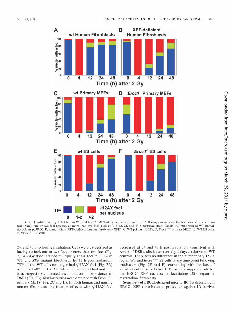
24, and 48 h following irradiation. Cells were categorized ashaving no foci, one or two foci, or more than two foci (Fig.2). A 2-Gy dose induced multiple �H2AX foci in 100% ofWT and XPF mutant fibroblasts. By 12 h postirradiation,75% of the WT cells no longer had �H2AX foci (Fig. 2A)whereas 60% of the XPF-deficient cells still had multiplefoci, suggesting continued accumulation or persistence ofDSBs (Fig. 2B). Similar results were obtained with Ercc1�/�
primary MEFs (Fig. 2C and D). In both human and murinemutant fibroblasts, the fraction of cells with �H2AX foci
decreased at 24 and 48 h postirradiation, consistent withrepair of DSBs, albeit substantially delayed relative to WTcontrols. There was no difference in the number of �H2AXfoci in WT and Ercc1�/� ES cells at any time point followingirradiation (Fig. 2E and F), correlating with the lack ofsensitivity of these cells to IR. These data support a role forthe ERCC1-XPF nuclease in facilitating DSB repair inmammalian fibroblasts.
Sensitivity of ERCC1-deficient mice to IR. To determine ifERCC1-XPF contributes to protection against IR in vivo,
FIG. 2. Quantitation of �H2AX foci in WT and ERCC1-XPF-deficient cells exposed to IR. Histograms indicate the fractions of cells with nofoci (blue), one or two foci (green), or more than two foci (red) at 0, 4, 12, 24, and 48 h postirradiation. Panels: A, immortalized WT humanfibroblasts (C5RO); B, immortalized XPF-deficient human fibroblasts (XFE); C, WT primary MEFs; D, Ercc1�/� primary MEFs; E, WT ES cells;F, Ercc1�/� ES cells.
VOL. 28, 2008 ERCC1-XPF FACILITATES DOUBLE-STRAND BREAK REPAIR 5085
on March 29, 2014 by guest
http://mcb.asm
.org/D
ownloaded from

6-week-old Ercc1�/� mice, which are hypomorphic forERCC1-XPF (14, 84), and WT littermates (n 6) wereexposed to a single dose of 6 Gy of total-body irradiation. Atthis age, Ercc1�/� mice are healthy and have no obvious ab-normal phenotype other than growth retardation (unpublisheddata). A 6-Gy dose of radiation did not acutely affect thesurvival of WT mice (Fig. 3A and reference 41) but caused100% lethality of Ercc1�/� mice by 11 days postirradiation.Although difficult to compare because of genetic backgrounddifferences, the sensitivity of Ercc1�/� mice is similar to that ofDNA-Pkcs
�/� mice (41) but less than that of Ku86�/� mice(56), both of which lack a protein required for NHEJ. There-fore, ERCC1-XPF makes a substantial contribution to protect-ing mammals from radiation-induced DNA damage in vivo.
To determine the cause of death of the Ercc1�/� mice, gas-trointestinal tract, bone marrow (BM), and liver tissue sectionsfrom WT and Ercc1�/� mice 11 days postirradiation were ex-amined. These tissues were indistinguishable between un-treated Ercc1�/� and WT littermates (Fig. 3B, D, and F).However, following IR, Ercc1�/� mice had dramatically fewerintestinal villi than WT mice (Fig. 3C). Immunostaining for theproliferation marker Ki67 (69) revealed considerably fewerpositive cells in Ercc1�/� crypts compared to WT mice at 11days postirradiation, but not at 2 days post-IR or in unexposedanimals (see Fig. S3 in the supplemental material). Postirradi-ation, the femoral BM of WT mice displayed hypercellularityof all hematopoietic lineages consistent with reactive hyperpla-sia (Fig. 3D and E). In contrast, the BM of Ercc1�/� mice wasmarkedly hypocellular with residual cells displaying dysplasticchanges including internuclear bridging, multinuclearity, andmegablastoid changes, most prominently affecting erythropoi-esis. Granulopoiesis displayed a shift to immature cell types. Inthe livers of Ercc1�/� but not WT mice, this dose of IR inducedcentrilobular necrosis (Fig. 3F and G). Immunostaining of liversections for �H2AX revealed nuclear foci in hepatocytes ofErcc1�/� mice but not WT littermates (Fig. 3H), indicative ofpersistent DSBs. Collectively, these data indicate that prolif-erative tissues of Ercc1�/� mice are vulnerable to IR-inducedgenotoxic stress and have diminished regenerative capacityfollowing damage.
ERCC1 deficiency causes embryonic lethality in a Ku86�/�
background. Very recent evidence indicates that ERCC1-XPFfacilitates SSA between direct repeats in mammalian cells (2)as do the orthologs Rad10-Rad1 in S. cerevisiae (28). In yeast,Rad10-Rad1 also participates in MMEJ, a Ku-independentmechanism of DSB repair (45). This pathway was recentlyconfirmed in mammals (87). To test if this function of ERCC1-
FIG. 3. Sensitivity of ERCC1-deficient mice to IR. (A) Six-week-old Ercc1�/� mice and WT littermates (n 6 per group) were exposedto 6 Gy of IR, and survival was recorded in days after exposure. (B toG) Tissue sections from 7- to 8-week-old WT and Ercc1�/� mice �exposure to 6 Gy of IR. (B) Small intestines of untreated mice.(C) Small intestines of mice exposed to IR demonstrating loss of villiin Ercc1�/� mice. (D) BM of untreated mice. (E) BM of mice exposedto IR demonstrating fatty replacement in Ercc1�/� mice. (F) Livers ofuntreated mice. (G) Livers of mice exposed to IR demonstrating cen-trilobular necrosis in Ercc1�/� mice. (H) Hepatocyte nuclei demon-strating �H2AX foci in Ercc1�/� mice 11 days after IR.
5086 AHMAD ET AL. MOL. CELL. BIOL.
on March 29, 2014 by guest
http://mcb.asm
.org/D
ownloaded from
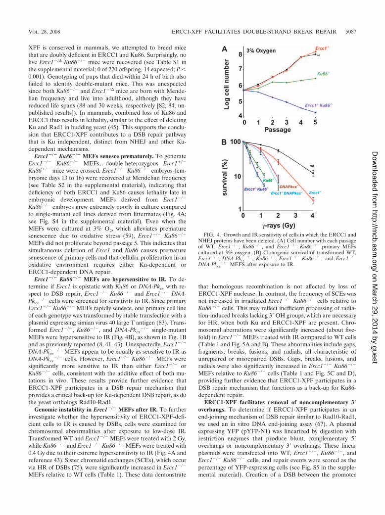
XPF is conserved in mammals, we attempted to breed micethat are doubly deficient in ERCC1 and Ku86. Surprisingly, nolive Ercc1�/� Ku86�/� mice were recovered (see Table S1 inthe supplemental material; 0 of 220 offspring, 14 expected; P �0.001). Genotyping of pups that died within 24 h of birth alsofailed to identify double-mutant mice. This was unexpectedsince both Ku86�/� and Ercc1�/� mice are born with Mende-lian frequency and live into adulthood, although they havereduced life spans (88 and 30 weeks, respectively [82, 84; un-published results]). In mammals, combined loss of Ku86 andERCC1 thus results in lethality, similar to the effect of deletingKu and Rad1 in budding yeast (45). This supports the conclu-sion that ERCC1-XPF contributes to a DSB repair pathwaythat is Ku independent, distinct from NHEJ and other Ku-dependent mechanisms.
Ercc1�/� Ku86�/� MEFs senesce prematurely. To generateErcc1�/� Ku86�/� MEFs, double-heterozygous Ercc1�/�
Ku86�/� mice were crossed. Ercc1�/� Ku86�/� embryos (em-bryonic days 13 to 16) were recovered at Mendelian frequency(see Table S2 in the supplemental material), indicating thatdeficiency of both ERCC1 and Ku86 causes lethality late inembryonic development. MEFs derived from Ercc1�/�
Ku86�/� embryos grew extremely poorly in culture comparedto single-mutant cell lines derived from littermates (Fig. 4A;see Fig. S4 in the supplemental material). Even when theMEFs were cultured at 3% O2, which alleviates prematuresenescence due to oxidative stress (59), Ercc1�/� Ku86�/�
MEFs did not proliferate beyond passage 5. This indicates thatsimultaneous deletion of Ercc1 and Ku86 causes prematuresenescence of primary cells and that cellular proliferation in anoxidative environment requires either Ku-dependent orERCC1-dependent DNA repair.
Ercc1�/� Ku86�/� MEFs are hypersensitive to IR. To de-termine if Ercc1 is epistatic with Ku86 or DNA-Pkcs with re-spect to DSB repair, Ercc1�/� Ku86�/� and Ercc1�/� DNA-Pkcs
�/� cells were screened for sensitivity to IR. Since primaryErcc1�/� Ku86�/� MEFs rapidly senesce, one primary cell lineof each genotype was transformed by stable transfection with aplasmid expressing simian virus 40 large T antigen (83). Trans-formed Ercc1�/�, Ku86�/�, and DNA-Pkcs
�/� single-mutantMEFs were hypersensitive to IR (Fig. 4B), as shown in Fig. 1Band as previously reported (8, 41, 43). Unexpectedly, Ercc1�/�
DNA-Pkcs�/� MEFs appear to be equally as sensitive to IR as
DNA-Pkcs�/� cells. However, Ercc1�/� Ku86�/� MEFs were
significantly more sensitive to IR than either Ercc1�/� orKu86�/� cells, consistent with the additive effect of both mu-tations in vivo. These results provide further evidence thatERCC1-XPF participates in a DSB repair mechanism thatprovides a critical back-up for Ku-dependent DSB repair, as dothe yeast orthologs Rad10-Rad1.
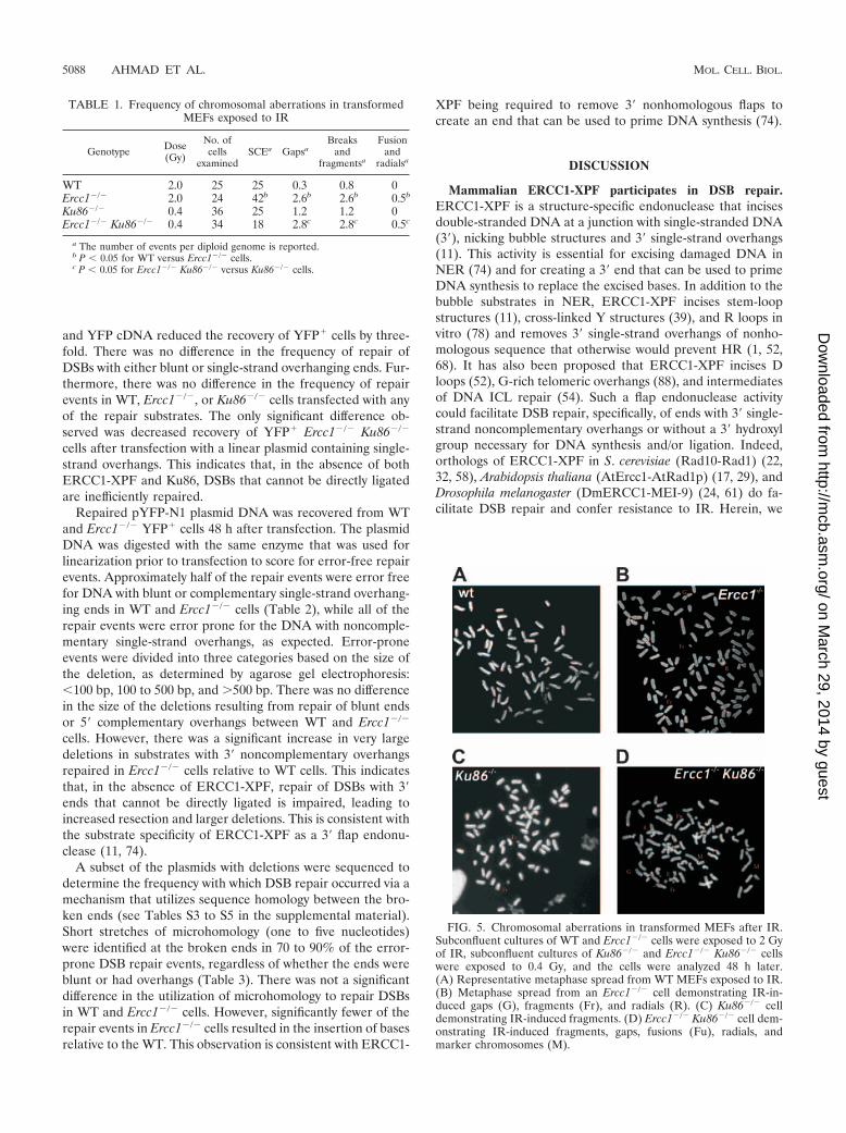
Genomic instability in Ercc1�/� MEFs after IR. To furtherinvestigate whether the hypersensitivity of ERCC1-XPF-defi-cient cells to IR is caused by DSBs, cells were examined forchromosomal abnormalities after exposure to low-dose IR.Transformed WT and Ercc1�/� MEFs were treated with 2 Gy,while Ku86�/� and Ercc1�/� Ku86�/� MEFs were treated with0.4 Gy due to their extreme hypersensitivity to IR (Fig. 4A andreference 43). Sister chromatid exchanges (SCEs), which occurvia HR of DSBs (75), were significantly increased in Ercc1�/�
MEFs relative to WT cells (Table 1). These data demonstrate
that homologous recombination is not affected by loss ofERCC1-XPF nuclease. In contrast, the frequency of SCEs wasnot increased in irradiated Ercc1�/� Ku86�/� cells relative toKu86�/� cells. This may reflect inefficient processing of radia-tion-induced breaks lacking 3� OH groups, which are necessaryfor HR, when both Ku and ERCC1-XPF are present. Chro-mosomal aberrations were significantly increased (about five-fold) in Ercc1�/� MEFs treated with IR compared to WT cells(Table 1 and Fig. 5A and B). These abnormalities include gaps,fragments, breaks, fusions, and radials, all characteristic ofunrepaired or misrepaired DSBs. Gaps, breaks, fusions, andradials were also significantly increased in Ercc1�/� Ku86�/�
MEFs relative to Ku86�/� cells (Table 1 and Fig. 5C and D),providing further evidence that ERCC1-XPF participates in aDSB repair mechanism that functions as a back-up for Ku86-dependent repair.
ERCC1-XPF facilitates removal of noncomplementary 3�overhangs. To determine if ERCC1-XPF participates in anend-joining mechanism of DSB repair similar to Rad10-Rad1,we used an in vitro DNA end-joining assay (67). A plasmidexpressing YFP (pYFP-N1) was linearized by digestion withrestriction enzymes that produce blunt, complementary 5�overhangs or noncomplementary 3� overhangs. These linearplasmids were transfected into WT, Ercc1�/�, Ku86�/�, andErcc1�/� Ku86�/� cells, and repair events were scored as thepercentage of YFP-expressing cells (see Fig. S5 in the supple-mental material). Creation of a DSB between the promoter
FIG. 4. Growth and IR sensitivity of cells in which the ERCC1 andNHEJ proteins have been deleted. (A) Cell number with each passageof WT, Ercc1�/�, Ku86�/�, and Ercc1�/� Ku86�/� primary MEFscultured at 3% oxygen. (B) Clonogenic survival of transformed WT,Ercc1�/�, DNA-Pkcs
�/�, Ku86�/�, Ercc1�/� Ku86�/�, and Ercc1�/�
DNA-Pkcs�/� MEFS after exposure to IR.
VOL. 28, 2008 ERCC1-XPF FACILITATES DOUBLE-STRAND BREAK REPAIR 5087
on March 29, 2014 by guest
http://mcb.asm
.org/D
ownloaded from
and YFP cDNA reduced the recovery of YFP� cells by three-fold. There was no difference in the frequency of repair ofDSBs with either blunt or single-strand overhanging ends. Fur-thermore, there was no difference in the frequency of repairevents in WT, Ercc1�/�, or Ku86�/� cells transfected with anyof the repair substrates. The only significant difference ob-served was decreased recovery of YFP� Ercc1�/� Ku86�/�
cells after transfection with a linear plasmid containing single-strand overhangs. This indicates that, in the absence of bothERCC1-XPF and Ku86, DSBs that cannot be directly ligatedare inefficiently repaired.
Repaired pYFP-N1 plasmid DNA was recovered from WTand Ercc1�/� YFP� cells 48 h after transfection. The plasmidDNA was digested with the same enzyme that was used forlinearization prior to transfection to score for error-free repairevents. Approximately half of the repair events were error freefor DNA with blunt or complementary single-strand overhang-ing ends in WT and Ercc1�/� cells (Table 2), while all of therepair events were error prone for the DNA with noncomple-mentary single-strand overhangs, as expected. Error-proneevents were divided into three categories based on the size ofthe deletion, as determined by agarose gel electrophoresis:�100 bp, 100 to 500 bp, and 500 bp. There was no differencein the size of the deletions resulting from repair of blunt endsor 5� complementary overhangs between WT and Ercc1�/�
cells. However, there was a significant increase in very largedeletions in substrates with 3� noncomplementary overhangsrepaired in Ercc1�/� cells relative to WT cells. This indicatesthat, in the absence of ERCC1-XPF, repair of DSBs with 3�ends that cannot be directly ligated is impaired, leading toincreased resection and larger deletions. This is consistent withthe substrate specificity of ERCC1-XPF as a 3� flap endonu-clease (11, 74).
A subset of the plasmids with deletions were sequenced todetermine the frequency with which DSB repair occurred via amechanism that utilizes sequence homology between the bro-ken ends (see Tables S3 to S5 in the supplemental material).Short stretches of microhomology (one to five nucleotides)were identified at the broken ends in 70 to 90% of the error-prone DSB repair events, regardless of whether the ends wereblunt or had overhangs (Table 3). There was not a significantdifference in the utilization of microhomology to repair DSBsin WT and Ercc1�/� cells. However, significantly fewer of therepair events in Ercc1�/� cells resulted in the insertion of basesrelative to the WT. This observation is consistent with ERCC1-
XPF being required to remove 3� nonhomologous flaps tocreate an end that can be used to prime DNA synthesis (74).
DISCUSSION
Mammalian ERCC1-XPF participates in DSB repair.ERCC1-XPF is a structure-specific endonuclease that incisesdouble-stranded DNA at a junction with single-stranded DNA(3�), nicking bubble structures and 3� single-strand overhangs(11). This activity is essential for excising damaged DNA inNER (74) and for creating a 3� end that can be used to primeDNA synthesis to replace the excised bases. In addition to thebubble substrates in NER, ERCC1-XPF incises stem-loopstructures (11), cross-linked Y structures (39), and R loops invitro (78) and removes 3� single-strand overhangs of nonho-mologous sequence that otherwise would prevent HR (1, 52,68). It has also been proposed that ERCC1-XPF incises Dloops (52), G-rich telomeric overhangs (88), and intermediatesof DNA ICL repair (54). Such a flap endonuclease activitycould facilitate DSB repair, specifically, of ends with 3� single-strand noncomplementary overhangs or without a 3� hydroxylgroup necessary for DNA synthesis and/or ligation. Indeed,orthologs of ERCC1-XPF in S. cerevisiae (Rad10-Rad1) (22,32, 58), Arabidopsis thaliana (AtErcc1-AtRad1p) (17, 29), andDrosophila melanogaster (DmERCC1-MEI-9) (24, 61) do fa-cilitate DSB repair and confer resistance to IR. Herein, we
FIG. 5. Chromosomal aberrations in transformed MEFs after IR.Subconfluent cultures of WT and Ercc1�/� cells were exposed to 2 Gyof IR, subconfluent cultures of Ku86�/� and Ercc1�/� Ku86�/� cellswere exposed to 0.4 Gy, and the cells were analyzed 48 h later.(A) Representative metaphase spread from WT MEFs exposed to IR.(B) Metaphase spread from an Ercc1�/� cell demonstrating IR-in-duced gaps (G), fragments (Fr), and radials (R). (C) Ku86�/� celldemonstrating IR-induced fragments. (D) Ercc1�/� Ku86�/� cell dem-onstrating IR-induced fragments, gaps, fusions (Fu), radials, andmarker chromosomes (M).
TABLE 1. Frequency of chromosomal aberrations in transformedMEFs exposed to IR
Genotype Dose(Gy)
No. ofcells
examinedSCEa Gapsa
Breaksand
fragmentsa
Fusionand
radialsa
WT 2.0 25 25 0.3 0.8 0Ercc1�/� 2.0 24 42b 2.6b 2.6b 0.5b
Ku86�/� 0.4 36 25 1.2 1.2 0Ercc1�/� Ku86�/� 0.4 34 18 2.8c 2.8c 0.5c
a The number of events per diploid genome is reported.b P � 0.05 for WT versus Ercc1�/� cells.c P � 0.05 for Ercc1�/� Ku86�/� versus Ku86�/� cells.
5088 AHMAD ET AL. MOL. CELL. BIOL.
on March 29, 2014 by guest
http://mcb.asm
.org/D
ownloaded from

provide in vitro, genetic and in vivo evidence that this functionis conserved in murine and human cells.
Role of ERCC1-XPF in DSB repair. ERCC1-XPF-deficientcells are exquisitely sensitive to cross-linking agents and accu-mulate DSBs in response to cross-link damage (54). We pos-tulated that in ICL repair, ERCC1-XPF is required to incisethe cross-link lesion after a DSB is created by stalled replica-tion, before the DSB can be repaired. Here we provide evi-dence for a distinct role for ERCC1-XPF in DSB repair.ERCC1-XPF-deficient murine and human cells are hypersen-sitive to IR (Fig. 1), similar to cells defective in HR-mediatedor NHEJ DSB repair (77). Murine fibroblasts, but not ES cells,are IR sensitive (Fig. 1). This pattern of cell type-specificsensitivity is similar to that of DNA-Pkcs mutant lines defectivein NHEJ (25) but the opposite of that of Rad54�/� cells de-fective in HR (19). This suggests that ERCC1-XPF may par-ticipate in an end-joining mechanism of DSB repair that ismore frequently utilized in differentiated cells than in ES cells.In further support of this, SCEs are significantly elevated inirradiated Ercc1�/� MEFs (Table 1), indicating that HR-me-diated DSB repair is possible in the absence of ERCC1-XPF.This increase in SCEs might reflect preferential repair of DSBsby HR when end-joining mechanisms are impaired (85).ERCC1-XPF, however, is clearly not part of the core NHEJmachinery, as there is no evidence of severe combined immu-
nodeficiency, a pathognomonic feature of NHEJ deficiency(87), in Ercc1�/� or Xpf�/� mice (70, 79) or humans with XPFdeficiency (53). Furthermore, Ercc1�/� MEFs are less sensitiveto IR than are DNA-Pkcs
�/� or Ku86�/� MEFs, in whichNHEJ is absent (Fig. 4B).
IR induces a variety of lesions in addition to DSBs (23).Thus, to confirm that the IR sensitivity of ERCC1-XPF-defi-cient cells is due, at least in part, to a defect in DSB repair, wequantitated �H2AX foci, a marker of DSBs (63), in cells fol-lowing IR. �H2AX foci persist in irradiated ERCC1-XPF-deficient fibroblasts (Fig. 2) and in hepatocytes of irradiatedERCC1-deficient mice (Fig. 3H) relative to congenic WT con-trols, consistent with impaired DSB repair in the absence ofERCC1-XPF both in vitro and in vivo. Furthermore, irradiatedErcc1�/� MEFs had increased gaps, breaks, fragments, andradial structures compared to WT cells (Table 1). These chro-mosome abnormalities are a consequence of DSBs (49) andthus demonstrate a defect in DSB repair in Ercc1�/� cells.Interestingly, there is no difference in the number of �H2AXfoci in Ercc1�/� and WT ES cells at any time point followingexposure to IR, correlating with their lack of sensitivity to IR.There is increasing evidence that homologous recombination isessential for genome stability in ES cells (6, 26, 62), furthersuggesting that HR occurs in ERCC1-deficient cells.
Biological significance of ERCC1-XPF in DSB repair. Asingle dose of 6 Gy of IR is universally lethal to mice hypo-morphic for ERCC1 (Fig. 3A), whereas 2 Gy is sublethal (datanot shown). This level of sensitivity is similar to that of mice inwhich DNA-PKcs, an essential component of NHEJ, is deleted(41), even though ERCC1-XPF expression is reduced only85% in the hypomorphic strain (84). This establishes an im-portant role for ERCC1-XPF in the radioprotection of mam-mals. IR causes loss of intestinal villi and BM hypocellularity inERCC1-deficient mice, as anticipated (Fig. 3). In addition,ERCC1-deficient mice display centrilobular necrosis of theliver, which is caused by occlusion of small venules with cellulardebris, following IR (20). Liver pathology is life limiting forErcc1�/� mice (72), explaining the particular hypersensitivityof Ercc1�/� mouse liver to IR. Mice defective in DSB repairhave impaired hematopoietic stem cell function and regener-ative capacity after stress (55, 64). We observed significantlyreduced proliferative reserve in the BM of Ercc1�/� mice (60)and in the intestine following IR (see Fig. S3 in the supple-
TABLE 2. Size of linear plasmid with blunt or overhanging DNA ends after repair in WT or Ercc1�/� cells
DNA ends and genotype n % Errorfree
% Errorprone
% with�100-bpdeletion
% with101- to 500-bp
deletion
% with500-bpdeletion
BluntWT 81 50 50 88 10 2Ercc1�/� 78 58 42 90 10 0
5� Overhangs, complementaryWT 65 43 57 60 40 0Ercc1�/� 66 42 58 60 35 5
3� Overhangs, noncomplementaryWT 94 0 100 63 31 6Ercc1�/� 82 0 100 65 14a 21a
a P � 0.05.
TABLE 3. Sequence analysis of blunt, complementary, andnoncomplementary DNA ends after repair in
WT or Ercc1�/� cells
DNA ends and genotype n% with
nucleotideadded
% Microhomologymediated
BluntWT 18 0 89Ercc1�/� 21 0 81
5� Overhangs, complementaryWT 18 6 72Ercc1�/� 17 6 71
3� Overhangs, noncomplementaryWT 30 37 80Ercc1�/� 31 10a 71
a P � 0.05.
VOL. 28, 2008 ERCC1-XPF FACILITATES DOUBLE-STRAND BREAK REPAIR 5089
on March 29, 2014 by guest
http://mcb.asm
.org/D
ownloaded from
mental material). Thus, the hypersensitivity of Ercc1�/� miceto IR likely is a consequence not only of increased cell death/senescence in response to unrepaired DNA damage but alsoreduced capacity to regenerate damaged tissues.
Although ERCC1 hypomorphic mice are equally as sensitiveto IR as DNA-Pkcs
�/� mice are, they are less sensitive thanKu86�/� mice (9, 56). DNA-Pkcs
�/� mice do not have a spon-taneous phenotype other than severe combined immunodefi-ciency (25, 41), whereas Ku86�/� mice display growth retarda-tion and progeroid symptoms in addition to immunodeficiency(82). Ercc1�/� mice have a more severe phenotype than eitherDNA-Pkcs
�/� or Ku86�/� mice, including growth retardation,liver dysfunction, renal insufficiency, epidermal atrophy, im-paired hematopoiesis, sarcopenia, kyphosis, and neurodegen-eration (48, 53, 60, 84), making it unlikely that the phenotypeof Ercc1�/� mice can be ascribed in its entirety to a defect inDSB repair. More plausibly, it is the combined defects inmultiple DNA repair pathways including global-genome andtranscription-coupled NER, ICL repair, and DSB repair, doc-umented here, that contribute to the severe phenotype ofErcc1�/� mice, XFE progeria (53), and a recently identifiedERCC1 patient (33).
ERCC1-XPF participates in error-prone, Ku-independentend joining of DSBs. ERCC1-XPF endonuclease facilitatesDSB repair and protects against IR in vivo. Yet the two majormechanisms of DSB repair in mammalian cells, HR andNHEJ, are intact in ERCC1-XPF-deficient cells. Thus, thefollowing question remains: how does this nuclease contributeto DSB repair? In yeast, the orthologs of ERCC1-XPF, Rad10-Rad1, are required for two error-prone DSB repair mecha-nisms: SSA (22, 32, 57) and MMEJ (45). Rad10-Rad1 arerequired to remove nonhomologous sequence from the 3� endsof the break to permit DNA synthesis and/or ligation. MMEJin yeast is independent of the HR protein Rad52 and theNHEJ protein Ku86 and critical for protection against IR (45).In rad52; yku70 strains, DSB repair is error prone, leading tolarge deletions and utilizing short homologies between ends(5). Recently, an alternative mechanism of end joining, distinctfrom NHEJ, was identified in mammals (87). This mechanismsupports class switch recombination in the absence of NHEJ, iserror prone, and utilizes microhomology to join broken ends,analogous to MMEJ in yeast. Linearized plasmids and I-Sce-I-induced genomic DSBs are repaired in cells deficient inNHEJ, most notably Ku86, leading to deletions and insertionsand utilizing microhomology (27, 35, 71). However, the bio-logical significance of this pathway in mammals, when NHEJ ispresent, remains uncertain.
Our data provide several lines of evidence that support theconclusion that ERCC1-XPF contributes to this alternativemechanism of end joining in mammalian cells. First, Ercc1�/�
Ku86�/� MEFs are hypersensitive to IR relative to single-mutant cells (Fig. 4B), similar to yeast (45). In addition,Ercc1�/� Ku86�/� cells accumulate significantly more chromo-somal aberrations in response to low-dose IR compared tocells defective in NHEJ only (Ku86�/� cells; Table 1). Further-more, Ercc1�/� Ku86�/� primary MEFs proliferate poorly,surviving only to passage 5, even when cultured at 3% oxygen(Fig. 4A). These data are consistent with the conclusion thatERCC1-XPF participates in a mechanism of DSB repair that isKu86 independent and important for protection against IR
and oxidative stress, analogous to the role of Rad10-Rad1 inyeast.
The second line of evidence that supports a role for ERCC1-XPF in MMEJ comes from the in vitro end-joining assay.Joining of blunt 5� or 3� overhangs is not significantly reducedin Ercc1�/� or Ku86�/� cells compared to WT cells (see Fig.S5 in the supplemental material). However, joining of endswith overhangs is significantly impaired in Ercc1�/� Ku86�/�
cells, suggesting loss of two distinct mechanisms of end joiningin these double-mutant cells. Ercc1�/� cells are defective injoining ends that cannot be directly ligated, i.e., ends with 3�noncomplementary overhangs but not blunt ends or ends with5� complementary overhangs. In the absence of ERCC1-XPF,joining of nonhomologous 3� overhangs leads to very largedeletions (Table 2), similar to yeast mutants (45). Further-more, in Ercc1�/� cells, insertion of nucleotides at the joints issignificantly reduced (Table 3), consistent with the conservedrole of this nuclease in creating a 3� end that can prime DNAsynthesis (58). In addition, there is a trend toward decreaseduse of microhomology in end joining in Ercc1�/� cells relativeto WT cells (Table 3). Deletion of Rad1 diminishes MMEJ toa greater extent in yeast but does not abrogate MMEJ, indi-cating the presence of redundant 3� flap endonucleases in yeast(45) and therefore likely also in mammals. Recently, it wasdemonstrated that telomere fusion likely occurs via a Ku-in-dependent end-joining mechanism (7). Thus, the participationof ERCC1-XPF in this alternative end-joining mechanism isalso supported by the fact that ERCC1-XPF trims unprotectedtelomeric overhangs, allowing telomeric fusion via end joining(88).
In total, these data demonstrate a novel function of ERCC1-XPF nuclease in a Ku86-independent mechanism of DSB re-pair. Further, our data, in combination with a recent reportestablishing a role for ERCC1-XPF in SSA (2), indicate thatRad10-Rad1 activities in DSB repair are conserved in mam-mals. The data also suggest that alternative end joining ofDSBs is important for protection against DSBs even whenNHEJ and HR repair can occur.
ACKNOWLEDGMENTS
This work was supported by the University of Pittsburgh CancerInstitute, NCI and NIA CA103730 and CA111525, the PennsylvaniaDepartment of Health, and The Ellison Medical Foundation (AG-NS-0303). J.H.J.H. is supported by the Nederlandse Organisatie voorWetenschappelijk Onderzoek (NOW), Cancer Genomics Center, theAssociation for International Cancer Research, and the EuropeanCommission (IP 512113), RISC-RAD contract F16R-CT-2003-508842.
We thank Richard D. Wood for careful reading of the manuscript.
REFERENCES
1. Adair, G. M., R. L. Rolig, D. Moore-Faver, M. Zabelshansky, J. H. Wilson,and R. S. Nairn. 2000. Role of ERCC1 in removal of long non-homologoustails during targeted homologous recombination. EMBO J. 19:5552–5561.
2. Al-Minawi, A. Z., N. Saleh-Gohari, and T. Helleday. 2008. The ERCC1/XPFendonuclease is required for efficient single-strand annealing and gene con-version in mammalian cells. Nucleic Acids Res. 36:1–9.
3. Baker, B. S., A. T. Carpenter, and P. Ripoll. 1978. The utilization duringmitotic cell division of loci controlling meiotic recombination and disjunctionin Drosophila melanogaster. Genetics 90:531–578.
4. Biggerstaff, M., D. E. Szymkowski, and R. D. Wood. 1993. Co-correction ofthe ERCC1, ERCC4 and xeroderma pigmentosum group F DNA repairdefects in vitro. EMBO J. 12:3685–3692.
5. Boulton, S. J., and S. P. Jackson. 1996. Saccharomyces cerevisiae Ku70potentiates illegitimate DNA double-strand break repair and serves as abarrier to error-prone DNA repair pathways. EMBO J. 15:5093–5103.
5090 AHMAD ET AL. MOL. CELL. BIOL.
on March 29, 2014 by guest
http://mcb.asm
.org/D
ownloaded from
6. Brugmans, L., R. Kanaar, and J. Essers. 2007. Analysis of DNA double-strand break repair pathways in mice. Mutat. Res. 614:95–108.
7. Capper, R., B. Britt-Compton, M. Tankimanova, J. Rowson, B. Letsolo, S.Man, M. Haughton, and D. M. Baird. 2007. The nature of telomere fusionand a definition of the critical telomere length in human cells. Genes Dev.21:2495–2508.
8. Chan, D. W., B. P. Chen, S. Prithivirajsingh, A. Kurimasa, M. D. Story, J.Qin, and D. J. Chen. 2002. Autophosphorylation of the DNA-dependentprotein kinase catalytic subunit is required for rejoining of DNA double-strand breaks. Genes Dev. 16:2333–2338.
9. Couedel, C., K. D. Mills, M. Barchi, L. Shen, A. Olshen, R. D. Johnson, A.Nussenzweig, J. Essers, R. Kanaar, G. C. Li, F. W. Alt, and M. Jasin. 2004.Collaboration of homologous recombination and nonhomologous end-join-ing factors for the survival and integrity of mice and cells. Genes Dev.18:1293–1304.
10. Decottignies, A. 2007. Microhomology-mediated end joining in fission yeastis repressed by pku70 and relies on genes involved in homologous recombi-nation. Genetics 176:1403–1415.
11. de Laat, W. L., E. Appeldoorn, N. G. Jaspers, and J. H. Hoeijmakers. 1998.DNA structural elements required for ERCC1-XPF endonuclease activity.J. Biol. Chem. 273:7835–7842.
12. de Vries, A., C. T. van Oostrom, F. M. Hofhuis, P. M. Dortant, R. J. Berg,F. R. de Gruijl, P. W. Wester, C. F. van Kreijl, P. J. Capel, H. van Steeg, etal. 1995. Increased susceptibility to ultraviolet-B and carcinogens of micelacking the DNA excision repair gene XPA. Nature 377:169–173.
13. de Waard, H., J. de Wit, J. O. Andressoo, C. T. van Oostrom, B. Riis, A.Weimann, H. E. Poulsen, H. van Steeg, J. H. Hoeijmakers, and G. T. van derHorst. 2004. Different effects of CSA and CSB deficiency on sensitivity tooxidative DNA damage. Mol. Cell. Biol. 24:7941–7948.
14. Dolle, M. E., R. A. Busuttil, A. M. Garcia, S. Wijnhoven, E. van Drunen, L. J.Niedernhofer, G. van der Horst, J. H. Hoeijmakers, H. van Steeg, and J. Vijg.2006. Increased genomic instability is not a prerequisite for shortened life-span in DNA repair deficient mice. Mutat. Res. 596:22–35.
15. Dronkert, M. L. G., H. B. Beverloo, R. D. Johnson, J. H. J. Hoeijmakers, M.Jasin, and R. Kanaar. 2000. Mouse RAD54 affects DNA double-strandbreak repair and sister chromatid exchange. Mol. Cell. Biol. 20:3147–3156.
16. Duan, D., Y. Yue, and J. F. Engelhardt. 2003. Consequences of DNA-dependent protein kinase catalytic subunit deficiency on recombinant adeno-associated virus genome circularization and heterodimerization in muscletissue. J. Virol. 77:4751–4759.
17. Dubest, S., M. E. Gallego, and C. I. White. 2002. Role of the AtRad1pendonuclease in homologous recombination in plants. EMBO Rep. 3:1049–1054.
18. Enzlin, J. H., and O. D. Scharer. 2002. The active site of the DNA repairendonuclease XPF-ERCC1 forms a highly conserved nuclease motif. EMBOJ. 21:2045–2053.
19. Essers, J., R. W. Hendriks, S. M. Swagemakers, C. Troelstra, J. de Wit, D.Bootsma, J. H. Hoeijmakers, and R. Kanaar. 1997. Disruption of mouseRAD54 reduces ionizing radiation resistance and homologous recombina-tion. Cell 89:195–204.
20. Fajardo, L. F. 2005. The pathology of ionizing radiation as defined bymorphologic patterns. Acta Oncol. 44:13–22.
21. Finkel, T., M. Serrano, and M. A. Blasco. 2007. The common biology ofcancer and ageing. Nature 448:767–774.
22. Fishman-Lobell, J., and J. E. Haber. 1992. Removal of nonhomologousDNA ends in double-strand break recombination: the role of the yeastultraviolet repair gene RAD1. Science 258:480–484.
23. Friedberg, E. C., G. C. Walker, W. Siede, R. D. Wood, R. A. Schultz, and T.Ellenberger. 2006. DNA repair and mutagenesis, 2nd ed. ASM Press, Wash-ington, DC.
24. Gamo, S., T. Megumi, and E. Nakashima-Tanaka. 1990. Sensitivity to etheranesthesia and to gamma-rays in mutagen-sensitive strains of Drosophilamelanogaster. Mutat. Res. 235:9–13.
25. Gao, Y., J. Chaudhuri, C. Zhu, L. Davidson, D. T. Weaver, and F. W. Alt.1998. A targeted DNA-PKcs-null mutation reveals DNA-PK-independentfunctions for KU in V(D)J. recombination. Immunity 9:367–376.
26. Griffin, C., H. Waard, B. Deans, and J. Thacker. 2005. The involvement ofkey DNA repair pathways in the formation of chromosome rearrangementsin embryonic stem cells. DNA Repair 4:1019–1027.
27. Guirouilh-Barbat, J., S. Huck, P. Bertrand, L. Pirzio, C. Desmaze, L. Saba-tier, and B. S. Lopez. 2004. Impact of the KU80 pathway on NHEJ-inducedgenome rearrangements in mammalian cells. Mol. Cell 14:611–623.
28. Haber, J. E. 2006. Transpositions and translocations induced by site-specificdouble-strand breaks in budding yeast. DNA Repair 5:998–1009.
29. Hefner, E., S. B. Preuss, and A. B. Britt. 2003. Arabidopsis mutants sensitiveto gamma radiation include the homologue of the human repair geneERCC1. J. Exp. Bot. 54:669–680.
30. Helleday, T., J. Lo, D. C. van Gent, and B. P. Engelward. 2007. DNAdouble-strand break repair: from mechanistic understanding to cancer treat-ment. DNA Repair 6:923–935.
31. Hesse, J. E., M. R. Lieber, M. Gellert, and K. Mizuuchi. 1987. Extrachro-
mosomal DNA substrates in pre-B cells undergo inversion or deletion atimmunoglobulin V-(D)-J. joining signals. Cell 49:775–783.
32. Ivanov, E. L., and J. E. Haber. 1995. RAD1 and RAD10, but not otherexcision repair genes, are required for double-strand break-induced recom-bination in Saccharomyces cerevisiae. Mol. Cell. Biol. 15:2245–2251.
33. Jaspers, N. G., A. Raams, M. C. Silengo, N. Wijgers, L. J. Niedernhofer,A. R. Robinson, G. Giglia-Mari, D. Hoogstraten, W. J. Kleijer, J. H. Hoei-jmakers, and W. Vermeulen. 2007. First reported patient with humanERCC1 deficiency has cerebro-oculo-facio-skeletal syndrome with a milddefect in nucleotide excision repair and severe developmental failure. Am. J.Hum. Genet. 80:457–466.
34. Jonnalagadda, V. S., T. Matsuguchi, and B. P. Engelward. 2005. Interstrandcrosslink-induced homologous recombination carries an increased risk ofdeletions and insertions. DNA Repair 4:594–605.
35. Kabotyanski, E. B., L. Gomelsky, J. O. Han, T. D. Stamato, and D. B. Roth.1998. Double-strand break repair in Ku86- and XRCC4-deficient cells. Nu-cleic Acids Res. 26:5333–5342.
36. Karran, P. 2000. DNA double strand break repair in mammalian cells. Curr.Opin. Genet. Dev. 10:144–150.
37. Khanna, K. K., and S. P. Jackson. 2001. DNA double-strand breaks:signaling, repair and the cancer connection. Nat. Genet. 27:247–254.
38. Kuhfittig-Kulle, S., E. Feldmann, A. Odersky, A. Kuliczkowska, W. Goede-cke, A. Eggert, and P. Pfeiffer. 2007. The mutagenic potential of non-homol-ogous end joining in the absence of the NHEJ core factors Ku70/80, DNA-PKcs and XRCC4-LigIV. Mutagenesis 22:217–233.
39. Kuraoka, I., W. R. Kobertz, R. R. Ariza, M. Biggerstaff, J. M. Essigmann, andR. D. Wood. 2000. Repair of an interstrand DNA cross-link initiated by ERCC1-XPF repair/recombination nuclease. J. Biol. Chem. 275:26632–26636.
40. Lee, K., and S. E. Lee. 2007. Saccharomyces cerevisiae Sae2- and Tel1-depen-dent single-strand DNA formation at DNA break promotes microhomology-mediated end joining. Genetics 176:2003–2014.
41. Li, X.-L., S.-R. Shen, S. Wang, H.-H. Ouyang, and G. C. Li. 2002. Restorationof T cell-specific V(D)J. recombination in DNA-PKcs�/� mice by ionizingradiation: the effects on survival, development, and tumorigenesis. ActaBiochim. Biophys. Sin. 34:149–157.
42. Liang, F., M. Han, P. J. Romanienko, and M. Jasin. 1998. Homology-directed repair is a major double-strand break repair pathway in mammaliancells. Proc. Natl. Acad. Sci. USA 95:5172–5177.
43. Lim, D.-S., H. Vogel, D. M. Willerford, A. T. Sands, K. A. Platt, and P. Hasty.2000. Analysis of ku80-mutant mice and cells with deficient levels of p53.Mol. Cell. Biol. 20:3772–3780.
44. Little, J. W. 1981. Lambda exonuclease. Gene Amplif. Anal. 2:135–145.45. Ma, J.-L., E. M. Kim, J. E. Haber, and S. E. Lee. 2003. Yeast Mre11 and
Rad1 proteins define a Ku-independent mechanism to repair double-strandbreaks lacking overlapping end sequences. Mol. Cell. Biol. 23:8820–8828.
46. Matsumura, Y., C. Nishigori, T. Yagi, S. Imamura, and H. Takebe. 1998.Characterization of molecular defects in xeroderma pigmentosum group F inrelation to its clinically mild symptoms. Hum. Mol. Genet. 7:969–974.
47. McHugh, P. J., V. J. Spanswick, and J. A. Hartley. 2001. Repair of DNAinterstrand crosslinks: molecular mechanisms and clinical relevance. LancetOncol. 2:483–490.
48. McWhir, J., J. Selfridge, D. J. Harrison, S. Squires, and D. W. Melton. 1993.Mice with DNA repair gene (ERCC-1) deficiency have elevated levels ofp53, liver nuclear abnormalities and die before weaning. Nat. Genet. 5:217–224.
49. Mills, K. D., D. O. Ferguson, J. Essers, M. Eckersdorff, R. Kanaar, and F. W.Alt. 2004. Rad54 and DNA ligase IV cooperate to maintain mammalianchromatid stability. Genes Dev. 18:1283–1292.
50. Murray, D., L. Vallee-Lucic, E. Rosenberg, and B. Andersson. 2002. Sensi-tivity of nucleotide excision repair-deficient human cells to ionizing radiationand cyclophosphamide. Anticancer Res. 22:21–26.
51. Newman, M., J. Murray-Rust, J. Lally, J. Rudolf, A. Fadden, P. P. Knowles,M. F. White, and N. Q. McDonald. 2005. Structure of an XPF endonucleasewith and without DNA suggests a model for substrate recognition. EMBO J.24:895–905.
52. Niedernhofer, L. J., J. Essers, G. Weeda, B. Beverloo, J. de Wit, M. Mui-jtjens, H. Odijk, J. H. Hoeijmakers, and R. Kanaar. 2001. The structure-specific endonuclease Ercc1-Xpf is required for targeted gene replacementin embryonic stem cells. EMBO J. 20:6540–6549.
53. Niedernhofer, L. J., G. A. Garinis, A. Raams, A. S. Lalai, A. R. Robinson, E.Appeldoorn, H. Odijk, R. Oostendorp, A. Ahmad, W. van Leeuwen, A. F.Theil, W. Vermeulen, G. T. van der Horst, P. Meinecke, W. J. Kleijer, J. Vijg,N. G. Jaspers, and J. H. Hoeijmakers. 2006. A new progeroid syndromereveals that genotoxic stress suppresses the somatotroph axis. Nature 444:1038–1043.
54. Niedernhofer, L. J., H. Odijk, M. Budzowska, E. van Drunen, A. Maas, A. F.Theil, J. de Wit, N. G. Jaspers, H. B. Beverloo, J. H. Hoeijmakers, and R.Kanaar. 2004. The structure-specific endonuclease Ercc1-Xpf is required toresolve DNA interstrand cross-link-induced double-strand breaks. Mol. Cell.Biol. 24:5776–5787.
55. Nijnik, A., L. Woodbine, C. Marchetti, S. Dawson, T. Lambe, C. Liu, N. P.Rodrigues, T. L. Crockford, E. Cabuy, A. Vindigni, T. Enver, J. I. Bell, P.
VOL. 28, 2008 ERCC1-XPF FACILITATES DOUBLE-STRAND BREAK REPAIR 5091
on March 29, 2014 by guest
http://mcb.asm
.org/D
ownloaded from
Slijepcevic, C. C. Goodnow, P. A. Jeggo, and R. J. Cornall. 2007. DNA repairis limiting for haematopoietic stem cells during ageing. Nature 447:686–690.
56. Nussenzweig, A., K. Sokol, P. Burgman, L. Li, and G. C. Li. 1997. Hyper-sensitivity of Ku80-deficient cell lines and mice to DNA damage: the effectsof ionizing radiation on growth, survival, and development. Proc. Natl. Acad.Sci. USA 94:13588–13593.
57. Paques, F., and J. E. Haber. 1999. Multiple pathways of recombinationinduced by double-strand breaks in Saccharomyces cerevisiae. Microbiol.Mol. Biol. Rev. 63:349–404.
58. Paques, F., and J. E. Haber. 1997. Two pathways for removal of nonhomolo-gous DNA ends during double-strand break repair in Saccharomyces cerevi-siae. Mol. Cell. Biol. 17:6765–6771.
59. Parrinello, S., E. Samper, A. Krtolica, J. Goldstein, S. Melov, and J.Campisi. 2003. Oxygen sensitivity severely limits the replicative lifespan ofmurine fibroblasts. Nat. Cell Biol. 5:741–747.
60. Prasher, J. M., A. S. Lalai, C. Heijmans-Antonissen, R. E. Ploemacher, J. H.Hoeijmakers, I. P. Touw, and L. J. Niedernhofer. 2005. Reduced hemato-poietic reserves in DNA interstrand crosslink repair-deficient Ercc1�/�
mice. EMBO J. 24:861–871.61. Radford, S. J., E. Goley, K. Baxter, S. McMahan, and J. Sekelsky. 2005.
Drosophila ERCC1 is required for a subset of MEI-9-dependent meioticcrossovers. Genetics 170:1737–1745.
62. Richardson, C., M. E. Moynahan, and M. Jasin. 1998. Double-strand breakrepair by interchromosomal recombination: suppression of chromosomaltranslocations. Genes Dev. 12:3831–3842.
63. Rogakou, E. P., C. Boon, C. Redon, and W. M. Bonner. 1999. Megabasechromatin domains involved in DNA double-strand breaks in vivo. J. CellBiol. 146:905–916.
64. Rossi, D. J., D. Bryder, J. Seita, A. Nussenzweig, J. Hoeijmakers, and I. L.Weissman. 2007. Deficiencies in DNA damage repair limit the function ofhaematopoietic stem cells with age. Nature 447:725–729.
65. Roth, D. B., and J. H. Wilson. 1986. Nonhomologous recombination inmammalian cells: role for short sequence homologies in the joining reaction.Mol. Cell. Biol. 6:4295–4304.
66. Rothkamm, K., I. Kruger, L. H. Thompson, and M. Lobrich. 2003. Pathwaysof DNA double-strand break repair during the mammalian cell cycle. Mol.Cell. Biol. 23:5706–5715.
67. Runger, T. M., and K. H. Kraemer. 1989. Joining of linear plasmid DNA isreduced and error-prone in Bloom’s syndrome cells. EMBO J. 8:1419–1425.
68. Sargent, R. G., J. L. Meservy, B. D. Perkins, A. E. Kilburn, Z. Intody, G. M.Adair, R. S. Nairn, and J. H. Wilson. 2000. Role of the nucleotide excisionrepair gene ERCC1 in formation of recombination-dependent rearrange-ments in mammalian cells. Nucleic Acids Res. 28:3771–3778.
69. Scholzen, T., and J. Gerdes. 2000. The Ki-67 protein: from the known andthe unknown. J. Cell Physiol. 182:311–322.
70. Schrader, C. E., J. Vardo, E. Linehan, M. Z. Twarog, L. J. Niedernhofer,J. H. Hoeijmakers, and J. Stavnezer. 2004. Deletion of the nucleotide exci-sion repair gene Ercc1 reduces immunoglobulin class switching and altersmutations near switch recombination junctions. J. Exp. Med. 200:321–330.
71. Secretan, M. B., Z. Scuric, J. Oshima, A. J. Bishop, N. G. Howlett, D. Yau,and R. H. Schiestl. 2004. Effect of Ku86 and DNA-PKcs deficiency onnon-homologous end-joining and homologous recombination using a tran-sient transfection assay. Mutat. Res. 554:351–364.
72. Selfridge, J., K. T. Hsia, N. J. Redhead, and D. W. Melton. 2001. Correctionof liver dysfunction in DNA repair-deficient mice with an ERCC1 transgene.Nucleic Acids Res. 29:4541–4550.
73. Sgouros, J., P. H. Gaillard, and R. D. Wood. 1999. A relationship between aDNA-repair/recombination nuclease family and archaeal helicases. TrendsBiochem. Sci. 24:95–97.
74. Sijbers, A. M., W. L. de Laat, R. R. Ariza, M. Biggerstaff, Y. F. Wei, J. G.Moggs, K. C. Carter, B. K. Shell, E. Evans, M. C. de Jong, S. Rademakers,J. de Rooij, N. G. Jaspers, J. H. Hoeijmakers, and R. D. Wood. 1996.Xeroderma pigmentosum group F caused by a defect in a structure-specificDNA repair endonuclease. Cell 86:811–822.
75. Sonoda, E., M. S. Sasaki, C. Morrison, Y. Yamaguchi-Iwai, M. Takata, andS. Takeda. 1999. Sister chromatid exchanges are mediated by homologousrecombination in vertebrate cells. Mol. Cell. Biol. 19:5166–5169.
76. Stark, J. M., A. J. Pierce, J. Oh, A. Pastink, and M. Jasin. 2004. Geneticsteps of mammalian homologous repair with distinct mutagenic conse-quences. Mol. Cell. Biol. 24:9305–9316.
77. Takata, M., M. S. Sasaki, E. Sonoda, C. Morrison, M. Hashimoto, H.Utsumi, Y. Yamaguchi-Iwai, A. Shinohara, and S. Takeda. 1998. Homolo-gous recombination and non-homologous end-joining pathways of DNAdouble-strand break repair have overlapping roles in the maintenance ofchromosomal integrity in vertebrate cells. EMBO J. 17:5497–5508.
78. Tian, M., and F. W. Alt. 2000. Transcription-induced cleavage of immuno-globulin switch regions by nucleotide excision repair nucleases in vitro.J. Biol. Chem. 275:24163–24172.
79. Tian, M., R. Shinkura, N. Shinkura, and F. W. Alt. 2004. Growth retarda-tion, early death, and DNA repair defects in mice deficient for the nucleotideexcision repair enzyme XPF. Mol. Cell. Biol. 24:1200–1205.
80. Tsodikov, O. V., J. H. Enzlin, O. D. Scharer, and T. Ellenberger. 2005.Crystal structure and DNA binding functions of ERCC1, a subunit of theDNA structure-specific endonuclease XPF-ERCC1. Proc. Natl. Acad. Sci.USA 102:11236–11241.
81. van Heemst, D., L. Brugmans, N. S. Verkaik, and D. C. van Gent. 2004.End-joining of blunt DNA double-strand breaks in mammalian fibroblasts isprecise and requires DNA-PK and XRCC4. DNA Repair 3:43–50.
82. Vogel, H., D. S. Lim, G. Karsenty, M. Finegold, and P. Hasty. 1999. Deletionof Ku86 causes early onset of senescence in mice. Proc. Natl. Acad. Sci. USA96:10770–10775.
83. Watanabe, N., H. Odagiri, E. Totsuka, and M. Sasaki. 2004. A new methodto immortalize primary cultured rat hepatocytes. Transplant Proc. 36:2457–2461.
84. Weeda, G., I. Donker, J. de Wit, H. Morreau, R. Janssens, C. J. Vissers, A.Nigg, H. van Steeg, D. Bootsma, and J. H. Hoeijmakers. 1997. Disruption ofmouse ERCC1 results in a novel repair syndrome with growth failure, nu-clear abnormalities and senescence. Curr. Biol. 7:427–439.
85. Weinstock, D. M., and M. Jasin. 2006. Alternative pathways for the repair ofRAG-induced DNA breaks. Mol. Cell. Biol. 26:131–139.
86. Wood, R. D., H. J. Burki, M. Hughes, and A. Poley. 1983. Radiation-inducedlethality and mutation in a repair-deficient CHO cell line. Int. J. Radiat. Biol.Relat. Stud. Phys. Chem. Med. 43:207–213.
87. Yan, C. T., C. Boboila, E. K. Souza, S. Franco, T. R. Hickernell, M. Murphy,S. Gumaste, M. Geyer, A. A. Zarrin, J. P. Manis, K. Rajewsky, and F. W. Alt.2007. IgH class switching and translocations use a robust non-classical end-joining pathway. Nature 449:478–482.
88. Zhu, X. D., L. Niedernhofer, B. Kuster, M. Mann, J. H. Hoeijmakers, and T.de Lange. 2003. ERCC1/XPF removes the 3� overhang from uncappedtelomeres and represses formation of telomeric DNA-containing doubleminute chromosomes. Mol. Cell 12:1489–1498.
5092 AHMAD ET AL. MOL. CELL. BIOL.
on March 29, 2014 by guest
http://mcb.asm
.org/D
ownloaded from