The Dermatomyositis-Specific Autoantigen Mi2 Is a Component of a Complex Containing Histone...
11
Cell, Vol. 95, 279–289, October 16, 1998, Copyright 1998 by Cell Press The Dermatomyositis-Specific Autoantigen Mi2 Is a Component of a Complex Containing Histone Deacetylase and Nucleosome Remodeling Activities the core histones (reviewed in Grunstein, 1997; Wade et al., 1997; Struhl, 1998). Extensive biochemical studies have led to the purifica- tion of several ATP-dependent nucleosome remodeling complexes from different organisms. These include the Yi Zhang,* Gary LeRoy,* Hans-Peter Seelig, ² William S. Lane, ‡ and Danny Reinberg* § * Howard Hughes Medical Institute Division of Nucleic Acid Enzymology Department of Biochemistry SWI/SNF and RSC complexes from yeast; the NURF, University of Medicine and Dentistry of New Jersey CHRAC, ACF, and Brahma complexes from Drosophila; Robert Wood Johnson Medical School the mammalian SWI/SNF complex from human (re- Piscataway, New Jersey 08854 viewed in Tsukiyama and Wu, 1997; Kadonaga, 1998; ² Institute of Immunology and Molecular Genetics Varga-Weisz and Becker, 1998); and the recently puri- Kriegsstrasse 99 fied RSF complex from human (G. L. et al., unpublished). D-76133, Karlsruhe Although these protein complexes have different com- Germany ponents and different properties, all contain a related ‡ Harvard Microchemistry Facility subunit that possesses a conserved SWI2/SNF2–type Harvard University helicase/ATPase domain (reviewed in Eisen et al., 1995). Cambridge, Massachusetts 02138 It has been postulated that this subunit might function as a processive, ATP-driven motor to disrupt DNA–histone interactions (Pazin and Kadonaga, 1997). Summary The notion that acetylation of core histones affects transcription comes from the observation that hypera- Histone acetylation and deacetylation were found to cetylated chromatin correlates with active genes and be catalyzed by structurally distinct, multisubunit com- hypoacetylated chromatin correlates with repressed plexes that mediate, respectively, activation and re- genes (Hebbes et al., 1988; Braunstein et al., 1993). pression of transcription. ATP-dependent nucleosome This general correlation is supported by the finding that remodeling, mediated by different multisubunit com- several transcriptional coactivators have histone acetyl- plexes, was thought to be involved only in transcription transferase (HAT) activity, while the transcriptional core- activation. Here we report the isolation of a protein pressor Sin3 is associated with histone deacetylases complex that contains both histone deacetylation and (reviewed in Grunstein, 1997; Wade et al., 1997; Struhl, ATP-dependent nucleosome remodeling activities. The 1998). In addition, targeting of Sin3-HDAC/Rpd3 com- complex contains the histone deacetylases HDAC1/2, plexes to promoter elements results in transcriptional histone-binding proteins, the dermatomyositis-spe- repression (Yang et al., 1996; Alland et al., 1997; Hassig cific autoantigen Mi2b, a polypeptide related to the et al., 1997; Heinzel et al., 1997; Kadosh and Struhl, 1997; Laherty et al., 1997; Nagy et al., 1997; Zhang et metastasis-associated protein 1, and a novel polypep- al., 1997), and targeting of yeast HAT complexes to tide of 32 kDa. Patients with dermatomyositis have a nucleosomes within the vicinity of the E4 promoter re- high rate of malignancy. The finding that Mi2b exists sults in transcriptional activation in vitro (Utley et al., in a complex containing histone deacetylase and nu- 1998). Moreover, mutagenesis studies with Gcn5 and cleosome remodeling activities suggests a role for Rpd3/HDAC1 strongly suggest that the histone acetyl- chromatin reorganization in cancer metastasis. transferase and deacetylase activities are required for transcriptional regulation (Hassig et al., 1998; Kadosh Introduction and Struhl, 1998; Kuo et al., 1998; Wang et al., 1998). Finally, Rpd3/Sin3–dependent repression has been The eukaryotic genome is compacted with histone and shown to be directly associated with the deacetylation other proteins to form chromatin (Van Holde, 1989), of lysine 5 of histone H4 in the promoters of UME6- which allows for efficient storage of genetic information. regulated genes (Rundlett et al., 1998). These studies However, this packaging also prevents the transcription have established the involvement of core histone ace- machinery from gaining access to the DNA template tylation/deacetylation in transcriptional regulation. How- (Paranjape et al., 1994). In order for the transcription ever, the mechanism by which histone acetylation machinery to access DNA, the compacted chromatin regulates transcription is still not clear. In addition, ex- structure needs to be altered. Recent studies have re- ceptions to the general correlation between core histone vealed two mechanisms that alter chromatin structure. acetylation and gene activation have also been reported. One mechanism, believed to function exclusively in tran- For example, loss of RPD3 function in yeast and Dro- scriptional activation, utilizes the energy derived from sophila results in enhanced heterochromatin silencing ATP hydrolysis to remodel nucleosome structure (re- (De Rubertis et al., 1996; Rundlett et al., 1996; Vannier viewed in Tsukiyama and Wu, 1997; Kadonaga, 1998; et al., 1996). Varga-Weisz and Becker, 1998). The other mechanism Toward a mechanistic understanding of how tran- involves posttranslational modification, in particular the scription is regulated by core histone acetylation, we acetylation of lysine residues at the N-terminal tails of have focused on purifying and characterizing histone deacetylase complexes from HeLa cell nuclear extracts. Using a combination of conventional and affinity chro- § To whom correspondence should be addressed (e-mail: reinbedf@ umdnj.edu). matography, we previously purified an HDAC1-containing
-
Upload
independent -
Category
Documents
-
view
3 -
download
0
Transcript of The Dermatomyositis-Specific Autoantigen Mi2 Is a Component of a Complex Containing Histone...
Cell, Vol. 95, 279–289, October 16, 1998, Copyright 1998 by Cell Press
The Dermatomyositis-Specific Autoantigen Mi2Is a Component of a Complex Containing HistoneDeacetylase and Nucleosome Remodeling Activities
the core histones (reviewed in Grunstein, 1997; Wadeet al., 1997; Struhl, 1998).
Extensive biochemical studies have led to the purifica-tion of several ATP-dependent nucleosome remodelingcomplexes from different organisms. These include the
Yi Zhang,* Gary LeRoy,* Hans-Peter Seelig,†William S. Lane,‡ and Danny Reinberg*§
*Howard Hughes Medical InstituteDivision of Nucleic Acid EnzymologyDepartment of Biochemistry
SWI/SNF and RSC complexes from yeast; the NURF,University of Medicine and Dentistry of New JerseyCHRAC, ACF, and Brahma complexes from Drosophila;Robert Wood Johnson Medical Schoolthe mammalian SWI/SNF complex from human (re-Piscataway, New Jersey 08854viewed in Tsukiyama and Wu, 1997; Kadonaga, 1998;† Institute of Immunology and Molecular GeneticsVarga-Weisz and Becker, 1998); and the recently puri-Kriegsstrasse 99fied RSF complex from human (G. L. et al., unpublished).D-76133, KarlsruheAlthough these protein complexes have different com-Germanyponents and different properties, all contain a related‡Harvard Microchemistry Facilitysubunit that possesses a conserved SWI2/SNF2–typeHarvard Universityhelicase/ATPase domain (reviewed in Eisen et al., 1995).Cambridge, Massachusetts 02138It has been postulated that this subunit might function asa processive, ATP-driven motor to disrupt DNA–histoneinteractions (Pazin and Kadonaga, 1997).
Summary The notion that acetylation of core histones affectstranscription comes from the observation that hypera-
Histone acetylation and deacetylation were found to cetylated chromatin correlates with active genes andbe catalyzed by structurally distinct, multisubunit com- hypoacetylated chromatin correlates with repressedplexes that mediate, respectively, activation and re- genes (Hebbes et al., 1988; Braunstein et al., 1993).pression of transcription. ATP-dependent nucleosome This general correlation is supported by the finding thatremodeling, mediated by different multisubunit com- several transcriptional coactivators have histone acetyl-plexes, was thought to be involved only in transcription transferase (HAT) activity, while the transcriptional core-activation. Here we report the isolation of a protein pressor Sin3 is associated with histone deacetylasescomplex that contains both histone deacetylation and (reviewed in Grunstein, 1997; Wade et al., 1997; Struhl,ATP-dependent nucleosome remodeling activities. The 1998). In addition, targeting of Sin3-HDAC/Rpd3 com-complex contains the histone deacetylases HDAC1/2, plexes to promoter elements results in transcriptionalhistone-binding proteins, the dermatomyositis-spe- repression (Yang et al., 1996; Alland et al., 1997; Hassigcific autoantigen Mi2b, a polypeptide related to the et al., 1997; Heinzel et al., 1997; Kadosh and Struhl,
1997; Laherty et al., 1997; Nagy et al., 1997; Zhang etmetastasis-associated protein 1, and a novel polypep-al., 1997), and targeting of yeast HAT complexes totide of 32 kDa. Patients with dermatomyositis have anucleosomes within the vicinity of the E4 promoter re-high rate of malignancy. The finding that Mi2b existssults in transcriptional activation in vitro (Utley et al.,in a complex containing histone deacetylase and nu-1998). Moreover, mutagenesis studies with Gcn5 andcleosome remodeling activities suggests a role forRpd3/HDAC1 strongly suggest that the histone acetyl-chromatin reorganization in cancer metastasis.transferase and deacetylase activities are required fortranscriptional regulation (Hassig et al., 1998; KadoshIntroductionand Struhl, 1998; Kuo et al., 1998; Wang et al., 1998).Finally, Rpd3/Sin3–dependent repression has beenThe eukaryotic genome is compacted with histone andshown to be directly associated with the deacetylationother proteins to form chromatin (Van Holde, 1989),of lysine 5 of histone H4 in the promoters of UME6-which allows for efficient storage of genetic information.regulated genes (Rundlett et al., 1998). These studiesHowever, this packaging also prevents the transcriptionhave established the involvement of core histone ace-machinery from gaining access to the DNA templatetylation/deacetylation in transcriptional regulation. How-(Paranjape et al., 1994). In order for the transcriptionever, the mechanism by which histone acetylation
machinery to access DNA, the compacted chromatinregulates transcription is still not clear. In addition, ex-
structure needs to be altered. Recent studies have re- ceptions to the general correlation between core histonevealed two mechanisms that alter chromatin structure. acetylation and gene activation have also been reported.One mechanism, believed to function exclusively in tran- For example, loss of RPD3 function in yeast and Dro-scriptional activation, utilizes the energy derived from sophila results in enhanced heterochromatin silencingATP hydrolysis to remodel nucleosome structure (re- (De Rubertis et al., 1996; Rundlett et al., 1996; Vannierviewed in Tsukiyama and Wu, 1997; Kadonaga, 1998; et al., 1996).Varga-Weisz and Becker, 1998). The other mechanism Toward a mechanistic understanding of how tran-involves posttranslational modification, in particular the scription is regulated by core histone acetylation, weacetylation of lysine residues at the N-terminal tails of have focused on purifying and characterizing histone
deacetylase complexes from HeLa cell nuclear extracts.Using a combination of conventional and affinity chro-§ To whom correspondence should be addressed (e-mail: reinbedf@
umdnj.edu). matography, we previously purified an HDAC1-containing

Cell280
Figure 1. Affinity Purification of Histone-Deacetylase-Containing Complexes
(A) Silver staining of a polyacrylamide–SDS gel containing aliquots of samples derived from affinity columns using antibodies against SAP30(lane 2), HDAC1 (lane 3), or GST (lane 1). Common polypeptides between the SAP30- and HDAC1-affinity-purified complexes are indicated(Sin3A, HDACs, RbAps, SAP30). Polypeptides uniquely retained in the anti-HDAC1 column (Mi2, p70, and p32) are also indicated. The aminoacid sequence at the top of the panel denotes 4 of the 43 peptide sequences obtained from the 230 kDa protein that allowed us to identifyit as Mi2b.(B) Western blot analysis using antibodies against an internal fragment of Mi2b. Input (lane 1) denotes the sample used in the affinity purificationprocedure. Lane 2 is the material that was purified using anti-HDAC1 antibodies.(C) Silver staining of a polyacrylamide–SDS gel comparing the polypeptides present in different affinity-purified samples. Antibodies used foraffinity purification are indicated on the top of the panel. Polypeptides retained in the different antibody columns are indicated on the rightof the panel. MTA2 denotes a polypeptide present in the anti-Mi2 and anti-HDAC1 affinity-purified samples. MTA2 was identified by peptidesequences from p70 (see [A]).(D) Western blot analysis of the different affinity-purified complexes, as indicated at the top of the panel. The antibodies used in the Westernblot are indicated on the left side of the panel.
histone deacetylase complex (Zhang et al., 1997). This a subset of the polypeptides present in the HDAC1 affin-ity-purified complex possessing histone deacetylasecomplex contains, in addition to the corepressor Sin3,
the histone deacetylases HDAC1 and HDAC2 (Taunton and ATP-dependent nucleosome remodeling activities.et al., 1996; Yang et al., 1996), the retinoblastoma (Rb)-associated proteins RbAp48 and RbAp46 (Qian et al., Results1993; Qian and Lee, 1995), and two novel Sin3-associ-ated polypeptides, SAP30 and SAP18. SAP30 was re- Mi2b Is a Component of a Histone
Deacetylase Complexcently found to be required for the establishment ofrepression at some promoters in yeast and mammals In order to isolate different HDAC1/2–containing com-
plexes, we attempted to identify the polypeptides asso-(Laherty et al., 1998; Zhang et al., 1998). The RbAp pro-teins are believed to function as molecular bridges that ciated with HDAC1 using antibodies against the C-termi-
nal fragment of HDAC1. The results of a representativebring histone/nucleosome–modifying enzymes to theirtargets. This assumption is based on the fact that the purification are shown in Figure 1A. Many polypeptides,
ranging in size from 230 to 30 kDa, were retained on theRbAp proteins interact with the core histones H2A andH4 and exist in protein complexes that modify core his- anti-HDAC1 column. These polypeptides appear to be
specific, since they were not retained on a control col-tones and alter nucleosome structure (Verreault et al.,1997). umn (Figures 1A and 1C). Moreover, antibodies against
SAP30, a component of the Sin3/HDAC complex, onlyDuring the process of purifying the Sin3/HDAC com-plex, we noticed that only a portion of the HDAC1/2 retained a subset of the polypeptides observed in the
anti-HDAC1 column (Figure 1A, compare lanes 2 andpolypeptides copurify with Sin3A. This led us to postu-late that more than one HDAC1/2–containing complex 3). Although Sin3A, HDAC1/2, RbAp48/46, and SAP30
were retained on both columns, several polypeptidesexists in cells. To characterize the different HDAC1/2–containing complexes, we utilized an affinity chroma- were only retained on the HDAC1 column. The three
most abundant HDAC1-specific polypeptides have ap-tography approach. Using antibodies against HDAC1,we isolated different HDAC1-associated polypeptides parent molecular weights of 230, 70, and 32 kDa (Figure
1A, compare lanes 2 and 3). The 230 kDa polypeptide(Zhang et al., 1998). Characterization of these HDAC1-associated polypeptides should allow us to characterize was subjected to in-gel tryptic digestion, and the
resulting peptides were sequenced by microcapillarythe different HDAC1-containing complexes. Here we re-port the identification of a protein complex containing HPLC ion trap mass spectrometry as described (Nash et

NuRD Complex Purification and Characterization281
Figure 2. Conventional Purification of the NuRD Complex
(A) Schematic representation of the steps used to purify NuRD.(B) Histone deacetylase assay of fractions derived from a Superose-6 column. An aliquot (2 ml) of the fractions (250 ml) derived from theSuperose-6 column was assayed for histone deacetylase activity as described in Experimental Procedures. The column was calibrated usingdifferent protein markers, and their elutions from the column are indicated.(C) Western blot analysis of the same samples described in (B). The antibodies used are indicated on the left side of the panel.
al., 1996). Forty-three peptide sequences derived from However, with the exception of the HDAC1/2 and thep230 were contained in a protein known as Mi2 (Seelig RbAp48/46 polypeptides, all other polypeptides (Mi2,et al., 1995). The identity of this polypeptide was also MTA2, and p32) were absent in the SAP30-affinity-puri-confirmed by Western blot analysis (Figure 1B). The pep- fied sample (Figure 1C, lane 2). Interestingly, the Mi2tide sequences obtained from p70 (Y. Z. et al., unpub- complex does not contain Sin3A or SAP30 (Figure 1C).lished) revealed that it is a novel protein highly related This result was confirmed by Western blot analysis (Fig-to the metastasis-associated protein MTA1 (Toh et al., ure 1D, lane 5). Furthermore, the SAP30 complex con-1994). Thus, we named p70 MTA2. The peptide se- tains SAP30 and Sin3A but is devoid of Mi2 and MTA2quences obtained from p32 by identical means revealed (Figure 1D, lane 3). However, the histone deacetylasesthat it is a novel protein. HDAC1/HDAC2 and the histone-binding proteins RbAp48/
Mi2 was originally identified as the dermatomyositis- 46 are present in all complexes (Figures 1C and 1D).specific autoantigen (Seelig et al., 1995). It contains The polypeptides present in each of the affinity-purifiedtwo PHD (plant homeodomain)–zinc-finger domains, samples are specific, as they are absent in the columntwo chromo domains, and a SWI2/SNF2–type helicase/ containing control antibodies (Figures 1C and 1D). TheATPase domain (Seelig et al., 1995; Woodage et al., above biochemical data collectively establish that Mi2b1997). Two closely related genes, Mi2a and Mi2b (also is a component of an HDAC1/2–containing histone deace-known as HsCHD3 and HsCHD4), have been described tylase complex.(Seelig et al., 1996; Woodage et al., 1997). Interestingly,while the 43 peptide sequences derived from the 230
The Affinity-Purified Mi2b Complex Is a SinglekDa polypeptide correspond to the sequence of Mi2b,Histone Deacetylase Complexonly 13 share identity with the Mi2a sequence. DespiteThe affinity purification approach described above couldan exhaustive effort combining manual interpretationnot distinguish whether the polypeptides associatedand correlation analysis using the Sequest algorithmwith Mi2b constitute one or multiple Mi2b-containing(Eng et al., 1994), we were unable to find any Mi2a-complexes. In addition, we could not rule out the possi-specific peptides. Therefore, we conclude that Mi2b isbility that the absence of Sin3A or SAP30 in the affinity-the 230 kDa polypeptide in the complex.purified Mi2b complex was due to their displacementIn order to identify which of the polypeptides retainedby the antibodies. To address these questions, we at-on the anti-HDAC1 column were specifically associatedtempted to purify the Mi2b complex from HeLa nuclearwith Mi2, we raised antibodies against a portion (aminoextracts using conventional chromatography (Figureacids 475–970) of Mi2b and used them in an affinity2A). Fractionation of the Mi2b-containing complex waspurification procedure. The polypeptides retained in thefollowed by Western blot analysis and histone deacety-anti-Mi2 column were visualized by silver staining andlase activity. We found that both Sin3A and SAP30 werewere compared to those retained in the anti-HDAC1 andseparated from Mi2b upon chromatography on Phenylanti-SAP30 columns (Figure 1C). Seven predominantSepharose and DEAE-5PW columns (data not shown).polypeptides were retained in the anti-Mi2 column (Fig-Therefore, the native Mi2b complex is devoid of Sin3Aure 1C, lane 4). All were present in the HDAC1-affinity-
purified sample (Figure 1C, compare lanes 3 and 4). and SAP30. Analysis of Superose-6 gel filtration column

Cell282
Figure 3. Elution Profile of the NuRD Com-plex from a Mono S Column
(A) Histone deacetylase assay. An aliquot (2ml) of the fractions (250 ml) derived from theMono S column was assayed for histonedeacetylase activity as described in Figure2B. The salt concentrations of the differentfractions are indicated.(B) Silver staining of a polyacrylamide–SDSgel containing an aliquot (10 ml) derived fromthe different fractions of the Mono S column.The identities of the polypeptides are indi-cated on the left side of the panel.(C) Western blot analysis of the fractions de-rived from the Mono S column. The antibod-ies used in the analysis are indicated on theleft side of the panel.(D) An aliquot (3 ml) of the fraction derivedfrom the Mono S column was analyzed fornucleosome remodeling activity as describedin Experimental Procedures. The panel showsa Southern blot of microccocal nuclease diges-tion (two different concentrations) productsdetected using a 32P-labeled probe that hy-bridizes within the promoter (Ad-ML).
fractions for histone deacetylase activity identified a The Mi2b-Containing Histone Deacetylase ComplexContains Nucleosome Remodeling Activitysingle peak of activity eluting with an apparent mass of
z1 MDa (Figure 2B). Moreover, Western blot analysis Mi2 contains a SWI2/SNF2–type helicase/ATPase do-main. Many proteins containing this domain have beendemonstrated that Mi2b, MTA2, HDAC1/2, and RbAp48/
46 coeluted with deacetylase activity (Figure 2C). found to exist in protein complexes that possess nucleo-some remodeling activity. This observation prompted usTo characterize the Mi2b-containing histone deacety-
lase complex further, the active fractions from the Su- to analyze whether the Mi2–HDAC1/2 complex containsnucleosome remodeling activity. Data presented in Fig-perose-6 column were pooled and fractionated on a
Mono S column. The Mono S–derived fractions were ure 3D demonstrates that the Mi2 complex possessesnucleosome remodeling activity, as indicated by the lossanalyzed by assaying histone deacetylase activity (Fig-
ure 3A), silver staining (Figure 3B), and Western blots of periodic nucleosome spacing between fractions 36and 45, which correlates with the elution profile of the(Figure 3C). Silver staining revealed that only seven poly-
peptides copurified with histone deacetylase activity. Mi2-containing histone deacetylase complex (Figure 3).All nucleosome remodeling protein complexes char-Western blot analysis confirmed that the seven polypep-
tides included Mi2b, MTA2, HDAC1/2, and RbAp48/46. acterized thus far require ATP hydrolysis for function.Moreover, most of the known nucleosome remodelingThis result is in perfect agreement with the result ob-
tained using the affinity purification approach (Figure complexes remodel nucleosomes in an activator (DNA-binding domain)-dependent manner. To examine whether1C). Therefore, we conclude that the seven-subunit
Mi2b-containing histone deacetylase complex purified the Mi2 complex has similar properties, we character-ized the nucleosome remodeling activity further. Weby both conventional and affinity approaches is a bona
fide histone deacetylase complex. found that the activity was dependent on ATP and a

NuRD Complex Purification and Characterization283
we analyzed the ability of the complex to hydrolyze ATPin the presence of equal amounts of either sonicatedcalf thymus DNA (200–600 bp), mononucleosomes, oroligonucleosomes purified from HeLa cells (Figure 4B).This analysis uncovered that the NuRD complex pos-sesses a DNA-dependent ATPase activity that was fur-ther stimulated by nucleosomes (approximately 3-foldfor mononucleosome and 6-fold for oligonucleosomewhen compared to DNA). The activity was not stimulatedby core histones.
The NuRD Complex Can DeacetylateNucleosomal HistonesPromoter-proximal remodeling by the NuRD complexsuggested that nucleosomal histone deacetylation byNuRD may require targeting of the complex to a pro-moter. An ideal system for testing targeted nucleosomalhistone deacetylation requires the assembly of a prop-erly spaced nucleosome array with acetylated histonesand a plasmid DNA containing a transcription factor–binding site. The presence of histones as well as histonedeacetylase activity in the Drosophila S190 extracts pre-vented us from using this assembly system to obtainFigure 4. Characterization of the Nucleosome Remodeling Activityhighly acetylated nucleosomal templates. In addition,Present in the NuRD Complexthe alternative salt dialysis method for nucleosome as-(A) Requirements for nucleosome remodeling activity by the NuRDsembly does not generate a properly spaced nucleosomecomplex. An aliquot (3 ml) of the Mono S–derived fraction was as-
sayed for activity as described in Figure 3D and in Experimental array. Thus, we used properly spaced, highly acetylatedProcedures. Additions were as indicated on the top of the panel nucleosomes isolated from sodium butyrate–treated(TSA was added to a final concentration of 400 ng/ml). The upper HeLa cells as a substrate. The limitation of using thesepanel shows a Southern blot hybridized with a 32P-labeled probe
substrates is that the NuRD complex cannot be targeted.hybridizing to sequences within the promoter. The bottom panelThe physiological substrate of the NuRD complex isshows a Southern blot hybridized with a 32P-labeled probe hybridiz-
most likely nucleosome arrays. However, for compari-ing approximately 1000 base pairs away from the promoter.(B) An aliquot (2 ml) of the Mono S pool was analyzed for ATPase son, acetylated core histone octamers and mononucleo-activity as described in Experimental Procedures. Reactions con- somes (Figure 5A) were also purified and used as sub-tained DNA, core histones, mononucleosomes, or oligonucleo- strates for histone deacetylase assays. In agreementsomes, as indicated at the bottom of the panel. Products of the
with the promoter proximal remodeling property of thereaction were separated by TLC, and the amount of 32Pi-releasedNuRD complex, we could not detect deacetylation ofwas quantitated and expressed as fold released over buffer afternucleosomes when using catalytic amounts of the NuRDdeduction of background without addition of the NuRD complex.complex. However, the NuRD complex was active indeacetylating core histone octamers under the sameconditions (data not shown, see below). This result sug-DNA-binding domain. Therefore, the remodeling activity
was only observed in the vicinity of the promoter, gests that nucleosomal histone deacetylation may re-quire targeted remodeling. Since high concentrations ofwhereas distal nucleosomes were not affected (Figure
4A). However, at high concentrations of the complex, the NuRD complex could remodel nucleosomes in atranscription factor (DNA-binding domain)–independentthe nucleosomes were remodeled in a transcription fac-
tor–independent manner (data not shown, see below). manner (data not shown), we predicted that a higherconcentration of the NuRD complex should be able toTo analyze whether the histone deacetylase activity of
this complex affects the nucleosome remodeling activ- deacetylate nucleosomal histones. Results shown inFigure 5B, using a triton-acid-urea (TAU) gel, confirmedity, we performed nucleosome remodeling assays in the
presence of the deacetylase inhibitor TSA. Results pre- this prediction. However, the histone deacetylase activ-ity was not dramatically affected by ATP (Figure 5B).sented in Figure 4A demonstrate that the remodeling
activity is not affected by TSA, indicating that the histone The TAU gel assay shown in Figure 5B revealed aninteresting feature about the histone deacetylase activ-deacetylase activity is not required for nucleosome re-
modeling. Collectively, the above results allow us to ity of the NuRD complex. Although the NuRD complexcould deacetylate all four core histones, the extent ofconclude that the Mi2b-containing histone deacetylase
complex has ATP-dependent nucleosome remodeling deacetylation on histone H4 varied depending on whetherH4 was present in an octamer, mononucleosome, oractivity that is independent of the deacetylase activity.
Thus, we have named this complex NuRD (nucleosome oligonucleosome. When the substrate was an octamer,histone H4 was completely deacetylated (Figure 5B,remodeling histone deacetylase complex).
To further characterize the ATP requirement for the lanes 2 and 3), indicating that all four lysines were tar-geted for deacetylation by NuRD. However, when thenucleosome remodeling activity of the NuRD complex,
Cell284
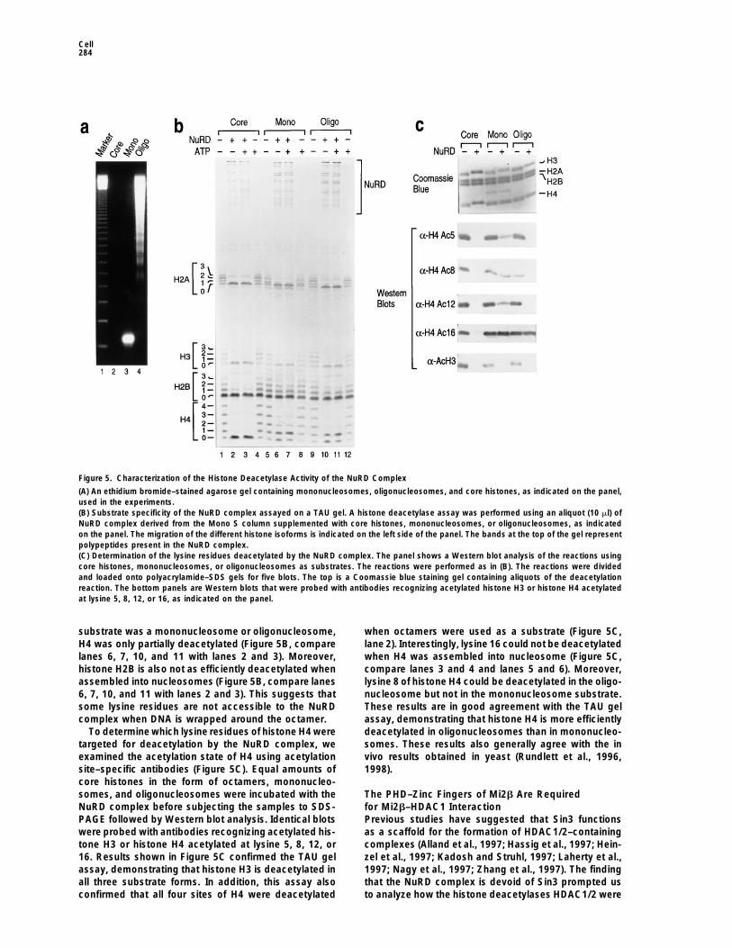
Figure 5. Characterization of the Histone Deacetylase Activity of the NuRD Complex
(A) An ethidium bromide–stained agarose gel containing mononucleosomes, oligonucleosomes, and core histones, as indicated on the panel,used in the experiments.(B) Substrate specificity of the NuRD complex assayed on a TAU gel. A histone deacetylase assay was performed using an aliquot (10 ml) ofNuRD complex derived from the Mono S column supplemented with core histones, mononucleosomes, or oligonucleosomes, as indicatedon the panel. The migration of the different histone isoforms is indicated on the left side of the panel. The bands at the top of the gel representpolypeptides present in the NuRD complex.(C) Determination of the lysine residues deacetylated by the NuRD complex. The panel shows a Western blot analysis of the reactions usingcore histones, mononucleosomes, or oligonucleosomes as substrates. The reactions were performed as in (B). The reactions were dividedand loaded onto polyacrylamide–SDS gels for five blots. The top is a Coomassie blue staining gel containing aliquots of the deacetylationreaction. The bottom panels are Western blots that were probed with antibodies recognizing acetylated histone H3 or histone H4 acetylatedat lysine 5, 8, 12, or 16, as indicated on the panel.
substrate was a mononucleosome or oligonucleosome, when octamers were used as a substrate (Figure 5C,lane 2). Interestingly, lysine 16 could not be deacetylatedH4 was only partially deacetylated (Figure 5B, compare
lanes 6, 7, 10, and 11 with lanes 2 and 3). Moreover, when H4 was assembled into nucleosome (Figure 5C,compare lanes 3 and 4 and lanes 5 and 6). Moreover,histone H2B is also not as efficiently deacetylated when
assembled into nucleosomes (Figure 5B, compare lanes lysine 8 of histone H4 could be deacetylated in the oligo-nucleosome but not in the mononucleosome substrate.6, 7, 10, and 11 with lanes 2 and 3). This suggests that
some lysine residues are not accessible to the NuRD These results are in good agreement with the TAU gelassay, demonstrating that histone H4 is more efficientlycomplex when DNA is wrapped around the octamer.
To determine which lysine residues of histone H4 were deacetylated in oligonucleosomes than in mononucleo-somes. These results also generally agree with the intargeted for deacetylation by the NuRD complex, we
examined the acetylation state of H4 using acetylation vivo results obtained in yeast (Rundlett et al., 1996,1998).site–specific antibodies (Figure 5C). Equal amounts of
core histones in the form of octamers, mononucleo-somes, and oligonucleosomes were incubated with the The PHD–Zinc Fingers of Mi2b Are Required
for Mi2b–HDAC1 InteractionNuRD complex before subjecting the samples to SDS-PAGE followed by Western blot analysis. Identical blots Previous studies have suggested that Sin3 functions
as a scaffold for the formation of HDAC1/2–containingwere probed with antibodies recognizing acetylated his-tone H3 or histone H4 acetylated at lysine 5, 8, 12, or complexes (Alland et al., 1997; Hassig et al., 1997; Hein-
zel et al., 1997; Kadosh and Struhl, 1997; Laherty et al.,16. Results shown in Figure 5C confirmed the TAU gelassay, demonstrating that histone H3 is deacetylated in 1997; Nagy et al., 1997; Zhang et al., 1997). The finding
that the NuRD complex is devoid of Sin3 prompted usall three substrate forms. In addition, this assay alsoconfirmed that all four sites of H4 were deacetylated to analyze how the histone deacetylases HDAC1/2 were

NuRD Complex Purification and Characterization285
Figure 6. The PHD–Zinc Fingers Are Required for Mi2b–HDAC1 Interaction
(A) Autoradiogram of a pull-down assay. Different proteins, as indicated on the panel, were attached to beads and incubated with invitro–translated Mi2b or luciferase. The beads were then washed three times with a buffer containing 400 mM KCl and 0.05% NP40 beforeelution with SDS loading buffer.(B) Schematic representation of the Mi2b polypeptide and different truncated proteins that were used in the analysis described in (C). The 1
symbol at the right of the panel denotes the protein that interacts with HDAC1.(C) Different truncated Mi2b expression vectors, described in (B), or a luciferase expression vector was transcribed/translated in vitro andincubated with HDAC1 as indicated at the top of the panel. The interaction between the different truncated Mi2b proteins and HDAC1 wasanalyzed by a pull-down assay as described in (A). HDAC1-Flag was attached to beads coated with monoclonal antibodies against the Flagepitope.
incorporated into the NuRD complex. Therefore, we ex- are dispensable for the interaction between Mi2 andHDAC1 (Figures 6B and 6C). However, deletion of theamined whether Mi2b interacts with HDAC1 and the
histone-binding proteins RbAp48/46. HDAC1 was puri- PHD-zinc fingers abrogated the interaction (Figure 6C,lane 12). To analyze whether the PHD-zinc fingers werefied from baculovirus-infected SF9 cells as an HDAC1-
FLAG fusion protein, while RbAp48/46 were purified sufficient for the interaction, polypeptides containingone or both fingers were produced in bacteria and ana-from E. coli as GST-fusion proteins. These proteins were
bound to beads and were incubated with in vitro– lyzed for their ability to interact with HDAC1. This analy-sis failed to demonstrate an interaction (data not shown).translated Mi2b protein. After extensive washing, the
proteins remaining bound to the beads were resolved Therefore, we conclude that the PHD-zinc fingers arerequired but not sufficient for the interaction. While theby SDS-PAGE and visualized by fluorography. We found
that Mi2b interacts with HDAC1 (Figure 6A, lane 5). This function of PHD-zinc fingers is not known, they havebeen implicated in the regulation of chromatin-mediatedinteraction appears to be specific, as GST and GST-
RbAp48/46 failed to pull down Mi2b (Figure 6A, lanes transcription (Aasland et al., 1995). The functional signifi-cance of PHD-zinc-finger domains has been highlighted2–4). Furthermore, HDAC1 failed to pull down in vitro–
translated luciferase (Figure 6A, lane 6). by the recent finding that mutations within the PHD-zincfinger of the transcriptional regulator ATRX are associ-Mi2b contains several interesting domains (Figure 6B).
To determine whether these domains are involved in the ated with the ATRX syndrome (Gibbons et al., 1997).Mi2/HDAC1 interaction, different Mi2 truncations wereanalyzed for their ability to interact with HDAC1. A sche- Discussionmatic representation of these truncations is shown inFigure 6B. The results from pull-down experiments, Using conventional chromatography and immuno-affin-
ity purification approaches, we have purified a novelshown in Figure 6C, demonstrate that amino acids (521–1912) at the C-terminal of the PHD-zinc fingers of Mi2 protein complex from human cells containing both

Cell286
nucleosome remodeling and histone deacetylase activi-ties. The complex, which we named NuRD, contains thetwo histone deacetylases HDAC1/2, the two histone-binding proteins RbAp48/46, the helicase/ATPase do-main–containing protein Mi2b, an MTA1-related protein,and a novel protein of 32 kDa. We show that the NuRDcomplex has DNA-dependent nucleosome-stimulatedATPase activity. In addition, we demonstrate that thiscomplex remodels nucleosomes in an ATP-dependentmanner. Furthermore, the NuRD complex is able to dea-cetylate nucleosomal histones.
The NuRD Complex Couples Histone Deacetylationwith Nucleosome RemodelingAs discussed above, two types of activities involvingATP hydrolysis and covalent modification of histonetails, respectively, have been discovered. However, aprotein complex possessing both activities has not beenreported. Here we demonstrate that the NuRD complexhas both nucleosome remodeling and histone deacety- Figure 7. A Speculative Model for the Function of the NuRD
Complexlation activities. Similar to NURF, the NuRD com-The NuRD complex is tethered to specific sites in promoters by aplex remodels nucleosomes in an ATP-dependent andsequence-specific DNA-binding protein interacting, directly or indi-transcription factor–dependent manner when catalyticrectly, with a component of the NuRD complex. Alternatively, oneamounts of the factor are used. At higher concentra-of the NuRD subunits may bind DNA directly. The ATP-dependent
tions, the NuRD complex loses transcription factor de- nucleosome remodeling activity of NuRD remodels the nucleo-pendence. Unlike other nucleosome remodeling factors some(s) and stabilizes the protein–DNA interaction. The remodelingsuch as ACF, CHRAC, and RSF, NuRD does not possess activity also permits the RbAp polypeptides to gain access to the
core histones. The histone deacetylases in the NuRD complex thenATP-dependent nucleosome spacing activity. With re-deacetylate (or maintain in a deacetylated state) the histone tailsgard to its deacetylase activity, the NuRD complex issuch that the neighboring nucleosomes are tightly packed and doable to deacetylate all four core histones (Figure 5B).not allow formation of transcription-competent complex. This model
However, the NuRD complex is not able to deacetylate permits the establishment and maintenance of a gene-specific re-lysine 16 of histone H4 when H4 is incorporated into pressed state.a nucleosome, although this lysine can be efficientlydeacetylated when histone octamers are used as a sub- case, the loss of histone deacetylase activity shouldstrate (Figure 5C). Interestingly, lysine 8 of histone H4 cause a general increase in histone acetylation, resultingalso becomes resistant to deacetylation when present in a global increase in gene expression. However, dis-in a mononucleosome, although it is efficiently deacety- ruption of RPD3 in yeast or treatment of human cellslated in oligonucleosomes (Figure 5C). These results with histone deacetylase inhibitors does not causeindicate that in addition to DNA, the nucleosome struc- global gene activation. In fact, only a limited number ofture also influences the accessibility of the NuRD com- genes are affected, and some genes are repressedplex to its target. rather than activated (Vidal and Gaber, 1991; Van Lint
The finding that both nucleosome remodeling and his- et al., 1996). One possible explanation is outlined intone deacetylation activities exist in the same complex Figure 7. A remodeling histone deacetylase complex,seems to be a paradox, since ATP-dependent nucleo- such as NuRD, is recruited to the promoter region of asome remodeling was thought to be involved in tran- subset of genes through the interaction with a DNA-scriptional activation, while histone deacetylation has binding protein. Alternatively, recruitment may be achievedbeen linked to transcriptional repression. However, we by the complex itself recognizing a specific DNA ele-believe that the coupling of a nucleosome remodeling ment. Upon recruitment, the complex uses energy fromactivity to HDAC or HAT activity may provide an efficient ATP hydrolysis to remodel adjacent nucleosomes, al-way for the cell to achieve transcriptional regulation. It lowing the histone tails to become accessible for deace-has been previously demonstrated that RbAp48 and tylation. Deacetylation of the core histone tails resultsRbAp46 cannot gain access to nucleosomal histones, in the formation of a more compacted nucleosomalalthough they efficiently bind to core histones (Verreault structure, leading to transcriptional repression. The dis-et al., 1997; Zhang et al., 1998). This is likely because covery that nucleosome remodeling and histone deace-helix 1 of histone H4, which is involved in RbAp binding tylase activities coexist in the same complex supports(Verreault et al., 1997), also interacts with DNA when this model. We observed that the NuRD complex dea-packaged into a nucleosome (Luger et al., 1997). There- cetylates nucleosomal histones in the absence of ATP.fore, in order for the RbAp proteins to access histones, This result does not seem to be compatible with thisthe nucleosome structure needs to be altered. If nucleo- model. However, this only occurs in the presence ofsomal histones were freely accessible to the NuRD or large amounts of the NuRD complex. When catalyticother complexes, one would expect that histone deace- amounts of the NuRD complex were used, no nucleoso-
mal histone deacetylation was observed, although coretylases would work on a genome-wide basis. In this

NuRD Complex Purification and Characterization287
using HeLa nuclear extracts fractionated on phosphocellulose andhistones were efficiently deacetylated. It is likely thatDEAE-52 columns. The affinity columns were prepared using puri-nucleosomal histone deacetylation by the NuRD com-fied antibodies against the C-terminal domain of HDAC1, againstplex requires targeting of this complex to the vicinity ofthe full-length SAP30, or against a fragment of Mi2b (amino acids
a specific nucleosome. We believe that it will be possible 475–970) coupled to 1 ml of protein A–agarose beads (Repligen) asto obtain ATP-dependent nucleosomal histone deacety- described (Harlow and Lane, 1988).lation if the NuRD complex is targeted to a specific site The procedure for conventional purification of the NuRD complex
is outlined in Figure 2A. Approximately 6 g of HeLa nuclear extractsin a reconstituted nucleosome array.was fractionated on phosphocellulose and DEAE-52 columns. TheDEAE-52-bound proteins were eluted with buffer C (20 mM Tris-HClThe Connection between Histone Deacetylase,[pH 7.9], 0.2 mM EDTA, 10 mM b-ME, 0.2 mM PMSF, and 10%Chromatin Structure, Dermatomyositis,glycerol) containing 350 mM KCl (BC350) and proteins were precipi-
and Cancer tated with saturated ammonium sulfate (final concentration of 45%).Chromatin is the in vivo substrate for all biological pro- Following centrifugation at 35,000 rpm for 1 hr, the pellets werecesses involving DNA. Thus, interfering with chromatin resuspended in buffer D (40 mM HEPES [pH 7.9], 0.2 mM EDTA, 1
mM DTT, 0.2 mM PMSF, and 10% glycerol), and the ammoniumstructure should influence many fundamental biologicalsulfate concentration was adjusted to 700 mM. The sample (approxi-processes, likely resulting in the development of specificmately 340 mg) was then loaded onto a 55 ml FPLC Phenyl Sepha-diseases. Several lines of evidence indicate that factorsrose column (Pharmacia). The column was then washed with 4 col-that alter chromatin structure could lead to malignancy.umn volumes (cv) of BD700. Proteins were eluted with a linear
First, histone deacetylase inhibitors, such as TSA, tra- gradient (20 cv) of ammonium sulfate from 700 to 0 mM in bufferpoxin, and depudecin, are able to cause cell cycle arrest D. The fractions containing the NuRD complex (150 to 400 mMand morphological changes in a number of tumor cell ammonium sulfate) were pooled and dialyzed against BD100. The
sample (75 mg) was then loaded onto an HPLC-DEAE-5PW columnlines (Yoshida et al., 1995; Kwon et al., 1998). In addition,(TosoHaas, 45 ml) equilibrated with BD100. Proteins bound to thehistone deacetylases may affect cell cycle regulationcolumn were eluted with a 10 cv linear gradient from 100 to 400through their association with the retinoblastoma tumormM ammonium sulfate in buffer D. Fractions containing the NuRDsuppressor protein (Brehm et al., 1998; Luo et al., 1998;complex (190 to 230 mM ammonium sulfate) were pooled (5 mg)
Magnaghi-Jaulin et al., 1998). Moreover, a chimeric mu- and precipitated with saturated ammonium sulfate to a final concen-tant of the retinoic acid receptor, which causes acute tration of 65%. After centrifugation at 35,000 rpm for 1 hr, the pelletspromyelocytic leukemia, was found to be associated were resuspended in BC500 and proteins were separated by chro-
matography on a Superose-6 column (Pharmacia). The NuRD com-with the histone deacetylase HDAC1 (Grignani et al.,plex eluted with an apparent mass of 1 MDa. The NuRD complex1998; Lin et al., 1998). Finally, mutations in hSNF5, apool (approximately 0.5 mg) was dialyzed against BC80 and loadedcomponent of the human SWI/SNF nucleosome remod-onto a 1 ml Mono S column (Pharmacia) equilibrated in BC80. Theeling factor, have recently been linked to the develop-proteins were then eluted with 20 cv linear gradient of buffer C from
ment of malignant rhabdoid tumors (Versteege et al., 80 to 450 mM KCl. The NuRD complex eluted between 200 to 3001998). The finding that the NuRD complex contains a mM KCl.component that is highly related to the metastasis-asso-ciated factor MTA1 reinforces the idea that changes in
Mass Spectrometric Peptide Sequencingchromatin structure, either by nucleosome remodelingAffinity-purified samples from anti-HDAC1 column was concen-factors or histone deacetylases, could affect the celltrated 10-fold using a centricon concentrator and was resolved oncycle.an 8% SDS-PAGE. After Coomassie staining, the 230 kDa band
MTA1 was initially identified by differential screening was subjected to in-gel reduction, S-carboxyamidomethylation, andusing the rat mammary adenocarcinoma metastatic sys- tryptic digestion (Promega), and a 10% aliquot of the resultant mix-tem (Toh et al., 1994). It was found that the expression ture was analyzed as described (Nash et al., 1996). Interpretationlevel of MTA1 correlates with the metastatic potential of the resulting MS/MS spectra of the peptides was facilitated by
searching the NCBI nr and dbest databases with the algorithmof several human cancer cell lines and tissues (Toh etSequest (Eng et al., 1994), followed by manual inspection. Forty-al., 1994, 1997). In agreement with a role for MTA2 in cellthree MS/MS spectra were individually confirmed to correlate withproliferation, we found that MTA2 is highly expressed insequences from Mi2.rapidly dividing cells (Y. Z. and D. R., unpublished data).
Another important component of the NuRD complexis Mi2b, an autoantigen for the autoimmune disease Nucleosomes and Core Histones Purification,dermatomyositis. While the relationship between the Histone Deacetylase Assays, and TAU Gelnormal function of Mi2b and the development of derma- Acetylated nucleosomes and core histones were purified from HeLa
cells as described (Zhang et al., 1998). Procedures for labeling,tomyositis is not clear, it has been reported that patientspurification of core histones, and electrophoresis on TAU gels werewith dermatomyositis also face an increased risk of ma-as described (Zhang et al., 1998). Histone deacetylase assay condi-lignancy (Airio et al., 1995; Shorr et al., 1997). Abouttions were modified from our previously published procedure (Zhang15% to 30% of patients with dermatomyositis developet al., 1998) to facilitate nucleosome remodeling. Briefly, about 10
cancer (Airio et al., 1995). The finding that the dermato-mg of 3H-labeled core histone octamers (Figures 2B and 3A) or 4
myositis-associated autoantigen Mi2b and the candi- mg of acetylated core histones and nucleosomes (Figure 5) weredate metastasis-associated protein MTA2 exist in the incubated with column fractions or purified NuRD complex at 308Csame protein complex may provide a molecular explana- for 1 hr in the presence or absence of 4 mM ATP in buffer containing
10 mM HEPES (pH 7.5), 5 mM MgCl2, 50 mM KCl, 2 mM DTT, andtion to the above observation.0.5 mM EDTA. The released [3H]acetate was extracted with ethylacetate and quantified by liquid scintillation counting (Figures 2BExperimental Proceduresand 3A). Alternatively, deacetylation was monitored by resolvinghistones on a TAU gel followed by Coomassie staining (Figure 5B)Purification of the NuRD Complexor by resolving on an SDS-PAGE followed by Coomassie stainingAffinity purification of the HDAC1, SAP30, and NuRD complexes
was based on a previously published procedure (Zhang et al., 1998) and Western blot (Figure 5C).

Cell288
Nucleosome Assembly, Nucleosome Remodeling, Cioce, M., Fanelli, M., Ruthardt, M., Ferrara, F.F., Zamir, I., et al.(1998). Fusion proteins of the retinoic acid receptor-a recruit histoneand ATPase Assaysdeacetylase in promyelocytic leukaemia. Nature 391, 815–818.Chromatin was assembled and purified as described (Bulger and
Kadonaga, 1994; Orphanides et al., 1998). The nucleosome remodel- Grunstein, M. (1997). Histone acetylation in chromatin structure anding assay was performed using a modification of a published proce- transcription. Nature 389, 349–352.dure (Tsukiyama and Wu, 1995). The ATPase activity of the NuRD Harlow, E., and Lane, D. (1988). Antibodies: A Laboratory Manualcomplex was analyzed using a modification of a published proce- (Cold Spring Harbor, New York: Cold Spring Harbor Laboratorydure (Tsukiyama and Wu, 1995). Briefly, 2 ml of purified NuRD com- Press).plex (Mono S fraction) was incubated with 500 ng of sonicated calf
Hassig, C.A., Fleischer, T.C., Billin, A.N., Schreiber, S.L., and Ayer,thymus DNA (200–600 bps), core histones, mononucleosomes, orD.E. (1997). Histone deacetylase activity is required for full transcrip-oligonucleosomes (purified from HeLa cells, see above section) intional repression by mSin3A. Cell 89, 331–347.the presence of 2 mM ATP, 8 mM MgCl2, 50 mM KCl, and 0.5 ml ofHassig, C.A., Tong, J.K., Fleischer, T.C., Owa, T., Grable, P.G., Ayer,[g-32p]ATP (3000 Ci/mmol, 5 mCi/ml, NEN Dupont) in a 10 ml volumeD.E., and Schreiber, S. (1998). A role for histone deacetylase activityat 288C for 30 min. The reactions were stopped by adding 5 ml ofin HDAC1-mediated transcriptional repression. Proc. Natl. Acad.0.5 M EDTA. Products were separated using a TLC glass chamberSci. USA 95, 3519–3524.presaturated with 1 M formic acid and LiCl. The TLC plates wereHebbes, T.R., Thorne, A.W., and Crane-Robinson, C. (1988). A directdried, exposed to X-ray film, and quantitated with an Image Masterlink between core histone acetylation and transcriptionally activeDensitometer (Pharmacia).chromatin. EMBO J. 7, 1395–1402.
Acknowledgments Heinzel, T., Lavinsky, R.M., Mullen, T.M., Soderstrom, M., Laherty,C.D., Torchia, J., Yang, W.M., Brard, G., Ngo, S.D., Davie, J.R., et
We are grateful to C. A. Hassig and S. Schreiber for baculoviruses al. (1997). A complex containing N-CoR, mSin3 and histone deacety-expressing HDAC1-Flag; to W.-M. Yang and E. Seto for anti-HDAC2 lase mediates transcription repression. Nature 387, 43–48.antibodies; to K. Cabane for help in generating Mi2b antibodies; Kadonaga, J.T. (1998). Eukaryotic transcription: an interlaced net-and to E. Spooner and R. Robinson for their technical expertise work of transcription factors and chromatin-modifying machines.in peptide sequencing. We also thank members of the Reinberg Cell 92, 307–313.laboratory for stimulating discussions during the course of this work,
Kadosh, D., and Struhl, K. (1997). Repression by Ume6 involvesand G. Orphanides for comments. Y. Z. is a recipient of an NIH recruitment of a complex containing Sin3 corepressor and Rpd3fellowship (1F32GM19515-01). D. R. was supported by grants from histone deacetylase to target promoters. Cell 89, 365–371.NIH (GM-48518) and the Howard Hughes Medical Institute.
Kadosh, D., and Struhl, K. (1998). Histone deacetylase activity ofRpd3 is important for transcriptional repression in vivo. Genes Dev.Received August 20, 1998; revised September 18, 1998.12, 797–805.
Kuo, M., Zhou, J., Jambeck, P., Churchill, M.E.A., and Allis, C.D.References(1998). Histone acetyltransferase activity of yeast Gcn5p is requiredfor the activation of target genes in vivo. Genes Dev. 12, 627–639.Aasland, R., Gibson, T.J., and Stewart, F.A. (1995). The PHD finger:Kwon, H.J., Owa, T., Hassig, C.A., Shimada, J., and Schreiber, S.L.implications for chromatin mediated transcriptional regulation.(1998). Depudecin induces morphological reversion of transformedTrends Biochem. 20, 56–59.fibroblasts via the inhibition of histone deacetylase. Proc. Natl. Acad.Airio, A., Pukkala, E., and Isomaki, H. (1995). Elevated cancer inci-Sci. USA 95, 3356–3361.dence in patients with dermatomyositis: a population based study.Laherty, C.D., Yang, W.-M., Sun, J.-M., Davie, J.R., Seto, E., andJ. Rheumatol. 22, 1300–1303.Eisenman, R.N. (1997). Histone deacetylases associated with theAlland, L., Muhle, R., Hou, H., Jr., Potes, J., Chin, L., Schreiber-mSin3 corepressor mediate Mad transcriptional repression. Cell 89,Agus, N., and DePinho, R.A. (1997). Role for N-CoR and histone349–356.deacetylase in Sin3-mediated transcriptional repression. NatureLaherty, C.D., Billin, A.N., Lavinsky, R.M., Yochum, G.S., Bush, A.C.,387, 49–55.Sun, J.M., Mullen, T.M., Davie, J.R., Rose, D.W., Glass, C.K., et al.Braunstein, M., Rose, A.B., Holmes, S.G., Allis, C.D., and Broach,(1998). SAP30, a component of the mSin3 corepressor complexJ.R. (1993). Transcriptional silencing in yeast is associated withinvolved in N-CoR-mediated repression by specific transcription
reduced nucleosome acetylation. Genes Dev. 7, 592–604.factors. Mol. Cell 2, 33–42.
Brehm, A., Miska, E.A., McCance, D.J., Reid, J.L., Bannister, A.J.,Lin, R.J., Nagy, L., Inoue, S., Shao, W., Miller, W.H., and Evans,
and Kouzarides, T. (1998). Retinoblastoma protein recruits histoneR.M. (1998). Role of the histone deacetylase in acute promyelocytic
deacetylase to repress transcription. Nature 391, 597–601. leukaemia. Nature 391, 811–814.Bulger, M., and Kadonaga, J.T. (1994). Biochemical reconstitution Luger, K., Mader, A.W., Richmond, R.K., Sargent, D.F., and Rich-of chromatin with physiological nucleosome spacing. Methods Mol. mond, T.J. (1997). Crystal structure of the nucleosome core particleGenet. 5, 241–262. at 2.8 A resolution. Nature 389, 251–260.De Rubertis, F., Kadosh, D., Henchoz, S., Pauli, D., Reuter, G., Struhl, Luo, R.X., Postigo, A.A., and Dean, D.C. (1998). Rb interacts withK., and Spierer, P. (1996). The histone deacetylase RPD3 counter- histone deacetylase to repress transcription. Cell 92, 463–473.acts genomic silencing in Drosophila and yeast. Nature 384,
Magnaghi-Jaulin, L., Groisman, R., Naguibneva, I., Robin, P., Lorain,589–591.
S., Le Villain, J.P., Troalen, F., Trouche, D., and Harel-Bellan, A.Eisen, J.A., Sweeder, K.S., and Hanawalt, P.C. (1995). Evolution of (1998). Retinoblastoma protein represses transcription by recruitingthe SNF2 family of proteins: subfamilies with distinct sequences a histone deacetylase. Nature 391, 601–605.and functions. Nucleic Acids Res. 23, 2715–2723. Nagy, L., Kao, H.-Y., Chakravarti, D., Lin, R.J., Hassig, C.A., Ayer,Eng, J.K., McCormick, A.L., and Yates, J.R., III. (1994). An approach D.E., Schreiber, S.L., and Evans, R.M. (1997). Nuclear receptor re-to correlate tandem mass spectral data of peptides with amino acid pression mediated by a complex containing SMRT, mSin3A, andsequences in a protein database. J. Am. Soc. Mass Spectrom. 5, histone deacetylase. Cell 89, 373–380.976–989. Nash, H.M., Bruner, S.D., Scharer, O.D., Kawate, T.A., Spooner, E.,Gibbons, R.J., Bachoo, S., Picketts, D.J., Aftimos, S., Asenbauer, Lane, W.S., and Verdine, G.L. (1996). Cloning of a yeast 8-oxogua-B., Bergoffen, J., Berry, S.A., Dahl, N., Fryer, A., Keppler, K., et al. nine DNA glycosylase reveals the existence of a base-excision DNA-(1997). Mutations in transcriptional regulator ATRX establish the repair protein superfamily. Curr. Biol. 6, 968–980.functional significance of a PHD-like domain. Nat. Genet. 17, Orphanides, G., LeRoy, G., Chang, C.-H., Luse, D., and Reinberg,146–148. D. (1998). FACT, a factor that facilitates transcript elongation through
nucleosomes. Cell 92, 105–116.Grignani, F., De Matteis, S., Nervi, C., Tomassoni, L., Gelmetti, V.,

NuRD Complex Purification and Characterization289
Paranjape, S.M., Kamakaka, R.T., and Kadonaga, J.T. (1994). Role required to achieve maximum positive and negative transcriptionalstates in Saccharomyces cerevisiae. Mol. Cell. Biol. 11, 6317–6327.of chromatin structure in the regulation of transcription by RNA
polymerase II. Annu. Rev. Biochem. 63, 265–297. Wade, P.A., Pruss, D., and Wolffe, A.P. (1997). Histone acetylation:chromatin in action. Trends Biochem. Sci. 22, 128–132.Pazin, M.J., and Kadonaga, J.T. (1997). SWI2/SNF2 and related pro-
teins: ATP-driven motors that disrupt protein–DNA interactions? Cell Wang, L., Liu, L., and Berger, S.L. (1998). Critical residues for histone88, 737–740. acetylation by Gcn5, functioning in Ada and SAGA complexes, are
also required for transcriptional function in vivo. Genes Dev. 12,Qian, Y.-W., and Lee, E.Y.-H.P. (1995). Dual retinoblastoma-bindingproteins with properties related to a negative regulator of Ras in 640–653.yeast. J. Biol. Chem. 270, 25507–25513. Woodage, T., Basarai, M.A., Baxevanis, A.D., Heiter, P., and Collins,
F.S. (1997). Characterization of the CHD family of proteins. Proc.Qian, Y.-W., Wang, Y.-C.J., Hollingsworth, R.E.J., Jones, D., Ling,N., and Lee, E.Y.-H.P. (1993). A retinoblastoma-binding protein re- Natl. Acad. Sci. USA 94, 11472–11477.lated to a negative regulator of Ras in yeast. Nature 364, 648–652. Yang, W.-M., Inouye, C., Zeng, Y.Y., Bearss, D., and Seto, E. (1996).
Transcriptional repression by YY1 is mediated by interaction withRundlett, S.E., Carmen, A.A., Kobayashi, R., Bavykin, S., Turner,B.M., and Grunstein, M. (1996). HDA1 and RPD3 are members of a mammalian homolog of the yeast global regulator RPD3. Proc.
Natl. Acad. Sci. USA 93, 12845–12850.distinct yeast histone deacetylase complexes that regulate silencingand transcription. Proc. Natl. Acad. Sci. USA 93, 14503–14508. Yoshida, M., Horinouchi, S., and Beppu, T. (1995). Trichostatin A and
trapoxin: novel chemical probes for the role of histone acetylation inRundlett, S.E., Carmen, A.A., Suka, N., Turner, B., and Grunstein, M.(1998). Transcriptional repression by UME6 involves deacetylation of chromatin structure and function. Bioessays 17, 423–430.lysine 5 of histone H4 by RPD3. Nature 392, 831–835. Zhang, Y., Iratni, R., Erdjument-Bromage, H., Tempst, P., and Rein-Seelig, H.P., Moosbrugger, I., Ehrfeld, H., Fink, T., Renz, M., and berg, D. (1997). Histone deacetylases and SAP18, a novel polypep-Genth, E. (1995). The major dermatomyositis-specific Mi-2 autoanti- tide, are components of a human sin3 complex. Cell 89, 357–364.gen is a presumed helicase involved in transcriptional activation. Zhang Y., Sun, Z.-W., Iratni, R., Erdjument-Bromage, H., Tempst,Arthritis Rheum. 38, 1389–1399. P., Hampsey, M., and Reinberg, D. (1998). SAP30, a novel proteinSeelig, H.P., Renz, M., Targoff, I.N., Ge, Q., and Frank, M.B. (1996). conserved between human and yeast, is a component of a histoneTwo forms of the major antigenic protein of the dermatomyositis- deacetylase complex. Mol. Cell 1, 1021–1031.specific Mi-2 autoantigen. Arthritis Rheum. 39, 1769–1771.
Shorr, A.F., Yacavone, M., Seguin, S., Jackson, L.W., and Dennis,G.J. (1997). Dermatomyositis and malignant melanoma. Am. J. Med.Sci. 313, 249–251.
Struhl, K. (1998). Histone acetylation and transcriptional regulatorymechanisms. Genes Dev. 12, 599–606.
Taunton, J., Hassig, C.A., and Schreiber, S.L. (1996). A mammalianhistone deacetylase related to the yeast transcriptional regulatorRPD3p. Science 272, 408–411.
Toh, Y., Pencil, S.D., and Nicolson, G.L. (1994). A novel candidatemetastasis-associated gene, mta1, differentially expressed in highlymetatastic mammary adenocarcinoma cell lines. cDNA cloning, ex-pression, and protein analyses. J. Biol. Chem. 269, 22958–22963.
Toh, Y., Oki, E., Oda, S., Eriko, T., Ohno, S., Maehara, Y., Nicolson,G.L., and Sugimachi, K. (1997). Overexpression of the MTA1 genein gastrointestinal carcinomas: correlation with invasion and metas-tasis. Int. J. Cancer 74, 459–463.
Tsukiyama, T., and Wu, C. (1995). Purification and properties of anATP-dependent nucleosome remodeling factor. Cell 83, 1011–1020.
Tsukiyama, T., and Wu, C. (1997). Chromatin remodeling and tran-scription. Curr. Opin. Genet. Dev. 7, 182–191.
Utley, R.T., Ikeda, K., Grant, P., Cote, J., Steger, D.J., Eberharter,A., John, S., and Workman, J.L. (1998). Transcriptional activatorsdirect histone acetyltransferase complexes to nucleosomes. Nature394, 498–502.
Van Holde, K.E. (1989). Chromatin, A. Rich, ed. (Berlin, Germany:Springer-Verlag).
Van Lint, C., Emiliani, S., and Verdin, E. (1996). The expression of asmall fraction of cellular genes is changed in response to histonehyperacetylation. Gene Expr. 5, 245–253.
Vannier, D., Balderes, D., and Shore, D. (1996). Evidence that thetranscriptional regulators SIN3 and RPD3, and a novel gene (SDS3)with similar functions, are involved in transcriptional silencing in S.cerevisiae. Genetics 144, 1343–1353.
Varga-Weisz, P.D., and Becker, P.B. (1998). Chromatin-remodelingfactors: machines that regulate? Curr. Opin. Cell Biol. 10, 346–353.
Verreault, A., Kaufman, P.D., Kobayashi, R., and Stillman, B. (1997).Nucleosomal DNA regulates the core-histone-binding subunit of thehuman hat1 acetyltransferase. Curr. Biol. 8, 96–108.
Versteege, I., Sevenet, N., Lange, J., Rousseau-Merck, M.-F.,Ambros, P., Handgretinger, R., Aurias, A., and Delattre, O. (1998).Truncating mutations of hSNF5/INI1 in aggressive paediatric cancer.Nature 394, 203–206.
Vidal, M., and Gaber, R.F. (1991). RPD3 encodes a second factor