Cell division autoantigen 1 plays a profibrotic role by modulating downstream signalling of TGF-β...
10
ARTICLE Cell division autoantigen 1 plays a profibrotic role by modulating downstream signalling of TGF-β in a murine diabetic model of atherosclerosis Y. Pham & Y. Tu & T. Wu & T. J. Allen & A. C. Calkin & A. M. Watson & J. Li & K. A. Jandeleit-Dahm & B-H. Toh & Z. Cao & M. E. Cooper & Z. Chai Received: 30 July 2009 / Accepted: 27 August 2009 / Published online: 22 October 2009 # Springer-Verlag 2009 Abstract Aims/hypothesis Excess accumulation of vascular extracellu- lar matrix (ECM) is an important pathological process in cardiovascular diseases including diabetes-associated athero- sclerosis. We explored how a recently identified molecule, cell division autoantigen 1 (CDA1), influences the profibrotic TGF-β pathway leading to vascular ECM accumulation. Methods Expression levels of genes encoding for CDA1, TGF-β and connective tissue growth factor (CTGF) were examined in aorta from Apoe -/- mice with or without diabetes. We used retroviral and adenoviral constructs to knockdown or overexpress Tspyl2, the gene encoding CDA1, in mouse vascular smooth muscle cells (VSMCs) with or without TGF-β treatment in order to demonstrate the role of CDA1 in TGF-β signalling. Results In vivo studies indicated that the mRNA levels of CDA1-encoding gene Tspyl2 and protein levels of CDA1 were elevated in the aorta of diabetic Apoe -/- mice, accompanied by increased levels of Tgf-β (also known as Tgfb1), Ctgf and ECM accumulation. In vitro studies in vascular cells showed that TGF-β treatment rapidly in- creased CDA1 protein levels, which then amplified TGF-β signalling leading to upregulation of ECM genes. Knock- down of CDA1-encoding gene Tspyl2 to reduce cellular CDA1 level markedly attenuated TGF-β-stimulated MAD homologue 3 (drosophila; SMAD3) phosphorylation and transcriptional activities. CDA1 overproduction increased and Tspyl2 knockdown decreased expression of TGF-β receptor type I, TβrI (also known as Tgfbr1), but not TGF- β receptor type II, TβrII (also known as Tgfbr2), providing a mechanism for CDA1’ s action in modulating TGF-β signalling. Knockdown of CDA1-encoding gene Tspyl2 also blocked the profibrotic effect of TGF-β in VSMCs. Conclusions/interpretation CDA1 plays an important role in vascular ECM accumulation by amplifying TGF-β signal- ling. This is critical for the profibrotic effect of TGF-β in the vasculature. CDA1 is therefore a potential target for attenuating vascular ECM accumulation caused by enhanced TGF-β action, as seen in diabetic atherosclerosis. Keywords Atherosclerosis . CDA1 . Diabetes . Fibrosis . TGF-β . Vascular ECM accumulation Abbreviations CDA1 Cell division autoantigen 1 CTGF Connective tissue growth factor ECM Extracellular matrix ERK Extracellular signal-regulated protein kinase MAPK Mitogen-activated protein kinase siRNA Short interfering RNA SMAD MAD homologue 3 (drosophila) VSMC Vascular smooth muscle cell Electronic supplementary material The online version of this article (doi:10.1007/s00125-009-1555-9) contains supplementary material, which is available to authorised users. Y. Pham : Y. Tu : T. Wu : T. J. Allen : A. C. Calkin : A. M. Watson : J. Li : K. A. Jandeleit-Dahm : Z. Cao : M. E. Cooper : Z. Chai (*) Diabetes and Metabolism Division, Baker IDI Heart and Diabetes Institute, 75 Commercial Rd, Melbourne, Victoria 3004, Australia e-mail: [email protected] B.-H. Toh Autoimmunity Laboratory, Centre for Inflammatory Diseases, Department of Medicine, Monash University, Clayton, Victoria, Australia Diabetologia (2010) 53:170–179 DOI 10.1007/s00125-009-1555-9
-
Upload
independent -
Category
Documents
-
view
2 -
download
0
Transcript of Cell division autoantigen 1 plays a profibrotic role by modulating downstream signalling of TGF-β...

ARTICLE
Cell division autoantigen 1 plays a profibrotic roleby modulating downstream signalling of TGF-β in a murinediabetic model of atherosclerosis
Y. Pham & Y. Tu & T. Wu & T. J. Allen & A. C. Calkin &
A. M. Watson & J. Li & K. A. Jandeleit-Dahm &
B-H. Toh & Z. Cao & M. E. Cooper & Z. Chai
Received: 30 July 2009 /Accepted: 27 August 2009 /Published online: 22 October 2009# Springer-Verlag 2009
AbstractAims/hypothesis Excess accumulation of vascular extracellu-lar matrix (ECM) is an important pathological process incardiovascular diseases including diabetes-associated athero-sclerosis.We explored how a recently identifiedmolecule, celldivision autoantigen 1 (CDA1), influences the profibroticTGF-β pathway leading to vascular ECM accumulation.Methods Expression levels of genes encoding for CDA1,TGF-β and connective tissue growth factor (CTGF) wereexamined in aorta from Apoe−/− mice with or withoutdiabetes. We used retroviral and adenoviral constructs toknockdown or overexpress Tspyl2, the gene encoding CDA1,in mouse vascular smooth muscle cells (VSMCs) with orwithout TGF-β treatment in order to demonstrate the role ofCDA1 in TGF-β signalling.Results In vivo studies indicated that the mRNA levels ofCDA1-encoding gene Tspyl2 and protein levels of CDA1were elevated in the aorta of diabetic Apoe−/− mice,accompanied by increased levels of Tgf-β (also known as
Tgfb1), Ctgf and ECM accumulation. In vitro studies invascular cells showed that TGF-β treatment rapidly in-creased CDA1 protein levels, which then amplified TGF-βsignalling leading to upregulation of ECM genes. Knock-down of CDA1-encoding gene Tspyl2 to reduce cellularCDA1 level markedly attenuated TGF-β-stimulated MADhomologue 3 (drosophila; SMAD3) phosphorylation andtranscriptional activities. CDA1 overproduction increasedand Tspyl2 knockdown decreased expression of TGF-βreceptor type I, TβrI (also known as Tgfbr1), but not TGF-β receptor type II, TβrII (also known as Tgfbr2), providing amechanism for CDA1’s action in modulating TGF-βsignalling. Knockdown of CDA1-encoding gene Tspyl2 alsoblocked the profibrotic effect of TGF-β in VSMCs.Conclusions/interpretation CDA1 plays an important role invascular ECM accumulation by amplifying TGF-β signal-ling. This is critical for the profibrotic effect of TGF-β in thevasculature. CDA1 is therefore a potential target forattenuating vascular ECM accumulation caused by enhancedTGF-β action, as seen in diabetic atherosclerosis.
Keywords Atherosclerosis . CDA1 . Diabetes . Fibrosis .
TGF-β . Vascular ECM accumulation
AbbreviationsCDA1 Cell division autoantigen 1CTGF Connective tissue growth factorECM Extracellular matrixERK Extracellular signal-regulated protein kinaseMAPK Mitogen-activated protein kinasesiRNA Short interfering RNASMAD MAD homologue 3 (drosophila)VSMC Vascular smooth muscle cell
Electronic supplementary material The online version of this article(doi:10.1007/s00125-009-1555-9) contains supplementary material,which is available to authorised users.
Y. Pham :Y. Tu : T. Wu : T. J. Allen :A. C. Calkin :A. M. Watson : J. Li :K. A. Jandeleit-Dahm : Z. Cao :M. E. Cooper : Z. Chai (*)Diabetes and Metabolism Division,Baker IDI Heart and Diabetes Institute,75 Commercial Rd,Melbourne, Victoria 3004, Australiae-mail: [email protected]
B.-H. TohAutoimmunity Laboratory, Centre for Inflammatory Diseases,Department of Medicine, Monash University,Clayton, Victoria, Australia
Diabetologia (2010) 53:170–179DOI 10.1007/s00125-009-1555-9

Introduction
Cardiovascular disease usually arises from vascular dys-function as a result of atherosclerosis and/or thrombosis,and is clinically associated with diabetes [1]. Excessaccumulation of extracellular matrix (ECM) in the vascu-lature appears to play an important role in the developmentand progression of the vasculopathy seen in diabetes [2–6].However, the underlying molecular mechanisms have notbeen fully delineated.
TGF-β is the major growth factor implicated in vascularmatrix accumulation via the SMAD signalling pathway [6, 7].TGF-β-dependent and -independent signalling pathwaysinvolving R-SMAD activation are the major pathways viawhich various pathological stimuli that appear to beenhanced in the diabetic milieu, e.g. TGF-β, angiotensin II,AGE, glucose and mechanical stretch, stimulate blood vesselECM accumulation and remodelling [6]. It has beenpostulated that targeting downstream signalling of TGF-βcould be beneficial by reducing ECM accumulation [8].Indeed, blockade of the renin–angiotensin system with drugssuch as ACE inhibitors has been shown to reduce fibrosis,partly via TGF-β-dependent pathways, and has beenreported to attenuate atherosclerosis in the setting of diabetes[4]. However, targeting ECM accumulation in the athero-sclerotic plaque at a later stage remains controversial since incertain contexts, such as in the fibrous cap, TGF-β may actparadoxically to reduce the risk of subsequent acute plaquerupture [9–11].
Cell division autoantigen 1 (CDA1) is a molecule that weidentified and named to reflect our initial finding that itarrested cell growth when overexpressed in HeLa cells [12].Its anti-proliferative action is supported by our furtherfindings as well as by those of other groups [13–15]. Wehave shown that CDA1 is able to activate the extracellularsignal-regulated protein kinase (ERK)/mitogen-activated pro-tein kinase (MAPK) pathway and transcriptionally upre-gulate the cyclin-dependent kinase inhibitor, p21Waf1/Cip1
[13]. Both the latter and ERK/MAPK are known to beregulated by TGF-β [16–21]. CDA1 has been shown to beupregulated by TGF-β and to increase luciferase activities ofTGF-β reporter constructs in some lung cancer cell lines[22–25]. These findings suggest that CDA1 may be involvedin TGF-β signalling and play a role in mediating thebiological functions of TGF-β. TGF-β upregulation isthought to be a pathological event in diabetes, leading toprominent ECM accumulation and thereby promoting vari-ous diabetic complications, including nephropathy andmacrovascular diseases [26, 27].
Here, we not only show overexpression of the CDA1-encoding gene Tspyl2 in diabetic Apoe−/− mice, whichexhibit accelerated atherosclerosis, but also identify a novelrole for CDA1 in modulating TGF-β signalling in vascular
smooth muscle cells (VSMCs), which leads to ECM geneexpression. Thus, these data show that CDA1 is anappropriate and potentially attractive molecular target toblock the profibrotic effect of TGF-β, thus preventing,retarding or treating diabetes-accelerated vascular ECMaccumulation and atherosclerosis.
Methods
Antibodies, reagents and plasmids Rabbit antibody, raisedto GST fusion protein of the C-terminal fragment of mouseCDA1, was affinity-purified using a method describedpreviously [12]. Antibodies to phospho-SMAD3 (Ser433/435) (#9514) and to SMAD2/3 (sc-8332) were from CellSignaling Technology (Beverly, MA, USA) and Santa CruzBiotechnology (Santa Cruz, CA, USA), respectively. Theantibody to α-tubulin (T5168) was from Sigma-Aldrich (StLouis, MO, USA) and recombinant human TGF-β1 fromUnited States Biological Inc. (Swampscott, MA, USA).Constructs of p3TP-Lux and p(CAGA)12-Luc have beendescribed previously [28, 29].
Cell culture Mouse VSMCs isolated from mouse aorta havebeen described previously [30]. The cells were cultured inDMEM/F12 plus 10% (vol./vol.) fetal bovine serum at 37°Cwith 5% CO2. VSMCs were stably transduced by vectorcontrol retrovirus or retrovirus expressing siRNA m957silencing the CDA1-encoding gene Tspyl2 as describedpreviously for the human CDA1-encoding gene TSPYL2 inHeLa cells [13]. Tspyl2 siRNA m957 targets a region of theopen reading frame conserved in human and mouse.
Adenovirus constructs Adenovirus constructs to overex-press mouse CDA1-encoding gene Tspyl2 or to expresshairpin-structured siRNA to silence Tspyl2 were preparedusing a vector system (AdEasy; Quantum Biotechnologies,Montreal, QC, Canada). Briefly, the DNA fragment encod-ing Myc-tagged mouse CDA1 was cloned into the transfervector, pAdTrack-CMV. The expression cascade containingH1 promoter and the DNA fragment coding for the hairpinstructured Tspyl2 siRNA m957 was cloned into the transfervector, pAdTrack. The linearised transfer vector constructswere co-transformed with pAdEasy-1 plasmid into E. colistrain BJ5183 allowing recombination to generate a re-combinant adenovirus plasmid, which was then used toproduce infectious, but replication-defective adenovirusesin HEK293 packaging cells. Viral particles were purified bydouble caesium chloride gradient ultra-centrifugations.
Initially, the HEK293 packaging cells failed to produceCDA1-producing adenovirus, probably due to overproduc-tion of CDA1 protein, which inhibits DNA synthesis andarrests cell proliferation, thereby adversely affecting ade-
Diabetologia (2010) 53:170–179 171

novirus production [12, 13]. To overcome this problem,HEK293 packaging cells were stably transduced with theretrovirus expressing Tspyl2 siRNA m957 [13], whichsignificantly reduced mRNA and protein levels of CDA1even when used to produce CDA1-producing adenoviruses.
Gene-specific mRNA quantitation by real-time RT-PCR Gene-specific mRNA levels were quantitatively determinedas described [13] using a PCR system (7500 Fast Real-Time PCR; Applied Biosystems, Foster City, CA, USA)and normalised against endogenous 18S ribosome RNAusing ribosomal RNA control reagents (TaqMan: AppliedBiosystems). Results are shown as fold change (arbitraryunit) relative to control. Probes and primers used to deter-mine gene-specific mRNA levels are listed in Electronicsupplementary material (ESM) Table 1.
Luciferase reporter assay The luciferase reporter assay hasbeen described previously [13]. Briefly, mouse VSMCsstably transduced with vector control retrovirus (pSUPER-retro-puromycin) or retrovirus expressing Tspyl2 siRNAm957 were co-transfected by electroporation with pGL3-BASIC (Promega, Madison, WI, USA), p3TP-Lux orp(CAGA)12-Luc (15 μg) and CMV-β-galactosidase (3 μg)plasmids. Complete medium was replaced with serum-freemedium 24 h after electroporation. TGF-β at final concen-tration of 1.25 ng/ml or no TGF-β was added to treat cellsfor a further 24 h. Luciferase and β-galactosidase activitiesof each sample were analysed using assay systems (Lucif-erase Assay, catalogue number E1501; β-GalactosidaseEnzyme Assay, catalogue number E2000; Promega).
Animal model The diabetic Apoe−/− mouse is an extensive-ly characterised model of diabetes-associated atherosclero-sis, as described previously [4, 31, 32]. Briefly, 6-week-oldApoe−/− male mice on a C57BL6 background were rendereddiabetic by five daily intraperitoneal injections of streptozo-tocin (Boehringer-Mannheim, Mannheim, Germany) at adose of 55 mg/kg in citrate buffer. Non-diabetic Apoe−/−
control animals were injected with buffer alone. Onlyanimals injected with streptozotocin and with blood glucoselevels >15 mmol/l at 1 week after injection were considereddiabetic. All animals were housed on a 12 h light/dark cyclewith free access to water and standard mouse chow(Specialty Feeds, WA, Australia). The animal studies wereapproved by the AMREP Animal Ethic Committee accord-ing to principles in line with international guidelinesincluding the ‘Principles of laboratory animal care’ (NationalInstitutes of Health, 1985). Animals were killed 10 and20 weeks after induction of diabetes. Body weight and bloodpressure (systolic BP) were measured prior to killing.Methods for the measurement of systolic BP by tail cuffplethysmography, HbA1c, plasma lipid and glucose levels, as
well as en face analysis of aortic plaque area (% intimal areastained with Sudan IV) have been described previously[33, 34]. Whole aortas were cleaned by removing fat andadventitia, and then used for isolation of total RNA or for enface analysis of aortic plaque area. Cleaned aortic tissueswere also used for preparation of paraffin sections asdescribed previously [4, 35, 36].
Assessment of plaque area The en face analysis of athero-sclerotic lesions was conducted as described previously [4].The plaque area was calculated as the proportion (%) of aorticintimal surface area occupied by red stained plaques (AdobePhotoshop 6.0.1; Adobe Systems, San Jose, CA, USA).
Immunohistochemical and Trichrome staining Immunohis-tochemical staining for various proteins in the paraffinsections was carried out as described previously [4]. Briefly,after incubation with primary antibodies (1:2,000 dilution forCDA1, 1:200 for connective tissue growth factor [CTGF]),biotinylated horse anti-rabbit or anti-mouse immunoglobulin(Vector Laboratories, Burlingame, CA, USA) was used as asecond antibody, then followed by horseradish peroxidase-conjugated streptavidin. Peroxidase activity was identified byreaction with 3,3′-diaminobenzidine tetrahydrochloride(Sigma Chemical, St Louis, MO, USA). Collagen contentwas shown by Masson’s Trichrome staining method [4].
Results
Phenotypic data of Apoe−/− mice with or without diabetes for20 weeks As summarised in Table 1, the Apoe−/− animalsafter 20 weeks of diabetes had significantly higher plasmaglucose concentrations and higher HbA1C levels than non-diabetic Apoe−/− mice. The body weight of the diabeticmice was significantly lower and plasma total cholesterol,HDL and LDL were higher than in control mice (Table 1).Diabetic Apoe−/− mice did not show a significant change inplasma triacylglycerol or systolic blood pressure whencompared with the non-diabetic group (Table 1).
Diabetes accelerates development of atherosclerosis inApoe−/− mice After 20 weeks of diabetes mice had in-creased levels of atherosclerosis compared with age matchednon-diabetic Apoe−/− mice (Fig. 1). Increased staining ofplaques in the arch, thoracic and abdominal aorta of diabeticmice was observed (Fig. 1). In diabetic mice, the arch regionof the aorta had a higher percentage plaque area than thethoracic and abdominal aortic regions (Fig. 1).
Concomitant increases in expression levels of Tspyl2 andprofibrotic growth factors in atherosclerotic aortas from
172 Diabetologia (2010) 53:170–179

diabetic Apoe−/− mice We found that mRNA levels ofCDA1-encoding gene Tspyl2 as well as of the profibroticgrowth factors Tgf-β and Ctgf increased 1.5 to twofold inaortas of Apoe−/− mice with 10 weeks of diabetes (Fig. 2a).Expression of these genes increased further, namely bymore than fivefold (Tspyl2), tenfold (Tgf-β) and more than15-fold (Ctgf), respectively, at 20 weeks of diabetes whencompared with age-matched non-diabetic animals (Fig. 2b).
CDA1 protein was strongly stained in vessels from miceafter 20 weeks of diabetes, being present extensively in theatherosclerotic plaques (Fig. 2c). In control Apoe−/− mice
there was much less atherosclerosis [4] and very littlestaining of CDA1 (Fig. 2c). Staining of CDA1 in thediabetic atherosclerotic plaque was removed by pre-absorption of the antibody with CDA1 antigen, confirmingthe specificity of CDA1 staining (Fig. 2c).
CTGF protein levels have been previously characterisedin detail and shown to be elevated in this animal model [4].Our immunohistochemical staining for CTGF showed asimilar result, with higher levels of CTGF in the diabetes-accelerated atherosclerotic plaque than in non-diabeticcontrols (Fig. 2d). This is consistent with a role for CDA1in promoting gene expression of profibrotic Ctgf in vivo
a
b
Total Arch Thora Abdom
Non-diabetic Diabetic
*
** *
0
5
10
15
20
25
Pla
que
area
(%
)
Fig. 1 Diabetes accelerates development of atherosclerosis in Apoe−/−
mice. a Representative view of atherosclerotic plaques stained bySudan IV (red) in arch, thoracic and abdominal (left to right) aortasfrom 20-week non-diabetic and diabetic Apoe−/− mice. b Analysis ofthe quantified plaque areas in total, arch, thoracic (Thora) andabdominal (Abdom) aortas from 20-week non-diabetic group (whitebar, n=10) and diabetic group (black bar, n=8) of Apoe−/− mice.Values are means±SE. SE (n=8–10) is shown as error bar. *p<0.001vs non-diabetic group
0 0
5
10
15
20
25
1
1.5
0.5
2.5
3
2
mR
NA
(ar
bitr
ary
units
)
mR
NA
(ar
bitr
ary
units
)
Tspyl2 Tgf- Ctgfβ Tspyl2 Tgf- Ctgfβ
**
* *
**
Con
Con Con
Dia
DiaDia
Dia+Ag
a
c
d e
b
Fig. 2 Aortic Tspyl2 expression is increased in diabetic Apoe−/− mice.a Relative mRNA levels of Tspyl2, Tgf-β and Ctgf as determined byquantitative real-time PCR in aorta of 10-week-old or (b) 20-week-olddiabetic (black bars) and non-diabetic (white bars) Apoe−/− mice.Values are means±SE. SE (n=5–7) is shown as error bar. *p<0.05 vsnon-diabetic group. c Immunohistochemical staining of CDA1 in 20-week diabetic (Dia) and non-diabetic control (Con) Apoe−/− mouseaorta sections. The diabetic aorta section was also stained with thesame anti-CDA1 antibody that was pre-absorbed with CDA1 antigen(Dia+Ag). d Immunohistochemical staining for CTGF and (e)Trichrome staining for ECM in 20-week non-diabetic (Con) anddiabetic (Dia) aorta sections
Table 1 Metabolic and blood pressure data for Apoe−/− mice
Variable Non-diabetic(n=9–20)
Diabetic(n=9–20)
Plasma glucose (mmol/l) 12.0±0.5 30.2±1.2a
HbA1c (%) 4.3±0.1 14.4±0.3a
Total cholesterol (mmol/l) 12.2±0.6 18.8±1.0a
HDL (mmol/l) 2.6±0.2 3.5±0.2b
LDL (mmol/l) 8.5±0.4 14.0±0.9a
Body weight (g) 32.7±0.5 24.4±0.5a
Triacylglycerol (mmol/l) 2.4±0.1 2.8±0.2
Blood pressure (mmHg) 106±3 110±5
Data are shown as mean±SEMa p<0.001; b p=0.02 vs non-diabetic Apoe−/− mice
Diabetologia (2010) 53:170–179 173

and is consistent with our in vitro finding that CDA1 isinvolved in TGF-β signalling and is able to promoteexpression of Ctgf.
Enhanced accumulation of ECM in diabetic atheroscleroticaortas of Apoe−/− mice Associated with upregulation ofCDA1 and TGF-β, the diabetic Apoe−/− atherosclerosismodel displayed increased accumulation of ECM in theplaque (Fig. 2e). This result is consistent with ourhypothesis that CDA1 has a role in enhancing TGF-βsignalling leading to upregulation of CTGF and ultimatelyleading to increased production and accumulation ofvascular ECM in vivo.
CDA1 knockdown abrogates TGF-β-stimulated SMAD3activation in VSMCs In order to establish a pathological rolefor overexpression of CDA1-encoding gene Tspyl2 leading tovascular ECM accumulation, we examined whether CDA1plays a role in TGF-β-stimulated SMAD phosphorylationand transcriptional activities. We stably transduced VSMCswith either a vector control retrovirus or a retrovirusexpressing hairpin-structured siRNA m957 [13] to reducethe CDA1 level. The cells were treated with TGF-β atspecified concentrations for 30 min. CDA1 was undetectablein mouse VSMCs with its gene being silenced by Tspyl2siRNA m957, whereas CDA1 protein levels in controlVSMCs were increased by TGF-β treatment for 30 min(Fig. 3a). SMAD3 phosphorylation was undetectable in cellswithout TGF-β treatment or with 0.1 ng/ml TGF-β. Incontrast, SMAD3 phosphorylation was markedly increasedwith 0.5 ng/ml TGF-β and further increased with 1.25 ng/mlin a dose-dependent manner. SMAD3 phosphorylationstimulated by TGF-β at the same concentrations wasmarkedly attenuated by the decrease in the CDA1 level dueto Tspyl2 siRNA knockdown, whereas total SMAD2/3 levelswere not reduced (Fig. 3a). Attenuation of TGF-β-stimulatedSMAD3 phosphorylation by Tspyl2 knockdown was alsoobserved with TGF-β treatment at higher concentrations(Fig. 3a). These results demonstrate that CDA1 participatesin TGF-β signalling and plays a critical role in amplifyingthe stimulation by TGF-β of SMAD3 phosphorylation; theyalso show that CDA1 protein levels play a pivotal role inmodulating SMAD phosphorylation and activation in re-sponse to TGF-β.
The role for CDA1 in mediating signalling and tran-scriptional activities of TGF-β was examined in control andTspyl2 knockdown VSMCs by measuring luciferase activ-ities of a TGF-β-responsive luciferase reporter construct,p3TP-Lux, and a SMAD3-specific luciferase reporterconstruct, p(CAGA)12-Luc (Fig. 3b). Background levelsof luciferase activities for the promoterless luciferase geneconstruct pGL3-BASIC in either vector retrovirus- orTspyl2 siRNA retrovirus-transduced VSMCs were not
changed by TGF-β treatment (Fig. 3b). p3TP-Lux activitywas stimulated >17-fold by TGF-β treatment at 2.5 ng/mlfor 24 h in vector control cells, whereas CDA1 knockdownattenuated the stimulatory effect of TGF-β by >70%(Fig. 3b). Similarly, TGF-β stimulated an eightfold increasein luciferase activity of the p(CAGA)12-Luc in vectorcontrol cells, but this increase was attenuated by more than60% in the Tspyl2 siRNA knockdown cells (Fig. 3b). Theseresults indicate a role for CDA1 in modulating TGF-βsignalling, leading to regulation of TGF-β-responsive genepromoters, a process involving SMAD3.
CDA1 protein levels rapidly increase in response to TGF-βtreatment To further confirm the response of CDA1 withincrease in CDA1 protein levels to the TGF-β treatment, asobserved in Fig. 3a, we treated VSMCs with TGF-β forvarious periods of time. TGF-β rapidly increased CDA1
a
b
c
Vect VectsiRNA siRNA
0 0.1 0.5 1.25 0 0 2.5 5 10 0 2.5 5 100.1 0.5 1.25 TGF-β (ng/ml)
CDA1
pSMAD3
pSMAD2/3
α-Tubulin
0
2
4
6
8
10
12
14
16
18
20
Luci
fera
se a
ctiv
ity (
fold
)
Vect siRNAVect siRNAVect siRNA
BASIC 3TP (CAGA)12
*
*
0
0 0.25 0.5 1 2 3 4 5
5 1510 20 25 (min)
(h)
CDA1
CDA1
pSMAD3
pSMAD3
α-Tubulin
α-Tubulin
Fig. 3 CDA1 is involved in TGF-β signalling. a Western blotshowing CDA1, phosphor-SMAD3, total SMAD2/3 and α-tubulin incontrol (Vect) and CDA1-encoding gene Tspyl2 siRNA (siRNA)knockdown VSMCs treated with TGF-β at specified concentrationsfor 30 min. b Luciferase activities of TGF-β-responsive reporterconstructs (3TP, CAGA) and a control (BASIC) in VSMCs treated ornot treated with TGF-β at 2.5 ng/ml for 24 h. Values are means±SE.SE (n=6) is shown as error bar. *p<0.001. c Western blot of CDA1,phosphor-SMAD3 and α-tubulin in VSMCs treated with TGF-β at1 ng/ml for specified times
174 Diabetologia (2010) 53:170–179

protein levels after 10 min and these progressively increasedup to 25 min (Fig. 3c). Following the rise in CDA1,phosphorylated SMAD3, detected by an antibody tophospho-SMAD3 (Ser433/435), rose to a detectable levelafter 15 and 20 min, and was markedly increased at 25 min(Fig. 3c). In a longer time-course study, the rapid induction ofCDA1 and SMAD3 phosphorylation by TGF-β was con-firmed at 15 and 30 min (Fig. 3c). Furthermore, CDA1 levelsdropped from the peak to the control level after 1 to 2 h ofTGF-β treatment, gradually rising again after 3 h and reachinga second peak after 5 h. SMAD3 phosphorylation peaked at1 h and then gradually decreased. These findings confirm thatTGF-β is able to rapidly increase CDA1 protein levels, whichfurther amplifies the ability of TGF-β to stimulate SMAD3phosphorylation and activities (Fig. 3a, b).
Physiological role for CDA1 in regulating Ctgf andECM genes We examined the effect of knockdown ofCDA1-encoding gene Tspyl2 on TGF-β target genes,including Ctgf and ECM genes in VSMCs. VSMCs wereun-transduced or stably transduced with a vector controlretrovirus or with a retrovirus expressing hairpin-structuredsiRNA targeting a human sequence that is not conservedin mouse genome. This retrovirus expressing hairpin-structured siRNA was for use as an irrelevant siRNAcontrol. The retrovirus expressing Tspyl2 siRNA m957 [13]is described in Fig. 3. Tspyl2 siRNA reduced Tspyl2 mRNAlevels by ∼40% in VSMCs (Fig. 4). Expression of Ctgf,collagen I, III and IV, and fibronectin was significantlydecreased in Tspyl2 siRNA knockdown cells by 20% to80% (Fig. 4). These results suggest a physiological role forendogenous CDA1 in regulating TGF-β target genes,including Ctgf and ECM genes, in VSMCs, consistent withits role in TGF-β signalling and transcriptional regulationof TGF-β-responsive gene promoters (Fig. 3a, b).
CDA1 is required for TGF-β to stimulate expression of Ctgfand ECM genes We used adenovirus constructs to eitheroverexpress or knockdown CDA1-encoding gene Tspyl2 inVSMCs. Uninfected VSMCs and VSMCs infected with avector adenovirus were used as controls (Fig. 5). After 24 h of
0
0.2
0.4
0.6
0.8
1
1.2
1.4
mR
NA
(ar
bitr
ary
units
)
Tspyl2 Ctgf Fn1 Col1a1 Col3a1 Col4a1
*
**
* **
Fig. 4 Knockdown of CDA1-encoding gene Tspyl2 results indecreased mRNA levels of Ctgf and various ECM genes. mRNAlevels for Tspyl2, Ctgf, Fn1 and Col1a1, Col3a1 and Col4a1 inVSMCs transduced with no virus (white bars), vector retrovirus(hatched bars), retrovirus expressing an irrelevant siRNA (grey bars)or retrovirus expressing siRNA silencing CDA1 (black bars). Valuesare means±SE. SE (n=6) is shown as error bar. *p<0.001 vs VSMCgroup with no virus
a b
c d
e f
0
2
4
6
8
10
12
mR
NA
(ar
bitr
ary
units
)
0
0.5
1
1.5
2
3
2.5
mR
NA
(ar
bitr
ary
units
)
0
0.5
1
1.5
2
3
2.5
mR
NA
(ar
bitr
ary
units
)
0
0.5
1
1.5
2
3
2.5
mR
NA
(ar
bitr
ary
units
)
0
0.5
1
1.5
2
3
2.5
mR
NA
(ar
bitr
ary
units
)
0
1
2
3
4
5
6
7
mR
NA
(ar
bitr
ary
units
)
Novirus
Con Tspyl2 siRNA
Adenovirus
Novirus
Con Tspyl2 siRNA
Adenovirus
Novirus
Con Tspyl2 siRNA
Adenovirus
Novirus
Con Tspyl2 siRNA
Adenovirus
Novirus
Con Tspyl2 siRNA
Adenovirus
Novirus
Con Tspyl2 siRNA
Adenovirus
Fig. 5 CDA1 is critical for TGF-β to regulate expression of Ctgf andvarious ECM genes. Mouse VSMCs were infected with no virus, avector adenovirus (Con adenovirus), an adenovirus expressing mouseCDA1-encoding gene Tspyl2 (Tspyl2 adenovirus) or an adenovirusexpressing siRNA m957 to silence Tspyl2 (siRNA adenovirus). Thesame quantity of adenoviruses (∼1×10⁹ plaque forming unit) was addedto ∼5×10⁵ VSMCs for 24 h to achieve the same levels of enhancedgreen fluorescent protein as assessed under a fluorescent microscope.After removal of the medium containing the adenoviruses, serum-freeDMEM/F12 medium with or without TGF-β at 1.25 ng/ml was addedand the cells were treated for a further 24 h. mRNA levels for CDA1-encoding gene Tspyl2 (a), Ctgf (b), and genes encoding for collagentypes I (Col1a1) (c), III (Col31a) (d) and IV (Col4a1) (e), andfibronectin (Fn1) (f) were determined by real-time RT-PCR and areshown as arbitrary units. Values are means±SE. SE (n=6) is shown aserror bar. *p<0.05 vs untreated group; †p<0.05 vs untreated group of novirus cells; ‡p<0.05 vs no virus and control adenovirus in the presenceof TGF-β; §p<0.05 vs no virus (white bar) and control adenovirus (Conadenovirus, white bar) in the absence of TGF-β treatment
Diabetologia (2010) 53:170–179 175

adenovirus infection, the cells were treated with TGF-β for afurther 24 h. As shown in Fig. 5, the Tspyl2-overexpressingadenovirus increased Tspyl2 mRNA levels by more thansixfold whereas the siRNA adenovirus reduced Tspyl2 mRNAlevels by >40%. TGF-β treatment moderately increasedendogenous Tspyl2 mRNA levels in the control groups aswell as in Tspyl2-adenovirus-infected VSMC group, but thiswas not observed in the siRNA adenovirus-infected VSMCs.TGF-β stimulated Ctgf mRNA levels more than threefold inthe two control groups. Overexpression of CDA1-encodinggene Tspyl2 also increased Ctgf mRNA levels by >70%.TGF-β treatment in the Tspyl2-adenovirus-infected VSMCwas associated with an even greater increase in Ctgfexpression of >500%. TGF-β failed to stimulate Ctgfexpression when CDA1-encoding gene Tspyl2 was silencedby the siRNA-adenovirus in VSMCs (Fig. 5). In a similarpattern to that seen with Ctgf, ECM genes including collagentypes I, III and IV, and fibronectin were stimulated moderatelyby TGF-β or CDA1 alone, and were stimulated to a greaterextent by the combination of TGF-β and CDA1.
In the presence of TGF-β, overexpression of the CDA1-encoding gene Tspyl2 significantly increased mRNA levelsof Ctgf and various ECM genes compared with the TGF-βtreatment groups with either no virus or with the controladenovirus. This demonstrates the ability of CDA1 toenhance the effect of TGF-β in upregulating these targetgenes. Furthermore, silencing of CDA1-encoding gene Tspyl2not only reduced basal mRNA levels of Ctgf and ECMgenes, consistent with the results seen with the retrovirus asshown in Fig. 4, but also prevented TGF-β from stimulatingthese genes (Fig. 5). The effect of Tspyl2 knockdown onTGF-β-stimulated gene expression is consistent with theTspyl2 knockdown-induced attenuation of TGF-β signalling(Fig. 3a, b).
CDA1 increases expression levels of Tgf-β and Tgf-βreceptor type I, but not type II To examine the mechanismwhereby CDA1 modulates the TGF-β signalling pathwayleading to regulation of TGF-β target genes such as Ctgfand various ECM genes, we determined the effects ofTspyl2 knockdown and overexpression on expression ofTgf-β and its receptors. We found that Tspyl2 knockdown,as described in Fig. 4, resulted in significant decreases inmRNA levels of Tgf-β by ∼30% and of the TGF-β receptortype I or TβrI (also known as Tgfbr1) by ∼50%, but not ofthe TGF-β receptor type II or TβrII (also known as Tgfbr2;Fig. 6a). In contrast, adenovirus-delivered CDA1 overpro-duction resulted in increase of Tgf-β by ∼150% and of theTβrI by ∼300%, but not of the TβrII (Fig. 6b). These resultsshow that CDA1 is able to increase the expression levels ofthe ligand and receptor levels of the TGF-β signallingpathway, providing a mechanism to explain the action ofCDA1 in mediating the profibrotic effect of TGF-β.
Discussion
This study used a well characterised animal model of diabetes-accelerated atherosclerosis, the streptozotocin induceddiabetic Apoe−/− mouse, which develops aortic plaques withpathological features relevant to human disease [32]. Ascharacterised in detail previously [4, 5], this model hasincreased vascular ECM accumulation, which is associatedwith accelerated vascular disease, manifested as rapiddevelopment of atherosclerotic plaques. ECM remodellingin blood vessels is an early event in response to pathologicalinsults, with vascular fibrosis likely to further contribute tothe subsequent development of atherosclerosis. Specifically,ECM accumulation may help increase plaque size, leading tonarrowing of the lumen of these vessels.
The present study reports a novel observation that ex-pression of Tspyl2, the gene encoding for CDA1, isincreased in the atherosclerotic aortas of diabetic Apoe−/−
mice. To further explore the potential biological role ofCDA1, we carried out a series of in vitro experimentsincreasing cellular levels of CDA1 in mouse VSMCs tomimic the in vivo diabetes context in order to demonstratethe profibrotic effect of CDA1. Our siRNA data specificallyshow the efficacy of targeting CDA1 in attenuating TGF-β-stimulated expression of ECM genes.
0
0.5
1
1.5
2
mR
NA
(ar
bitr
ary
units
)
0
0.5
1
1.5
2
3
3.5
2.5
mR
NA
(ar
bitr
ary
units
)
Tgf-β Tβrl Tβrll
Tgf-β Tβrl Tβrll
a
b*
*
* *
Fig. 6 CDA1 increases expression of Tgf-β and TβrI, but not TβrII.The mRNA levels of the above are shown in the same VSMCs with(a) CDA1 knockdown and controls as described for Fig. 4 (keyidentical) or with (b) overexpression of CDA1-encoding gene Tspyl2as described in Fig. 5, but without TGF-β treatment, including novirus (white bars), vector (hatched bars) and CDA1-producingadenovirus (black bars) infected cells. Values are means±SE. SE(n=6) is shown as error bar. *p<0.001 vs no virus group
176 Diabetologia (2010) 53:170–179
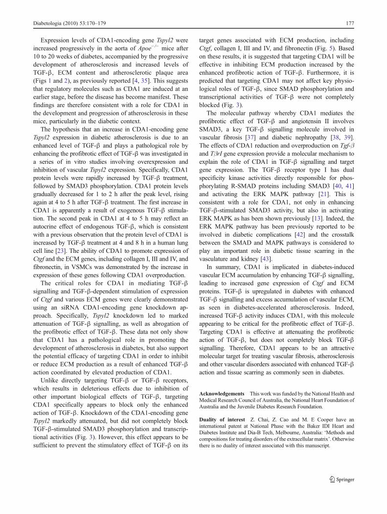
Expression levels of CDA1-encoding gene Tspyl2 wereincreased progressively in the aorta of Apoe−/− mice after10 to 20 weeks of diabetes, accompanied by the progressivedevelopment of atherosclerosis and increased levels ofTGF-β, ECM content and atherosclerotic plaque area(Figs 1 and 2), as previously reported [4, 35]. This suggeststhat regulatory molecules such as CDA1 are induced at anearlier stage, before the disease has become manifest. Thesefindings are therefore consistent with a role for CDA1 inthe development and progression of atherosclerosis in thesemice, particularly in the diabetic context.
The hypothesis that an increase in CDA1-encoding geneTspyl2 expression in diabetic atherosclerosis is due to anenhanced level of TGF-β and plays a pathological role byenhancing the profibrotic effect of TGF-β was investigated ina series of in vitro studies involving overexpression andinhibition of vascular Tspyl2 expression. Specifically, CDA1protein levels were rapidly increased by TGF-β treatment,followed by SMAD3 phosphorylation. CDA1 protein levelsgradually decreased for 1 to 2 h after the peak level, risingagain at 4 to 5 h after TGF-β treatment. The first increase inCDA1 is apparently a result of exogenous TGF-β stimula-tion. The second peak in CDA1 at 4 to 5 h may reflect anautocrine effect of endogenous TGF-β, which is consistentwith a previous observation that the protein level of CDA1 isincreased by TGF-β treatment at 4 and 8 h in a human lungcell line [23]. The ability of CDA1 to promote expression ofCtgf and the ECM genes, including collagen I, III and IV, andfibronectin, in VSMCs was demonstrated by the increase inexpression of these genes following CDA1 overproduction.
The critical roles for CDA1 in mediating TGF-βsignalling and TGF-β-dependent stimulation of expressionof Ctgf and various ECM genes were clearly demonstratedusing an siRNA CDA1-encoding gene knockdown ap-proach. Specifically, Tspyl2 knockdown led to markedattenuation of TGF-β signalling, as well as abrogation ofthe profibrotic effect of TGF-β. These data not only showthat CDA1 has a pathological role in promoting thedevelopment of atherosclerosis in diabetes, but also supportthe potential efficacy of targeting CDA1 in order to inhibitor reduce ECM production as a result of enhanced TGF-βaction coordinated by elevated production of CDA1.
Unlike directly targeting TGF-β or TGF-β receptors,which results in deleterious effects due to inhibition ofother important biological effects of TGF-β, targetingCDA1 specifically appears to block only the enhancedaction of TGF-β. Knockdown of the CDA1-encoding geneTspyl2 markedly attenuated, but did not completely blockTGF-β-stimulated SMAD3 phosphorylation and transcrip-tional activities (Fig. 3). However, this effect appears to besufficient to prevent the stimulatory effect of TGF-β on its
target genes associated with ECM production, includingCtgf, collagen I, III and IV, and fibronectin (Fig. 5). Basedon these results, it is suggested that targeting CDA1 will beeffective in inhibiting ECM production increased by theenhanced profibrotic action of TGF-β. Furthermore, it ispredicted that targeting CDA1 may not affect key physio-logical roles of TGF-β, since SMAD phosphorylation andtranscriptional activities of TGF-β were not completelyblocked (Fig. 3).
The molecular pathway whereby CDA1 mediates theprofibrotic effect of TGF-β and angiotensin II involvesSMAD3, a key TGF-β signalling molecule involved invascular fibrosis [37] and diabetic nephropathy [38, 39].The effects of CDA1 reduction and overproduction on Tgf-βand TβrI gene expression provide a molecular mechanism toexplain the role of CDA1 in TGF-β signalling and targetgene expression. The TGF-β receptor type I has dualspecificity kinase activities directly responsible for phos-phorylating R-SMAD proteins including SMAD3 [40, 41]and activating the ERK MAPK pathway [21]. This isconsistent with a role for CDA1, not only in enhancingTGF-β-stimulated SMAD3 activity, but also in activatingERK MAPK as has been shown previously [13]. Indeed, theERK MAPK pathway has been previously reported to beinvolved in diabetic complications [42] and the crosstalkbetween the SMAD and MAPK pathways is considered toplay an important role in diabetic tissue scarring in thevasculature and kidney [43].
In summary, CDA1 is implicated in diabetes-inducedvascular ECM accumulation by enhancing TGF-β signalling,leading to increased gene expression of Ctgf and ECMproteins. TGF-β is upregulated in diabetes with enhancedTGF-β signalling and excess accumulation of vascular ECM,as seen in diabetes-accelerated atherosclerosis. Indeed,increased TGF-β activity induces CDA1, with this moleculeappearing to be critical for the profibrotic effect of TGF-β.Targeting CDA1 is effective at attenuating the profibroticaction of TGF-β, but does not completely block TGF-βsignalling. Therefore, CDA1 appears to be an attractivemolecular target for treating vascular fibrosis, atherosclerosisand other vascular disorders associated with enhanced TGF-βaction and tissue scarring as commonly seen in diabetes.
Acknowledgements This work was funded by the National Health andMedical Research Council of Australia, the National Heart Foundation ofAustralia and the Juvenile Diabetes Research Foundation.
Duality of interest Z. Chai, Z. Cao and M. E Cooper have aninternational patent at National Phase with the Baker IDI Heart andDiabetes Institute and Dia-B Tech, Melbourne, Australia: ‘Methods andcompositions for treating disorders of the extracellular matrix’. Otherwisethere is no duality of interest associated with this manuscript.
Diabetologia (2010) 53:170–179 177

References
1. Basson M (2008) Cardiovascular disease. Nature 451:9032. Rumble JR, Cooper ME, Soulis T et al (1997) Vascular
hypertrophy in experimental diabetes. Role of advanced glycationend products. J Clin Invest 99:1016–1027
3. Cao ZM, Hulthen UL, Allen TJ, Cooper ME (1998) Angiotensinconverting enzyme inhibition and calcium antagonism attenuatestreptozotocin-diabetes-associated mesenteric vascular hypertro-phy independently of their hypotensive action. J Hypertens16:793–799
4. Candido R, Jandeleit-Dahm KA, Cao Z et al (2002) Prevention ofaccelerated atherosclerosis by angiotensin-converting enzymeinhibition in diabetic apolipoprotein E-deficient mice. Circulation106:246–253
5. Candido R, Allen TJ, Lassila M et al (2004) Irbesartan but notamlodipine suppresses diabetes-associated atherosclerosis. Circu-lation 109:1536–1542
6. Ruiz-Ortega M, Rodriguez-Vita J, Sanchez-Lopez E, Carvajal G,Egido J (2007) TGF-beta signaling in vascular fibrosis. CardiovascRes 74:196–206
7. Leask A, Abraham DJ (2004) TGF-beta signaling and the fibroticresponse. Faseb J 18:816–827
8. Sorescu D (2006) Smad3 mediates angiotensin II- and TGF-beta1-induced vascular fibrosis: Smad3 thickens the plot. Circ Res 98:988–989
9. Hayden MR, Tyagi SC (2000) Arteriogenesis: angiogenesis withinunstable atherosclerotic plaque—interactions with extracellularmatrix. Curr Interv Cardiol Rep 2:218–227
10. Schmidt A, Lorkowski S, Seidler D, Breithardt G, Buddecke E(2006) TGF-beta1 generates a specific multicomponent extracellularmatrix in human coronary SMC. Eur J Clin Invest 36:473–482
11. Rudijanto A (2007) The role of vascular smooth muscle cells onthe pathogenesis of atherosclerosis. Acta Med Indones 39:86–93
12. Chai Z, Sarcevic B, Mawson A, Toh BH (2001) SET-related celldivision autoantigen-1 (CDA1) arrests cell growth. J Biol Chem276:33665–33674
13. Tu Y, Wu W, Wu T et al (2007) Antiproliferative autoantigenCDA1 transcriptionally upregulates p21Wafl/Cip1 by activating p53andMEK/ERK1/2MAPKpathways. J Biol Chem 282:11722–11731
14. Ito T, Tsukumo S, Suzuki N et al (2004) A constitutively activearylhydrocarbon receptor induces growth inhibition of jurkat Tcells through changes in the expression of genes related toapoptosis and cell cycle arrest. J Biol Chem 279:25204–25210
15. Radke JR, Donald RG, Eibs A et al (2006) Changes in theexpression of human cell division autoantigen-1 influenceToxoplasma gondii growth and development. PLoS Pathog 2:e105
16. Datto MB, Li Y, Panus JF, Howe DJ, Xiong Y, Wang XF (1995)Transforming growth factor beta induces the cyclin-dependentkinase inhibitor p21 through a p53-independent mechanism. ProcNatl Acad Sci USA 92:5545–5549
17. Datto MB, Yu Y, Wang X-F (1995) Functional analysis of thetransforming growth factor beta responsive elements in the WAF1/Cip1/p21 promoter. J Biol Chem 270:28623–28628
18. Hu PP, Shen X, Huang D, Liu Y, Counter C, Wang XF (1999) TheMEK pathway is required for stimulation of p21(WAF1/CIP1) bytransforming growth factor-beta. J Biol Chem 274:35381–35387
19. Kim YK, Bae GU, Kang JK et al (2006) Cooperation of H2O2-mediated ERK activation with Smad pathway in TGF-beta1induction of p21WAF1/Cip1. Cell Signal 18:236–243
20. Goldberg HJ, Huszar T, Mozes MM, Rosivall L, Mucsi I (2002)Overexpression of the type II transforming growth factor-betareceptor inhibits fibroblasts proliferation and activates extracellu-
lar signal regulated kinase and c-Jun N-terminal kinase. Cell BiolInt 26:165–174
21. Lee MK, Pardoux C, Hall MC et al (2007) TGF-beta activates ErkMAP kinase signalling through direct phosphorylation of ShcA.Embo J 26:3957–3967
22. Ozbun LL, Martinez A, Angdisen J et al (2003) Differentiallyexpressed nucleolar TGF-beta1 target (DENTT) in mouse devel-opment. Dev Dyn 226:491–511
23. Ozbun LL, Martinez A, Jakowlew SB (2005) Differentiallyexpressed nucleolar TGF-beta1 target (DENTT) shows tissue-specific nuclear and cytoplasmic localization and increases TGF-beta1-responsive transcription in primates. Biochim Biophys Acta1728:163–180
24. Ozbun LL, You L, Kiang S, Angdisen J, Martinez A, JakowlewSB (2001) Identification of differentially expressed nucleolarTGF-beta1 target (DENTT) in human lung cancer cells that is anew member of the TSPY/SET/NAP-1 superfamily. Genomics73:179–193
25. Martinez A, Ozbun LL, Angdisen J, Jakowlew SB (2002)Expression of differentially expressed nucleolar transforminggrowth factor-beta1 target (DENTT) in adult mouse tissues. DevDyn 224:186–199
26. Yokoyama H, Deckert T (1996) Central role of TGF-beta in thepathogenesis of diabetic nephropathy and macrovascular compli-cations: a hypothesis. Diabet Med 13:313–320
27. Twigg SM, Cooper ME (2004) The time has come to targetconnective tissue growth factor in diabetic complications. Dia-betologia 47:965–968
28. Wrana JL, Attisano L, Carcamo J et al (1992) TGF beta signalsthrough a heteromeric protein kinase receptor complex. Cell71:1003–1014
29. Dennler S, Itoh S, Vivien D, ten Dijke P, Huet S, Gauthier JM(1998) Direct binding of Smad3 and Smad4 to critical TGF beta-inducible elements in the promoter of human plasminogenactivator inhibitor-type 1 gene. Embo J 17:3091–3100
30. Ling S, Dai A, Dilley RJ et al (2004) Endogenous estrogendeficiency reduces proliferation and enhances apoptosis-relateddeath in vascular smooth muscle cells: insights from thearomatase-knockout mouse. Circulation 109:537–543
31. Park L, Raman KG, Lee KJ et al (1998) Suppression ofaccelerated diabetic atherosclerosis by the soluble receptor foradvanced glycation endproducts. Nat Med 4:1025–1031
32. Hsueh W, Abel ED, Breslow JL et al (2007) Recipes for creatinganimal models of diabetic cardiovascular disease. Circ Res100:1415–1427
33. Krege JH, Hodgin JB, Hagaman JR, Smithies O (1995) Anoninvasive computerized tail-cuff system for measuring bloodpressure in mice. Hypertension 25:1111–1115
34. Soro-Paavonen A, Watson AM, Li J et al (2008) Receptor foradvanced glycation end products (RAGE) deficiency attenuates thedevelopment of atherosclerosis in diabetes. Diabetes 57:2461–2469
35. Lassila M, Allen TJ, Cao Z et al (2004) Imatinib attenuatesdiabetes-associated atherosclerosis. Arterioscler Thromb Vasc Biol24:935–942
36. Calkin AC, Forbes JM, Smith CM et al (2005) Rosiglitazoneattenuates atherosclerosis in a model of insulin insufficiencyindependent of its metabolic effects. Arterioscler Thromb VascBiol 25:1903–1909
37. Wang W, Huang XR, Canlas E et al (2006) Essential role ofSmad3 in angiotensin II-induced vascular fibrosis. Circ Res98:1032–1039
38. Wang A, Ziyadeh FN, Lee EY et al (2007) Interference with TGF-beta signaling by Smad3-knockout in mice limits diabetic
178 Diabetologia (2010) 53:170–179

glomerulosclerosis without affecting albuminuria. Am J PhysiolRenal Physiol 293:F1657–F1665
39. Li JH, Huang XR, Zhu HJ, Johnson R, Lan HY (2003) Role ofTGF-beta signaling in extracellular matrix production under highglucose conditions. Kidney Int 63:2010–2019
40. Macias-Silva M, Abdollah S, Hoodless PA, Pirone R, Attisano L,Wrana JL (1996) MADR2 is a substrate of the TGFbeta receptorand its phosphorylation is required for nuclear accumulation andsignaling. Cell 87:1215–1224
41. Liu X, Sun Y, Constantinescu SN, Karam E, Weinberg RA,Lodish HF (1997) Transforming growth factor beta-induced
phosphorylation of Smad3 is required for growth inhibition andtranscriptional induction in epithelial cells. Proc Natl Acad SciUSA 94:10669–10674
42. Li JH, Wang W, Huang XR et al (2004) Advanced glycation endproducts induce tubular epithelial-myofibroblast transitionthrough the RAGE-ERK1/2 MAP kinase signaling pathway. AmJ Pathol 164:1389–1397
43. Li JH, Huang XR, Zhu HJ et al (2004) Advanced glycation endproducts activate Smad signaling via TGF-beta-dependent andindependent mechanisms: implications for diabetic renal andvascular disease. FASEB J 18:176–178
Diabetologia (2010) 53:170–179 179