Antimalarial Activity of Phenylthiazolyl-Bearing Hydroxamate-Based Histone Deacetylase Inhibitors
12
Published Ahead of Print 21 July 2008. 10.1128/AAC.00439-08. 2008, 52(10):3467. DOI: Antimicrob. Agents Chemother. Thanh N. Tran and Alan P. Kozikowski Johnson, Qigui Li, Victor Melendez, Nicanor Obaldia III, Diana Caridha, Lucia Gerena, Montip Gettayacamin, Jacob Geoffrey S. Dow, Yufeng Chen, Katherine T. Andrews, Inhibitors Hydroxamate-Based Histone Deacetylase Phenylthiazolyl-Bearing Antimalarial Activity of http://aac.asm.org/content/52/10/3467 Updated information and services can be found at: These include: SUPPLEMENTAL MATERIAL Supplemental material REFERENCES http://aac.asm.org/content/52/10/3467#ref-list-1 at: This article cites 37 articles, 17 of which can be accessed free CONTENT ALERTS more» articles cite this article), Receive: RSS Feeds, eTOCs, free email alerts (when new http://journals.asm.org/site/misc/reprints.xhtml Information about commercial reprint orders: http://journals.asm.org/site/subscriptions/ To subscribe to to another ASM Journal go to: on February 27, 2014 by guest http://aac.asm.org/ Downloaded from on February 27, 2014 by guest http://aac.asm.org/ Downloaded from
-
Upload
independent -
Category
Documents
-
view
4 -
download
0
Transcript of Antimalarial Activity of Phenylthiazolyl-Bearing Hydroxamate-Based Histone Deacetylase Inhibitors
Published Ahead of Print 21 July 2008. 10.1128/AAC.00439-08.
2008, 52(10):3467. DOI:Antimicrob. Agents Chemother. Thanh N. Tran and Alan P. KozikowskiJohnson, Qigui Li, Victor Melendez, Nicanor Obaldia III,Diana Caridha, Lucia Gerena, Montip Gettayacamin, Jacob Geoffrey S. Dow, Yufeng Chen, Katherine T. Andrews, InhibitorsHydroxamate-Based Histone DeacetylasePhenylthiazolyl-Bearing Antimalarial Activity of
http://aac.asm.org/content/52/10/3467Updated information and services can be found at:
These include:
SUPPLEMENTAL MATERIAL Supplemental material
REFERENCEShttp://aac.asm.org/content/52/10/3467#ref-list-1at:
This article cites 37 articles, 17 of which can be accessed free
CONTENT ALERTS more»articles cite this article),
Receive: RSS Feeds, eTOCs, free email alerts (when new
http://journals.asm.org/site/misc/reprints.xhtmlInformation about commercial reprint orders: http://journals.asm.org/site/subscriptions/To subscribe to to another ASM Journal go to:
on February 27, 2014 by guest
http://aac.asm.org/
Dow
nloaded from
on February 27, 2014 by guest
http://aac.asm.org/
Dow
nloaded from
ANTIMICROBIAL AGENTS AND CHEMOTHERAPY, Oct. 2008, p. 3467–3477 Vol. 52, No. 100066-4804/08/$08.00�0 doi:10.1128/AAC.00439-08Copyright © 2008, American Society for Microbiology. All Rights Reserved.
Antimalarial Activity of Phenylthiazolyl-Bearing Hydroxamate-BasedHistone Deacetylase Inhibitors�†
Geoffrey S. Dow,1* Yufeng Chen,2 Katherine T. Andrews,3,4,5 Diana Caridha,1 Lucia Gerena,1Montip Gettayacamin,6 Jacob Johnson,1 Qigui Li,1 Victor Melendez,1 Nicanor Obaldia III,7
Thanh N. Tran,4 and Alan P. Kozikowski2*Division of Experimental Therapeutics, Walter Reed Army Institute of Research, 503 Robert Grant Ave, Silver Spring, Maryland 209101;Drug Discovery Program, Department of Medicinal Chemistry and Pharmacognosy, University of Illinois at Chicago, 833 South Wood,
Chicago, Illinois 606122; Queensland Institute of Medical Research, Herston, Queensland, Australia3; Griffith Medical Research College,Joint Program of Griffith University and the Queensland Institute of Medical Research, Herston, Queensland, Australia4;
Australian Centre for International and Tropical Health, University of Queensland, Queensland, Australia5; Department ofVeterinary Medicine, United States Army Component, Armed Forces Institute of Medical Sciences, 315/6, Rajthevi,
Bangkok, Thailand 104006; and Tropical Medicine Research/Gorgas Memorial Institute,Ave. Justo Arosemena no. 3530, Panama City, Panama7
Received 2 April 2008/Returned for modification 2 May 2008/Accepted 7 July 2008
The antimalarial activity and pharmacology of a series of phenylthiazolyl-bearing hydroxamate-basedhistone deacetylase inhibitors (HDACIs) was evaluated. In in vitro growth inhibition assays approximately 50analogs were evaluated against four drug resistant strains of Plasmodium falciparum. The range of 50%inhibitory concentrations (IC50s) was 0.0005 to >1 �M. Five analogs exhibited IC50s of <3 nM, and three ofthese exhibited selectivity indices of >600. The most potent compound, WR301801 (YC-2-88) was shown tocause hyperacetylation of P. falciparum histones, which is a marker for HDAC inhibition in eukaryotic cells.The compound also inhibited malarial and mammalian HDAC activity in functional assays at low nanomolarconcentrations. WR301801 did not exhibit cures in P. berghei-infected mice at oral doses as high as 640mg/kg/day for 3 days or in P. falciparum-infected Aotus lemurinus lemurinus monkeys at oral doses of 32mg/kg/day for 3 days, despite high relative bioavailability. The failure of monotherapy in mice may be due toa short half-life, since the compound was rapidly hydrolyzed to an inactive acid metabolite by loss of itshydroxamate group in vitro (half-life of 11 min in mouse microsomes) and in vivo (half-life in mice of 3.5 h aftera single oral dose of 50 mg/kg). However, WR301801 exhibited cures in P. berghei-infected mice when combinedat doses of 52 mg/kg/day orally with subcurative doses of chloroquine. Next-generation HDACIs with greatermetabolic stability than WR301801 may be useful as antimalarials if combined appropriately with conventionalantimalarial drugs.
Considerable research activity has focused on understandingthe “histone code,” and in particular on the design of hydrox-amate-based histone deacetylase inhibitors (HDACIs) as noveltherapeutics for the treatment of a wide range of disorders,including cancer and neurodegenerative diseases (26). HDACIsowe their action to their ability to reactivate silenced genes bymodulating the condensation status of DNA. The posttransla-tional acetylation status of chromatin is determined by thecompeting activities of two classes of enzymes, histone acetyl-transferases (HATs) and histone deacetylases (HDACs) whichcontrol the acetylation of lysine residues on histone tails. Ingeneral, HATs function to acetylate lysine groups in nuclearhistones, resulting in neutralization of the charges on the his-
tones and a more open, transcriptionally active chromatinstructure, while the HDACs function to deacetylate and sup-press transcription. A shift in the balance of acetylation onchromatin may result in changes in the regulation of patternsof gene expression (16, 24, 37, 39). Since many cancers areassociated with aberrant transcriptional activity, and HDACscan affect transcription factors and gene regulation, these en-zymes have been identified as attractive targets for cancertherapy. Indeed, chemical inhibitors of HDACs have beenshown to inhibit tumor cell growth and induce differentiationand cell death (28). Several such inhibitory agents, includingsuberoylanilide hydroxamic acid (SAHA) and depsipeptide(FR901228) have reached clinical trials (6, 20, 35), and SAHAhas been approved by the U.S. Food and Drug Administrationfor use in treating cutaneous T-cell lymphoma. HDACIs arethought to function by allowing the transcription and expres-sion of genes, including tumor suppressor genes. HDACIsalso enhance the cytotoxic effects of therapeutic agents usedin cancer treatment, including radiation and chemothera-peutic drugs (25, 27).
Initial studies suggest that HDAC may be an attractive tar-get in P. falciparum. The antimalarial activity of HDACIs wasfirst reported by Darkin-Rattray et al. (11). Apicidin, a fungal
* Corresponding author. Mailing address for G. S. Dow: Division ofExperimental Therapeutics, Walter Reed Army Institute of Research,503 Robert Grant Ave., Silver Spring, MD 20910. Phone: (301) 319-9009. Fax: (301) 319-9954. E-mail: [email protected] address for A. Kozikowski: Drug Discovery Program,Department of Medicinal Chemistry and Pharmacognosy, University ofIllinois at Chicago, 833 South Wood, Chicago, IL 60612. Phone: (312)996-7577. Fax: (312) 413-1701. E-mail: [email protected].
† Supplemental material for this article may be found at http://aac.asm.org/.
� Published ahead of print on 21 July 2008.
3467
on February 27, 2014 by guest
http://aac.asm.org/
Dow
nloaded from
metabolite, was found to exhibit an MIC of 125 ng/ml againstP. falciparum and exhibited a suppressive effect on blood-stageparasites when administered intraperitoneally at a dose rate of50 mg/kg in mice. Functional studies suggested that the targetof apicidin was indeed malarial HDAC. The compound did notappear to have much selectivity for malaria, since its 50%inhibitory concentration (IC50) against proliferating mamma-lian cells was 50 to 100 nM. Subsequently, Joshi et al. (18)cloned an HDAC directly from P. falciparum (HDAC-1) andshowed that it localized in the parasite nucleus and was ex-pressed primarily in mature asexual blood stages and gameto-cytes. A subsequent study (2) also demonstrated that otherputative HDACIs were active in vitro and in vivo. These stud-ies suggest that HDAC is a promising target. However, theseinitial findings have not yet been properly exploited in order todevelop potential antimalarial drugs for clinical use. Recently,our research groups reported that certain triazolylphenyl (9)-and 2-aminosuberic acid (1)-based hydroxamates have nano-molar potency against drug-resistant strains of P. falciparum invitro. We have also reported the synthesis and screening ofphenylthiazolyl-based hydroxamate inhibitors as agents againstpancreatic cancer (21). Here we report that this same group ofcompounds exhibits the most potent and selective antimalarialactivity of all of the HDACIs tested thus far.
MATERIALS AND METHODS
Synthesis of HDACIs. Approximately 50 phenylthiazolyl-hydroxamate-basedHDACIs were synthesized. Those with IC50s � 10 nM (C2A strain) or with IC50s �100 nM that revealed useful information about structure activity relationshipsare shown in Table 1. Synthetic schemes and characterization data are presentedas Fig. S1 and S2 and Table S1 in the supplemental material, respectively.Compounds are referred to by their WR designations throughout (the originallaboratory designations have been left associated with the characterization datain Table S1 in the supplemental material).
In vitro susceptibility and toxicity assays. The in vitro activities of HDACIsagainst P. falciparum strains W2, D6, TM91C235, and TM90C2A were evaluatedby using the labeled hypoxanthine assay of Desjardins et al. (12) as modified byMilhous et al. (30) and described in one of our earlier reports (14). W2 ischloroquine resistant and mefloquine sensitive, D6 is chloroquine sensitive butnaturally less susceptible to mefloquine, and TM91C235 is resistant tomefloquine, chloroquine, and pyrimethamine, as is TM90C2A; however, thislatter parasite is a two pfmdr1 copy strain. The data for WR301801, WR308298,WR308291, WR308296, and WR308294 against the D6, W2, and C235 strainsare reported as the averages of duplicate IC50s in Table 1. The remaining IC50sfor other compounds against all strains are reported as single values for eachcompound in Table 1. We routinely run mefloquine and chloroquine as controlsto ensure assay validity. The IC50s and standard deviations of chloroquine andmefloquine (for the last 15 assays) for each strain are reported in Table 1.Selectivity was defined at the cellular rather than enzymatic level (see Discus-sion). Selectivity was assessed by first determining the LC50s of three of the fivemost potent compounds against macrophages (RAW cell line) using the 3-(4,5-dimethylthiazole-2-yl)-2,5-diphenyltetrazolium bromide (MTT) assay as de-scribed in one of our earlier studies (7). Selectivity indices were determined byusing the following formula: 50% lethal concentration (LC50) (RAW cells)/IC50
(P. falciparum).Histone hyperacetylation assay. The effect of compounds on P. falciparum
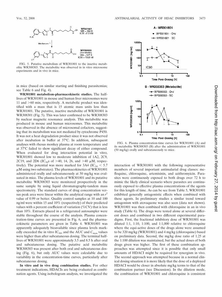
histone acetylation status was determined as previously described (1). Approxi-mately 3 � 109 late-trophozoite-stage 3D7-infected erythrocytes (a mefloquine-resistant clone of W2) were incubated with different concentrations of com-pounds or vehicle alone (0.01% dimethyl sulfoxide) for 3.5 h at 37°C, beforehistones were prepared. An additional untreated control sample was taken at thestart of the assay. Histone extracts (ca. 6 � 108 parasites per lane) were separatedby sodium dodecyl sulfate–15% polyacrylamide gel electrophoresis (SDS–15%PAGE), and equal loading was confirmed using silver staining. To detect changesin acetylation patterns after treatment, SDS-PAGE separated samples weretransferred to a polyvinylidene difluoride membrane (Roche) at 80 V for 1 h in25 mM Tris-HCl–0.2 M glycine–20% methanol. Membranes were blocked in
phosphate-buffered saline (PBS) containing 3% skim milk powder for at least 1 hbefore anti-(tetra)acetyl H4 histone antibody or anti-(di)acetyl-H3 antiserum(Upstate; 1:2,000 dilution in PBS–3% skim milk powder) was added, followed byincubation for 2 h at room temperature. After three washes in deionized water,the membranes were incubated for 1 h in goat anti-rabbit antibody conjugated tohorseradish peroxidase (Zymed; 1:5,000 dilution in PBS–3% skim milk powder).The membranes were washed again, and signal was detected with enhancedchemiluminescence (GE Healthcare, United Kingdom) and autoradiography.The same membrane was then stripped and rehybridized with anti-(di)acetyl-H3antisera (Upstate; 1:2,000 dilution in PBS–3% skim milk powder) using theconditions described above.
Functional HDAC assay. The effect of WR301801 on malarial and mammalianHDAC was evaluated by using a commercially available fluorometric activityassay in 96-well plates according to the manufacturer’s instructions (kit AK-503;Biomol International) as follows. For the mammalian assay, confluent Vero cellswere used. Approximately 106 isolated parasites (W2, late trophozoites) wereused for the malaria assay. Cells were incubated with a cell-permeable substrate(Fleur de Lys) for 3 to 4 h. During this time the substrate accumulates intracel-lularly and is deacetylated. After incubation, the cells were treated for 20 minwith a developer. This generates a fluorescent signal from the deacetylatedsubstrate and induces cell lysis. Plates were read in a Tecan fluorescence reader(excitation, 350 to 380 nm; emission, 440 to 460 nm). HDAC inhibition ismanifested as a reduction in the arbitrary fluorescence units relative to drug-freecontrols. Each drug concentration was tested in triplicate at least twice. Tricho-statin A was used as a control in the mammalian assay and exhibited the expectedresults (data not shown).
In vitro isobologram studies. The in vitro drug combination activity ofHDACIs with known antimalarial drugs against P. falciparum strains W2 and D6were evaluated by using the malaria Sybr green I fluorescence-based assay ofSmilkstein et al. (36) and as described in one of our earlier studies (17). Briefly,each antimalarial drug was tested alone and at fixed ratios with 12 twofold serialdilutions. The starting concentration (for serial dilutions across the microtiterplate) was assigned so that the IC50 of each drug would be in the center of theplate. The IC50s were calculated for each drug alone and for their respectivefixed concentration ratios. The IC50s of drug A and drug B were standardized byallocating a value of 1 to each drug that was tested alone and pro rata values foreach fixed concentration ratio. The individual and sum 50% fractional inhibitoryconcentrations (FIC50 and �FIC50, respectively) were determined as describedby Berenbaum (3). Isobolograms were constructed from the FIC50s of drug Aand drug B at different fixed concentration ratios: a straight line representsadditivity (�FIC � 1), a concave line denotes a trend toward synergy (�FIC �1) or synergy (�FIC � 0.5), and a convex curve represents a trend towardantagonism (�FIC � 1) or antagonism (�FIC � 2). The data are reported hereas the average �FIC (across all drug dilutions) or the average maximum �FICat any dilution for at least two replicate experiments for each drug combination.
In vivo efficacy studies. The antimalarial activity of WR301801 alone and incombination with chloroquine against blood stage Plasmodium berghei in miceand P. falciparum in Aotus lemurinus lemurinus monkeys was evaluated as de-scribed by us in an earlier publication (13). Briefly, in the mouse assay, all micewere inoculated intraperitoneally with 106 P. berghei-parasitized erythrocytes.WR301801 (0.32 to 640 mg/kg/day for 3 days) alone or in combination withchloroquine at subtherapeutic doses (2 to 64 mg/kg/day for 3 days) orally onceper day on days 3, 4, and 5 postinfection in a hydroxethylcellulose-Tween 20vehicle (n � 5 mice per dose group). Blood smears were obtained on study days0, 3, 6, 10, 13, 17, 20, 24, 27, and 31. The efficacy was assessed by monitoringintermittently the effect of the drugs on parasitemia and cure rates. A cure wasdefined as the absence of microscopic parasites in blood smears without his-topathological evidence of malaria infection on day 31 postinfection. In otherexperiments WR301801 was given subcutaneously or over 7 days. In the Aotusstudies, the five monkeys selected for the study had active infections of thechloroquine-resistant FVO strain of P. falciparum. The animals had previouslybeen treated unsuccessfully with a single 25-mg/kg dose of an experimentalquinoline methanol. Once parasitemias reached 5,000/�l (or as deemed appro-priate by the attending veterinarian), they were treated orally with WR30801 at8, 16, or 32 mg/kg/day for 3 days. When these treatments failed they were rescuedwith a single 20-mg/kg dose of mefloquine orally). The Aotus monkeys werecared for and maintained at Gorgas Memorial Institute Aotus colony as previ-ously described (34). All animal experiments were conducted in compliance withthe Animal Welfare Act and other federal statutes and regulations relating toanimals and experiments involving animals and adhere to principles stated in theGuide for the Care and Use of Laboratory Animals (32).
Metabolism-pharmacokinetic studies. The half-life of WR30801 in mouse andhuman liver microsomes was determined as described in our earlier study (13).
3468 DOW ET AL. ANTIMICROB. AGENTS CHEMOTHER.
on February 27, 2014 by guest
http://aac.asm.org/
Dow
nloaded from
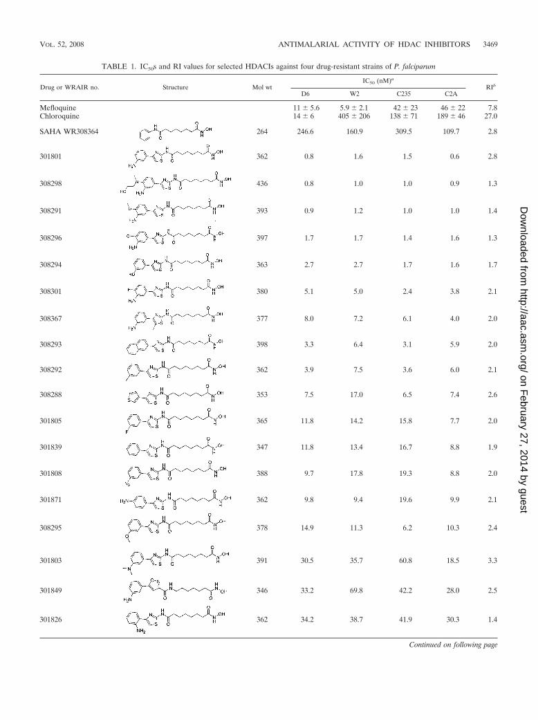
TABLE 1. IC50s and RI values for selected HDACIs against four drug-resistant strains of P. falciparum
Drug or WRAIR no. Structure Mol wtIC50 (nM)a
RIb
D6 W2 C235 C2A
Mefloquine 11 � 5.6 5.9 � 2.1 42 � 23 46 � 22 7.8Chloroquine 14 � 6 405 � 206 138 � 71 189 � 46 27.0
SAHA WR308364 264 246.6 160.9 309.5 109.7 2.8
301801 362 0.8 1.6 1.5 0.6 2.8
308298 436 0.8 1.0 1.0 0.9 1.3
308291 393 0.9 1.2 1.0 1.0 1.4
308296 397 1.7 1.7 1.4 1.6 1.3
308294 363 2.7 2.7 1.7 1.6 1.7
308301 380 5.1 5.0 2.4 3.8 2.1
308367 377 8.0 7.2 6.1 4.0 2.0
308293 398 3.3 6.4 3.1 5.9 2.0
308292 362 3.9 7.5 3.6 6.0 2.1
308288 353 7.5 17.0 6.5 7.4 2.6
301805 365 11.8 14.2 15.8 7.7 2.0
301839 347 11.8 13.4 16.7 8.8 1.9
301808 388 9.7 17.8 19.3 8.8 2.0
301871 362 9.8 9.4 19.6 9.9 2.1
308295 378 14.9 11.3 6.2 10.3 2.4
301803 391 30.5 35.7 60.8 18.5 3.3
301849 346 33.2 69.8 42.2 28.0 2.5
301826 362 34.2 38.7 41.9 30.3 1.4
Continued on following page
VOL. 52, 2008 ANTIMALARIAL ACTIVITY OF HDAC INHIBITORS 3469
on February 27, 2014 by guest
http://aac.asm.org/
Dow
nloaded from
Drug interaction potential was evaluated in vitro by using BD Gentest (FranklinLakes, NJ) CYP450 inhibition kits as recommended by the manufacturer. Plasmastability was determined by individually incubating the compounds for 0, 1, and2 h at room temperature and 37°C using a lab rocker and a water bath, respec-tively, set at 90 rpm. The data were compared to similarly treated acetonitrile-water samples. Plasma samples were extracted and analyzed as described for thepharmacokinetic studies below. In the pharmacokinetic studies, WR301801 wasadministered as a single dose orally and subcutaneously at a dose of 50 mg/kg tothree groups of six 8-week-old male ICR mice for each dosing route. At 15, 30,and 60 min and at 3, 6, and 24 h, one mouse from each group (six mice total) wassacrificed, and plasma was collected and frozen at �80°C. Plasma proteins wereprecipitated by adding 2 volumes of acetonitrile to 100-�l plasma samples,followed by high-speed vortexing for 10 to 12 s and centrifugation at 13,000 rpmfor 10 min at 4°C. Then, 5 �l of the undisturbed supernatants was analyzedby liquid chromatography-tandem mass spectrometry (LEAP Technologies,Carrboro, NC, and ThermoFissher, Waltham, MA) based on standard curves ofWR301801 and WR308301 in the range of 0 to 500 ng/ml and quality controls (10and 100, n � 4 each) prepared by serial dilutions using blank plasma. Sampleswere chromatographically separated using a shallow acetonitrile-water gradientran trough a 3.5-�m, 2.1-by-50-mm, C-18 XTerra MS column from Waters(Milford, MA) at a flow rate of 0.3 ml/min. Quantification was done by using theExcalibur (ThermoFisher) software. For the purposes of determining pharma-cokinetic parameters, each group of six mice was considered to be a single“animal.” Plasma concentration-time data for WR301801 for each animal werefitted to a two-compartment open model using a nonlinear, extended least-squares fitting procedure (WinNonlin 5.1; Scientific Consulting, Inc., Apex, NC)using weighted (1/concentration) nonlinear regression. The area under the curve(AUC) was determined by the linear trapezoidal rule with extrapolation toinfinity based on the concentration of the last time point divided by the terminalrate constant. Extrapolations to time zero were done using zero concentrationfor oral or subcutaneous dosing and using C0 values determined from the two-compartment model equation at time zero. The elimination half-life (t1/2) wascalculated as ln(2)Vss/Vmax[Km�C]. Cmax and Tmax were determined individuallyfor each “animal.” Cmax and Tmax were determined individually for each “ani-mal.” For WR308303, the AUC was determined after fitting plasma-concentra-tion time curves to a noncompartmental model. Significant inter-“animal” vari-ability prevented the determination of other pharmacokinetic parameters.
RESULTS
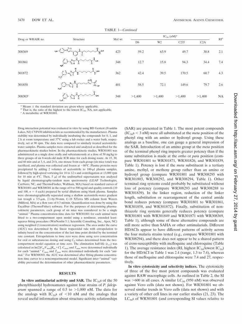
In vitro antimalarial activity and SAR. The IC50s of the 50phenylthiazolyl hydroxamates against four strains of P. falcip-arum spanned a range of 0.5 to �1,000 nM. The data forthe analogs with IC50s of �10 nM and the analogs thatreveal useful information about structure-activity relationships
(SAR) are presented in Table 1. The most potent compounds(IC50s � 3 nM) were all substituted at the meta position of thephenyl ring with an amino or hydroxyl group. Using theseanalogs as a baseline, one can gauge a general impression ofthe SAR. Introduction of an amino group at the meta positionof the terminal phenyl ring imparts greater potency than if thesame substitution is made at the ortho or para position (com-pare WR301801 to WR301871, WR301826, and WR301839,Table 1). Potency is lost if the meta substituent is a tertiaryamine, methyl, or methoxy group rather than an amino orhydroxyl group (compare WR301801 and WR308295 withWR301803, WR308292, and WR308294, Table 1). Otherterminal ring systems could probably be substituted withoutloss of potency (compare WR308293 and WR308288 toWR301839). In the linker region, reduction of the linkerlength, substitution or rearrangement of the central amidebond reduces potency (compare WR301801 to WR301861,WR301858, and WR301872). Finally, substitution of non-thiazole ring systems generally reduces potency (compareWR301801 with WR301849 and WR301871 with WR308369,Table 1), although some of these alternative compounds arestill more active than SAHA or other antimalarials. DifferentHDACIs appear to have different patterns of activity acrossthe four malaria strains tested (e.g., compare WR301801 withWR308294), and there does not appear to be a shared patternof cross-susceptibility with mefloquine and chloroquine (Table1). The average resistance index (RI, highest IC50/lowest IC50)for the HDACI in Table 1 was 2.4 (range, 1.3 to 7.4), whereasthose of mefloquine and chloroquine were 7.4 and 27, respec-tively.
In vitro cytotoxicity and selectivity indices. The cytotoxicityof three of the five most potent compounds was evaluatedagainst RAW macrophage cells. As outlined in Table 2, the SIwas �600 in all cases. A similar LC50 (950 nM) was observedagainst Vero cells (data not shown). For WR301801 we ob-served similar trends in Vero cells (data not shown) and witha variety of other cell lines in our earlier studies (21, 23). TheLC50s of WR301801 (and corresponding SI values relative to
TABLE 1—Continued
Drug or WRAIR no. Structure Mol wtIC50 (nM)a
RIb
D6 W2 C235 C2A
308369 423 59.2 63.9 49.7 30.8 2.1
301861 348 12.7 15.8 94.3 34.4 7.4
301872 362 42.4 39.5 170.5 60.7 4.3
301858 401 58.5 72.1 149.6 79.7 2.6
308303c 348 �1,400 �1,400 �1,400 �1,400 NA
a Means � the standard deviation are given where applicable.b That is, the ratio of the highest to the lowest IC50. NA, not applicable.c A metabolite of WR301801.
3470 DOW ET AL. ANTIMICROB. AGENTS CHEMOTHER.
on February 27, 2014 by guest
http://aac.asm.org/
Dow
nloaded from
P. falciparum) are lower against the specific pancreatic cancercells against which this series was originally optimized (21, 23).
WR301801 (YC-2-88) alters histone acetylation in P. falcip-arum parasites. Histone hyperacetylation is the marker ofHDAC inhibition in eukaryotic cells, including Plasmodiumspp. (1, 11, 15, 19). The natural product derivative apicidin andsynthetic hydroxamic acid-based HDACIs based on 2-aminosuberic acid cause hyperacetylation of P. falciparum histones(1, 11) and, conversely, the HAT inhibitor curcumin hypo-acetylates P. falciparum histone H3 (10). Here we demonstratethat WR301801 also alters P. falciparum histone acetylationprofiles (Fig. 1). After treatment with WR301801 for 3.5 h, anincrease in acetylated histone-H3 and -H4 was detected usinganti-(di)acetyl H3 and anti-(tetra)acetyl H4 antibodies, respec-tively. The anti-(tetra)acetyl H4 antibody recognizes a majorspecies at ca. 12 to 13 kDa in control sample lanes, which
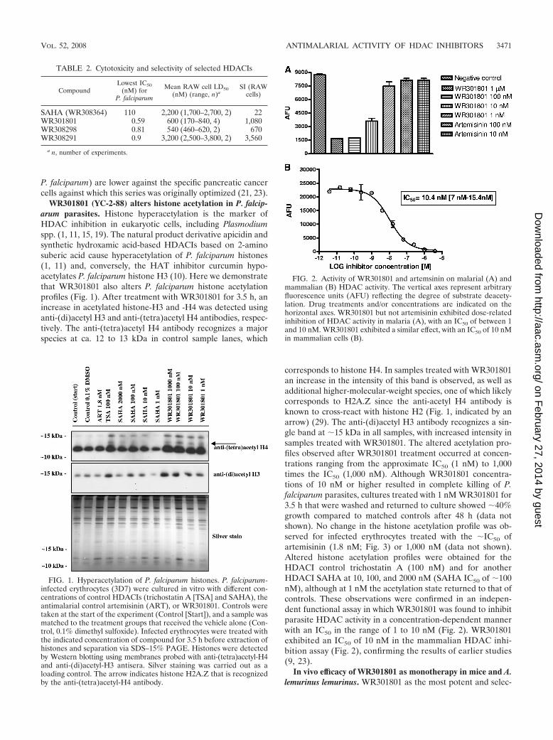
corresponds to histone H4. In samples treated with WR301801an increase in the intensity of this band is observed, as well asadditional higher-molecular-weight species, one of which likelycorresponds to H2A.Z since the anti-acetyl H4 antibody isknown to cross-react with histone H2 (Fig. 1, indicated by anarrow) (29). The anti-(di)acetyl H3 antibody recognizes a sin-gle band at 15 kDa in all samples, with increased intensity insamples treated with WR301801. The altered acetylation pro-files observed after WR301801 treatment occurred at concen-trations ranging from the approximate IC50 (1 nM) to 1,000times the IC50 (1,000 nM). Although WR301801 concentra-tions of 10 nM or higher resulted in complete killing of P.falciparum parasites, cultures treated with 1 nM WR301801 for3.5 h that were washed and returned to culture showed 40%growth compared to matched controls after 48 h (data notshown). No change in the histone acetylation profile was ob-served for infected erythrocytes treated with the IC50 ofartemisinin (1.8 nM; Fig. 3) or 1,000 nM (data not shown).Altered histone acetylation profiles were obtained for theHDACI control trichostatin A (100 nM) and for anotherHDACI SAHA at 10, 100, and 2000 nM (SAHA IC50 of 100nM), although at 1 nM the acetylation state returned to that ofcontrols. These observations were confirmed in an indepen-dent functional assay in which WR301801 was found to inhibitparasite HDAC activity in a concentration-dependent mannerwith an IC50 in the range of 1 to 10 nM (Fig. 2). WR301801exhibited an IC50 of 10 nM in the mammalian HDAC inhi-bition assay (Fig. 2), confirming the results of earlier studies(9, 23).
In vivo efficacy of WR301801 as monotherapy in mice and A.lemurinus lemurinus. WR301801 as the most potent and selec-
TABLE 2. Cytotoxicity and selectivity of selected HDACIs
CompoundLowest IC50
(nM) forP. falciparum
Mean RAW cell LD50(nM) (range, n)a
SI (RAWcells)
SAHA (WR308364) 110 2,200 (1,700–2,700, 2) 22WR301801 0.59 600 (170–840, 4) 1,080WR308298 0.81 540 (460–620, 2) 670WR308291 0.9 3,200 (2,500–3,800, 2) 3,560
a n, number of experiments.
FIG. 1. Hyperacetylation of P. falciparum histones. P. falciparum-infected erythrocytes (3D7) were cultured in vitro with different con-centrations of control HDACIs (trichostatin A [TSA] and SAHA), theantimalarial control artemisinin (ART), or WR301801. Controls weretaken at the start of the experiment (Control [Start]), and a sample wasmatched to the treatment groups that received the vehicle alone (Con-trol, 0.1% dimethyl sulfoxide). Infected erythrocytes were treated withthe indicated concentration of compound for 3.5 h before extraction ofhistones and separation via SDS–15% PAGE. Histones were detectedby Western blotting using membranes probed with anti-(tetra)acetyl-H4and anti-(di)acetyl-H3 antisera. Silver staining was carried out as aloading control. The arrow indicates histone H2A.Z that is recognizedby the anti-(tetra)acetyl-H4 antibody.
FIG. 2. Activity of WR301801 and artemsinin on malarial (A) andmammalian (B) HDAC activity. The vertical axes represent arbitraryfluorescence units (AFU) reflecting the degree of substrate deacety-lation. Drug treatments and/or concentrations are indicated on thehorizontal axes. WR301801 but not artemisinin exhibited dose-relatedinhibition of HDAC activity in malaria (A), with an IC50 of between 1and 10 nM. WR301801 exhibited a similar effect, with an IC50 of 10 nMin mammalian cells (B).
VOL. 52, 2008 ANTIMALARIAL ACTIVITY OF HDAC INHIBITORS 3471
on February 27, 2014 by guest
http://aac.asm.org/
Dow
nloaded from

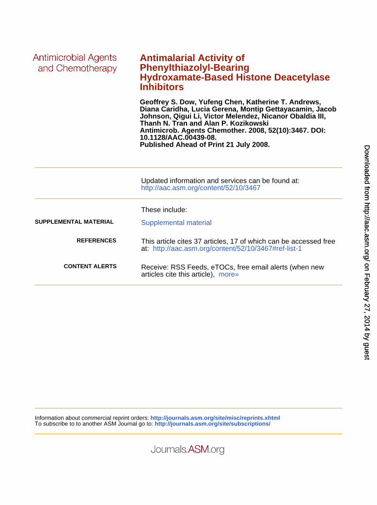
tive compound was selected for in vivo studies in the P. berghei-mouse blood schizonticidal model. The compound was testedfrom doses of 40 to 640 mg/kg/day for 3 days given once perday by oral gavage (Table 3). Control animals typically died orwere euthanized at the moribund early endpoint between day6 to 10 postinfection. No animals were cured using this regi-men, although posttreatment parasitemia relative to controlsdeclined with dose. We attempted to induce cures throughsplit-dosing regimes, longer periods of administration, subcu-taneous dosing, and administration at the time of parasiteinoculation. None of these attempts were successful. Split dos-ing actually decreased the efficacy marginally, whereas subcu-taneous dosing was associated with toxicity (Table 3). Theinduction of clearance with the once-a-day regimen is theoret-ically possible at the next highest dose of 1,280 mg/kg/day for3 days based on the observed dose response (Fig. 3) but notpractical. Toxicity was observed only after subcutaneous dosing(Table 3) and in one of five mice given a split oral dose of 130mg/kg/day for 3 days in preliminary experiments (data notshown). Given that the metabolic instability of WR301801 wasgreater in rodent than in primate microsomes (see below) andtoxicity was not observed after once daily oral administration,we considered a focused set of Aotus experiments to be rea-sonable. Five Aotus monkeys infected with P. falciparum FVO,in which an earlier course of experimental quinoline methanolshad failed to induce cures, were given WR301801 at once daily
oral doses of 8, 16, or 32 mg/kg/day for 3 days. AlthoughWR301801 was not effective, there did appear to be a dose-related effect on parasitemia (Fig. 4). The 32-mg/kg/day dosestabilized parasitemia during the treatment period despite arelatively high starting level. In contrast, the parasitemia ofanimals given lower doses increased. This is evident whenparasitemias are averaged for each dose group and normalizedrelative to starting parasitemias (Fig. 4B). The 32-mg/kg/daydosing regimen is probably equivalent to a 160-mg/kg/day dose
TABLE 3. Efficacy and toxicity of WR301801 in P. berghei-infected mice
Daily dose(mg/kg/day) Route No. of
days
Single (S) orsplit (D)
daily dose
Fraction ofposttreatment
controlparasitemiaon day 6a
No. of animals/total no. in group
No. ofcures Toxic deaths
40 Oral 3 D 0.32 0/5 0/580 Oral 3 D 0.05 0/5 0/5160 Oral 3 D 0.03 0/5 0/5320 Oral 3 D 0.004 0/5 0/5640 Oral 3 D 0.001 0/5 0/520 Oral 3 S 0.57 0/5 0/540 Oral 3 S 0.12 0/5 0/580 Oral 3 S 0.096 0/5 0/5130 Oral 7 D 0.01 0/5 0/5130 Subcutaneous 3 D NA 0/5 5/5 (day5)
a Calculated as the parasitemia in the treatment group on day 6/the parasitemia in control group on day 6. NA, not applicable.
FIG. 3. Dose-related parasite killing by WR301801 in vivo. Whenadministered in a once daily oral regimen, the fraction of parasitesremaining relative to controls declines linearly with increasing dose (40to 640 mg/kg/day) on a log scale. A further increase of the dose to1,280 mg/kg/day would probably result in a decline in parasitemiabelow the limit of detection but is impractical.
FIG. 4. Effect of WR301801 on P. falciparum FVO in Aotus.(A) Five Aotus monkeys previously administered a failed regimen of anexperimental quinoline methanol were given WR301801 at doses of 8,16, or 32 mg/kg/day orally for 3 days as indicated by the black bar. Twodays after the third dose the animals were rescued with a single 20-mg/kg dose of mefloquine, since the WR301801 treatments failed.However, WR301801 did exhibit a dose-related effect on parasitemia.(B) This is evident when parasitemias are averaged for each dosegroup and normalized relative to the starting values.
3472 DOW ET AL. ANTIMICROB. AGENTS CHEMOTHER.
on February 27, 2014 by guest
http://aac.asm.org/
Dow
nloaded from
in mice (based on similar starting and finishing parasitemias;see Table 6 and Fig. 4).
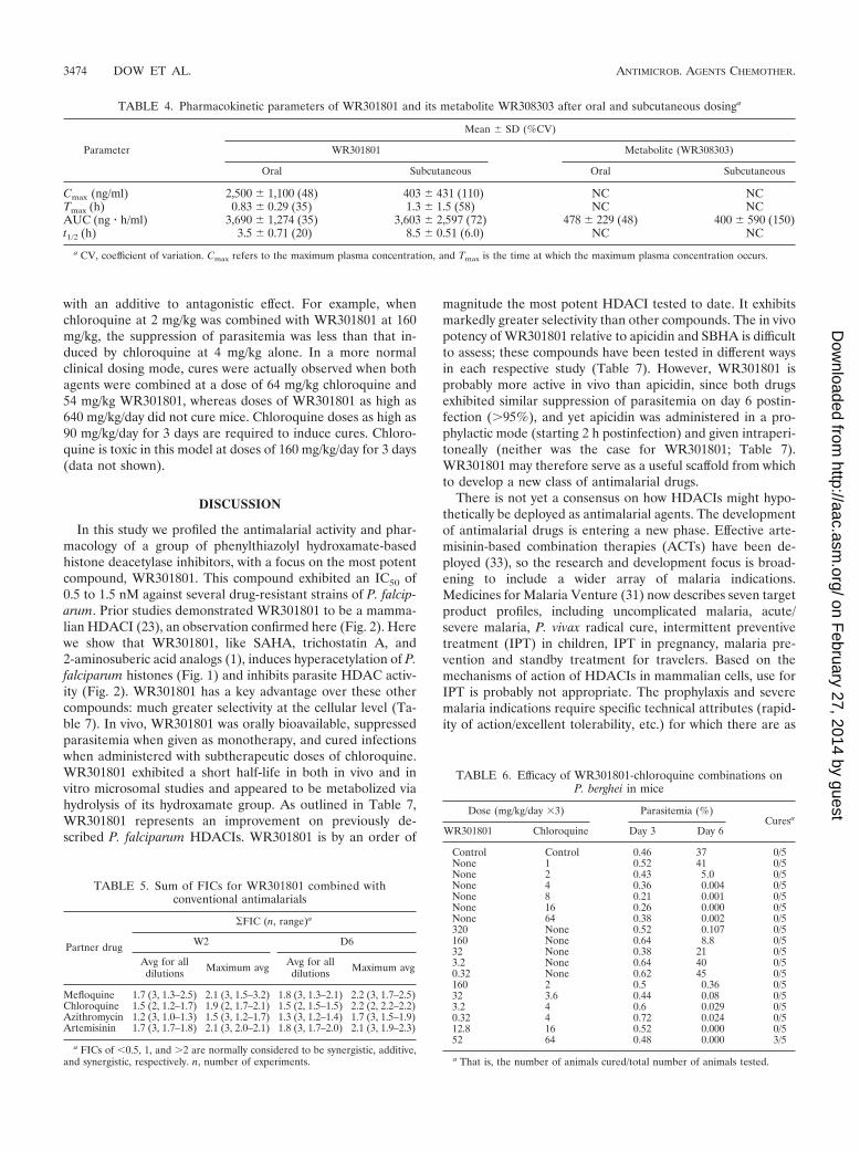
WR301801 metabolism-pharmacokinetic studies. The half-lives of WR301801 in mouse and human liver microsomes were11 and �60 min, respectively. A metabolic product was iden-tified with a mass that is 15 atomic mass units less thanWR301801. The putative, inactive metabolite of WR301801 isWR308301 (Fig. 5). This was later confirmed to be WR308303by nuclear magnetic resonance analysis. This metabolite wasproduced in mouse and human microsomes. This metabolitewas observed in the absence of microsomal cofactors, suggest-ing that its metabolism was not mediated by cytochrome P450.It was not a heat degradation product since it was not observedafter incubation in buffer at 37°C. In addition, subsequentanalyses with rhesus monkey plasma at room temperature andat 37°C failed to show significant decay of either compound.When evaluated for drug interaction potential in vitro,WR301801 showed low to moderate inhibition of 1A2, 2C9,2C19, and 2D6 (IC50s of �40, 14, 26, and �40 �M, respec-tively). The potential was more marked for 3A4 (IC50 � 5.7�M using two substrates). The pharmacokinetics of WR301801administered orally and subcutaneously at 50 mg/kg was eval-uated in mice. The plasma levels of WR301801 and its putativemetabolite WR308303 were monitored prospectively in thesame sample by using liquid chromatography-tandem massspectrometry. The standard curves of drug concentration ver-sus peak area were linear within the analytical range with an r2
value of 0.99 or better. Quality control samples at 10 and 100ng/ml were within 15 and 10% (respectively) of their predictedvalues with a percent coefficient of variation (%CV) that is lessthan 10%. Extracts placed in a refrigerated autosampler werestable throughout the course of the analysis. Plasma concen-tration-time curves are presented in Fig. 6, and the pharma-cokinetic parameters are given in Table 4. WR301801 wasapparently adequately bioavailable since plasma levels mark-edly exceeded the in vitro IC90, and the AUC and Cmax valueswere higher than after subcutaneous dosing. The terminal half-lives of WR301801 were approximately 3.5 and 8.5 h after oraland subcutaneous dosing. The putative acid metaboliteWR308303 was present after both oral and subcutaneous dos-ing (Fig. 6), but only AUC values were calculable due tovariability in the concentration-time curves, particularly aftersubcutaneous dosing.
In vitro and in vivo drug combination studies. For othertreatment indications, HDACIs are being evaluated as combi-nation agents. Using isobologram analysis, we investigated the
interaction of WR301801 with the following representativemembers of several important antimalarial drug classes: me-floquine, chloroquine, artemisinin, and azithromyicin. Para-sites were continuously exposed to both drugs over 72 h tomimic the likely clinical scenario where parasites are continu-ously exposed to effective plasma concentrations of the agentsfor this length of time. As can be see from Table 5, WR301801exhibited generally antagonistic effects when combined withthese agents. In preliminary studies a similar trend towardantagonism with atovaquone was also seen (data not shown).WR301801 was then combined with chloroquine in an in vivostudy (Table 6). The drugs were tested alone at several differ-ent doses and combined in two different experimental para-digms. First, the fractional inhibitory dose of WR301801 wasdiluted 1:1, 1:10, 1:100, and 1:1,000 relative to chloroquinewhere the equi-active doses of the drugs alone were assumedto be 320 mg/kg (WR301801) and 4 mg/kg (chloroquine) basedon preliminary data. Second, the mg/kg dose ratio of 1.25 atthe 1:100 dilution was maintained, but the actual doses of bothdrugs given was higher. The first of these combination ap-proaches was attempted since it is possible that only smallamounts of HDACI might be required for synergism in vivo.The second approach was attempted because in a normal clin-ical dosing situation it is more likely that the dose of a deployedHDACI would be closer in absolute mg/kg terms to that of thecombination partner (see Discussion). In the dilution mode,the combination of WR301801 and chloroquine is consistent
FIG. 5. Putative metabolism of WR301801 to the inactive metab-olite WR308303. The metabolite was observed in in vitro microsomeexperiments and in vivo in mice.
FIG. 6. Plasma concentration-time curves for WR301801 (A) andits metabolite WR308303 (B) after the administration of WR301801(50 mg/kg) orally and subcutaneously to mice.
VOL. 52, 2008 ANTIMALARIAL ACTIVITY OF HDAC INHIBITORS 3473
on February 27, 2014 by guest
http://aac.asm.org/
Dow
nloaded from
with an additive to antagonistic effect. For example, whenchloroquine at 2 mg/kg was combined with WR301801 at 160mg/kg, the suppression of parasitemia was less than that in-duced by chloroquine at 4 mg/kg alone. In a more normalclinical dosing mode, cures were actually observed when bothagents were combined at a dose of 64 mg/kg chloroquine and54 mg/kg WR301801, whereas doses of WR301801 as high as640 mg/kg/day did not cure mice. Chloroquine doses as high as90 mg/kg/day for 3 days are required to induce cures. Chloro-quine is toxic in this model at doses of 160 mg/kg/day for 3 days(data not shown).
DISCUSSION
In this study we profiled the antimalarial activity and phar-macology of a group of phenylthiazolyl hydroxamate-basedhistone deacetylase inhibitors, with a focus on the most potentcompound, WR301801. This compound exhibited an IC50 of0.5 to 1.5 nM against several drug-resistant strains of P. falcip-arum. Prior studies demonstrated WR301801 to be a mamma-lian HDACI (23), an observation confirmed here (Fig. 2). Herewe show that WR301801, like SAHA, trichostatin A, and2-aminosuberic acid analogs (1), induces hyperacetylation of P.falciparum histones (Fig. 1) and inhibits parasite HDAC activ-ity (Fig. 2). WR301801 has a key advantage over these othercompounds: much greater selectivity at the cellular level (Ta-ble 7). In vivo, WR301801 was orally bioavailable, suppressedparasitemia when given as monotherapy, and cured infectionswhen administered with subtherapeutic doses of chloroquine.WR301801 exhibited a short half-life in both in vivo and invitro microsomal studies and appeared to be metabolized viahydrolysis of its hydroxamate group. As outlined in Table 7,WR301801 represents an improvement on previously de-scribed P. falciparum HDACIs. WR301801 is by an order of
magnitude the most potent HDACI tested to date. It exhibitsmarkedly greater selectivity than other compounds. The in vivopotency of WR301801 relative to apicidin and SBHA is difficultto assess; these compounds have been tested in different waysin each respective study (Table 7). However, WR301801 isprobably more active in vivo than apicidin, since both drugsexhibited similar suppression of parasitemia on day 6 postin-fection (�95%), and yet apicidin was administered in a pro-phylactic mode (starting 2 h postinfection) and given intraperi-toneally (neither was the case for WR301801; Table 7).WR301801 may therefore serve as a useful scaffold from whichto develop a new class of antimalarial drugs.
There is not yet a consensus on how HDACIs might hypo-thetically be deployed as antimalarial agents. The developmentof antimalarial drugs is entering a new phase. Effective arte-misinin-based combination therapies (ACTs) have been de-ployed (33), so the research and development focus is broad-ening to include a wider array of malaria indications.Medicines for Malaria Venture (31) now describes seven targetproduct profiles, including uncomplicated malaria, acute/severe malaria, P. vivax radical cure, intermittent preventivetreatment (IPT) in children, IPT in pregnancy, malaria pre-vention and standby treatment for travelers. Based on themechanisms of action of HDACIs in mammalian cells, use forIPT is probably not appropriate. The prophylaxis and severemalaria indications require specific technical attributes (rapid-ity of action/excellent tolerability, etc.) for which there are as
TABLE 4. Pharmacokinetic parameters of WR301801 and its metabolite WR308303 after oral and subcutaneous dosinga
Parameter
Mean � SD (%CV)
WR301801 Metabolite (WR308303)
Oral Subcutaneous Oral Subcutaneous
Cmax (ng/ml) 2,500 � 1,100 (48) 403 � 431 (110) NC NCTmax (h) 0.83 � 0.29 (35) 1.3 � 1.5 (58) NC NCAUC (ng � h/ml) 3,690 � 1,274 (35) 3,603 � 2,597 (72) 478 � 229 (48) 400 � 590 (150)t1/2 (h) 3.5 � 0.71 (20) 8.5 � 0.51 (6.0) NC NC
a CV, coefficient of variation. Cmax refers to the maximum plasma concentration, and Tmax is the time at which the maximum plasma concentration occurs.
TABLE 5. Sum of FICs for WR301801 combined withconventional antimalarials
Partner drug
FIC (n, range)a
W2 D6
Avg for alldilutions Maximum avg Avg for all
dilutions Maximum avg
Mefloquine 1.7 (3, 1.3–2.5) 2.1 (3, 1.5–3.2) 1.8 (3, 1.3–2.1) 2.2 (3, 1.7–2.5)Chloroquine 1.5 (2, 1.2–1.7) 1.9 (2, 1.7–2.1) 1.5 (2, 1.5–1.5) 2.2 (2, 2.2–2.2)Azithromycin 1.2 (3, 1.0–1.3) 1.5 (3, 1.2–1.7) 1.3 (3, 1.2–1.4) 1.7 (3, 1.5–1.9)Artemisinin 1.7 (3, 1.7–1.8) 2.1 (3, 2.0–2.1) 1.8 (3, 1.7–2.0) 2.1 (3, 1.9–2.3)
a FICs of �0.5, 1, and �2 are normally considered to be synergistic, additive,and synergistic, respectively. n, number of experiments.
TABLE 6. Efficacy of WR301801-chloroquine combinations onP. berghei in mice
Dose (mg/kg/day �3) Parasitemia (%)Curesa
WR301801 Chloroquine Day 3 Day 6
Control Control 0.46 37 0/5None 1 0.52 41 0/5None 2 0.43 5.0 0/5None 4 0.36 0.004 0/5None 8 0.21 0.001 0/5None 16 0.26 0.000 0/5None 64 0.38 0.002 0/5320 None 0.52 0.107 0/5160 None 0.64 8.8 0/532 None 0.38 21 0/53.2 None 0.64 40 0/50.32 None 0.62 45 0/5160 2 0.5 0.36 0/532 3.6 0.44 0.08 0/53.2 4 0.6 0.029 0/50.32 4 0.72 0.024 0/512.8 16 0.52 0.000 0/552 64 0.48 0.000 3/5
a That is, the number of animals cured/total number of animals tested.
3474 DOW ET AL. ANTIMICROB. AGENTS CHEMOTHER.
on February 27, 2014 by guest
http://aac.asm.org/
Dow
nloaded from
yet insufficient data to determine whether HDACIs would be alogical match. The uncomplicated malaria and traveler standbytreatment indications seem the most appropriate at this stagegiven the results we and others have shown. If the currentlydeployed ACTs fail en mass, it will likely be due to the devel-opment of artemisinin resistance. It therefore seems appropri-ate to explore the possibility that HDACIs might serve as afunctional replacement for artemisinin compounds in futuredrug combinations regimens.
What kind of HDACI drug combination might be used inlight of the in vitro and in vivo drug combination data? In thein vitro isobologram studies, WR301801 exhibited apparentantagonism with chloroquine, atovaquone, azithromycin, andmefloquine. This also appeared to be the case in vivo when thefractional inhibitory dose of the WR301801 component of thecombination was diluted relative to choroquine. In this respect,the in vitro data can be seen as predictive of some of the in vivodata. If this trend remains true, it would indicate that combi-nation of small dose of an HDACI with a clinically effectivedose of a conventional antimalarial might have limited utility.However, there was a clear difference in outcomes when thedose combination used approached mg/kg parity; the “64/52mg/kg/day for 3 days” chloroquine-WR301801 combination re-sulted in cures where 10-fold-higher doses of WR301801 wereineffective and chloroquine doses of 90 mg/kg/day for 3 dayswere required to induce cures. These data are generally sup-portive of the use of HDACIs in future drug combinations. Wedo not know how to interpret these data in the context of theantagonism observed in vitro. The in vivo situation is compli-cated by metabolism, species differences, and changing ratiosof WR301801 and chloroquine plasma concentrations overtime. The utility of the isobologram approach for predicting invivo outcomes may, perhaps unsurprisingly, be limited. Ourfuture combination studies will focus on mouse studies in orderto provide the rationale for a more definitive set of Aotusexperiments (discussed below).
A drug deployed for treatment of uncomplicated malariamust be effective in an oral administration route, in a once-daily regimen, for three consecutive days (31). The expectedcure rate is �99% (31). However, this is not the case for theartemisinin component of ACTs administered as mono-
therapy. Artemisinin compounds administered even as a 7-dayregimen result in a 10% failure rate (33), and this failure rateis higher with shorter regimens. However, the rapidity of actionof artemisinins allows dramatic reductions in the parasite bur-den (33). When combined, this enhances the clinical efficacy oftheir combination partner even against a background of resis-tance (e.g., mefloquine). How then might we test the utility ofthis approach for an HDACI? Experimentally, the most ap-propriate model would be evaluation of a drug combinationagainst drug-resistant P. falciparum strain in Aotus monkeys.Strains resistant to antifolates and chloroquine, but not thecurrently used ACTs, have been adapted to Aotus. Therefore,chloroquine is a logical partner drug with which to test thehypothesis in a proof-of-principle experiment. The combina-tion experiments described here represent the first in a seriesrequired to establish a rationale for future Aotus testing. How-ever, WR301801 is not the compound with which this proof ofprinciple should be demonstrated since the drug does not clearparasitemia like artemisinin when used alone as monotherapy.We would expect that WR301801 would clear parasitemias ata dose of 1,280 mg/kg/day in mice (Fig. 4), and we know that adose of 32 mg/kg/day for 3 days is tolerated in Aotus (Fig. 5)and is equivalent to 160 mg/kg/day for 3 days in mice (Table 6).A new HDACI therefore would need to be at least eightfoldmore potent than WR301801 in vivo and similarly well toler-ated to be considered for progression to Aotus studies.
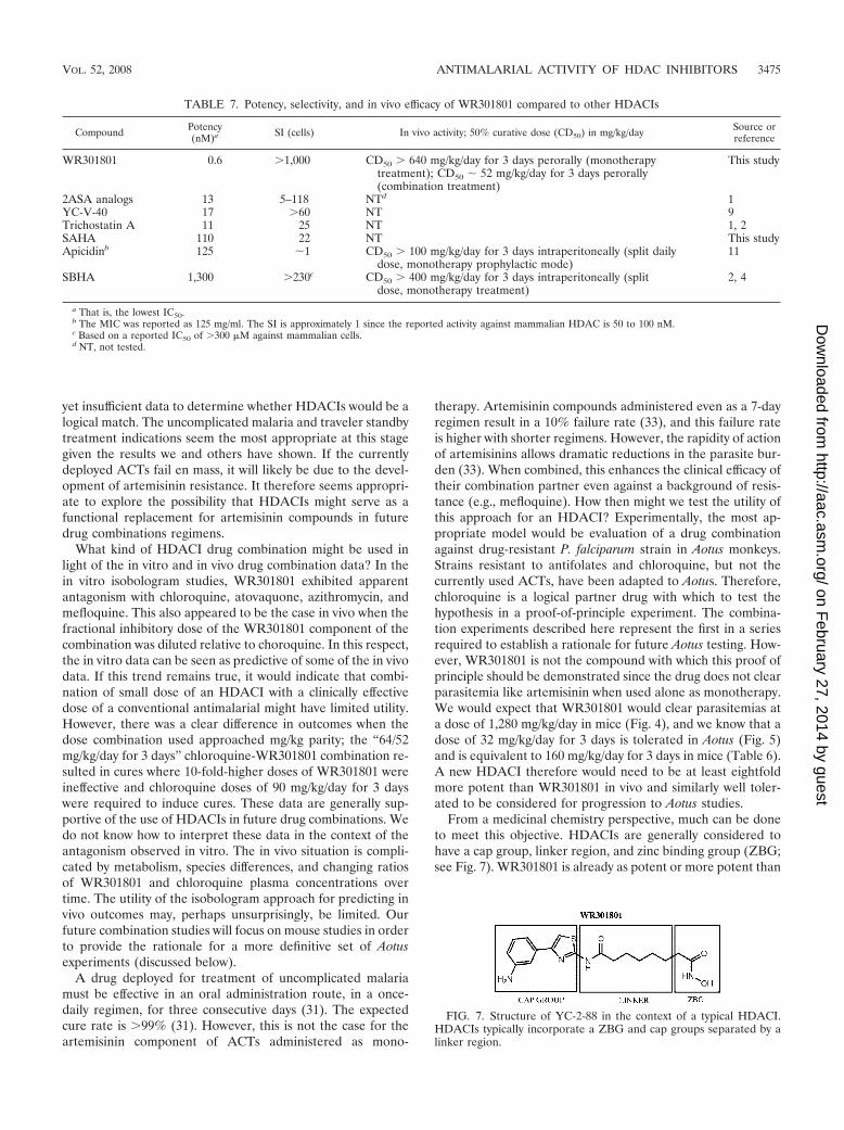
From a medicinal chemistry perspective, much can be doneto meet this objective. HDACIs are generally considered tohave a cap group, linker region, and zinc binding group (ZBG;see Fig. 7). WR301801 is already as potent or more potent than
TABLE 7. Potency, selectivity, and in vivo efficacy of WR301801 compared to other HDACIs
Compound Potency(nM)a SI (cells) In vivo activity; 50% curative dose (CD50) in mg/kg/day Source or
reference
WR301801 0.6 �1,000 CD50 � 640 mg/kg/day for 3 days perorally (monotherapytreatment); CD50 52 mg/kg/day for 3 days perorally(combination treatment)
This study
2ASA analogs 13 5–118 NTd 1YC-V-40 17 �60 NT 9Trichostatin A 11 25 NT 1, 2SAHA 110 22 NT This studyApicidinb 125 1 CD50 � 100 mg/kg/day for 3 days intraperitoneally (split daily
dose, monotherapy prophylactic mode)11
SBHA 1,300 �230c CD50 � 400 mg/kg/day for 3 days intraperitoneally (splitdose, monotherapy treatment)
2, 4
a That is, the lowest IC50.b The MIC was reported as 125 mg/ml. The SI is approximately 1 since the reported activity against mammalian HDAC is 50 to 100 nM.c Based on a reported IC50 of �300 �M against mammalian cells.d NT, not tested.
FIG. 7. Structure of YC-2-88 in the context of a typical HDACI.HDACIs typically incorporate a ZBG and cap groups separated by alinker region.
VOL. 52, 2008 ANTIMALARIAL ACTIVITY OF HDAC INHIBITORS 3475
on February 27, 2014 by guest
http://aac.asm.org/
Dow
nloaded from
existing antimalarials and is relatively selective at a whole-celllevel (discussed in more detail below). Therefore, further de-rivatization of the head or linker regions to yield these prop-erties may not be productive. WR301801 appears to be bio-available. Thus, our hypothesis is that metabolic instability,mediated through hydrolysis of the hydroxamate group, is oneof the reasons for the lack of desired efficacy in vivo. There aretwo logical approaches to address this deficiency. First, thehalf-life of WR301801 in mouse microsomes in vitro is mark-edly shorter than that of SAHA (data not shown), despite thefact that both compounds contain a hydroxamate ZBG. There-fore, a spectrum of metabolic stability may exist within thisclass of compounds, so WR308298, WR308291, WR308296,and WR308294 may be worth testing in vivo if it turns out theyhave greater metabolic stability in vitro than WR301801. Al-ternatively, it may be worth attempting to replace the hydrox-amate moiety with an alternative ZBG. This approach hasbeen used successfully by our group for alternate indications(8, 22).
We have not yet considered the issue of the mechanism ofaction, toxicity, and selectivity in detail. Earlier studies haveshown that WR301801 is a nM inhibitor of mammalian HDAC(9, 23). This was confirmed in a functional assay here (Fig. 2).Putatively, this will also be the case in P. falciparum, as sug-gested indirectly by the hyperacetylation and functional datareported here (Fig. 1 and 2). As with most antimalarial drugs,it is likely that WR301801 will also exhibit additional mecha-nisms of action. The issue of the selectivity of HDACIs iscomplicated. There are 11 zinc-based isoforms of mammalianHDAC and at least two class I/II HDACs are expressed inblood-stage P. falciparum parasites (5, 18, 38). Development ofan HDACI selective for malaria parasites at the enzymaticlevel could therefore be a daunting task, although recent insilico modeling studies have provided useful information forrational development of new compounds designed to target P.falciparum HDAC-1 (1). The HDACIs in clinical developmentdo not target the known mammalian isozymes with any greatselectivity (38). Rather, their selectivity is based on the biolog-ical context; cancer cells are thought to be more vulnerable tointermittent HDAC inhibition than normal cells. Similarly, inour view, selectivity of HDACIs for infectious diseases shouldbe determined at the cellular level rather than enzyme level. Inthis respect, WR301801 exhibits suitable selectivity. Our in vivoand pharmacokinetic studies offer some insights into the toxi-cology of the compound in vivo. Substantial toxicity was ob-served only after subcutaneous dosing. The half-lives ofWR301801 after oral and subcutaneous doses of 50 mg/kgwere 3.5 and 8.5 h, respectively (Table 4). This suggests thatthe duration of exposure may contribute to toxicity. It will beimportant to determine whether this is the case in any newseries of more metabolically stable HDACIs.
ACKNOWLEDGMENTS
This manuscript was reviewed by the Walter Reed Army Institute ofResearch and the U.S. Army Medical Research and Materiel Com-mand, and there is no objection to its publication or dissemination.The opinions expressed herein are those of the authors and do notreflect the views of opinions of the Department of the Army and theDepartment of Defense. All animal experiments were conducted incompliance with the Animal Welfare Act and other federal statutesand regulations relating to animals and experiments involving animals
and adhere to the principles stated in the Guide for the Care and Useof Laboratory Animals (32).
Y.C. synthesized the compounds designated with the prefix YC.We gratefully acknowledge the technical assistance of Jason Sousa,
Arsen Gaysin, Subhasish Tapadar, Miriam Lopez Sanchez, NormaRoncal, Ricky Denull, Drew Reinbold, Anchalee Tungtaeng, and Con-stance Asher, without whose efforts the execution of the work con-tained herein would not have been feasible. We thank William Mc-Calmont and Rong He for helpful review of the manuscript.
K.T.A. was supported by Griffith University (GURG) and theQueensland Institute of Medical Research (Directors Initiative Fund-ing). T.N.T. was supported by an ANZ Trustees Medical ResearchScholarship. The WRAIR, AFRIMS, and Gorgas Institute componentof this effort was supported by the U.S. Military Infectious DiseasesResearch Program through the funding of core screening capabilities.
REFERENCES
1. Andrews, K. T., T. N. Tran, A. J. Lucke, P. Kahnberg, G. T. Le, G. M. Boyle,D. L. Gardiner, T. S. Skinner-Adams, and D. P. Fairlie. 2008. Potent anti-malarial activity of histone deacetylase inhibitor analogues. Antimicrob.Agents Chemother. 52:1454–1461.
2. Andrews, K. T., A. Walduck, M. J. Kelso, D. P. Fairlie, A. Saul, and P. G.Parsons. 2000. Anti-malarial effect of histone deacetylation inhibitors andmammalian tumour cytodifferentiating agents. Int. J. Parasitol. 30:761–768.
3. Berenbaum, M. C. 1978. A method for testing for synergy with any numberof agents. J. Infect. Dis. 137:122–130.
4. Brinkmann, H., A. L. Dahler, C. Popa, M. M. Serewko, P. G. Parsons, B. G.Gabrielli, A. J. Burgess, and N. A. Saunders. 2001. Histone hyperacetylationinduced by histone deacetylase inhibitors is not sufficient to cause growthinhibition in human dermal fibroblasts. J. Biol. Chem. 276:22491–22499.
5. Butler, K. V., and A. P. Kozikowski. 2008. Chemical origins of isoformselectivity in histone deacetylase inhibitors. Curr. Pharm. Des. 14:505–528.
6. Carducci, M. A., J. Gilbert, M. K. Bowling, D. Noe, M. A. Eisenberger, V.Sinibaldi, Y. Zabelina, T. L. Chen, L. B. Grochow, and R. C. Donehower.2001. A phase I clinical and pharmacological evaluation of sodium phenyl-butyrate on an 120-h infusion schedule. Clin. Cancer Res. 7:3047–3055.
7. Caridha, D., D. Yourick, M. Cabezas, L. Wolf, T. H. Hudson, and G. S. Dow.2008. Mefloquine-induced disruption of calcium homeostasis in mammaliancells is similar to that induced by ionomycin. Antimicrob. Agents Chemother.52:684–693.
8. Chen, B., P. A. Petukhov, M. Jung, A. Velena, E. Eliseeva, A. Dritschilo, andA. P. Kozikowski. 2005. Chemistry and biology of mercaptoacetamides asnovel histone deacetylase inhibitors. Bioorg. Med. Chem. Lett. 15:1389–1392.
9. Chen, Y., M. Lopez-Sanchez, D. N. Savoy, D. D. Billadeau, G. S. Dow, andA. P. Kozikowski. 2008. A series of potent and selective, triazolylphenyl-based histone deacetylases inhibitors with activity against pancreatic cancercells and Plasmodium falciparum. J. Med. Chem. 51:3437–3448.
10. Cui, L., and J. Miao. 2007. Cytotoxic effect of curcumin on malaria parasitePlasmodium falciparum: inhibition of histone acetylation and generation ofreactive oxygen species. Antimicrob. Agents Chemother. 51:488–494.
11. Darkin-Rattray, S. J., A. M. Gurnett, R. W. Myers, P. M. Dulski, T. M.Crumley, J. J. Allocco, C. Cannova, P. T. Meinke, S. L. Colletti, M. A.Bednarek, S. B. Singh, M. A. Goetz, A. W. Dombrowski, J. D. Polishook, andD. M. Schmatz. 1996. Apicidin: a novel antiprotozoal agent that inhibitsparasite histone deacetylase. Proc. Natl. Acad. Sci. USA 93:13143–13147.
12. Desjardins, R. E., C. J. Canfield, J. D. Haynes, and J. D. Chulay. 1979.Quantitative assessment of antimalarial activity in vitro by a semiautomatedmicrodilution technique. Antimicrob. Agents Chemother. 16:710–718.
13. Dow, G. S., T. N. Heady, A. K. Bhattacharjee, D. Caridha, L. Gerena, M.Gettayacamin, C. A. Lanteri, N. Obaldia III, N. Roncal, T. Shearer, P. L.Smith, A. Tungtaeng, L. Wolf, M. Cabezas, D. Yourick, and K. S. Smith.2006. Utility of alkylaminoquinolinyl methanols as new antimalarial drugs.Antimicrob. Agents Chemother. 50:4132–4143.
14. Dow, G. S., M. L. Koenig, L. Wolf, L. Gerena, M. Lopez-Sanchez, T. H.Hudson, and A. K. Bhattacharjee. 2004. The antimalarial potential of4-quinolinecarbinolamines may be limited due to neurotoxicity and cross-resistance in mefloquine-resistant Plasmodium falciparum strains. Antimi-crob. Agents Chemother. 48:2624–2632.
15. Glenn, M. P., P. Kahnberg, G. M. Boyle, K. A. Hansford, D. Hans, A. C.Martyn, P. G. Parsons, and D. P. Fairlie. 2004. Antiproliferative and phe-notype-transforming antitumor agents derived from cysteine. J. Med. Chem.47:2984–2994.
16. Grunstein, M. 1997. Histone acetylation in chromatin structure and tran-scription. Nature 389:349–352.
17. Johnson, J. D., R. A. Dennull, L. Gerena, M. Lopez-Sanchez, N. E. Roncal,and N. C. Waters. 2007. Assessment and continued validation of the malariaSybr green I-based fluorescence assay for use in malaria drug screening.Antimicrob. Agents Chemother. 51:1926–1933.
18. Joshi, M. B., D. T. Lin, P. H. Chiang, N. D. Goldman, H. Fujioka, M. Aikawa,
3476 DOW ET AL. ANTIMICROB. AGENTS CHEMOTHER.
on February 27, 2014 by guest
http://aac.asm.org/
Dow
nloaded from
and C. Syin. 1999. Molecular cloning and nuclear localization of a histonedeacetylase homologue in Plasmodium falciparum. Mol. Biochem. Parasitol.99:11–19.
19. Kahnberg, P., A. J. Lucke, M. P. Glenn, G. M. Boyle, J. D. Tyndall, P. G.Parsons, and D. P. Fairlie. 2006. Design, synthesis, potency, and cytoselec-tivity of anticancer agents derived by parallel synthesis from alpha-amino-suberic acid. J. Med. Chem. 49:7611–7622.
20. Kelly, W. K., V. M. Richon, O. O’Connor, T. Curley, B. MacGregor-Curtelli,W. Tong, M. Klang, L. Schwartz, S. Richardson, E. Rosa, M. Drobnjak, C.Cordon-Cordo, J. H. Chiao, R. Rifkind, P. A. Marks, and H. Scher. 2003.Phase I clinical trial of histone deacetylase inhibitor: suberoylanilide hydrox-amic acid administered intravenously. Clin. Cancer Res. 9:3578–3588.
21. Kozikowski, A. P., Y. Chen, A. Gaysin, D. D. Billadeau, and K. H. Kim. 2008.Chemistry, biology, and QSAR studies of substituted biaryl hydroxamatesand mercaptoacetamides as HDAC inhibitors: nanomolar potency inhibitorsof pancreatic cancer cell growth. ChemMedChem 3:487–501.
22. Kozikowski, A. P., Y. Chen, A. Gaysin, B. Chen, M. A. D’Annibale, C. M.Suto, and B. C. Langley. 2007. Functional differences in epigenetic modu-lators-superiority of mercaptoacetamide-based histone deacetylase inhibi-tors relative to hydroxamates in cortical neuron neuroprotection studies.J. Med. Chem. 50:3054–3061.
23. Kozikowski, A. P., S. Tapadar, D. N. Luchini, K. H. Kim, and D. D. Bil-ladeau. 2008. Use of the nitric oxide cycloaddition (NOC) reaction formolecular probe generation: a new class of enzyme selective histone deacety-lase inhibitors (HDACIs) showing picomolar activity at HDAC6. J. Med.Chem. 51:4370–4373.
24. Kurdistani, S. K., and M. Grunstein. 2003. Histone acetylation and deacety-lation in yeast. Nat. Rev. Mol. Cell. Biol. 4:276–284.
25. Marks, P., R. A. Rifkind, V. M. Richon, R. Breslow, T. Miller, and W. K.Kelly. 2001. Histone deacetylases and cancer: causes and therapies. Nat.Rev. Cancer 1:194–202.
26. Marks, P. A., and M. Dokmanovic. 2005. Histone deacetylase inhibitors:discovery and development as anticancer agents. Expert Opin. Investig.Drugs 14:1497–1511.
27. Marks, P. A., V. M. Richon, T. Miller, and W. K. Kelly. 2004. Histonedeacetylase inhibitors. Adv. Cancer Res. 91:137–168.
28. Marks, P. A., V. M. Richon, and R. A. Rifkind. 2000. Histone deacetylaseinhibitors: inducers of differentiation or apoptosis of transformed cells.J. Natl. Cancer Inst. 92:1210–1216.
29. Miao, J., Q. Fan, L. Cui, and J. Li. 2006. The malaria parasite Plasmodiumfalciparum histones: organization, expression, and acetylation. Gene 369:53–65.
30. Milhous, W. K., N. F. Weatherly, J. H. Bowdre, and R. E. Desjardins. 1985.In vitro activities of and mechanisms of resistance to antifol antimalarialdrugs. Antimicrob. Agents Chemother. 27:525–530.
31. Medicines for Malaria Venture. 2008, posting date. Medicines for malaria ven-ture (MMV) product profiles for antimalarial drugs. Medicines for MalariaVenture, Geneva, Switzerland. http://www.mmu.org/IMG/pdf/PRODUCT_PROFILE_with_logo.pdf/.
32. National Academy Press. 1996. Guide for the care and use of laboratoryanimals. National Academy Press, Washington, DC.
33. Nosten, F., and N. J. White. 2007. Artemisinin-based combination treatmentof falciparum malaria. Am. J. Trop. Med. Hyg. 77:181–192.
34. Obaldia, N., III. 1991. Detection of Klebsiella pneumoniae antibodies inAotus l. lemurinus (Panamanian owl monkey) using an enzyme linked im-munosorbent assay (ELISA) test. Lab. Anim. 25:133–141.
35. Sasakawa, Y., Y. Naoe, T. Inoue, T. Sasakawa, M. Matsuo, T. Manda, and S.Mutoh. 2002. Effects of FK228, a novel histone deacetylase inhibitor, onhuman lymphoma U-937 cells in vitro and in vivo. Biochem. Pharmacol.64:1079–1090.
36. Smilkstein, M., N. Sriwilaijaroen, J. X. Kelly, P. Wilairat, and M. Riscoe.2004. Simple and inexpensive fluorescence-based technique for high-throughput antimalarial drug screening. Antimicrob. Agents Chemother.48:1803–1806.
37. Struhl, K., and Z. Moqtaderi. 1998. The TAFs in the HAT. Cell 94:1–4.38. Suzuki, T., and N. Miyata. 2005. Non-hydroxamate histone deacetylase in-
hibitors. Curr. Med. Chem. 12:2867–2880.39. Wolffe, A. P., and D. Guschin. 2000. Review: chromatin structural features
and targets that regulate transcription. J. Struct. Biol. 129:102–122.
VOL. 52, 2008 ANTIMALARIAL ACTIVITY OF HDAC INHIBITORS 3477
on February 27, 2014 by guest
http://aac.asm.org/
Dow
nloaded from