HDAC6 is a specific deacetylase of peroxiredoxins and is involved in redox regulation
7
HDAC6 is a specific deacetylase of peroxiredoxins and is involved in redox regulation R. B. Parmigiani*, W. S. Xu*, G. Venta-Perez*, H. Erdjument-Bromage † , M. Yaneva † , P. Tempst † , and P. A. Marks* ‡ *Cell Biology and † Molecular Biology Programs, Memorial Sloan–Kettering Cancer Center, 1275 York Avenue, New York, NY 10065 Contributed by Paul A. Marks, April 17, 2008 (sent for review April 6, 2008) Eighteen histone deacetylases (HDACs) are present in humans, cate- gorized into two groups: zinc-dependent enzymes (HDAC1–11) and NAD -dependent enzymes (sirtuins 1–7). Among zinc-dependent HDACs, HDAC6 is unique. It has a cytoplasmic localization, two catalytic sites, a ubiquitin-binding site, and it selectively deacetylases -tubulin and Hsp90. Here, we report the discovery that the redox regulatory proteins, peroxiredoxin (Prx) I and Prx II are specific targets of HDAC6. Prx are antioxidants enzymes whose main function is H2 O 2 reduction. Prx are elevated in many cancers and neurodegenerative diseases. The acetylated form of Prx accumulates in the absence of an active HDAC6. Acetylation of Prx increases its reducing activity, its resistance to superoxidation, and its resistance to transition to high- molecular-mass complexes. Thus, HDAC6 and Prx are targets for modulating intracellular redox status in therapeutic strategies for disorders as disparate as cancers and neurodegenerative diseases. acetylation hydrogen peroxide histone deacetylase inhibitors E ighteen histone deacetylases (HDACs) have been identified in humans and classified based on homologies to yeast HDACs (1, 2). Class I (HDACs 1, 2, 3, and 8), class II (HDACs 4, 5, 7, and 9), class IIB (HDACs 6 and 10), and class IV (HDAC11) are zinc-dependent deacetylases. Class III HDACs (sirtuins 1–7) are not zinc-dependent deacetylases and have an absolute requirement for NAD for their activity (3). HDAC6 is unique among the zinc-dependent HDACs (4 –12). It has a primary cytoplasmic localization, full duplication of its two catalytic sites, and a ubiquitin-binding domain at the C terminus. Inhibition of HDAC6 activity by the specific inhibitor, tubacin, or its down-regulation by siRNA, can increase accumu- lation of acetylated -tubulin (6, 7) and can alter cellular mobility and can increase acetylated Hsp90 (8–10), inducing client protein degradation. The ubiquitin-binding activity of HDAC6 mediates the recruitment of autophagic material to aggresomes, decreasing the cytotoxic effects of these aggregates (11, 12). Thus, HDAC6 functions in various cellular processes that are dependent and independent of its catalytic activity and affects cell growth, migration, and cell death. In this work, we made the discovery that HDAC6 has an important role in redox regulation and cellular stress response. We found that the redox regulatory proteins peroxiredoxin I (Prx I) and II (Prx II) are specific targets of HDAC6 deacetylase. Acetylated Prx I and Prx II accumulate in cells lacking HDAC6 deacetylase activity. We found that the prostate cancer cell (LAPC4) does not express HDAC6 protein. This finding was confirmed by siRNA knockdown of HDAC6 and by the specific inhibition of HDAC6 with tubacin (13) in a cell line, which expresses HDAC6 protein (LNCaP). Prx I and Prx II are highly homologous 2-cysteine members of the Prx protein family that function as antioxidants at low resting H 2 O 2 levels (14, 15). At higher levels of H 2 O 2, the cysteine residue can be oxidized to sulfonic acid, with transformation of these proteins to high- molecular-mass protein complexes. Prx I and Prx II are reported to be elevated in many cancers and in various neurodegenerative disorders (16–23). In cancer cells, Prx I and Prx II can confer resistance to chemotherapy and radiation therapy (15–20). Stress-related cellular dysfunction caused by reactive oxygen species (ROS) appears to be involved in the development of various neurodegenerative diseases (22, 23). We found that acetylation of Prx I and Prx II proteins increases their activity in reducing H 2 O 2 and increases their relative resistance to superoxidation and to transition to high-molecular- mass complexes. At high levels of H 2 O 2 , Prx are transformed to large-molecular-mass complexes (14, 15). HDAC6 and its spe- cific targets Prx I and Prx II are shown to play an important role in modulating response to H 2 O 2 -induced cellular stress. This understanding of the specific deacetylase function of HDAC6 with Prx I and Prx II suggests that manipulating HDAC6 activity and, in turn, the redox activities of the 2-Cys Prx proteins, Prx I and Prx II, have an important potential in therapeutic strategies for cancers, neurodegenerative diseases, and other disorders that may involve cellular apoptosis. Results -Tubulin and 50- to 22-kDa Proteins are HDAC6 Targets. In studies (24) that determined the expression of the zinc-dependent HDACs in transformed and normal cells, we found that a human prostate cancer cell, LAPC4, did not express HDAC6 protein (Fig. 1A, lane 1). All other zinc-dependent HDACs of class I, II, and IV were expressed in both the normal and transformed cells examined (24). To determine the basis of LAPC4 lack of expression of HDAC6 protein, Northern blot analyses were performed on lysates of these cells. No transcript of this protein was detected (data not shown). By using RT-PCR, HDAC6 transcript was detectable with two pairs of primers designed on the 3 origin [supporting information (SI) Fig. S1 A]. Using primers covering the entire transcript, we could not amplify the 5 end of the transcript corresponding to the CDS1 and CDS2 fragments compared with LNCaP cells (Fig. S1 A). This finding is consistent with expression of a truncated HDAC6 transcript that could not be translated into a HDAC6 protein in LAPC4 cells. Using an anti-acetylated -tubulin antibody, we found that LAPC4 cells (Fig. 1 A, lane 1) accumulated acetylated -tubulin and other acetylated proteins, ranging in size from 50 to 22 kDa. In three human prostate cancer cell lines that express HDAC6, Du145, LNCaP, and PC3, there was a low level of acetylated -tubulin but no detectable acetylated proteins corresponding to 50 –22 kDa (Fig. 1 A). Noteworthy, SIRT2, an -tubulin deacetylase (25), is present in LAPC4 cells but apparently does not deacetylate the 50- to 22-kDa proteins (data not shown). Author contributions: R.B.P. and P.A.M. designed research; R.B.P., G.V.-P., H.E.-B., M.Y., and P.T. performed research; H.E.-B., M.Y., and P.T. contributed new reagents/analytic tools; R.B.P., W.S.X., and P.A.M. analyzed data; and R.B.P. and P.A.M. wrote the paper. Conflict of interest statement: Memorial Sloan–Kettering Cancer Center and Columbia jointly hold patents on SAHA and related compounds that were exclusively licensed to ATON Pharma, acquired by Merck in April 2004. P.A.M. was a founder of ATON and has a financial interest in the further development of SAHA (vorinostat) by Merck. Freely available online through the PNAS open access option. ‡ To whom correspondence should be addressed. E-mail: [email protected]. This article contains supporting information online at www.pnas.org/cgi/content/full/ 0803749105/DCSupplemental © 2008 by The National Academy of Sciences of the USA www.pnas.orgcgidoi10.1073pnas.0803749105 PNAS July 15, 2008 vol. 105 no. 28 9633–9638 CELL BIOLOGY
-
Upload
independent -
Category
Documents
-
view
3 -
download
0
Transcript of HDAC6 is a specific deacetylase of peroxiredoxins and is involved in redox regulation
HDAC6 is a specific deacetylase of peroxiredoxinsand is involved in redox regulationR. B. Parmigiani*, W. S. Xu*, G. Venta-Perez*, H. Erdjument-Bromage†, M. Yaneva†, P. Tempst†, and P. A. Marks*‡
*Cell Biology and †Molecular Biology Programs, Memorial Sloan–Kettering Cancer Center, 1275 York Avenue, New York, NY 10065
Contributed by Paul A. Marks, April 17, 2008 (sent for review April 6, 2008)
Eighteen histone deacetylases (HDACs) are present in humans, cate-gorized into two groups: zinc-dependent enzymes (HDAC1–11) andNAD�-dependent enzymes (sirtuins 1–7). Among zinc-dependentHDACs, HDAC6 is unique. It has a cytoplasmic localization, twocatalytic sites, a ubiquitin-binding site, and it selectively deacetylases�-tubulin and Hsp90. Here, we report the discovery that the redoxregulatory proteins, peroxiredoxin (Prx) I and Prx II are specific targetsof HDAC6. Prx are antioxidants enzymes whose main function is H2O2
reduction. Prx are elevated in many cancers and neurodegenerativediseases. The acetylated form of Prx accumulates in the absence ofan active HDAC6. Acetylation of Prx increases its reducing activity, itsresistance to superoxidation, and its resistance to transition to high-molecular-mass complexes. Thus, HDAC6 and Prx are targets formodulating intracellular redox status in therapeutic strategies fordisorders as disparate as cancers and neurodegenerative diseases.
acetylation � hydrogen peroxide � histone deacetylase inhibitors
E ighteen histone deacetylases (HDACs) have been identifiedin humans and classified based on homologies to yeast
HDACs (1, 2). Class I (HDACs 1, 2, 3, and 8), class II (HDACs4, 5, 7, and 9), class IIB (HDACs 6 and 10), and class IV(HDAC11) are zinc-dependent deacetylases. Class III HDACs(sirtuins 1–7) are not zinc-dependent deacetylases and have anabsolute requirement for NAD� for their activity (3).
HDAC6 is unique among the zinc-dependent HDACs (4–12).It has a primary cytoplasmic localization, full duplication of itstwo catalytic sites, and a ubiquitin-binding domain at the Cterminus. Inhibition of HDAC6 activity by the specific inhibitor,tubacin, or its down-regulation by siRNA, can increase accumu-lation of acetylated �-tubulin (6, 7) and can alter cellularmobility and can increase acetylated Hsp90 (8–10), inducingclient protein degradation. The ubiquitin-binding activity ofHDAC6 mediates the recruitment of autophagic material toaggresomes, decreasing the cytotoxic effects of these aggregates(11, 12). Thus, HDAC6 functions in various cellular processesthat are dependent and independent of its catalytic activity andaffects cell growth, migration, and cell death.
In this work, we made the discovery that HDAC6 has animportant role in redox regulation and cellular stress response.We found that the redox regulatory proteins peroxiredoxin I(Prx I) and II (Prx II) are specific targets of HDAC6 deacetylase.Acetylated Prx I and Prx II accumulate in cells lacking HDAC6deacetylase activity. We found that the prostate cancer cell(LAPC4) does not express HDAC6 protein. This finding wasconfirmed by siRNA knockdown of HDAC6 and by the specificinhibition of HDAC6 with tubacin (13) in a cell line, whichexpresses HDAC6 protein (LNCaP). Prx I and Prx II are highlyhomologous 2-cysteine members of the Prx protein family thatfunction as antioxidants at low resting H2O2 levels (14, 15). Athigher levels of H2O2, the cysteine residue can be oxidized tosulfonic acid, with transformation of these proteins to high-molecular-mass protein complexes. Prx I and Prx II are reportedto be elevated in many cancers and in various neurodegenerativedisorders (16–23). In cancer cells, Prx I and Prx II can conferresistance to chemotherapy and radiation therapy (15–20).Stress-related cellular dysfunction caused by reactive oxygen
species (ROS) appears to be involved in the development ofvarious neurodegenerative diseases (22, 23).
We found that acetylation of Prx I and Prx II proteins increasestheir activity in reducing H2O2 and increases their relativeresistance to superoxidation and to transition to high-molecular-mass complexes. At high levels of H2O2, Prx are transformed tolarge-molecular-mass complexes (14, 15). HDAC6 and its spe-cific targets Prx I and Prx II are shown to play an important rolein modulating response to H2O2-induced cellular stress. Thisunderstanding of the specific deacetylase function of HDAC6with Prx I and Prx II suggests that manipulating HDAC6 activityand, in turn, the redox activities of the 2-Cys Prx proteins, Prx Iand Prx II, have an important potential in therapeutic strategiesfor cancers, neurodegenerative diseases, and other disorders thatmay involve cellular apoptosis.
Results�-Tubulin and 50- to 22-kDa Proteins are HDAC6 Targets. In studies(24) that determined the expression of the zinc-dependentHDACs in transformed and normal cells, we found that a humanprostate cancer cell, LAPC4, did not express HDAC6 protein(Fig. 1A, lane 1). All other zinc-dependent HDACs of class I, II,and IV were expressed in both the normal and transformed cellsexamined (24).
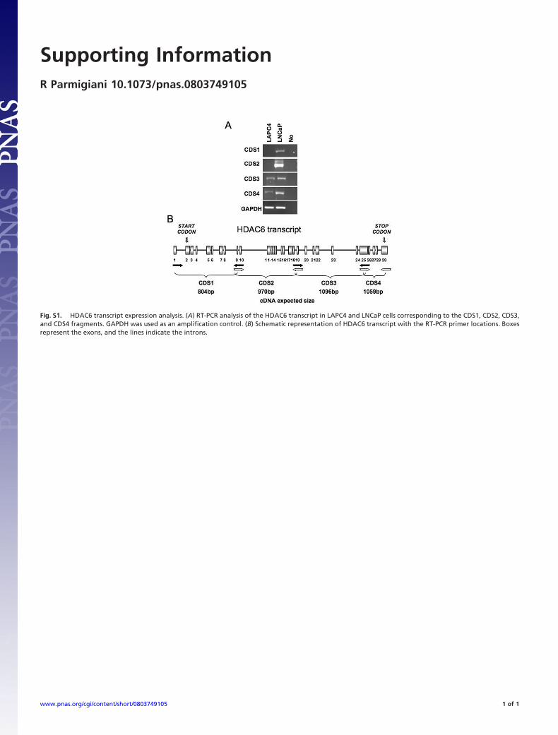
To determine the basis of LAPC4 lack of expression ofHDAC6 protein, Northern blot analyses were performed onlysates of these cells. No transcript of this protein was detected(data not shown). By using RT-PCR, HDAC6 transcript wasdetectable with two pairs of primers designed on the 3� origin[supporting information (SI) Fig. S1 A]. Using primers coveringthe entire transcript, we could not amplify the 5� end of thetranscript corresponding to the CDS1 and CDS2 fragmentscompared with LNCaP cells (Fig. S1 A). This finding is consistentwith expression of a truncated HDAC6 transcript that could notbe translated into a HDAC6 protein in LAPC4 cells.
Using an anti-acetylated �-tubulin antibody, we found thatLAPC4 cells (Fig. 1A, lane 1) accumulated acetylated �-tubulin andother acetylated proteins, ranging in size from �50 to 22 kDa. Inthree human prostate cancer cell lines that express HDAC6, Du145,LNCaP, and PC3, there was a low level of acetylated �-tubulin butno detectable acetylated proteins corresponding to 50–22 kDa (Fig.1A). Noteworthy, SIRT2, an �-tubulin deacetylase (25), is presentin LAPC4 cells but apparently does not deacetylate the 50- to22-kDa proteins (data not shown).
Author contributions: R.B.P. and P.A.M. designed research; R.B.P., G.V.-P., H.E.-B., M.Y., andP.T. performed research; H.E.-B., M.Y., and P.T. contributed new reagents/analytic tools;R.B.P., W.S.X., and P.A.M. analyzed data; and R.B.P. and P.A.M. wrote the paper.
Conflict of interest statement: Memorial Sloan–Kettering Cancer Center and Columbiajointly hold patents on SAHA and related compounds that were exclusively licensed toATON Pharma, acquired by Merck in April 2004. P.A.M. was a founder of ATON and has afinancial interest in the further development of SAHA (vorinostat) by Merck.
Freely available online through the PNAS open access option.
‡To whom correspondence should be addressed. E-mail: [email protected].
This article contains supporting information online at www.pnas.org/cgi/content/full/0803749105/DCSupplemental
© 2008 by The National Academy of Sciences of the USA
www.pnas.org�cgi�doi�10.1073�pnas.0803749105 PNAS � July 15, 2008 � vol. 105 � no. 28 � 9633–9638
CELL
BIO
LOG
Y
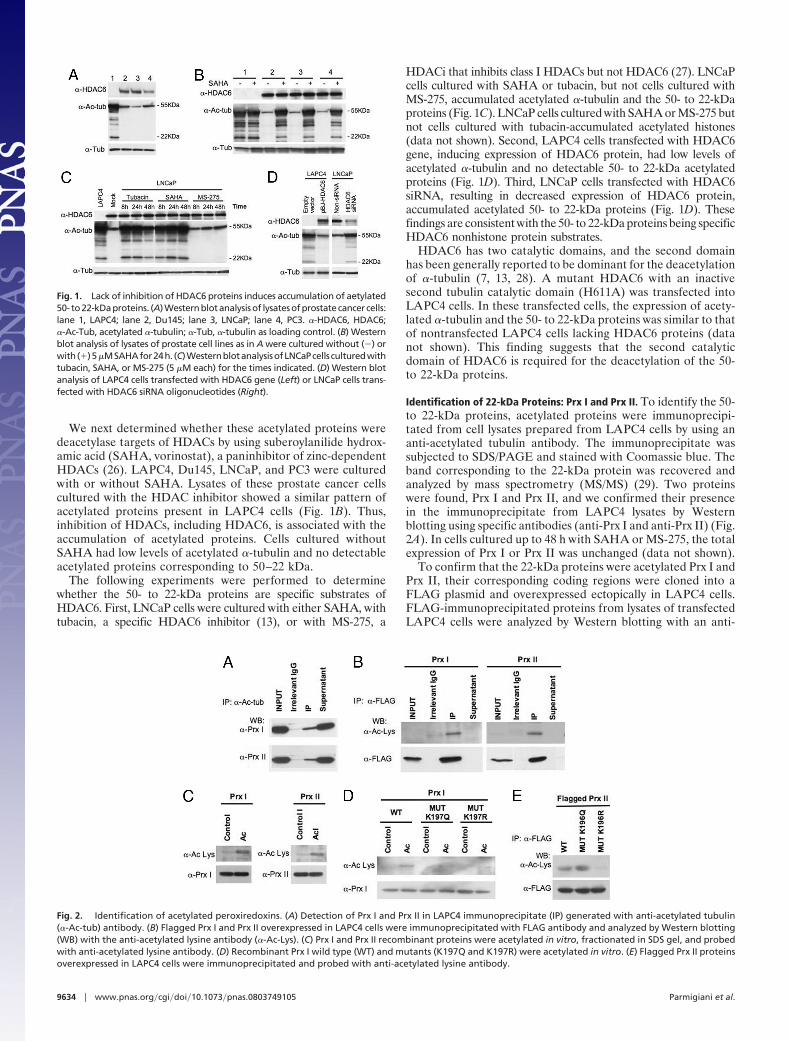
We next determined whether these acetylated proteins weredeacetylase targets of HDACs by using suberoylanilide hydrox-amic acid (SAHA, vorinostat), a paninhibitor of zinc-dependentHDACs (26). LAPC4, Du145, LNCaP, and PC3 were culturedwith or without SAHA. Lysates of these prostate cancer cellscultured with the HDAC inhibitor showed a similar pattern ofacetylated proteins present in LAPC4 cells (Fig. 1B). Thus,inhibition of HDACs, including HDAC6, is associated with theaccumulation of acetylated proteins. Cells cultured withoutSAHA had low levels of acetylated �-tubulin and no detectableacetylated proteins corresponding to 50–22 kDa.
The following experiments were performed to determinewhether the 50- to 22-kDa proteins are specific substrates ofHDAC6. First, LNCaP cells were cultured with either SAHA, withtubacin, a specific HDAC6 inhibitor (13), or with MS-275, a
HDACi that inhibits class I HDACs but not HDAC6 (27). LNCaPcells cultured with SAHA or tubacin, but not cells cultured withMS-275, accumulated acetylated �-tubulin and the 50- to 22-kDaproteins (Fig. 1C). LNCaP cells cultured with SAHA or MS-275 butnot cells cultured with tubacin-accumulated acetylated histones(data not shown). Second, LAPC4 cells transfected with HDAC6gene, inducing expression of HDAC6 protein, had low levels ofacetylated �-tubulin and no detectable 50- to 22-kDa acetylatedproteins (Fig. 1D). Third, LNCaP cells transfected with HDAC6siRNA, resulting in decreased expression of HDAC6 protein,accumulated acetylated 50- to 22-kDa proteins (Fig. 1D). Thesefindings are consistent with the 50- to 22-kDa proteins being specificHDAC6 nonhistone protein substrates.
HDAC6 has two catalytic domains, and the second domainhas been generally reported to be dominant for the deacetylationof �-tubulin (7, 13, 28). A mutant HDAC6 with an inactivesecond tubulin catalytic domain (H611A) was transfected intoLAPC4 cells. In these transfected cells, the expression of acety-lated �-tubulin and the 50- to 22-kDa proteins was similar to thatof nontransfected LAPC4 cells lacking HDAC6 proteins (datanot shown). This finding suggests that the second catalyticdomain of HDAC6 is required for the deacetylation of the 50-to 22-kDa proteins.
Identification of 22-kDa Proteins: Prx I and Prx II. To identify the 50-to 22-kDa proteins, acetylated proteins were immunoprecipi-tated from cell lysates prepared from LAPC4 cells by using ananti-acetylated tubulin antibody. The immunoprecipitate wassubjected to SDS/PAGE and stained with Coomassie blue. Theband corresponding to the 22-kDa protein was recovered andanalyzed by mass spectrometry (MS/MS) (29). Two proteinswere found, Prx I and Prx II, and we confirmed their presencein the immunoprecipitate from LAPC4 lysates by Westernblotting using specific antibodies (anti-Prx I and anti-Prx II) (Fig.2A). In cells cultured up to 48 h with SAHA or MS-275, the totalexpression of Prx I or Prx II was unchanged (data not shown).
To confirm that the 22-kDa proteins were acetylated Prx I andPrx II, their corresponding coding regions were cloned into aFLAG plasmid and overexpressed ectopically in LAPC4 cells.FLAG-immunoprecipitated proteins from lysates of transfectedLAPC4 cells were analyzed by Western blotting with an anti-
Fig. 1. Lack of inhibition of HDAC6 proteins induces accumulation of aetylated50- to22-kDaproteins. (A)Westernblotanalysisof lysatesofprostatecancer cells:lane 1, LAPC4; lane 2, Du145; lane 3, LNCaP; lane 4, PC3. �-HDAC6, HDAC6;�-Ac-Tub, acetylated �-tubulin; �-Tub, �-tubulin as loading control. (B) Westernblot analysis of lysates of prostate cell lines as in A were cultured without (�) orwith(�)5�MSAHAfor24h. (C)WesternblotanalysisofLNCaPcells culturedwithtubacin, SAHA, or MS-275 (5 �M each) for the times indicated. (D) Western blotanalysis of LAPC4 cells transfected with HDAC6 gene (Left) or LNCaP cells trans-fected with HDAC6 siRNA oligonucleotides (Right).
Fig. 2. Identification of acetylated peroxiredoxins. (A) Detection of Prx I and Prx II in LAPC4 immunoprecipitate (IP) generated with anti-acetylated tubulin(�-Ac-tub) antibody. (B) Flagged Prx I and Prx II overexpressed in LAPC4 cells were immunoprecipitated with FLAG antibody and analyzed by Western blotting(WB) with the anti-acetylated lysine antibody (�-Ac-Lys). (C) Prx I and Prx II recombinant proteins were acetylated in vitro, fractionated in SDS gel, and probedwith anti-acetylated lysine antibody. (D) Recombinant Prx I wild type (WT) and mutants (K197Q and K197R) were acetylated in vitro. (E) Flagged Prx II proteinsoverexpressed in LAPC4 cells were immunoprecipitated and probed with anti-acetylated lysine antibody.
9634 � www.pnas.org�cgi�doi�10.1073�pnas.0803749105 Parmigiani et al.

acetylated lysine antibody. The anti-acetylated lysine antibodyspecifically recognized Prx I and Prx II (Fig. 2B).
Using recombinant Prx proteins, acetylated Prx I and Prx IIwere prepared by reacting them with histone acetyltransferase(HAT) and acetylcoenzyme A (acetyl-CoA). The acetylation ofPrx I and Prx II was confirmed by Western blotting, usinganti-acetylated lysine antibody (Fig. 2C).
The intact positive charge of Lys191 in the C terminus of yeastPrx is important for the reducing activity and resistance of theprotein to superoxidation by H2O2 (15, 30, 31). We next testedwhether the lysine residues in human Prx (Lys197 in Prx I; Lys196
in Prx II), corresponding to Lys191 of yeast Prx, were acetylationsites. Mutants of recombinant Prx I were generated: K197Q (Glnis substituted for Lys and mimics acetylated lysine) and K197R(Arg is substituted for Lys and is a nonacetylated mimic) andreacted with HAT and acetyl-CoA (Fig. 2D). A weak signal wasdetected in Prx mutant K197Q and in K197R, compared withwild-type Prx I. These findings indicate that the Prx I Lys197 is anacetylation site. We also generated Prx II mutants (K196Q andK196R) cloned in a FLAG plasmid and transfected them intoLAPC4 cells. The immunoprecipitates were analyzed by West-ern blotting using an anti-acetylated lysine antibody. The wild-type Prx II and the K196Q mutant presented strong positivesignals, whereas the K196R mutant presented no detectablesignal (Fig. 2E). These results confirmed that Prx II Lys196 is alsoa site of acetylation. The present results do not rule out that theremay be other lysines in each protein that are acetylated.
Acetylation of Prx Increases Reducing Activity. We next evaluated theeffect of acetylation of Prx I on its H2O2-reducing activity, usingthe peroxidase reaction with the thioredoxin (Trx) system as anelectron donor, Trx reductase, and NADPH (32–34). The activityof recombinant Prx I protein in reducing H2O2 was assayed bymeasuring NADPH oxidation (absorbance at 340 nm) (30, 34). Thein vitro acetylated Prx I was more active than the nonacetylated PrxI in reducing H2O2 over a range of 0.1–2.0 mM (Fig. 3A).
Prx I and Prx II both regulate intracellular H2O2 levels whileat the same time are regulated by H2O2. Under normal redoxconditions, Prx I and Prx II scavenge H2O2 by building disulfidebridges that can be reduced via the thioredoxin system (30,32–36). At high levels of H2O2, Prx-reducing activity is lostthrough overoxidation of the active sulfhydryl site to sulfinic orsulfonic acid (33–35). Upon overoxidation, Prx undergoes a
transition to a high-molecular-mass complex (14). Using ananti-Prx-SO3H antibody that specifically recognizes both sulfinicand sulfonic forms of overoxidized cysteine in Prx, we found thatthe acetylated Prx I was more resistant to inactivation by H2O2than the nonacetylated form of the protein (Fig. 3B).
To test whether acetylation of Lys197 was critical in determin-ing of Prx H2O2-reducing activity, the effect of in vitro acetylationon the reducing activity of mutant Prx I proteins, K197Q andK197R, was assayed. Acetylation of the mutant proteins did notincrease their reducing activity as it did in the wild-type protein(Fig. 3C). The acetylated mimic mutant (K197Q) had a lowerlevel of oxidized protein (assayed by reaction with anti-Prx-SO3H antibody) compared with the wild-type and K197R mu-tant proteins (Fig. 3D). These findings indicate that acetylationof Lys197 is involved in determining Prx-reducing activity andprotection against overoxidation.
Overoxidation of Prx I or Prx II causes the loss of H2O2-reducingactivity and the transition to high-molecular-mass complex protein(14, 15). LNCaP cells exposed to concentrations of 50 and 100 �MH2O2 for 20 min accumulate high-molecular-mass complexes of�480 kDa (Fig. 4A). By comparison, in LAPC4 cells, which haveacetylated Prx, the high-molecular-mass complexes accumulateonly when they were exposed to 100 �M H2O2, but not 50 �M H2O2
Fig. 3. Acetylation of Prx increases its reducing activity and resistance to overoxidation. (A) Activity of recombinant Prx I, control (F), and acetylated forms (■ )was assayed for H2O2 reduction by measuring NADPH oxidation (A340 nm) (21). (B) Acetylated Prx I is more resistant to being overoxidized (anti-Prx-SO3H antibody).(C) H2O2-reducing activity of acetylated recombinant Prx I, WT, and mutants (K197Q and K197R). The percentage change in 340 mM, O.D., per minute, per amountof protein in 20 min is shown. Activity of the nonacetylated protein in 0.1 mM H2O2 was taken as 100%. (D) Western blot analysis of oxidized Prx I after 1-hexposure to H2O2 in a peroxidase reaction in WT, MUTK197Q, and MUTK197R.
Fig. 4. High-molecular-mass complexes in LAPC4 and LNCaP cells. (A) Nativegels showing Prx I high-molecular-mass complexes induced by exposure toH2O2 for 20 min in LNCaP and LAPC4 cells. Concentration of H2O2 (lanes): 1,none; 2, 25 �M; 3, 50 �M; 4, 100 �M; 5, 100 �M � fluid change and recoveryfor 1 h. (Lower) Denaturing gels showing the levels of oxidized Prx (�-Prx-SO3H), Prx I (�-Prx I), and �-tubulin (�-Tub) in the LNCaP and LAPC4 lysates. (B)Flagged Prx II high-molecular-mass complexes in LNCaP cells treated with theindicated concentrations of H2O2; WT, and transfected with MUTQ (K196Q) orMUTR (K196R) are shown (see Results).
Parmigiani et al. PNAS � July 15, 2008 � vol. 105 � no. 28 � 9635
CELL
BIO
LOG
Y

(Fig. 4A). Similar results were found for Prx II (data not shown).The level of overoxidized Prx (anti-Prx-SO3H) was lower in LAPC4cells compared with LNCaP cells (Fig. 4A), which is consistent withacetylated Prx in LAPC4 cells being more active in H2O2-reducingactivity and being more resistant to overoxidation and transition tothe high-molecular-mass complexes.
To confirm that the lower level of oxidized Prx in LAPC4H2O2-treated cells was associated with Prx acetylation, Prx II-flagged proteins (wild type and mutants) were overexpressed inLNCaP cells and then treated with H2O2. The Prx II K196Qacetylated mimic formed high-molecular-mass complexes only at100 �M H2O2 whereas in the wild type and K196R mutant thecomplexes formed at 50 and 100 �M H2O2 (Fig. 4B). Acetylationof Prx is a major determinant in its protection from overoxidation.
Sensitivity to H2O2-Induced Cell Death of LAPC4, LNCaP, and NormalHuman Foreskin (HFS) Cells. ROS are generated in cells in responseto several types of environmental stress that lead to apoptosis andcell death (37). Prx I and Prx II play a role in modulating cellularresponse to ROS (30, 31). LAPC4 cells, in which there is anaccumulation of acetylated Prx I, H2O2 concentrations (0.5 mM)that induce 100% of LNCaP and HFS cells to undergo apoptosisand cell death (Fig. 5A) induces �10% LAPC4 cell death. LNCaP(Fig. 1A) and HFS cells (data not shown) lack detectable acetylatedPrx (22-kDa) proteins. When LAPC4 cells were transiently trans-fected with HDAC6 gene and expressed HDAC6 protein (Fig. 1D),H2O2 (0.5 mM) induced �40% of the cells to undergo cell death(data not shown). When HDAC6 was inhibited in LNCaP cells bytubacin or transient transfection with HDAC6 siRNA, acetylatedPrx accumulated (Fig. 1 C and D). LNCaP cells were significantlymore resistant to H2O2-induced cell death when HDAC6 activitywas inhibited by tubacin (at 24 h, P � 0.004) (Fig. 5C Upper) or byHDAC6 siRNA compared with control cells (at 24 h, P � 0.014)(Fig. 5C Lower). The loss of viability was greater in cultures of cellstransfected with siRNA than cells exposed to tubacin, which may
reflect the manipulation required for siRNA transfection. In bothstudies, inhibiting HDAC6 was associated with significant increasedresistance to H2O2.
Poly(ADP-ribose) polymerase (PARP) degradation is amarker of cellular apoptosis (37). PARP was assayed in lysatesof LNCaP cells cultured with and without tubacin for 4 h andthen exposed to 0.1 mM H2O2 for 48 h. Cells cultured withtubacin plus H2O2 had nondegraded PARP (Fig. 5D), similar tothe pattern observed in cells not exposed to H2O2. LNCaP cellsexposed to H2O2 without prior culture with tubacin had de-graded PARP (Fig. 5D). Thus, inhibition of HDAC6 activity isassociated with resistance to H2O2-induced apoptotic cell death.
DiscussionThis work describes the discovery of a function for HDAC6,namely the specific deacetylation of redox regulatory proteinsPrx I and Prx II. Previous studies established that HDAC6 is thedeacetylase for �-tubulin, cortactin, an actin binding protein andthe chaperone protein Hsp90 (6–10). In addition to its deacety-lase activity, HDAC6 has a ubiquitin binding site at its Cterminus that can play a role in facilitating autophagic degra-dation of potentially noxious proteins (11, 12).
The present discovery that the redox regulatory proteins PrxI and Prx II, whose main function is cellular protection from freeradical accumulation, are specific substrates of HDAC6 adds animportant understanding of the functions of this deacetylase thathas implications for diseases as disparate as cancers and neuro-degenerative disorders.
The human prostate cancer cell LAPC4 lacks HDAC6 proteinand accumulates the previously unrecognized substrates ofHDCA6, acetylated Prx I and Prx II. Inhibition of HDAC6 withtubacin or its down-regulation with siRNA in both transformedcells (LNCaP, PC3, Du145) as well as normal cells (HFS) wasassociated with an accumulation of acetylated and Prx I and Prx II.
Fig. 5. HDAC6 inhibition reduces sensitivity to H2O2-induced cell death. (A) Cell growth (Lower) and viability (Upper) of LNCaP, HFS, and LAPC4 cells culturedwith H2O2 at the concentrations and times indicated. (B) (Left) LNCaP cells were precultured with 8 �M tubacin for 4 h and then exposed to H2O2 for timesindicated. (Right) LNCaP cells were transfected with non-silencing RNA (non-siRNA) or HDAC6 siRNA 48 h before H2O2 treatment for times indicated. (C) Analysisof PARP degradation in lysates of LNCaP cells: no additions, treated with 0.1 mM H2O2, and precultured with 8 �M tubacin, and exposed to 0.1 mM H2O2 for thetimes indicated.
9636 � www.pnas.org�cgi�doi�10.1073�pnas.0803749105 Parmigiani et al.
Prx I are elevated in many cancers including esophageal,pancreatic, melanoma, thyroid, and lung cancers (16–20). Ele-vated levels of Prx I and Prx II are associated with resistance tocancer therapy and promote aggressive survival phenotypes ofcancer cells.
Prx have also been reported to be overexpressed and/oraberrantly expressed in several neurodegenerating disorders,including Alzheimer’s, Pick’s disease, and others associated withprogressive aggregate formation (22, 23). The present workfound that the acetylated form of Prx is more active in reducingH2O2 than the nonacetylated form. Thus, inhibition of HDAC6deacetylase activity with a consequent accumulation of acety-lated Prx could lead to a beneficial increase in antioxidantactivity in neurodegenerative disorders. HDAC inhibitors suchas SAHA, trichostatin A, and sodium butyrate have been shownto ameliorate disease progression in rodent models of Hunting-ton’s disease (38), spinal and bulbar muscular atrophy (39),amyotrophic lateral sclerosis (40), and Parkinson’s disease (41).Although the mechanisms of the beneficial effects of the HDACinhibitors in these neurodegenerative diseases are not known,inhibition of HDAC6 with consequent increase in Prx reducingactivity may, in part, explain these effects.
However, in cancer cells, the increased reducing activity of Prxassociated with inhibition of HDAC6 could contribute to resis-tance to therapy. This hypothesis would suggest that as part ofan anticancer therapeutic regimen, inactivating Prx activitymight be beneficial (42–45).
In summary, the discovery that HDAC6 is a specific deacetylasefor the redox regulatory proteins Prx I and Prx II whose activity isregulated, in part, by acetylation, suggests that the activity of thisdeacetylase and of the redox proteins can be useful targets fortherapeutic strategies in these disparate disorders. The redoxproteins, Prx, have a role in both the resistance of certain cancersto therapy and quite a different role in possibly slowing theprogression of neurodegenerative diseases. Developing inhibitorsof Prx acetylation may be a useful therapeutic strategy for cancers.However, an agent such as a specific HDAC6 inhibitor thatenhances the accumulation of acetylated Prx and protects theseproteins from overoxidation may be useful in treating neurodegen-erative disorders that involve apoptotic cell death.
MethodsCell Lines, Reagents, and Antibodies. LNCaP, Du145, PC3, and HFS wereobtained from American Type Culture Collection. LAPC4 cells were kindlyprovided by Charles Sawyers (Memorial Sloan–Kettering Cancer Center). An-tibodies used were: anti-acetylated tubulin (Sigma), anti-HDAC6 (Santa CruzBiotechnology), anti-tubulin, (Calbiochem), anti-acetylated lysine (Cell Signal-ing), anti-Prx I and Prx II (Upstate), anti-Prx-SO3H (Abcam), anti-PARP (BDBiosciences). MS-275 was obtained from Calbiochem. Tubacin and SAHA were
kindly provided by Stuart Schreiber (Harvard University, Cambridge, MA) andRonald Breslow (Columbia University, New York), respectively.
Western Blotting and Immunoprecipitation. Western blotting was performed aspublished in ref. 24. Immunoprecipitation was performed either by using theanti-acetylated tubulin antibody, which was covalently bound to CNBr-activatedSepharose 4B beads according to the manufacturer’s instructions (Amersham), orby using anti-FLAG beads (Sigma). Cell lysate and beads were incubated over-night at 4°C. Bound proteins were eluted by competition with 100 �g/ml specificpeptide. For protein identification, different elution fractions were fractionatedin SDS/polyacrylamide gels. Gels were stained with Coomassie blue R-250, andvisible bands were cut and submitted to mass spectrometry analyses (29).
Transfections. HDAC6 vectors were kindly provided by Stuart Schreiber. Prx Iand Prx II coding region sequences were amplified by using cDNA from LAPC4cells. The primers used for RT-PCR were: Prx I forward (5�-ggaagcttatgtcttcag-gaaatg-3�), Prx I reverse (5�-ccgaattctcacttctgcttggag-3�), Prx II forward (5�-ggaagcttatggcctccggtaacg-3�) and Prx II reverse (5�-ccgaattcctaattgtgtttggag-3�). They were cloned into the pFLAG-CMV-4 vector (Sigma). Prx mutants weregenerated by using the QuikChange II site-directed mutagenesis kit (Strat-agene). HDAC6 siRNA and nonsilencing oligonucleotides were obtained fromQiagen. Cells were transfected by using the Nucleofactor kit (Amaxa), follow-ing the manufacturer’s instructions.
In Vitro Acetylation. In vitro acetylation of Prx was performed by using 10 �gof Prx recombinant protein incubated at 30°C for 20 min with 2 �g of p300 HATdomain (Upstate) and 1.2 mM acetyl-CoA (Sigma) in a buffer containing 50mM Tris�HCl (pH 8.0), 10% glycerol, 0.1 mM EDTA, and 3 mM DTT, in a 45-�lfinal volume.
Peroxiredoxin Activity. Peroxidase activity was measured by monitoring theoxidation of NADPH by the decrease of absorbance at 340 nm (21). The reactionwas carried out by using 50 mM Hepes-NaOH (pH 7.4), 3 �g of Trx reductase(Sigma),4 �gofTrx (Sigma),0.5mMNADPH(Sigma),5 �gofPrx I (BPSBioscience),and different concentrations of H2O2 in a 150-�l final volume.
H2O2 Treatment and Prx High-Molecular-Mass Complex Detection. Cells wereseeded in 24-well plates at 5 � 104 cells per well and treated the day after, asfollows. Chemical inhibition of HDAC6 activity was done by pretreatment with8 �M tubacin for 4 h before H2O2 treatment. In the HDAC6 down-regulationexperiment, cells were transiently transfected with HDAC6 siRNA or non-siRNA oligonucleotides and allowed to recover for 2 days and then treatedwith H2O2. Cell viability was evaluated by Trypan blue assay (45). Prx oligomerswere identified after cells were treated with H2O2 for the appropriate timesand concentrations. Cell lysates were fractionated in nondenaturing gels(Invitrogen) and analyzed by Western blotting as described in ref. 24.
ACKNOWLEDGMENTS. We are grateful to Dr. James Bradner and StuartSchreiber, Harvard University, for providing the HDAC6 clones and tubacin.Studies reported here were supported in part by National Institutes of HealthGrant P30CA08748-41, The Jack and Susan Rudin Foundation, David KochFoundation, and Experimental Therapeutics Center at Memorial Sloan–Kettering Cancer Center.
1. Lehrmann H, Pritchard LL, Harel-Bellan, A (2002) Histone acetyltransferases anddeacetylases in the control of cell proliferation and differentiation. Adv Cancer Res86:41–65.
2. Dokmanovic M, Clarke C, Marks PA (2007) Histone deacetylase inhibitors: Overviewand perspectives. Mol Cancer Res 5:981–989.
3. Sauve AA, Wolberger C, Schramm VL, Boeke JD (2006) The biochemistry of sirtuins.Annu Rev Biochem 75(1):435–465.
4. Grozinger CM, Hassig CA, Schreiber SL (1999) Three proteins define a class of humanhistone deacetylases related to yeast Hda1p. Proc Natl Acad Sci USA 96:4868–4873.
5. Verdel A, et al. (2000) Active maintenance of mHDA2/mHDAC6 histone deacetylase inthe cytoplasm. Curr Biol 10:747–749.
6. Hubbert C, et al. (2002) HDAC6 is a microtubule-associated deacetylase. Nature417:455–458.
7. Zhang Y, et al. (2003) HDAC-6 interacts with and deacetylases tubulin and microtubulesin vivo. EMBO J 22:1168–1179.
8. Bali P, et al. (2005) Inhibition of histone deacetylase 6 acetylates and disrupts thechaperone function of heat shock protein 90: A novel basis for antileukemia activity ofhistone deacetylase inhibitors. J Biol Chem 280:26729–26734.
9. Kovacs JJ, et al. (2005) HDAC6 regulates Hsp90 acetylation and chaperone-dependentactivation of glucocorticoid receptor. Mol Cell 18:601–607.
10. Aoyagi S, Archer TK (2005) Modulating molecular chaperone Hsp90 functions throughreversible acetylation. Trends Cell Biol 15:565–567.
11. Iwata A, Riley BE, Johnston JA, Kopito RR (2005) HDAC6 and microtubules arerequired for autophagic degradation of aggregated huntingtin. J Biol Chem280:40282– 40292.
12. Pandey UB, Batlevi Y, Baehrecke EH, Taylor JP (2007) HDAC6 at the intersection ofautophagy, the ubiquitin-proteasome system and neurodegeneration. Autophagy3:643–645.
13. Haggarty SJ, et al. (2003) Domain-selective small-molecule inhibitor of histonedeacetylase 6 (HDAC6)-mediated tubulin deacetylation. Proc Natl Acad Sci USA100:4389–4394.
14. Moon JC, et al. (2005) Oxidative stress-dependent structural and functional switchingof a human 2-Cys peroxiredoxin isotype II that enhances HeLa cell resistance toH2O2-induced cell death. J Biol Chem 280:28775–28784.
15. Wood ZA, Schroder E, Robin Harris J, Poole LB (2003) Structure, mechanism andregulation of peroxiredoxins. Trends Biochem Sci 28:32–40.
16. Kinnula VL, et al. (2002) Overexpression of peroxiredoxins I, II, III, V, and VI in malignantmesothelioma. J Pathol 196:316–323.
17. Noh DY, et al. (2001) Overexpression of peroxiredoxin in human breast cancer.Anticancer Res 21:2085–2090.
18. Karihtala P, et al. (2003) Peroxiredoxins in breast carcinoma. Clin Cancer Res 9:3418–3424.
19. Chen WC, et al. (2002) Induction of radioprotective peroxiredoxin-I by ionizing irra-diation. J Neurosci Res 70:794–798.
Parmigiani et al. PNAS � July 15, 2008 � vol. 105 � no. 28 � 9637
CELL
BIO
LOG
Y

20. Nonn L, Berggren M, Powis G (2003) Increased expression of mitochondrial peroxire-doxin-3 (thioredoxin peroxidase-2) protects cancer cells against hypoxia and drug-induced hydrogen peroxide-dependent apoptosis. Mol Cancer Res 1:682–689.
21. Lee W, et al. (2007) Human peroxiredoxin 1 and 2 are not duplicate proteins: Theunique presence of Cys-83 in Prx1 underscores the structural and functional differencesbetween Prx1 and Prx2. J Biol Chem 282:22011–22022.
22. Multhaup G, et al. (1997) Reactive oxygen species and Alzheimer’s disease. BiochemPharmacol 54:533–539.
23. Krapfenbauer K, et al. (2003) Aberrant expression of peroxiredoxin subtypes in neu-rodegenerative disorders. Brain Res 967:152–160.
24. Dokmanovic M, et al. (2007) Histone deacetylase inhibitors selectively suppress expres-sion of HDAC7. Mol Cancer Ther 6:2525–2534.
25. North BJ, et al. (2003) The human Sir2 ortholog, SIRT2, is an NAD�-dependent tubulindeacetylase. Mol Cell 11:437–444.
26. Marks PA, Breslow R (2007) Dimethyl sulfoxide to vorinostat: Development of thishistone deacetylase inhibitor as an anticancer drug. Nat Biotech 25:84–90.
27. Dokmanovic M, Marks PA (2005) Prospects: Histone deacetylase inhibitors. J CellBiochem 96:293–304.
28. Zou H, Wu Y, Navre M, Sang BC (2006) Characterization of the two catalytic domainsin histone deacetylase 6. Biochem Biophys Res Commun 341:45–50.
29. Winkler GS, et al. (2002) Isolation and mass spectrometry of transcription factorcomplexes. Methods 26:260–269.
30. Oktyabrsky ON, Smirnova GV (2007) Redox regulation of cellular functions. Biochem-istry (Mosc) 72:132–145.
31. Koo KH, et al. (2002) Regulation of thioredoxin peroxidase activity by C-terminaltruncation. Arch Biochem Biophys 397:312–318.
32. Seaver LC, Imlay JA (2001) Alkyl hydroperoxide reductase is the primary scavenger ofendogenous hydrogen peroxide in Escherichia coli. J Bacteriol 183:7173–7181.
33. Immenschuh S, Baumgart-Vogt E (2005) Peroxiredoxins, oxidative stress, and cellproliferation. Antioxid Redox Signal 7:768–777.
34. Arner ES, Holmgren A (2000) Physiological functions of thioredoxin and thioredoxinreductase. Eur J Biochem 267:6102–6109.
35. Rabilloud T, et al. (2002) Proteomics analysis of cellular response to oxidative stress:Evidence for in vivo overoxidation of peroxiredoxins at their active site. J Biol Chem277:19396–19401.
36. Wagner E, et al. (2002) A method for detection of overoxidation of cysteines: Perox-iredoxins are oxidized in vivo at the active-site cysteine during oxidative stress.Biochem J 366:777–785.
37. Xu WS, Parmigiani RB, Marks PA (2007) Histone deacetylase inhibitors: Molecularmechanisms of action. Oncogene 26:5541–5552.
38. Hockly E, et al. (2003) Suberoylanilide hydroxamic acid, a histone deacetylase inhibitor,ameliorates motor deficits in a mouse model of Huntington’s disease. Proc Natl AcadSci USA 100:2041–2046.
39. Minamiyama M, et al. (2004) Sodium butyrate ameliorates phenotypicexpression in a transgenic mouse model of spinal and bulbar muscular atrophy.Hum Mol Genet 13:1183–1192.
40. Petri S, et al. (2006) Additive neuroprotective effects of a histone deacetylase inhibitorand a catalytic antioxidant in a transgenic mouse model of amyotrophic lateralsclerosis. Neurobiol Dis 22:40–49.
41. Gardian G, et al. (2005) Neuroprotective effects of phenylbutyrate in the N171–82Qtransgenic mouse model of Huntington’s disease. J Biol Chem 280:556–563.
42. Engel RH, Evens AM (2006) Oxidative stress and apoptosis: A new treatment paradigmin cancer. Front Biosci 11:300–312.
43. Fang J, Nakamura H, Iyer AK (2007) Tumor-targeted induction of oxystress for cancertherapy. J Drug Target 15:475–486.
44. Powis G, Mustacich D, Coon A (2000) The role of the redox protein thioredoxin in cellgrowth and cancer. Free Radic Biol Med 29:312–322.
45. Ungerstedt JS, et al. (2005) Role of thioredoxin in the response of normal and transformedcells to histone deacetylase inhibitors. Proc Natl Acad Sci USA 102:673–678.
9638 � www.pnas.org�cgi�doi�10.1073�pnas.0803749105 Parmigiani et al.
Supporting InformationR Parmigiani 10.1073/pnas.0803749105
Fig. S1. HDAC6 transcript expression analysis. (A) RT-PCR analysis of the HDAC6 transcript in LAPC4 and LNCaP cells corresponding to the CDS1, CDS2, CDS3,and CDS4 fragments. GAPDH was used as an amplification control. (B) Schematic representation of HDAC6 transcript with the RT-PCR primer locations. Boxesrepresent the exons, and the lines indicate the introns.
www.pnas.org/cgi/content/short/0803749105 1 of 1