The Role of Redox Chemistry in Mussel Byssus
92
UNIVERSITY OF CALIFORNIA Santa Barbara The Role of Redox Chemistry in Mussel Byssus A dissertation submitted in partial satisfaction of the requirements for the degree Doctor of Philosophy in Biochemistry and Molecular Biology by Eric Valois Committee in charge: Professor J. Herbert Waite, Chair Professor Alison Butler Professor Denis Clegg Professor Max Wilson March 2020
-
Upload
khangminh22 -
Category
Documents
-
view
0 -
download
0
Transcript of The Role of Redox Chemistry in Mussel Byssus
UNIVERSITY OF CALIFORNIA
Santa Barbara
The Role of Redox Chemistry in Mussel Byssus
A dissertation submitted in partial satisfaction of the
requirements for the degree Doctor of Philosophy
in Biochemistry and Molecular Biology
by
Eric Valois
Committee in charge:
Professor J. Herbert Waite, Chair
Professor Alison Butler
Professor Denis Clegg
Professor Max Wilson
March 2020
The dissertation of Eric Valois is approved.
____________________________________________ Dr. Alison Butler ____________________________________________ Dr. Denis Clegg ____________________________________________ Dr. Max Wilson ____________________________________________ Dr. J. Herbert Waite, Committee Chair
December 2019
iii
[This page is optional]
The Role of Redox Chemistry in Mussel Byssus
Copyright © 2019
by
Eric Valois

iv
“When you have eliminated all which is impossible, then whatever remains, however
improbable, must be the truth.”
-Sherlock Holmes
v
ACKNOWLEDGEMENTS
The pursuit of my Ph.D. began in 2013 as a master’s student. It was during that time I
arrived in Dr. Waite’s Protein Physical Biochemistry and promptly received 3 points of a
possible 10 on the first homework assignment. From then on, I knew I was in for a
challenging yet rewarding adventure. Despite my early struggles, Herb never lost sight of
my potential. Now, 6 years along, under Herb’s guidance I have become a competent
scientist who appreciates the virtues of basic sciences and knows that nature will always
have more to teach us. I want to thank Herb for his unwavering support despite all of my
‘distractions’. Being an advisor is no easy task. An advisor must walk the fine line between
intervention and letting their students fail; no one is better at this than Herb. As a result,
Herb’s students gain a complete knowledge of the scientific process from question asking to
experimental design to data interpretation. Most importantly, as Herb always says, “Its
more important to be a diver than a surfer”. A reference to the fact that transformative
science involves asking deep questions that most overlook in favor of the superficial.
I also want to thank William Wonderly and Dr. Daniel DeMartini. The success of any
Ph.D. student is largely dependent upon those around them. These two share my successful
completion of this degree. Primarily due to their willingness to always talk about science, no
matter how outlandish the idea may have been. I could also always count on them for a
mental break, whether it was an unnecessary walk to the UCen or hunting for mushrooms
and worms.
I want to thank my parents, Diane and Gerry for their unwavering support. My journey
to a Ph.D. was certainly not a linear path. It began at Cuesta, then moved to UC Davis which
vi
was accompanied with a major change from biology to nutrition and then a second change to
Neuroscience. When I said I wanted to go to Santa Barbara, with no real plan, they
supported me in my venture, always claiming they knew I would land on my feet. Despite
more than a decade of schooling, they continue to encourage my endeavors.
Lastly, I want to thank Dr. Jacklyn Valois. When our journey began together, we were
merely two college students bumbling round the world void of any degrees. From our early
beginnings at Davis, we have grown and matured together, enduring extremely difficult
times during each of our pursuits for doctorate degrees. You have always pushed me to
pursue my dreams, often times at the sacrifice of your own happiness, and for that I will
never be able to repay you. It is with your love and support that I have been able work my
way through one of my life’s most difficult tasks. Our path toward my completion of this
dissertation, while not always parallel, has gone something like this. Just two college kids
who met in the most unlikely of ways, to college graduates living together, to two people
independently pursuing doctorates, and finally, to engagement and marriage. And at the end
of it all, we are two Doctors, maturing together in a pursuit to make the world a better place.
Thank you for all that you have done for me over the years.

vii
Curriculum Vitae of Eric Valois October 2019
Education: Ph.D. Biomolecular Science and Engineering, 2019 University of California at Santa Barbara,
B.S. Neurology, Physiology and Behavior, 2012 University of California at Davis,
Research Experience: Waite Lab, UC Santa Barbara 2013 – 2019
Ph.D. Candidate
• Design and execution of experiments characterizing the redox chemistry of the mussel byssus as well as characterization of biochemical and mechanical properties of proteinaceous load bearing biomaterials.
• Proposal writing for funding sources and synchrotron access.
Genentech, Inc. 2014 Graduate Student Intern • Member of Research and Early Development Team (gRED) in their
Protein Sciences Division • Design and manufacture of multifunctional antibodies to be used in the
treatment of macular degeneration.
M.I.N.D. Institute 2010 Undergraduate Researcher • Member of the Noctor Lab, our research focused on the
neurodegenerative disease Fragile-X • Duties involved preparation of embryonic mice brain tissue for
histological and immunohistochemical staining.
Publications: Valois E, Hoffman C, Demartini DG, Waite H. The Thiol-Rich Interlayer in the Shell/Core Architecture of Mussel Byssal Threads. Langmuir. 2019. DOI: 10.1021/acs.langmuir.9b01844
Degen G, Lewis R, Andersen-Eguiluz R, Valois E, Butler A. Impact of Molecular Architecture and Adsorption Density on Adhesion of Mussel-Inspired Surface Primers with Catechol-Cation Synergy. J. Am. Chem. Soc. 2019. DOI: 10.1021/jacs.9b04337
Mukherjee S, Xie R, Reynolds V.G., Uchiyama T, Levi A.E., Valois E, Wang H, Chabinyc M, Bates C.M., Universal Approach to Photo-Crosslink Bottlebrush Polymers. Macromolecules. Accepted, Dec 2019
Valois E, Mirshafian R, Waite H. Phase-Separated Redox Batteries in Mussel Adhesion. Science Advances. 2019. In Revisions, Sept 2019.
viii
Wonderly W, Demartini D, Phan T, Valois E, Helgeson J, Waite J.H Catalyst, template, and copolymer in film formation: A multitasking polypeptide from bloodworm jaws.. In preparation
Presentations: E. Valois (2019) Phase-separated Redox Reservoirs in Mussel Adhesion. Presented
at Adhesion ’19, Bristol, United Kingdom.
E. Valois. (2018) Cysteine Rich Mechanical Interphase of the Mussel Byssus Thread. Presented at the Gordon Research Conference on Biomimetic Materials, Les Diablerets, Switzerland.
E. Valois. (2017) Cysteine Rich Mechanical Buffer of the Mussel Byssus Thread. Presented at the Gordon Research Conference on Biological Interfaces, Hong Kong, China.
E. Valois (2014) PEGylating Anti-fD: Foreseeing the Future in the Treatment of Geographic Atrophy. Presented at Genentech’s Interns Day. San Francisco, USA.
Teaching Experience: Instructor of Record Biochemistry S, F 2016 Teaching Assistant Protein Processing S, 2016 Teaching Assistant Protein Physical Biochemistry W 2016 Teaching Assistant Biochemistry S, F, 2015 Teaching Assistant Oncogenesis S, 2015 Teaching Assistant Introductory Biology Lab F, 2013, 2014,2015

ix
ABSTRACT
The Role of Redox Chemistry in Mussel Byssus
By
Eric Valois
The mussel byssus is a collection of extra-organismal, acellular, proteinaceous load
bearing structures that are radial displayed and utilized by marine mussels to secure
themselves to a multitude of substrates. A single byssal thread can be subdivided into the
loading bearing thread and adhesive plaque, which provide tensile strength and adhesive
strength respectively. Both regions of the byssus face their own unique challenges and have
devised independent mechanisms to protect themselves against oxidative stresses. Here we
present evidence the mussel utilizes isolated redox compartments to protect 3, 4-
dihydroxyphenylalanine (Dopa) from oxidative damage in both the thread and plaque,
permitting long lasting mechanical performance of the byssus.
The byssus thread is an extremely tough core-shelled fiber that dissipates substantial
amounts of energy during tensile loading. The mechanical performance of the shell is
critically reliant on Dopa’s ability to form reversible iron-catecholate complexes at pH 8.
However, the formation of these coordinate crosslinks is undercut by Dopa’s oxidation to
Dopa-quinone, a spontaneous process at seawater conditions. Using a combination of
x
electron and atomic force microscopy we identify a previously undescribed stratum situated
between the core and shell. Spectroscopy results indicate this region is rich in thiol and thus
will be called the thiol rich layer (TRL). We propose the TRL acts as an electron sink to
protect the shell against oxidation. Additionally, indentation type atomic force microscopy
reveals the TRL has intermediate mechanical properties which act as a mechanical buffer
between the shell and core.
The adhesive plaque is also reliant on Dopa. Dopa in the plaque is primarily
responsible for strong adhesion but only if protected from oxidation at the adhesive-
substratum interface. Dopa oxidation is thermodynamically favorable in seawater yet barely
detectable in mature plaques. Experiments were designed to understand how plaques
insulate Dopa-containing mfps against oxidation. Spectrometry and confocal fluorescence
results indicate seawater sulfate triggers a mfp3 and mfp6 liquid-liquid phase separation
(LLPS). Subsequently, cyclic voltammetry of LLPS material demonstrates DOPA’s redox
potential is phase dependent. Furthermore, mass spectrometry and redox exchange assays
indicate Dopa-containing mfp-3 and mfp-6 in phase-separated droplets remain stable despite
rapid oxidation in the equilibrium solution. Taken together, the results suggest that a cohort
of oxidation-prone proteins is endowed with phase-dependent redox stability. Moreover, in
forming LLPS compartments, Dopa-proteins become reservoirs of chemical energy which
can be called upon in the event of oxidative damage.
xi
TABLE OF CONTENTS
List of Figures ………………………………………………………………………..xii
I. Background
a. Mytilus californianus ................................................................................... 1
b. The mussel byssus ....................................................................................... 2
c. Ultrastructure ............................................................................................... .2
i. Core ..................................................................................................... 2
ii. Cuticle ................................................................................................. 3
iii. Plaque ................................................................................................. 3
d. Mechanics ..................................................................................................... 4
i. Thread ................................................................................................ 4
ii. Plaque ................................................................................................. 5
e. Biochemistry ................................................................................................. 7
i. Core .................................................................................................... 7
ii. Cuticle ................................................................................................. 9
iii. Plaque ............................................................................................... 11
f. References ................................................................................................... 15
II. Thiol Rich Layer in the Core-Shell Architectures of byssus threads
a. Abstract ..................................................................................................... 21
b. Introduction ............................................................................................... 22
c. Materials and Methods ............................................................................... 24
xii
i. Transmission Electron Microscopy ................................................. 24
ii. Atomic Force Microscopy .............................................................. 25
iii. Scanning Electron Microscopy ..................................................... 25
iv. Confocal Fluorescence Microscopy .............................................. 26
v. Granule Isolation and Tandem Mass Spectrometry ...................... 27
vi. micro-X-ray Absorption Near Edge Structure .............................. 28
vii. Secondary Ion Mass Spectrometry ............................................... 29
viii. Redox Assay (DPPH) ................................................................. 29
d. Results ........................................................................................................ 30
e. Conclusions ................................................................................................ 41
f. References ................................................................................................... 43
III. Phase-separated redox batteries in mussel adhesion
a. Abstract… ................................................................................................ 45
b. Introduction…. ......................................................................................... 46
c. Materials and Methonds…. ..................................................................... 46
i. Plaque Collection…. ..................................................................... 46
ii. Transmission Electron Microscopy…. .......................................... 46
iii. Cyclic Voltammetry…. .................................................................. 49
iv. Electro-Transfers of Plaque Proteins…. ....................................... 50
v. MALDI Mass Spectrometry…. ...................................................... 51
vi. Amino acid analysis of plaque footprint….. ................................. 52
vii. PAGE of plaque proteins….. ......................................................... 53
viii. Thiol and Dopa quantification…. ................................................. 53
xiii
ix. XPS of plaque footprint…… .......................................................... 54
x. Redox Assays with DPPH….. ........................................................ 55
xi. Confocal Fluorescence of plaque protein coacervates…. ............ 55
d. Results…. ................................................................................................. 57
e. Conclusions….. ......................................................................................... 68
f. References…. ........................................................................................... 69
IV. Conclusions, Future Directions and Closing Remarks
a. Conclusions… ........................................................................................... 73
b. Future Directions…. ................................................................................. 75
c. Closing Remarks…. .................................................................................. 75

xiv
xv
LIST OF FIGURES
Figure 2.1. Thread Ultrastructure ............................................................................... 31
Figure 2.2. Collagen Core Architecture ..................................................................... 32
Figure 2.3. Secretory Granule Isolation ..................................................................... 34
Figure 2.4. Thiol Rich Layer (TRL) Biochemistry .................................................... 36
Figure 2.5. Secondary Ion Mass spectrometry of Sulfur in the Thread ..................... 37
Figure 2.6 Thiol Rich Layer (TRL) Reducing Capability .......................................... 38
Figure 2.7 Region Specific Nano-mechanics ............................................................ 40
Figure 3.1 Dopa Function in MFPs ........................................................................... 47
Figure 3.2 Plaque Macro and Microstructure ............................................................. 58
Figure 3.3 Chemical Analysis of Plaque Interface ..................................................... 60
Figure 3.4 Sulfate Induced Coacervates Protect Against Oxidation. ......................... 63
Figure 3.5 Coacervate Identity and Redox. ................................................................ 64
Figure 3.6. Phase Dependent Cyclic Voltammetry .................................................... 65
Figure 3.7 Isolated Redox Compartments Model ...................................................... 67
1
Chapter I
Background
Mytilus californianus
From an evolutionary perspective, Mytilus is notable for its adaptive emergence from
the benthos where many other bivalve varieties still reside 1. The origins of Mytilus can be
traced to the Northwest Pacific Ocean where Mytilus californianus, Mytilus edulis, Mytilus
galloprovincialis and Mytilus trossulus shared a common ancestor ~7.6 million years ago2.
While the later three comprise the ‘blue mussel’ species complex, sharing a common
ancestor as recently as 3.5 million years ago, Mytilus californianus represents a distinct
evolutionary lineage that can be found in an expansive geographical range in the northern
Pacific arcing from Alaska to Baja2.
Mytilus californianus (M.cali), also known as the California sea mussel, is a sessile
marine mussel that is the most abundant bivalve found in the intertidal zones of the
California coast 2. Its shell can be distinguished from other mytilids by the presence of
protruding radiating ridges3 and the presence of its umbo at the extreme anterior end 3 . M.
cali can reach lengths up to 250mm in length and live up to 50 years 4.
2
The Mussel Byssus:
Emergence into the littoral habitat is complicated by waves crashing at 20m/s,
predation, cyclic dehydration and other environmental stresses 5, yet the mussel spends the
majority of its life securely anchored to an intertidal substrate1. In order to achieve this
remarkable feat, the marine mussel has devised a holdfast called the byssus. The word
byssus or βύσσος which means flax linen 1,6 in Greek originates from a mistranslation by
Theodorus Gaza of Aristotle’s description of Pinna in his “History of Animals” in which
Aristotle actually states that Pinna is secured to the bottom or βυσσός (as in abyss
bottomless). The byssus is a collection of radially displayed threads with adhesive plaques
and is comprised of three distinct regions; stem, thread and plaque. While the thread itself
can be subdivided into the proximal and distal region, only the distal portion will be
discussed henceforth.
Ultrastructure of the thread-plaque system
Core:
Byssus threads, which tether the mussel to its substrate, are 2-5cm in length and 100-
200 um in diameter 7. The principal component of the thread, comprising 96% by dry
weight, are extensible collagen-like proteins called preCols 7-9. preCols possess a triple
helical core flanked on either side by highly extensible domains with semblance to silk 8,9.
preCol triple helical monomers are high aspect ratio proteins (1.5nm in width 200 nm in
length), which assemble into hexagonal 6+1 bundles ~8.5 nm in width 10-12. These 6+1
bundles serve as the ‘building block’ of the thread, further assembling into fibers. Whilst
sharing many similarities with mammalian collagens, mussel collagen fibers lack the
classical 67 nm banding pattern visible by transmission electron microscopy (TEM) and
3
atomic force microscopy (AFM) 8. Despite the lack of visible banding, small angle X-ray
scattering shows semi-crystallinity between bundles with an axial spacing of 13.2 nm and
bundle center to center distance of 20.4 nm 10,13.
Cuticle:
The core is ensheathed by a thiol rich layer (TRL) discussed in a later chapter.
Enveloping the core and sheath, and in direct contact with the external environment, is a 2-5
um layer known as the cuticle 14. The cuticle, who’s major protein component is mfp-115, is
a granular composite material. Granules contained in the cuticle of M. cali are
approximately 200 nm in diameter and are regularly distributed throughout 14. Surrounding
the granules is a homogenous matrix resulting in a 1:1 volume fraction ratio of matrix to
granules 16.
Plaque:
Continuous with the distal end of the thread is the adhesive plaque. Macroscopically, the
plaque made by adult mussels is an ellipsoid disc approximately 3mm wide by 5 mm long 17.
At the periphery, the plaque thins to >50um, forming an almost smooth continuum with the
substrate 17. Cross-sections of the plaque reveal the three distinct phases. First, a fibrous
extension of the distal thread exists at the center of the plaque. In this region, collagen fibers
penetrate the plaque, reaching to within 3 𝜇m of the substrate 17. Second, the bulk of the
plaque is a homogenous material of interconnected trabeculae, or foam. Two characteristic
length scales comprise the foam; a dense meshwork with ~100nm struts interpenetrated by
pores larger than 1 𝜇m 18. Third, the adhesive surface is a contiguous dense film.
4
Mechanics of the thread-plaque system
Thread:
A mussel’s survival is dependent upon its ability to withstand displacement by waves
that exert hydrodynamic forces in excess of 10N 19. The mussel byssus has evolved over 300
million years with the sole purpose of firmly securing the mussel to solid supports in its
habitat. Under tensile loading, the distal thread behaves like a typical elastomer with three
unique phases: 1) initial elastic regime, 2) yield followed by zero stiffness extension, and 3)
post-yield stiffening prior to failure 20.
The elastic regime of a tensile stress-strain curve exists at low strains (>10%).
Within this regime, the thread has a measured modulus of approximately 850 MPa 20-22.
Furthermore, and perhaps more importantly, at low strains the distal thread shows
exceptional resilience (up to 90%), with cyclic loads being indistinguishable from the
pervious. Resilience is a measure of elastic efficiency and should be 100% for perfectly
elastic materials22.
When strain is increased beyond 20% the thread yields 21, transitioning from elastic
to plastic deformation. The post yield phase is characterized by zero force extension, i.e. 0
MPa stiffness, from 20-40% strain. In this regime, the material experiences molecular re-
orientation (Vincent), which is evident by X-ray scattering 9. In these experiments, as
threads are strained from 0 to 45%, a shift from high to low q-space is observable,
representing an increase in real-space of the flanking domains. While elastically deformed
threads exhibit high resilience, plastically deformed threads are only ~30% resilient, with a
majority of the potential energy being dissipated as molecular friction 21. The ability to
undergo reversible yield, thought to be the most important mechanical attribute of the
5
mussel byssus20, serves three critical roles in byssus performance. First, it increases the
toughness of individual threads by preventing brittle fracture. Second, it permits
reorientation of the thread parallel to the direction of the applied load 20. Third, it promotes
engagement of additional threads. Consequently, the load is distributed over a larger cross-
sectional area, effectively reducing the stress experienced by any individual thread 20-22.
At strains exceeding the critical yield strain the byssal thread experiences stress
softening. Stress softening, known as the Mullin’s effect, is common in many materials and
is typically the result of irreversible bond breakage 23. When the distal thread is repetitively
cycled to strains beyond yield, each cycle becomes less stiff than the previous 21. However,
contrary to typical materials, the distal thread is capable of time dependent recovery of
initial stiffness in a process termed ‘self-healing’ 9,21. When left unstressed, 50-60% of initial
stiffness is recovered after only 30 min, though 100% recovery takes more than 24hours
7,9,21. Exceptionally, byssus self-healing occurs without extrinsic inputs, namely cellular aid
9. Given the predominantly proteinaceous composition, self-healing can be attributed to
reversible structural and biochemical contributions only.
Plaque:
As described by Bell et al., the byssus as a whole can be thought of as a series of
chain links 20 starting with the stem and terminating at the plaque. As the saying goes, a
chain is only as strong as its weakest link: for the byssus, that link is the adhesive plaque 20.
Three modes of plaque failure are easily observable; adhesive failure, cohesive failure and
plaque-thread junction failure 19,24. For plaques pulled at 45 degrees (from horizontal),
adhesive failure is most commonly observed 19. Plaques pulled in this way experience
strains below the detectable limit at the periphery 19. However, strains at thread plaque-
6
junction exceeded 100% 19. In mushroom shaped structures, much like the mussel plaque-
thread, when the distance from the axis of the fiber (q) is larger than the radius of the fiber
(r) such that q/r>1, the stress field at the edges is slightly compressive. Subsequently, slight
defects in the compressive region of adhesive contact will not propagate 25. Conversely,
stress states are concentrated in regions were q/r<1, resulting in high stress/strain near the
fiber stalk which ultimately lead to adhesive failure propagation from the center outward 19.
Decreasing the pull angle to 20 degrees (from horizontal) results in the largest forces
necessary to initiate adhesive failure. As a result, the frequency at which adhesive failure is
observed decreases but cohesive failure increases. According to Kendall’s approximation for
peeling of adhesive of thin films, adhesion energy scales linearly scales with 1/(1-cos(q)) 26,
with q denoting the pull angle with respect to normal. Thus, small peel angles produce the
largest adhesion energies.
While both Kendall et al. and Desmond et al. argue adhesion strength and failure
mode are largely geometry dependent, Burkett et al. recognized that surface chemistry may
alter failure mode independent of geometry. Plaques adhered to low surface energy
substrates, such as polyvinyl chloride (PVC), failed adhesively ~98% of the time.
Conversely, plaques adhered to high surface energy substrates, such as glass, failed at the
plaque thread junction ~91% of the time 24.
The energy required to remove a single plaque from the surface is approximately 95
J/m^2 yet the plaque’s most adhesive protein, mfp-5, has an adhesion energy 5 orders or
magnitude less at ~14 mJ/m^2. This dramatic discrepancy indicates the supramolecular
structure of the plaque is capable of dissipating tremendous amounts of energy.
7
Furthermore, it reveals there is an important synergy between geometry and surface
chemistry, which will be discussed in the section pertaining to biochemistry of the plaque.
Biochemistry
The unique mechanical properties of byssus derive from its composite architecture at
multiple length scales. In this section I will discuss the biochemistry of these structures.
Core:
The core of the thread is composed of collagen like molecules whose block
composition from N- to C- terminus includes a histidine rich domain, variable flank domain
A, central collagen domain, an acidic patch, variable flank domain B and a second histidine
rich domain12. The central collagen domain comprises between ~50-60% of the total preCol
sequence and maintains a typical G-X-Y collagen motif capable of forming a triple PPII-
type helix 27.
It is known that when Proline (Pro,P) and Hydroxyproline (HPro) occupy positions
X and Y the helix is thermodynamically stable and that any aberrations are destabilizing 28.
M cali contains three destabilizing deviations from the canonical sequence. 28. Two of these
changes are Gly deletions, similar to those observed in M gallo. and M edulis, however, the
third is not seen in any other species. This novel change is the introduction of a four amino
acid insert, Val-Val-Gly-Gly 28. These changes in Gly-X-Y typically result in kinks in the
rod structure 29,30, yet what mechanical advantage these kinks provide is still a mystery28.
Flanking the central collagen domain on both the N- and C- termini are the variable
domains. Specific to preCol-D, there are 7 poly-alanine runs between 11 and 14 residues in
8
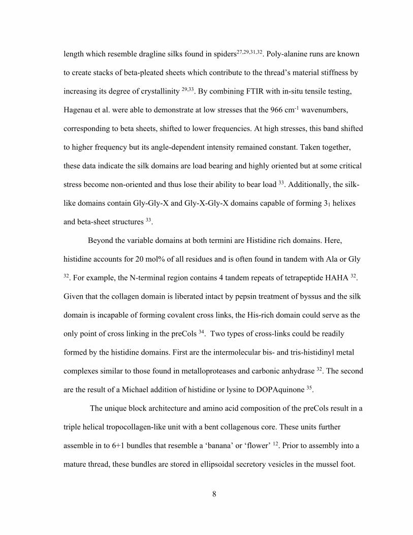
length which resemble dragline silks found in spiders27,29,31,32. Poly-alanine runs are known
to create stacks of beta-pleated sheets which contribute to the thread’s material stiffness by
increasing its degree of crystallinity 29,33. By combining FTIR with in-situ tensile testing,
Hagenau et al. were able to demonstrate at low stresses that the 966 cm-1 wavenumbers,
corresponding to beta sheets, shifted to lower frequencies. At high stresses, this band shifted
to higher frequency but its angle-dependent intensity remained constant. Taken together,
these data indicate the silk domains are load bearing and highly oriented but at some critical
stress become non-oriented and thus lose their ability to bear load 33. Additionally, the silk-
like domains contain Gly-Gly-X and Gly-X-Gly-X domains capable of forming 31 helixes
and beta-sheet structures 33.
Beyond the variable domains at both termini are Histidine rich domains. Here,
histidine accounts for 20 mol% of all residues and is often found in tandem with Ala or Gly
32. For example, the N-terminal region contains 4 tandem repeats of tetrapeptide HAHA 32.
Given that the collagen domain is liberated intact by pepsin treatment of byssus and the silk
domain is incapable of forming covalent cross links, the His-rich domain could serve as the
only point of cross linking in the preCols 34. Two types of cross-links could be readily
formed by the histidine domains. First are the intermolecular bis- and tris-histidinyl metal
complexes similar to those found in metalloproteases and carbonic anhydrase 32. The second
are the result of a Michael addition of histidine or lysine to DOPAquinone 35.
The unique block architecture and amino acid composition of the preCols result in a
triple helical tropocollagen-like unit with a bent collagenous core. These units further
assemble in to 6+1 bundles that resemble a ‘banana’ or ‘flower’ 12. Prior to assembly into a
mature thread, these bundles are stored in ellipsoidal secretory vesicles in the mussel foot.
9
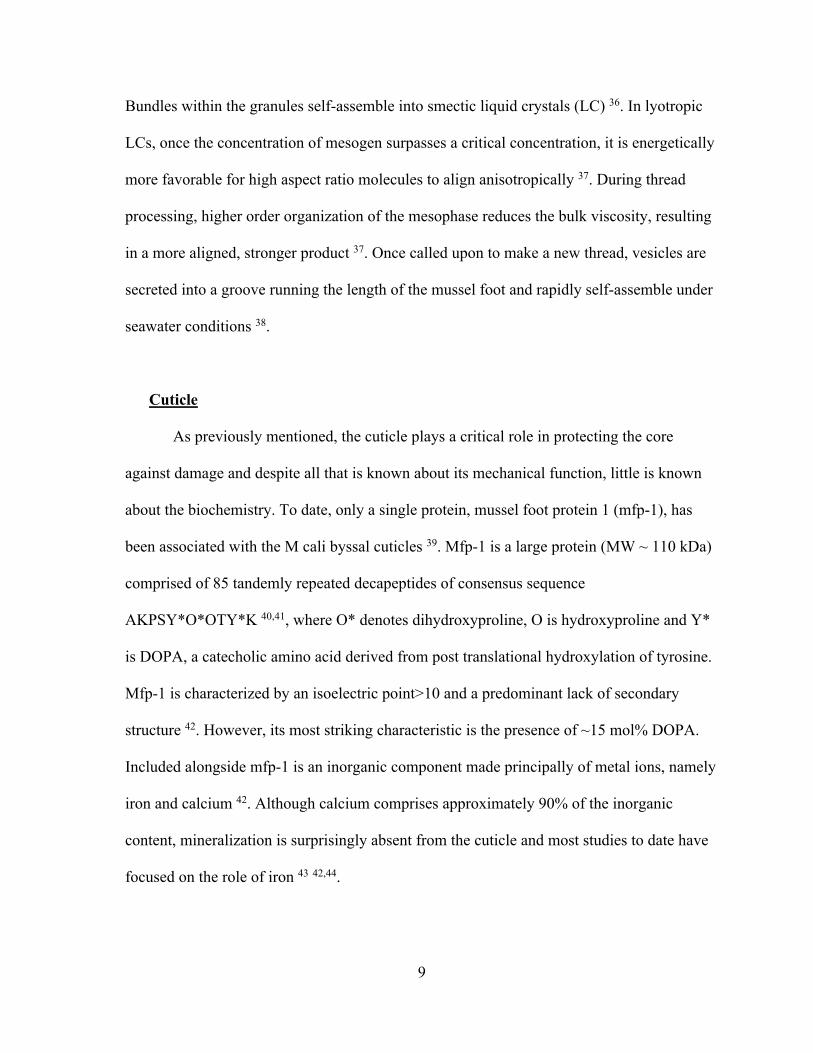
Bundles within the granules self-assemble into smectic liquid crystals (LC) 36. In lyotropic
LCs, once the concentration of mesogen surpasses a critical concentration, it is energetically
more favorable for high aspect ratio molecules to align anisotropically 37. During thread
processing, higher order organization of the mesophase reduces the bulk viscosity, resulting
in a more aligned, stronger product 37. Once called upon to make a new thread, vesicles are
secreted into a groove running the length of the mussel foot and rapidly self-assemble under
seawater conditions 38.
Cuticle
As previously mentioned, the cuticle plays a critical role in protecting the core
against damage and despite all that is known about its mechanical function, little is known
about the biochemistry. To date, only a single protein, mussel foot protein 1 (mfp-1), has
been associated with the M cali byssal cuticles 39. Mfp-1 is a large protein (MW ~ 110 kDa)
comprised of 85 tandemly repeated decapeptides of consensus sequence
AKPSY*O*OTY*K 40,41, where O* denotes dihydroxyproline, O is hydroxyproline and Y*
is DOPA, a catecholic amino acid derived from post translational hydroxylation of tyrosine.
Mfp-1 is characterized by an isoelectric point>10 and a predominant lack of secondary
structure 42. However, its most striking characteristic is the presence of ~15 mol% DOPA.
Included alongside mfp-1 is an inorganic component made principally of metal ions, namely
iron and calcium 42. Although calcium comprises approximately 90% of the inorganic
content, mineralization is surprisingly absent from the cuticle and most studies to date have
focused on the role of iron 43 42,44.
10
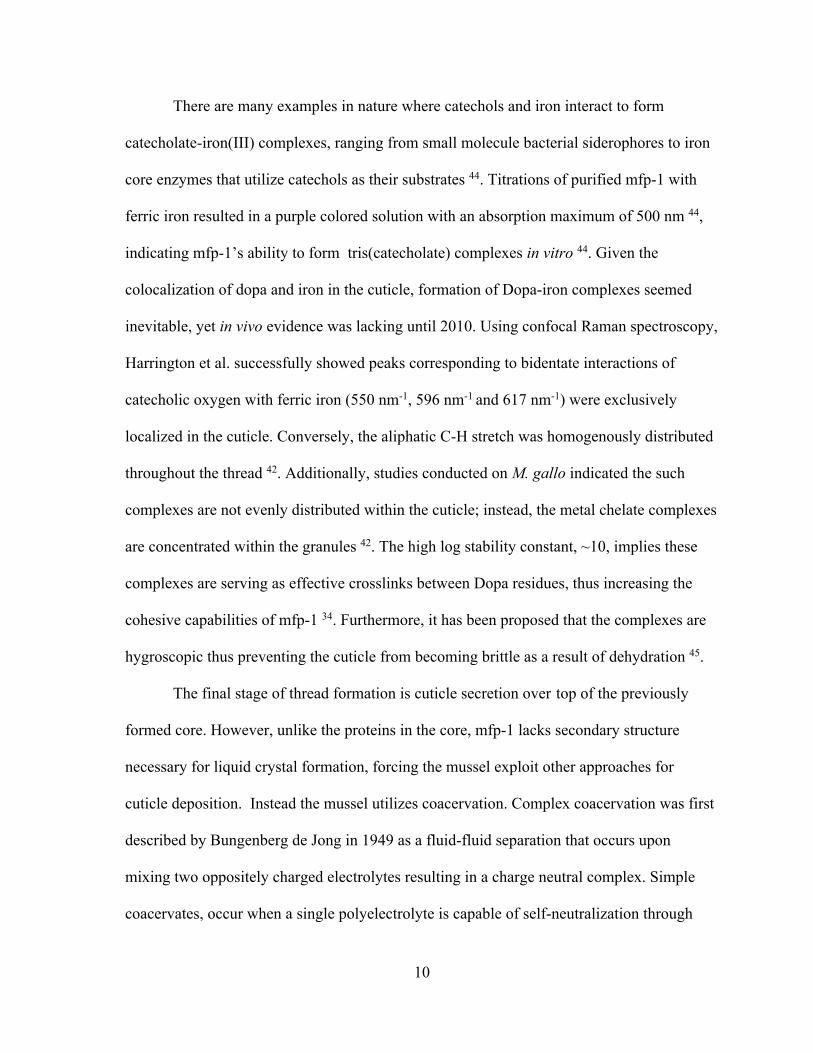
There are many examples in nature where catechols and iron interact to form
catecholate-iron(III) complexes, ranging from small molecule bacterial siderophores to iron
core enzymes that utilize catechols as their substrates 44. Titrations of purified mfp-1 with
ferric iron resulted in a purple colored solution with an absorption maximum of 500 nm 44,
indicating mfp-1’s ability to form tris(catecholate) complexes in vitro 44. Given the
colocalization of dopa and iron in the cuticle, formation of Dopa-iron complexes seemed
inevitable, yet in vivo evidence was lacking until 2010. Using confocal Raman spectroscopy,
Harrington et al. successfully showed peaks corresponding to bidentate interactions of
catecholic oxygen with ferric iron (550 nm-1, 596 nm-1 and 617 nm-1) were exclusively
localized in the cuticle. Conversely, the aliphatic C-H stretch was homogenously distributed
throughout the thread 42. Additionally, studies conducted on M. gallo indicated the such
complexes are not evenly distributed within the cuticle; instead, the metal chelate complexes
are concentrated within the granules 42. The high log stability constant, ~10, implies these
complexes are serving as effective crosslinks between Dopa residues, thus increasing the
cohesive capabilities of mfp-1 34. Furthermore, it has been proposed that the complexes are
hygroscopic thus preventing the cuticle from becoming brittle as a result of dehydration 45.
The final stage of thread formation is cuticle secretion over top of the previously
formed core. However, unlike the proteins in the core, mfp-1 lacks secondary structure
necessary for liquid crystal formation, forcing the mussel exploit other approaches for
cuticle deposition. Instead the mussel utilizes coacervation. Complex coacervation was first
described by Bungenberg de Jong in 1949 as a fluid-fluid separation that occurs upon
mixing two oppositely charged electrolytes resulting in a charge neutral complex. Simple
coacervates, occur when a single polyelectrolyte is capable of self-neutralization through

11
modulation of ionic strength, pH and polyelectrolyte concentration. Sheer-thinning viscous
properties and low interfacial tension allow coacervates to easily be secreted through an
intricate duct system and rapidly coat surfaces, making it an ideal mechanism for cuticle
delivery 46. Many factors contribute to this phenomenon such as charge density,
hydrophobicity, polymer weight, ionic strength, temperature, etc. Mfp-1 is capable of both
complex and simple coacervation as demonstrated by Miller et al. and Kim et al. Studies
with hyaluronic acid and mfp-1 demonstrate the ability to form complex coacervates via
charge neutralization 47. It is currently hypothesized that in vivo mfp-1 may complex with an
anionic Ca2+ binding protein, yet direct evidence is lacking 47. Simple coacervation of
recombinant mfp-1 (rmfp-1) can be induced by increasing the ionic strength of the solution
to ~0.7M 48. Typically, high ionic strength solutions disrupt the coacervate phase by
screening electrostatic interactions between oppositely charge poly-electrolytes 48-50.
However, in the mussel system, high ionic strength reduces electrostatic repulsions that arise
from the predominant cationic nature of mfp-1 48. Subsequently, allowing for cation-𝜋
interactions to drive and stabilize the coacervate 48.
Plaque:
At the plaque-substrate interface exists a complex assortment of mussel foot
proteins, namely -2, -3, -4, -5 and -6 that are responsible for long term adhesion51. For
purposes of this dissertation, I will focus the discussion on mfps -3, -5 and -6.
Mfp-3 and mfp-5 are considered to be priming proteins found at the adhesive
interface. The mfp-3 family contains more then 30 variants divided into 2 families known as
‘Fast’ and ‘Slow’. The fast variants contain more positive charges than slow family and
12
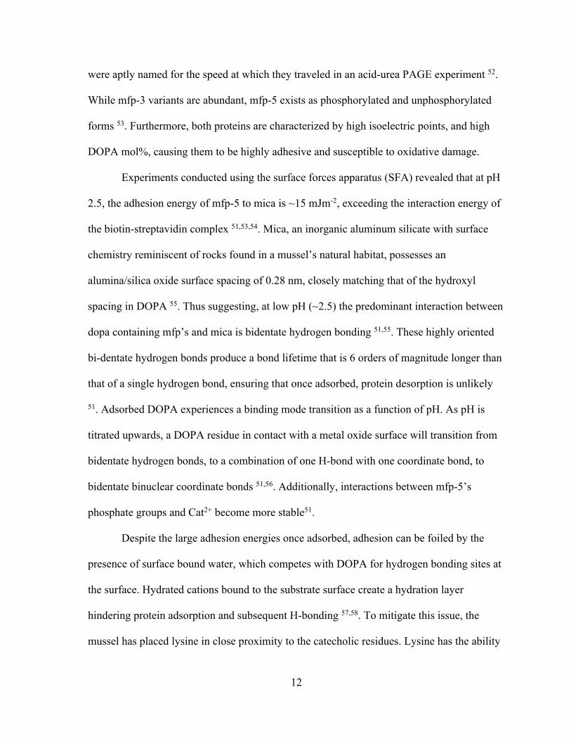
were aptly named for the speed at which they traveled in an acid-urea PAGE experiment 52.
While mfp-3 variants are abundant, mfp-5 exists as phosphorylated and unphosphorylated
forms 53. Furthermore, both proteins are characterized by high isoelectric points, and high
DOPA mol%, causing them to be highly adhesive and susceptible to oxidative damage.
Experiments conducted using the surface forces apparatus (SFA) revealed that at pH
2.5, the adhesion energy of mfp-5 to mica is ~15 mJm-2, exceeding the interaction energy of
the biotin-streptavidin complex 51,53,54. Mica, an inorganic aluminum silicate with surface
chemistry reminiscent of rocks found in a mussel’s natural habitat, possesses an
alumina/silica oxide surface spacing of 0.28 nm, closely matching that of the hydroxyl
spacing in DOPA 55. Thus suggesting, at low pH (~2.5) the predominant interaction between
dopa containing mfp’s and mica is bidentate hydrogen bonding 51,55. These highly oriented
bi-dentate hydrogen bonds produce a bond lifetime that is 6 orders of magnitude longer than
that of a single hydrogen bond, ensuring that once adsorbed, protein desorption is unlikely
51. Adsorbed DOPA experiences a binding mode transition as a function of pH. As pH is
titrated upwards, a DOPA residue in contact with a metal oxide surface will transition from
bidentate hydrogen bonds, to a combination of one H-bond with one coordinate bond, to
bidentate binuclear coordinate bonds 51,56. Additionally, interactions between mfp-5’s
phosphate groups and Cat2+ become more stable51.
Despite the large adhesion energies once adsorbed, adhesion can be foiled by the
presence of surface bound water, which competes with DOPA for hydrogen bonding sites at
the surface. Hydrated cations bound to the substrate surface create a hydration layer
hindering protein adsorption and subsequent H-bonding 57,58. To mitigate this issue, the
mussel has placed lysine in close proximity to the catecholic residues. Lysine has the ability
13
to evict hydrated surface bound cations, subsequently reducing the hydration layer 57 and
paving the way for long lived bidentate hydrogen bond formation.
Deposition can be further enhanced by involving liquid-liquid phase separation such
as coacervation. Plaque proteins are injected into the acidified distal depression as solutes 59,
and undergo a phase transition upon exposure to seawater conditions60. In vitro, mfp-3
coacervate deposition resulted in four times more material binding to the surface compared
with the solution state 61, as well as decreased desorption upon rinsing. Additionally, ATR-
FTIR indicates increased dehydration of the substrate surface especially when using Dopa-
containing proteins 61.
Although equilibration toward seawater conditions may be beneficial for deposition
it is detrimental to adhesion. For instance, Mfp-5 adhesion diminishes 75% at pH 5.5 and is
effectively abolished at pH 7.5 54,62. Simply lowering the pH was insufficient at restoring
adhesion 63. Losses in adhesion were attributed to oxygen dependent oxidation of DOPA to
DOPAquinone, which is not capable of the metal coordination and H-bond formation
essential for stable bonding 54,62,64,65. Subsequent SFA tests combining mfp-3 at acidic pH
with the strong oxidizer periodate produced nominal adhesion, thus confirming the
correlation between quinone formation and adhesion loss 63.
Seawater contains large amounts of dissolved oxygen and high trace metal content
resulting in a standard redox potential of approximately 600 mV, substantially higher than
that of DOPA 51,66. As a result, DOPA oxidation via oxygen reduction is a likely process,
however DOPA remains detectable for upwards of 1 month in mature plaques 67. To combat
oxidation and oxidative losses, the mussel incorporates reducing compartments and
antioxidant proteins, chiefly mfp-6 64.
14
Mfp-6 is a 11.5kDa protein with a composition that is ~11 mol% cysteine giving rise
to a reducing capacity of 17 e- per protein 64,68. When introduced to oxidized mfp-3 films,
mfp-6 was able to fully restore adhesion 63. Interestingly, reducing activity of mfp-6 persists
at pHs as low as 3, suggesting some of mfp-6’s thiols possess pKa’s well below the typical
8-9 63. The rescue process requires two subsequent nucleophilic attacks initiated by a
cysteinyl thiolate. First, a thiolate anion adds to carbon 5 in a Michael addition reaction,
followed by attack of the thioether by a second thiolate resulting in a restored DOPA and
cystine 69,70. The question of mfp-6’s ability to maintain DOPA is certainly one of
stoichiometry. As the number of reducing thiols is consumed, a buildup of 5-S-
cysteinylDOPA occurs 68. To date, redox poise has been somewhat of a mystery. How do
thiols persist for extended periods of time? How does the mussel protect both thiols and
DOPA from immediate oxidation despite existing in highly oxidizing seawater? These
questions will be addressed in Chapter 3.
15
References:
1. Yonge CM. On The Primitive Significance of the Byssus in the Bivalvia and its Effects in Evolution. https://doiorg/101017/S0025315400004495. December 2008:1-13. doi:10.1017/S0025315400004495.
2. Lockwood BL, Connor KM, Gracey AY. The environmentally tuned transcriptomes of Mytilus mussels. J Exp Biol. 2015;218(Pt 12):1822-1833. doi:10.1242/jeb.118190.
3. Jones TL, Richman JR. On Mussels: Mytilus Californianusas a Prehistoric Resource. North American Archaeologist. 1995;16(1):33-58. doi:10.2190/G5TT-YFHP-JE6A-P2TX.
4. Shaw WN. Species Profiles: Life Histories and Environmental Requirement of Coastal Fishes and Invertebrates (Pacific Southwest): California Sea Mussel and Bay Mussel. Biological Report Fish and Wildlife Servies. 1988:1-24.
5. Carrington E. Drag and dislodgment of an intertidal macroalga: consequences of morphological variation in Mastocarpus papillatus Kutzing. Journal of Experimental Marine Biology and Ecology. 1990;139:185-200.
6. Waite JH. Adhesion a la Moule. Integrative and Comparative Biology. 2002;42(6):1172-1180. doi:10.1093/icb/42.6.1172.
7. Harrington MJ, Gupta HS, Fratzl P, Waite JH. Collagen insulated from tensile damage by domains that unfold reversibly: in situ X-ray investigation of mechanical yield and damage repair in the mussel byssus. Journal of Structural Biology. 2009;167(1):47-54. doi:10.1016/j.jsb.2009.03.001.
8. Qin X, Waite JH. Exotic collagen gradients in the byssus of the mussel Mytilus edulis. Journal of Experimental Biology. 1995;198(Pt 3):633-644.
9. Krauss S, Metzger TH, Fratzl P, Harrington MJ. Self-Repair of a Biological Fiber Guided by an Ordered Elastic Framework. Biomacromolecules. 2013;14(5):1520-1528. doi:10.1021/bm4001712.
10. Krauss S, Metzger TH, Fratzl P, Harrington MJ. Self-Repair of a Biological Fiber Guided by an Ordered Elastic Framework. Biomacromolecules. 2013;14(5):1520-1528. doi:10.1021/bm4001712.
11. Zucarrello-Vitellaro, L., 1980. The collagen gland of Mytilus galloprovincialis: an ultrastructural and cytochemical study on secretory granules. Tissue & Cell or J. Ultrastruct. Res Elsevier. 1980;73(2):135-147. doi:10.1016/S0022-5320(80)90119-7.
16
12. Hassenkam T, Gutsmann T, Hansma P, Sagert J, Waite JH. Giant Bent-Core Mesogens in the Thread Forming Process of Marine Mussels. Biomacromolecules. 2004;5(4):1351-1355. doi:10.1021/bm049899t.
13. Bairati A, research LZCAT, 1976. The ultrastructure of the byssal apparatus of Mytilus galloprovincialis. Springer. doi:10.1007/BF00227043.
14. Holten-Andersen N, Zhao H, Waite JH. Stiff Coatings on Compliant Biofibers: The Cuticle of Mytilus californianusByssal Threads †,‡. Biochemistry. 2009;48(12):2752-2759. doi:10.1021/bi900018m.
15. Holten-Andersen N, Slack N, Proceedings FZMO, 2004. Nano-mechanical investigation of the byssal cuticle, a protective coating of a bio-elastomer. cambridgeorg. doi:10.1557/PROC-841-R3.7/Y3.7.
16. Monnier CA, DeMartini DG, Waite JH. Intertidal exposure favors the soft-studded armor of adaptive mussel coatings. Nature Communications. August 2018:1-9. doi:10.1038/s41467-018-05952-5.
17. Tamarin A, Lewis P, Askey J. The structure and formation of the byssus attachment plaque in Mytilus. J Morphol. 1976;149(2):199-221. doi:10.1002/jmor.1051490205.
18. Filippidi E, DeMartini DG, Malo de Molina P, et al. The microscopic network structure of mussel (Mytilus) adhesive plaques. J R Soc Interface. 2015;12(113):20150827–10. doi:10.1098/rsif.2015.0827.
19. Desmond KW, Zacchia NA, Waite JH, Valentine MT. Dynamics of mussel plaque detachment. Soft Matter. 2015;11(34):6832-6839. doi:10.1039/c5sm01072a.
20. Bell E, Gosline J. Mechanical design of mussel byssus: material yield enhances attachment strength. Journal of Experimental Biology. 1996;199(4):1005-1017.
21. Carrington E, Gosline JM. Mechanical design of mussell byssus: Load cycle and strain rate dependence. 2004.
22. Gosline J, Lillie M, Carrington E, Guerette P, Ortlepp C, Savage K. Elastic proteins: biological roles and mechanical properties. Bailey AJ, Macmillan J, Shrewry PR, Tatham AS, eds. Philosophical Transactions of the Royal Society of London B: Biological Sciences. 2002;357(1418):121-132. doi:10.1098/rstb.2001.1022.
23. Bueche F. Molecular basis for the mullins effect. Journal of Applied Polymer Science. 1960;4(10):107-114. doi:10.1002/app.1960.070041017.
17
24. Burkett JR, Wojtas JL, Cloud JL, Wilker JJ. A Method for Measuring the Adhesion Strength of Marine Mussels. The Journal of Adhesion. 2009;85(9):601-615. doi:10.1080/00218460902996903.
25. Spuskanyuk AV, McMeeking RM, Deshpande VS, Arzt E. The effect of shape on the adhesion of fibrillar surfaces. Acta Biomaterialia. 2008;4(6):1669-1676. doi:10.1016/j.actbio.2008.05.026.
26. Kendall K. The adhesion and surface energy of elastic solids. J Phys D: Appl Phys. 2002;4(8):1186-1195. doi:10.1088/0022-3727/4/8/320.
27. Waite JH, Vaccaro E, Sun C, Lucas JM. Elastomeric gradients: a hedge against stress concentration in marine holdfasts? Philosophical Transactions of the Royal Society of London B: Biological Sciences. 2002;357(1418):143-153. doi:10.1098/rstb.2001.1025.
28. Harrington MJ, Waite JH. Holdfast heroics: comparing the molecular and mechanical properties of Mytilus californianus byssal threads. Journal of Experimental Biology. 2007;210(24):4307-4318. doi:10.1242/jeb.009753.
29. Waite JH, Qin X-X, Coyne KJ. The peculiar collagens of mussel byssus. Matrix Biol. 1998;17(2):93-106. doi:10.1016/S0945-053X(98)90023-3.
30. Kilchherr E, Hofmann H, molecular WSJO, 1985. Structural model of the collagen-like region of C1q comprising the kink region and the fibre-like packing of the six triple helices. Elsevier. 1985;186(2):403-415. doi:10.1016/0022-2836(85)90114-7.
31. Hagenau A, Scheidt HA, Serpell L, Huster D, Scheibel T. Structural Analysis of Proteinaceous Components in Byssal Threads of the Mussel Mytilus galloprovincialis. Taubert A, ed. Macromol Biosci. 2009;9(2):162-168. doi:10.1002/mabi.200800271.
32. Qin X-X, Coyne KJ, Waite JH. Tough Tendons MUSSEL BYSSUS HAS COLLAGEN WITH SILK-LIKE DOMAINS. Journal of Biological Chemistry. 1997;272(51):32623-32627. doi:10.1074/jbc.272.51.32623.
33. Hagenau A, Papadopoulos P, Kremer F, Scheibel T. Mussel collagen molecules with silk-like domains as load-bearing elements in distal byssal threads. Journal of Structural Biology. 2011;175(3):339-347. doi:10.1016/j.jsb.2011.05.016.
34. Das S, Miller DR, Kaufman Y, et al. Tough Coating Proteins: Subtle Sequence Variation Modulates Cohesion. Biomacromolecules. 2015;16(3):1002-1008. doi:10.1021/bm501893y.
35. Xu REA. Characterization of Products from the Reactions of N-Acetyldopamine Quinone with N-Acetylhistidine. April 1996:1-9.
18
36. Priemel T, Degtyar E, Dean MN, Harrington MJ. Rapid self-assembly of complex biomolecular architectures during mussel byssus biofabrication. Nature Communications. 2017;8:1-12. doi:10.1038/ncomms14539.
37. Harrington MJ, Waite JH. pH-Dependent Locking of Giant Mesogens in Fibers Drawn from Mussel Byssal Collagens. Biomacromolecules. 2008;9(5):1480-1486. doi:10.1021/bm8000827.
38. Renner-Rao M, Clark M, ORCID: 0000-0003-1417-9251 MJH. Fiber Formation from Liquid Crystalline Collagen Vesicles Isolated from Mussels. Langmuir. August 2019:1-10. doi:10.1021/acs.langmuir.9b01932.
39. Lee BP, Messersmith PB, Israelachvili JN, Waite JH. Mussel-Inspired Adhesives and Coatings. Annu Rev Mater Res. 2011;41(1):99-132. doi:10.1146/annurev-matsci-062910-100429.
40. Sun C, Waite JH. Mapping chemical gradients within and along a fibrous structural tissue, mussel byssal threads. Journal of Biological Chemistry. 2005;280(47):39332-39336. doi:10.1074/jbc.M508674200.
41. Taylor SW, Chase DB, Emptage MH, Nelson MJ, Waite JH. Ferric Ion Complexes of a DOPA-Containing Adhesive Protein from Mytilus edulis. Inorg Chem. 1996;35(26):7572-7577. doi:10.1021/ic960514s.
42. Harrington MJ, Masic A, Holten-Andersen N, Waite JH, Fratzl P. Iron-Clad Fibers: A Metal-Based Biological Strategy for Hard Flexible Coatings. Science. 2010;328(5975):216-220. doi:10.1126/science.1181044.
43. Holten-Andersen N, Mates TE, Toprak MS, Stucky GD, Zok FW, Waite JH. Metals and the Integrity of a Biological Coating: The Cuticle of Mussel Byssus. Langmuir. 2009;25(6):3323-3326. doi:10.1021/la8027012.
44. Taylor SW, Chase DB, Emptage MH, Nelson MJ, Waite JH. Ferric ion complexes of a DOPA-containing adhesive protein from Mytilus edulis. Inorg Chem. 1996;35(26):7572-7577. doi:10.1021/ic960514s.
45. Monnier CA, DeMartini DG, Waite JH. Intertidal exposure favors the soft-studded armor of adaptive mussel coatings. Nature Communications. August 2018:1-9. doi:10.1038/s41467-018-05952-5.
46. Yang B, Jin S, Park Y, Jung YM, Cha HJ. Coacervation of Interfacial Adhesive Proteins for Initial Mussel Adhesion to a Wet Surface. Small. 2018;14(52):1803377–12. doi:10.1002/smll.201803377.
47. Miller DR, Das S, Huang K-Y, Han S, Israelachvili JN, Waite JH. Mussel Coating Protein-Derived Complex Coacervates Mitigate Frictional Surface Damage. ACS Biomater Sci Eng. 2015;1(11):1121-1128. doi:10.1021/acsbiomaterials.5b00252.
19
48. Kim S, Yoo HY, Huang J, et al. Salt Triggers the Simple Coacervation of an Underwater Adhesive When Cations Meet Aromatic π Electrons in Seawater. ACS Nano. 2017;11(7):6764-6772. doi:10.1021/acsnano.7b01370.
49. Elbaum-Garfinkle S, Kim Y, Szczepaniak K, et al. The disordered P granule protein LAF-1 drives phase separation into droplets with tunable viscosity and dynamics. Proc Natl Acad Sci USA. 2015;112(23):7189-7194. doi:10.1073/pnas.1504822112.
50. Danielsen SPO, McCarty J, Shea J-E, Delaney KT, Fredrickson GH. Small ion effects on self-coacervation phenomena in block polyampholytes. The Journal of Chemical Physics. July 2019:1-19. doi:10.1063/1.5109045.
51. Waite JH. Mussel adhesion - essential footwork. J Exp Biol. 2017;220(Pt 4):517-530. doi:10.1242/jeb.134056.
52. Zhao H, Robertson NB, Jewhurst SA, Waite JH. Probing the adhesive footprints of Mytilus californianus byssus. Journal of Biological Chemistry. 2006;281(16):11090-11096. doi:10.1074/jbc.M510792200.
53. Lu Q, Danner E, Waite JH, Israelachvili JN, Zeng H, Hwang DS. Adhesion of mussel foot proteins to different substrate surfaces. J R Soc Interface. 2013;10(79):20120759–11. doi:10.1098/rsif.2012.0759.
54. Danner EW, Kan Y, Hammer MU, Israelachvili JN, Waite JH. Adhesion of Mussel Foot Protein Mefp-5 to Mica: An Underwater Superglue. Biochemistry. 2012;51(33):6511-6518. doi:10.1021/bi3002538.
55. Yu J, Wei W, Danner E, Israelachvili JN, Waite JH. Effects of Interfacial Redox in Mussel Adhesive Protein Films on Mica. Adv Mater. 2011;23(20):2362-2366. doi:10.1002/adma.201003580.
56. Yu J, Kan Y, Rapp M, the EDPO, 2013. Adaptive hydrophobic and hydrophilic interactions of mussel foot proteins with organic thin films. National Acad Sciences. doi:10.1073/pnas.1315015110/-/DCSupplemental.
57. Maier GP, Rapp MV, Waite JH, Israelachvili JN, 2015. Adaptive synergy between catechol and lysine promotes wet adhesion by surface salt displacement. sciencesciencemagorg. doi:10.1126/science.aab3175.
58. Pashley RM. Hydration forces between mica surfaces in electrolyte solutions. Advances in Colloid and Interface Science. 1982;16(1):57-62. doi:10.1016/0001-8686(82)85006-9.
59. Martinez Rodriguez NR, Das S, Kaufman Y, Israelachvili JN, Waite JH. Interfacial pH during mussel adhesive plaque formation. Biofouling. 2015;31(2):221-227. doi:10.1080/08927014.2015.1026337.

20
60. Wei W, Tan Y, Rodriguez NRM, Yu J, Israelachvili JN, Waite JH. A mussel-derived one component adhesive coacervate. Acta Biomaterialia. 2014;10(4):1663-1670. doi:10.1016/j.actbio.2013.09.007.
61. Wei W, Petrone L, Tan Y, et al. An Underwater Surface‐Drying Peptide Inspired by a Mussel Adhesive Protein. Advanced Functional Materials. March 2016:n/a–n/a. doi:10.1002/adfm.201600210.
62. Nicklisch SCT, Waite JH. Mini-review: The role of redox in Dopa-mediated marine adhesion. Biofouling. 2012;28(8):865-877. doi:10.1080/08927014.2012.719023.
63. Yu J, Wei W, Danner E, Ashley RK, Israelachvili JN, Waite JH. Mussel protein adhesion depends on interprotein thiol-mediated redox modulation. Nat Chem Biol. 2011;7(9):588-590. doi:10.1038/nchembio.630.
64. Nicklisch SCT, Spahn JE, Zhou H, Gruian CM, Waite JH. Redox Capacity of an Extracellular Matrix Protein Associated with Adhesion in Mytilus californianus. Biochemistry. 2016;55(13):2022-2030. doi:10.1021/acs.biochem.6b00044.
65. Anderson TH, Yu J, Estrada A, Hammer MU, Waite JH, Israelachvili JN. The Contribution of DOPA to Substrate-Peptide Adhesion and Internal Cohesion of Mussel-Inspired Synthetic Peptide Films. Advanced Functional Materials. 2010;20(23):4196-4205. doi:10.1002/adfm.201000932.
66. Cooper LHN. OXIDA TION-REDUCTION POTENTIAL IN SEA WATER By L. H. N. Cooper, Ph.D., F.I.C. https://doiorg/101017/S0025315400011929. December 2008:1-10. doi:10.1017/S0025315400011929.
67. Miller DR, Spahn JE, Waite JH. The staying power of adhesion-associated antioxidant activity in Mytilus californianus. J R Soc Interface. 2015;12(111):20150614-20150617. doi:10.1098/rsif.2015.0614.
68. Zhao H, Waite JH. Linking adhesive and structural proteins in the attachment plaque of Mytilus californianus. Journal of Biological Chemistry. 2006;281(36):26150-26158. doi:10.1074/jbc.M604357200.
69. Inaba K. Structural basis of protein disulfide bond generation in the cell. Genes Cells. 2010;15(9):935-943. doi:10.1111/j.1365-2443.2010.01434.x.
70. Yu J, Wei W, Danner E, Ashley RK, Israelachvili JN, Waite JH. Mussel protein adhesion depends on interprotein thiol-mediated redox modulation. Nat Chem Biol. 2011;7(9):586-588. doi:10.1038/nchembio.630.
21
Chapter II.
Thiol Rich Layer in the Core-Shell Architectures of byssus threads
Reproduced in part with permission from: Valois, E. and Waite, J.H. (2019). The Thiol-
Rich Interlayer in the Shell/Core Architecture of Mussel Byssal Threads. Langmuir. Article
ASAP. DOI: 10.1021/acs.langmuir.9b01844
A. Abstract
The mussel byssus thread is an extremely tough core-shelled fiber that dissipates
substantial amounts of energy during tensile loading. The mechanical performance of the shell
is critically reliant on 3, 4-dihydroxyphenylalanine’s (Dopa) ability to form reversible iron-
catecholate complexes at pH 8. However, the formation of these coordinate crosslinks is
undercut by Dopa’s oxidation to Dopa-quinone, a spontaneous process at seawater conditions.
The large mechanical mismatch between the cuticle and the core lends itself to further
complications. Despite these challenges, the mussel byssus thread performs its tethering
function over long periods of time. Here we address these two major questions. 1) How does
the mussel slow/prevent oxidation in the cuticle and 2) how is the mechanical mismatch at the
core-shell interface mitigated? By combining a number of microscopy and spectroscopy
techniques we have discerned a previously undescribed layer. Our results indicate this
interlayer is thiol rich and thus will be called the thiol-rich interlayer (TRL). We propose the
TRL serves as a long-lasting redox reservoir as well as a mechanical barrier.
22
B. Introduction.
Materials with core-shell architectures are pervasive throughout nature and technology.
Indeed, in nature there may be no functional material without both core and shell. The
utility of a core/shell design is self-evident: the core provides bulk properties e.g. tensile or
compressive strength in load bearing materials, while the shell protects the core from
corrosion, light, desiccation, microbial attack, and/or abrasion in the ambient environment.
Because the disparate functions of shell and core usually lead to incompatibilities between
the two, additional adaptive layers are introduced to enhance their union. These are
increasingly referred to as self-stratifying coatings 1, and, once again, were evolved by
biology, e.g. plant cutins, long before becoming a design concept in engineered materials.
Some, but by no means all, biological coatings may thus provide valuable insights for
stimulating improvements in manufactured coatings.
Mussel byssal threads have core-shell architectures in which the shell is 5-6 times stiffer
than the core2. The thread core consists of anisotropic bundles of collagen fibers not unlike
tendon, whereas the shell, known as the cuticle, is a composite of granules dispersed in a
continuous, amorphous matrix. The granules are largely composed of a Dopa-rich mussel
foot protein, which, at pH 5-8, readily binds iron to form cross-linking bis- and tris-
catecholate-iron complexes 3,4. Given the reversible nature of metal-coordinate bonds, tris
catecholate-iron complexes are reversible sacrificial bonds, dissipating energy by enabling
greater strain during deformation. During unloading, the tris-catecholate-iron complexes
self-heal to repair damage sustained during deformation, but only in a Dopa-rich
environment6. The high dissolved oxygen content and abundance of trace metals make
seawater a powerful oxidant of Dopa 7 thus undermining its integrity in byssal threads, yet
23
Dopa-iron complexes remain stable in the cuticle. How does the mussel protect the cuticle
against oxidative damage and consequent loss of function?
Our structural, chemical and nanomechanical analyses of byssal threads suggest that
thread redox is maintained by cuticle stratification in combination with thiol enrichment in a
subsurface layer, i.e. the thiol rich layer (TRL). A combination of microscopy techniques
including transmission electron microscopy (TEM), scanning electron microscopy (SEM)
and atomic force microscopy were used to characterize the location and morphology of this
TRL. Furthermore, fluorescence microscopy, X-ray near edge spectroscopy (XANES) and
secondary ion mass spectrometry were used to speciate the thiol.
Prior studies have identified the cuticle as being significantly stiffer than the core5.
While this unique combination of stiff cuticle and compliant core results in high toughness,
it creates the possibility of contact damage at the interface between the two mechanically
mismatched materials. Here, we characterize the nano-mechanical characteristics under
biologically relevant conditions using indentation type atomic force microscopy. Although
our measurements confirm a large discrepancy in elastic modulus between cuticle and core,
they also reveal that the inclusion of the TRL creates a step gradient in hardness and
abrasion resistance. By building on many years of byssus research, the present study
addresses two major gaps in our understanding. First, how the cuticle copes with an
oxidizing environment, and second, how the byssus successfully joins two mechanically
mismatched materials.
24
C. Materials and Methods
TEM/STEM
Threads less than 48 hours old were excised from the animal and fixed according to
the following procedure: All fixation and washing steps were performed on ice. Threads
were fixed with 2% formaldehyde, 2.5% glutaraldehyde in fixation buffer (200 mM sodium
cacodylate, 300 mM NaCl, pH 7.2) for 2 h. The samples were washed three times (10 min
each) in degassed fixation buffer and then post-fixed in 2% osmium tetroxide in degassed
fixation buffer for 2 h. The samples were then washed 4 times (10 min each) in degassed
deionized water and then dehydrated through a graded series of ethanol washes (25, 50, 75,
90, 100, 100, 100% ethanol, 10 min each). Solvent was then switched to propylene oxide by
washing in 33, 66, 100, 100, 100% propylene oxide in ethanol. The samples were then
infiltrated with epoxy resin (Embed812, Electron Microscopy Sciences, Hatfield, USA)
incubating the sample in resin diluted in propylene oxide as follows: 33% (2 h), 66% (16 h),
and 100% (4 h). Finally, samples were placed in molds and cured at 60 °C for 24 h. Thin
sections (60–80 nm) for TEM were cut on an EM UC6 ultramicrotome (Leica Biosystems).
Sections were mounted on copper TEM grids and post-stained on drops of uranyl acetate
and lead citrate following standard protocols. All samples were investigated with a Tecnai
G2 transmission electron microscope (FEI) operating at 200 kV, and micrographs were
recorded with a Gatan Ultrascan CCD camera (2048 × 2048 pixels. STEM images were
acquired using a Titan transmission electron microscope operating at 300 kV. Collagen
spacing was calculated using Fiji’s (v. 2.0.0-rc-65) nearest neighbor distance (NND) plugin.
Prior to analysis, each image was smoothed, and a threshold was applied resulting in a
binary image.
25
Atomic Force Microscopy (AFM)
Images and force measurements were conducted on a MFP-3D Bio (Asylum
Research, Santa Barbara, CA). FORTA silicon tips (APPNano, Mountain View, CA) were
used in all experiments. Fresh threads (<48 hours old) were imbedded in Neg-50
cryoprotectant (Rachard-Allan Scientific, San Diego, CA) and frozen to -20º C. 10 µm thick
sagittal sections were cut using a Leica Cryostat. Sections were placed on top of a drop of
water on a poly-lysine slide. The water was subsequently removed with a gel loading pipette
tip causing the section to lie flat on the slide. Once mounted, sections were allowed to dry
for 1 hour prior to measurement, ensuring sufficient adherence to the slide. 20 µm2 topology
images were generate using AC mode at a rate of 0.7 HZ. For submerged samples, threads
were allowed to equilibrate in seawater for 1 hour prior to imaging. Prior to force
measurements, the cantilever spring constant was experimentally determined by the thermal
tune method usually ranging between 2 and 3 N/m. Deflection sensitivity was calculated
using a glass slide as an indefinitely stiff calibrant material. Force spectroscopic maps of
32X32 pixels were recorded over previously acquired topology images. Force spectroscopy
measurements were conducted at 250 nm/s load/unload rate with a maximal loading force of
500 nN. Stiffness and hardness values were calculated by Asylum Research’s Elastic
Analysis Tool by fitting the upper 1/3 of the loading curve, an indenter half angle of 20 º,
and a Poisson ratio of 0.33 to the Hertz Model.
SEM
Fresh threads were fixed with 2% formaldehyde, 2.5% glutaraldehyde in fixation
buffer (200 mM sodium cacodylate, 300 mM NaCl, pH 7.2) for 2 h. Fixed threads were

26
imbedded in Neg-50 cryoprotectant (Rachard-Allan Scientific, San Diego, CA) and frozen
to -20º C. 10 µm thick sagittal sections were cut using a Leica Cryostat. Sections were
washed 3 times, 10 min each, in milli-Q water to remove any excess cryoprotectant. Solvent
exchange from water to ethanol was performed in steps of 30%, 50%, 60%, 90%, 100% X3
ethanol. Samples were dried and secured to aluminum imaging stub with double sided
carbon tape. A thin layer of gold-palladium (60:40) was sputter coated onto the sample for
100 seconds using an Anatech USA Hummer 6.2 coater. Specimens were viewed at 3kV
with a FEI Nova Nano 650 FEG SEM.
Fluorescence Microscopy
Transverse cross sections of fresh mussel threads were prepared as described above.
Sections were washed 3 times (1 min each) to remove excess cryoprotectant followed by 3
times 5 min each in phosphate buffered saline (PBS) pH 7.4 (Gibco Life Technologies).
Washed sections were incubated in 1µM Alexa Fluor555 C2 Maleimide (Thermo Fisher
Scientific, Waltham, MA) in PBS for hours at room temperature. Alexa Fluor 555 C2
Maleimide stock was prepared by dissolving received product in dimethyl sulfoxide to a
final concentration 10mM. Following staining, sections were rinsed 3 times (10 min each) in
PBS to remove excess dye and subsequently mounted onto glass slides using ProLong Gold
Antifade Mountant (Thermo Fisher Scientific, Waltham, MA). Confocal images were
collected using a Leica SP8 confocal microscope (Leica Microsystems) fitted with
63X/1.4NA oil immersion objective and hybrid detectors (HyD). Thread cuticle was
visualized via its intrinsic fluorescence when excited with a 405 nm laser. Alexa Fluor555
stained TRL was visualized using the white light laser tuned to 550 nm excitation.
27
Comparative fluorescent intensity was calculated with Fiji’s analyze plot profile function.
Alkylation of cysteine residues was accomplished using iodoacetamide (IAM). In control
threads, alkylation was done prior to Alexa Fluor555 C2 Maleimide addition by incubating
thread sections in 60 µM IAM in PBS pH 7.4 for 1 hour.
Accessory and Collagen Granule Isolation and Tandem MS
Live Mytilus californianus were collected from Goleta Pier in Goleta, CA and stored
in maricultural tanks with an open seawater circulation system. Feet were excised from the
mussel and sliced into sections approximately 3 mm in length using a No. 10 scalpel. The
distal portion of the foot surrounding the distal depression was discarded in order to avoid
contamination from phenol gland granules. Gland tissue at the center of the foot was
carefully separated from the pigmented epithelium and homogenized in 1mL per foot of
0.05M sodium phosphate pH 7.4 with 0.45M NaCl (mPBS). Granules were separated from
insoluble tissue by successive centrifugation through a 40 µm mesh followed by 5 µm mesh
(2X 10 min each at 500g). If necessary, additional mPBS was used to rinse the mesh in
between spin cycles to prevent clogging. After the final centrifugation, granules were
pelleted at 1000g for 10 min and the supernatant was removed. Pelleted granules were
resuspended in 0.1M Citrate pH 5.0 with 0.6M Sucrose 0.005M Ascorbic Acid and 0.001M
EDTA. Resuspended granules were then subjected to differential centrifugation gradient of
10 to 50% OptiPrep (Alere Technologies, Waltham, MA) for 16 hours at 31,000rpm, 4°C
(Beckman L8-80M Ultracentrifuge, Beckman Coulter, Brea, CA). Granules contained in
40% Optiprep were collected for trypsinization. Optiprep was removed from the sample by
diluting 1:10 with mPBS and re-pelleted at 1000g for 5 min X 3. Trpysinization was done in
28
50 mM Tris- HCl (pH 8), 5 mM DTT, and 8 M urea at 37°C for 4 hours. Resulting peptides
were sent to UC Davis mass spectrometry facility for sequencing (Davis, CA). Peptides
were quantified and 1.0μg was injected into the Xevo G2 QT mass spectrometer coupled to
a nano-UPLC system (Waters, Milford, MA. USA). Peptides were injected into a nanoTile,
housing a C18 column and eluted off the column in 1h gradient from 2 to 60% acetonitrile.
Mass spectrometry analysis was done using MSe, a data independent acquisition (DIA)
method. Data analysis was done using Protein Lynx Global Server and subsequently
analyzed in Scaffold-4 (Proteome Software, Portland, OR USA)
µXANES
For determining in situ sulfur species, synchrotron-based sulfur X-ray absorption
near-edge spectroscopy (S XANES) were measured at beamline (BL) 14-3 at the Stanford
Synchrotron Radiation Lightsource (SSRL) using a Si(111) (Φ = 90) double crystal
monochromator. The monochromator was calibrated at the thiol pre-edge energy of a
sodium thiosulfate powder to 2472.02 eV. The fluorescence lines of interest were measured
with a Si Vortex Si drift detector (SII Nano Technology) using Xspress3 pulse processing
electronics (Quantum Detectors) that deliver superior count rates without significant dead
times. The X-ray beam was focused using Kirkpatrick-Baez (K−B) mirrors to a size of ~3 ×
3 μm at a flux of ∼2 × 10¹⁰ photons per second. To determine sulfur speciation at specific
points within the sample, absorption spectra (S XANES) were collected from 2460 to 2536
at 0.2 eV steps to capture the range of energies for the absorption edges of possible common
sulfur compounds. Multiple spectra were acquired at each point to confirm that the
29
absorption edge energies did not shift to lower energies because of photoreduction by beam
damage to the sample.
SIMS
Sagittal sections of threads less than 48 hours old were excised from the animal and
prepared according to TEM procedures. Thin sections were then mounted to a silicon wafer.
To ensure a flat sample surface, sections were floated on a small drop of water placed on top
of a silicon wafer. The water droplet was slowly evaporated by heating the wafer to 50ºC
with a heating block. Elemental mapping was conducted using a Cameca IMS 7f-Auto
SIMS. Negative ions (32S- and 26CN-) were mapped with a primary cesium gun using 15 kV
impact energy and spot size of 1.5um.
DPPH Assay
Given the light sensitivity of DPPH, all reaction vessels containing DPPH were
wrapped in foil. 2mM stock solution was made by resuspending DPPH in 99.5% ethanol.
100µL of stock was added to 900µL of ethanol. The resulting 0.2mM DPPH solution was
mixed 1:1 with 0.1M Tris buffer (pH 7.4). 5mg of sectioned threads were added to the
solution and mixed by tube inversion. The absorbance at 517 nm was monitored over time.
A blank solution was prepared by replacing 100µL stock with 100µL ethanol. As a control,
100µL stock was replaced with 100µM cysteine. The percentage of DPPH remaining was
calculated by the following: % remaining = (Abssample,t/Abscontrol,t x 100)
30
D. Results
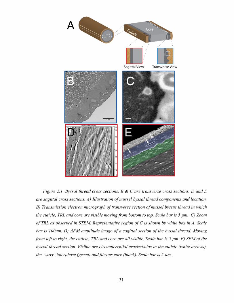
Ultrastructural characterization of distal byssus
Transverse cross-sections of distal byssal threads from local Mytilus californianus reveal
the three distinct regions: 1) cuticle, 2) TRL and 3) core. The granular structure of the
cuticle is readily visible with TEM and is characterized by the presence of ~500 nm (diam)
electro-lucent granules surrounded by an electron-dense protein matrix at a fill ratio of 1:1
(Figure 2.1.B) 8. Granules are the primary location of iron-catecholate complexes in the
byssal thread 4 and function to increase the abrasion resistance of the exterior coating and
prevent dehydration during times of exposure to atmosphere 8. The core of each byssal
thread consists of anisotropic microbundles of collagen fiber bundles oriented parallel to the
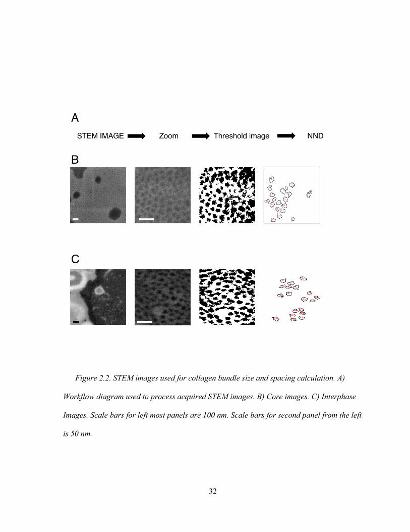
long axis of the thread (Figure 2.1.C,D,E). In this study, high resolution transmission
electron microscopy (STEM) was employed to measure an average diameter of 13.6 ± 1 nm
(Figure 2.2).
Sandwiched between the cuticle and core, an electron dense homogenous material is
discernable. We shall refer to this as the thiol rich layer (TRL). However, as revealed by
STEM, true homogeneity is complicated by interfacial penetration of TRL by collagen fibers
Figure 1C). Fibers within the TRL maintain a diameter similar to that of the core, 13.6 ± 0.7
nm (Figure 2.2.B,C). Furthermore, fibrillar center to center distance, indicative of collagen
packing density, remains unchanged; 22.3 and 23.7 nm for the TRL and core, respectively.
We interpret this to mean that collagen in the TRL is not significantly different from that of
the core.
31
Figure 2.1. Byssal thread cross sections. B & C are transverse cross sections. D and E
are sagittal cross sections. A) Illustration of mussel byssal thread components and location.
B) Transmission electron micrograph of transverse section of mussel byssus thread in which
the cuticle, TRL and core are visible moving from bottom to top. Scale bar is 5 µm. C) Zoom
of TRL as observed in STEM. Representative region of C is shown by white box in A. Scale
bar is 100nm. D) AFM amplitude image of a sagittal section of the byssal thread. Moving
from left to right, the cuticle, TRL and core are all visible. Scale bar is 5 µm. E) SEM of the
byssal thread section. Visible are circumferential cracks/voids in the cuticle (white arrows),
the ‘wavy’ interphase (green) and fibrous core (black). Scale bar is 5 µm.
32
Figure 2.2. STEM images used for collagen bundle size and spacing calculation. A)
Workflow diagram used to process acquired STEM images. B) Core images. C) Interphase
Images. Scale bars for left most panels are 100 nm. Scale bars for second panel from the left
is 50 nm.
33
In thin (<100nm) transverse sections, collagen bundles contained in the TRL appear to
maintain their anisotropy. However, imaging of sagittal cross sections demonstrates that
fibers in the TRL are more isotropic (Figure 2.1D). Furthermore, voids and cracks that form
in
the cuticle as the result of normal deformation become visible. Interestingly, these cracks are
deflected by the TRL and only propagate parallel to the long axis of the fibers (Figure
2.1.E).
Biochemical Characterization:
The byssal thread is formed by a processive injection molding of secretory vesicles from
two specific glands in the ventral groove of the foot 11. The collagen gland contains prolate
ellipsoid vesicles containing collagen mesophases, which self-assemble into mature collagen
fibers during molding of the core (Figure 2.3.A-E). The coating or cuticle is formed when
the accessory gland secretes spherical vesicles with a “marbled” internal structure 11 that
flow over and coat the nascent collagen core. Recently, in-depth analysis of the accessory
gland transcriptome revealed, at the mRNA level, the presence of four novel mussel foot
proteins (mfp’s); mfp -16, -17, -18, -19. This cohort of mfps contained cysteine levels
ranging from 7 to 20 mole percent12. By isolating cuticle and collagen vesicles from their
respective glands, we directly confirmed one of the aforementioned proteins (Figure 2.3.F).
A dispersion of granules prepared from the accessory gland was trypsinized and subjected to
ESI-TOF-MSMS. A positive identification of the sequence HDDYSSDPYTEPQK was
made to residues 85-99 of a 21-kDa protein, mfp-1712. Full cysteine-enriched sequence is
shown in Figure 2.3.H. Sequences for other proteins known to exist in the isolated secretory
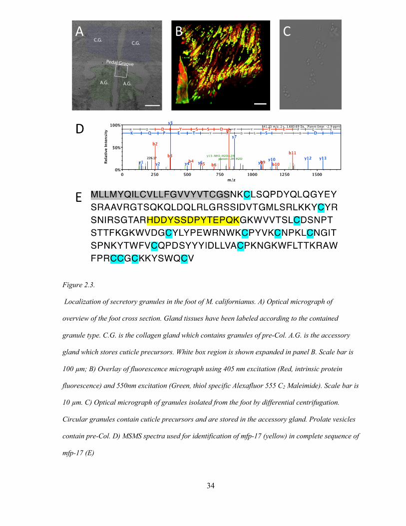
34
Figure 2.3.
Localization of secretory granules in the foot of M. californianus. A) Optical micrograph of
overview of the foot cross section. Gland tissues have been labeled according to the contained
granule type. C.G. is the collagen gland which contains granules of pre-Col. A.G. is the accessory
gland which stores cuticle precursors. White box region is shown expanded in panel B. Scale bar is
100 µm; B) Overlay of fluorescence micrograph using 405 nm excitation (Red, intrinsic protein
fluorescence) and 550nm excitation (Green, thiol specific Alexafluor 555 C2 Maleimide). Scale bar is
10 µm. C) Optical micrograph of granules isolated from the foot by differential centrifugation.
Circular granules contain cuticle precursors and are stored in the accessory gland. Prolate vesicles
contain pre-Col. D) MSMS spectra used for identification of mfp-17 (yellow) in complete sequence of
mfp-17 (E)
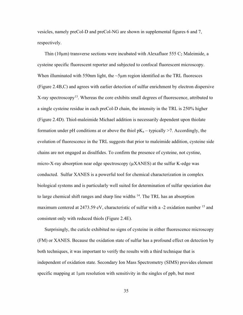
35
vesicles, namely preCol-D and preCol-NG are shown in supplemental figures 6 and 7,
respectively.
Thin (10µm) transverse sections were incubated with Alexafluor 555 C2 Maleimide, a
cysteine specific fluorescent reporter and subjected to confocal fluorescent microscopy.
When illuminated with 550nm light, the ~5µm region identified as the TRL fluoresces
(Figure 2.4B,C) and agrees with earlier detection of sulfur enrichment by electron dispersive
X-ray spectroscopy13. Whereas the core exhibits small degrees of fluorescence, attributed to
a single cysteine residue in each preCol-D chain, the intensity in the TRL is 250% higher
(Figure 2.4D). Thiol-maleimide Michael addition is necessarily dependent upon thiolate
formation under pH conditions at or above the thiol pKa – typically >7. Accordingly, the
evolution of fluorescence in the TRL suggests that prior to maleimide addition, cysteine side
chains are not engaged as disulfides. To confirm the presence of cysteine, not cystine,
micro-X-ray absorption near edge spectroscopy (µXANES) at the sulfur K-edge was
conducted. Sulfur XANES is a powerful tool for chemical characterization in complex
biological systems and is particularly well suited for determination of sulfur speciation due
to large chemical shift ranges and sharp line widths 14. The TRL has an absorption
maximum centered at 2473.59 eV, characteristic of sulfur with a -2 oxidation number 15 and
consistent only with reduced thiols (Figure 2.4E).
Surprisingly, the cuticle exhibited no signs of cysteine in either fluorescence microscopy
(FM) or XANES. Because the oxidation state of sulfur has a profound effect on detection by
both techniques, it was important to verify the results with a third technique that is
independent of oxidation state. Secondary Ion Mass Spectrometry (SIMS) provides element
specific mapping at 1µm resolution with sensitivity in the singles of ppb, but most
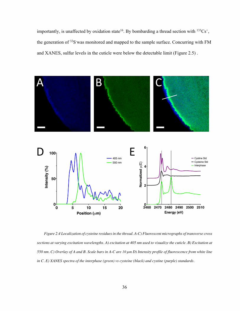
36
importantly, is unaffected by oxidation state16. By bombarding a thread section with 133Cs+,
the generation of 32S-was monitored and mapped to the sample surface. Concurring with FM
and XANES, sulfur levels in the cuticle were below the detectable limit (Figure 2.5) .
Figure 2.4 Localization of cysteine residues in the thread. A-C) Fluorescent micrographs of transverse cross
sections at varying excitation wavelengths. A) excitation at 405 nm used to visualize the cuticle. B) Excitation at
550 nm. C) Overlay of A and B. Scale bars in A-C are 10 µm D) Intensity profile of fluorescence from white line
in C. E) XANES spectra of the interphase (green) vs cysteine (black) and cystine (purple) standards.
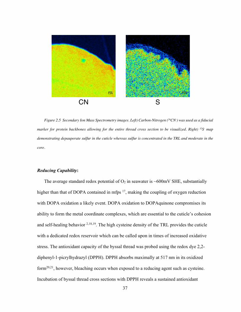
37
Figure 2.5 Secondary Ion Mass Spectrometry images. Left) Carbon-Nitrogen (26CN-) was used as a fiducial
marker for protein backbones allowing for the entire thread cross section to be visualized. Right) 32S- map
demonstrating depauperate sulfur in the cuticle whereas sulfur is concentrated in the TRL and moderate in the
core.
Reducing Capability:
The average standard redox potential of O2 in seawater is ~600mV SHE, substantially
higher than that of DOPA contained in mfps 17, making the coupling of oxygen reduction
with DOPA oxidation a likely event. DOPA oxidation to DOPAquinone compromises its
ability to form the metal coordinate complexes, which are essential to the cuticle’s cohesion
and self-healing behavior 2,18,19. The high cysteine density of the TRL provides the cuticle
with a dedicated redox reservoir which can be called upon in times of increased oxidative
stress. The antioxidant capacity of the byssal thread was probed using the redox dye 2,2-
diphenyl-1-picrylhydrazyl (DPPH). DPPH absorbs maximally at 517 nm in its oxidized
form20,21, however, bleaching occurs when exposed to a reducing agent such as cysteine.
Incubation of byssal thread cross sections with DPPH reveals a sustained antioxidant
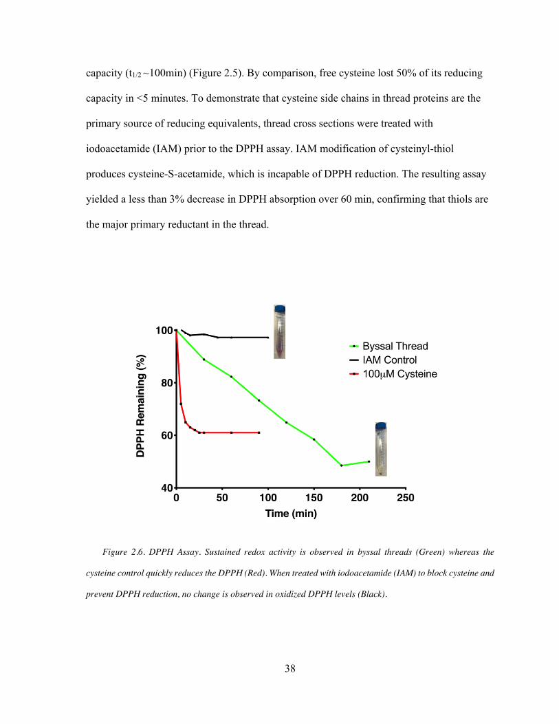
38
capacity (t1/2 ~100min) (Figure 2.5). By comparison, free cysteine lost 50% of its reducing
capacity in <5 minutes. To demonstrate that cysteine side chains in thread proteins are the
primary source of reducing equivalents, thread cross sections were treated with
iodoacetamide (IAM) prior to the DPPH assay. IAM modification of cysteinyl-thiol
produces cysteine-S-acetamide, which is incapable of DPPH reduction. The resulting assay
yielded a less than 3% decrease in DPPH absorption over 60 min, confirming that thiols are
the major primary reductant in the thread.
Figure 2.6. DPPH Assay. Sustained redox activity is observed in byssal threads (Green) whereas the
cysteine control quickly reduces the DPPH (Red). When treated with iodoacetamide (IAM) to block cysteine and
prevent DPPH reduction, no change is observed in oxidized DPPH levels (Black).
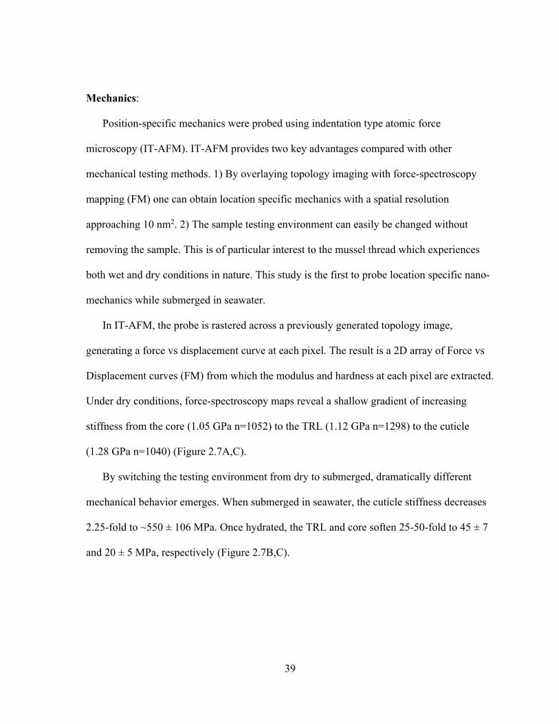
39
Mechanics:
Position-specific mechanics were probed using indentation type atomic force
microscopy (IT-AFM). IT-AFM provides two key advantages compared with other
mechanical testing methods. 1) By overlaying topology imaging with force-spectroscopy
mapping (FM) one can obtain location specific mechanics with a spatial resolution
approaching 10 nm2. 2) The sample testing environment can easily be changed without
removing the sample. This is of particular interest to the mussel thread which experiences
both wet and dry conditions in nature. This study is the first to probe location specific nano-
mechanics while submerged in seawater.
In IT-AFM, the probe is rastered across a previously generated topology image,
generating a force vs displacement curve at each pixel. The result is a 2D array of Force vs
Displacement curves (FM) from which the modulus and hardness at each pixel are extracted.
Under dry conditions, force-spectroscopy maps reveal a shallow gradient of increasing
stiffness from the core (1.05 GPa n=1052) to the TRL (1.12 GPa n=1298) to the cuticle
(1.28 GPa n=1040) (Figure 2.7A,C).
By switching the testing environment from dry to submerged, dramatically different
mechanical behavior emerges. When submerged in seawater, the cuticle stiffness decreases
2.25-fold to ~550 ± 106 MPa. Once hydrated, the TRL and core soften 25-50-fold to 45 ± 7
and 20 ± 5 MPa, respectively (Figure 2.7B,C).
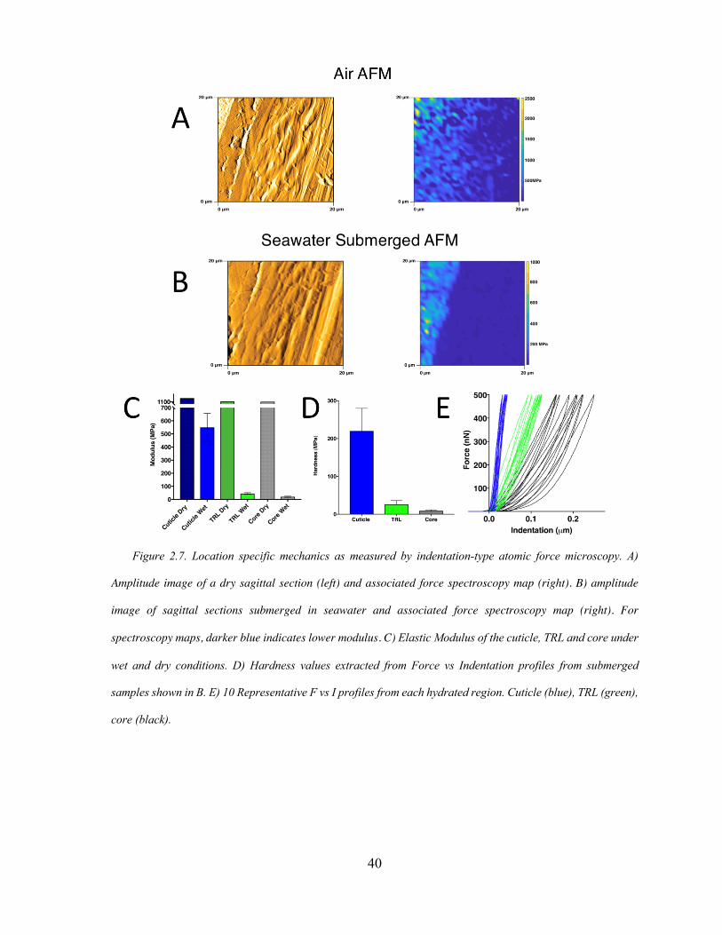
40
Figure 2.7. Location specific mechanics as measured by indentation-type atomic force microscopy. A)
Amplitude image of a dry sagittal section (left) and associated force spectroscopy map (right). B) amplitude
image of sagittal sections submerged in seawater and associated force spectroscopy map (right). For
spectroscopy maps, darker blue indicates lower modulus. C) Elastic Modulus of the cuticle, TRL and core under
wet and dry conditions. D) Hardness values extracted from Force vs Indentation profiles from submerged
samples shown in B. E) 10 Representative F vs I profiles from each hydrated region. Cuticle (blue), TRL (green),
core (black).
41
Functional gradients are designed to create high-performance materials by confining and
insulating specific compositions and architectures to regions of need 22. As is the case with
most core-shelled fibers, the shell is usually designated as a protective outer coating.
Consistent with this, the mussel cuticle’s stiff granular structure has a hardness (H) of 220 ±
60 MPa (Figure 2.7D). Using Ashby plots of critical properties, the abrasion resistance of a
material can be comparatively assessed by plotting H vs E. Materials which fall on a straight
line of slope 2/3 have equivalent performance and can be grouped according to their H3/E2
23. The cuticle ‘H’ and ‘E’ yield a H3/E2 of 35 MPa, putting it in the realm of most
engineering epoxies. In the TRL, both E and H decrease by an order of magnitude to 26 ±10
MPa. However, the resulting H3/E2 is 9MPa, only a 4-fold decrease from the cuticle. Given
that the core’s primary function is to dissipate tensile energy, the need for hardness is not
readily apparent. Fittingly, the measured hardness of the core is 9MPa, with a H3/E2 of 1.5
MPa.
E. Conclusion:
Prior studies have classified the byssal thread as a coated, fiber-filled composite material
with distinct shell and core domains. By combining microscopy, spectroscopy and
mechanical analysis techniques we introduce a third, multi-functional thiol rich layer. The
TRL is an approximately 5 µm thick region intercalated between the cuticle and core and
characterized by an increase in cysteine density compared with the two adjoining domains.
Additionally, we show that cysteine residues in the TRL persist in their reduced state.
Cysteine clustering has been shown in a number of proteins and often increases in proteins
designed to deal with high oxidative stress 24, such as that experienced by the byssus. Given

42
that the mechanical performance of the cuticle necessarily relies on Dopa’s ability to form
tris-Dopa-iron complexes, which deteriorate upon Dopa oxidization, we propose that the
TRL provides a reservoir of electrons to create a sustained reducing environment to protect
the cuticle against oxidative damage. Despite being submerged in seawater for 24-48 hours,
the threads used for these studies indicated no cysteine oxidation. Further studies to
investigate the aging process of the TRL are underway. Given its thiol rich nature, the
formation of a variety of covalent bonds including disulfide and cysteinyl-dopa25 adducts are
possible over time. The penetration of collagen fibers into this dense thiol-rich matrix
creates a mechanically distinct barrier between the cuticle and the core. The inclusion of this
interlayer forms a step gradient in hardness and abrasion resistance from the cuticle to the
core which helps mitigate contact damage. Furthermore, composite structures of stiff outer
materials underlaid by a gradient of compliant materials favor superficial circumferential
cracking as opposed to deep penetrating radial cracking 22. Prevention of radial cracking
prevents cracks from entering the core, which would result in catastrophic failure under
tension. The multifunctionality of the TRL addresses two major problem areas of the mussel
byssus; oxidation and mechanical mismatch. By further understanding the role of redox
chemistry at biological interfaces, we gained valuable knowledge, which may lead to smart
design principles for layered load bearing materials.
43
F. References
(1) Beaugendre, A.; Degoutin, S.; Bellayer, S.; Pierlot, C.; Duquesne, S.; Casetta, M.; Jimenez, M. Self-Stratifying Coatings: a Review. Progress in Organic Coatings 2017, 110, 210–241.
(2) Holten-Andersen, N.; Waite, J. H. Mussel-Designed Protective Coatings for Compliant Substrates. J. Dent. Res. 2008, 87 (8), 701–709.
(3) Taylor, S. W.; Chase, D. B.; Emptage, M. H.; Nelson, M. J.; Waite, J. H. Ferric Ion Complexes of a DOPA-Containing Adhesive Protein From Mytilus Edulis. Inorg. Chem. 1996, 35 (26), 7572–7577.
(4) Harrington, M. J.; Masic, A.; Holten-Andersen, N.; Waite, J. H.; Fratzl, P. Iron-Clad Fibers: a Metal-Based Biological Strategy for Hard Flexible Coatings. Science 2010, 328 (5975), 216–220.
(5) Holten-Andersen, N.; Fantner, G. E.; Hohlbauch, S.; Waite, J. H.; Zok, F. W. Protective Coatings on Extensible Biofibres. Nat Mater 2007, 6 (9), 669–672.
(6) Xu, Z. Mechanics of Metal-Catecholate Complexes: the Roles of Coordination State and Metal Types. Scientific Reports 2013, 3 (1), 216–217.
(7) Cooper, L. H. N. OXIDA TION-REDUCTION POTENTIAL in SEA WATER by L. H. N. Cooper, Ph.D., F.I.C. https://doi.org/10.1017/S0025315400011929 2008, 1–10.
(8) Monnier, C. A.; DeMartini, D. G.; Waite, J. H. Intertidal Exposure Favors the Soft-Studded Armor of Adaptive Mussel Coatings. Nature Communications 2018, 1–9.
(9) Hassenkam, T.; Gutsmann, T.; Hansma, P.; Sagert, J.; Waite, J. H. Giant Bent-Core Mesogens in the Thread Forming Process of Marine Mussels. Biomacromolecules 2004, 5 (4), 1351–1355.
(10) Krauss, S.; Metzger, T. H.; Fratzl, P.; Harrington, M. J. Self-Repair of a Biological Fiber Guided by an Ordered Elastic Framework. Biomacromolecules 2013, 14 (5), 1520–1528.
(11) Priemel, T.; Degtyar, E.; Dean, M. N.; Harrington, M. J. Rapid Self-Assembly of Complex Biomolecular Architectures During Mussel Byssus Biofabrication. Nature Communications 2017, 8, 1–12.
(12) DeMartini, D. G.; Errico, J. M.; Sjoestroem, S.; Fenster, A.; Waite, J. H. A Cohort of New Adhesive Proteins Identified From Transcriptomic Analysis of Mussel Foot Glands. J. R. Soc. Interface 2017, 14 (131), 20170151–14.
(13) Sun, C.; Waite, J. H. Mapping Chemical Gradients Within and Along a Fibrous Structural Tissue, Mussel Byssal Threads. Journal of Biological Chemistry 2005, 280 (47), 39332–39336.
(14) Hullebusch, E. V.; Rossano, S.; Farges, F.; Lenz, M.; Labanowski, J.; Lagarde, P.; Flank, A.-M.; Lens, P. Sulfur K-Edge XANES Spectroscopy as a Tool for Understanding Sulfur Chemical State in Anaerobic Granular Sludge. J. Phys.: Conf. Ser. 2009, 190, 012184–012185.
(15) Almkvist, G.; Boye, K.; Persson, I. K-Edge XANES Analysis of Sulfur
Compounds: an Investigation of the Relative Intensities Using Internal
44
Calibration. J. Synchrotron Rad (2010). 17, 683-688 [doi:10.1107/S0909049510022946] 2010, 1–6.
(16) Ray, S.; Shard, A. G. Secondary Ion Mass Spectrometry of Proteins. International Journal of Physical and Mathmatical Sciences 2011, 5 (4), 552–556.
(17) Waite, J. H. Mussel Adhesion - Essential Footwork. J. Exp. Biol. 2017, 220 (Pt 4), 517–530.
(18) Li, Y.; Wen, J.; Qin, M.; Cao, Y.; Ma, H.; Wang, W. Single-Molecule Mechanics of Catechol-Iron Coordination Bonds. ACS Biomater. Sci. Eng. 2017, 3 (6), 979–989.
(19) Zeng, H.; Hwang, D. S.; the, J. I. P. O.; 2010. Strong Reversible Fe3+-Mediated Bridging Between Dopa-Containing Protein Films in Water. National Acad Sciences.
(20) Nicklisch, S. C. T.; Waite, J. H. Optimized DPPH Assay in a Detergent-Based Buffer System for Measuring Antioxidant Activity of Proteins. MethodsX 2014, 1, 233–238.
(21) Shimamura, T.; Sumikura, Y.; Yamazaki, T.; Tada, A.; Kashiwagi, T.; Ishikawa, H.; Matsui, T.; Sugimoto, N.; Akiyama, H.; Ukeda, H. Applicability of the DPPH Assay for Evaluating the Antioxidant Capacity of Food Additives - Inter-Laboratory Evaluation Study -. Anal Sci 2014, 30 (7), 717–721.
(22) Liu, Z.; Meyers, M. A.; Zhang, Z.; Ritchie, R. O. Functional Gradients and Heterogeneities in Biological Materials: Design Principles, Functions, and Bioinspired Applications. Progress in Materials Science 2017, 88, 467–498.
(23) Moses, D. N.; Mattoni, M. A.; Slack, N. L.; Waite, J. H.; Zok, F. W. Role of Melanin in Mechanical Properties of Glycera Jaws. Acta Biomaterialia 2006, 2 (5), 521–530.
(24) Poole, L. B. The Basics of Thiols and Cysteines in Redox Biology and Chemistry. Free Radical Biology and Medicine 2015, 80, 148–157.
(25) Nicklisch, S. C. T.; Spahn, J. E.; Zhou, H.; Gruian, C. M.; Waite, J. H. Redox Capacity of an Extracellular Matrix Protein Associated with Adhesion in Mytilus Californianus. Biochemistry 2016, 55 (13), 2022–2030.
45
Chapter III
Phase-separated Redox Batteries in Mussel Adhesion
Reproduced in part with permission from: Valois, E. and Waite, J.H. (2019). Phase-
separated Redox Batteries in Mussel Adhesion. Science Advances. Article ASAP. DOI:
XXXXXXXXXXXX.
A. Abstract
Catecholic 3, 4-dihydroxyphenylalanine (Dopa) residues in mussel foot proteins (mfp)
contribute critically to mussel (Mytilus californianus) plaque adhesion, but only if protected
from oxidation at the adhesive-substratum interface. Dopa oxidation is thermodynamically
favorable in seawater yet barely detectable in plaques, therefore we investigated how
plaques insulate Dopa-containing mfps against oxidation. Seawater sulfate triggers a mfp3
and mfp6 liquid-liquid phase separation (LLPS). By combining plaque cyclic voltammetry
with electrophoresis, mass spectrometry and redox exchange chemistry, we show that Dopa-
containing mfp3 and mfp6 in phase-separated droplets remain stable despite rapid oxidation
in the equilibrium solution. The results suggest that a cohort of oxidation-prone proteins is
endowed with phase-dependent redox stability. Moreover, in forming LLPS compartments,
Dopa-proteins become reservoirs of chemical energy.
46
B. Introduction
Liquid-liquid phase changes in biomacromolecular assemblages are not confined to
living cells where they facilitate dynamic compartmentalization associated with time- and
location-specific reactions (1). A common extracellular and extra-organismic liquid-liquid
phase change known as coacervation is involved in the formation of biomolecular materials
as diverse as arteries (2), silks (3), beaks (4), butterfly wings (5), and marine adhesives (6,7).
The formation of adhesive plaques by mussels (Mytilus californianus) involves a cohort of
mostly disordered proteins known as mussel foot proteins (mfps) (8). Several mfps exhibit
liquid-liquid phase separation (LLPS) (7, 9-11) triggered by specific chemical cues in the
ambient seawater. Adaptive advantages of coacervation include concentrating adhesive
proteins as fluids, shear thinning to facilitate fluid transport, and low interfacial energy for
spontaneous spreading underwater (4, 9, 12-13).
An iconic chemical feature of all mfps is 3, 4-dihydroxyphenyl-L-alanine (Dopa),
a catecholic amino acid that is post- or co-translationally introduced into the proteins prior to
deposition onto target surfaces. Dopa contributes significantly to wet adhesion but only
when redox poise is locally controlled. Under reducing conditions (Fig. 3.1), Dopa-
containing proteins dehydrate surfaces, form stable π-cation interactions and bidentate H-
bonds, coordinate with metal ions and metal oxide surfaces, and, in synergy with lysine,
evict adsorbed salts from rock surfaces (14, 15). Under oxidizing conditions, Dopa becomes
a quinone with strong cross-linking tendencies, but metal oxide/mineral surface binding and
metal ion complexation are significantly diminished (15) (Fig. 3.1). Known interim
adaptations to reduce oxidative losses, including electron-withdrawing effects on the
catechol moiety (16), surface deposition of mfps at acidic pH (17-18) and cosecretion of
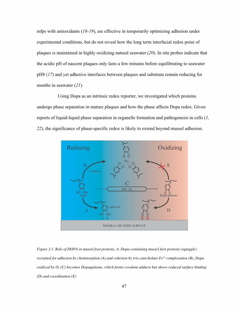
47
mfps with antioxidants (18-19), are effective in temporarily optimizing adhesion under
experimental conditions, but do not reveal how the long term interfacial redox poise of
plaques is maintained in highly oxidizing natural seawater (20). In situ probes indicate that
the acidic pH of nascent plaques only lasts a few minutes before equilibrating to seawater
pH8 (17) and yet adhesive interfaces between plaques and substrata remain reducing for
months in seawater (21).
Using Dopa as an intrinsic redox reporter, we investigated which proteins
undergo phase separation in mature plaques and how the phase affects Dopa redox. Given
reports of liquid-liquid phase separation in organelle formation and pathogenesis in cells (1,
22), the significance of phase-specific redox is likely to extend beyond mussel adhesion.
Figure 3.1. Role of DOPA in mussel foot proteins. A. Dopa-containing mussel foot proteins (squiggle)
recruited for adhesion by chemisorption (A) and cohesion by tris-catecholato Fe3+ complexation (B). Dopa
oxidized by O2 (C) becomes Dopaquinone, which forms covalent adducts but shows reduced surface binding
(D) and coordination (E)
48
C. Materials and Methods.
Plaque collections.
California mussels Mytilus californianus Conrad harvested from Goleta Pier (34.414088,
-119.828690) were held in tanks with circulating natural seawater at 12-16˚C. To prepare the
intact mussel plaques for analysis (Figure 1b), the mussels were tethered over target
substrates such as mica coated with nanometer thick gold films, mica, glass slides or, for
easy sectioning, Saranä wrap. In the case of gold plated mica, the gold layer remained
firmly attached to the plaque but was easily detached from mica surface by gentle tension on
the plaque. This approach keeps intact the interfacial plaque chemistry and structure.
Transmission electron microscopy of phase transitions.
All plaques intended for TEM were collected from the same individual and deposited
onto Saranä wrap. Half were processed directly for electron microscopy after optimizing
the fixation conditions. Half were placed in 5% acetic acid (15˚C) for 30 min before
processing. All fixation and washing steps were performed on ice. Fresh plaques were fixed
in 2% formaldehyde, 2.5% glutaraldehyde, and fixation buffer (200mM sodium cacodylate,
300mM NaCl, pH 7.2) for 2 h. The samples were washed three times (10 min each) in
degassed fixation buffer and then post-fixed in 2% osmium tetroxide in degassed fixation
buffer for 2 h. The samples were then washed 4 times (10 min each) in degassed deionized
water and then dehydrated through a graded series of ethanol washes (25, 50, 75, 90, 100,
100, 100% ethanol, 10 min each). Solvent was then switched to propylene oxide by washing
in 33, 66, 100, 100, 100% propylene oxide in ethanol. The samples were then infiltrated
with epoxy resin (Embed812, Electron Microscopy Sciences, Hatfield, USA) incubating the
49
sample in resin diluted in propylene oxide as follows: 33% (2 h), 66% (16 h), and 100% (4
h). Finally, samples were placed in molds and cured at 60 °C for 24 h. Thin sections (60–80
nm) for TEM and semithin sections (300 nm) for electron tomography were cut on a EM
UC6 ultramicrotome (Leica Biosystems). Sections were mounted on copper TEM grids and
post-stained on drops of uranyl acetate and lead citrate following standard protocols (30).
All samples were investigated with a Tecnai G2 transmission electron microscope (FEI)
operating at 200 kV, and micrographs were recorded with a Gatan Ultrascan CCD camera
(2048 Å~ 2048 pixels).
Cyclic Voltammetry
Cyclic voltammetry was performed at room temperature on a Versastat 3 Potentiostat
(Ametek Co) with a typical three-electrode cell using Pt wire as the counter electrode,
Ag/AgCl reference electrode, and gold or glassy carbon (3mm disc electrodes from BASi,
W Lafayette, IN) as the working electrode at 22˚C and 1 ATM pressure. To avoid
interference by adsorbed chemicals on the surface of the working electrode, the following
cleaning method was applied to gold electrodes before each experiment: 1) mechanical
polishing of the electrode was done using a diamond particle abrasion solution of 1µm,
0.25µm and 0.05 µm particle sizes (Buehler, Lake Bluff, IL) for 2min each on a nylon disk.
The electrode was rinsed with water and sonicated for 2min in a shallow vessel containing
water to remove excess diamond particles; 2) The electrode was then stripped by repetitive
cycling between -400 to +1400 mV (vs. Ag/AgCl) at a rate of 100 mV/s in 50 mM sulfuric
acid until the CV became stable (typically 12 cycles). For abrasive polishing of glassy
carbon electrodes, diamond particles were replaced with alumina particles of equivalent size.
50
Analyte autoxidation was minimized by performing all electrochemical experiments with
degassed deionized water in a glove-bag under argon. Some CV measurements required
notable adaptations: for detached plaques, for example, we used dialysis membranes
(Spectrapor MWCO 1000) with 2 O-rings to affix the plaque interface against the working
electrode. For reconstituted coacervates, the reference electrode was enclosed in a sleeve of
dialysis tubing. Without it, adsorbed coacervates caused significant drift.
Electro-transfer of Plaque Proteins. To capture proteins undergoing a phase change
from a condensed state to solution, plaques naturally deposited by mussels onto glass slides
(Fisher Scientific 12-544-7) were collected by severing the connecting distal thread within
24 h of deposition. While attached to the slide, each plaque was rinsed for 60 sec with 18
megohm pure water (MilliPore). Following rinsing, plaques were incubated in either 5%
acetic acid or filtered sea water for 30 min at room temperature with shaking at 45 rpm.
Seawater treated plaques required additional washing step post treatment to remove excess
salts, which interfere with the electrophoretic process. These plaques were washed 3 X
@min each with transfer buffer. Following incubation, plaques were peeled from the slide
with a clean single edge razor blade. Excised plaques were placed between two 0.45-micron
nitrocellulose membranes (NitroPure WP4HY08250) and loaded into a semidry transfer cell
(BioRad Transblot SD #1703940). 5% acetic acid conditioned plaques were electrophoresed
in 0.7% acetic acid at 12 volts for 20 min. Seawater conditioned plaques were
electrophoresed in 121mM Tris-glycinate pH 8.2 at 12 volts for 20 min. Although the
direction of electrophoresis was initially performed in both directions, soluble components
51
migrated only toward the cathode. As a result, the plaque-substrate interface must face the
cathode during acidic transfers and the anode during basic transfers.
Mass spectrometry
Proteins transferred to nitrocellulose membranes were investigated in situ by MALDI-
TOF MS. For post-electroblotting transfer, the membrane was cut around the perimeter of
the plaque footprint using a clean #11 scalpel. With fine nose tweezers, the plaque was
removed from the membrane, which is subsequently affixed to a MSP 96 MALDI Target
(Bruker) with adhesive carbon tape. 10 µL of MALDI-matrix, a saturated solution of α-
cyano-4-hydroxycinnamic acid in 30% acetonitrile 1% trifluoroacetic acid, was applied to
the membrane and dried in a vacuum desiccator (Nalgene). Mass spec analysis was
conducted using a Microflex LRF MALDI-TOF (Bruker). Sample membranes were
irradiated using a Nitrogen laser at 337 nm and a pulse length of 3ns with a repetition rate of
20 Hz. Detection occurred in linear mode between 2-20kDa at sampling rate of 1 Gs/s.
Protein Calibration Standard 1 (Bruker #206355) was used as an internal calibration.
Oxidative damage to proteins was assessed by mass spectrometry. Plaques less than 48 h
old were homogenized in 10mM acetic acid at a ratio of ~50mg wet weight to 1 mL acetic
acid using a Dounce homogenizer. The homogenate was clarified via centrifugation at
5000xg for 5 min after which the soluble supernatant was carefully separated from the
insoluble fraction. The concentration of protein was assessed via the Bradford assay kit
(Thermo Fisher Pierce) and adjusted to 1 mg/mL with 10mM acetic acid. The solution was
aliquoted into 5 samples and adjusted to assay conditions using the following stock buffers.
52
1) 10mM acetic acid 2) 10 mM acetic acid + 10mM sodium metaperiodate (Sigma Aldrich) 3) 50 mM Tris pH 8.0 4) 50 mM Tris pH 8.0 (coacervate) 5) 10 mM acetic Acid (coacervate) + 10mM sodium metaperiodate Oxidation was tracked by acquiring spectra as time elapsed. The sample was mixed with
α-cyano-4-hydroxycinnamic acid (HCCA) in 50% (v/v) aqueous acetonitrile with 0.1%
trifluoroacetic acid at a ratio 1:9 and spotted onto a stainless steel MALDI target.
Amino acid analysis of electro-transferred footprints
Electro-transfer was performed as above except that NitroPure membranes were
replaced with PVDF membranes (Immobilon IPVH00005). This change avoided
nitrocellulose degradation in 6N HCl. Five blotted footprints were excised from the excess
membrane and loaded into a 1mL glass ampule (Wheaton Glass, #651502) and submerged
with 250 μL of constant boiling 6N HCl with 10 μL phenol. Acid hydrolysis of electro-
transferred proteins was done at 110°C for 24 h under vacuum. After 24 hours the sample
was removed from the vial and loaded into a LoBind microcentrifuge tube (Eppendorf,
#Z666505). The hydrolysis ampule was washed with 500μL X2 of 18 megohm pure water to
ensure all amino acids were transferred to the microcentrifuge tube. The now dilute HCl was
removed via flash evaporation with a SpeedVac vacuum concentrator with vapor trap
(Savant, Holbrook, NY). Once dried, amino acids were resuspended in 200 μL of 0.01N HCl
and loaded into the automated amino acid analyzer (Hitachi High-Technologies model L-
8900).
53
Polyacrylamide Gel Electrophoresis of Plaque Extracts
Proteins can be electrokinetically extracted from single plaques for microanalysis by
mass spectrometry and amino acid analysis, but these quantities are insufficient to see on a
gel or to coacervate. For gels, plaques (<24 h) old were collected and pooled for bulk
homogenization at a ratio of ~50mg wet weight to 1 mL of either 0.7% acetic acid pH3 or
121mM Tris-glycinate pH 8.2 to match electro-transfer conditions. The homogenate was
clarified via centrifugation at 5000g for 10 min at which point the supernatant solution was
carefully separated from the insoluble fraction. The pH 8.2 homogenate was then dialyzed
against 0.7% acetic acid >4h at 4°C. Both homogenates were mixed 1:1 with gel loading
buffer (5% Acetic acid 8M Urea) (31) and 10μL was loaded onto a preequilibrated 7.5%
Acid-Urea gel (10 mA for 2 hours). The gel was run with 5% acetic acid serving as the tank
buffer in a RioRad Mini Protean Tetra Cell at a constant current of 12 mA for 30min using a
GE EPS 301 power supply. Post electrophoresis, the gel was stained with 0.1% CBB G-250
in 40% methanol 20% acetic acid for 30 min and destained with 20% methanol 10% acetic
acid until the gel background became completely transparent.
Analyses of bulk extracted plaque proteins.
Thiol and Dopa Quantification
Because Dopa and Cysteine are important to protein redox these functionalities were
independently quantified for comparison to amino acid analysis. Plaques (<48 h old) were
homogenized in 10mM acetic acid at a ratio of ~50mg wet weight to 1 mL acetic acid using
a Dounce homogenizer on ice. The homogenate was clarified via centrifugation at 5000xg
for 10 min effectively separating the soluble supernatant from the insoluble pellet. A stock

54
of Ellman’s Reagent (Thermo Fisher) was made 4mg/mL in dimethyl sulfoxide. Guanidine-
HCl (Fisher) was dissolved in reaction buffer (RB, 0.1M HEPES, 1mM EDTA pH 8) to a
final concentration of 6M. 107μL of RB was added to 30 μL guanidine and 40μL
homogenate and mixed via aspiration to ensure homogeneity of the solution. 10 μL of stock
Ellman’s Reagent was added to the mixture and allowed to react for 15 min prior to
measuring the absorbance at 412 nm (33). Concentration of thiols in the homogenate was
calculated from a standard curve of cysteine-HCl monohydrate. The standard curve was
created by make 2-fold serial dilutions of cysteine from 2mM to 0.08mM. Dopa was
quantified in aliquots of the 10mM extract supernatants using an adaptation of the Arnow
assay (34) with L-Dopa or 4-methyl catechol as calibration standards.
X-Ray Photoelectron Spectroscopy (Sulfate Identification)
Sulfate-mediated coacervation of mfps in plaques is not biologically relevant unless
sulfate levels are elevated at the plaque interface. A two-component 5-min epoxy was mixed
and deposited into a 6 x 6 mm square face mold (Electron Microscopy Sciences, Hatfield,
PA). A fresh mussel plaque was excised from the mussel using a clean single edge razor and
mounted with the thread down (plaque interface up) into the exposed underside of partially
cured epoxy. Sulfate was detected via X-ray photoelectron spectroscopy using a Thermo
Fisher ESCALAB XI spectrometer with Al Kα X–ray source. An overview spectrum was
gathered from 700 to 0 eV with a 0.5 eV step size and 100 ms dwell time. A high-resolution
spectrum of the sulfur K-edge was then collected from 175 to 144 eV with a 0.1 eV step size
and 250ms dwell time and summed over 6 scans. Sulfate was identified using the Casa
software, fitting the S 2p3/2 with a Gaussian function centered at 168.12 eV with a full width
55
height mean of 1.35 eV separated from 2p1/2 by 1.18 eV (169.3eV) constrained to an
intensity ratio of 2:1.
Redox assays using diphenylpicrylhydrazyl radical (DPPH)
DPPH (redox potential = +0.77V SHE see Table S3) is a redox dye that can be used to
quantify a wide variety of reducing compounds with lower redox potentials. Plaques (< 48 h
old) were collected and homogenized in 10mM acetic acid at ~50mg wet weight per 1 mL
acetic acid using a Dounce homogenizer. The homogenate was clarified via sedimentation at
5000xg for 5 min after which the soluble supernatant was carefully decanted from the
insoluble fraction. The concentration of protein was determined by the Bradford assay and
adjusted to 1 mg/mL. The redox capacity of the solution was assessed using a published
DPPH assay protocol (32). 50µL of protein solution was added to 900 µL of assay buffer
(100 mM HEPES pH 7.4). For coacervate assays, an aliquot of 1M Na2SO4 stock was added
to a final concentration of 30mM to solutions containing 1 mg/mL soluble protein and
mixed with assay buffer by rapid in-out pipetting. To begin the redox reaction, 50 µL of
2mM stock DPPH (Sigma Aldrich) solution (in methanol) was added to the protein
containing solution. The absorbance at 517nm was monitored as a function of time. The
percentage of oxidized DPPH reagent remaining as a function of time was calculated as %
DPPH remaining = (Abssample,t/Abscontrol,t x 100).
Reconstituted coacervation and labeling of plaque proteins
Coacervation requires mg amounts of protein particularly for doing coacervate
electrochemistry and redox assays with DPPH. In vitro coacervation of plaque protein
56
extracts was effected by sulfate addition, which Bungenberg de Jong referred to as “complex
coacervation” between microanions and macrocations. Notably, two groups have
investigated on the phase behavior of recombinant mfp-3 variants coacervated by
microanions such as sulfate, phosphate and citrate (9, 10). At a protein concentration of
1mg/ml, both reported a coacervate phase boundary at 20-40 mM sulfate, which
approximates the sulfate concentration in seawater. 100 mg wet weight of plaques were
collected and rinsed 3X 1min with de-ionized water and then 1X with marine phosphate
buffered saline (mPBS). Plaques were homogenized in 10mM acetic acid at a ratio of
100mg/1mL. The homogenate was clarified via centrifugation at 5000g for 10min. The
resulting clarified homogenate was filtered with a 0.22 µm filter and adjusted to 1mg/mL
protein with 10mM acetic acid as necessary. Coacervation was induced by adding stock 1M
sodium sulfate to a final concentration of 30 mM. Confocal images of phase separation were
collected using a Leica SP8 confocal microscope (Leica Microsystems) fitted with
63X/1.4NA oil immersion objective and hybrid detectors (HyD). Coacervates were
visualized via the intrinsic fluorescence of mfp3 excited with a 405 nm laser (7). In a
parallel experiment in coacervating mfps in which two distinct fluorophores are present,
rinsed plaques were then incubated in 0.05 µM Alexa Fluor555 C2 maleimide (Thermo Fisher
Scientific, Waltham, MA) in mPBS at room temp for 2 hours while rotating on a mixer.
Alexa Fluor555 C2 maleimide specifically targets Cys residues which are limited to mfp6.
Following incubation, plaques were rinsed 3X 1 min with 10 mL of deionized water to
remove excess fluorophore. Alexa Fluor555 stained coacervates were visualized using the
white light laser tuned to 550 nm excitation.
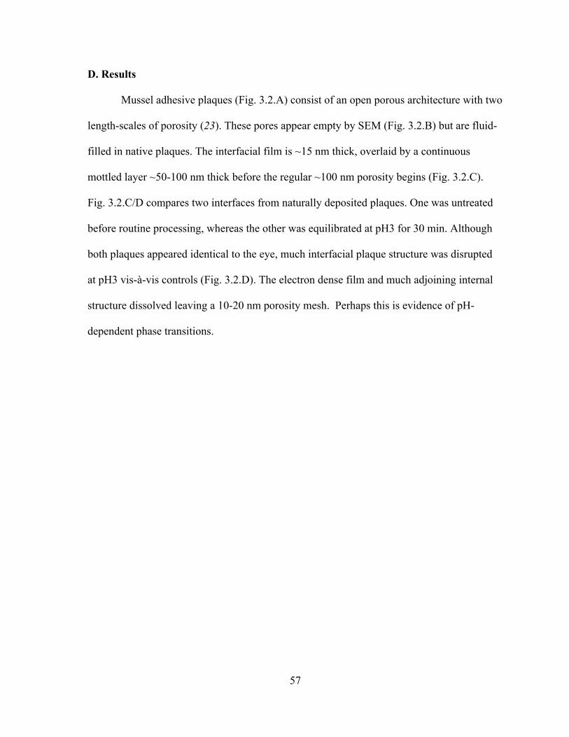
57
D. Results
Mussel adhesive plaques (Fig. 3.2.A) consist of an open porous architecture with two
length-scales of porosity (23). These pores appear empty by SEM (Fig. 3.2.B) but are fluid-
filled in native plaques. The interfacial film is ~15 nm thick, overlaid by a continuous
mottled layer ~50-100 nm thick before the regular ~100 nm porosity begins (Fig. 3.2.C).
Fig. 3.2.C/D compares two interfaces from naturally deposited plaques. One was untreated
before routine processing, whereas the other was equilibrated at pH3 for 30 min. Although
both plaques appeared identical to the eye, much interfacial plaque structure was disrupted
at pH3 vis-à-vis controls (Fig. 3.2.D). The electron dense film and much adjoining internal
structure dissolved leaving a 10-20 nm porosity mesh. Perhaps this is evidence of pH-
dependent phase transitions.
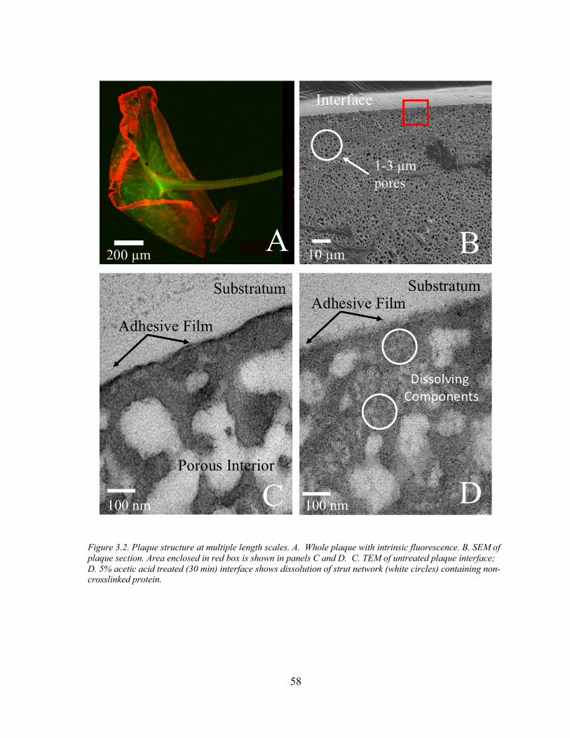
58
Figure 3.2. Plaque structure at multiple length scales. A. Whole plaque with intrinsic fluorescence. B. SEM of plaque section. Area enclosed in red box is shown in panels C and D. C. TEM of untreated plaque interface; D. 5% acetic acid treated (30 min) interface shows dissolution of strut network (white circles) containing non-crosslinked protein.
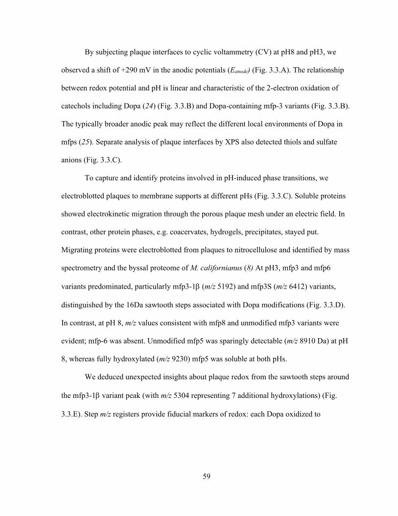
59
By subjecting plaque interfaces to cyclic voltammetry (CV) at pH8 and pH3, we
observed a shift of +290 mV in the anodic potentials (Eanode) (Fig. 3.3.A). The relationship
between redox potential and pH is linear and characteristic of the 2-electron oxidation of
catechols including Dopa (24) (Fig. 3.3.B) and Dopa-containing mfp-3 variants (Fig. 3.3.B).
The typically broader anodic peak may reflect the different local environments of Dopa in
mfps (25). Separate analysis of plaque interfaces by XPS also detected thiols and sulfate
anions (Fig. 3.3.C).
To capture and identify proteins involved in pH-induced phase transitions, we
electroblotted plaques to membrane supports at different pHs (Fig. 3.3.C). Soluble proteins
showed electrokinetic migration through the porous plaque mesh under an electric field. In
contrast, other protein phases, e.g. coacervates, hydrogels, precipitates, stayed put.
Migrating proteins were electroblotted from plaques to nitrocellulose and identified by mass
spectrometry and the byssal proteome of M. californianus (8) At pH3, mfp3 and mfp6
variants predominated, particularly mfp3-1b (m/z 5192) and mfp3S (m/z 6412) variants,
distinguished by the 16Da sawtooth steps associated with Dopa modifications (Fig. 3.3.D).
In contrast, at pH 8, m/z values consistent with mfp8 and unmodified mfp3 variants were
evident; mfp-6 was absent. Unmodified mfp5 was sparingly detectable (m/z 8910 Da) at pH
8, whereas fully hydroxylated (m/z 9230) mfp5 was soluble at both pHs.
We deduced unexpected insights about plaque redox from the sawtooth steps around
the mfp3-1b variant peak (with m/z 5304 representing 7 additional hydroxylations) (Fig.
3.3.E). Step m/z registers provide fiducial markers of redox: each Dopa oxidized to
60
Figure 3.3 Chemical analysis of plaque interfaces. (A) Dependence of catechol redox on pH, CV plaque, pH 3 (5% acetic acid, Black) and pH 8 (seawater, Red) showing anodic peak potentials. Glassy carbon electrode at 50 mV/s scan rate. (B) Cyclic voltammetry of purified mfp3.1b and mfp3-4 (S variant) (0.1 µM) at pH3. Gold working electrode and 100 mV/s scan rate at 20 ˚C and 1 ATM. Inset, plot of Ea vs pH in plaques is linear according to the Nernst equation for reversible redox reactions that involve H+ and e-. Red data points were measured potentials whereas the others were predicted from the pH 3 and 8 endpoints. (C) XPS of plaque foot print on glass with sulfur speciation as thiol and sulfate peaks. (D) Electroblotting strategy used to move soluble proteins: plaque is positioned with adhesive interface facing nitrocellulose (NC) membrane and the cathode. NC was soaked in matrix, dried, and subjected to mass spectrometry. (E) Mass spectrum of electrokinetic proteins at pH 8 (red, tris glycinate) and pH 3 (black acetic acid). Top Inset,enlarged m/z of mfp3-1b showing mass register with mfp3-1b standard (purple). Bottom inset extends m/z range to 13,000.
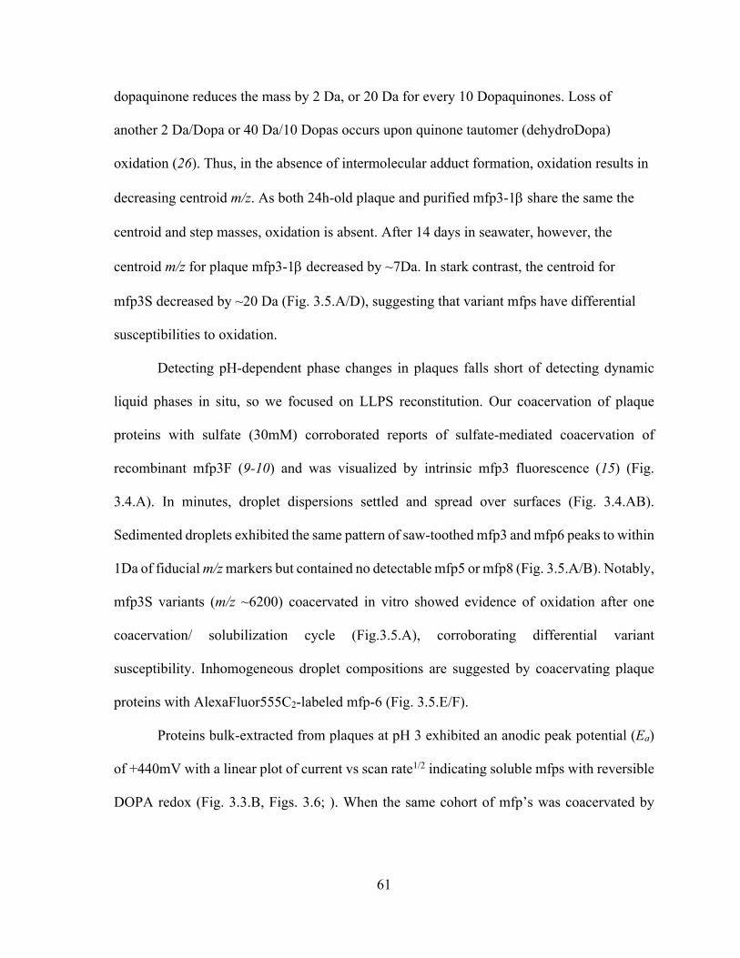
61
dopaquinone reduces the mass by 2 Da, or 20 Da for every 10 Dopaquinones. Loss of
another 2 Da/Dopa or 40 Da/10 Dopas occurs upon quinone tautomer (dehydroDopa)
oxidation (26). Thus, in the absence of intermolecular adduct formation, oxidation results in
decreasing centroid m/z. As both 24h-old plaque and purified mfp3-1b share the same the
centroid and step masses, oxidation is absent. After 14 days in seawater, however, the
centroid m/z for plaque mfp3-1b decreased by ~7Da. In stark contrast, the centroid for
mfp3S decreased by ~20 Da (Fig. 3.5.A/D), suggesting that variant mfps have differential
susceptibilities to oxidation.
Detecting pH-dependent phase changes in plaques falls short of detecting dynamic
liquid phases in situ, so we focused on LLPS reconstitution. Our coacervation of plaque
proteins with sulfate (30mM) corroborated reports of sulfate-mediated coacervation of
recombinant mfp3F (9-10) and was visualized by intrinsic mfp3 fluorescence (15) (Fig.
3.4.A). In minutes, droplet dispersions settled and spread over surfaces (Fig. 3.4.AB).
Sedimented droplets exhibited the same pattern of saw-toothed mfp3 and mfp6 peaks to within
1Da of fiducial m/z markers but contained no detectable mfp5 or mfp8 (Fig. 3.5.A/B). Notably,
mfp3S variants (m/z ~6200) coacervated in vitro showed evidence of oxidation after one
coacervation/ solubilization cycle (Fig.3.5.A), corroborating differential variant
susceptibility. Inhomogeneous droplet compositions are suggested by coacervating plaque
proteins with AlexaFluor555C2-labeled mfp-6 (Fig. 3.5.E/F).
Proteins bulk-extracted from plaques at pH 3 exhibited an anodic peak potential (Ea)
of +440mV with a linear plot of current vs scan rate1/2 indicating soluble mfps with reversible
DOPA redox (Fig. 3.3.B, Figs. 3.6; ). When the same cohort of mfp’s was coacervated by
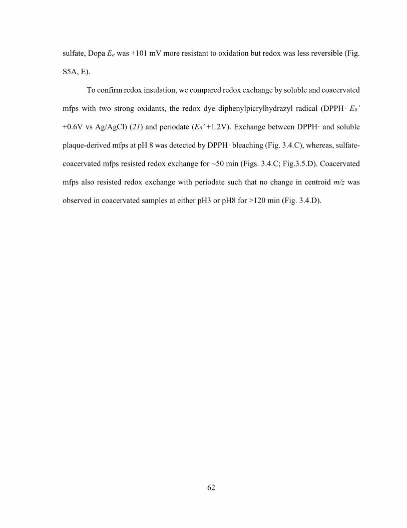
62
sulfate, Dopa Ea was +101 mV more resistant to oxidation but redox was less reversible (Fig.
S5A, E).
To confirm redox insulation, we compared redox exchange by soluble and coacervated
mfps with two strong oxidants, the redox dye diphenylpicrylhydrazyl radical (DPPH· E0’
+0.6V vs Ag/AgCl) (21) and periodate (E0’ +1.2V). Exchange between DPPH· and soluble
plaque-derived mfps at pH 8 was detected by DPPH· bleaching (Fig. 3.4.C), whereas, sulfate-
coacervated mfps resisted redox exchange for ~50 min (Figs. 3.4.C; Fig.3.5.D). Coacervated
mfps also resisted redox exchange with periodate such that no change in centroid m/z was
observed in coacervated samples at either pH3 or pH8 for >120 min (Fig. 3.4.D).
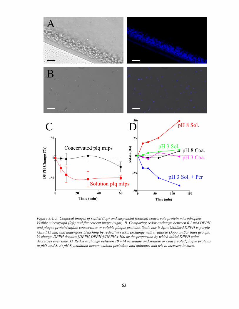
63
Figure 3.4. A. Confocal images of settled (top) and suspended (bottom) coacervate protein microdroplets. Visible micrograph (left) and fluorescent image (right). B. Comparing redox exchange between 0.1 mM DPPH and plaque protein/sulfate coacervates or soluble plaque proteins. Scale bar is 5𝜇𝑚 Oxidized DPPH is purple (lmax 515 nm) and undergoes bleaching by reductive redox exchange with available Dopa and/or thiol groups. % change DPPH denotes [DPPH-DPPH2]/DPPH x 100 or the proportion by which initial DPPH color decreases over time. D. Redox exchange between 10 mM periodate and soluble or coacervated plaque proteins at pH3 and 8. At pH 8, oxidation occurs without periodate and quinones add tris to increase in mass.
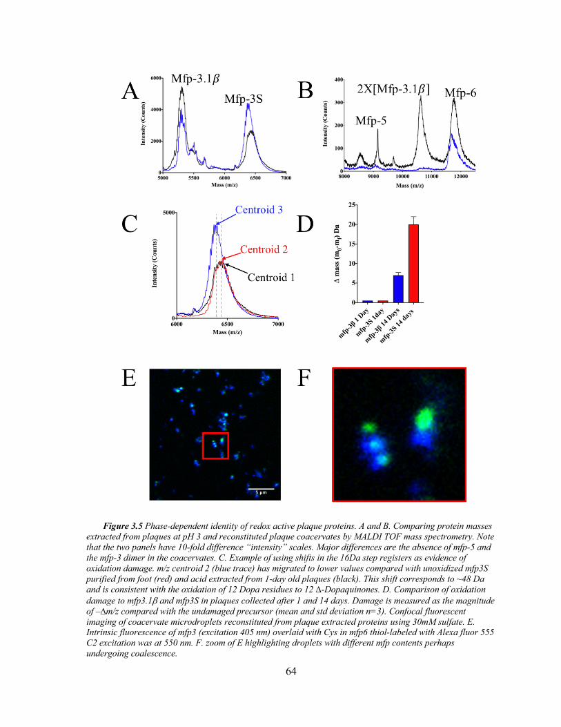
64
Figure 3.5 Phase-dependent identity of redox active plaque proteins. A and B. Comparing protein masses extracted from plaques at pH 3 and reconstituted plaque coacervates by MALDI TOF mass spectrometry. Note that the two panels have 10-fold difference “intensity” scales. Major differences are the absence of mfp-5 and the mfp-3 dimer in the coacervates. C. Example of using shifts in the 16Da step registers as evidence of oxidation damage. m/z centroid 2 (blue trace) has migrated to lower values compared with unoxidized mfp3S purified from foot (red) and acid extracted from 1-day old plaques (black). This shift corresponds to ~48 Da and is consistent with the oxidation of 12 Dopa residues to 12 ∆-Dopaquinones. D. Comparison of oxidation damage to mfp3.1b and mfp3S in plaques collected after 1 and 14 days. Damage is measured as the magnitude of –∆m/z compared with the undamaged precursor (mean and std deviation n=3). Confocal fluorescent imaging of coacervate microdroplets reconstituted from plaque extracted proteins using 30mM sulfate. E. Intrinsic fluorescence of mfp3 (excitation 405 nm) overlaid with Cys in mfp6 thiol-labeled with Alexa fluor 555 C2 excitation was at 550 nm. F. zoom of E highlighting droplets with different mfp contents perhaps undergoing coalescence.
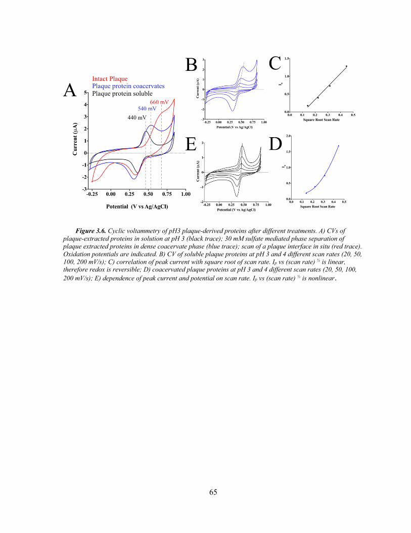
65
Figure 3.6. Cyclic voltammetry of pH3 plaque-derived proteins after different treatments. A) CVs of plaque-extracted proteins in solution at pH 3 (black trace); 30 mM sulfate mediated phase separation of plaque extracted proteins in dense coacervate phase (blue trace); scan of a plaque interface in situ (red trace). Oxidation potentials are indicated. B) CV of soluble plaque proteins at pH 3 and 4 different scan rates (20, 50, 100, 200 mV/s); C) correlation of peak current with square root of scan rate. Ip vs (scan rate) ½ is linear, therefore redox is reversible; D) coacervated plaque proteins at pH 3 and 4 different scan rates (20, 50, 100, 200 mV/s); E) dependence of peak current and potential on scan rate. Ip vs (scan rate) ½ is nonlinear.
66
Mussels are masters of interfacial Dopa redox control in plaques. Success may rely
on maintaining two phase-separated and redox-regulated compartments (C) of Dopa in the
plaque: C1 is a 15 nm-thick interfacial film of mfp-5 and mfp-8 with a high Ean; C2 is a 10-
20 nm open-pore mesh filled with coacervate droplets of mfp-3 and mfp-6 and a Ean ~120
mV lower than C1. We propose the following redox interplay to maintain Dopa-dependent
adhesion (Fig. 3.7.A/B): i) Dopa-containing interfacial mfps in C1 regularly sustain damage
by oxidation to quinone; ii) unable to rebind the surface, the C1 quinone H-bonds to
accessible Dopa in C2, and, given the significantly more positive Ean of C1 Dopa, undergoes
redox exchange to restore Dopa for adhesion; iii) C2 Mfp-3 now has Dopaquinone, which it
recycles to catechol using 2e- + 2H+ from two thiols in mfp-6, or by simple tautomerization
to the vinylcatechol (26). Dopa and thiol groups in C2 thus provide a lifeline of electrons
and protons for oxidized Dopa in C1 until the reservoir is depleted. Indeed, mfp-3 and mfp-6
droplets resemble dedicated batteries for maintaining Dopa-dependent adhesion in the film.

67
Fig. 3.7. Phase-specific Dopa and thiol redox in coacervated plaque proteins. A. Schematic plaque interface; B. Enlarged droplet with mfp3 and mfp6 variants. C1 is the film below the droplet and contains adhesive primers mfp5 and mfp8. When C1 Dopa is oxidized to Dopaquinone (Ea +660 mV Ag/AgCl; Table S3), the damage is repaired with 2H++2e- donated by C2 (Ea +540 mV). C2 Dopaquinone is reduced back to Dopa by thiols in mfp6 (Ea -200 mV), but when thiols are depleted, Dopa derivatives (∆-Dopa) provide reducing poise. Bidentate hydroquinone-quinone interactions actuate redox exchange in many cellular pathways (29).
68
E. Conclusions
Our results reveal that mussel plaques are core-shell structures with the adhesive film
as the shell and a coacervate-filled hydrogel as the core. Although hydrogels are receiving
much scrutiny for engineering tough interfaces (27), replacement of the water with
coacervates in these gels is a novel twist that merits particular scrutiny. Coacervation as a
natural strategy for protecting redox-sensitive functionalities from oxidation has a profound
bearing on improved translation of wet adhesion (40-41) as well as phase-specific redox in
phase-separated compartments elsewhere in biology (1, 28). Evidently, by sequestering
oxidation prone groups within fluid-filled inclusions in a porous structure like the plaque,
the groups can be shielded and recruited for redox exchange with external functionalities
damaged by oxidation.
69
F. References
1. A.A. Hyman, C.A. Weber, F. Jülicher, Liquid-liquid phase separation in biology. Annu. Rev. Cell. Dev. Biol. 30, 39-58 (2014).
2. B. Vrhovski, S. Jensen, A.S. Weiss, Coacervation characteristics of recombinant human tropoelastin. FEBS Journal 250, 92-98 (1997).
3. H. J. Jin, D. L. Kaplan, Mechanism of silk processing in insects and spiders. Nature 424, 1057-61 (2003).
4. Y. P. Tan, et al., Infiltration of a chitin by protein coacervates defines the squid beak mechanical gradient. Nature Chem Biol 11, 489-495 (2015).
5. M. Yamanaka, Y. Ishizaki, T. Nagakawa, A. Taoka, Y. Fukumori, Purification and characterization of coacervate-forming cuticular proteins from Papilio xuthus pupae. Zool. Sci. 30, 534-542 (2013).
6. H. Zhao, C.J. Sun, R.J. Stewart, J.H. Waite, Cement proteins of the tube building polychaete Phragmatopoma californica. J. Biol. Chem. 280, 42938-42944 (2005).
7. W. Wei, Y. Tan, N. R. Martinez, J. Yu, J. N. Israelachvili, J.H. Waite, A mussel-derived one-component adhesive coacervate. Acta Biomaterialia, 10, 1663-1670 (2013).
8. D. G. DeMartini, J.M. Errico, S. Sjöström, A. Fenster, J.H. Waite, A cohort of new adhesive proteins identified from transcriptomic analysis of mussel foot glands. J. R. Soc. Interface 14, 20170151 (2017).
9. B. Yang, S. Jin, Y. Park, Y.M. Jung, H. J. Cha, Coacervation of interfacial adhesive proteins for initial adhesion to a wet surface. Small 14, 1803377 (2018).
10. J. Wang, T. Scheibel, Coacervation of the recombinant Mytilus galloprovincialis foot protein-3b. Biomacromolecules 19, 3612-3619 (2018).
11. S. Kim, H. Y. Yoo, J. Huang, Y. Lee, S. Park, S. Park, S. Jin, Y.M. Jung, H. B. Zeng, D.S. Hwang, Y.S. Jho, Salt triggers the simple coacervation of an underwater adhesive when cations meet aromatic π electrons in seawater. ACS Nano 11, 6764-6772 (2017).
12. D. S. Hwang, H. Zeng, A. Srivastava, M. Tirrell, J. N. Israelachvili, J. H. Waite, Complex coacervation of a recombinant mussel adhesive protein and hyaluronic acid - viscous and interfacial properties. Soft Matter 6, 3232-3236 (2010).
13. I. Kaminker, W. Wei, A.M. Schrader, Y. Talmon, M.T. Valentine, J.N. Israelachvili, J.H. Waite, S. Han, Simple peptide coacervates adapted for rapid pressure sensitive wet adhesion. Soft Matter 13, 9122e (2017).
14. J. H. Waite, Mussel adhesion – essential footwork. J. Exp. Biol. 220, 517-530 (2017).
15. J. Saiz-Poseu, J. Mancebo-Aracil, F. Nador, F. Busque, D. Ruiz-Molina, The chemistry behind catechol-based adhesion. Angew. Chem. Int. Ed., 58, 696-714 (2019).
16. G.P. Maier, C.M. Bernt, A. Butler, Catechol oxidation: considerations in the design of wet adhesive materials. Biomaterials Science doi: 10.1039/c7bm00884h (2017).
70
17. N.R. Martinez Rodriguez, S. Das, Y. Kaufman, J.N. Israelachvili, J. H. Waite, Interfacial pH during mussel adhesive plaque formation. Biofouling 31, 221-227 (2015).
18. J. Yu, E. Danner, R. K. Ashley, J.N. Israelachvili, J. H. Waite, Mussel protein adhesion depends on interprotein thiol-mediated redox modulation. Nature Chem. Biol. 7, 588-590 (2011).
19. C. Zhong, T. Gurry, A.A. Cheng, J. Downey, Z. Deng, C.M. Stultz, T.K. Lu, Strong underwater adhesives made by self-assembling multiprotein nanofibres. Nature Nanotech. 9, 858-866 (2014).
20. L. H. N. Cooper, Oxidation-reduction potential of seawater. J. Mar. Biol. Ass. UK, 22, 167-176 (1937).
21. D. R. Miller, J. E. Spahn, J. H. Waite, The staying power of adhesion-associated antioxidant activity in Mytilus californianus. J. R. Soc. Interface 12, 20150614 (2015).
22. A. Jain, R. D. Vale, RNA Phase transitions in repeat expansion disorders. Nature 56, 243-247 (2018).
23. E. Filippidi, D.G. DeMartini, P. Malo de Molina, E.W. Danner, J. Kim, M.E. Helgeson, J.H. Waite, M. T. Valentine, Fine microscopic network structure of mussel (Mytilus) plaques. J. R. Soc. Interface 12, 20150827 (2015).
24. G. M. Proudfoot, I.M. Ritchie, Cyclic voltammetric study of some 4-substituted benzene-1, 2 diols. Austral. J. Chem. 36, 885-894 (1983).
25. W. Wei, J. Yu, C. C. Broomell, J. N. Israelachvili, J. H. Waite, Hydrophobic enhancement of Dopa mediated adhesion in a mussel foot protein. J. Am. Chem. Soc. 135, 377-383 (2013).
26. R. Mirshafian, W. Wei, J.N. Israelachvili, J. H. Waite, a, b-Dehydro-Dopa: a hidden participant in mussel adhesion. Biochemistry 55, 743-750 (2016).
27. J. Yang, R. Bai, B. Chen, Z. Suo, Hydrogel adhesion: a supramolecular synergy of chemistry, topology, and mechanics. Adv. Func. Mat. 1901693 (2019).
28. Y Shin, C.P. Brangwynne, Liquid phase condensation in cell physiology and disease. Science 491, 357 (2017).
29. J. Regeimbal, S. Gleiter, B.L. Trumpower, C. Yu, M. Diwakar, D.P. Ballou, J.C.A. Bardwell, Disulfide bond formation involves a quinhydrone-type charge transfer complex. PNAS 100, 13779-13784 (2003).
30. C.A. Monnier, D. DeMartini, J.H. Waite, Adaptive mussel coatings: intertidal exposure favors soft studded armors. Nature Commun. 9: article 3424 (2018).
31. S. Panyim, R. Chalkley, High resolution acrylamide gel electrophoresis of histones. Arch. Biochem. Biophys. 130, 337-346 (1969).
32. S.C.T. Nicklisch, J.E. Spahn, H. Zhou, C.M. Gruian, J.H. Waite, Redox capacity of an extracellular matrix protein associated with adhesion in Mytilus californianus. Biochemistry 55, 2022-2030 (2016).
71
33. T. E. Creighton, in Protein Structure – A Practical Approach (ed Creighton, T.E.) 155-168 (IRL Press, Oxford, UK, 1989).
34. J. H. Waite, M.L. Tanzer, Specific colorimetric detection of o-diphenols and 3, 4-dihydroxycontaining peptides. Anal. Biochem. 111, 131-136 (1981).
35. H. Zhao, J.H. Waite, Linking adhesive and structural proteins in the attachment plaque of Mytilus californianus. J. Biol. Chem. 281 26150-26158 (2006).
H. Zhao, N.B. Robertson, S.A. Jewhurst, J.H. Waite, missing reference
72
Chapter IV
Conclusions, Future Directions and Closing Remarks
Conclusions:
The most prominent chemistry in the mussel byssus is based on the catechol moiety.
Specifically, 3,4-dihydroxyphenlalanine (DOPA), a catecholic nonstandard amino acid is
incorporated in a majority of mussel foot proteins (mfp) and is relied upon to participate
both cohesively and adhesively. Furthermore, DOPA must perform its duties at seawater
conditions (pH 8.0 ionic strength 0.7M), at which DOPA oxidation is a thermodynamically
favorable process when coupled to oxygen reduction. Additionally, the byssus cannot rely
on cellular support to maintain functional integrity. Despite these shortcomings, byssus
functional lifespan is on the order of months.
In 2011, Yu et al first suggested that mussels incorporated antioxidants, namely mfp-6,
into the byssus to protect the plaque against oxidative damage. While mfp-6 demonstrated
the ability to reverse oxidative damage at pH 8 in vitro, the strategy was short lived. Given
the propensity of DOPA to oxidize in seawater simply incorporating an antioxidant is
insufficient at providing long term performance.
The dissertation work outlined above, demonstrates the mussel utilizes a system of
kinetically trapped redox reservoirs to insulate redox active DOPA from the surrounding
environment. More specifically, in the plaque, the mussel uses liquid-liquid phase separated
compartments to house specific DOPA containing proteins. By doing so it accomplishes two
primary goals.
73
1) It creates discrete redox compartments in which the DOPA proteins have varying
redox potentials. Mfp-5, found in a gel like state at the substrate interface has DOPA with
the highest redox potential. Mfp-3, contained in coacervate like droplets, has DOPA with a
redox potential approximately 100mV less then mfp-5, creating a redox potential cascade. In
doing so mfp-3-DOPA may reduce damaged mfp-5-DOPAqunine. Additionally, when
present as LLPS material, DOPA exhibited prolonged redox lifetime compared to solution
state DOPA.
2) It overcomes a stochiometric deficit experienced by mfp-6’s redox capacity. While
cysteine is a powerful antioxidant, the concentration of redox available thiol is
approximately 25 mM which is substantially less than the more than 200mM DOPA
contained in the plaque. This puts the reducing equivalents at a ~15-fold deficit. Specific
compartmentalization of DOPA alleviates cysteine’s responsibility as the sole antioxidant
amino acid.
With respect to the thread, the mussel incorporates a region of elevated sulfur content.
Results from this dissertation indicate that the sulfur is contained as reduced thiols. Hence
hereinafter this region will be known as the thiol rich region (TRL). Situated between the
cuticle and the core, the TRL may serve as an electron reservoir to be called upon when
DOPA in the cuticle is compromised. Beyond its redox responsibilities, the TRL serves as a
mechanical barrier, protecting the core from contact damage propagating from the cuticle.
In conclusion, this dissertation has demonstrated the mussel is capable of controlling
redox poise in the byssus through location specific mechanisms.
74
Future Directions:
In 2006 Hasheen Lee, Norbert Scherer and Phillip Messersmith published a hallmark
manuscript on DOPA’s role in mussel adhesion. In 2011, the importance of antioxidants
effects on DOPA adhesion was highlighted and a proposed mechanism was put forth. Now,
in 2019, we have expanded on that antioxidant model. While substantial progress has been
made many questions still remain. Future endeavors should focus on elucidating the
following gaps in knowledge pertaining to redox chemistry in the plaque: we know DOPA
selectively partitions into coacervates and these coacervates can control redox poise, we do
not know what acts as the “redox gate”. That is to say, what interaction drives delivery of
electrons from DOPA to DOPAquinone. A few possibilities are patently evident.
1. Any DOPAquinone-DOPA interaction is sufficient to drive electron transfer.
2. It must be mfp-3 à mfp-5 interaction to drive electron transfer.
a. Catechols at the coacervate interface come into contact with quinones
at the substrate interface inducing electron transfer.
b. Phosphate groups on mfp-5 interact with basic residues in mfp-3 pulling
the quinone into the coacervate leading to electron transfer.
Independent of the redox gate scenario, the mechanism of electron transfer should be
investigated. One plausible configuration is the formation of a quinhydrone like structure in
which a charge transfer complex is formed between aromatic stacks and stabilized via
hydrogen bonding. Additionally, the effect of local protein sequence on DOPA’s redox
potential should be thoroughly explored.
With respect to the thread, a cohort of cysteine rich proteins, namely mfp-16 -17 -18 and
-19 were identified in the transcriptome. Mfp-17 was positively identified at the protein
75
level in the collagen granules purified from the foot, but localization in the mature thread is
currently lacking. Future experiments should focus on localization of the putative thiol rich
proteins. MALDI-mapping may provide a viable platform for localization. Additionally,
given cysteines extreme reactivity, efforts should be made to identify covalent crosslinks.
Given the proximity of DOPA in the cuticle to thiol in the TRL, 5-cysteinylDOPA is a
possible candidate.
Additionally, another crosslinking possibility, all be it a far-fetched exotic idea, is an
isopeptide bond. Mfp-1 in the cuticle contains elevated levels of lysine, a prime candidate
for isopeptide linkages. Isopeptide bond formation is often catalyzed by transglutaminase, an
enzyme containing a cys-his-asp catalytic triad, and in some instances utilizes calcium as a
cofactor. While Kim et al. successfully demonstrated lysine’s involvement in cation pi-
interactions in the cuticle, isopeptide bonds could potentially serve to bond the cuticle to the
TRL/core. Interestingly, all of the components necessary for isopeptide bond formation are
found in elevated levels directly adjacent to the TRL. Anti-isopeptide antibodies are
commercially available thus could serve as a “low hanging fruit” opportunity experiment.
Closing Remarks:
Mussel byssus projects have spawned many successful careers around the globe,
with Dr. Waite being a central figure in each one. Herb’s success is largely due to his
unwavering dedication to his principles and moral compass. Accompanying his dedication is
a level of humility rarely displayed by others. This combination of attributes has allowed
Herb to produce meaningful science guided principally by curiosity and substantive

76
questions asking. As a result, Herb’s work has been increasingly used as inspiration for
biologists and engineers alike, evident by the following.
Anyone who has spent an extended period of time with Herb knows he has
many “Herbisms” but perhaps the most penetrating and long-lasting ism; “Be a diver
not a surfer”. Translation; in order to do something truly transformative and
impactful, you must go beyond the superficial and dive deep into the meaning of the
77
question. This is becoming increasingly difficult as funding agencies look toward
application-based science. Despite this, as Herb trainees we have a responsibility to
continue to push ‘basic’ research projects and to promote collaborations between
fundamentalists and “engineers”. To close, I will leave you with the following quote
from Herb.
“A significant lesson emerging from research on mussel adhesion that is widely
applicable to other organisms is that there is no substitute for penetrating biological
exploration at many length and time scales. Indeed, this exploration is as essential
for good translation as it is for discovery. As reviewers and funding agencies place
more and more emphasis on translation for its own sake we should stand together
to insist that a retreat from the biology is counter-productive and may even be a
misuse of taxpayer money. Translations driven by superficial impressions or
fantasies about biology will not stand the test of time because they have missed the
point.”
- J. Herbert Waite (2019)