Redox Regulation of Cell Survival by the Thioredoxin Superfamily: An Implication of Redox Gene...
18
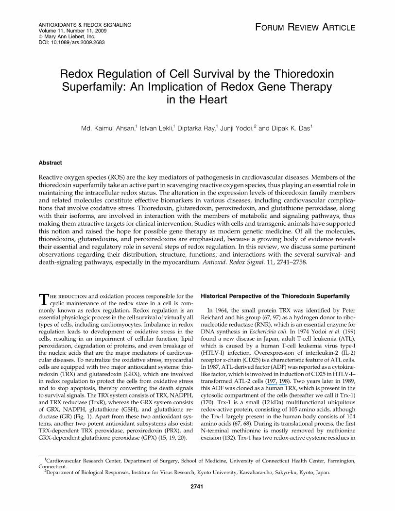
FORUM REVIEW ARTICLE Redox Regulation of Cell Survival by the Thioredoxin Superfamily: An Implication of Redox Gene Therapy in the Heart Md. Kaimul Ahsan, 1 Istvan Lekli, 1 Diptarka Ray, 1 Junji Yodoi, 2 and Dipak K. Das 1 Abstract Reactive oxygen species (ROS) are the key mediators of pathogenesis in cardiovascular diseases. Members of the thioredoxin superfamily take an active part in scavenging reactive oxygen species, thus playing an essential role in maintaining the intracellular redox status. The alteration in the expression levels of thioredoxin family members and related molecules constitute effective biomarkers in various diseases, including cardiovascular complica- tions that involve oxidative stress. Thioredoxin, glutaredoxin, peroxiredoxin, and glutathione peroxidase, along with their isoforms, are involved in interaction with the members of metabolic and signaling pathways, thus making them attractive targets for clinical intervention. Studies with cells and transgenic animals have supported this notion and raised the hope for possible gene therapy as modern genetic medicine. Of all the molecules, thioredoxins, glutaredoxins, and peroxiredoxins are emphasized, because a growing body of evidence reveals their essential and regulatory role in several steps of redox regulation. In this review, we discuss some pertinent observations regarding their distribution, structure, functions, and interactions with the several survival- and death-signaling pathways, especially in the myocardium. Antioxid. Redox Signal. 11, 2741–2758. T he reduction and oxidation process responsible for the cyclic maintenance of the redox state in a cell is com- monly known as redox regulation. Redox regulation is an essential physiologic process in the cell survival of virtually all types of cells, including cardiomyocytes. Imbalance in redox regulation leads to development of oxidative stress in the cells, resulting in an impairment of cellular function, lipid peroxidation, degradation of proteins, and even breakage of the nucleic acids that are the major mediators of cardiovas- cular diseases. To neutralize the oxidative stress, myocardial cells are equipped with two major antioxidant systems: thio- redoxin (TRX) and glutaredoxin (GRX), which are involved in redox regulation to protect the cells from oxidative stress and to stop apoptosis, thereby converting the death signals to survival signals. The TRX system consists of TRX, NADPH, and TRX reductase (TrxR), whereas the GRX system consists of GRX, NADPH, glutathione (GSH), and glutathione re- ductase (GR) (Fig. 1). Apart from these two antioxidant sys- tems, another two potent antioxidant subsystems also exist: TRX-dependent TRX peroxidase, peroxiredoxin (PRX), and GRX-dependent glutathione peroxidase (GPX) (15, 19, 20). Historical Perspective of the Thioredoxin Superfamily In 1964, the small protein TRX was identified by Peter Reichard and his group (67, 97) as a hydrogen donor to ribo- nucleotide reductase (RNR), which is an essential enzyme for DNA synthesis in Escherichia coli. In 1974 Yodoi et al. (199) found a new disease in Japan, adult T-cell leukemia (ATL), which is caused by a human T-cell leukemia virus type-I (HTLV-I) infection. Overexpression of interleukin-2 (IL-2) receptor a-chain (CD25) is a characteristic feature of ATL cells. In 1987, ATL-derived factor (ADF) was reported as a cytokine- like factor, which is involved in induction of CD25 in HTLV-I– transformed ATL-2 cells (197, 198). Two years later in 1989, this ADF was cloned as a human TRX, which is present in the cytosolic compartment of the cells (hereafter we call it Trx-1) (170). Trx-1 is a small (12 kDa) multifunctional ubiquitous redox-active protein, consisting of 105 amino acids, although the Trx-1 largely present in the human body consists of 104 amino acids (67, 68). During its translational process, the first N-terminal methionine is mostly removed by methionine excision (132). Trx-1 has two redox-active cysteine residues in 1 Cardiovascular Research Center, Department of Surgery, School of Medicine, University of Connecticut Health Center, Farmington, Connecticut. 2 Department of Biological Responses, Institute for Virus Research, Kyoto University, Kawahara-cho, Sakyo-ku, Kyoto, Japan. ANTIOXIDANTS & REDOX SIGNALING Volume 11, Number 11, 2009 ª Mary Ann Liebert, Inc. DOI: 10.1089=ars.2009.2683 2741
-
Upload
independent -
Category
Documents
-
view
1 -
download
0
Transcript of Redox Regulation of Cell Survival by the Thioredoxin Superfamily: An Implication of Redox Gene...
FORUM REVIEW ARTICLE
Redox Regulation of Cell Survival by the ThioredoxinSuperfamily: An Implication of Redox Gene Therapy
in the Heart
Md. Kaimul Ahsan,1 Istvan Lekli,1 Diptarka Ray,1 Junji Yodoi,2 and Dipak K. Das1
Abstract
Reactive oxygen species (ROS) are the key mediators of pathogenesis in cardiovascular diseases. Members of thethioredoxin superfamily take an active part in scavenging reactive oxygen species, thus playing an essential role inmaintaining the intracellular redox status. The alteration in the expression levels of thioredoxin family membersand related molecules constitute effective biomarkers in various diseases, including cardiovascular complica-tions that involve oxidative stress. Thioredoxin, glutaredoxin, peroxiredoxin, and glutathione peroxidase, alongwith their isoforms, are involved in interaction with the members of metabolic and signaling pathways, thusmaking them attractive targets for clinical intervention. Studies with cells and transgenic animals have supportedthis notion and raised the hope for possible gene therapy as modern genetic medicine. Of all the molecules,thioredoxins, glutaredoxins, and peroxiredoxins are emphasized, because a growing body of evidence revealstheir essential and regulatory role in several steps of redox regulation. In this review, we discuss some pertinentobservations regarding their distribution, structure, functions, and interactions with the several survival- anddeath-signaling pathways, especially in the myocardium. Antioxid. Redox Signal. 11, 2741–2758.
The reduction and oxidation process responsible for thecyclic maintenance of the redox state in a cell is com-
monly known as redox regulation. Redox regulation is anessential physiologic process in the cell survival of virtually alltypes of cells, including cardiomyocytes. Imbalance in redoxregulation leads to development of oxidative stress in thecells, resulting in an impairment of cellular function, lipidperoxidation, degradation of proteins, and even breakage ofthe nucleic acids that are the major mediators of cardiovas-cular diseases. To neutralize the oxidative stress, myocardialcells are equipped with two major antioxidant systems: thio-redoxin (TRX) and glutaredoxin (GRX), which are involvedin redox regulation to protect the cells from oxidative stressand to stop apoptosis, thereby converting the death signalsto survival signals. The TRX system consists of TRX, NADPH,and TRX reductase (TrxR), whereas the GRX system consistsof GRX, NADPH, glutathione (GSH), and glutathione re-ductase (GR) (Fig. 1). Apart from these two antioxidant sys-tems, another two potent antioxidant subsystems also exist:TRX-dependent TRX peroxidase, peroxiredoxin (PRX), andGRX-dependent glutathione peroxidase (GPX) (15, 19, 20).
Historical Perspective of the Thioredoxin Superfamily
In 1964, the small protein TRX was identified by PeterReichard and his group (67, 97) as a hydrogen donor to ribo-nucleotide reductase (RNR), which is an essential enzyme forDNA synthesis in Escherichia coli. In 1974 Yodoi et al. (199)found a new disease in Japan, adult T-cell leukemia (ATL),which is caused by a human T-cell leukemia virus type-I(HTLV-I) infection. Overexpression of interleukin-2 (IL-2)receptor a-chain (CD25) is a characteristic feature of ATL cells.In 1987, ATL-derived factor (ADF) was reported as a cytokine-like factor, which is involved in induction of CD25 in HTLV-I–transformed ATL-2 cells (197, 198). Two years later in 1989,this ADF was cloned as a human TRX, which is present in thecytosolic compartment of the cells (hereafter we call it Trx-1)(170). Trx-1 is a small (12 kDa) multifunctional ubiquitousredox-active protein, consisting of 105 amino acids, althoughthe Trx-1 largely present in the human body consists of 104amino acids (67, 68). During its translational process, the firstN-terminal methionine is mostly removed by methionineexcision (132). Trx-1 has two redox-active cysteine residues in
1Cardiovascular Research Center, Department of Surgery, School of Medicine, University of Connecticut Health Center, Farmington,Connecticut.
2Department of Biological Responses, Institute for Virus Research, Kyoto University, Kawahara-cho, Sakyo-ku, Kyoto, Japan.
ANTIOXIDANTS & REDOX SIGNALINGVolume 11, Number 11, 2009ª Mary Ann Liebert, Inc.DOI: 10.1089=ars.2009.2683
2741
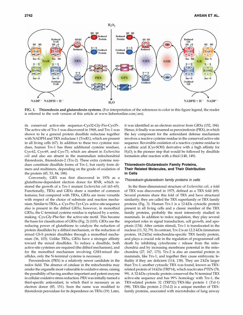
its conserved active-site sequence:-Cys32-Gly-Pro-Cys35-.The active site of Trx-1 was discovered in 1968, and Trx-1 wasshown to be a general protein disulfide reductase togetherwith NADPH and TRX reductase 1 (TrxR1), which are presentin all living cells (67). In addition to these two cysteine resi-dues, human Trx-1 has three additional cysteine residues,Cys-62, Cys-69, and Cys-73, which are absent in Escherichiacoli and also are absent in the mammalian mitochondrialthioredoxin, thioredoxin-2 (Trx-2). These extra cysteine resi-dues constitute disulfide forms of Trx-1, but rarely form di-mers and multimers, depending on the grade of oxidation ofthe protein (45, 53, 84, 188).
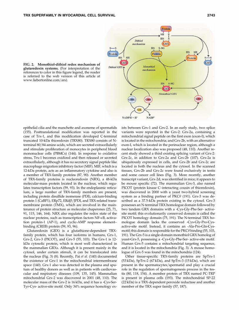
Conversely, GRX was first discovered in 1976 as aglutathione-dependent electron donor for RNR, which re-stored the growth of a Trx-1 mutant Escherichia coli (63–65).Functionally, TRXs and GRXs share a number of commonfeatures; but compared with TRXs, GRXs are more versatilewith respect of the choice of substrate and reaction mecha-nism. Similar to TRXs, a -Cys-Pro-Tyr-Cys- active-site sequencealso is present in the dithiol GRXs; however, in monothiolGRXs, the C-terminal cysteine residue is replaced by a serine,making -Cys-Gly-Phe-Ser- the active-site motif. This becamethe basis for classification of GRXs (Fig. 2) (103). GRXs use thereducing power of glutathione to catalyze the reduction ofprotein disulfides by a dithiol mechanism, or the reduction ofmixed GS-S protein disulfides through a monothiol mecha-nism (36, 103). Unlike TRXs, GRXs have a stronger affinitytoward the mixed disulfides. To reduce a disulfide, bothactive-site cysteines are required (the dithiol mechanism), andfor the monothiol mechanism involving GSH-mixed dis-ulfides, only the N-terminal cysteine is necessary.
Peroxiredoxin (PRX) is a relatively newer candidate in theredox field. The absence of catalase in the mitochondria mayrender the organelle most vulnerable to oxidative stress, raisingthe possibility of having another important and potent enzymein cellular oxidative stress reduction. PRX was initially named athiol-specific antioxidant, in which thiol is necessary as anelectron donor (85, 151). Soon the name was modified tothioredoxin peroxidase for its dependence on TRXs (19). Later,
it was identified as an electron receiver from GRXs (152, 184).Hence, it finally was renamed as peroxiredoxin (PRX), in whichthe key component for the antioxidant defense mechanisminvolves a reactive cysteine residue in the conserved active-sitesequence. Reversible oxidation of a reactive cysteine residue toa sulfinic acid (Cys-SOH) derivative with a high affinity forH2O2 is the pioneer step that would be followed by disulfideformation after reaction with a thiol (148, 149).
Thioredoxin-Glutaredoxin Family Proteins,Their Related Molecules, and Their Distributionin Cells
Thioredoxin-glutaredoxin family proteins in cells
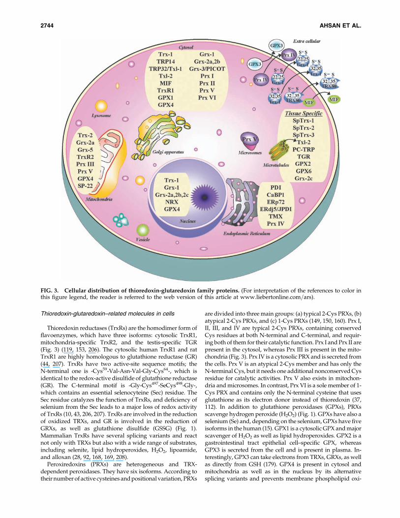
In the three-dimensional structure of Escherichia coli, a foldof TRX was discovered in 1975, defined as a TRX fold (69).Several proteins share this fold of TRX and have structuralsimilarity; they are called the TRX superfamily or TRX familyproteins (Fig. 3). Human Trx-1 is a 12-kDa cytosolic proteinpresent in all living cells and a classic member of the TRXfamily proteins, probably the most intensively studied inmammals. In addition to redox regulation, they play severalimportant roles in signal transduction, cell growth, and apo-ptosis (134). After certain stimuli, Trx-1 is translocated in thenucleus (11, 52, 79). In contrast, Trx-2 is an 12.2-kDa (immatureprotein, 18.2 kDa) mitochondria-specific TRX family protein,and plays a crucial role in the regulation of programmed celldeath by inhibiting cytochrome c release from the mito-chondria and by increasing membrane potential in the mito-chondria (27, 167, 173). Trx-2 is also an essential protein inmammals, like Trx-1, and together they cause embryonic le-thality if they are deficient (114, 138). They are 2 kDa largerthan Trx-1; another cytosolic TRX was found, known as TRX-related protein of 14 kDa (TRP14), which reactivates PTEN (78,99). A 32-kDa cytosolic protein conserved the N-terminal TRXactive-site sequence and has 99% homology with Trx-1, theTRX-related protein 32 (TRP32)=TRX-like protein 1 (Txl-1)(98). TRX-like protein 2 (Txl-2) is a unique member of TRX-family proteins, associated with microtubules of lung airway
FIG. 1. Thioredoxin and glutaredoxin systems. (For interpretation of the references to color in this figure legend, the readeris referred to the web version of this article at www.liebertonline.com=ars).
2742 AHSAN ET AL.
epithelial cilia and the manchette and axoneme of spermatids(155). Posttranslational modification was reported in thecase of Trx-1, and this modification developed C-terminaltruncated 10-kDa thioredoxin (TRX80). TRX80 consists of N-terminal 80=84 amino acids, which are secreted extracellularlyand stimulate proliferation of monocytes in peripheral bloodmononuclear cells (PBMCs) (144). In response to oxidativestress, Trx-1 becomes oxidized and then released or secretedextracellularly, although it has no secretory signal peptide likemacrophage migration inhibitory factor (MIF). MIF, which is a12-kDa protein, acts as an inflammatory cytokine and also isa member of TRX-family proteins (87, 90). Another memberof TRX-family proteins is nucleoredoxin (NRX), a 48-kDamolecular-mass protein located in the nucleus, which regu-lates transcription factors (59, 93). In the endoplasmic reticu-lum, a large number of TRX-family members are present,including protein disulfide isomerase (PDI), calcium-bindingprotein 1 (CaBP1), ERp72, ERdj5=JPDI, and TRX-related trans-membrane protein (TMX), which are involved in the main-tenance of protein structure as molecular chaperones (25, 71,91, 115, 146, 164). NRX also regulates the redox state of thenuclear proteins, such as transcription factors NF-kB, activa-tion protein-1 (AP-1), and cyclic-AMP response element–binding (CREB) protein (59, 93, 96).
Glutaredoxin (GRX) is a glutathione-dependent TRX-family protein, which has four isoforms in humans, Grx-1,Grx-2, Grx-3 (PICOT), and Grx-5 (55, 103). The Grx-1 is 12-kDa cytosolic protein, which is most well characterized inthe mammalian GRXs. Although it is present mainly in thecytosol, under certain stimuli, it can be translocated intothe nucleus (Fig. 3) (8). Recently, Pai et al. (140) documentedthe existence of Grx-1 in the mitochondrial intermembranespace (140). Grx-1 also was detected in the plasma and spu-tum of healthy donors as well as in patients with cardiovas-cular and respiratory diseases (109, 135, 145). Mammalianmitochondrial Grx-2 was discovered in 2001 (48, 110). Themolecular mass of the Grx-2 is 16 kDa, and it has a -Cys-Ser-Tyr-Cys- active-site motif. Only 34% sequence homology ex-
ists between Grx-1 and Grx-2. In an early study, two splicevariants were reported in the Grx-2: Grx-2a, containing amitochondrial signal peptide on the first exon (exon-I), whichis located in the mitochondria; and Grx-2b, with an alternativeexon-I, which is located in the perinuclear region, although anuclear localization also was proposed (48, 110). Another re-cent study showed a third existing splicing variant of Grx-2:Grx-2c, in addition to Grx-2a and Grx-2b (107). Grx-2a isubiquitously expressed in cells, and Grx-2b and Grx-2c arelocated in both the nucleus and the cytosol. In the scannedtissues, Grx-2b and Grx-2c were found exclusively in testisand some cancer cell lines (Fig. 3). More recently, anothertranscript variant, Grx-2d, was identified in mice; it appears tobe mouse specific (72). The mammalian Grx-3, also namedPICOT (protein kinase C–interacting cousin of thioredoxin),was discovered in 2000 with a yeast two-hybrid screeningsystem as a binding partner of PKCy (191). Grx-3 was de-scribed as a 37.5-kDa protein existing in the cytosol. Grx-3possesses an N-terminal TRX homologue domain followed bytwo tandem GRX domains with a -Cys-Gly-Phe-Ser- active-site motif; this evolutionarily conserved domain is called thePICOT homology domain (75, 191). The N-terminal TRX ho-mologue domain lacks the conserved -Cys-Gly-Pro-Cys-active-site motif. Instead, it contains an -Ala-Pro-Gln-Cys-motif; this domain is responsible for the PKCy binding (55, 103,191). The Grx-5 is a single-domain monothiol GRX homolog ofyeast-Grx-5, possessing a -Cys-Gly-Phe-Ser- active-site motif.Human Grx-5 contains a mitochondrial targeting sequence,and it is located in the mitochondria (Fig. 3). A mouse homo-logue of Grx-5 was found in the mitochondria (124).
Other tissue-specific TRX-family proteins are SpTrx-1(53 kDa), SpTrx-2 (67 kDa), and SpTrx-3 (15 kDa), which arepresent in the spermatocytes=spermatid and play a crucialrole in the regulation of spermatogenesis process in the tes-tis (80, 118, 154). A member protein of TRX named PC-TRPis present in plasma cells (193). The mitochondrial SP-22(22 kDa) is a TRX-dependent peroxide reductase and anothermember of the TRX super family (57, 187).
FIG. 2. Monothiol-dithiol redox mechanism ofglutaredoxin systems. (For interpretation of thereferences to color in this figure legend, the readeris referred to the web version of this article atwww.liebertonline.com=ars).
TRX SUPERFAMILY IN MYOCARDIAL CELL SURVIVAL 2743
Thioredoxin-glutaredoxin–related molecules in cells
Thioredoxin reductases (TrxRs) are the homodimer form offlavoenzymes, which have three isoforms: cytosolic TrxR1,mitochondria-specific TrxR2, and the testis-specific TGR(Fig. 3) (119, 153, 206). The cytosolic human TrxR1 and ratTrxR1 are highly homologous to glutathione reductase (GR)(44, 207). TrxRs have two active-site sequence motifs; theN-terminal one is -Cys59-Val-Asn-Val-Gly-Cys64-, which isidentical to the redox-active disulfide of glutathione reductase(GR). The C-terminal motif is -Gly-Cys497-SeCys498-Gly-,which contains an essential selenocyteine (Sec) residue. TheSec residue catalyzes the function of TrxRs, and deficiency ofselenium from the Sec leads to a major loss of redox activityof TrxRs (10, 43, 206, 207). TrxRs are involved in the reductionof oxidized TRXs, and GR is involved in the reduction ofGRXs, as well as glutathione disulfide (GSSG) (Fig. 1).Mammalian TrxRs have several splicing variants and reactnot only with TRXs but also with a wide range of substrates,including selenite, lipid hydroperoxides, H2O2, lipoamide,and alloxan (28, 92, 168, 169, 208).
Peroxiredoxins (PRXs) are heterogeneous and TRX-dependent peroxidases. They have six isoforms. According totheir number of active cysteines and positional variation, PRXs
are divided into three main groups: (a) typical 2-Cys PRXs, (b)atypical 2-Cys PRXs, and (c) 1-Cys PRXs (149, 150, 160). Prx I,II, III, and IV are typical 2-Cys PRXs, containing conservedCys residues at both N-terminal and C-terminal, and requir-ing both of them for their catalytic function. Prx I and Prx II arepresent in the cytosol, whereas Prx III is present in the mito-chondria (Fig. 3). Prx IV is a cytosolic PRX and is secreted fromthe cells. Prx V is an atypical 2-Cys member and has only theN-terminal Cys, but it needs one additional nonconserved Cysresidue for catalytic activities. Prx V also exists in mitochon-dria and microsomes. In contrast, Prx VI is a sole member of 1-Cys PRX and contains only the N-terminal cysteine that usesglutathione as its electron donor instead of thioredoxin (37,112). In addition to glutathione peroxidases (GPXs), PRXsscavenge hydrogen peroxide (H2O2) (Fig. 1). GPXs have also aselenium (Se) and, depending on the selenium, GPXs have fiveisoforms in the human (15). GPX1 is a cytosolic GPX and majorscavenger of H2O2 as well as lipid hydroperoxides. GPX2 is agastrointestinal tract epithelial cell–specific GPX, whereasGPX3 is secreted from the cell and is present in plasma. In-terestingly, GPX3 can take electrons from TRXs, GRXs, as wellas directly from GSH (179). GPX4 is present in cytosol andmitochondria as well as in the nucleus by its alternativesplicing variants and prevents membrane phospholipid oxi-
FIG. 3. Cellular distribution of thioredoxin-glutaredoxin family proteins. (For interpretation of the references to color inthis figure legend, the reader is referred to the web version of this article at www.liebertonline.com=ars).
2744 AHSAN ET AL.

dation. GPX6 is a newly found GPX, and it is also tissue spe-cific to olfactory mucosa and embryonic tissue (15, 141).
In this review, we focus mainly on TRXs (Trx-1 and Trx-2),GRXs (Grx-1 and Grx-2), and PRXs, concentrating in the areasrelated to myocardial cell survival and cardiovascular dis-eases and their diagnosis, including possible treatment plans.
Thioredoxin Superfamily in the Heart
Thioredoxins (TRXs) have been implicated in a large num-ber of cardiovascular diseases, including ischemic heart dis-ease, cardiac hypertrophy, diabetes, obesity, atherosclerosis,and hypertension. Plasma TRX levels are either elevated ordepressed in the pathologic heart. A brief description of therole of TRXs in the diseased heart is furnished later.
Ischemic heart diseases
The TRX system regulates the postischemic redox imbalanceelicited by oxidative stress, contributing to the pathogenic re-modeling of the Kþ channel, which underlies arrhythmogen-esis and contractile dysfunction in the falling heart (101). Thechronic intermittent hypoxia reduces ischemia=reperfusion–induced myocardial injury by enhancing the expression level ofTrx-1. However, the short-term intermittent hypoxia enhancesmyocardial injury, whereas the expression level of Trx-1 isminimal (142). The same study shows that ischemia=reperfusion–induced myocardial injury is enhanced by the in-hibition TrxR1, suggesting that postischemic injury is regulatedby alteration of Trx-1 levels (142). Plasma levels of Trx-1 of thepatients undergoing open-heart surgery are significantly re-duced (135). Euryale ferox, a ROS scavenger, exerts its postis-chemic cardioprotective effect by enhancing the expressionlevel of Trx-1 and TRP32 (29). The mitochondrial protein SP-22exerts its cardioprotective effect in rat heart as an antioxidantmolecule (5). However, the higher expression level of serum orplasma Trx-1 is indicative of a risk factor for the patients withacute myocardial infarction (AMI), in which Trx-1 could en-hance platelet aggregability and reduce the left ventricularejection fraction (123, 165).
Cardiac hypertrophy
Higher levels of ROS and ROS-induced cardiomyocyte ap-optosis play an important role in developing cardiac hyper-trophy. Trx-1 attenuates cardiac hypertrophy not only byscavenging ROS but also by regulating the protein expressionin the signal-transduction pathway (3). Trx-1 attenuates cardiachypertrophy by upregulating DnaJb5 and heat-shock protein40 and by forming multiple protein complexes with DnaJb5and class II histone deacetylases (HDACs) (2). Apoptosis sig-nal-regulating kinase 1 (ASK1) is involved in cardiac apoptosis,leading to cardiac remodeling and hypertrophy. Both cardiachypertrophy and fibrosis are associated with the down-regulation of Trx-1 and the upregulation of ASK1=caspasesignaling in the menopausal mouse model (33). Trx-1 and Trx-2directly bind with the N-terminal regulatory domain of ASK1and inhibit the ASK1-induced cardiac apoptosis as well ascardiac hypertrophy (137). The antioxidative properties of es-trogen prevent congestive heart failure by inhibiting ASK1through upregulation of Trx-1 and TrxR1 (158). Trx-1 inhibitscardiac hypertrophy by enhancing mitochondrial functionsthrough the upregulation of mitochondrial proteins PGC-1aand nuclear respiratory factors (NRFs) (4). PICOT=Grx-3 at-
tenuates cardiac hypertrophy by inhibiting hypertrophic signaltransduction and by disrupting calcineurin-NFAT signalingby dissociating muscle LIM protein (MLP)–calcineurin inter-action (76, 77, 157). a-Adrenergic receptor–stimulated myo-cardial hypertrophy in the adult rat is mediated through Trx-1–sensitive posttranslational oxidative modification of thiols onthe Ras-Raf-MEK1=2-ERK1=2 signaling pathway (94).
Diabetes
Diabetes is associated with increased oxidative stress andinflammation. Chronic diabetic hyperglycemia can cause ex-cessive oxidative stress, leading to cardiac complications, in-cluding hypertension, left ventricular hypertrophy, dilatedcardiomyopathy, and myocardial infarction. Diabetic cardio-myopathy causes hyperglycemia-induced left ventriculardysfunction, leading to myocardial apoptosis, cardiac hyper-trophy, and fibrosis, mediated by hyperactivity of ASK1. The14-3-3 protein regulates ASK1 signaling and protects the heartfrom diabetic cardiomyopathy by upregulating the activityof TrxR1 (177). Resveratrol exerts its potent cardioprotectiveeffect on streptozotocin-induced diabetic cardiomyopathythrough upregulation of Trx-1, HO-1, and NO (178). The ac-tivities of the antioxidant system TRX and GRX are down-regulated in streptozotocin-induced diabetic rat heart (102),indicating that TRX superfamily members are critically in-volved in diabetes-induced myocardial complications.
Atherosclerosis and hypertension
Diabetes-induced oxidative stress and oxidative stress–mediated formation of oxidized LDL and the activation ofmonocytes and macrophages are closely involved in thepathogenesis of atherosclerosis, leading to narrowing ofvessel structure that finally develops hypertension. Hyper-homocysteinemia impairs Trx-1 function, leading to increasedROS production and secretion of MCP-1 from macrophages inthe ApoE�=� mice and accelerates the atherogenesis process(26). Uptake of oxidized LDL by macrophages upregulates theexpression of the TRX and GRX systems, indicating that thecellular defense mechanism against oxidized LDL is mediatedthrough the TRXs and GRXs systems (49, 139).
Thioredoxins in the Vascular Smooth Muscleand Endothelial Cells
TRX superfamily in smooth muscle cells
Redox regulation by Trx-1 is critically important for theprevention of mechanical stress–induced cardiac hypertrophyor cardiac smooth muscle cells (CSMCs) growth. Over-expression of Trx-1 increases DNA synthesis in the CSMCs,suggesting the growth-promoting role of Trx-1 (159). Themechanical pressure overload–induced upregulation of Trx-1enhances the proliferation of CSMCs in the myocardium andcardiac hypertrophy. An endogenous negative regulator ofTrx-1, Trx-1–binding protein-2 (TBP-2), also known as Trx-1–interacting protein (TXNIP) or vitamin D3 upregulated pro-tein 1 (VDUP1), prevented smooth muscle cell growth byinhibition of functional activity of Trx-1, indicating criticalregulation of biomechanical signaling of Trx-1 by TBP-2 (201).The endogenous Trx-1 is an essential component for themyocardial antioxidant system and plays a vital role in reg-ulation of oxidative-stress homeostasis in the myocardium. A
TRX SUPERFAMILY IN MYOCARDIAL CELL SURVIVAL 2745
transgene of the dominant-negative double mutant (C32S=C35S) of Trx-1 enhances accumulation of markers of oxida-tive stress and causes simultaneous hypertrophy in the basalcondition or pressure overload or both through the redox-sensitive mechanisms (194).
TRX superfamily in vascular endothelial cells
Vascular endothelial–specific overexpression of a memberof TRX superfamily, Trx-2, plays a crucial role in preservingthe proper function of endothelium to protect atheroscleroticlesions by reducing oxidative stress and by increasing nitricoxide (NO) (203). The study showed that Trx-2 improvedendothelial cell function and reduced atherosclerotic lesionsin the apolipoprotein-E–deficient mouse. In another study,Trx-1 stimulated endothelial cell growth and vascular smoothmuscle cell migration through the activation of ERK1=2 (192).A significant number of articles exist in the literature show-ing that mitochondrial ROS significantly contribute to endo-thelial cell dysfunction and the progression of atherosclerosis,and mitochondrial Trx-2 plays a role in this process (7, 143).A cysteine-modified mutant of recombinant human Trx-1(rhTrx-1-C35S) was found to bind to human umbilical veinendothelial cells (HUVECs) and to enter these cells via lipidrafts (51). The same study showed that endogenous Trx-1 wasexpressed on the surface of HUVECs, including lipid rafts
(51). In another related study, overexpression of Trx-1 pre-vented NO-induced reduction of NO synthase activity in lungendothelial cells (204).
Redox Regulation by Thioredoxin-Glutaredoxinand Related Molecules in Myocardial Cell Survival
Thioredoxin and related molecules as biomarkersof cardiovascular diseases
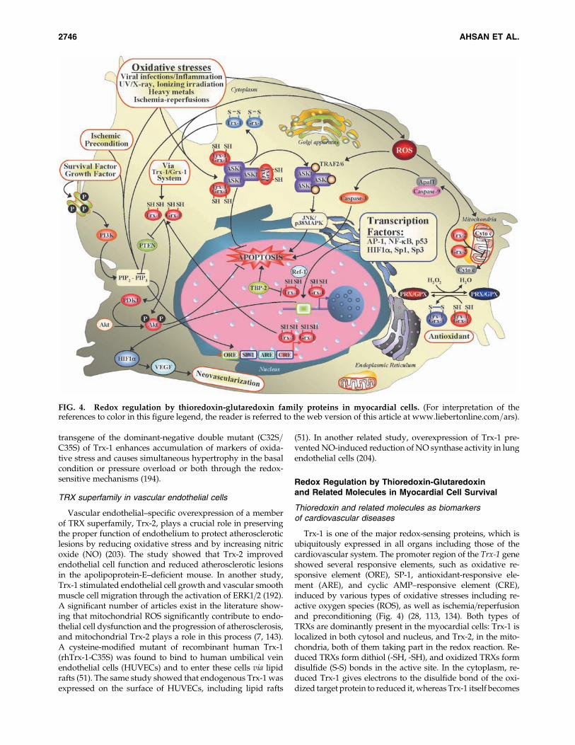
Trx-1 is one of the major redox-sensing proteins, which isubiquitously expressed in all organs including those of thecardiovascular system. The promoter region of the Trx-1 geneshowed several responsive elements, such as oxidative re-sponsive element (ORE), SP-1, antioxidant-responsive ele-ment (ARE), and cyclic AMP–responsive element (CRE),induced by various types of oxidative stresses including re-active oxygen species (ROS), as well as ischemia=reperfusionand preconditioning (Fig. 4) (28, 113, 134). Both types ofTRXs are dominantly present in the myocardial cells: Trx-1 islocalized in both cytosol and nucleus, and Trx-2, in the mito-chondria, both of them taking part in the redox reaction. Re-duced TRXs form dithiol (-SH, -SH), and oxidized TRXs formdisulfide (S-S) bonds in the active site. In the cytoplasm, re-duced Trx-1 gives electrons to the disulfide bond of the oxi-dized target protein to reduced it, whereas Trx-1 itself becomes
FIG. 4. Redox regulation by thioredoxin-glutaredoxin family proteins in myocardial cells. (For interpretation of thereferences to color in this figure legend, the reader is referred to the web version of this article at www.liebertonline.com=ars).
2746 AHSAN ET AL.
oxidized. Oxidized Trx-1 reversibly changes to the reducedform by TrxR1 and NADPH (13, 62, 66). Trx-1–dependentperoxiredoxin (PRX) scavenges hydrogen peroxide (Fig. 1).Thus, cytoplasmic Trx-1 also possesses an antioxidant effecttogether with NADPH, TrxR1, and PRX (Prx I and Prx II).Similarly, mitochondrial Trx-2 also shows an antioxidant effecttogether with NADPH, TrxR2, and Prx III, and it is stronglyexpressed in heart tissue (39, 173). Trx-1 and Trx-2 show redox-regulatory functions in signal transduction of myocardial cellsurvival and prevent apoptosis. Apoptosis signal–regulatingkinase 1 (ASK1) is a mitogen-activated protein kinase kinasekinase (MAPKKK), which is associated with the reduced formof Trx-1 in cytosol and Trx-2 in mitochondria in the normalstate of a cell to prevent cellular apoptosis (156). During oxi-dative stress–induced apoptosis in the cells, TRXs becomeoxidized, followed by the dissociation of ASK1 from TRXs,and then ASK1 interacts with TRAF2=6, inducing the phos-phorylation of JNK and p38 MAPK to transduce the apoptosissignal (Fig. 4) (38, 205). TRXs also interfere with the Ras=Raf=ERK pathway to inhibit apoptosis (94, 95). Reduced Trx-1also inhibits apoptosis by regulating or preserving phos-phorylation status of Akt, or both, by inhibiting PTEN, whichis known as a PI3K-Akt pathway inhibitor or by upregulationof survivin expression (83, 99, 172). Trx-1 is known to be in-volved in redox regulation of a numbers of transcriptionalfactors, including Ref-1, AP-1, NF-kB, HIF-1a, Sp1, and Sp3.During redox regulation, the promoter of Trx-1 binds the Sp1and Sp3 to enhance the expression of Trx-1 (14). The DNAbinding of AP-1, NF-kB, p53, and HIF1a is regulated by Trx-1–or Ref-1–dependent reduction of their key cysteine residues, orboth (34, 58, 60). DNA binding of NF-kB regulates several geneexpressions in response to inflammatory cytokines, infections,ROS, carcinogenesis, cellular stress, and apoptosis inducers(47, 189). Transactivation of p53 causes upregulation of p21expression in the cells transiently transfected with Trx-1 (181).The glucocorticoid receptor is a transcription factor, the
activity of which also is regulated by Trx-1 (117). The mito-chondrial Trx-2 (12.2 kDa) plays a crucial role in the regula-tion of programmed cell death by inhibition of cytochrome crelease from the mitochondria and by increasing mitochon-drial membrane potential, leading to the inhibition of thecaspase-mediated apoptosis pathway (Fig. 4) (27, 173). Trx-1enhances cardioprotection by postmyocardial neovascular-ization through the Trx-1–HO-1–VEGF pathway (82).
Several lines of evidence indicate that Trx-1 reduces myo-cardial injury by reducing both oxidative and nitrative stress.For example, treatment of heat cells with Trx-1 significantlyreduced myocardial apoptosis and upregulated MnSOD(176). In this study, Trx-1 also reduced peroxynitrite donorSIN-1 (3-morpholinosydnonimine)-induced cardiomyocyteapoptosis. In another study, high glucose was found to sen-sitize cardiomyocytes to ischemia=reperfusion injury due tonitrative inactivation of Trx-1 (108). The treatment of car-diomyocytes with rhTrx-1 exerts its cardioprotective effectby suppressing the ischemia=reperfusion-induced nitrativestress (176).
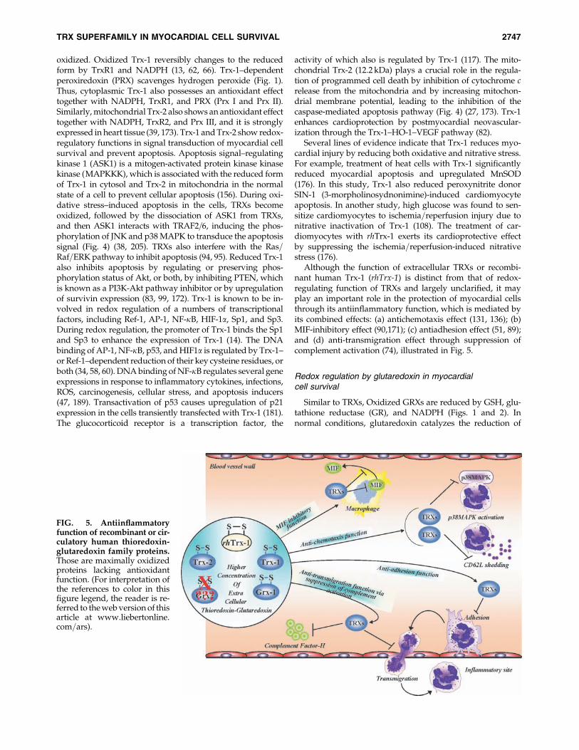
Although the function of extracellular TRXs or recombi-nant human Trx-1 (rhTrx-1) is distinct from that of redox-regulating function of TRXs and largely unclarified, it mayplay an important role in the protection of myocardial cellsthrough its antiinflammatory function, which is mediated byits combined effects: (a) antichemotaxis effect (131, 136); (b)MIF-inhibitory effect (90,171); (c) antiadhesion effect (51, 89);and (d) anti-transmigration effect through suppression ofcomplement activation (74), illustrated in Fig. 5.
Redox regulation by glutaredoxin in myocardialcell survival
Similar to TRXs, Oxidized GRXs are reduced by GSH, glu-tathione reductase (GR), and NADPH (Figs. 1 and 2). Innormal conditions, glutaredoxin catalyzes the reduction of
FIG. 5. Antiinflammatoryfunction of recombinant or cir-culatory human thioredoxin-glutaredoxin family proteins.Those are maximally oxidizedproteins lacking antioxidantfunction. (For interpretation ofthe references to color in thisfigure legend, the reader is re-ferred to the web version of thisarticle at www.liebertonline.com=ars).
TRX SUPERFAMILY IN MYOCARDIAL CELL SURVIVAL 2747
some protein disulfides and GSH-mixed disulfides, but incertain conditions, also can catalyze the reverse reaction,leading to GS-S-protein mixed disulfide formation (23, 41,161).
Like Trx-1, Grx-1 also was shown to bind and negativelyto regulate the activation of ASK1, and the overexpression ofGrx1 represses the ASK1=SEK1=JNK1 pathway and pre-vents cytotoxicity induced by metabolic oxidative stress in aglutathione-dependent manner (Fig. 4) (166). Overexpressionof Grx1 maintains the redox status of Akt, facilitates cellsurvival, and suppresses apoptosis (127). 17=b Estrogen (E2)induces Grx1 and protects H9c2 from apoptosis by the sameAkt pathway (182). Conversely, Grx1 can regulate the Ras=Raf=Erk pathway as a response to hypertrophic stimuli. TheS-glutathionylation as a response to hypertrophic stimuli ofRas causes activation of the Raf=Mek=Erk pathway, lead-ing to hypertrophy. Overexpression of Grx-1 inhibited theS-glutathionylation of the Ras, repressed the activation of theErk, and suppressed the increased protein synthesis inducedby strain stimulation (147). Grx-1 also suppresses the activa-tion of p38 induced by angiotensin II (1).
Grx-2 plays a central role in the maintenance of the mito-chondrial redox environment. Depending on the ratio ofthe GSH to GSSG, Grx-2 can catalyze glutathionylation anddeglutathionylation of the protein. Unlike Grx-1, Grx-2 is asubstrate for mitochondrial TrxR2. During oxidative stress,when the GSH=GSSG ratio is decreased, this alternative re-ducing mechanism becomes operative, suggesting an im-portant role for Grx-2 (81). Grx-2 regulates the activity ofmitochondrial proteins like complex I by reversible S-glutathionylation (9). Human Grx-2 is the first member ofTRX superfamily characterized as an iron–sulfur protein.Grx-2 forms a (2Fe-2S) bridged dimer, which is enzymaticallyinactive, but the (2Fe 2S) cluster serves as a redox sensor forGrx-2 (12, 103, 104). Overexpression of Grx-2 protects the cellsagainst apoptosis induced by 2-deoxy-glucose and doxoru-bicin, by inhibiting the release of cytochrome c as well theactivation of caspase 3, and the loss of cardiolipin (Fig. 4) (35,105). Furthermore, overexpression of Grx-2 promotes the ac-tivation of survival protein Akt, redox-sensitive transcriptionfactor NF-kB, and antiapoptotic Bcl2 (129).
Witte et al. (191) reported that the overexpression of Grx-3in T cells inhibited the activation of AP-1 and NF-kB in a dose-dependent manner (191). In response to oxidative stress,Grx-3=PICOT is translocated into the nucleus (6). Cardiac-specific overexpression of PICOT diminished the hypertro-phic response of the hearts after pressure overload. PICOTwas found to enhance the sensitivity of the myofilaments to-ward Ca2þ, and increased the Ca2þ reuptake by SR. ExcessivePICOT transfection repressed the activation of PKCa, PKCe,and PKCz after hypertrophic insults, whereas the activationERK and JNK was decreased (76). Direct interaction betweenthe PICOT GRX domain (PICOD-HD) and muscle LIM pro-tein (MLP) has been identified. PICOT competes with calci-neurin for binding to MLP in a dose-dependent manner.Overexpressed PICOT extruded the calcineurin from bindingwith MLP, and this inhibited the PE-induced increasedphosphatase activity of calcineurin, as well the binding, de-phosphorylation, and nuclear translocation of NFAT withcalcineurin (18, 76, 77). Grx-5 deficiency leads to inappropri-ate Fe-S cluster synthesis and further deflects the heme syn-thesis. Grx-5 deficiency in humans causes anemia with iron
overload (17, 190). All of these studies suggested that redoxregulation by GRXs plays an essential role in myocardial cellsurvival.
Redox regulation by peroxiredoxinin myocardial cell survival
Peroxiredoxin (PRX) is a potent antioxidant that scavengesH2O2 and produces by-product H2O with the help of reducedTRX (Fig. 4). Prx I is present in both cytosol and nuclearcompartments of a cell. Cytosolic Prx I inhibits NF-kB p50activation and prevents nuclear translocation. However, nu-clear Prx I does not affect the nuclear translocation but pro-motes the activity of NF-kB receptor (50). Decreased Prx I isassociated with increased ROS production, leading to p53activation, which regulates apoptosis through the activationof the caspase-mediated pathway (185). Overexpression ofPrx I also is associated with increased cell proliferation, in-dicating that overexpression of Prx I prevents apoptosis byscavenging ROS (73, 125). PRX also scavenges=reduces per-oxynitrite, acting as a peroxynitrite reductase (183). Prx IIregulates inflammatory responses through the NF-kB andMAP kinase pathways, and thereby controls the macrophageresponse to pro-inflammatory stimuli, like LPS and cytokines(195). Prx III is the mitochondrial isoform of the PRX family,which serves as the first line of defense against ROS genera-tion and subsequent myocardial cell damage (21). In vivotransfer of the Prx III gene prevents cell death induced byoxidative stress (54). Depletion of Prx III results in increasedintracellular H2O2, which makes the cells prone to apoptosisby TNF-a or staurosporine (21). Prx IV regulates the expres-sion of the TP-b receptor and prevents oxidative stress (46).Prx V also prevents p53-dependent generation of ROS andp53-induced apoptosis (209). These two mechanisms areinvolved in inactivation of all PRXs, phosphorylation ofthreonine-90 residue by the cyclin-dependant kinase cdc2,and hyperoxidation of cysteine residues of the active-site se-quence (21, 196). These studies suggest that redox regulationby PRXs is an essential factor for myocardial cell survivalagainst ROS and ROS-induced apoptosis.
Thioredoxin-Glutaredoxin and Related Moleculesas Biomarkers of Cardiovascular Diseases
Thioredoxin and related molecules as biomarkersof cardiovascular diseases
Cardiovascular diseases are among the major causes ofdeath all over the world and involve oxidative stress. Oxi-dative stress is deeply involved in atherosclerotic plaqueformation and uptake of oxidized LDL (oxLDL) by macro-phages and leads to induction of Trx-1 (139). Higher oxLDLlevels in serum are associated with higher serum levels ofTrx1, indicating its absence as a risk factor of coronary ath-erosclerotic diseases (Ahsan M.K. et al., unpublished data).TrxR1 was also induced in atherosclerotic plaques (40).Plasma Trx-1 is elevated during reperfusion of the post-cardioplegic heart because of systemic oxidative stress (133).Serum Trx-1 levels are elevated in the patients with acutecoronary syndrome and dilated cardiomyopathy but not instable angina patients (86, 163). The serum levels of Trx-1 arehighly elevated in the acute phase but not in the chronic phaseof patients with fulminant myocarditis, Trx-1 is expressed in
2748 AHSAN ET AL.
inflammatory cells as well as in cardiomyocytes in the biopsysamples of the left ventricle in patients with fulminantmyocarditis (162). Plasma Trx-1 levels are elevated in pa-tients with unstable angina, spastic angina, and acute myo-cardial infraction compared with those in stable exertionalangina and chest-pain syndrome (61, 120, 165). ElevatedTrx-1 levels in acute myocardial infraction are associatedwith hyperaggregation of platelet, which indicates furtherrisk as well as pathogenesis of ischemic heart diseases (123).Highly elevated plasma or serum Trx-1 levels are reportedin the patients with diabetes mellitus (DM), especially inDM type II as well as in patients with glucose intolerance(IGT) and patients with hypertension, hypercholesterolemia,and atherosclerosis, all of these being the major risk factorsfor cardiovascular diseases (121, 122, 139). Conversely, inspontaneous hypertensive rats, the Trx-1 expression is de-creased in the aorta or in the aortic arch (174). In the heart,preconditioning-induced hormesis is involved in cGMP-dependent induction of redox protein Trx-1 and Trx-2, in-cluding MnSOD and heat-shock protein 70 (HSP70), leadingto cardioprotection (22). Therefore, the postischemic heartdisease–induced induction of Trx-1 and Trx-2 in serum mightshow a good adaptive response for the heart, although we donot have any direct evidence. Therefore, plasma or serumTRXs levels may be good markers to identify the risk factoror pathologic state or postadaptive signs of cardiovasculardiseases.
Glutaredoxin and related molecules as biomarkersof cardiovascular diseases
Only a handful number of studies investigated the detectionof extracellular levels of Grx-1. Nakamura et al. (135), throughELISA assays, found reduced plasma levels of Grx-1 in pre-operative cardiac patients. Lundberg and colleagues (109) alsoused sandwich ELISA to determinate the cellular and plasmalevels of Grx-1 and Grx-2; the Grx-2 level was not detectable inthe plasma of healthy donors (109). The study revealed that theGrx-1 level was increased during cardiopulmonary bypasssurgery, probably because of the increased hemolysis. Thisstudy also reported that peripheral blood mononuclearcells secrete Grx-1 without stimulation. A related study re-ported that the plasma levels of Grx-1 of the control patients(angiography group) were noted to be higher compared withthose of the angiography group. However, this study couldnot find a correlation between the plasma level of Grx-1 beforeand after angioplasty (186). It was also reported that bloodglutathione reductase (GR) was decreased in patients withmyocardial infarction (32). Therefore, plasma or serum levelsof GRXs and related molecules may also to be good markersto identify the risk factor or pathologic state or postadaptiveresponse of cardiovascular diseases. Further studies mustbe conducted to investigate the extracellular level and the roleand the source of GRXs and related molecules in diverse car-diovascular disease conditions compared with normal condi-tions to confirm that GRXs and the related molecules can beused as biomarkers of cardiovascular diseases.
Peroxiredoxin as a biomarkerof cardiovascular diseases
A recent study shows that the plasma levels of oxidized PrxII and VI are increased in Alzheimer disease patient (200).
Although oxidized Prx II and VI already are established asbiomarkers of Alzheimer disease, in cardiovascular diseases,the role PRXs as biomarkers is yet to be confirmed. We maydetect PRXs levels in plasma by using the assay kits in severalcardiovascular disease states to establish PRXs as biomark-ers of cardiovascular diseases. This hypothesis was furtherstrengthened by Brixius et al. (16); they showed a selectivedownregulation of some PRX isoforms (Prx III to VI) in pa-tients with dilated cardiomyopathy (16). Further studiesmight establish whether PRXs could be used as biomarkers ofcardiovascular diseases.
Implication of Thioredoxin, Glutaredoxin,and Peroxiredoxin Gene Therapyin Cardiovascular Diseases
Gene delivery of thioredoxin
Since recombinant human Trx-1 (rhTrx-1) was shown topossess antioxidant, antiinflammatory, and antiapoptoticfunctions that demonstrated its cytoprotective effect, Trx-1has became one of the better therapeutic agents for the dis-eases related to oxidative stress, acute inflammation, andapoptosis=necrosis. Most of the cardiovascular diseases arerelated to oxidative stress, acute inflammation, and apoptosis=necrosis (56, 70, 88, 100). Moreover, resveratrol, a drug formyocardial infarction (MI), improves neovascularization inthe infarct myocardium, and temocapril, a cardiovascular-protective drug, exerts cytoprotective effect through upregu-lation of Trx-1 and Trx-2 expression (82, 202). In addition,an animal model shows that endogenous Trx-1 has a protec-tive role in the ischemic heart (180). The Trx-2–transgenicmice model shows reduced ROS, increased nitric oxide (NO)bioavailability, leading to reduced vasoconstriction and in-creased vasodilatation, indicating that Trx-2 may have abeneficiary effect in hypertension. Trx-2 also improves ECfunction and reduces atherosclerotic lesions in the apolipo-protein E–deficient mouse model (203). Conversely, cardiaccell–specific ablation of TrxR2 results in fatal dilated cardio-myopathy and death shortly after birth in a mice model,indicating that TrxR2 as well as Trx-2 may have also benefi-cial effects in dilated cardiomyopathy (24). S-nitrosylation ofrhTrx-1 shows more potential cardioprotective effects againstischemic hearts in a mouse model (175, 176). rhTrx-1 amelio-rates myosin-induced autoimmune myocarditis in mice (106).All these reports suggest that TRXs might be good therapeuticagents for clinical application against cardiovascular diseases.However, developing the drug-delivery technique and itsbioavailability is one of the important steps to making a suc-cessful treatment plan. The half-life of rhTrx-1 in plasma isroughly 1 h in mice, 2 h in rats, and 8 h in monkeys (130).Moreover, our research experiment shows that a continuousflow of rhTrx-1 is needed for the maximal therapeutic effi-cacy of TRXs. To elongate the half-life of TRXs in plasma, apossible modification has been considered: conjugation withpolyethylene glycol (PEG), which bridges with cysteine resi-dues. This modification is not recommended for clinical trial,because these cysteine residues of TRXs play a crucial rolein redox regulation. Therefore, one of the best techniques isgene therapy or gene delivery to make a successful treatmentplan by using these redox proteins in cardiovascular diseases.This study is continuing in our Cardiovascular ResearchCenter.
TRX SUPERFAMILY IN MYOCARDIAL CELL SURVIVAL 2749
Gene delivery of glutaredoxin
Overexpression of both Grx-1 and Grx-2 renders the heartresistant to ischemia=reperfusion injury (42, 111, 129). Ourrecent studies showed that several natural food products, likeresveratrol or broccoli, induce Grx-1 and Grx-2 and protectthe heart from ischemia=reperfusion injury (31, 126). Doxor-ubicin, a known anticancer drug, exerts cardiac cell damageduring chemotherapy. Overexpression of Grx-2 amelioratesdoxorubicin-induced cardiac cell damage as well as ventric-ular remodeling of the heart (30). These results suggest Grx-2as a possible therapeutic candidate for the cancer patient whois undergoing treatment with doxorubicin. The Grx-3 nega-tively regulates left ventricular hypertrophy, suggesting apossible therapeutic role for Grx-3 in left ventricular hyper-trophy (18, 76, 77). Moreover, Grx-1 gene therapy can preventischemia=reperfusion injury in the early phase in the diabeticmouse model (Lekli I and Das DK et al., unpublished data).Based on all these studies, we may assume that GRX over-expression could have therapeutic value in different heartdiseases. Although we observed all these data after theoverexpression of GRXs, like a preventive medicine, furtherstudy is needed to address the therapeutic value of GRXs as amodern medicine after the disease condition develops. Theoverexpression GRXs could be done by direct gene delivery.
Gene delivery of peroxiredoxin
As mentioned earlier, ROS-induced apoptosis=necrosisand inflammation are involved in pathogenesis of a major-ity of cardiovascular diseases. All the isoforms of PRXs areinvolved in scavenging ROS and prevent ROS-inducedapoptosis=necrosis, thus indicating the possible therapeuticvalues of PRXs in cardiovascular diseases. Deficiency of PrxVI makes the heart more susceptible to ischemia=reperfusion–induced injury and leads to an increase in infarct size andapoptotic cell death, suggesting a protective role of Prx VI onischemia=reperfusion injury as well as myocardial infarction(MI) (128). Moreover, overexpression of mitochondrial Prx IIIin another mouse model shows resistance to MI and preventsventricular remodeling and post-MI heart failure (116). Thus,PRXs should also be a potent therapeutic agent for cardio-vascular diseases.
Concluding Remarks
The review is focused on the vital cellular antioxidantdefense mechanism comprising TRXs, GRXs, and PRXs,along with their isoforms and their effects on the myocar-dium. Ischemia=reperfusion-induced injury generates a hugeamount of ROS. Uncontrolled increment of ROS brings tur-bulence to the cellular microenvironment that ultimatelytriggers apoptosis. Given the fact that modulation of ROS is ofimmense importance for the maintenance of cardiovascularhealth. Overexpression of the few isoforms of TRXs, GRXs,and PRXs have been shown to be beneficial for the stressedmyocardium, which works in redox chain to scavenge theROS. Added to that, all these proteins seem to have a role inantiinflammatory activities. All our summarized informationsuggests myocardial cell survival as an effect of TRXs-, GRXs-,and PRXs-mediated redox regulation. It appears reasonableto speculate that improve cardiac health is possible by over-expressing these proteins by a successful and clinically ap-plicable gene therapy.
Acknowledgments
We thank Dr. Monika Das, Dr. Narasimman Gurusamy,Subhendu Mukherjee. and Jocelyn Dudley (all in the De-partment of Surgery, UCONN Health Center) for their criticalreviews of the manuscript. This study was supported bygrants-in-aid from the National Institutes of Health: NIH HL34360, HL 22559, and HL 33889.
References
1. Adachi T, Pimentel DR, Heibeck T, Hou X, Lee YJ, Jiang B,Ido Y, and Cohen RA. S-glutathiolation of Ras mediatesredox-sensitive signaling by angiotensin II in vascularsmooth muscle cells. J Biol Chem 279: 29857–29862, 2004.
2. Ago T, Liu T, Zhai P, Chen W, Li H, Molkentin JD, VatnerSF, and Sadoshima J. A redox-dependent pathway forregulating class II HDACs and cardiac hypertrophy. Cell133: 978–993, 2008.
3. Ago T and Sadoshima J. Thioredoxin1 as a negative regu-lator of cardiac hypertrophy. Antioxid Redox Signal 9: 679–687, 2007.
4. Ago T, Yeh I, Yamamoto M, Schinke-Braun M, Brown JA,Tian B, and Sadoshima J. Thioredoxin1 upregulates mito-chondrial proteins related to oxidative phosphorylationand TCA cycle in the heart. Antioxid Redox Signal 8: 1635–1650, 2006.
5. Araki M, Nanri H, Ejima K, Murasato Y, Fujiwara T,Nakashima Y, and Ikeda M. Antioxidant function of themitochondrial protein SP-22 in the cardiovascular system.J Biol Chem 274: 2271–2278, 1999.
6. Babichev Y and Isakov N. Tyrosine phosphorylation ofPICOT and its translocation to the nucleus in response ofhuman T cells to oxidative stress. Adv Exp Med Biol 495: 41–45, 2001.
7. Ballinger SW, Patterson C, Knight-Lozano CA, Burow DL,Conklin CA, Hu Z, Reuf J, Horaist C, Lebovitz R, HunterGC, McIntyre K, and Runge MS. Mitochondrial integrityand function in atherogenesis. Circulation 106: 544–549,2002.
8. Bandyopadhyay S, Starke DW, Mieyal JJ, and GronostajskiRM. Thioltransferase (glutaredoxin) reactivates the DNA-binding activity of oxidation-inactivated nuclear factor I.J Biol Chem 273: 392–397, 1998.
9. Beer SM, Taylor ER, Brown SE, Dahm CC, Costa NJ,Runswick MJ, and Murphy MP. Glutaredoxin 2 catalyzesthe reversible oxidation and glutathionylation of mito-chondrial membrane thiol proteins: implications for mito-chondrial redox regulation and antioxidant DEFENSE. J BiolChem 279: 47939–47951, 2004.
10. Berggren M, Gallegos A, Gasdaska J, and Powis G. Cellularthioredoxin reductase activity is regulated by selenium.Anticancer Res 17: 3377–3380, 1997.
11. Berggren MM and Powis G. Alternative splicing is associ-ated with decreased expression of the redox proto-oncogenethioredoxin-1 in human cancers. Arch Biochem Biophys 389:144–149, 2001.
12. Berndt C, Hudemann C, Hanschmann EM, Axelsson R,Holmgren A, and Lillig CH. How does iron-sulfur clustercoordination regulate the activity of human glutaredoxin 2?Antioxid Redox Signal 9: 151–157, 2007.
13. Bjornstedt M, Xue J, Huang W, Akesson B, and HolmgrenA. The thioredoxin and glutaredoxin systems are efficientelectron donors to human plasma glutathione peroxidase.J Biol Chem 269: 29382–29384, 1994.
2750 AHSAN ET AL.
14. Bloomfield KL, Osborne SA, Kennedy DD, Clarke FM, andTonissen KF. Thioredoxin-mediated redox control of thetranscription factor Sp1 and regulation of the thioredoxingene promoter. Gene 319: 107–116, 2003.
15. Brigelius-Flohe R. Glutathione peroxidases and redox-regulated transcription factors. Biol Chem 387: 1329–1335,2006.
16. Brixius K, Schwinger RH, Hoyer F, Napp A, Renner R,Bolck B, Kumin A, Fischer U, Mehlhorn U, Werner S,and Bloch W. Isoform-specific downregulation of pero-xiredoxin in human failing myocardium. Life Sci 81: 823–831, 2007.
17. Camaschella C, Campanella A, De Falco L, Boschetto L,Merlini R, Silvestri L, Levi S, and Iolascon A. The humancounterpart of zebrafish shiraz shows sideroblastic-likemicrocytic anemia and iron overload. Blood 110: 1353–1358,2007.
18. Cha H, Kim JM, Oh JG, Jeong MH, Park CS, Park J, JeongHJ, Park BK, Lee YH, Jeong D, Yang DK, Bernecker OY,Kim do H, Hajjar RJ, and Park WJ. PICOT is a criticalregulator of cardiac hypertrophy and cardiomyocyte con-tractility. J Mol Cell Cardiol 45: 796–803, 2008.
19. Chae HZ, Chung SJ, and Rhee SG. Thioredoxin-dependentperoxide reductase from yeast. J Biol Chem 269: 27670–27678, 1994.
20. Chae HZ, Kim HJ, Kang SW, and Rhee SG. Characteriza-tion of three isoforms of mammalian peroxiredoxin thatreduce peroxides in the presence of thioredoxin. DiabetesRes Clin Pract 45: 101–112, 1999.
21. Chang TS, Cho CS, Park S, Yu S, Kang SW, and Rhee SG.Peroxiredoxin III, a mitochondrion-specific peroxidase, reg-ulates apoptotic signaling by mitochondria. J Biol Chem 279:41975–41984, 2004.
22. Chiueh CC, Andoh T, and Chock PB. Induction ofthioredoxin and mitochondrial survival proteins mediatespreconditioning-induced cardioprotection and neuropro-tection. Ann N Y Acad Sci 1042: 403–418, 2005.
23. Chrestensen CA, Starke DW, and Mieyal JJ. Acute cad-mium exposure inactivates thioltransferase (glutaredoxin),inhibits intracellular reduction of protein-glutathionyl-mixed disulfides, and initiates apoptosis. J Biol Chem 275:26556–26565, 2000.
24. Conrad M, Jakupoglu C, Moreno SG, Lippl S, Banjac A,Schneider M, Beck H, Hatzopoulos AK, Just U, Sinowatz F,Schmahl W, Chien KR, Wurst W, Bornkamm GW, andBrielmeier M. Essential role for mitochondrial thioredoxinreductase in hematopoiesis, heart development, and heartfunction. Mol Cell Biol 24: 9414–9423, 2004.
25. Cunnea PM, Miranda-Vizuete A, Bertoli G, Simmen T,Damdimopoulos AE, Hermann S, Leinonen S, Huikko MP,Gustafsson JA, Sitia R, and Spyrou G. ERdj5, an endo-plasmic reticulum (ER)-resident protein containing DnaJand thioredoxin domains, is expressed in secretory cells orfollowing ER stress. J Biol Chem 278: 1059–1066, 2003.
26. Dai J, Wang X, Feng J, Kong W, Xu Q, and Shen X. Reg-ulatory role of thioredoxin in homocysteine-induced mono-cyte chemoattractant protein-1 secretion in monocytes=macrophages. FEBS Lett 582: 3893–3898, 2008.
27. Damdimopoulos AE, Miranda-Vizuete A, Pelto-Huikko M,Gustafsson JA, and Spyrou G. Human mitochondrialthioredoxin: involvement in mitochondrial membrane po-tential and cell death. J Biol Chem 277: 33249–33257, 2002.
28. Das DK. Thioredoxin regulation of ischemic precondition-ing. Antioxid Redox Signal 6: 405–412, 2004.
29. Das S, Der P, Raychaudhuri U, Maulik N, and Das DK. Theeffect of Euryale ferox (Makhana), an herb of aquatic origin,on myocardial ischemic reperfusion injury. Mol Cell Bio-chem 289: 55–63, 2006.
30. Diotte NM, Xiong Y, Gao J, Chua BH, and Ho YS. Attenua-tion of doxorubicin-induced cardiac injury by mitochon-drial glutaredoxin 2. Biochim Biophys Acta 1793: 427–438,2009.
31. Dudley J, Das S, Mukherjee S, and Das DK. Resveratrol, aunique phytoalexin present in red wine, delivers eithersurvival signal or death signal to the ischemic myocardiumdepending on dose. J Nutr Biochem 20: 443–452, 2009.
32. Dwivedi VK, Chandra M, Misra PC, Misra A, and MisraMK. Status of some free radical scavenging enzymes in theblood of myocardial infarction patients. J Enzyme Inhib MedChem 21: 43–46, 2006.
33. Ebrahimian T, Sairam MR, Schiffrin EL, and Touyz RM.Cardiac hypertrophy is associated with altered thioredoxinand ASK-1 signaling in a mouse model of menopause. Am JPhysiol Heart Circ Physiol 295: H1481–H1488, 2008.
34. Ema M, Hirota K, Mimura J, Abe H, Yodoi J, Sogawa K,Poellinger L, and Fujii-Kuriyama Y. Molecular mechanismsof transcription activation by HLF and HIF1alpha in re-sponse to hypoxia: their stabilization and redox signal-induced interaction with CBP=p300. EMBO J 18: 1905–1914,1999.
35. Enoksson M, Fernandes AP, Prast S, Lillig CH, HolmgrenA, and Orrenius S. Overexpression of glutaredoxin 2 at-tenuates apoptosis by preventing cytochrome c release.Biochem Biophys Res Commun 327: 774–779, 2005.
36. Fernandes AP and Holmgren A. Glutaredoxins: glutathione-dependent redox enzymes with functions far beyond asimple thioredoxin backup system. Antioxid Redox Signal 6:63–74, 2004.
37. Fisher AB, Dodia C, Manevich Y, Chen JW, and FeinsteinSI. Phospholipid hydroperoxides are substrates for non-selenium glutathione peroxidase. J Biol Chem 274: 21326–21334, 1999.
38. Fujino G, Noguchi T, Matsuzawa A, Yamauchi S, Saitoh M,Takeda K, and Ichijo H. Thioredoxin and TRAF familyproteins regulate reactive oxygen species-dependent acti-vation of ASK1 through reciprocal modulation of theN-terminal homophilic interaction of ASK1. Mol Cell Biol27: 8152–8163, 2007.
39. Funato Y and Miki H. Nucleoredoxin, a novel thioredoxinfamily member involved in cell growth and differentiation.Antioxid Redox Signal 9: 1035–1057, 2007.
40. Furman C, Rundlof AK, Larigauderie G, Jaye M, Bricca G,Copin C, Kandoussi AM, Fruchart JC, Arner ES, and RouisM. Thioredoxin reductase 1 is upregulated in atheroscle-rotic plaques: specific induction of the promoter in humanmacrophages by oxidized low-density lipoproteins. FreeRadic Biol Med 37: 71–85, 2004.
41. Gallogly MM and Mieyal JJ. Mechanisms of reversibleprotein glutathionylation in redox signaling and oxidativestress. Curr Opin Pharmacol 7: 381–391, 2007.
42. Gallogly MM, Starke DW, Leonberg AK, Ospina SM, andMieyal JJ. Kinetic and mechanistic characterization andversatile catalytic properties of mammalian glutaredoxin 2:implications for intracellular roles. Biochemistry 47: 11144–11157, 2008.
43. Gasdaska JR, Harney JW, Gasdaska PY, Powis G, and BerryMJ. Regulation of human thioredoxin reductase expres-sion and activity by 3’-untranslated region selenocysteine
TRX SUPERFAMILY IN MYOCARDIAL CELL SURVIVAL 2751
insertion sequence and mRNA instability elements. J BiolChem 274: 25379–25385, 1999.
44. Gasdaska PY, Gasdaska JR, Cochran S, and Powis G.Cloning and sequencing of a human thioredoxin reductase.FEBS Lett 373: 5–9, 1995.
45. Gasdaska PY, Oblong JE, Cotgreave IA, and Powis G. Thepredicted amino acid sequence of human thioredoxin isidentical to that of the autocrine growth factor human adultT-cell derived factor (ADF): thioredoxin mRNA is elevatedin some human tumors. Biochim Biophys Acta 1218: 292–296,1994.
46. Giguere P, Turcotte ME, Hamelin E, Parent A, Brisson J,Laroche G, Labrecque P, Dupuis G, and Parent JL.Peroxiredoxin-4 interacts with and regulates the throm-boxane A(2) receptor. FEBS Lett 581: 3863–3868, 2007.
47. Gilmore TD. Introduction: the Rel=NF-kappaB signaltransduction pathway. Semin Cancer Biol 8: 61–62, 1997.
48. Gladyshev VN, Liu A, Novoselov SV, Krysan K, Sun QA,Kryukov VM, Kryukov GV, and Lou MF. Identificationand characterization of a new mammalian glutaredoxin(thioltransferase), Grx2. J Biol Chem 276: 30374–30380, 2001.
49. Hagg D, Englund MC, Jernas M, Schmidt C, Wiklund O,Hulten LM, Ohlsson BG, Carlsson LM, Carlsson B, andSvensson PA. Oxidized LDL induces a coordinated up-regulation of the glutathione and thioredoxin systems inhuman macrophages. Atherosclerosis 185: 282–289, 2006.
50. Hansen JM, Moriarty-Craige S, and Jones DP. Nuclear andcytoplasmic peroxiredoxin-1 differentially regulate NF-kappaB activities. Free Radic Biol Med 43: 282–288, 2007.
51. Hara T, Kondo N, Nakamura H, Okuyama H, Mitsui A,Hoshino Y, and Yodoi J. Cell-surface thioredoxin-1: possi-ble involvement in thiol-mediated leukocyte-endothelialcell interaction through lipid rafts. Antioxid Redox Signal 9:1427–1437, 2007.
52. Hariharan J, Hebbar P, Ranie J, Philomena, Sinha AM, andDatta S. Alternative forms of the human thioredoxinmRNA: identification and characterization. Gene 173: 265–270, 1996.
53. Hashemy SI and Holmgren A. Regulation of the catalyticactivity and structure of human thioredoxin 1 via oxidationand S-nitrosylation of cysteine residues. J Biol Chem 283:21890–21898, 2008.
54. Hattori F, Murayama N, Noshita T, and Oikawa S. Mito-chondrial peroxiredoxin-3 protects hippocampal neuronsfrom excitotoxic injury in vivo. J Neurochem 86: 860–868,2003.
55. Herrero E and de la Torre-Ruiz MA. Monothiol glutar-edoxins: a common domain for multiple functions. Cell MolLife Sci 64: 1518–1530, 2007.
56. Hiraoka Y, Kishimoto C, Takada H, Kurokawa M, OchiaiH, Shiraki K, and Sasayama S. Role of oxygen derived freeradicals in the pathogenesis of coxsackievirus B3 myocar-ditis in mice. Cardiovasc Res 27: 957–961, 1993.
57. Hiroi T, Watabe S, Takimoto K, Yago N, Yamamoto Y, andTakahashi SY. The cDNA sequence encoding bovine SP-22,a new defence system against reactive oxygen species inmitochondria. DNA Seq 6: 239–242, 1996.
58. Hirota K, Matsui M, Iwata S, Nishiyama A, Mori K, andYodoi J. AP-1 transcriptional activity is regulated by a di-rect association between thioredoxin and Ref-1. Proc NatlAcad Sci U S A 94: 3633–3638, 1997.
59. Hirota K, Matsui M, Murata M, Takashima Y, Cheng FS,Itoh T, Fukuda K, and Yodoi J. Nucleoredoxin, glutar-edoxin, and thioredoxin differentially regulate NF-kappaB,
AP-1, and CREB activation in HEK293 cells. Biochem Bio-phys Res Commun 274: 177–182, 2000.
60. Hirota K, Murata M, Sachi Y, Nakamura H, Takeuchi J,Mori K, and Yodoi J. Distinct roles of thioredoxin in thecytoplasm and in the nucleus: a two-step mechanism ofredox regulation of transcription factor NF-kappaB. J BiolChem 274: 27891–27897, 1999.
61. Hokamaki J, Kawano H, Soejima H, Miyamoto S, KajiwaraI, Kojima S, Sakamoto T, Sugiyama S, Yoshimura M, Na-kamura H, Yodoi J, and Ogawa H. Plasma thioredoxinlevels in patients with unstable angina. Int J Cardiol 99: 225–231, 2005.
62. Holmgren A. Enzymatic reduction-oxidation of proteindisulfides by thioredoxin. Methods Enzymol 107: 295–300,1984.
63. Holmgren A. Glutathione-dependent synthesis of deoxyri-bonucleotides: characterization of the enzymatic mechanismof Escherichia coli glutaredoxin. J Biol Chem 254: 3672–3678,1979.
64. Holmgren A. Glutathione-dependent synthesis of deoxyri-bonucleotides: purification and characterization of glutar-edoxin from Escherichia coli. J Biol Chem 254: 3664–3671,1979.
65. Holmgren A. Hydrogen donor system for Escherichia coliribonucleoside-diphosphate reductase dependent uponglutathione. Proc Natl Acad Sci U S A 73: 2275–2279, 1976.
66. Holmgren A. Reduction of disulfides by thioredoxin: ex-ceptional reactivity of insulin and suggested functions ofthioredoxin in mechanism of hormone action. J Biol Chem254: 9113–9119, 1979.
67. Holmgren A. Thioredoxin. Annu Rev Biochem 54: 237–271,1985.
68. Holmgren A. Thioredoxin and glutaredoxin systems. J BiolChem 264: 13963–13966, 1989.
69. Holmgren A, Soderberg BO, Eklund H, and Branden CI.Three-dimensional structure of Escherichia coli thioredoxin-S2 to 2.8 A resolution. Proc Natl Acad Sci U S A 72: 2305–2309, 1975.
70. Hoshino Y, Shioji K, Nakamura H, Masutani H, and YodoiJ. From oxygen sensing to heart failure: role of thioredoxin.Antioxid Redox Signal 9: 689–699, 2007.
71. Hosoda A, Kimata Y, Tsuru A, and Kohno K. JPDI, a novelendoplasmic reticulum-resident protein containing both aBiP-interacting J-domain and thioredoxin-like motifs. J BiolChem 278: 2669–2676, 2003.
72. Hudemann C, Lonn ME, Godoy JR, Zahedi Avval F, Ca-pani F, Holmgren A, and Lillig CH. Identification, expres-sion pattern, and characterization of mouse glutaredoxin 2isoforms. Antioxid Redox Signal 11: 1–14, 2009.
73. Immenschuh S and Baumgart-Vogt E. Peroxiredoxins, ox-idative stress, and cell proliferation. Antioxid Redox Signal 7:768–777, 2005.
74. Inomata Y, Tanihara H, Tanito M, Okuyama H,Hoshino Y, Kinumi T, Kawaji T, Kondo N, Yodoi J, andNakamura H. Suppression of choroidal neovascularizationby thioredoxin-1 via interaction with complement factor H.Invest Ophthalmol Vis Sci 49: 5118–5125, 2008.
75. Isakov N, Witte S, and Altman A. PICOT-HD: a highlyconserved protein domain that is often associated withthioredoxin and glutaredoxin modules. Trends Biochem Sci25: 537–539, 2000.
76. Jeong D, Cha H, Kim E, Kang M, Yang DK, Kim JM, YoonPO, Oh JG, Bernecker OY, Sakata S, Le TT, Cui L, Lee YH,Kim do H, Woo SH, Liao R, Hajjar RJ, and Park WJ.
2752 AHSAN ET AL.
PICOT inhibits cardiac hypertrophy and enhances ventric-ular function and cardiomyocyte contractility. Circ Res 99:307–314, 2006.
77. Jeong D, Kim JM, Cha H, Oh JG, Park J, Yun SH, Ju ES, JeonES, Hajjar RJ, and Park WJ. PICOT attenuates cardiac hy-pertrophy by disrupting calcineurin-NFAT signaling. CircRes 102: 711–719, 2008.
78. Jeong W, Yoon HW, Lee SR, and Rhee SG. Identificationand characterization of TRP14, a thioredoxin-related pro-tein of 14 kDa: new insights into the specificity of thior-edoxin function. J Biol Chem 279: 3142–3150, 2004.
79. Jimenez A and Miranda-Vizuete A. Purification and char-acterization of delta3Trx-1, a splicing variant of humanthioredoxin-1 lacking exon 3. Protein Expr Purif 27: 319–324,2003.
80. Jimenez A, Zu W, Rawe VY, Pelto-Huikko M, Flickinger CJ,Sutovsky P, Gustafsson JA, Oko R, and Miranda-VizueteA. Spermatocyte=spermatid-specific thioredoxin-3, a novelGolgi apparatus-associated thioredoxin, is a specific markerof aberrant spermatogenesis. J Biol Chem 279: 34971–34982,2004.
81. Johansson C, Lillig CH, and Holmgren A. Human mito-chondrial glutaredoxin reduces S-glutathionylated proteinswith high affinity accepting electrons from either glutathi-one or thioredoxin reductase. J Biol Chem 279: 7537–7543,2004.
82. Kaga S, Zhan L, Matsumoto M, and Maulik N. Resveratrolenhances neovascularization in the infarcted rat myo-cardium through the induction of thioredoxin-1, hemeoxygenase-1 and vascular endothelial growth factor. J MolCell Cardiol 39: 813–822, 2005.
83. Kaimul Ahsan M, Nakamura H, Tanito M, Yamada K,Utsumi H, and Yodoi J. Thioredoxin-1 suppresses lunginjury and apoptosis induced by diesel exhaust particles(DEP) by scavenging reactive oxygen species and by in-hibiting DEP-induced downregulation of Akt. Free RadicBiol Med 39: 1549–1559, 2005.
84. Kaimul Ahsan M, Nakamura H, Masutani H, and Yodoi J.Thioredoxin and thioredoxin-binding protein-2 in cancerand metabolic syndrome. Free Radic Biol Med 43: 861–868,2007.
85. Kim K, Rhee SG, and Stadtman ER. Nonenzymatic cleav-age of proteins by reactive oxygen species generated bydithiothreitol and iron. J Biol Chem 260: 15394–15397, 1985.
86. Kishimoto C, Shioji K, Nakamura H, Nakayama Y, Yodoi J,and Sasayama S. Serum thioredoxin (TRX) levels in pa-tients with heart failure. Jpn Circ J 65: 491–494, 2001.
87. Kleemann R, Kapurniotu A, Mischke R, Held J, and Bern-hagen J. Characterization of catalytic centre mutants ofmacrophage migration inhibitory factor (MIF) and com-parison to Cys81Ser MIF. Eur J Biochem 261: 753–766, 1999.
88. Kobayashi-Miura M, Shioji K, Hoshino Y, Masutani H,Nakamura H, and Yodoi J. Oxygen sensing and redoxsignaling: the role of thioredoxin in embryonic develop-ment and cardiac diseases. Am J Physiol Heart Circ Physiol292: H2040–H2050, 2007.
89. Kondo N, Ishii Y, Kwon YW, Tanito M, Sakakura-Nishiyama J, Mochizuki M, Maeda M, Suzuki S, Kojima M,Kim YC, Son A, Nakamura H, and Yodoi J. Lipidraft-mediated uptake of cysteine-modified thioredoxin-1:apoptosis enhancement by inhibiting the endogenousthioredoxin-1. Antioxid Redox Signal 9: 1439–1448, 2007.
90. Kondo N, Ishii Y, Son A, Sakakura-Nishiyama J, KwonYW, Tanito M, Nishinaka Y, Matsuo Y, Nakayama T,
Taniguchi M, and Yodoi J. Cysteine-dependent immuneregulation by TRX and MIF=GIF family proteins. ImmunolLett 92: 143–147, 2004.
91. Krause G, Lundstrom J, Barea JL, Pueyo de la Cuesta C,and Holmgren A. Mimicking the active site of proteindisulfide-isomerase by substitution of proline 34 in Es-cherichia coli thioredoxin. J Biol Chem 266: 9494–9500, 1991.
92. Kumar S, Bjornstedt M, and Holmgren A. Selenite is asubstrate for calf thymus thioredoxin reductase and thior-edoxin and elicits a large non-stoichiometric oxidation ofNADPH in the presence of oxygen. Eur J Biochem 207: 435–439, 1992.
93. Kurooka H, Kato K, Minoguchi S, Takahashi Y, Ikeda J,Habu S, Osawa N, Buchberg AM, Moriwaki K, Shisa H,and Honjo T. Cloning and characterization of the nucleor-edoxin gene that encodes a novel nuclear protein related tothioredoxin. Genomics 39: 331–339, 1997.
94. Kuster GM, Pimentel DR, Adachi T, Ido Y, Brenner DA,Cohen RA, Liao R, Siwik DA, and Colucci WS. Alpha-adrenergic receptor-stimulated hypertrophy in adult rat ven-tricular myocytes is mediated via thioredoxin-1-sensitiveoxidative modification of thiols on Ras. Circulation 111:1192–1198, 2005.
95. Kuster GM, Siwik DA, Pimentel DR, and Colucci WS. Roleof reversible, thioredoxin-sensitive oxidative protein mod-ifications in cardiac myocytes. Antioxid Redox Signal 8:2153–2159, 2006.
96. Laughner BJ, Sehnke PC, and Ferl RJ. A novel nuclearmember of the thioredoxin superfamily. Plant Physiol 118:987–996, 1998.
97. Laurent TC, Moore EC, and Reichard P. Enzymatic syn-thesis of deoxyribonucleotides. IV: isolation and character-ization of thioredoxin, the hydrogen donor from Escherichiacoli B. J Biol Chem 239: 3436–3444, 1964.
98. Lee KK, Murakawa M, Takahashi S, Tsubuki S, KawashimaS, Sakamaki K, and Yonehara S. Purification, molecularcloning, and characterization of TRP32, a novel thioredoxin-related mammalian protein of 32 kDa. J Biol Chem 273:19160–19166, 1998.
99. Lee SR, Yang KS, Kwon J, Lee C, Jeong W, and Rhee SG.Reversible inactivation of the tumor suppressor PTEN byH2O2. J Biol Chem 277: 20336–20342, 2002.
100. Lefer DJ and Granger DN. Oxidative stress and cardiacdisease. Am J Med 109: 315–323, 2000.
101. Li X, Tang K, Xie B, Li S, and Rozanski GJ. Regulation ofKv4 channel expression in failing rat heart by the thior-edoxin system. Am J Physiol Heart Circ Physiol 295: H416–H424, 2008.
102. Li X, Xu Z, Li S, and Rozanski GJ. Redox regulation of Itoremodeling in diabetic rat heart. Am J Physiol Heart CircPhysiol 288: H1417–H1424, 2005.
103. Lillig CH, Berndt C, and Holmgren A. Glutaredoxin sys-tems. Biochim Biophys Acta 1980: 1304–1317, 2008.
104. Lillig CH, Berndt C, Vergnolle O, Lonn ME, Hudemann C,Bill E, and Holmgren A. Characterization of human glu-taredoxin 2 as iron-sulfur protein: a possible role as redoxsensor. Proc Natl Acad Sci U S A 102: 8168–8173, 2005.
105. Lillig CH, Lonn ME, Enoksson M, Fernandes AP, andHolmgren A. Short interfering RNA-mediated silencing ofglutaredoxin 2 increases the sensitivity of HeLa cells to-ward doxorubicin and phenylarsine oxide. Proc Natl AcadSci U S A 101: 13227–13232, 2004.
106. Liu W, Nakamura H, Shioji K, Tanito M, Oka S, Ahsan MK,Son A, Ishii Y, Kishimoto C, and Yodoi J. Thioredoxin-1
TRX SUPERFAMILY IN MYOCARDIAL CELL SURVIVAL 2753

ameliorates myosin-induced autoimmune myocarditis bysuppressing chemokine expressions and leukocyte chemo-taxis in mice. Circulation 110: 1276–1283, 2004.
107. Lonn ME, Hudemann C, Berndt C, Cherkasov V, Capani F,Holmgren A, and Lillig CH. Expression pattern of humanglutaredoxin 2 isoforms: identification and characterizationof two testis=cancer cell-specific isoforms. Antioxid RedoxSignal 10: 547–557, 2008.
108. Luan R, Liu S, Yin T, Lau WB, Wang Q, Guo W, Wang H,and Tao L. High glucose sensitizes adult cardiomyocytes toischaemia=reperfusion injury through nitrative thioredoxininactivation. Cardiovasc Res 83: 294–302, 2009.
109. Lundberg M, Fernandes AP, Kumar S, and Holmgren A.Cellular and plasma levels of human glutaredoxin 1 and 2detected by sensitive ELISA systems. Biochem Biophys ResCommun 319: 801–809, 2004.
110. Lundberg M, Johansson C, Chandra J, Enoksson M,Jacobsson G, Ljung J, Johansson M, and Holmgren A.Cloning and expression of a novel human glutaredoxin(Grx2) with mitochondrial and nuclear isoforms. J BiolChem 276: 26269–26275, 2001.
111. Malik G, Nagy N, Ho YS, Maulik N, and Das DK. Role ofglutaredoxin-1 in cardioprotection: an insight with Glrx1transgenic and knockout animals. J Mol Cell Cardiol 44: 261–269, 2008.
112. Manevich Y, Sweitzer T, Pak JH, Feinstein SI, MuzykantovV, and Fisher AB. 1-Cys peroxiredoxin overexpressionprotects cells against phospholipid peroxidation-mediatedmembrane damage. Proc Natl Acad Sci U S A 99: 11599–11604, 2002.
113. Masutani H, Nishiyama A, Kown YW, Kim YC,Nakamura H, and Yodoi J. Redox regulation of gene ex-pression and transcription factors in response to environ-mental oxidants. In: Enviromental stressors in health anddisease, edited by Fuchs J and Packer L. New York: Dekker,2001, pp. 115–134.
114. Matsui M, Oshima M, Oshima H, Takaku K, Maruyama T,Yodoi J, and Taketo MM. Early embryonic lethality causedby targeted disruption of the mouse thioredoxin gene. DevBiol 178: 179–185, 1996.
115. Matsuo Y, Akiyama N, Nakamura H, Yodoi J, Noda M, andKizaka-Kondoh S. Identification of a novel thioredoxin-related transmembrane protein. J Biol Chem 276: 10032–10038, 2001.
116. Matsushima S, Ide T, Yamato M, Matsusaka H, Hattori F,Ikeuchi M, Kubota T, Sunagawa K, Hasegawa Y, KuriharaT, Oikawa S, Kinugawa S, and Tsutsui H. Overexpressionof mitochondrial peroxiredoxin-3 prevents left ventricularremodeling and failure after myocardial infarction in mice.Circulation 113: 1779–1786, 2006.
117. Maulik N and Das DK. Emerging potential of thioredoxinand thioredoxin interacting proteins in various diseaseconditions. Biochim Biophys Acta 1780: 1368–1382, 2008.
118. Miranda-Vizuete A, Ljung J, Damdimopoulos AE,Gustafsson JA, Oko R, Pelto-Huikko M, and Spyrou G.Characterization of Sptrx, a novel member of the thior-edoxin family specifically expressed in human spermato-zoa. J Biol Chem 276: 31567–31574, 2001.
119. Miranda-Vizuete A, Sadek CM, Jimenez A, Krause WJ,Sutovsky P, and Oko R. The mammalian testis-specificthioredoxin system. Antioxid Redox Signal 6: 25–40, 2004.
120. Miwa K, Kishimoto C, Nakamura H, Makita T, Ishii K,Okuda N, Taniguchi A, Shioji K, Yodoi J, and Sasayama S.Increased oxidative stress with elevated serum thioredoxin
level in patients with coronary spastic angina. Clin Cardiol26: 177–181, 2003.
121. Miwa K, Kishimoto C, Nakamura H, Makita T, Ishii K,Okuda N, Yodoi J, and Sasayama S. Serum thioredoxin andalpha-tocopherol concentrations in patients with major riskfactors. Circ J 69: 291–294, 2005.
122. Miyamoto S, Kawano H, Hokamaki J, Soejima H, Kojima S,Kudoh T, Nagayoshi Y, Sugiyama S, Sakamoto T, Yoshi-mura M, Nakamura H, Yodoi J, and Ogawa H. Increasedplasma levels of thioredoxin in patients with glucose in-tolerance. Intern Med 44: 1127–1132, 2005.
123. Miyamoto S, Sakamoto T, Soejima H, Shimomura H, Kaji-wara I, Kojima S, Hokamaki J, Sugiyama S, Yoshimura M,Ozaki Y, Nakamura H, Yodoi J, and Ogawa H. Plasmathioredoxin levels and platelet aggregability in patientswith acute myocardial infarction. Am Heart J 146: 465–471,2003.
124. Mootha VK, Bunkenborg J, Olsen JV, Hjerrild M, Wis-niewski JR, Stahl E, Bolouri MS, Ray HN, Sihag S, KamalM, Patterson N, Lander ES, and Mann M. Integratedanalysis of protein composition, tissue diversity, and generegulation in mouse mitochondria. Cell 115: 629–640, 2003.
125. Mu C, Zhao J, Wang L, Song L, Zhang H, Li C, Qiu L, andGai Y. Molecular cloning and characterization of peroxir-edoxin 6 from Chinese mitten crab Eriocheir sinensis. FishShellfish Immunol 26: 821–827, 2009.
126. Mukherjee S, Gangopadhyay H, and Das DK. Broccoli: aunique vegetable that protects mammalian hearts throughthe redox cycling of the thioredoxin superfamily. J AgricFood Chem 56: 609–617, 2008.
127. Murata H, Ihara Y, Nakamura H, Yodoi J, Sumikawa K,and Kondo T. Glutaredoxin exerts an antiapoptotic effectby regulating the redox state of Akt. J Biol Chem 278: 50226–50233, 2003.
128. Nagy N, Malik G, Fisher AB, and Das DK. Targeted dis-ruption of peroxiredoxin 6 gene renders the heart vulner-able to ischemia-reperfusion injury. Am J Physiol Heart CircPhysiol 291: H2636–H2640, 2006.
129. Nagy N, Malik G, Tosaki A, Ho YS, Maulik N, and Das DK.Overexpression of glutaredoxin-2 reduces myocardial celldeath by preventing both apoptosis and necrosis. J Mol CellCardiol 44: 252–260, 2008.
130. Nakamura H. Extracellular functions of thioredoxin. No-vartis Found Symp 291: 184–192; discussion 192–185, 221–184, 2008.
131. Nakamura H, Herzenberg LA, Bai J, Araya S, Kondo N,Nishinaka Y, and Yodoi J. Circulating thioredoxin sup-presses lipopolysaccharide-induced neutrophil chemotaxis.Proc Natl Acad Sci U S A 98: 15143–15148, 2001.
132. Nakamura H, Masutani H, and Yodoi J. Extracellularthioredoxin and thioredoxin-binding protein 2 in control ofcancer. Semin Cancer Biol 16: 444–451, 2006.
133. Nakamura H, Matsuda M, Furuke K, Kitaoka Y, Iwata S,Toda K, Inamoto T, Yamaoka Y, Ozawa K, and Yodoi J.Adult T cell leukemia-derived factor=human thioredoxinprotects endothelial F-2 cell injury caused by activatedneutrophils or hydrogen peroxide. Immunol Lett 42: 75–80,1994.
134. Nakamura H, Nakamura K, and Yodoi J. Redox regulationof cellular activation. Annu Rev Immunol 15: 351–369, 1997.
135. Nakamura H, Vaage J, Valen G, Padilla CA, Bjornstedt M,and Holmgren A. Measurements of plasma glutaredoxinand thioredoxin in healthy volunteers and during open-heart surgery. Free Radic Biol Med 24: 1176–1186, 1998.
2754 AHSAN ET AL.

136. Nakamura T, Hoshino Y, Yamada A, Teratani A, FurukawaS, Okuyama H, Ueda S, Wada H, Yodoi J, and NakamuraH. Recombinant human thioredoxin-1 becomes oxidized incirculation and suppresses bleomycin-induced neutrophilrecruitment in the rat airway. Free Radic Res 41: 1089–1098,2007.
137. Nishida K and Otsu K. The role of apoptosis signal-regulating kinase 1 in cardiomyocyte apoptosis. AntioxidRedox Signal 8: 1729–1736, 2006.
138. Nonn L, Williams RR, Erickson RP, and Powis G. Theabsence of mitochondrial thioredoxin 2 causes massiveapoptosis, exencephaly, and early embryonic lethality inhomozygous mice. Mol Cell Biol 23: 916–922, 2003.
139. Okuda M, Inoue N, Azumi H, Seno T, Sumi Y, Hirata K,Kawashima S, Hayashi Y, Itoh H, Yodoi J, and YokoyamaM. Expression of glutaredoxin in human coronary arteries:its potential role in antioxidant protection against athero-sclerosis. Arterioscler Thromb Vasc Biol 21: 1483–1487, 2001.
140. Pai HV, Starke DW, Lesnefsky EJ, Hoppel CL, and MieyalJJ. What is the functional significance of the unique locationof glutaredoxin 1 (GRx1) in the intermembrane space ofmitochondria? Antioxid Redox Signal 9: 2027–2033, 2007.
141. Pappas AC, Zoidis E, Surai PF, and Zervas G. Selenopro-teins and maternal nutrition. Comp Biochem Physiol B Bio-chem Mol Biol 151: 361–372, 2008.
142. Park AM and Suzuki YJ. Effects of intermittent hypoxia onoxidative stress-induced myocardial damage in mice. J ApplPhysiol 102: 1806–1814, 2007.
143. Patwari P and Lee RT. Thioredoxins, mitochondria, andhypertension. Am J Pathol 170: 805–808, 2007.
144. Pekkari K and Holmgren A. Truncated thioredoxin: phys-iological functions and mechanism. Antioxid Redox Signal 6:53–61, 2004.
145. Peltoniemi MJ, Rytila PH, Harju TH, Soini YM, SalmenkiviKM, Ruddock LW, and Kinnula VL. Modulation of glu-taredoxin in the lung and sputum of cigarette smokers andchronic obstructive pulmonary disease. Respir Res 7: 133,2006.
146. Peter F, Nguyen Van P, and Soling HD. Different sorting ofLys-Asp-Glu-Leu proteins in rat liver. J Biol Chem 267:10631–10637, 1992.
147. Pimentel DR, Adachi T, Ido Y, Heibeck T, Jiang B, LeeY, Melendez JA, Cohen RA, and Colucci WS. Strain-stimulated hypertrophy in cardiac myocytes is mediated byreactive oxygen species-dependent Ras S-glutathiolation.J Mol Cell Cardiol 41: 613–622, 2006.
148. Rabilloud T, Heller M, Gasnier F, Luche S, Rey C,Aebersold R, Benahmed M, Louisot P, and Lunardi J.Proteomics analysis of cellular response to oxidative stress:evidence for in vivo overoxidation of peroxiredoxins attheir active site. J Biol Chem 277: 19396–19401, 2002.
149. Rhee SG, Chae HZ, and Kim K. Peroxiredoxins: a historicaloverview and speculative preview of novel mechanismsand emerging concepts in cell signaling. Free Radic Biol Med38: 1543–1552, 2005.
150. Rhee SG, Kang SW, Chang TS, Jeong W, and Kim K. Per-oxiredoxin, a novel family of peroxidases. IUBMB Life 52:35–41, 2001.
151. Rhee SG, Kim KH, Chae HZ, Yim MB, Uchida K, Netto LE,and Stadtman ER. Antioxidant defense mechanisms: a newthiol-specific antioxidant enzyme. Ann N Y Acad Sci 738:86–92, 1994.
152. Rouhier N, Gelhaye E, and Jacquot JP. Glutaredoxin-dependent peroxiredoxin from poplar: protein-protein
interaction and catalytic mechanism. J Biol Chem 277:13609–13614, 2002.
153. Rundlof AK and Arner ES. Regulation of the mammalianselenoprotein thioredoxin reductase 1 in relation to cellularphenotype, growth, and signaling events. Antioxid RedoxSignal 6: 41–52, 2004.
154. Sadek CM, Damdimopoulos AE, Pelto-Huikko M, Gus-tafsson JA, Spyrou G, and Miranda-Vizuete A. Sptrx-2, afusion protein composed of one thioredoxin and three tan-demly repeated NDP-kinase domains is expressed in humantestis germ cells. Genes Cells 6: 1077–1090, 2001.
155. Sadek CM, Jimenez A, Damdimopoulos AE, Kieselbach T,Nord M, Gustafsson JA, Spyrou G, Davis EC, Oko R, vander Hoorn FA, and Miranda-Vizuete A. Characterizationof human thioredoxin-like 2: a novel microtubule-bindingthioredoxin expressed predominantly in the cilia of lungairway epithelium and spermatid manchette and axoneme.J Biol Chem 278: 13133–13142, 2003.
156. Saitoh M, Nishitoh H, Fujii M, Takeda K, Tobiume K,Sawada Y, Kawabata M, Miyazono K, and Ichijo H.Mammalian thioredoxin is a direct inhibitor of apoptosissignal-regulating kinase (ASK) 1. EMBO J 17: 2596–2606,1998.
157. Samarel AM. PICOT: a multidomain scaffolding inhibitorof hypertrophic signal transduction. Circ Res 102: 625–627,2008.
158. Satoh M, Matter CM, Ogita H, Takeshita K, Wang CY,Dorn GW 2nd, and Liao JK. Inhibition of apoptosis-regulated signaling kinase-1 and prevention of conges-tive heart failure by estrogen. Circulation 115: 3197–3204,2007.
159. Schulze PC, De Keulenaer GW, Yoshioka J, Kassik KA, andLee RT. Vitamin D3-upregulated protein-1 (VDUP-1) reg-ulates redox-dependent vascular smooth muscle cell pro-liferation through interaction with thioredoxin. Circ Res 91:689–695, 2002.
160. Seo MS, Kang SW, Kim K, Baines IC, Lee TH, and Rhee SG.Identification of a new type of mammalian peroxiredoxinthat forms an intramolecular disulfide as a reaction inter-mediate. J Biol Chem 275: 20346–20354, 2000.
161. Shelton MD and Mieyal JJ. Regulation by reversibleS-glutathionylation: molecular targets implicated in inflam-matory diseases. Mol Cells 25: 332–346, 2008.
162. Shioji K, Kishimoto C, Nakamura H, Masutani H, Yuan Z,Oka S, and Yodoi J. Overexpression of thioredoxin-1 intransgenic mice attenuates Adriamycin-induced cardio-toxicity. Circulation 106: 1403–1409, 2002.
163. Shioji K, Nakamura H, Masutani H, and Yodoi J. Redoxregulation by thioredoxin in cardiovascular diseases. Anti-oxid Redox Signal 5: 795–802, 2003.
164. Shorrosh BS and Dixon RA. Molecular characterization andexpression of an alfalfa protein with sequence similarity tomammalian ERp72, a glucose-regulated endoplasmic re-ticulum protein containing active site sequences of proteindisulphide isomerase. Plant J 2: 51–58, 1992.
165. Soejima H, Suefuji H, Miyamoto S, Kajiwaram I, Kojima S,Hokamaki J, Sakamoto T, Yoshimura M, Nakamura H,Yodoi J, and Ogawa H. Increased plasma thioredoxin inpatients with acute myocardial infarction. Clin Cardiol 26:583–587, 2003.
166. Song JJ, Rhee JG, Suntharalingam M, Walsh SA, Spitz DR,and Lee YJ. Role of glutaredoxin in metabolic oxidativestress: glutaredoxin as a sensor of oxidative stress mediatedby H2O2. J Biol Chem 277: 46566–46575, 2002.
TRX SUPERFAMILY IN MYOCARDIAL CELL SURVIVAL 2755
167. Spyrou G, Enmark E, Miranda-Vizuete A, and GustafssonJ. Cloning and expression of a novel mammalian thior-edoxin. J Biol Chem 272: 2936–2941, 1997.
168. Sun QA, Kirnarsky L, Sherman S, and Gladyshev VN. Se-lenoprotein oxidoreductase with specificity for thioredoxinand glutathione systems. Proc Natl Acad Sci U S A 98: 3673–3678, 2001.
169. Sun QA, Zappacosta F, Factor VM, Wirth PJ, Hatfield DL,and Gladyshev VN. Heterogeneity within animal thior-edoxin reductases: evidence for alternative first exon splic-ing. J Biol Chem 276: 3106–3114, 2001.
170. Tagaya Y, Maeda Y, Mitsui A, Kondo N, Matsui H,Hamuro J, Brown N, Arai K, Yokota T, Wakasugi H, andYodoi J. ATL-derived factor (ADF), an IL-2 receptor=Tacinducer homologous to thioredoxin; possible involvementof dithiol-reduction in the IL-2 receptor induction. EMBO J8: 757–764, 1989.
171. Tamaki H, Nakamura H, Nishio A, Nakase H, Ueno S, UzaN, Kido M, Inoue S, Mikami S, Asada M, Kiriya K, Kita-mura H, Ohashi S, Fukui T, Kawasaki K, Matsuura M, IshiiY, Okazaki K, Yodoi J, and Chiba T. Human thioredoxin-1ameliorates experimental murine colitis in association withsuppressed macrophage inhibitory factor production. Gas-troenterology 131: 1110–1121, 2006.
172. Tan A, Nakamura H, Kondo N, Tanito M, Kwon YW,Ahsan MK, Matsui H, Narita M, and Yodoi J. Thioredoxin-1 attenuates indomethacin-induced gastric mucosal injuryin mice. Free Radic Res 41: 861–869, 2007.
173. Tanaka T, Hosoi F, Yamaguchi-Iwai Y, Nakamura H,Masutani H, Ueda S, Nishiyama A, Takeda S, Wada H,Spyrou G, and Yodoi J. Thioredoxin-2 (TRX-2) is an es-sential gene regulating mitochondria-dependent apoptosis.EMBO J 21: 1695–1703, 2002.
174. Tanito M, Nakamura H, Kwon YW, Teratani A, MasutaniH, Shioji K, Kishimoto C, Ohira A, Horie R, and Yodoi J.Enhanced oxidative stress and impaired thioredoxin ex-pression in spontaneously hypertensive rats. Antioxid RedoxSignal 6: 89–97, 2004.
175. Tao L, Gao E, Bryan NS, Qu Y, Liu HR, Hu A, ChristopherTA, Lopez BL, Yodoi J, Koch WJ, Feelisch M, and Ma XL.Cardioprotective effects of thioredoxin in myocardial is-chemia and reperfusion: role of S-nitrosation [corrected].Proc Natl Acad Sci U S A 101: 11471–11476, 2004.
176. Tao L, Gao E, Hu A, Coletti C, Wang Y, Christopher TA,Lopez BL, Koch W, and Ma XL. Thioredoxin reduces post-ischemic myocardial apoptosis by reducing oxidative=nitrative stress. Br J Pharmacol 149: 311–318, 2006.
177. Thandavarayan RA, Watanabe K, Ma M, Veeraveedu PT,Gurusamy N, Palaniyandi SS, Zhang S, Muslin AJ, KodamaM, and Aizawa Y. 14-3-3 protein regulates Ask1 signalingand protects against diabetic cardiomyopathy. BiochemPharmacol 75: 1797–1806, 2008.
178. Thirunavukkarasu M, Penumathsa SV, Koneru S, Juhasz B,Zhan L, Otani H, Bagchi D, Das DK, and Maulik N. Re-sveratrol alleviates cardiac dysfunction in streptozotocin-induced diabetes: role of nitric oxide, thioredoxin, andheme oxygenase. Free Radic Biol Med 43: 720–729, 2007.
179. Trachootham D, Lu W, Ogasawara MA, Nilsa RD, andHuang P. Redox regulation of cell survival. Antioxid RedoxSignal 10: 1343–1374, 2008.
180. Turoczi T, Chang VW, Engelman RM, Maulik N, Ho YS,and Das DK. Thioredoxin redox signaling in the ischemicheart: an insight with transgenic mice overexpressing Trx1.J Mol Cell Cardiol 35: 695–704, 2003.
181. Ueno M, Masutani H, Arai RJ, Yamauchi A, Hirota K,Sakai T, Inamoto T, Yamaoka Y, Yodoi J, and Nikaido T.Thioredoxin-dependent redox regulation of p53-mediatedp21 activation. J Biol Chem 274: 35809–35815, 1999.
182. Urata Y, Ihara Y, Murata H, Goto S, Koji T, Yodoi J, InoueS, and Kondo T. 17Beta-estradiol protects against oxida-tive stress-induced cell death through the glutathione=glutaredoxin-dependent redox regulation of Akt in myo-cardiac H9c2 cells. J Biol Chem 281: 13092–13102, 2006.
183. Uwayama J, Hirayama A, Yanagawa T, Warabi E, Sugi-moto R, Itoh K, Yamamoto M, Yoshida H, Koyama A, andIshii T. Tissue Prx I in the protection against Fe-NTA andthe reduction of nitroxyl radicals. Biochem Biophys ResCommun 339: 226–231, 2006.
184. Vergauwen B, Pauwels F, Jacquemotte F, Meyer TE,Cusanovich MA, Bartsch RG, and Van Beeumen JJ. Char-acterization of glutathione amide reductase from Chroma-tium gracile: identification of a novel thiol peroxidase (Prx=Grx) fueled by glutathione amide redox cycling. J Biol Chem276: 20890–20897, 2001.
185. Vogelstein B, Lane D, and Levine AJ. Surfing the p53 net-work. Nature 408: 307–310, 2000.
186. Wahlgren CM and Pekkari K. Elevated thioredoxin afterangioplasty in peripheral arterial disease. Eur J Vasc En-dovasc Surg 29: 281–286, 2005.
187. Watabe S, Hiroi T, Yamamoto Y, Fujioka Y, HasegawaH, Yago N, and Takahashi SY. SP-22 is a thioredoxin-dependent peroxide reductase in mitochondria. Eur J Bio-chem 249: 52–60, 1997.
188. Watson WH, Pohl J, Montfort WR, Stuchlik O, Reed MS,Powis G, and Jones DP. Redox potential of human thior-edoxin 1 and identification of a second dithiol=disulfidemotif. J Biol Chem 278: 33408–33415, 2003.
189. White DW and Gilmore TD. Transcription factors, onco-genes, and apoptosis. Science 276: 185, 1997.
190. Wingert RA, Galloway JL, Barut B, Foott H, Fraenkel P,Axe JL, Weber GJ, Dooley K, Davidson AJ, Schmid B, PawBH, Shaw GC, Kingsley P, Palis J, Schubert H, Chen O,Kaplan J, and Zon LI. Deficiency of glutaredoxin 5 revealsFe-S clusters are required for vertebrate haem synthesis.Nature 436: 1035–1039, 2005.
191. Witte S, Villalba M, Bi K, Liu Y, Isakov N, and Altman A.Inhibition of the c-Jun N-terminal kinase=AP-1 and NF-kappaB pathways by PICOT, a novel protein kinase C-interacting protein with a thioredoxin homology domain.J Biol Chem 275: 1902–1909, 2000.
192. World C and Berk B. Reactive oxygen species-mediatedregulation of thioredoxin translocation in vascular smoothmuscle cells. Vasc Pharmacol 45: 188–188, 2006.
193. Wrammert J, Kallberg E, and Leanderson T. Identificationof a novel thioredoxin-related protein, PC-TRP, which ispreferentially expressed in plasma cells. Eur J Immunol 34:137–146, 2004.
194. Yamamoto M, Yang G, Hong C, Liu J, Holle E, Yu X,Wagner T, Vatner SF, and Sadoshima J. Inhibition of en-dogenous thioredoxin in the heart increases oxidativestress and cardiac hypertrophy. J Clin Invest 112: 1395–1406,2003.
195. Yang CS, Lee DS, Song CH, An SJ, Li S, Kim JM, Kim CS,Yoo DG, Jeon BH, Yang HY, Lee TH, Lee ZW, El-Benna J,Yu DY, and Jo EK. Roles of peroxiredoxin II in the regu-lation of proinflammatory responses to LPS and protectionagainst endotoxin-induced lethal shock. J Exp Med 204:583–594, 2007.
2756 AHSAN ET AL.
196. Yang KS, Kang SW, Woo HA, Hwang SC, Chae HZ, KimK, and Rhee SG. Inactivation of human peroxiredoxin Iduring catalysis as the result of the oxidation of the cata-lytic site cysteine to cysteine-sulfinic acid. J Biol Chem 277:38029–38036, 2002.
197. Yodoi J, Okada M, Tagaya Y, Taniguchi Y, TeshigawaraK, Kasahara T, Dinarello CA, Matsushima K, Honko T, andUchiyama T. IL-2 receptor gene activation by ATL-derivedfactor (ADF). Adv Exp Med Biol 213: 139–148, 1987.
198. Yodoi J, Tagaya Y, Okada M, Taniguchi Y, Hirata M,Naramura M, and Maeda M. Interleukin-2 receptor-inducing factor(s) in adult T cell leukemia. Acta Haematol78(suppl 1): 56–63, 1987.
199. Yodoi J, Takatsuki K, and Masuda T. Letter: Two cases ofT-cell chronic lymphocytic leukemia in Japan. N Engl J Med290: 572–573, 1974.
200. Yoshida Y, Yoshikawa A, Kinumi T, Ogawa Y, Saito Y,Ohara K, Yamamoto H, Imai Y, and Niki E. Hydroxyoc-tadecadienoic acid and oxidatively modified peroxiredox-ins in the blood of Alzheimer’s disease patients and theirpotential as biomarkers. Neurobiol Aging 30: 174–185, 2009.
201. Yoshioka J, Schulze PC, Cupesi M, Sylvan JD, MacGillivrayC, Gannon J, Huang H, and Lee RT. Thioredoxin-interactingprotein controls cardiac hypertrophy through regulation ofthioredoxin activity. Circulation 109: 2581–2586, 2004.
202. Yuan Z, Kishimoto C, Shioji K, Nakamura H, Yodoi J, andSasayama S. Temocapril treatment ameliorates autoimmunemyocarditis associated with enhanced cardiomyocyte thio-redoxin expression. Mol Cell Biochem 248: 185–192, 2003.
203. Zhang H, Luo Y, Zhang W, He Y, Dai S, Zhang R, HuangY, Bernatchez P, Giordano FJ, Shadel G, Sessa WC, andMin W. Endothelial-specific expression of mitochondrialthioredoxin improves endothelial cell function and re-duces atherosclerotic lesions. Am J Pathol 170: 1108–1120,2007.
204. Zhang J, Li YD, Patel JM, and Block ER. Thioredoxinoverexpression prevents NO-induced reduction of NOsynthase activity in lung endothelial cells. Am J Physiol 275:L288–L293, 1998.
205. Zhang R, Al-Lamki R, Bai L, Streb JW, Miano JM, Bradley J,and Min W. Thioredoxin-2 inhibits mitochondria-locatedASK1-mediated apoptosis in a JNK-independent manner.Circ Res 94: 1483–1491, 2004.
206. Zhong L, Arner ES, and Holmgren A. Structure and mech-anism of mammalian thioredoxin reductase: the active siteis a redox-active selenolthiol=selenenylsulfide formed fromthe conserved cysteine-selenocysteine sequence. Proc NatlAcad Sci U S A 97: 5854–5859, 2000.
207. Zhong L, Arner ES, Ljung J, Aslund F, and Holmgren A.Rat and calf thioredoxin reductase are homologous toglutathione reductase with a carboxyl-terminal elonga-tion containing a conserved catalytically active penulti-mate selenocysteine residue. J Biol Chem 273: 8581–8591,1998.
208. Zhong L and Holmgren A. Essential role of selenium in thecatalytic activities of mammalian thioredoxin reductaserevealed by characterization of recombinant enzymes withselenocysteine mutations. J Biol Chem 275: 18121–18128,2000.
209. Zhou Y, Kok KH, Chun AC, Wong CM, Wu HW, Lin MC,Fung PC, Kung H, and Jin DY. Mouse peroxiredoxin V is athioredoxin peroxidase that inhibits p53-induced apoptosis.Biochem Biophys Res Commun 268: 921–927, 2000.
Address correspondence to:Prof. Dipak K. Das, Ph.D., Sc.D., FAHA
Cardiovascular Research CenterDepartment of Surgery
School of MedicineUniversity of Connecticut Health Center
263 Farmington AvenueFarmington, CT 06030-1110
E-mail: [email protected]
Date of first submission to ARS Central, May 28, 2009; date offinal revised submission, July 1, 2009; date of acceptance, July7, 2009.
Abbreviations Used
ADF¼ATL-derived factorAP-1¼ activation protein-1ARE¼ antioxidant responsive element
ASK1¼ apoptosis signal regulating kinase 1ATL¼ adult T-cell leukemia
CaBP1¼ calcium-binding protein 1CD25¼ inteleukin-2 receptor a-chain
CRE¼ cyclic-AMP responsive elementCREB¼ cyclic-AMP response
element binding proteinCSMCs¼ cardiac smooth muscle cells
DM¼diabetes mellitusGPX¼ glutathione peroxidase
GR¼ glutathione reductaseGRP 78¼ 78-kDa glucose regulated proteinGRP 94¼ 94-kDa glucose regulated protein
GRX¼ glutaredoxinGSH¼ glutathione
HDACs¼histone deacetylasesHDL¼high density lipoprotein
HIF1a¼hypoxia inducing factor 1 alphaHO-1¼heme oxygenase-1
HSP70¼heat shock protein 70HTLV-I¼human T-cell leukemia virus type-I
HUVECs¼human umbilical veinendothelial cells
IGT¼ glucose intoleranceIL-2¼ interleukin-2
LAD¼ left anterior descendentcoronary artery
LDL¼ low density lipoproteinLPS¼ lipopolysaccharide
MAPKKK¼mitogen-activatedprotein kinase kinase
MCP-1¼monocyte chemoattractant protein-1MEF¼mouse embryonic fibroblast
MI¼myocardial infarctionMIF¼macrophage migration
inhibitory factorMLP¼muscle LIM protein
NF-kB¼nuclear factor kappa BNO¼nitric oxide
NRFs¼nuclear respiratory factorsNRX¼nucleoredoxinORE¼ oxidative responsive element
TRX SUPERFAMILY IN MYOCARDIAL CELL SURVIVAL 2757

Abbreviations Used (cont.)
PBMC¼peripheral blood mononuclear cellsPDGF¼platelet derived growth factor
PDI¼protein disulfide isomerasePGC-1a¼peroxisome proliferators-activated
receptor gamma co-activator-1 alphaPICOT¼protein kinase C-interacting
cousin of thioredoxinPICOT-HD¼protein kinase C-interacting cousin
of thioredoxin glutaredoxin domainPRX¼peroxiredoxin
rhTrx-1¼ recombinant human thioredoxin-1RNR¼ ribonucleotide reductaseROS¼ reactive oxygen species
Sec¼ selenocysteineSIN-1¼ 3-morpholinosydnonimineSOD¼ superoxide dismutase
TBP-2¼ thioredoxin-1-binding protein-2TMP¼ thioredoxin-related
transmembrane proteinTMX¼ thioredoxin-related
transmembrane proteinTNF-a¼ tumor necrosis factor alphaTRP14¼ thioredoxin-related protein of 14TRP32¼ thioredoxin-related protein 32
TRX¼ thioredoxinTrxR¼ thioredoxin reductase
TRX80¼ 10-kDa thioredoxin=truncated thioredoxin
Txl¼ thioredoxin-like proteinTXNIP¼ thioredoxin-1-interacting protein
UPR¼unfolded protein responseVDUP1¼vitamin-D3 up-regulated protein 1
2758 AHSAN ET AL.