Redox Control of Vascular Smooth Muscle Migration
16
Redox Control of Vascular Smooth Muscle Migration Alejandra San Martı ´n and Kathy K. Griendling Abstract Vascular smooth muscle cell migration is important during vascular development and contributes to lesion formation in the adult vasculature. The mechanisms regulating migration of this cell type are therefore of great interest. Recent work has shown that reactive oxygen species (ROS) derived from NADPH oxidases are im- portant mediators of promigratory signaling pathways. ROS regulate the intracellular signals responsible for lamellipodia formation, actin cytoskeleton remodeling, focal adhesion turnover, and contraction of the cell body. In addition, they contribute to matrix remodeling, a critical step to initiate and support vascular smooth muscle cell motility. Despite these recent advances in our understanding of the redox mechanisms that contribute to migration, additional work is needed to evaluate fully the potential of ROS-sensitive molecular signals as thera- peutic targets to prevent inappropriate smooth muscle cell migration. Antioxid. Redox Signal. 12, 625–640. Introduction D irected cell migration is an integrated, dynamic, and cyclic process that guides the morphogenesis of the embryo during development. In the adult, cell migration plays a key role in mounting immune responses and the repair of injured tissues. In vascular remodeling associated with diseases such as hypertension, atherosclerosis, hyperlipid- emia, diabetes, and postangioplasty restenosis, one of the most relevant cellular events underlying this process is the dedifferentiation of vascular smooth muscle cells (VSMCs) into a synthetic phenotype. A major characteristic of this latter phenotype is that it recoups its capacity to migrate and pro- liferate in response to a variety of extracellular stimuli. Reactive oxygen species (ROS) production has been im- plicated in nearly every cardiovascular pathology, from hy- pertension to atherosclerosis and restenosis after angioplasty (53). Indeed, ROS mediate neointimal hyperplasia during restenosis (95, 171), angiotensin II–induced hypertension (39, 146), impaired endothelium-dependent vasorelaxation (96), and heart failure (16). Moreover, the expression of NADPH oxidase subunits is upregulated in aortas of hypertensive animals (46) and during restenosis in experimental models (175), suggesting an association between these oxidases and ROS-mediated events. The strong relation of oxidant stress with vascular re- modeling establishes a connection between ROS production and VSMC proliferation, hypertrophy, and migration. Al- though the role of ROS in vascular growth has been investi- gated in detail, surprisingly, only limited information is available regarding the role of ROS in VSMC migration. This review summarizes the current knowledge of the im- pact of ROS-mediated signaling on a variety of molecular targets that participate in VSMC migration. The repercussions for pathology and the potential directions for future research are discussed. Reactive Oxygen Species ROS is the common name given to a heterogeneous group of highly reactive small molecules. An important fraction of these compounds have unpaired valence shell electrons in the oxy- gen atom, explaining, in part, their increased reactivity. An- other important ROS is hydrogen peroxide (H 2 O 2 ), which is not a free radical. Although it is one of the most stable ROS, it has a strong oxidizing capacity based on its high reduction po- tential. The reaction of H 2 O 2 with thiol-containing proteins is a key redox-signaling event (202). Very often, the term ROS also is used to refer to reactive nitrogen species (RNS) such as nitric oxide (NO) and peroxynitrite (ONOO ), which are also highly reactive and have high oxidant capacity, but instead depend on the presence of a reactive nitrogen atom within the molecule. ROS are produced from a sequential one- or two-electron reduction of molecular oxygen. Among the major sources of intracellular ROS are the mitochondria, where they are pro- duced as a by-product of the electron-transport chain (prin- cipally in complexes I and III) during cell metabolism (124). In addition to the mitochondrial respiratory chain, VSMCs contain abundant sources of ROS, including xanthine oxidase (205), lipoxygenases (126), nitric oxide synthases (155), and NADPH oxidases (51). VSMCs also contain hemoxygenases (122), which produce the Fe 2þ that reacts with H 2 O 2 to create Division of Cardiology, Emory University, Atlanta, Georgia. ANTIOXIDANTS & REDOX SIGNALING Volume 12, Number 5, 2010 ª Mary Ann Liebert, Inc. DOI: 10.1089=ars.2009.2852 625
-
Upload
independent -
Category
Documents
-
view
1 -
download
0
Transcript of Redox Control of Vascular Smooth Muscle Migration

Redox Control of Vascular Smooth Muscle Migration
Alejandra San Martın and Kathy K. Griendling
Abstract
Vascular smooth muscle cell migration is important during vascular development and contributes to lesionformation in the adult vasculature. The mechanisms regulating migration of this cell type are therefore of greatinterest. Recent work has shown that reactive oxygen species (ROS) derived from NADPH oxidases are im-portant mediators of promigratory signaling pathways. ROS regulate the intracellular signals responsible forlamellipodia formation, actin cytoskeleton remodeling, focal adhesion turnover, and contraction of the cell body.In addition, they contribute to matrix remodeling, a critical step to initiate and support vascular smooth musclecell motility. Despite these recent advances in our understanding of the redox mechanisms that contribute tomigration, additional work is needed to evaluate fully the potential of ROS-sensitive molecular signals as thera-peutic targets to prevent inappropriate smooth muscle cell migration. Antioxid. Redox Signal. 12, 625–640.
Introduction
Directed cell migration is an integrated, dynamic, andcyclic process that guides the morphogenesis of the
embryo during development. In the adult, cell migrationplays a key role in mounting immune responses and the repairof injured tissues. In vascular remodeling associated withdiseases such as hypertension, atherosclerosis, hyperlipid-emia, diabetes, and postangioplasty restenosis, one of themost relevant cellular events underlying this process is thededifferentiation of vascular smooth muscle cells (VSMCs)into a synthetic phenotype. A major characteristic of this latterphenotype is that it recoups its capacity to migrate and pro-liferate in response to a variety of extracellular stimuli.
Reactive oxygen species (ROS) production has been im-plicated in nearly every cardiovascular pathology, from hy-pertension to atherosclerosis and restenosis after angioplasty(53). Indeed, ROS mediate neointimal hyperplasia duringrestenosis (95, 171), angiotensin II–induced hypertension (39,146), impaired endothelium-dependent vasorelaxation (96),and heart failure (16). Moreover, the expression of NADPHoxidase subunits is upregulated in aortas of hypertensiveanimals (46) and during restenosis in experimental models(175), suggesting an association between these oxidases andROS-mediated events.
The strong relation of oxidant stress with vascular re-modeling establishes a connection between ROS productionand VSMC proliferation, hypertrophy, and migration. Al-though the role of ROS in vascular growth has been investi-gated in detail, surprisingly, only limited information isavailable regarding the role of ROS in VSMC migration.
This review summarizes the current knowledge of the im-pact of ROS-mediated signaling on a variety of moleculartargets that participate in VSMC migration. The repercussionsfor pathology and the potential directions for future researchare discussed.
Reactive Oxygen Species
ROS is the common name given to a heterogeneous group ofhighly reactive small molecules. An important fraction of thesecompounds have unpaired valence shell electrons in the oxy-gen atom, explaining, in part, their increased reactivity. An-other important ROS is hydrogen peroxide (H2O2), which isnot a free radical. Although it is one of the most stable ROS, ithas a strong oxidizing capacity based on its high reduction po-tential. The reaction of H2O2 with thiol-containing proteins is akey redox-signaling event (202). Very often, the term ROS alsois used to refer to reactive nitrogen species (RNS) such as nitricoxide (NO) and peroxynitrite (ONOO�), which are also highlyreactive and have high oxidant capacity, but instead depend onthe presence of a reactive nitrogen atom within the molecule.
ROS are produced from a sequential one- or two-electronreduction of molecular oxygen. Among the major sources ofintracellular ROS are the mitochondria, where they are pro-duced as a by-product of the electron-transport chain (prin-cipally in complexes I and III) during cell metabolism (124). Inaddition to the mitochondrial respiratory chain, VSMCscontain abundant sources of ROS, including xanthine oxidase(205), lipoxygenases (126), nitric oxide synthases (155), andNADPH oxidases (51). VSMCs also contain hemoxygenases(122), which produce the Fe2þ that reacts with H2O2 to create
Division of Cardiology, Emory University, Atlanta, Georgia.
ANTIOXIDANTS & REDOX SIGNALINGVolume 12, Number 5, 2010ª Mary Ann Liebert, Inc.DOI: 10.1089=ars.2009.2852
625

the highly reactive hydroxyl radical through the Fenton re-action (103). Sources of ROS and the possible interactionsamong them are summarized in Fig. 1.
Although they were originally known for their detrimentalrole in oxidation of biomolecules, such as proteins, lipids, andDNA, it is now widely accepted that ROS function as im-portant intracellular and intercellular second messengers tomodulate many downstream signaling molecules. ROS in-fluence signaling molecules by altering the intracellular redoxstate and by oxidative modification of proteins, such as pro-tein tyrosine phosphatases (30, 31), protein tyrosine kinases(52), transcription factors (155), mitogen-activated proteinkinases (189), and ion channels (104, 151, 176). It is now wellestablished that ROS such as superoxide (O2
·�) and H2O2 playimportant roles regulating physiologic and pathophysiologicprocesses in vascular biology (52). Of importance for ourreview, they have been shown to have profound effects onVSMC growth and migration (65, 99, 119, 157, 173, 199, 208).
We and others have established that the ROS responsible forPDGF-induced migratory signaling is H2O2 (22, 173, 199). Thismakes sense because of its longer half-life and lower reactivitythan other ROS. As noted earlier, despite its higher stability,H2O2 can induce protein oxidation, such as thiol modifications,that alter the activation state of proteins (207). Certain proteinscontain cysteine thiols with a low pKa that are easily oxidizedby H2O2 to form sulfenic (SOH), sulfinic (SO2H), and sulfonic(SO3H) acids or protein disulfides (PrSSPr).
Among the nonmitochondrial oxidases, NADPH oxidasesare a major source of O2
·� and H2O2 within the vessel wall(117, 145, 185, 208). NADPH oxidases produce ROS both ex-tracellularly and intracellularly within endosomal compart-ments (113, 133). As a result, NADPH oxidase–derived ROShave been shown to modulate intracellular signaling path-ways in a paracrine, autocrine, or even intracrine manner (145,187, 193, 194). NADPH oxidases are multisubunit enzymes ofwhich the catalytic subunit consists of one of the Nox proteins.
The best-studied NADPH oxidase mediates the respiratoryburst of neutrophils. The catalytic moiety of this enzyme isgp91phox (Nox2), which contains one FAD and two hemes, andcatalyzes NADPH-dependent reduction of O2 to form O2
·�. Itis dormant in resting neutrophils and becomes activated onassembly with the cytosolic regulatory proteins p47phox,p67phox, and the small GTPase Rac (9).
In VSMCs, NADPH oxidase activity is centered aroundnovel gp91phox homologues as the catalytic subunits (93).VSMCs from large arteries express Nox1 and Nox4, whereasresistance and coronary arteries express Nox2 (56, 185), andhuman VSMCs also express Nox5 (14). In all vascular cells,these oxidases are low-output enzymes whose capacity isabout one third that of the neutrophil (54), making them goodcandidates for participation in signaling. The kinetics of acti-vation with cellular stimulation are also unique: O2
·� is pro-duced in minutes to hours, rather than in seconds to minutes asin the neutrophil (51, 134, 137). Like Nox2, Nox1 and Nox4 alsointeract with p22phox (4, 62, 190), and agonist-stimulated Nox1activity requires Rac1 activation (92, 163, 197). Although theexact identity of the vascular Nox1 complex has not beenproven in a single study, Lavigne et al. (97) showed that geneticdeletion of p47phox attenuates angiotensin II– and PDGF-induced radical production in aortic VSMCs (which has beenshown to be Nox1 dependent), whereas Ambasta et al. (4)found that p67phox mRNA is barely detectable in VSMCs and isinstead functionally replaced by the p67phox homologue Nox-activator 1 (Noxa1). Taken together, these studies suggest thatthe VSMC Nox1 complex consists of Nox1, p22phox, p47phox,Noxa1, and Rac1. In contrast, Nox4 does not require any of theknown cytosolic subunits for activity (17) but instead usesPolip2 as an activator (105). Structures of NADPH oxidasesexpressed in VSMCs are shown in Fig. 2.
The need for more than one NADPH oxidase complexwithin the same cell is somewhat paradoxic. One likelyexplanation may be that the location of O2
·� production is
FIG. 1. VSMCs contain mul-tiple sources of ROS. ROS-producing enzymes includeNADPH oxidases, lipoxygena-ses, xanthine oxidase, iNOS, mi-tochondrial electron-transportchain, and cytochrome p450 mo-noxygenase.VSMCsalsocontainhemoxygenase, which producesFe that can react with H2O2 andgenerate hydroxyl radical (·OH)(For interpretation of the refer-ences to color in this figurelegend, the reader is referred tothe web version of this article atwww.liebertonline.com=ars).
626 SAN MARTIN AND GRIENDLING

important, as expected when dealing with a signaling moleculewith an extremely short half-life and diffusion distance. Wehave found that VSMC Noxes have different subcellular local-izations (68) and differential regulation by agonists (92), sug-gesting different functions in VSMC biology as well. Similarobservations have been made for Nox2 and Nox4 in endothelialcells (5). This is potentially extremely important for the regu-lation of migration, because, as noted later, subcellular locationis exquisitely important in migratory signaling.
ROS and VSMC Migration: An Overview
The first clue that ROS might be important in VSMC mi-gration came from a seminal study of Sundaresan et al. (173),who showed that H2O2 was required for PDGF-induced mi-gration in VSMCs (173). We and others have expanded theseobservations, showing that PDGF-induced ROS production isdependent on Nox1 activation (22, 173, 181, 199). Subse-quently, it was shown that migration in response to otheragonists, such as phenylephrine and VEGF, is also ROSsensitive, as it is prevented by catalase treatment and anti-oxidants [N-acetylcysteine (NAC) and pyrrolidine dithiocar-bamate] (127, 195). Moreover, thrombin-stimulated migrationis blocked by the flavin-containing oxidase inhibitor diphe-nylene iodonium (DPI) and the NADPH oxidase inhibitorapocynin, implicating NADPH oxidase–derived ROS in thisresponse (196). Recently, it was proven that the Nox1-basedNADPH oxidase is required for VSMC migration induced bybasic fibroblast growth factor (161) and PDGF (99, 157). Al-though Nox1 seems to be unequivocally involved in agonist-induced migration in VSMCs, Nox4 may play a role as well. Itwas recently published that Nox4 mediates insulin-likegrowth factor-I–induced migration (111) and angiotensinII–induced myofibroblast migration (65).
Peroxiredoxins are a family of multifunctional antioxidantthioredoxin-dependent peroxidases that eliminate H2O2. Oneof the members of this family, peroxiredoxin II (Prx II), hasbeen shown to be a negative regulator of PDGF signaling. Prx IIoverexpression in VSMCs inhibits migration in vitro (32).
In parallel with the in vitro studies, data obtained in animalmodels support the role of ROS in VSMC migration. Super-oxide and lipid peroxidation are elevated immediately aftervascular injury, during the migratory phase of neointimalformation (8, 132, 171). In addition, an increase in nitrotyr-osine, which is formed by reaction of nitric oxide and O2
·�,has been detected with immunohistochemistry after vascularinjury (184). Functionally, a number of investigators haveshown that administration of antioxidants (75, 76, 89, 99, 132,180), treatment with the NADPH oxidase inhibitor gp91ds-tat(74), genetic deletion of NADPH oxidase homologues (99), ortreatment with the xanthine oxidase inhibitor allopurinol(205) significantly reduces neointimal hyperplasia formationduring repair of vascular injury, a response that is heavilydependent on VSMC migration and proliferation. Likewise,it was recently reported that gene transfer of redox factor 1inhibits neointimal formation in vivo because it blocks ROS-mediated protein tyrosine kinase activity in VSMCs (98).Similar results were obtained by adenovirus-mediated over-expression of peroxisome proliferator–activated receptor-gcoactivator (PGC)-1a, a protein that regulates mitochondrialantioxidant capacity and biogenesis. PGC-1a overexpressiongreatly reduced neointima formation in balloon-injured ratcarotid artery (143). Conversely, wire-injured carotid arteriesfrom Prx II�=� animals develop a thicker layer of neointimawhen compared with wild-type animals (32).
The Cycle of Migration
Our knowledge of the molecular mechanisms regulatingVSMC migration is still somewhat limited, but much can beinferred from studies in fibroblasts. Fibroblast migration is adynamic process that requires specialized signaling domainsat the front and rear of the cell (11, 25, 144). First, a cell mustsense a gradient and establish polarity (94). Plasma mem-brane is then extended in the direction of eventual movementin the form of lamellipodia (168). New focal complexes areestablished in the front of the cell under the protrusion byrestructuring of the actin cytoskeleton. Then, a mechanical
FIG. 2. Structures of NADPH oxi-dases found in VSMCs. NADPH oxi-dases are a family of multisubunitenzymes whose catalytic subunit con-sists of one of the Nox proteins. VSMCsfrom large arteries express Nox1 andNox4, as well as Nox5 in humans.VSMCs from resistance arteries expressNox2 and Nox4. The Nox1 NADPHoxidase associates with two cytosolicfactors, p47phox and Nox activator 1(Noxa1), as well as the small-molecular-weight G protein Rac. Nox2 is regulatedby p47phox and p67phox, whereas Nox4 isnot. In the case of Nox1 and 2, activityrequires assembly with the cytosolicsubunits (For interpretation of the ref-erences to color in this figure legend, thereader is referred to the web versionof this article at www.liebertonline.com=ars).
REDOX CONTROL OF VASCULAR SMOOTH MUSCLE MIGRATION 627

contraction force is induced by phosphorylation of myosin II,and the body of the cell contracts, moving it forward. Subse-quently, focal adhesions in the rear of the cell are detached,and the trailing edge retracts. Finally, adhesion receptors arerecycled by endocytosis and vesicular transport (164). Theseindividual events are directed by activation of specific signalsin the relevant subcellular compartment. Therefore, special-ized signaling domains exist that serve to distinguish the frontand rear of the cell (11, 25, 144). Successful migration is thusdependent on many molecules, the activation and actions ofwhich are carefully timed in the pertinent subcellular com-partments. In the remainder of this review, we consider howROS regulate each of these steps in migration, starting withtheir effects on the actin cytoskeleton and microtubules,which are involved in all aspects of migration.
Cytoskeleton Dynamics and ROS
Actin-filament dynamics and reorganization are essentialfor cell-shape change, polarity formation, and all phases of cellmigration (59). Organized and directed movement of the cellis based on an exquisite local and temporal regulation of theactin cytoskeleton. The Rho family of low-molecular-weightG proteins (especially, cdc42, Rho, and Rac) are intimatelyinvolved in most aspects of actin-filament turnover and as-sembly. Depending on the identity of the GTPase, differentchanges in the actin cytoskeleton will be induced. Cdc42 ac-tivation induces the formation of actin-rich surface protru-sions called filopodia (90, 131). Rho activation leads to theassembly of contractile actin–myosin filaments (stress fibers)and of associated focal adhesion complexes (148). Finally, Racinduces the assembly of a meshwork of actin filaments at thecell periphery to produce lamellipodia and membrane ruffles(129–131, 148). As discussed in more detail later, ROS caninfluence actin dynamics both directly and indirectly throughthe alteration of intracellular signaling pathways duringspecific phases of migration.
ROS as Direct Regulators of Actin Polymerization
As noted earlier, H2O2 can oxidize reactive thiols in pro-teins. Oxidized thiols can also react with glutathione (GSH) toform glutathiolated disulfides (PrSSG). S-glutathiolation isreversible by enzymatic reduction through glutaredoxins,thioredoxin, or peroxiredoxins (207). Obviously, a number ofproteins involved in cytoskeletal reorganization are potentialtargets for oxidation or glutathiolation, but oxidation of only afew has been verified, including Src (49), C-terminal Src ki-nase (Csk) (114), actin (35), and a number of phosphatases(PTP-PEST, LMW-PTP, and SHP-2) (30, 177). Of importance,b-actin itself can be directly oxidized, and this posttransla-tional modification has been shown to affect polymerization.In vitro treatment of b-actin with high (millimolar) concen-trations of H2O2 or tert-butyl hydroperoxide decreases themaximal rate of polymerization, increases both the delay timeand the time required for half-maximal assembly, decreasesthe elongation rate, increases the critical monomer concen-tration for polymerization, and inhibits binding of the actincapping protein filamin (35, 36, 115). Extensive mutationaland mass-spectrometry analysis showed that the C-terminalcysteine (Cys374) of a- or b-actin can be oxidized in eitherG-actin monomers or after polymerization of F-actin (35, 36).Of importance, this C-terminal region of the molecule is the
binding site for several actin-binding proteins (152). Cys374has also been shown to be glutathiolated (81), which alsoleads to a reduced rate of polymerization, a relative instabilityof F-actin filaments, and a corresponding enhancement ofsteady-state ATPase activity in vitro (40, 172).
This effect of oxidants to cause cytoskeletal disorganiza-tion or impairment of actin–myosin functionality has alsobeen demonstrated in cells and tissues treated withstrong oxidants. Incubation of cardiomyocytes with 2,2-dithiodipyridine reduces contractile-force generation in par-allel with oxidation of actin (67), whereas treatment of per-meabilized rabbit psoas muscle fibers with 50 mM H2O2
decreases fiber contractility and impairs actomyosin enzymeactivity (142). However, disruption of the actin cytoskele-ton by oxidants is not a universal finding. Treatment ofmacrophage-like P388D1 cells with 1–5 mM H2O2 increasesstress-fiber formation while decreasing actin nucleation ac-tivity (136). Slow oxidation of G-actin produces intermolecu-lar disulfide-bonded actin dimers that can be incorporatedinto F-actin during polymerization, generating cross-linksbetween actin filaments and thus enhancing the elasticity ofthe F-actin network (178). Moreover, additional work hasshown that when oxidizing conditions favor sulfhydryl oxi-dation, a greater rate and extent of actin polymerization isobserved (69).
It should be noted that all of these studies were performedwith very high concentrations of oxidants, which do notnecessarily mimic the physiologic state. When intact cells areexposed to millimolar concentrations of H2O2, cells undergoapoptosis or cell cycle arrest but not migration (38, 101). Con-versely, generation of lower, physiologically relevant con-centrations of H2O2 seems to promote actin polymerizationand formation of stress fibers. For example, endothelial cellsactively migrating into a wound produce elevated levels ofROS, and reduction of these molecules with DPI or the SOD-mimetic MnTMPyP abolishes actin monomer incorporation atthe barbed end of growing actin filaments (118). Because thesestudies were performed in intact cells, it was not possible todetermine whether ROS exert their effects by directly oxi-dizing actin or by affecting the oxidation state, phosphoryla-tion, or binding of actin-binding proteins. However, theeffects of oxidants on the actin cytoskeleton may be cell-typespecific. Huot et al. (71) showed that the same concentration ofH2O2 that induces fragmentation of F-actin in fibroblasts in-duces a reorganization of F-actin in endothelial cells, leadingto the accumulation of stress fibers, the recruitment of vin-culin to focal adhesions, and the loss of membrane ruffles.Fiaschi et al. (42) found that administration of an inhibitor ofROS generation during cell adhesion and spreading on fi-bronectin prevents the necessary remodeling of the actin cy-toskeleton. They found that engagement of integrin receptorsresults in a transient glutathiolation of actin that is requiredfor cytoskeletal reorganization. Similarly, our work showedthat depletion of Nox4 by using siRNA results in dissolutionof smooth muscle a-actin–based stress fibers (33), but themechanism remains unclear. Clearly, more work is needed todetermine the potential role of actin oxidation in VSMCs.
The relation between the actin cytoskeleton and ROS seemsto work both ways. Thus, cortactin, an actin-binding proteinthat has traditionally been found to regulate polymerizationof the actin cortex, has also been shown to mediate p47phox
translocation to the membrane during angiotensin II– and
628 SAN MARTIN AND GRIENDLING

hyperoxia-induced of NADPH oxidase activation (186, 188).Moreover, actin activates Nox2 in neutrophils in a cell-freesystem, implying a direct effect on NADPH oxidase enzymeactivity, and destabilization of the actin cytoskeleton robustlyenhances the neutrophil respiratory burst activity (19, 121). Amore complete understanding of this bidirectional relationbetween NADPH oxidases and the actin cytoskeleton mayshed further light on how ROS mediate migration.
Microtubules, ROS, and Migration
Active remodeling of microtubules also is required duringmultiple phases of migration. Microtubules are the strongestof the cytoskeletal polymers and are made up of a=b-tubulinheterodimers. Microtubules are essential, not only becausethey reorganize the microtubule cytoskeleton during cell-cycle progression and cell motility, but also because theyparticipate in the modulation of signal transduction withinthe cell and regulate remodeling of the actin cytoskeleton.
To migrate directionally, cells must be polarized. Themicrotubule-organizing center (MTOC) and other microtubule-containing apparatuses orient toward the direction of mi-gration. Treatment with the microtubule-stabilizing agenttaxol has been shown to inhibit VSMC migration in vivo andin vitro (7, 169). The pathways involved in microtubule dy-namics in VSMCs have not been well studied, and no directevidence suggests that ROS participate in those dynamics.Most of the available mechanistic information has been in-ferred from the mechanism of action of microtubule inhibi-tors. As in the relation between actin and NADPH oxidases,microtubules seem both to influence ROS production and tobe regulated by it. It has been reported that in melanoma cells,taxol induces downregulation of uncoupling protein 2, thusincreasing mitochondrial ROS production, in a mechanismthat involves activation of the JNK and p38 pathways, and isblocked by N-acetylcysteine (NAC) (162). In addition, it hasbeen shown that taxol promotes ROS generation by enhanc-ing the activity of NADPH oxidase in neurons (77) and incancer cells through translocation of Rac1 (3). Moreover, theflavoenzyme inhibitor diphenylene iodonium (DPI), whichblocks NADPH oxidases and mitochondrial ROS production,has been shown to inhibit mitotic cell division by impairmentof centrosome maturation (159). Finally, depolymerization ofmicrotubules activates NF-kB (150), a transcription factorwidely implicated in the regulation of oxidative stress–relatedproteins. Because of the critical role of microtubules in mi-gration in other cell types, clearly the relation of ROSwith microtubules during VSMC migration deserves furtherstudy.
ROS and the Initiation of Migration
VSMC migration is influenced by many factors, but in vivo,PDGF is the major promigratory stimulus (55, 73, 78), largelyas a consequence of PDGF-b receptor activation (24). PDGF-aand PDGF-b are expressed at very low or undetectable levelsin normal vessels (66). Likewise, PDGF-a and PDGF-b re-ceptor mRNAs are present in SMCs in the vessel wall (108),but their proteins are barely detectable (43, 153). In athero-sclerosis, during the initial response to injury or even duringthe phenotypic transformation of VSMCs in culture, PDGF-breceptor synthesis is induced (15, 108). This may in part bemediated by ROS as a result of a positive feedback of the
increased ROS production in these conditions (63, 138).Agents that inhibit the PDGF-induced ROS formation inVSMCs (139) are also capable of blocking autocrine PDGF-bsynthesis in mesangial cells (154). Furthermore, preincubationof VSMCs with NADPH oxidase inhibitors DPI and apocyninpartially blocks PDGF-induced PDGF-b–receptor phosphor-ylation (98), suggesting that ROS may be involved as early asthe initial activation of the receptor.
Several migratory stimuli can induce a positive redoxfeedback in the expression of other migratory signals. Forinstance, after the engagement of PDGF with its receptor,Nox1-mediated ROS are produced over minutes to hours (92,99). Not only do these ROS mediate cytoskeletal-associatedsignal transduction (see later), but they also participate in theinduction of other promigratory growth factors (109), such asFGF-2 (21, 140). Likewise, angiotensin II, which is a weakmigratory factor that also activates Nox1, can enhance EGF-receptor expression levels through an ROS-mediated mecha-nism in a nontumorigenic human keratinocyte cell line (125).Thus, in the course of their action, and most likely throughROS-dependent pathways, growth factors and cytokines canstimulate the synthesis of other promigratory stimuli, ampli-fying their cellular responses.
The production of ROS extracellularly can also increasemigratory signals. For instance, nitrotyrosine can stimulateVSMC migration through a mechanism blocked by antioxi-dants or the SOD mimetic MnTBAP (123). Similarly, oxidantstress can increase levels of homocysteine (Hcys), an aminoacid associated with a high risk for atherosclerosis and rest-enosis after angioplasty (110). Hcys activates MAP kinases andinduces migration in VSMCs by a mechanism blocked bypretreatment with the flavoenzyme inhibitor DPI and the free-radical scavenger NAC (102). Moreover, in cultured VSMCs,Hcys can upregulate monocyte chemoattractant protein-1(MCP-1), a potent chemokine that stimulates VSMC migra-tion (192).
Finally, biomechanical forces such as hemodynamic chan-ges also can affect VSMC migration (58). Although themechanisms remain to be elucidated, focal adhesion sites,integrins, and cellular junctions can act as sensors of thesemechanical changes, which have been reported to activateROS-sensitive signal-transduction pathways (88).
Lamellipodium Formation and ROS
After the cell senses a signal, lamellipodia formation, orlocalized protrusion of the cell membrane in the directionof the chemotactic stimulus, is driven by the extension of F-actin–rich fibers (1, 28, 191, 204). Protrusion of such actin-richlamellipodia in moving cells requires cycles of actin poly-merization and depolymerization (actin polymerizationtransients). ROS-dependent pathways leading to lamellipo-dium formation are summarized in Fig. 3.
In lamellipodia, chemoattractants bind to receptors to ac-tivate a specific guanine nucleotide exchange factor (GEF),leading to an increase in the GTP-bound active form of Rac(10, 94, 116). Rac stimulates actin polymerization by severalmechanisms, including NADPH oxidase–mediated ROSproduction (118), nucleation of new actin filaments by acti-vation of WAVE=Arp2=3 (106, 112), or barbed-end uncappingand extension of existing filaments (64).
Lamellipodium formation in vivo has been shown to beessentially dependent on the formation of free barbed ends on
REDOX CONTROL OF VASCULAR SMOOTH MUSCLE MIGRATION 629
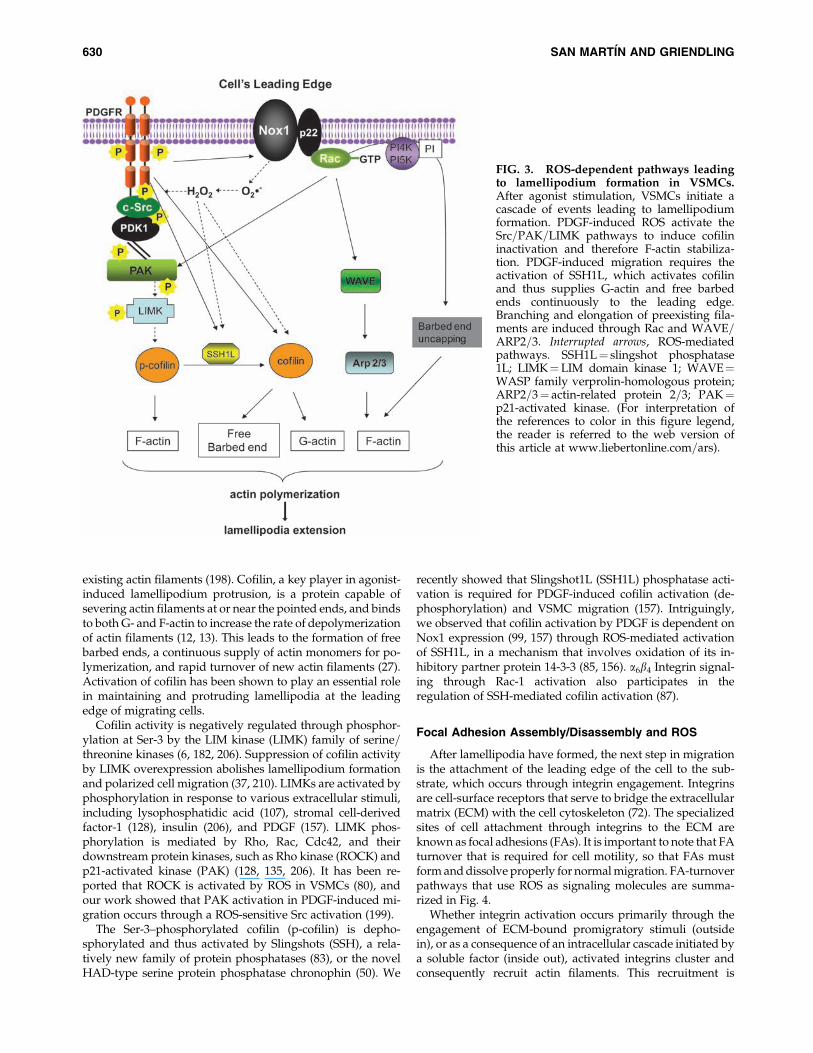
existing actin filaments (198). Cofilin, a key player in agonist-induced lamellipodium protrusion, is a protein capable ofsevering actin filaments at or near the pointed ends, and bindsto both G- and F-actin to increase the rate of depolymerizationof actin filaments (12, 13). This leads to the formation of freebarbed ends, a continuous supply of actin monomers for po-lymerization, and rapid turnover of new actin filaments (27).Activation of cofilin has been shown to play an essential rolein maintaining and protruding lamellipodia at the leadingedge of migrating cells.
Cofilin activity is negatively regulated through phosphor-ylation at Ser-3 by the LIM kinase (LIMK) family of serine=threonine kinases (6, 182, 206). Suppression of cofilin activityby LIMK overexpression abolishes lamellipodium formationand polarized cell migration (37, 210). LIMKs are activated byphosphorylation in response to various extracellular stimuli,including lysophosphatidic acid (107), stromal cell-derivedfactor-1 (128), insulin (206), and PDGF (157). LIMK phos-phorylation is mediated by Rho, Rac, Cdc42, and theirdownstream protein kinases, such as Rho kinase (ROCK) andp21-activated kinase (PAK) (128, 135, 206). It has been re-ported that ROCK is activated by ROS in VSMCs (80), andour work showed that PAK activation in PDGF-induced mi-gration occurs through a ROS-sensitive Src activation (199).
The Ser-3–phosphorylated cofilin (p-cofilin) is depho-sphorylated and thus activated by Slingshots (SSH), a rela-tively new family of protein phosphatases (83), or the novelHAD-type serine protein phosphatase chronophin (50). We
recently showed that Slingshot1L (SSH1L) phosphatase acti-vation is required for PDGF-induced cofilin activation (de-phosphorylation) and VSMC migration (157). Intriguingly,we observed that cofilin activation by PDGF is dependent onNox1 expression (99, 157) through ROS-mediated activationof SSH1L, in a mechanism that involves oxidation of its in-hibitory partner protein 14-3-3 (85, 156). a6b4 Integrin signal-ing through Rac-1 activation also participates in theregulation of SSH-mediated cofilin activation (87).
Focal Adhesion Assembly/Disassembly and ROS
After lamellipodia have formed, the next step in migrationis the attachment of the leading edge of the cell to the sub-strate, which occurs through integrin engagement. Integrinsare cell-surface receptors that serve to bridge the extracellularmatrix (ECM) with the cell cytoskeleton (72). The specializedsites of cell attachment through integrins to the ECM areknown as focal adhesions (FAs). It is important to note that FAturnover that is required for cell motility, so that FAs mustform and dissolve properly for normal migration. FA-turnoverpathways that use ROS as signaling molecules are summa-rized in Fig. 4.
Whether integrin activation occurs primarily through theengagement of ECM-bound promigratory stimuli (outsidein), or as a consequence of an intracellular cascade initiated bya soluble factor (inside out), activated integrins cluster andconsequently recruit actin filaments. This recruitment is
FIG. 3. ROS-dependent pathways leadingto lamellipodium formation in VSMCs.After agonist stimulation, VSMCs initiate acascade of events leading to lamellipodiumformation. PDGF-induced ROS activate theSrc=PAK=LIMK pathways to induce cofilininactivation and therefore F-actin stabiliza-tion. PDGF-induced migration requires theactivation of SSH1L, which activates cofilinand thus supplies G-actin and free barbedends continuously to the leading edge.Branching and elongation of preexisting fila-ments are induced through Rac and WAVE=ARP2=3. Interrupted arrows, ROS-mediatedpathways. SSH1L¼ slingshot phosphatase1L; LIMK¼LIM domain kinase 1; WAVE¼WASP family verprolin-homologous protein;ARP2=3¼ actin-related protein 2=3; PAK¼p21-activated kinase. (For interpretation ofthe references to color in this figure legend,the reader is referred to the web version ofthis article at www.liebertonline.com=ars).
630 SAN MARTIN AND GRIENDLING

achieved as the cytoplasmic domains of integrins associatewith a group of effectors, which include talin, vinculin,a-actinin, filamin, and paxillin (34, 91, 209). One of the twohomologues of LIMK, LIMK1, is also localized mainly to focaladhesions (2). Interestingly, in Drosophila, paxillin can nega-tively modulate LIMK function within focal adhesions byregulating the Rho pathway (26), which is activated by ROS inVSMCs (79, 80). These findings suggest a role for LIMK in FAsat the time that it participates in the formation of lamellipodia.
The FA protein complex organizes the actomyosin contractileapparatus and attracts signaling molecules such as focal adhe-sion kinase (FAK), Src family kinases, and integrin-linked ki-nase (91, 209). These kinases link integrins to the actincytoskeleton and coordinate the formation and strengthening ofFAs in the lamellipodium, as well as their recycling from therear of the cell. FAK is of particular importance. Integrin-mediated activation of FAK leads to phosphorylation of paxillinand p130Cas, thereby regulating their translocation to FAs (203)and enhancing FA formation. At the same time, FAK autop-hosphorylates on tyrosine 397, which is essential for FAK-induced FA disassembly (60). Depletion of FAK in fibroblastsresults in enhanced FAs and impaired migration (167).
It is well established that integrin signaling involves ROS,and, at the same time, that ROS can mediate integrin acti-vation (29, 174). In different cell types, integrins have beenshown to activate small Rho-GTPases (20, 61, 141). Duringfibronectin (FN)=integrin-mediated cell adhesion, ROS aredramatically increased by a Rac1-dependent activation ofNADPH oxidase (31). Other sources of integrin-inducedROS include mitochondria (201) and lipoxygenase (177). Asa result of this oxidative burst, the activity of low-molecular-weight protein tyrosine phosphatase (LMW-PTP) is tran-siently inhibited by thiol oxidation within the active site ofthe enzyme (30, 31). LMW-PTP also has been shown to as-sociate with, dephosphorylate, and thus inactivate FAK(149). Therefore, LMW-PTP activity must be tightly regu-lated, probably through ROS-dependent mechanisms, toensure proper focal complex=adhesion dynamics. Indeed,overexpression of LMW-PTP has been shown to inhibitVSMC migration (165). Similarly, the participation of ROSmodulating the activity of PTPs has been studied in an in vivomodel in which the participation of PDGF-induced ROS in-hibition of PTP activity was reversed with the use of anti-oxidants (84).
FIG. 4. ROS-dependent path-ways leading to focal adhesionformation in VSMCs. Integrinstimulation through inside-outor outside-in stimuli inducesclustering of FA proteins andstrengthening of stress fibers.Simultaneous stimulation withPDGF activates a series of ty-rosine kinases and inhibitsphosphatases that contributeto FA formation. Both PDGF-mediated signaling and localproduction of ROS by Nox4in FAs coordinate FA forma-tion. Interrupted arrows, ROS-mediated pathways. FAs¼ focaladhesions. (For interpretationof the references to color in thisfigure legend, the reader is re-ferred to the web version ofthis article at www.liebertonline.com=ars).
REDOX CONTROL OF VASCULAR SMOOTH MUSCLE MIGRATION 631

FAs provide a support against which cell contraction canoccur. They begin life as focal complexes, and the mechanismsregulating the conversion of focal complexes to FAs are un-clear. Our work has shown that in angiotensin II–treated cells,ROS regulate the Src-dependent activation of PDK1, which isessential for this process (179). In addition, stress fiber for-mation and contraction, which are involved in FA strength-ening, require activation of Rho (130, 200). Shinohara et al.(166) found that in Ras-transformed cells, Nox1-generatedROS mediate downregulation of Rho activity through oxi-dative inactivation of the LMW-PTP.
We recently found that Nox4 is also a key player in theregulation of stress fiber formation and focal adhesion turn-over in VSMCs (33). Our group just reported the identificationof Poldip2, a new regulator of Nox4 (105). Poldip2 is an acti-vator of Nox4-mediated ROS production in VSMCs, and ei-ther upregulation or downregulation of Poldip2=Nox4negatively affects FA turnover and inhibits VSMCs migration.These findings suggest a potentially novel mechanism, localROS production, by which FA turnover is coordinated.
Contraction and ROS
The next phase in SMC motility is contraction of the cellbody to create forward movement. As with cell contraction toregulate vascular tone, ATPase activity associated withmyosin II is required for contraction to occur. After phos-phorylation of the myosin regulatory light chain by a calcium-
calmodulin (Ca2þ=CaM)-dependent myosin light-chain ki-nase (MLCK), actin activates myosin II ATPase activity, andcontraction proceeds (47, 48, 170). In Fig. 5, ROS-mediatedsignaling pathways leading to VSMCs contraction are sum-marized.
In VSMCs, ROS appear to be both upstream and down-stream of intracellular Ca2þ release and calcium influx. Aftergrowth-factor stimulation, VSMCs exhibit waves of cytosoliccalcium release that are required for migration (160); blockersof calcium channels reduce both migration and ROS pro-duction. However, it also was shown that H2O2 and super-oxide can increase the intracellular Ca2þ concentration inVSMCs and endothelial cells (104, 151, 176), apparently byregulating Ca2þ release from 1,4,5-trisphosphate–sensitiveCa2þ stores.
Although Ca2þ-dependent activation of MLCK is the majormechanism initiating cell contraction, additional pathwayshave been shown to regulate actin–myosin function. One suchpathway in VSMCs is PAK. After activation by either Rac orCdc42, PAK1 phosphorylates MLCK, resulting in decreasedMLCK activity, thereby inhibiting myosin light chain (MLC)phosphorylation and cell contractility (158). This mechanismmay be ROS mediated, because PAK1 activation in VSMCs isdependent on Nox1-derived ROS (99). Another importantmechanism that regulates the contractile apparatus is theRho=ROCK pathway, which has shown to be activated byROS in both aorta and VSMCs (79, 80). Importantly, thispathway promotes MLC phosphorylation by phosphorylat-
FIG. 5. ROS-dependent pathways leading to cell-body contraction in VSMCs. The final step in motility is the productionof forward movement through regulation of myosin II phosphorylation and actin–myosin interaction. Several major regu-lators of myosin phosphorylation are ROS sensitive, including mobilization of calcium from intracellular or extracellularcompartments, Src activation, and the Rho-ROCK pathway. Interrupted arrows, ROS-mediated pathways. Src¼Rous sarcomavirus kinase homologue; ROCK¼Rho-associated protein kinase. (For interpretation of the references to color in this figurelegend, the reader is referred to the web version of this article at www.liebertonline.com=ars).
632 SAN MARTIN AND GRIENDLING

ing and thus inhibiting the regulatory subunit of myosin lightchain phosphatase (79, 80, 86), and by direct ROCK-mediatedmyosin II phosphorylation in fibroblasts (183).
Actual movement of the cell occurs through engagement ofactin–myosin interactions. As the body of the cell moves for-ward, the newly formed FAs become stronger and arrive atthe rear of the cell, where they are dissociated, allowing theircomponents to recycle to the leading edge of the cell for thenext wave of migration (11). Virtually nothing is known aboutthe role of ROS in FA dissociation.
Extracellular Matrix and ROS
As mentioned earlier, cell adhesion and migration are de-pendent on integrin binding to the extracellular matrix(ECM). Cell migration is, in its essence, an invasive processthat requires degradation of the ECM. This is achieved byactivation of matrix metalloproteinases (MMPs) and simul-taneous inhibition of tissue inhibitors of metalloproteinases(TIMPs). Accordingly, MMP inhibitors have been shown toattenuate migration and delay neointimal formation (18).Moreover, genetic deletion of either MMP-2 or MMP-9 re-duces VSMC migration (82). MMP activity is regulated bytranscriptional and posttranscriptional mechanisms, both ofwhich are mediated by ROS. Although ROS has been reportedto downregulate MMP2 and 14 activities (41), most of thecompelling data indicate that ROS can directly or indirectlyactivate MMPs. In VSMCs, ROS activate MMP-9 (120) andMMP-2 (70). The stimulation of MMP-9 activity by direct in-cubation with H2O2 (147) proves unequivocally that the redoxstate of MMPs is at least part of their mechanism of regulation.MMP-2 activity also is increased by H2O2, as well as byONOO� (147). Conversely, MnSOD and NO inhibit IL-1b–stimulated MMP-9 activity (57). It should be noted that MMP-7, an MMP with high degradative ability, is activated (45) orinactivated (44) by hypochloric acid (HOCl�) depending onthe system.
Like activity, expression of MMPs has shown to be sensitiveto ROS. MMP-1, which is important in collagen degradation,is increased by angiotensin II stimulation through the redox-sensitive transcription factors NF-kB and activating protein-1(AP-1) (23). TNF-a stimulation has similar effects (23). Simi-larly, 4-hydroxynonenal (HNE), a by-product of oxidativedamage that frequently accumulates in atherosclerotic le-sions, increases mitochondrial ROS production, and conse-quently enhances MMP-2 activity in VSMCs by a mechanismthat involves the Akt=NF-kB signaling pathway (100). Thus,ROS not only regulate the mechanics of cell migration, butalso regulate the expression and activity of the enzymesnecessary to create a path for the migrating cell.
Conclusions and Future Directions
The findings discussed herein undoubtedly support a keyrole of ROS as signaling molecules that regulate VSMC mi-gration. Because migration requires carefully coordinated,tightly regulated signaling within particular subcellular lo-cations, ROS are potentially excellent candidates for suchregulation. They have short half-lives and are degradedshortly after being produced, most likely only a few atomicratios away from the site of production.
As is evident throughout this review, much work remainsto be done to understand better the participation of ROS at
different levels of cell migration. Particularly interesting willbe to understand how the redox state of actin and actin-associated proteins affects their protein function=polymeri-zation properties. It will also be important to understand thespatial and temporal relations of ROS production from spe-cific sources of ROS and their specific subcellular targets. Atthis point, one of the major challenges is to be able to visualizethese localized events. New live-cell imaging techniques andnew probes that allow us to study this process in real time areessential.
We also need to understand the contribution of variationsin cell type as well as particular extracellular environmentsthat can differentially affect cellular movement. Our presentparadigm of VSMC migration is based on a model developedin other cell types, principally, but not exclusively, fibroblasts.It is very likely that VSMCs, and even potentially VSMCsfrom different vascular beds, have unique regulatory mech-anisms for their migratory behavior. Ultimately, knowledgegained in the in vitro systems will have to be translated toanimal models to allow us to understand how ROS-mediatedsignaling contributes to phenotypic modulation and woundhealing.
Acknowledgments
This work was supported by National Institutes of Healthgrants HL38206, HL093115, and HL058863.
References
1. Abercrombie M, Heaysman JE, and Pegrum SM. The lo-comotion of fibroblasts in culture, 3: Movements of parti-cles on the dorsal surface of the leading lamella. Exp CellRes 62: 389–398, 1970.
2. Acevedo K, Moussi N, Li R, Soo P, and Bernard O. LIMkinase 2 is widely expressed in all tissues. J Histochem Cy-tochem 54: 487–501, 2006.
3. Alexandre J, Hu Y, Lu W, Pelicano H, and Huang P. Novelaction of paclitaxel against cancer cells: bystander effectmediated by reactive oxygen species. Cancer Res 67: 3512–3517, 2007.
4. Ambasta RK, Schreiber JG, Janiszewski M, Busse R, andBrandes RP. Noxa1 is a central component of the smoothmuscle NADPH oxidase in mice. Free Radic Biol Med 41:193–201, 2006.
5. Anilkumar N, Weber R, Zhang M, Brewer A, and ShahAM. Nox4 and nox2 NADPH oxidases mediate distinctcellular redox signaling responses to agonist stimulation.Arterioscler Thromb Vasc Biol 28: 1347–1354, 2008.
6. Arber S, Barbayannis FA, Hanser H, Schneider C, StanyonCA, Bernard O, and Caroni P. Regulation of actin dynamicsthrough phosphorylation of cofilin by LIM-kinase. Nature393: 805–809, 1998.
7. Axel DI, Kunert W, Goggelmann C, Oberhoff M, Herdeg C,Kuttner A, Wild DH, Brehm BR, Riessen R, Koveker G, andKarsch KR. Paclitaxel inhibits arterial smooth muscle cellproliferation and migration in vitro and in vivo using localdrug delivery. Circulation 96: 636–645, 1997.
8. Azevedo LC, Pedro MA, Souza LC, de Souza HP, Ja-niszewski M, da Luz PL, and Laurindo FR. Oxidative stressas a signaling mechanism of the vascular response to in-jury: the redox hypothesis of restenosis. Cardiovasc Res 47:436–445, 2000.
9. Babior BM. The respiratory burst oxidase. Curr Opin He-matol 2: 55–60, 1995.
REDOX CONTROL OF VASCULAR SMOOTH MUSCLE MIGRATION 633

10. Bailly M, Condeelis JS, and Segall JE. Chemoattractant-induced lamellipod extension. Microsc Res Tech 43: 433–443,1998.
11. Ballestrem C, Hinz B, Imhof BA, and Wehrle-Haller B.Marching at the front and dragging behind: differentialalphaVbeta3-integrin turnover regulates focal adhesionbehavior. J Cell Biol 155: 1319–1332, 2001.
12. Bamburg JR. Proteins of the ADF=cofilin family: essentialregulators of actin dynamics. Annu Rev Cell Dev Biol 15:185–230, 1999.
13. Bamburg JR, McGough A, and Ono S. Putting a new twiston actin: ADF=cofilins modulate actin dynamics. TrendsCell Biol 9: 364–370, 1999.
14. Banfi B, Molnar G, Maturana A, Steger K, Hegedus B,Demaurex N, and Krause KH. A Ca(2þ)-activated NADPHoxidase in testis, spleen, and lymph nodes. J Biol Chem 276:37594–37601, 2001.
15. Barrett TB and Benditt EP. Platelet-derived growth factorgene expression in human atherosclerotic plaques and nor-mal artery wall. Proc Natl Acad Sci U S A 85: 2810–2814, 1988.
16. Bauersachs J, Bouloumie A, Fraccarollo D, Hu K, Busse R,and Ertl G. Endothelial dysfunction in chronic myocardialinfarction despite increased vascular endothelial nitricoxide synthase and soluble guanylate cyclase expression:role of enhanced vascular superoxide production. Circula-tion 100: 292–298, 1999.
17. Bedard K and Krause KH. The NOX family of ROS-generating NADPH oxidases: physiology and pathophysi-ology. Physiol Rev 87: 245–313, 2007.
18. Bendeck MP, Irvin C, and Reidy MA. Inhibition of matrixmetalloproteinase activity inhibits smooth muscle cell mi-gration but not neointimal thickening after arterial injury.Circ Res 78: 38–43, 1996.
19. Bengtsson T, Orselius K, and Wettero J. Role of the actincytoskeleton during respiratory burst in chemoattractant-stimulated neutrophils. Cell Biol Int 30: 154–163, 2006.
20. Bialkowska K, Kulkarni S, Du X, Goll DE, Saido TC, andFox JE. Evidence that beta3 integrin-induced Rac activationinvolves the calpain-dependent formation of integrin clus-ters that are distinct from the focal complexes and focaladhesions that form as Rac and RhoA become active. J CellBiol 151: 685–696, 2000.
21. Black SM, DeVol JM, and Wedgwood S. Regulation of fi-broblast growth factor-2 expression in pulmonary arterialsmooth muscle cells involves increased reactive oxygenspecies generation. Am J Physiol Cell Physiol 294: C345–C354, 2008.
22. Brandes RP, Viedt C, Nguyen K, Beer S, Kreuzer J, Busse R,and Gorlach A. Thrombin-induced MCP-1 expression in-volves activation of the p22phox-containing NADPH oxi-dase in human vascular smooth muscle cells. ThrombHaemost 85: 1104–1110, 2001.
23. Browatzki M, Larsen D, Pfeiffer CA, Gehrke SG, Schmidt J,Kranzhofer A, Katus HA, and Kranzhofer R. Angiotensin IIstimulates matrix metalloproteinase secretion in humanvascular smooth muscle cells via nuclear factor-kappaB andactivator protein 1 in a redox-sensitive manner. J Vasc Res42: 415–423, 2005.
24. Buetow BS, Tappan KA, Crosby JR, Seifert RA, and Bowen-Pope DF. Chimera analysis supports a predominant role ofPDGFRbeta in promoting smooth-muscle cell chemotaxisafter arterial injury. Am J Pathol 163: 979–984, 2003.
25. Carpenter CL. Actin cytoskeleton and cell signaling. CritCare Med 28: N94–N99, 2000.
26. Chen GC, Turano B, Ruest PJ, Hagel M, Settleman J, andThomas SM. Regulation of Rho and Rac signaling to theactin cytoskeleton by paxillin during Drosophila develop-ment. Mol Cell Biol 25: 979–987, 2005.
27. Chen H, Bernstein BW, and Bamburg JR. Regulating actin-filament dynamics in vivo. Trends Biochem Sci 25: 19–23, 2000.
28. Chen P, Gupta K, and Wells A. Cell movement elicited byepidermal growth factor receptor requires kinase and auto-phosphorylation but is separable from mitogenesis. J CellBiol 124: 547–555, 1994.
29. Chiarugi P and Fiaschi T. Redox signalling in anchorage-dependent cell growth. Cell Signal 19: 672–682, 2007.
30. Chiarugi P, Fiaschi T, Taddei ML, Talini D, Giannoni E,Raugei G, and Ramponi G. Two vicinal cysteines confer apeculiar redox regulation to low molecular weight proteintyrosine phosphatase in response to platelet-derivedgrowth factor receptor stimulation. J Biol Chem 276: 33478–33487, 2001.
31. Chiarugi P, Pani G, Giannoni E, Taddei L, Colavitti R,Raugei G, Symons M, Borrello S, Galeotti T, and Ramponi G.Reactive oxygen species as essential mediators of cell adhe-sion: the oxidative inhibition of a FAK tyrosine phosphataseis required for cell adhesion. J Cell Biol 161: 933–944, 2003.
32. Choi MH, Lee IK, Kim GW, Kim BU, Han YH, Yu DY, ParkHS, Kim KY, Lee JS, Choi C, Bae YS, Lee BI, Rhee SG, andKang SW. Regulation of PDGF signalling and vascular re-modelling by peroxiredoxin II. Nature 435: 347–353, 2005.
33. Clempus RE, Sorescu D, Dikalova AE, Pounkova L, Jo P,Sorescu GP, Schmidt HH, Lassegue B, and Griendling KK.Nox4 is required for maintenance of the differentiatedvascular smooth muscle cell phenotype. Arterioscler ThrombVasc Biol 27: 42–48, 2007.
34. Critchley DR. Focal adhesions: the cytoskeletal connection.Curr Opin Cell Biol 12: 133–139, 2000.
35. DalleDonne I, Milzani A, and Colombo R. H2O2-treatedactin: assembly and polymer interactions with cross-linkingproteins. Biophys J 69: 2710–2719, 1995.
36. DalleDonne I, Milzani A, and Colombo R. The tert-butylhydroperoxide-induced oxidation of actin Cys-374 is cou-pled with structural changes in distant regions of the pro-tein. Biochemistry 38: 12471–12480, 1999.
37. Dawe HR, Minamide LS, Bamburg JR, and Cramer LP.ADF=cofilin controls cell polarity during fibroblast migra-tion. Curr Biol 13: 252–257, 2003.
38. Deshpande NN, Sorescu D, Seshiah P, Ushio-Fukai M,Akers M, Yin Q, and Griendling KK. Mechanism of hy-drogen peroxide-induced cell cycle arrest in vascularsmooth muscle. Antioxid Redox Signal 4: 845–854, 2002.
39. Dikalova A, Clempus R, Lassegue B, Cheng G, McCoy J,Dikalov S, San Martin A, Lyle A, Weber DS, Weiss D,Taylor WR, Schmidt HH, Owens GK, Lambeth JD, andGriendling KK. Nox1 overexpression potentiates angiotensinII-induced hypertension and vascular smooth muscle hyper-trophy in transgenic mice. Circulation 112: 2668–2676, 2005.
40. Drewes G and Faulstich H. The enhanced ATPase activity ofglutathione-substituted actin provides a quantitative approachto filament stabilization. J Biol Chem 265: 3017–3021, 1990.
41. Elliot S, Catanuto P, Stetler-Stevenson W, and CousinsSW. Retinal pigment epithelium protection from oxidant-mediated loss of MMP-2 activation requires both MMP-14and TIMP-2. Invest Ophthalmol Vis Sci 47: 1696–1702, 2006.
42. Fiaschi T, Cozzi G, Raugei G, Formigli L, Ramponi G, andChiarugi P. Redox regulation of beta-actin during integrin-mediated cell adhesion. J Biol Chem 281: 22983–22991, 2006.
634 SAN MARTIN AND GRIENDLING

43. Floege J, Hudkins KL, Davis CL, Schwartz SM, and AlpersCE. Expression of PDGF alpha-receptor in renal arterio-sclerosis and rejecting renal transplants. J Am Soc Nephrol 9:211–223, 1998.
44. Fu X, Kassim SY, Parks WC, and Heinecke JW. Hypo-chlorous acid generated by myeloperoxidase modifies ad-jacent tryptophan and glycine residues in the catalyticdomain of matrix metalloproteinase-7 (matrilysin): an oxi-dative mechanism for restraining proteolytic activity dur-ing inflammation. J Biol Chem 278: 28403–28409, 2003.
45. Fu X, Kassim SY, Parks WC, and Heinecke JW. Hypo-chlorous acid oxygenates the cysteine switch domain ofpro-matrilysin (MMP-7): a mechanism for matrix metallo-proteinase activation and atherosclerotic plaque rupture bymyeloperoxidase. J Biol Chem 276: 41279–41287, 2001.
46. Fukui T, Ishizaka N, Rajagopalan S, Laursen JB, Capers QT,Taylor WR, Harrison DG, de Leon H, Wilcox JN, andGriendling KK. p22phox mRNA expression and NADPHoxidase activity are increased in aortas from hypertensiverats. Circ Res 80: 45–51, 1997.
47. Gallagher PJ and Herring BP. The carboxyl terminus of thesmooth muscle myosin light chain kinase is expressed as anindependent protein, telokin. J Biol Chem 266: 23945–23952,1991.
48. Gallagher PJ, Herring BP, Griffin SA, and Stull JT. Molecularcharacterization of a mammalian smooth muscle myosinlight chain kinase. J Biol Chem 266: 23936–23944, 1991.
49. Giannoni E, Buricchi F, Raugei G, Ramponi G, and Chiar-ugi P. Intracellular reactive oxygen species activate Srctyrosine kinase during cell adhesion and anchorage-dependent cell growth. Mol Cell Biol 25: 6391–6403, 2005.
50. Gohla A, Birkenfeld J, and Bokoch GM. Chronophin,a novel HAD-type serine protein phosphatase, regulatescofilin-dependent actin dynamics. Nat Cell Biol 7: 21–29, 2005.
51. Griendling KK, Minieri CA, Ollerenshaw JD, and Alex-ander RW. Angiotensin II stimulates NADH and NADPHoxidase activity in cultured vascular smooth muscle cells.Circ Res 74: 1141–1148, 1994.
52. Griendling KK, Sorescu D, Lassegue B, and Ushio-Fukai M.Modulation of protein kinase activity and gene expressionby reactive oxygen species and their role in vascularphysiology and pathophysiology. Arterioscler Thromb VascBiol 20: 2175–2183, 2000.
53. Griendling KK, Sorescu D, and Ushio-Fukai M. NAD(P)Hoxidase: role in cardiovascular biology and disease. Circ Res86: 494–501, 2000.
54. Griendling KK and Ushio-Fukai M. Redox control of vas-cular smooth muscle proliferation. J Lab Clin Med 132: 9–15,1998.
55. Grotendorst GR, Seppa HE, Kleinman HK, and Martin GR.Attachment of smooth muscle cells to collagen and theirmigration toward platelet-derived growth factor. Proc NatlAcad Sci U S A 78: 3669–3672, 1981.
56. Gupte SA, Kaminski PM, George S, Kouznestova L, OlsonSC, Matthew R, Hintze TH, and Wolin MS. Peroxide gen-eration by p47phox-Src activation of Nox2 has a key role inprotein kinase C-induced arterial smooth muscle contrac-tion. Am J Physiol Heart Circ Physiol 296: H1048–H1057, 2009.
57. Gurjar MV, Deleon J, Sharma RV, and Bhalla RC. Role ofreactive oxygen species in IL-1 beta-stimulated sustainedERK activation and MMP-9 induction. Am J Physiol HeartCirc Physiol 281: H2568–H2574, 2001.
58. Halka AT, Turner NJ, Carter A, Ghosh J, Murphy MO,Kirton JP, Kielty CM, and Walker MG. The effects of stretch
on vascular smooth muscle cell phenotype in vitro. Cardi-ovasc Pathol 17: 98–102, 2008.
59. Hall A. Rho GTPases and the actin cytoskeleton. Science279: 509–514, 1998.
60. Hamadi A, Bouali M, Dontenwill M, Stoeckel H, Takeda K,and Ronde P. Regulation of focal adhesion dynamics anddisassembly by phosphorylation of FAK at tyrosine 397.J Cell Sci 118: 4415–4425, 2005.
61. Hamelers IH, Olivo C, Mertens AE, Pegtel DM, van derKammen RA, Sonnenberg A, and Collard JG. The Rac ac-tivator Tiam1 is required for (alpha)3(beta)1-mediatedlaminin-5 deposition, cell spreading, and cell migration.J Cell Biol 171: 871–881, 2005.
62. Hanna IR, Hilenski LL, Dikalova A, Taniyama Y, Dikalov S,Lyle A, Quinn MT, Lassegue B, and Griendling KK. Func-tional association of nox1 with p22phox in vascular smoothmuscle cells. Free Radic Biol Med 37: 1542–1549, 2004.
63. Harrison D, Griendling KK, Landmesser U, Hornig B, andDrexler H. Role of oxidative stress in atherosclerosis. Am JCardiol 91: 7A–11A, 2003.
64. Hartwig JH, Bokoch GM, Carpenter CL, Janmey PA, TaylorLA, Toker A, and Stossel TP. Thrombin receptor ligationand activated Rac uncap actin filament barbed endsthrough phosphoinositide synthesis in permeabilizedhuman platelets. Cell 82: 643–653, 1995.
65. Haurani MJ, Cifuentes ME, Shepard AD, and Pagano PJ. Nox4oxidase overexpression specifically decreases endogenousNox4 mRNA and inhibits angiotensin II-induced adventitialmyofibroblast migration. Hypertension 52: 143–149, 2008.
66. Heldin CH and Westermark B. Mechanism of action andin vivo role of platelet-derived growth factor. Physiol Rev79: 1283–1316, 1999.
67. Hertelendi Z, Toth A, Borbely A, Galajda Z, van der VeldenJ, Stienen GJ, Edes I, and Papp Z. Oxidation of myofilamentprotein sulfhydryl groups reduces the contractile force andits Ca2þ sensitivity in human cardiomyocytes. AntioxidRedox Signal 10: 1175–1184, 2008.
68. Hilenski LL, Clempus RE, Quinn MT, Lambeth JD, andGriendling KK. Distinct subcellular localizations of Nox1and Nox4 in vascular smooth muscle cells [see comment].Arterioscler Thromb Vasc Biol 24: 677–683, 2004.
69. Hinshaw DB, Burger JM, Beals TF, Armstrong BC, andHyslop PA. Actin polymerization in cellular oxidant injury.Arch Biochem Biophys 288: 311–316, 1991.
70. Hu T, Luan R, Zhang H, Lau WB, Wang Q, Zhang Y, WangHC, and Tao L. Hydrogen peroxide enhances the osteo-pontin expression and matrix metalloproteinase activity inaortic vascular smooth muscle cells. Clin Exp PharmacolPhysiol 36: 626–630, 2008.
71. Huot J, Houle F, Marceau F, and Landry J. Oxidative stress-induced actin reorganization mediated by the p38 mitogen-activated protein kinase=heat shock protein 27 pathway invascular endothelial cells. Circ Res 80: 383–392, 1997.
72. Hynes RO. Integrins: versatility, modulation, and signalingin cell adhesion. Cell 69: 11–25, 1992.
73. Jackson CL, Raines EW, Ross R, and Reidy MA. Role ofendogenous platelet-derived growth factor in arterialsmooth muscle cell migration after balloon catheter injury.Arterioscler Thromb 13: 1218–1226, 1993.
74. Jacobson GM, Dourron HM, Liu J, Carretero OA, ReddyDJ, Andrzejewski T, and Pagano PJ. Novel NAD(P)H oxi-dase inhibitor suppresses angioplasty-induced superoxideand neointimal hyperplasia of rat carotid artery. Circ Res92: 637–643, 2003.
REDOX CONTROL OF VASCULAR SMOOTH MUSCLE MIGRATION 635

75. Jagadeesha DK, Lindley TE, Deleon J, Sharma RV, Miller F,and Bhalla RC. Tempol therapy attenuates medial smoothmuscle cell apoptosis and neointima formation after bal-loon catheter injury in carotid artery of diabetic rats. Am JPhysiol Heart Circ Physiol 289: H1047–H1053, 2005.
76. Jagadeesha DK, Miller FJ Jr, and Bhalla RC. Inhibition ofapoptotic signaling and neointimal hyperplasia by tempoland nitric oxide synthase following vascular injury. J VascRes 46: 109–118, 2008.
77. Jang HJ, Hwang S, Cho KY, Kim do K, Chay KO, and KimJK. Taxol induces oxidative neuronal cell death by en-hancing the activity of NADPH oxidase in mouse corticalcultures. Neurosci Lett 443: 17–22, 2008.
78. Jawien A, Bowen-Pope DF, Lindner V, Schwartz SM, andClowes AW. Platelet-derived growth factor promotessmooth muscle migration and intimal thickening in a ratmodel of balloon angioplasty. J Clin Invest 89: 507–511, 1992.
79. Jernigan NL, Walker BR, and Resta TC. Reactive oxygen spe-cies mediate RhoA=Rho kinase-induced Ca2þ sensitization inpulmonary vascular smooth muscle following chronic hyp-oxia. Am J Physiol Lung Cell Mol Physiol 295: L515–L529, 2008.
80. Jin L, Ying Z, and Webb RC. Activation of Rho=Rho kinasesignaling pathway by reactive oxygen species in rat aorta.Am J Physiol Heart Circ Physiol 287: H1495–H1500, 2004.
81. Johansson M and Lundberg M. Glutathionylation of beta-actin via a cysteinyl sulfenic acid intermediary. BMC Bio-chem 8: 26, 2007.
82. Johnson C and Galis ZS. Matrix metalloproteinase-2 and�9 differentially regulate smooth muscle cell migration andcell-mediated collagen organization. Arterioscler ThrombVasc Biol 24: 54–60, 2004.
83. Kaji N, Ohashi K, Shuin M, Niwa R, Uemura T, and Mi-zuno K. Cell cycle-associated changes in Slingshot phos-phatase activity and roles in cytokinesis in animal cells.J Biol Chem 278: 33450–33455, 2003.
84. Kappert K, Sparwel J, Sandin A, Seiler A, Siebolts U, Lep-panen O, Rosenkranz S, and Ostman A. Antioxidants re-lieve phosphatase inhibition and reduce PDGF signaling incultured VSMCs and in restenosis. Arterioscler Thromb VascBiol 26: 2644–2651, 2006.
85. Kim JS, Huang TY, and Bokoch GM. Reactive oxygenspecies regulate a slingshot-cofilin activation pathway. MolBiol Cell 20: 2650–2660, 2009.
86. Kimura K, Ito M, Amano M, Chihara K, Fukata Y, NakafukuM, Yamamori B, Feng J, Nakano T, Okawa K, Iwamatsu A,and Kaibuchi K. Regulation of myosin phosphatase by Rhoand Rho-associated kinase (Rho-kinase). Science 273: 245–248, 1996.
87. Kligys K, Claiborne JN, Debiase PJ, Hopkinson SB, Wu Y,Mizuno K, and Jones JC. The slingshot family of phos-phatases mediates Rac1 regulation of cofilin phosphoryla-tion, laminin-332 organization and motility behavior ofkeratinocytes. J Biol Chem 282: 32520–32528, 2007.
88. Koller A. Signaling pathways of mechanotransduction inarteriolar endothelium and smooth muscle cells in hyper-tension. Microcirculation 9: 277–294, 2002.
89. Konneh MK, Rutherford C, Li SR, Anggard EE, and FernsGA. Vitamin E inhibits the intimal response to ballooncatheter injury in the carotid artery of the cholesterol-fedrat. Atherosclerosis 113: 29–39, 1995.
90. Kozma R, Ahmed S, Best A, and Lim L. The Ras-relatedprotein Cdc42Hs and bradykinin promote formation ofperipheral actin microspikes and filopodia in Swiss 3T3fibroblasts. Mol Cell Biol 15: 1942–1952, 1995.
91. Kumar CC. Signaling by integrin receptors. Oncogene 17:1365–1373, 1998.
92. Lassegue B, Sorescu D, Szocs K, Yin Q, Akers M, Zhang Y,Grant SL, Lambeth JD, and Griendling KK. Novelgp91(phox) homologues in vascular smooth muscle cells:nox1 mediates angiotensin II-induced superoxide forma-tion and redox-sensitive signaling pathways. Circ Res 88:888–894, 2001.
93. Lassegue B and Clempus RE. Vascular NAD(P)H oxidases:specific features, expression, and regulation. Am J PhysiolRegul Integr Comp Physiol 285: R277–R297, 2003.
94. Lauffenburger DA and Horwitz AF. Cell migration: aphysically integrated molecular process. Cell 84: 359–369,1996.
95. Laurindo FR, da Luz PL, Uint L, Rocha TF, Jaeger RG, andLopes EA. Evidence for superoxide radical-dependentcoronary vasospasm after angioplasty in intact dogs. Cir-culation 83: 1705–1715, 1991.
96. Laursen JB, Rajagopalan S, Galis Z, Tarpey M, Freeman BA,and Harrison DG. Role of superoxide in angiotensin II-induced but not catecholamine-induced hypertension. Cir-culation 95: 588–593, 1997.
97. Lavigne MC, Malech HL, Holland SM, and Leto TL. Ge-netic requirement of p47phox for superoxide production bymurine microglia. FASEB J 15: 285–287, 2001.
98. Lee HM, Jeon BH, Won KJ, Lee CK, Park TK, Choi WS, BaeYM, Kim HS, Lee SK, Park SH, Irani K, and Kim B. Genetransfer of redox factor-1 inhibits neointimal formation:involvement of platelet-derived growth factor-{beta} re-ceptor signaling via the inhibition of reactive oxygenspecies-mediated syk pathway. Circ Res 104: 219–227, 2008.
99. Lee MY, San Martin A, Mehta PK, Dikalova AE, GarridoAM, Lyons E, Krause KH, Banfi B, Lambeth JD, Lassegue B,and Griendling KK. Mechanisms of vascular smooth mus-cle NADPH oxidase 1 (nox1) contribution to injury-inducedneointimal formation. Arterioscler Thromb Vasc Biol 29: 480–487, 2009.
100. Lee SJ, Seo KW, Yun MR, Bae SS, Lee WS, Hong KW, andKim CD. 4-Hydroxynonenal enhances MMP-2 productionin vascular smooth muscle cells via mitochondrial ROS-mediated activation of the Akt=NF-kappaB signalingpathways. Free Radic Biol Med 45: 1487–1492, 2008.
101. Li J, Li W, Su J, Liu W, Altura BT, and Altura BM. Hy-drogen peroxide induces apoptosis in cerebral vascularsmooth muscle cells: possible relation to neurodegenerativediseases and strokes. Brain Res Bull 62: 101–106, 2003.
102. Li L, Gao PJ, Xi R, Wu CF, Zhu DL, Yan J, and Lu GP.Pioglitazone inhibits homocysteine-induced migration ofvascular smooth muscle cells through a peroxisome pro-liferator-activated receptor gamma-independent mecha-nism. Clin Exp Pharmacol Physiol 35: 1471–1476, 2008.
103. Liochev SL. The role of iron-sulfur clusters in in vivohydroxyl radical production. Free Radic Res 25: 369–384,1996.
104. Lounsbury KM, Hu Q, and Ziegelstein RC. Calcium sig-naling and oxidant stress in the vasculature. Free Radic BiolMed 28: 1362–1369, 2000.
105. Lyle AN, Deshpande NN, Taniyama Y, Seidel-Rogol B,Pounkova L, Du P, Papaharalambus C, Lassegue B, andGriendling KK. Poldip2, a novel regulator of nex1 andcytoskeletal integrity in vascular smooth muscle cells. CircRes 105: 249–259, 2009.
106. Machesky LM and Insall RH. Scar1 and the relatedWiskott-Aldrich syndrome protein, WASP, regulate the
636 SAN MARTIN AND GRIENDLING

actin cytoskeleton through the Arp2=3 complex. Curr Biol 8:1347–1356, 1998.
107. Maekawa M, Ishizaki T, Boku S, Watanabe N, Fujita A,Iwamatsu A, Obinata T, Ohashi K, Mizuno K, and Nar-umiya S. Signaling from Rho to the actin cytoskeletonthrough protein kinases ROCK and LIM-kinase. Science285: 895–898, 1999.
108. Majesky MW, Reidy MA, Bowen-Pope DF, Hart CE, Wil-cox JN, and Schwartz SM. PDGF ligand and receptor geneexpression during repair of arterial injury. J Cell Biol 111:2149–2158, 1990.
109. Marmur JD, Poon M, Rossikhina M, and Taubman MB.Induction of PDGF-responsive genes in vascular smoothmuscle: implications for the early response to vessel injury.Circulation 86: III53–III60, 1992.
110. McCully KS. Vascular pathology of homocysteinemia: im-plications for the pathogenesis of arteriosclerosis. Am JPathol 56: 111–128, 1969.
111. Meng D, Lv DD, and Fang J. Insulin-like growth factor-Iinduces reactive oxygen species production and cell mi-gration through Nox4 and Rac1 in vascular smooth musclecells. Cardiovasc Res 80: 299–308, 2008.
112. Miki H, Suetsugu S, and Takenawa T. WAVE, a novelWASP-family protein involved in actin reorganization in-duced by Rac. EMBO J 17: 6932–6941, 1998.
113. Miller FJ, Jr., Filali M, Huss GJ, Stanic B, Chamseddine A,Barna TJ, and Lamb FS. Cytokine activation of nuclearfactor kappa B in vascular smooth muscle cells requiressignaling endosomes containing Nox1 and ClC-3. Circ Res101: 663–671, 2007.
114. Mills JE, Whitford PC, Shaffer J, Onuchic JN, Adams JA,and Jennings PA. A novel disulfide bond in the SH2 do-main of the C-terminal Src kinase controls catalytic activity.J Mol Biol 365: 1460–1468, 2007.
115. Milzani A, DalleDonne I, and Colombo R. Prolonged oxi-dative stress on actin. Arch Biochem Biophys 339: 267–274,1997.
116. Mitchison TJ and Cramer LP. Actin-based cell motility andcell locomotion. Cell 84: 371–379, 1996.
117. Mohazzab KM and Wolin MS. Sites of superoxide anionproduction detected by lucigenin in calf pulmonary arterysmooth muscle. Am J Physiol 267: L815–L822, 1994.
118. Moldovan L, Moldovan NI, Sohn RH, Parikh SA, andGoldschmidt-Clermont PJ. Redox changes of cultured endo-thelial cells and actin dynamics. Circ Res 86: 549–557, 2000.
119. Montezano AC, Callera GE, Yogi A, He Y, Tostes RC, He G,Schiffrin EL, and Touyz RM. Aldosterone and angiotensinII synergistically stimulate migration in vascular smoothmuscle cells through c-Src-regulated redox-sensitive RhoApathways. Arterioscler Thromb Vasc Biol 28: 1511–1518, 2008.
120. Moon SK, Kang SK, and Kim CH. Reactive oxygen speciesmediates disialoganglioside GD3-induced inhibition ofERK1=2 and matrix metalloproteinase-9 expression invascular smooth muscle cells. FASEB J 20: 1387–1395, 2006.
121. Morimatsu T, Kawagoshi A, Yoshida K, and Tamura M.Actin enhances the activation of human neutrophilNADPH oxidase in a cell-free system. Biochem Biophys ResCommun 230: 206–210, 1997.
122. Morita T, Perrella MA, Lee ME, and Kourembanas S.Smooth muscle cell-derived carbon monoxide is a regulatorof vascular cGMP. Proc Natl Acad Sci U S A 92: 1475–1479,1995.
123. Mu H, Wang X, Lin P, Yao Q, and Chen C. Nitrotyrosinepromotes human aortic smooth muscle cell migration
through oxidative stress and ERK1=2 activation. BiochimBiophys Acta 1783: 1576–1584, 2008.
124. Murphy MP. How mitochondria produce reactive oxygenspecies. Biochem J 417: 1–13, 2009.
125. Nakai K, Yoneda K, Igarashi J, Moriue T, Kosaka H, andKubota Y. Angiotensin II enhances EGF receptor expres-sion levels via ROS formation in HaCaT cells. J Dermatol Sci51: 181–189, 2008.
126. Natarajan R and Nadler JL. Lipoxygenases and lipid sig-naling in vascular cells in diabetes. Front Biosci 8: s783–s795,2003.
127. Nishio E and Watanabe Y. The involvement of reactiveoxygen species and arachidonic acid in alpha 1-adrenoceptor-induced smooth muscle cell proliferation and migration.Br J Pharmacol 121: 665–670, 1997.
128. Nishita M, Aizawa H, and Mizuno K. Stromal cell-derivedfactor 1alpha activates LIM kinase 1 and induces cofilinphosphorylation for T-cell chemotaxis. Mol Cell Biol 22:774–783, 2002.
129. Nobes CD and Hall A. Rho GTPases control polarity,protrusion, and adhesion during cell movement. J Cell Biol144: 1235–1244, 1999.
130. Nobes CD and Hall A. Rho, rac and cdc42 GTPases: reg-ulators of actin structures, cell adhesion and motility. Bio-chem Soc Trans 23: 456–459, 1995.
131. Nobes CD and Hall A. Rho, rac, and cdc42 GTPases reg-ulate the assembly of multimolecular focal complexes as-sociated with actin stress fibers, lamellipodia, andfilopodia. Cell 81: 53–62, 1995.
132. Nunes GL, Sgoutas DS, Redden RA, Sigman SR, GravanisMB, King SB 3rd, and Berk BC. Combination of vitamins Cand E alters the response to coronary balloon injury in thepig. Arterioscler Thromb Vasc Biol 15: 156–165, 1995.
133. Oakley FD, Abbott D, Li Q, and Engelhardt J. Signalingcomponents of redox active endosomes: the redoxosomes.Antioxid Redox Signal 11: 1313–1333, 2008.
134. Ohara Y, Peterson TE, and Harrison DG. Hypercholester-olemia increases endothelial superoxide anion production.J Clin Invest 91: 2546–2551, 1993.
135. Ohashi K, Nagata K, Maekawa M, Ishizaki T, Narumiya S,and Mizuno K. Rho-associated kinase ROCK activates LIM-kinase 1 by phosphorylation at threonine 508 within theactivation loop. J Biol Chem 275: 3577–3582, 2000.
136. Omann GM, Harter JM, Burger JM, and Hinshaw DB.H2O2-induced increases in cellular F-actin occur withoutincreases in actin nucleation activity. Arch Biochem Biophys308: 407–412, 1994.
137. Pagano PJ, Chanock SJ, Siwik DA, Colucci WS, and ClarkJK. Angiotensin II induces p67phox mRNA expression andNADPH oxidase superoxide generation in rabbit aorticadventitial fibroblasts. Hypertension 32: 331–337, 1998.
138. Papaharalambus CA and Griendling KK. Basic mechanismsof oxidative stress and reactive oxygen species in cardio-vascular injury. Trends Cardiovasc Med 17: 48–54, 2007.
139. Park J, Ha H, Seo J, Kim MS, Kim HJ, Huh KH, Park K, andKim YS. Mycophenolic acid inhibits platelet-derivedgrowth factor-induced reactive oxygen species and mito-gen-activated protein kinase activation in rat vascularsmooth muscle cells. Am J Transplant 4: 1982–1990, 2004.
140. Pintucci G, Yu PJ, Saponara F, Kadian-Dodov DL, Gallo-way AC, and Mignatti P. PDGF-BB induces vascularsmooth muscle cell expression of high molecular weightFGF-2, which accumulates in the nucleus. J Cell Biochem 95:1292–1300, 2005.
REDOX CONTROL OF VASCULAR SMOOTH MUSCLE MIGRATION 637

141. Price LS, Leng J, Schwartz MA, and Bokoch GM. Activationof Rac and Cdc42 by integrins mediates cell spreading. MolBiol Cell 9: 1863–1871, 1998.
142. Prochniewicz E, Lowe DA, Spakowicz DJ, Higgins L,O’Conor K, Thompson LV, Ferrington DA, and ThomasDD. Functional, structural, and chemical changes in myosinassociated with hydrogen peroxide treatment of skeletalmuscle fibers. Am J Physiol Cell Physiol 294: C613–C626, 2008.
143. Qu A, Jiang C, Xu M, Zhang Y, Zhu Y, Xu Q, Zhang C, andWang X. PGC-1a attenuates neointimal formation via in-hibition of vascular smooth muscle cell migration in theinjured rat carotid artery. Am J Physiol Cell Physiol 297:C645–C653, 2009.
144. Raftopoulou M and Hall A. Cell migration: Rho GTPaseslead the way. Dev Biol 265: 23–32, 2004.
145. Rajagopalan S, Kurz S, Munzel T, Tarpey M, Freeman BA,Griendling KK, and Harrison DG. Angiotensin II-mediatedhypertension in the rat increases vascular superoxide pro-duction via membrane NADH=NADPH oxidase activation:contribution to alterations of vasomotor tone. J Clin Invest97: 1916–1923, 1996.
146. Rajagopalan S, Kurz S, Munzel T, Tarpey M, Freeman BA,Griendling KK, and Harrison DG. Angiotensin II-mediatedhypertension in the rat increases vascular superoxide pro-duction via membrane NADH=NADPH oxidase activation:contribution to alterations of vasomotor tone. J Clin Invest97: 1916–1923, 1996.
147. Rajagopalan S, Meng XP, Ramasamy S, Harrison DG, andGalis ZS. Reactive oxygen species produced by macro-phage-derived foam cells regulate the activity of vascularmatrix metalloproteinases in vitro: implications for ath-erosclerotic plaque stability. J Clin Invest 98: 2572–2579,1996.
148. Ridley AJ and Hall A. The small GTP-binding protein rhoregulates the assembly of focal adhesions and actin stressfibers in response to growth factors. Cell 70: 389–399, 1992.
149. Rigacci S, Rovida E, Dello Sbarba P, and Berti A. Low Mr
phosphotyrosine protein phosphatase associates and de-phosphorylates p125 focal adhesion kinase, interfering withcell motility and spreading. J Biol Chem 277: 41631–41636,2002.
150. Rosette C and Karin M. Cytoskeletal control of gene ex-pression: depolymerization of microtubules activates NF-kappa B. J Cell Biol 128: 1111–1119, 1995.
151. Roveri A, Coassin M, Maiorino M, Zamburlini A, vanAmsterdam FT, Ratti E, and Ursini F. Effect of hydrogenperoxide on calcium homeostasis in smooth muscle cells.Arch Biochem Biophys 297: 265–270, 1992.
152. Rozycki MD, Myslik JC, Schutt CE, and Lindberg U.Structural aspects of actin-binding proteins. Curr Opin CellBiol 6: 87–95, 1994.
153. Rubin K, Tingstrom A, Hansson GK, Larsson E, Ronn-strand L, Klareskog L, Claesson-Welsh L, Heldin CH,Fellstrom B, and Terracio L. Induction of B-type receptorsfor platelet-derived growth factor in vascular inflamma-tion: possible implications for development of vascularproliferative lesions. Lancet 1: 1353–1356, 1988.
154. Sabuda-Widemann D, Grabensee B, Schwandt C, andBlume C. Mycophenolic acid inhibits the autocrine PDGF-Bsynthesis and PDGF-BB-induced mRNA expression of Egr-1 in rat mesangial cells. Nephrol Dial Transplant 24: 52–61,2009.
155. San Martin A, Foncea R, Laurindo FR, Ebensperger R,Griendling KK, and Leighton F. Nox1-based NADPH oxi-
dase-derived superoxide is required for VSMC activationby advanced glycation end-products. Free Radic Biol Med42: 1671–1679, 2007.
156. San Martin A, Lee MY, and Griendling KK. Novel nox1-mediated mechanism of SShil activation in VSMC: role incell migration. Atheroscler Thromb Vasc Biol 28: e109, 2008.
157. San Martin A, Lee MY, Williams HC, Mizuno K, LassegueB, and Griendling KK. Dual regulation of cofilin activity byLIM kinase and slingshot-1L phosphatase controls platelet-derived growth factor-induced migration of human aorticsmooth muscle cells. Circ Res 102: 432–438, 2008.
158. Sanders LC, Matsumura F, Bokoch GM, and de Lanerolle P.Inhibition of myosin light chain kinase by p21-activatedkinase. Science 283: 2083–2085, 1999.
159. Scaife RM. G2 cell cycle arrest, down-regulation of cyclin B,and induction of mitotic catastrophe by the flavoproteininhibitor diphenyleneiodonium. Mol Cancer Ther 3: 1229–1237, 2004.
160. Scherberich A, Campos-Toimil M, Ronde P, Takeda K, andBeretz A. Migration of human vascular smooth muscle cellsinvolves serum-dependent repeated cytosolic calciumtransients. J Cell Sci 113: 653–662, 2000.
161. Schroder K, Helmcke I, Palfi K, Krause KH, Busse R, andBrandes RP. Nox1 mediates basic fibroblast growth factor-induced migration of vascular smooth muscle cells. Arter-ioscler Thromb Vasc Biol 27: 1736–1743, 2007.
162. Selimovic D, Hassan M, Haikel Y, and Hengge UR. Taxol-induced mitochondrial stress in melanoma cells is medi-ated by activation of c-Jun N-terminal kinase ( JNK) andp38 pathways via uncoupling protein 2. Cell Signal 20: 311–322, 2008.
163. Seshiah PN, Weber DS, Rocic P, Valppu L, Taniyama Y,and Griendling KK. Angiotensin II stimulation ofNAD(P)H oxidase activity: upstream mediators. Circ Res91: 406–413, 2002.
164. Sheetz MP, Felsenfeld DP, and Galbraith CG. Cell migra-tion: regulation of force on extracellular-matrix-integrincomplexes. Trends Cell Biol 8: 51–54, 1998.
165. Shimizu H, Shiota M, Yamada N, Miyazaki K, Ishida N,Kim S, and Miyazaki H. Low M(r) protein tyrosine phos-phatase inhibits growth and migration of vascular smoothmuscle cells induced by platelet-derived growth factor.Biochem Biophys Res Commun 289: 602–607, 2001.
166. Shinohara M, Shang WH, Kubodera M, Harada S, Mit-sushita J, Kato M, Miyazaki H, Sumimoto H, and KamataT. Nox1 redox signaling mediates oncogenic Ras-induceddisruption of stress fibers and focal adhesions by down-regulating Rho. J Biol Chem 282: 17640–17648, 2007.
167. Sieg DJ, Hauck CR, and Schlaepfer DD. Required role offocal adhesion kinase (FAK) for integrin-stimulated cellmigration. J Cell Sci 112: 2677–2691, 1999.
168. Small JV, Stradal T, Vignal E, and Rottner K. The lamelli-podium: where motility begins. Trends Cell Biol 12: 112–120,2002.
169. Sollott SJ, Cheng L, Pauly RR, Jenkins GM, Monticone RE,Kuzuya M, Froehlich JP, Crow MT, Lakatta EG, and Ro-winsky EK. Taxol inhibits neointimal smooth muscle cellaccumulation after angioplasty in the rat. J Clin Invest 95:1869–1876, 1995.
170. Somlyo AP and Somlyo AV. Signal transduction and reg-ulation in smooth muscle. Nature 372: 231–236, 1994.
171. Souza HP, Souza LC, Anastacio VM, Pereira AC, JunqueiraML, Krieger JE, da Luz PL, Augusto O, and Laurindo FR.Vascular oxidant stress early after balloon injury: evidence
638 SAN MARTIN AND GRIENDLING

for increased NAD(P)H oxidoreductase activity. Free RadicBiol Med 28: 1232–1242, 2000.
172. Stournaras C, Drewes G, Blackholm H, Merkler I, andFaulstich H. Glutathionyl(cysteine-374) actin forms fila-ments of low mechanical stability. Biochim Biophys Acta1037: 86–91, 1990.
173. Sundaresan M, Yu ZX, Ferrans VJ, Irani K, and Finkel T.Requirement for generation of H2O2 for platelet-derivedgrowth factor signal transduction. Science 270: 296–299,1995.
174. Svineng G, Ravuri C, Rikardsen O, Huseby NE, and Win-berg JO. The role of reactive oxygen species in integrin andmatrix metalloproteinase expression and function. ConnectTissue Res 49: 197–202, 2008.
175. Szocs K, Lassegue B, Sorescu D, Hilenski LL, Valppu L,Couse TL, Wilcox JN, Quinn MT, Lambeth JD, andGriendling KK. Upregulation of Nox-based NAD(P)H ox-idases in restenosis after carotid injury. Arterioscler ThrombVasc Biol 22: 21–27, 2002.
176. Tabet F, Savoia C, Schiffrin EL, and Touyz RM. Differentialcalcium regulation by hydrogen peroxide and superoxidein vascular smooth muscle cells from spontaneously hy-pertensive rats. J Cardiovasc Pharmacol 44: 200–208, 2004.
177. Taddei ML, Parri M, Mello T, Catalano A, Levine AD,Raugei G, Ramponi G, and Chiarugi P. Integrin-mediatedcell adhesion and spreading engage different sources ofreactive oxygen species. Antioxid Redox Signal 9: 469–481,2007.
178. Tang JX, Janmey PA, Stossel TP, and Ito T. Thiol oxidationof actin produces dimers that enhance the elasticity of theF-actin network. Biophys J 76: 2208–2215, 1999.
179. Taniyama Y, Weber DS, Rocic P, Hilenski L, Akers ML,Park J, Hemmings BA, Alexander RW, and Griendling KK.Pyk2- and Src-dependent tyrosine phosphorylation ofPDK1 regulates focal adhesions. Mol Cell Biol 23: 8019–8029,2003.
180. Tardif JC, Cote G, Lesperance J, Bourassa M, Lambert J,Doucet S, Bilodeau L, Nattel S, and de Guise P. Probucoland multivitamins in the prevention of restenosis aftercoronary angioplasty: Multivitamins and Probucol StudyGroup. N Engl J Med 337: 365–372, 1997.
181. ten Freyhaus H, Huntgeburth M, Wingler K, Schnitker J,Baumer AT, Vantler M, Bekhite MM, Wartenberg M, SauerH, and Rosenkranz S. Novel Nox inhibitor VAS2870 at-tenuates PDGF-dependent smooth muscle cell chemotaxis,but not proliferation. Cardiovasc Res 71: 331–341, 2006.
182. Toshima J, Toshima JY, Amano T, Yang N, Narumiya S,and Mizuno K. Cofilin phosphorylation by protein kinasetesticular protein kinase 1 and its role in integrin-mediatedactin reorganization and focal adhesion formation. Mol BiolCell 12: 1131–1145, 2001.
183. Totsukawa G, Wu Y, Sasaki Y, Hartshorne DJ, Yamakita Y,Yamashiro S, and Matsumura F. Distinct roles of MLCKand ROCK in the regulation of membrane protrusions andfocal adhesion dynamics during cell migration of fibro-blasts. J Cell Biol 164: 427–439, 2004.
184. Toumpoulis IK, Malamou-Mitsi VD, Michalis LK, Kat-souras C, Gloustianou G, Galaris D, Bai M, Vardakas D,Agnantis NJ, and Sideris DA. Apoptosis bcl-2 and ni-trotyrosine expression in an angioplasty-restenosis rabbit:an experimental model. Int J Surg 5: 260–266, 2007.
185. Touyz RM, Chen X, Tabet F, Yao G, He G, Quinn MT,Pagano PJ, and Schiffrin EL. Expression of a functionallyactive gp91phox-containing neutrophil-type NAD(P)H oxi-
dase in smooth muscle cells from human resistance arteries:regulation by angiotensin II. Circ Res 90: 1205–1213, 2002.
186. Touyz RM, Yao G, Quinn MT, Pagano PJ, and Schiffrin EL.p47phox associates with the cytoskeleton through cortactinin human vascular smooth muscle cells: role in NAD(P)Hoxidase regulation by angiotensin II. Arterioscler ThrombVasc Biol 25: 512–518, 2005.
187. Tummala PE, Chen XL, Sundell CL, Laursen JB, HammesCP, Alexander RW, Harrison DG, and Medford RM. An-giotensin II induces vascular cell adhesion molecule-1 ex-pression in rat vasculature: a potential link between therenin-angiotensin system and atherosclerosis. Circulation100: 1223–1229, 1999.
188. Usatyuk PV, Romer LH, He D, Parinandi NL, KleinbergME, Zhan S, Jacobson JR, Dudek SM, Pendyala S, GarciaJG, and Natarajan V. Regulation of hyperoxia-inducedNADPH oxidase activation in human lung endothelial cellsby the actin cytoskeleton and cortactin. J Biol Chem 282:23284–23295, 2007.
189. Ushio-Fukai M, Alexander RW, Akers M, and GriendlingKK. p38 Mitogen-activated protein kinase is a criticalcomponent of the redox-sensitive signaling pathways acti-vated by angiotensin II: role in vascular smooth muscle cellhypertrophy. J Biol Chem 273: 15022–15029, 1998.
190. Ushio-Fukai M, Zafari AM, Fukui T, Ishizaka N, andGriendling KK. p22phox is a critical component of thesuperoxide-generating NADH=NADPH oxidase system andregulates angiotensin II-induced hypertrophy in vascularsmooth muscle cells. J Biol Chem 271: 23317–23321, 1996.
191. Verschueren H, Dewit J, De Braekeleer J, Schirrmacher V,and De Baetselier P. Motility and invasive potency of mu-rine T-lymphoma cells: effect of microtubule inhibitors. CellBiol Int 18: 11–19, 1994.
192. Wang G, Siow YL, and O K. Homocysteine stimulatesnuclear factor kappaB activity and monocyte chemoat-tractant protein-1 expression in vascular smooth-musclecells: a possible role for protein kinase C. Biochem J 352:817–826, 2000.
193. Wang HD, Pagano PJ, Du Y, Cayatte AJ, Quinn MT, Bre-cher P, and Cohen RA. Superoxide anion from the adven-titia of the rat thoracic aorta inactivates nitric oxide. Circ Res82: 810–818, 1998.
194. Wang HD, Xu S, Johns DG, Du Y, Quinn MT, Cayatte AJ,and Cohen RA. Role of NADPH oxidase in the vascularhypertrophic and oxidative stress response to angiotensinII in mice [see comment]. Circ Res 88: 947–953, 2001.
195. Wang Z, Castresana MR, and Newman WH. Reactive ox-ygen and NF-kappaB in VEGF-induced migration ofhuman vascular smooth muscle cells. Biochem Biophys ResCommun 285: 669–674, 2001.
196. Wang Z, Castresana MR, and Newman WH. Reactive ox-ygen species-sensitive p38 MAPK controls thrombin-induced migration of vascular smooth muscle cells. J MolCell Cardiol 36: 49–56, 2004.
197. Wassmann S, Laufs U, Muller K, Konkol C, Ahlbory K,Baumer AT, Linz W, Bohm M, and Nickenig G. Cellularantioxidant effects of atorvastatin in vitro and in vivo.Arterioscler Thromb Vasc Biol 22: 300–305, 2002.
198. Wear MA, Schafer DA, and Cooper JA. Actin dynamics:assembly and disassembly of actin networks. Curr Biol 10:R891–R895, 2000.
199. Weber DS, Taniyama Y, Rocic P, Seshiah PN, Dechert MA,Gerthoffer WT, and Griendling KK. Phosphoinositide-dependent kinase 1 and p21-activated protein kinase
REDOX CONTROL OF VASCULAR SMOOTH MUSCLE MIGRATION 639

mediate reactive oxygen species-dependent regulation ofplatelet-derived growth factor-induced smooth muscle cellmigration. Circ Res 94: 1219–1226, 2004.
200. Weiss S, Frischknecht K, Greutert H, Payeli S, Steffel J,Luscher TF, Carrel TP, and Tanner FC. Different migrationof vascular smooth muscle cells from human coronary ar-tery bypass vessels: role of Rho=ROCK pathway. J Vasc Res44: 149–156, 2007.
201. Werner E and Werb Z. Integrins engage mitochondrialfunction for signal transduction by a mechanism dependenton Rho GTPases. J Cell Biol 158: 357–368, 2002.
202. Winterbourn CC and Hampton MB. Thiol chemistry andspecificity in redox signaling. Free Radic Biol Med 45: 549–561, 2008.
203. Wozniak MA, Modzelewska K, Kwong L, and Keely PJ.Focal adhesion regulation of cell behavior. Biochim BiophysActa 1692: 103–119, 2004.
204. Xie H, Turner T, Wang MH, Singh RK, Siegal GP, andWells A. In vitro invasiveness of DU-145 human prostatecarcinoma cells is modulated by EGF receptor-mediatedsignals. Clin Exp Metastasis 13: 407–419, 1995.
205. Yamamoto Y, Ogino K, Igawa G, Matsuura T, Kaetsu Y,Sugihara S, Matsubara K, Miake J, Hamada T, Yoshida A,Igawa O, Yamamoto T, Shigemasa C, and Hisatome I. Al-lopurinol reduces neointimal hyperplasia in the carotidartery ligation model in spontaneously hypertensive rats.Hypertens Res 29: 915–921, 2006.
206. Yang N, Higuchi O, Ohashi K, Nagata K, Wada A, Kan-gawa K, Nishida E, and Mizuno K. Cofilin phosphorylationby LIM-kinase 1 and its role in Rac-mediated actin reor-ganization. Nature 393: 809–812, 1998.
207. Ying J, Clavreul N, Sethuraman M, Adachi T, and CohenRA. Thiol oxidation in signaling and response to stress:detection and quantification of physiological and patho-physiological thiol modifications. Free Radic Biol Med 43:1099–1108, 2007.
208. Zafari AM, Ushio-Fukai M, Akers M, Yin Q, Shah A,Harrison DG, Taylor WR, and Griendling KK. Role ofNADH=NADPH oxidase-derived H2O2 in angiotensin II-induced vascular hypertrophy. Hypertension 32: 488–495,1998.
209. Zaidel-Bar R, Ballestrem C, Kam Z, and Geiger B. Earlymolecular events in the assembly of matrix adhesions at theleading edge of migrating cells. J Cell Sci 116: 4605–4613,2003.
210. Zebda N, Bernard O, Bailly M, Welti S, Lawrence DS, andCondeelis JS. Phosphorylation of ADF=cofilin abolishesEGF-induced actin nucleation at the leading edge andsubsequent lamellipod extension. J Cell Biol 151: 1119–1128,2000.
Address correspondence to:Kathy K. Griendling, Ph.D.
Emory UniversityDivision of Cardiology
1639 Pierce Drive, 319 WMBAtlanta, GA 30087
E-mail: [email protected]
Date of first submission to ARS Central, August 27, 2009; dateof final revised submission, September 1, 2009; date ofacceptance, September 5, 2009.
Abbreviations Used
AP-1¼ activating protein-1Arp 2=3¼ actin-related protein 2=3
Cdc42¼ cell-division cycle 42Csk¼C-terminal Src kinaseDPI¼diphenylene iodonium
ECM¼ extracellular matrixFA¼ focal adhesion
FAD¼flavin adenine dinucleotideFAK¼ focal adhesion kinase
FN¼fibronectinGEF¼ guanine nucleotide exchange factorGSH¼ glutathioneHcys¼homocysteineH2O2¼hydrogen peroxide
HOCl� ¼hypochloric acidJNK¼ JUN NH2-terminal kinase
LIMK¼LIM-domain kinaseLMW-PTP¼ low-molecular-weight protein tyrosine
phosphataseMCP-1¼monocyte chemoattractant protein-1
MLC¼myosin light chainMLCK¼myosin light-chain kinase
MMP¼metalloproteinaseMnTBAP¼Mn(III)tetrakis(4-benzoic acid) porphyrin
chlorideMTOC¼microtubule organizing center
NAC¼N-acetyl cysteineNADPH¼nicotinamide adenine dinucleotide phosphate
NF-kB¼nuclear factor kBNO¼nitric oxide
Noxa1¼NADPH oxidase-activator 1O2
·� ¼ superoxide radicalONOO� ¼peroxynitrite
p38¼p38 mitogen-activated protein kinasePAK¼p21-activated kinase
PDGF¼platelet-derived growth factorPDK1¼ 3’-phosphoinositide-dependent kinase-1
PGC-1a¼peroxisome proliferator–activated receptor-gcoactivator
Poldip2¼polymerase (DNA-directed), delta-interactingprotein 2
PrSSPr¼protein disulfidePrx II¼peroxiredoxin II
PTP-PEST¼PEST sequence containing protein tyrosinephosphatase
Rho¼Ras homologueRNS¼ reactive nitrogen species
ROCK¼Rho-associated kinaseROS¼ reactive oxygen species
SHP-2¼ Src homology 2–containing protein tyrosinephosphatase
SOD¼ superoxide dismutaseSOH¼ sulfenic acid
SO2H¼ sulfinic acidSO3H¼ sulfonic acid
SSH1L¼ slingshot 1LTIMP¼ tissue inhibitor of metalloproteinase
TNF-a¼ tumor necrosis factor aVEGF¼vascular endothelial growth factor
VSMCs¼vascular smooth muscle cellWave¼Wiskott-Aldrich syndrome protein
640 SAN MARTIN AND GRIENDLING