Characterisation of the components of the thioredoxin system in the archaeon Sulfolobus solfataricus
This article appeared in a journal published by Elsevier. The attachedcopy is furnished to the author for internal non-commercial researchand education use, including for instruction at the authors institution
and sharing with colleagues.
Other uses, including reproduction and distribution, or selling orlicensing copies, or posting to personal, institutional or third party
websites are prohibited.
In most cases authors are permitted to post their version of thearticle (e.g. in Word or Tex form) to their personal website orinstitutional repository. Authors requiring further information
regarding Elsevier’s archiving and manuscript policies areencouraged to visit:
http://www.elsevier.com/copyright

Author's personal copy
Shrimp thioredoxin is a potent antioxidant protein
Emmanuel Aispuro-Hernandez, Karina D. Garcia-Orozco, Adriana Muhlia-Almazan,Lizette del-Toro-Sanchez, Rosario M. Robles-Sanchez, Jesus Hernandez, Gustavo Gonzalez-Aguilar,Gloria Yepiz-Plascencia, Rogerio R. Sotelo-Mundo ⁎
Centro de Investigación en Alimentación y Desarrollo, A.C., Carretera a Ejido La Victoria Km 0.6, Hermosillo, Sonora 83000, Mexico
A B S T R A C TA R T I C L E I N F O
Article history:Received 19 February 2008Received in revised form 29 March 2008Accepted 31 March 2008Available online 7 April 2008
Keywords:ThioredoxinShrimpLitopenaeus vannameiAntioxidant capacityTEACcDNA
Thioredoxin (TRX) is a main component of the redox homeostasis machinery in the cell and it is required forribonucleotide reductase function among others. In invertebrates, the redox balance is compromised duringdisease and changes in the physiological state and it is one of the components of the innate immuneresponse. In this work, the shrimp (Litopenaeus vannamei) LvTRX cDNA was sequenced, cloned and over-expressed in bacteria to further characterize the function of the recombinant protein. LvTRX was able toreduce insulin disulfides and it was a better antioxidant compared to reduced glutathione and ascorbic acid,by means of the Trolox Equivalent Antioxidant Capacity (TEAC) assay. Interestingly, LvTRX contains aside ofthe canonical active site CXXC disulfide motif, one Cys (C73) residue in the interface of a putative dimerpreviously reported for human TRX. Using qRT-PCR, we found that shrimp LvTRX is mainly expressed in gillsand pleopods; the variation of LvTRX mRNA upon hypoxia and re-oxygenation is not statistically significant.LvTRX stands as an important antioxidant that must be considered in future physiological and immunechallenges studies.
© 2008 Elsevier Inc. All rights reserved.
1. Introduction
The Pacific white shrimp (Litopenaeus vannamei) is one of the mostcultivated and traded fishery resources worldwide, and these shrimpsare commonly exposed to continuous environmental changes. Vary-ing conditions of temperature, pressure, salinity, oxygen and nutrientsactivate mechanisms to maintain vital functions. Moreover, the expo-sure to solar radiation, pollution, microorganisms and pathogens alsolead to activation of immune defense responses including the pro-duction of reactive oxygen species (ROS). Therefore, an oxidativestress occurs when the production and accumulation of these reactivespecies is beyond the organism capacity to remove or quench them,leading to cellular lipids, proteins and DNA damage (Nappi andOttaviani, 2000; Lesser, 2006).
The immune response of invertebrates lacks memory effectorssuch as immunoglobulins and lymphoid cells. The arthropods defensesystem is based on the innate humoral and cellular components of thecirculatory system to eliminate foreign invaders (Bachere, 2000;Nappi and Ottaviani, 2000). Therefore, in organisms without thevertebrate adaptive immune system, it is crucial to study the innatefactors that play a role in cell protection against foreign and self-produced toxic substances, such as the redox systems. Grant (2001)
reviewed the importance of the sulphydryl groups (–SH) in the res-ponse to oxidative stress and highlighted the role of the glutathione/glutaredoxin and thioredoxin systems in the maintenance of the cellredox homeostasis.
The thioredoxin system is formed by the small ubiquitous redoxprotein thioredoxin (TRX) and the enzyme thioredoxin reductase (TR)and it is conserved throughout all the species, from Archebacteria tohumans (Holmgren, 1985). TR catalyzes the reduction of TRX withelectrons donated by NADH. Hence, TRX is involved in multiple keybiological processes including the selective redox regulation of sometranscription factors, DNA synthesis via ribonucleotide reductase,extracellular growth factor activity, protein disulfide reduction andprotection against oxidative stress (Powis and Montfort, 2001; Lilligand Holmgren, 2007). Most of the TRX biological functions depend onthe dithiol group presence, formed by the two conserved cysteineresidues located in its active site. Thus, disulfide exchange leads to areduced target. Moreover, TRX is an efficient electron donor to humanplasma glutathione peroxidase, and also acts as a cofactor to thio-redoxin peroxidases, known as peroxiredoxins, leading to effectivedetoxification processes (Powis and Montfort, 2001; Watson et al.,2004). There are two TRX isoforms, the cytosolic TRX-1 and the mito-chondrial TRX-2. The cytosolic TRX isoform is secreted by a leaderlesspathway in mammals by an undescribed mechanism (Powis andMontfort, 2001).
Although invertebrate thioredoxins have been recently studied,their antioxidant properties have been tested as a protecting agentagainstmetal-catalyzed oxidation of proteins (Kim et al., 2007) or DNA
Comparative Biochemistry and Physiology, Part C 148 (2008) 94–99
⁎ Corresponding author. Aquatic Molecular Biology Laboratory, Centro de Investiga-ción en Alimentación y Desarrollo, A.C., Carretera a Ejido La Victoria Km 0.6, Hermosillo,Sonora 83000, Mexico. Tel.: +52 662 289 2400; fax: +52 662 280 0421.
E-mail address: [email protected] (R.R. Sotelo-Mundo).
1532-0456/$ – see front matter © 2008 Elsevier Inc. All rights reserved.doi:10.1016/j.cbpc.2008.03.013
Contents lists available at ScienceDirect
Comparative Biochemistry and Physiology, Part C
j ourna l homepage: www.e lsev ie r.com/ locate /cbpc

Author's personal copy
(De Zoysa et al., 2008). The recently reported crystal structure ofDrosophila melanogaster TRX has the cognate (α/β)-fold associatedwith disulfide redox proteins (Wahl et al., 2005). Specifically inCrustacea, there are no reports on decapod TRX sequence or function.The most related work has focused on the peroxiredoxin protein(Zhang et al., 2007; Bacano Maningas et al., 2008). Therefore, theobjective of the present research was to clone, over-express andcharacterize the L. vannamei thioredoxin (LvTRX) using molecularbiology and biochemical approaches.
2. Materials and methods
2.1. LvTRX cDNA cloning and sequence analysis
The thioredoxin cDNA from L. vannamei was assembled from ESTsfrom a gill cDNA library previously described (Clavero-Salas et al.,2007). DNA sequencing was done at the Laboratory of Molecular andSystematic Evolution Facility at the University of Arizona (Tucson, AZ,USA) and the Laboratory of Parasitic Diseases of the National Instituteof Allergy and Infectious Diseases (NIAID-NIH, Bethesda, MD, USA).The identity of the contig was assigned using the BLASTX algorithmand identities were obtained after alignment with CLUSTALW. Thededuced amino acid sequence was modeled using the SwissModel(Schwede et al., 2003) and the Drosophila melanogaster TRX (PDB1XWA) as the structural template, while the figures were made usingPyMOL 1.0 (DeLano 2002).
2.2. Recombinant expression of LvTRX in E. coli
LvTRX was over-expressed in E. coli using the pET11a vector(Novagen). The coding region of the polypeptide was amplified fromthe cDNA clone using a high-fidelity DNA polymerase (Invitrogen) andprimers TRX-F (5′-GGAATTCCATATGGTTTACCAAGTTAAAG-3′) andTRX-R (5′-CGGGATCCTTACTTGTTCTTCTC-3′) containing the restrictionsites NdeI and BamHI, respectively. Restriction sites are in italics ineach primer. The recombinant clone was sequenced on both strandsand used to transform E. coli BL21(DE3)SI competent cells (Novagen).
A preparation of 1 L of bacterial culture was made. Fermentationwas done in Fernbach flasks with 25 mL of an overnight culture asinoculum. Briefly, bacteriawere grown in Luria-Bertani broth (withoutNaCl) containing ampicillin (100 µg mL−1) until an optical density of0.6 at 600 nmwas reached, in an orbital shaker at 250 rpm. Expressionwas induced with addition of NaCl to a final concentration of 0.3 M.After 4 h of growth at 37 °C, the cells were harvested and washed in0.9% NaCl (w/v). The bacterial pellet was sonicated (four pulses of 10 sat 0 °C) in lysis buffer (50 mM Tris–HCl pH 8.0, 1 mM EDTA, 5 mMbenzamidine, 5 mM dithiothreitol (DTT), 30 mM NaCl, 10 mM MgCl2and 0.5 mM phenylmethylsulfonyl fluoride), and the lysate wascentrifuged at 35,000 g for 20 min at 4 °C for debris removal. LvTRXwas soluble, as determined by SDS-PAGE analysis of soluble andinsoluble fractions of the bacterial lysate.
2.3. LvTRX chromatographic purification
The recombinant thioredoxin was purified by ion-exchangechromatography. The debris-free supernatant was fractionated bytwo consecutive precipitation steps with 50 and 85% ammoniumsulfate saturation. The precipitate was dissolved in 10 mM Tris–HCl,63 mMNaPO4, 2 mM EDTA and 5 mMDTT, pH 7.5 (buffer A), heated to70 °C for 15 min and clarified by centrifugation. The ammoniumsulfate was further removed by dialysis against buffer A. The clarifiedsolution was loaded on an 8 mL Fast Flow Q-Sepharose column (GEHealthcare) equilibrated with buffer A, in an ÄKTA Basic chromato-grapher (GE Healthcare). The bound proteins were eluted using a saltgradient from 0 to 1 M NaCl in buffer A and 2 mL fractions werecollected and monitored by absorbance at 280 nm. Samples from
elution peaks were analyzed by SDS-PAGE and assayed for thioredoxinactivity (see below). Those samples with thioredoxin activity and an11-kDa band in the gel were pooled. Protein concentration wasdetermined using the bicinchoninic acid assay (Smith et al., 1985) withbovine serum albumin (BSA) as the standard. Purity and homogeneityof the protein samples were determined by SDS-PAGE. Gels were runin a Mini-Protean 3 electrophoresis system (Bio-Rad), and stainedwith 1% Coomassie Brilliant Blue R250 or with silver stain.
2.4. Identification of the recombinant LvTRX
To confirm the identity of the recombinant LvTRX, the sample wasdigested with sequencing-grade trypsin and the peptides wereseparated using high-performance liquid chromatography coupledto a matrix-assisted laser desorption ionization/time of flight MALDI-TOF mass spectrometer (LC-MS/MS) at the Proteomics facility at theBIO5 Institute, University of Arizona (Tucson, AZ, USA) as previouslydescribed (Zakharyan et al., 2005).
2.5. LvTRX disulfide reducing activity
The activity of the LvTRX was measured using a modified turbi-dimetric assay, based on the insulin disulfides reduction by thioredoxin(Holmgren 1979;Martinez-Galisteo et al.,1993). The turbidity producedby the insoluble reduced free-insulin polypeptides was followed by anincrease in absorbance at 650 nm. The reactionwas performed at 25 °C,and started with the addition of LvTRX. Final concentrations were0.328 µM LvTRX, 5 mM Tris, 63 mMNaPO4, 1 mMDTT, 2 mM EDTA and1 mg mL−1 insulin, at pH 7.0. Absorbance at 650 nm was continuallyread for 30 min against a blank solution by triplicate, which includedall reagents except LvTRX. Slopes were obtained by a linear regressionto calculate specific activity as ΔA650 nm mg LvTRX−1 min−1.
2.6. Trolox equivalent antioxidant capacity (TEAC) activity of LvTRX
TROLOX (6-hydroxy-2,5,7,8-tetramethylchroman-2-carboxylicacid) was purchased from Aldrich (Milwaukee, WI, USA). ABTS (2,2-azino-bis(3-ethylbenzthiazoline-6-sulfonic acid), potassium persul-fate (K2S2O8), reduced glutathione (GSH), ascorbic acid (vitamin C)and hen egg white lysozyme (HEWL) were obtained from Sigma-Aldrich Chemical Co. (St. Louis, MO, USA). All other reagents were ofanalytical grade or better.
LvTRX-containing samples were dialyzed against 50 mM phos-phate, 150 mMNaCl buffered saline (PBS), pH 7.4, and concentrated byultrafiltration through a 1 kDamolecular weight cutoff filter (Amicon).All four samples (LvTRX, GSH, ascorbic acid and HEWL) wereevaluated at a 335 µM concentration using a modified TEAC method(Re et al., 1999) to compare their antioxidant activity.
TEAC assay is based on the ability of the antioxidants to scavengethe blue-greenABTSU+ radical cation relative to the scavenging capacityof thewater soluble vitamin E analogue TROLOX (Pellegrini et al.,1999;Re et al.,1999). TheABTSU+ radical cationwas generated bymixing 5mLof a 7 mM ABTS solution and 88 µL of a 140 mM K2S2O8 solution. OnemLof theABTSU+, was dissolved in 88mLof PBS. The reaction contained3900 µL of ABTSU+ plus 100 µL of each antioxidant sample or severaldilutions of the TROLOX standard. The absorbance was monitoredduring 30 min after the initial mixing. Calculations were made cor-relating the scavenging capacity of TROLOX vs. each sample (Pellegriniet al.,1999; Re et al.,1999). The antioxidant capacitywas expressed as gequivalent TROLOX per mol of each antioxidant tested.
2.7. Relative quantification of LvTRX gene expression in tissues and underhypoxia
The mRNA levels of LvTRX was investigated in four differentshrimp tissues. Pleopods, gills, hepatopancreas and muscle were
95E. Aispuro-Hernandez et al. / Comparative Biochemistry and Physiology, Part C 148 (2008) 94–99

Author's personal copy
individually dissected from three adult intermolt shrimp randomlycollected from a laboratory maintained stock of experimental animalsunder controlled conditions. Each tissue was homogenized, and totalRNA was isolated using TRIzol (Invitrogen) following manufacturer’sspecifications. Total RNA integrity was analyzed by 1% agarose-formaldehyde gel electrophoresis (Sambrook and Russell, 2001), andconcentrations of total RNA samples were determined by absorbanceat 280 nm using a ND-1000 spectrophotometer (NanoDrop). Twomicrograms of total RNA samples were treated with DNAse I (Sigma)(1 U/μg RNA) to eliminate any genomic DNA contamination. LvTRXand ribosomal protein L8 GenBank accession number DQ316258(Gomez-Anduro et al., 2006) mRNA levels were analyzed by real timeRT-PCR. Total RNA was reverse transcribed and amplified in a iQ5thermocycler (Bio-Rad), each sample reaction was carried out induplicates in a total volume reaction of 25 μL using 12.5 μL of 2XBrilliant II QRT-PCR one-step master mix (Stratagene), 1 μL of RT/RNAse block enzyme mix (Stratagene), 250 ng of total RNA, 1.25 μL ofprimer mix including 18 μM of each forward and reverse primers, and5 μM of a specific LvTRX or L8 Taqman probe using 5-carboxyfluor-escein (FAM) as reporter dye and a non-fluorescent quencher (NFQ)(Applied Biosystems). After denaturing at 95 °C for 2.5 min, reversetranscription was done at 50 °C for 30 min followed by incubation at95 °C for 15 min and then, PCR amplification using 40 cycles at 95 °Cfor 15 s, and 60 °C for 1 min. A single fluorescence measurement wastaken at 60 °C. Controls without template were included for each geneduring the run. The following primers and probes were used: L8-SHRIMP-Fw 5′-ATTTGCAACCTTGAGGAGAAGACT-3′; L8-SHRIMP-Rv5′-TGGGCAATGACCTGAGCATAATT-3′; L8-SHRIMP-Probe 5′-FAM ATCC-ACGGGCAATACG-NFQ-3′; TRX-SHRIMP-Fw 5′-GTGGATGTGGATGAA-TGTGAAGAC-3′; TRX-SHRIMP-Rv 5′-CAAGCTTCTGGCCATTCTTCATG-3′;TRX-SHRIMP-Probe 5′-FAM ATTGCCCAAGATAACC-NFQ-3′.
Ribosomal protein L8 was used to normalize LvTRX mRNA ex-pression levels and the relative expression was calculated using theEquation 2−ΔΔCT (Livak and Schmittgen, 2001).
The effect of hypoxiawas evaluated in gills. Fifteen shrimp of 30.58±2.5 g were randomly distributed in three tanks containing fiveanimals and 300 L of sea water each. The shrimp were maintained foracclimatization under controlled laboratory conditions for a period of24 h (water temperature 28 °C, salinity 34 ppt, and dissolved oxygenconcentration of 6 mg L–1). After acclimatization, one tank was
maintained under normoxic conditions as a control (6 mg L–1). The airstones were retired from the other two tanks and oxygen concentra-tion in water was continuously monitored using a submersibleoxymeter. The lowest concentration of dissolved oxygen persistentlyregistered in water in each tank was 2 mg L–1 with the same shrimpdensity and water content. Shrimp from the second and third tankswere maintained in hypoxia (2 mg L–1) for 3 h. The animals werecollected from the control (6 mg L–1) and second (2 mg L–1) tanks. Thethird tank was re-oxygenated using the air stones and after 2 h, theoxygen concentration of the water reached 7 mg L–1 and the shrimpwere sampled 3 h later.
Once each experimental group was exposed up to 3 h to eachcondition (6, 2 and 7mg L–1), the gills of three shrimp from each groupwere individually dissected, homogenized in TRIzol reagent (Invitro-gen) and total RNA obtained. Each RNA sample was treated withDNAse I and mRNA levels determined as mentioned before.
2.8. Statistical analysis
mRNA expressionwas analyzed using one-way ANOVA followed byTukey’s Multiple Comparison test. All the statistical analyses werecarried out using the Statistica software V 6.0.
3. Results
3.1. Shrimp TRX cDNA cloning
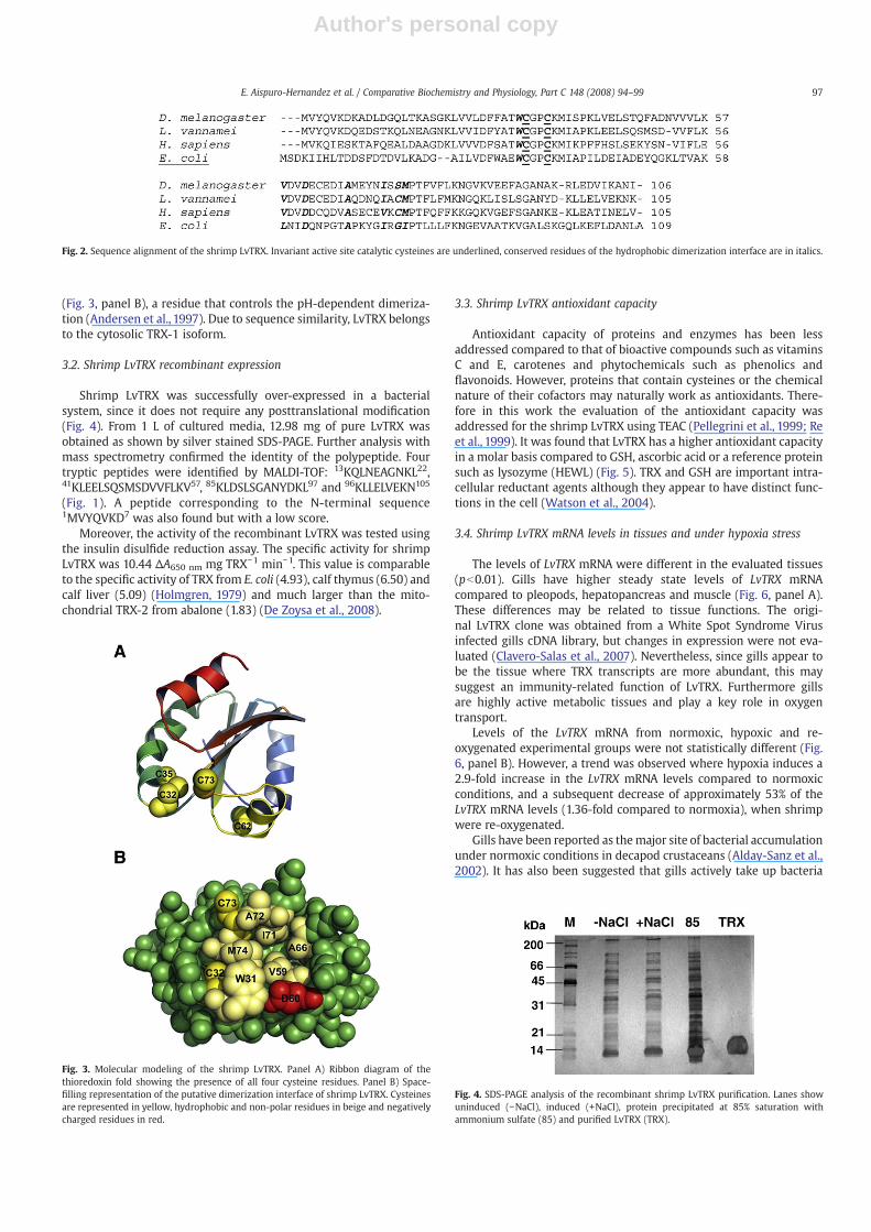
The deduced amino acid sequence of shrimp LvTRX was obtainedfrom a gill cDNA library prepared for a massive sequencing project togenerate shrimp ESTs (Clavero-Salas et al., 2007). The cDNA anddeduced amino acid sequence were deposited in GenBank underaccession number EU499301 (Fig. 1). The identity of LvTRXwith insectTRXs is around 60–65%, and it is 53% identical to human TRX (Fig. 2).After molecular modeling of the LvTRX, it was found that the aminoacid sequence is consistent with the canonical α/β thioredoxin foldpresent in disulfide redox proteins such as glutaredoxins, peroxir-edoxins and all of the thioredoxin family members (Fig. 3, panel A).
The presence of a C73 residue in LvTRX suggests that this protein isable to form covalent dimers. Many conserved residues in mammalianTRXs are conserved in the LvTRX dimer interface, including D60
Fig. 1. cDNA nucleotide and deduced amino acid sequence of L. vannamei LvTRX. Peptides of the recombinant protein identified by mass spectrometry are underlined.
96 E. Aispuro-Hernandez et al. / Comparative Biochemistry and Physiology, Part C 148 (2008) 94–99
Author's personal copy
(Fig. 3, panel B), a residue that controls the pH-dependent dimeriza-tion (Andersen et al., 1997). Due to sequence similarity, LvTRX belongsto the cytosolic TRX-1 isoform.
3.2. Shrimp LvTRX recombinant expression
Shrimp LvTRX was successfully over-expressed in a bacterialsystem, since it does not require any posttranslational modification(Fig. 4). From 1 L of cultured media, 12.98 mg of pure LvTRX wasobtained as shown by silver stained SDS-PAGE. Further analysis withmass spectrometry confirmed the identity of the polypeptide. Fourtryptic peptides were identified by MALDI-TOF: 13KQLNEAGNKL22,41KLEELSQSMSDVVFLKV57, 85KLDSLSGANYDKL97 and 96KLLELVEKN105
(Fig. 1). A peptide corresponding to the N-terminal sequence1MVYQVKD7 was also found but with a low score.
Moreover, the activity of the recombinant LvTRX was tested usingthe insulin disulfide reduction assay. The specific activity for shrimpLvTRX was 10.44 ΔA650 nm mg TRX−1 min−1. This value is comparableto the specific activity of TRX from E. coli (4.93), calf thymus (6.50) andcalf liver (5.09) (Holmgren, 1979) and much larger than the mito-chondrial TRX-2 from abalone (1.83) (De Zoysa et al., 2008).
3.3. Shrimp LvTRX antioxidant capacity
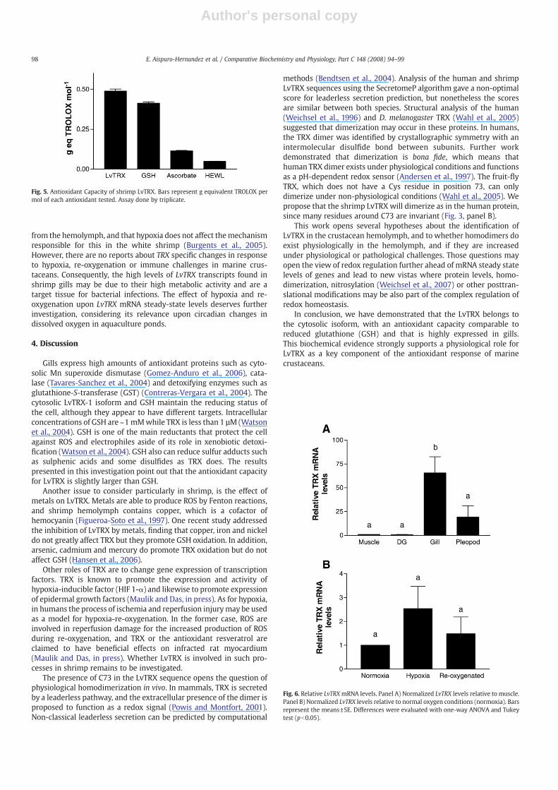
Antioxidant capacity of proteins and enzymes has been lessaddressed compared to that of bioactive compounds such as vitaminsC and E, carotenes and phytochemicals such as phenolics andflavonoids. However, proteins that contain cysteines or the chemicalnature of their cofactors may naturally work as antioxidants. There-fore in this work the evaluation of the antioxidant capacity wasaddressed for the shrimp LvTRX using TEAC (Pellegrini et al., 1999; Reet al., 1999). It was found that LvTRX has a higher antioxidant capacityin a molar basis compared to GSH, ascorbic acid or a reference proteinsuch as lysozyme (HEWL) (Fig. 5). TRX and GSH are important intra-cellular reductant agents although they appear to have distinct func-tions in the cell (Watson et al., 2004).
3.4. Shrimp LvTRX mRNA levels in tissues and under hypoxia stress
The levels of LvTRX mRNA were different in the evaluated tissues(pb0.01). Gills have higher steady state levels of LvTRX mRNAcompared to pleopods, hepatopancreas and muscle (Fig. 6, panel A).These differences may be related to tissue functions. The origi-nal LvTRX clone was obtained from a White Spot Syndrome Virusinfected gills cDNA library, but changes in expression were not eva-luated (Clavero-Salas et al., 2007). Nevertheless, since gills appear tobe the tissue where TRX transcripts are more abundant, this maysuggest an immunity-related function of LvTRX. Furthermore gillsare highly active metabolic tissues and play a key role in oxygentransport.
Levels of the LvTRX mRNA from normoxic, hypoxic and re-oxygenated experimental groups were not statistically different (Fig.6, panel B). However, a trend was observed where hypoxia induces a2.9-fold increase in the LvTRX mRNA levels compared to normoxicconditions, and a subsequent decrease of approximately 53% of theLvTRX mRNA levels (1.36-fold compared to normoxia), when shrimpwere re-oxygenated.
Gills have been reported as themajor site of bacterial accumulationunder normoxic conditions in decapod crustaceans (Alday-Sanz et al.,2002). It has also been suggested that gills actively take up bacteria
Fig. 3. Molecular modeling of the shrimp LvTRX. Panel A) Ribbon diagram of thethioredoxin fold showing the presence of all four cysteine residues. Panel B) Space-filling representation of the putative dimerization interface of shrimp LvTRX. Cysteinesare represented in yellow, hydrophobic and non-polar residues in beige and negativelycharged residues in red.
Fig. 4. SDS-PAGE analysis of the recombinant shrimp LvTRX purification. Lanes showuninduced (−NaCl), induced (+NaCl), protein precipitated at 85% saturation withammonium sulfate (85) and purified LvTRX (TRX).
Fig. 2. Sequence alignment of the shrimp LvTRX. Invariant active site catalytic cysteines are underlined, conserved residues of the hydrophobic dimerization interface are in italics.
97E. Aispuro-Hernandez et al. / Comparative Biochemistry and Physiology, Part C 148 (2008) 94–99
Author's personal copy
from the hemolymph, and that hypoxia does not affect themechanismresponsible for this in the white shrimp (Burgents et al., 2005).However, there are no reports about TRX specific changes in responseto hypoxia, re-oxygenation or immune challenges in marine crus-taceans. Consequently, the high levels of LvTRX transcripts found inshrimp gills may be due to their high metabolic activity and are atarget tissue for bacterial infections. The effect of hypoxia and re-oxygenation upon LvTRX mRNA steady-state levels deserves furtherinvestigation, considering its relevance upon circadian changes indissolved oxygen in aquaculture ponds.
4. Discussion
Gills express high amounts of antioxidant proteins such as cyto-solic Mn superoxide dismutase (Gomez-Anduro et al., 2006), cata-lase (Tavares-Sanchez et al., 2004) and detoxifying enzymes such asglutathione-S-transferase (GST) (Contreras-Vergara et al., 2004). Thecytosolic LvTRX-1 isoform and GSH maintain the reducing status ofthe cell, although they appear to have different targets. Intracellularconcentrations of GSH are ~1mMwhile TRX is less than 1 µM (Watsonet al., 2004). GSH is one of the main reductants that protect the cellagainst ROS and electrophiles aside of its role in xenobiotic detoxi-fication (Watson et al., 2004). GSH also can reduce sulfur adducts suchas sulphenic acids and some disulfides as TRX does. The resultspresented in this investigation point out that the antioxidant capacityfor LvTRX is slightly larger than GSH.
Another issue to consider particularly in shrimp, is the effect ofmetals on LvTRX. Metals are able to produce ROS by Fenton reactions,and shrimp hemolymph contains copper, which is a cofactor ofhemocyanin (Figueroa-Soto et al., 1997). One recent study addressedthe inhibition of LvTRX by metals, finding that copper, iron and nickeldo not greatly affect TRX but they promote GSH oxidation. In addition,arsenic, cadmium and mercury do promote TRX oxidation but do notaffect GSH (Hansen et al., 2006).
Other roles of TRX are to change gene expression of transcriptionfactors. TRX is known to promote the expression and activity ofhypoxia-inducible factor (HIF 1-α) and likewise to promote expressionof epidermal growth factors (Maulik and Das, in press). As for hypoxia,in humans the process of ischemia and reperfusion injurymay be usedas a model for hypoxia-re-oxygenation. In the former case, ROS areinvolved in reperfusion damage for the increased production of ROSduring re-oxygenation, and TRX or the antioxidant resveratrol areclaimed to have beneficial effects on infracted rat myocardium(Maulik and Das, in press). Whether LvTRX is involved in such pro-cesses in shrimp remains to be investigated.
The presence of C73 in the LvTRX sequence opens the question ofphysiological homodimerization in vivo. In mammals, TRX is secretedby a leaderless pathway, and the extracellular presence of the dimer isproposed to function as a redox signal (Powis and Montfort, 2001).Non-classical leaderless secretion can be predicted by computational
methods (Bendtsen et al., 2004). Analysis of the human and shrimpLvTRX sequences using the SecretomeP algorithm gave a non-optimalscore for leaderless secretion prediction, but nonetheless the scoresare similar between both species. Structural analysis of the human(Weichsel et al., 1996) and D. melanogaster TRX (Wahl et al., 2005)suggested that dimerization may occur in these proteins. In humans,the TRX dimer was identified by crystallographic symmetry with anintermolecular disulfide bond between subunits. Further workdemonstrated that dimerization is bona fide, which means thathuman TRX dimer exists under physiological conditions and functionsas a pH-dependent redox sensor (Andersen et al., 1997). The fruit-flyTRX, which does not have a Cys residue in position 73, can onlydimerize under non-physiological conditions (Wahl et al., 2005). Wepropose that the shrimp LvTRX will dimerize as in the human protein,since many residues around C73 are invariant (Fig. 3, panel B).
This work opens several hypotheses about the identification ofLvTRX in the crustacean hemolymph, and to whether homodimers doexist physiologically in the hemolymph, and if they are increasedunder physiological or pathological challenges. Those questions mayopen the view of redox regulation further ahead of mRNA steady statelevels of genes and lead to new vistas where protein levels, homo-dimerization, nitrosylation (Weichsel et al., 2007) or other posttran-slational modifications may be also part of the complex regulation ofredox homeostasis.
In conclusion, we have demonstrated that the LvTRX belongs tothe cytosolic isoform, with an antioxidant capacity comparable toreduced glutathione (GSH) and that is highly expressed in gills.This biochemical evidence strongly supports a physiological role forLvTRX as a key component of the antioxidant response of marinecrustaceans.
Fig. 6. Relative LvTRXmRNA levels. Panel A) Normalized LvTRX levels relative to muscle.Panel B) Normalized LvTRX levels relative to normal oxygen conditions (normoxia). Barsrepresent the means±SE. Differences were evaluated with one-way ANOVA and Tukeytest (pb0.05).
Fig. 5. Antioxidant Capacity of shrimp LvTRX. Bars represent g equivalent TROLOX permol of each antioxidant tested. Assay done by triplicate.
98 E. Aispuro-Hernandez et al. / Comparative Biochemistry and Physiology, Part C 148 (2008) 94–99

Author's personal copy
Acknowledgements
This research was sponsored by a CONACYT grant 45964 to G.Yepiz-Plascencia. E. Aispuro-Hernandez, L. del-Toro-Sanchez and R. M.Robles-Sanchez received scholarships from CONACYT (NationalResearch and Technology Council, Mexico). We thank MonicaResendiz (CIAD) for technical assistance with the qRT-PCR experi-ments, and Dr. Maria Islas-Osuna for review and critical comments tothe manuscript. E.A-H designed and performed research, analyzeddata and wrote the paper, K.D.G-O designed experiments, analyzeddata and wrote the paper, A.M-A designed and performed research,analyzed data and wrote the paper, L.d-T-S and R.M. R-S designed andperformed research, J.H designed experiments, analyzed data andcontributed new reagents, G.G-A designed experiments, contributedreagents and analytical tools and wrote the paper, G.Y-P designedexperiments, contributed reagents and wrote the paper, R.R.S-Mdesigned experiments, analyzed data and wrote the paper.
References
Alday-Sanz, V., Roque, A., Turnbull, J.F., 2002. Clearing mechanisms of Vibrio vulnificusbiotype I in the black tiger shrimp Penaeus monodon. Dis. Aquat. Org. 48, 91–99.
Andersen, J.F., Sanders, D.A., Gasdaska, J.R., Weichsel, A., Powis, G., Montfort, W.R., 1997.Human thioredoxin homodimers: regulation by pH, role of aspartate 60, and crystalstructure of the aspartate 60 –N asparagine mutant. Biochemistry 36, 13979–13988.
Bacano Maningas, M.B., Koyama, T., Kondo, H., Hirono, I., Aoki, T., 2008. A peroxiredoxinfrom kuruma shrimp, Marsupenaeus japonicus, inhibited by peptidoglycan. Dev.Comp. Immunol. 32, 198–203.
Bachere, E., 2000. Shrimp immunity and disease control — introduction. Aquaculture191, 3–11.
Bendtsen, J.D., Jensen, L.J., Blom, N., Von Heijne, G., Brunak, S., 2004. Feature-basedprediction of non-classical and leaderless protein secretion. Protein Eng. Des. Sel. 17,349–356.
Burgents, J.E., Burnett, K.G., Burnett, L.E., 2005. Effects of hypoxia and hypercapnichypoxia on the localization and the elimination of Vibrio campbellii in Litopenaeusvannamei, the Pacific white shrimp. Biol. Bull. 208, 159–168.
Clavero-Salas, A., Sotelo-Mundo, R.R., Gollas-Galvan, T., Hernandez-Lopez, J., Peregrino-Uriarte, A.B., Muhlia-Almazan, A., Yepiz-Plascencia, G., 2007. Transcriptome analysisof gills from the white shrimp Litopenaeus vannamei infected with White SpotSyndrome Virus. Fish Shellfish Immunol. 23, 459–472.
Contreras-Vergara, C.A., Harris-Valle, C., Sotelo-Mundo, R.R., Yepiz-Plascencia, G., 2004.A mu-class glutathione S-transferase from the marine shrimp Litopenaeusvannamei: molecular cloning and active-site structural modeling. J. Biochem. Mol.Toxicol. 18, 245–252.
DeLano, W.L., 2002. The PyMOL molecular graphics system. In DeLano Scientific. SanCarlos, CA, USA.
De Zoysa, M., Pushpamali, W.A., Whang, I., Kim, S.J., Lee, J., 2008. Mitochondrialthioredoxin-2 from disk abalone (Haliotis discus discus): molecular characterization,tissue expression and DNA protection activity of its recombinant protein. Comp.Biochem. Physiol. B, doi:10.1016/j.cbpb.2007.12.009.
Figueroa-Soto, C.G., Calderón de la Barca, A.M., Vázquez-Moreno, L., Higuera-Ciapara, I.,Yepiz-Plascencia, G., 1997. Purification of hemocyanin from the white shrimp(Penaeus vannamei Boone), by immobilized metal affinity chromatography. Comp.Biochem. Physiol. B 117, 203–208.
Gomez-Anduro, G.A., Barillas-Mury, C.V., Peregrino-Uriarte, A.B., Gupta, L., Gollas-Galvan, T., Hernandez-Lopez, J., Yepiz-Plascencia, G., 2006. The cytosolic manganesesuperoxide dismutase from the shrimp Litopenaeus vannamei: molecular cloningand expression. Dev. Comp. Immunol. 30, 893–900.
Grant, C.M., 2001. Role of the glutathione/glutaredoxin and thioredoxin systems in yeastgrowth and response to stress conditions. Mol. Microbiol. 39, 533–541.
Hansen, J.M., Zhang, H., Jones, D.P., 2006. Differential oxidation of thioredoxin-1,thioredoxin-2, and glutathione by metal ions. Free Radic. Biol. Med. 40, 138–145.
Holmgren, A., 1979. Thioredoxin catalyzes the reduction of insulin disulfides bydithiothreitol and dihydrolipoamide. J. Biol. Chem. 254, 9627–9632.
Holmgren, A., 1985. Thioredoxin. Annu. Rev. Biochem. 54, 237–271.Kim, Y.J., Lee, K.S., Kim, B.Y., Choo, Y.M., Sohn, H.D., Jin, B.R., 2007. Thioredoxin from the
silkworm, Bombyx mori: cDNA sequence, expression, and functional characteriza-tion. Comp. Biochem. Physiol. B 147, 574–581.
Lesser, M.P., 2006. Oxidative stress in marine environments: biochemistry andphysiological ecology. Annu. Rev. Physiol. 68, 253–278.
Lillig, C.H., Holmgren, A., 2007. Thioredoxin and related molecules—from biology tohealth and disease. Antioxid. Redox Signal. 9, 25–47.
Livak, K.J., Schmittgen, T.D., 2001. Analysis of relative gene expression data using real-time quantitative PCR and the 2−ΔΔCT method. Methods 25, 402–408.
Martinez-Galisteo, E., Padilla, C.A., Garcia-Alfonso, C., Lopez-Barea, J., Barcena, J.A., 1993.Purification and properties of bovine thioredoxin system. Biochimie 75, 803–809.
Maulik, N., Das, D.K., 2008. Emerging potential of thioredoxin and thioredoxininteracting proteins in various disease conditions. Biochim Biophys Acta. (inpress). doi:10.1016/j.bbagen.2007.12.008.
Nappi, A.J., Ottaviani, E., 2000. Cytotoxicity and cytotoxic molecules in invertebrates.Bioessays 22, 469–480.
Pellegrini, N., Re, R., Yang, M., Rice-Evans, C., 1999. Screening of dietary carotenoids andcarotenoid-rich fruit extracts for antioxidant activities applying 2,2′-Azinobis(3-ethylenebenzothiazoline-6-sulfonic acid radical cation decolorization assay.Methods Enzymol. 299, 379–389.
Powis, G., Montfort, W.R., 2001. Properties and biological activities of thioredoxins.Annu. Rev. Pharmacol. Toxicol. 41, 261–295.
Re, R., Pellegrini, N., Proteggente, A., Pannala, A., Yang, M., Rice-Evans, C., 1999.Antioxidant activity applying an improved ABTS radical cation decolorization assay.Free Radic. Biol. Med. 26, 1231–1237.
Sambrook, J., Russell, D.W., 2001. Molecular Cloning: A Laboratory Manual. Cold SpringHarbor Laboratory Press.
Schwede, T., Kopp, J., Guex, N., Peitsch, M.C., 2003. SWISS-MODEL: an automatedprotein homology-modeling server. Nucleic Acids Res. 31, 3381–3385.
Smith, P.K., Krohn, R.I., Hermanson, G.T., Mallia, A.K., Gartner, F.H., Provenzano, M.D.,Fujimoto, E.K., Goeke, N.M., Olson, B.J., Klenk, D.C., 1985. Measurement of proteinusing bicinchoninic acid. Anal. Biochem. 150, 76–85.
Tavares-Sanchez, O.L., Gomez-Anduro, G.A., Felipe-Ortega, X., Islas-Osuna, M.A., Sotelo-Mundo, R.R., Barillas-Mury, C., Yepiz-Plascencia, G., 2004. Catalase from the whiteshrimp Penaeus (Litopenaeus) vannamei: molecular cloning and protein detection.Comp. Biochem. Physiol. B 138, 331–337.
Wahl, M.C., Irmler, A., Hecker, B., Schirmer, R.H., Becker, K., 2005. Comparative structuralanalysis of oxidized and reduced thioredoxin from Drosophila melanogaster. J. Mol.Biol. 345, 1119–1130.
Watson, W.H., Yang, X., Choi, Y.E., Jones, D.P., Kehrer, J.P., 2004. Thioredoxin and its rolein toxicology. Toxicol. Sci. 78, 3–14.
Weichsel, A., Gasdaska, J.R., Powis, G., Montfort, W.R., 1996. Crystal structures ofreduced, oxidized, and mutated human thioredoxins: evidence for a regulatoryhomodimer. Structure 4, 735–751.
Weichsel, A., Brailey, J.L., Montfort, W.R., 2007. Buried S-nitrosocysteine revealed incrystal structures of human thioredoxin. Biochemistry 46, 1219–1227.
Zakharyan, R.A., Tsaprailis, G., Chowdhury, U.K., Hernandez, A., Aposhian, H.V., 2005.Interactions of sodium selenite, glutathione, arsenic species, and omega classhuman glutathione transferase. Chem. Res. Toxicol. 18, 1287–1295.
Zhang, Q., Li, F., Zhang, J., Wang, B., Gao, H., Huang, B., Jiang, H., Xiang, J., 2007. Molecularcloning, expression of a peroxiredoxin gene in Chinese shrimp Fenneropenaeuschinensis and the antioxidant activity of its recombinant protein. Mol. Immunol. 44,3501–3509.
99E. Aispuro-Hernandez et al. / Comparative Biochemistry and Physiology, Part C 148 (2008) 94–99
Copyright © 2022 FDOKUMEN