
A Novel Antioxidant Phenyl Disaccharide from Populus tremula Knotwood
Upload
khangminh22Category
view
1download
0
1 / 64
Journal of Medicinal Chemistry 2019; 62 (11): 5382-5403
DOI: 10.1021/acs.jmedchem.9b00003
Potent 5-Cyano-6-Phenyl-Pyrimidin-Based Derivatives Targeting
DCN1-UBE2M Interaction
Wenjuan Zhou†, ‡, #, Liying Ma†, #, Lina Ding†, Qian Guo†, Zhangxu He†, Jing Yang†,
Hui Qiao†, Lingyu Li†, Jie Yang†, Shimin Yu†, Lili Zhao†, Shaomeng Wang†, §,
Hong-Min Liu†,*, Zhenhe Suo‡, *, Wen Zhao†, *
†State Key Laboratory of Esophageal Cancer Prevention and Treatment; Key Laboratory of
Advanced Pharmaceutical Technology, Ministry of Education of China; School of
Pharmaceutical Sciences, Zhengzhou University, 100 Kexue Avenue, Zhengzhou, Henan,
450001, China
‡Department of Pathology, Oslo University Hospital; Faculty of Medicine, University of Oslo,
Oslo, 0379, Norway
§The Rogel Cancer Center and Departments of Internal Medicine, Pharmacology, Medicinal
Chemistry and Pathology, University of Michigan Medical School, Ann Arbor,
Michigan, 48109, United States
#: Both authors contribute equally to this work.
*: These senior authors contribute equally to this work.
*: Corresponding to:
Wen Zhao, Email: [email protected], [email protected]; Phone, 86-15003898187
Zhenhe Suo, Email: [email protected]; Phone, 47-98280093
Hong-Min Liu, Email: [email protected]; Phone, 86-371-67781739.
2 / 64
ABSTRACT
Neddylation of the Cullin-RING E3 ligases (CRLs) regulates the homeostasis of
approximately 20% of cellular proteins. Defective in cullin neddylation 1(DCN1), as a co-E3
ligase, interacts with UBE2M to enhance the activation of CRLs and this interaction is emerging
as a therapeutic target for human diseases. Here, we present a series of pyrimidin-based small
molecular inhibitors targeting DCN1-UBE2M interaction. After finding a novel inhibitor DC-
1 with IC50 = 1.2µM, we performed a series of chemical optimization, which finally led to the
discovery of a potent thiazole contained 5-cyano-6-phenylpyrimidin-based inhibitor DC-2
(IC50 = 15nM). Next, using protein and cellular thermal shift assays, co-immunoprecipitation,
molecular docking and site specific mutation experiments, we further proved that DC-2
specifically inhibited the interaction of UBE2M and DCN1 at molecule and cellular levels,
resulting in the decrease of cullin3 neddylation and accumulation of its substrate: NRF2. Our
findings indicate that DC-2 may serve as a novel lead compound for specific derivatives
targeting DCN1-UBE2M interaction.
3 / 64
INTRODUCTION
The ubiquitin–proteasome system (UPS) is integral to maintaining cellular protein
homeostasis by regulating degradation of intracellular proteins.1-7 The ubiquitylation pathway
is executed by the coordinated efforts of the E1 (ubiquitin-activating enzyme), E2 (ubiquitin-
conjugating enzyme) and E3 (ubiquitin ligase enzyme) proteins.5 Like ubiquitination,8
neddylation is a novel type of posttranslational modification, in which the ubiquitin-like
molecule NEDD8 is added to its target proteins and thus regulates their functions.9, 10
Neddylation also consists of a tripartite enzymatic cascade, including the NEDD8-activating
enzyme E1 (NAE, APPBP1 (NAE1) and UBA3 heterodimer), two NEDD8-conjugating
enzymes E2s (UBE2F and UBE2M, also known as UBC12) and NEDD8-E3 ligases,9, 10 with
cullins being the best-characterized substrates.11-22 Up to now, neddylation has been recognized
to be highly activated in various cancers, 20, 27-33 which draws much attraction for oncologic drug
discovery, especially on the finding of NEDD8 E1 inhibitor Pevonedistat(MLN4924).10, 23-26
MLN4924 covalently binds to NEDD8 E1, blocking all CRLs neddylation,7 thus causing
accumulation of CRL substrates.7, 10 Since MLN4924 possesses immense anti-cancer effects
both in vitro and in vivo,10, 27, 28-43 it has been approved for Phase II clinical trials for treatment
of human acute myeloid leukemia, non-small cell lung cancer and mesothelioma.29 However,
as a result of its broad ablation of neddylation, MLN4924 has a series of toxicities. Therefore,
alternative targeting specific CRLs may be more potential and safer for cancer treatment.30-32
DCN1 (defective in cullin neddylation 1), also called DCUN1D1, DCNL1 or SCCRO, is a
4 / 64
highly conserved gene and amplified along the 3q26.3 in most squamous cell carcinomas and
some other human cancers.33-39 As a co-E3 ligase, it binds to the activation complex of Cullin-
RBX1-UBE2M-NEDD8 to increase neddylation efficiency.16, 40-42 Researchers have found that
blocking UBE2M-DCN1 interaction can reduce cullin3 neddylation.30, 32, 43 Moreover, DCN1
overexpression has been reported to be associated with poor survival outcome, and DCN1
depression significantly reduces cancer cell growth and invasive capacity. 10,33-35, 42, 44-51
Therefore, DCN1 is considered as a promising and attractive anti-cancer target.
From the X-ray crystallographic structure of DCN1 with UBE2M, researchers have
confirmed that during neddylation, the N-terminal acetylated UBE2M can dock into the
hydrophobic pocket of DCN1 and this pocket could be used for designing DCN1 inhibitors.21,
52 Up to now, four papers have reported the discovery of DCN1-UBE2M interaction
inhibitors,30-32, 43 also known as DCN1 inhibitor. NAcM-COV, the first discovered inhibitor,
showed highly specific effects on blocking the N-terminal acetylation-dependent interaction of
UBE2M with DCN1, resulting in the reduction of CUL1 and CUL3 neddylation in a squamous
lung cancer cell line (HCC95).31,43 At the same time, Zhou et al, discovered two high-affinity
inhibitors DI-591 and DI-404, which can selectively inhibit the neddylation of cullin 3 over
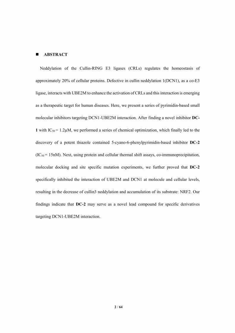
other cullins, leading to the accumulation of cullin 3’s substrate:NRF2 (Figure 1). 30, 32 In this
study, we screened our in-house structurally diverse molecular library (ca.1000 compounds)
and finally found a novel and potent pyrimidine-based DCN1-UBE2M interaction mediator
DC-1, using both fluorescence polarization (FP) and homogeneous time resolved fluorescence
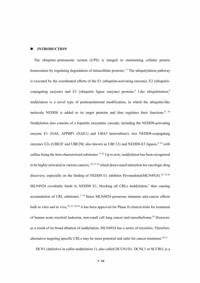
(HTRF)53 assays. After extensive structure-activity relationship (SAR) efforts, a potent thiazole
contained 5-cyano-6-phenylpyrimidin-based inhibitor DC-2 was discovered (Figure 2). DC-2
5 / 64
specifically targets DCN1-UBE2M interaction, leading to the inhibition of cullin3 neddylation
and the accumulation of NRF2 and NRF2’ downstream proteins: HO-1 and NQO1. Our overall
findings indicate that DC-2 may serve as a novel lead compound for specific derivatives
targeting DCN1-UBE2M interaction.
Figure 1. Representative examples of DCN1 inhibitors.
Figure 2. Discovery of potent thiazole contained pyrimidin-based inhibitor DC-2.
RESULTS AND DISCUSSION
Chemistry. The general synthesis route of the target pyrimidine-thiourea hybrids is depicted
in Scheme 1. Benzaldehydes 1a-m, ethyl cyanoacetate 2, and thiourea 3 were prolonged heated
in ethanol containing potassium carbonate to obtain 6-aryl-5-cyano-2-thiouracils 4a-m. Then,
compounds 4a-m reacted with the 3-bromoprop-1-yne, 3-bromoprop-1-ene, 1-bromopropane
and phosphorus oxychloride in dioxane to obtain the target derivatives 5a-o.54 Compound 53
was prepared via click reaction of compound 5a with benzyl azide. After that, these highly
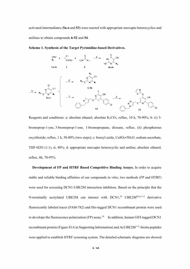
6 / 64
activated intermediates (5a-o and 53) were reacted with appropriate mercapto heterocyclics and
anilines to obtain compounds 6-52 and 54.
Scheme 1. Synthesis of the Target Pyrimidine-based Derivatives.
Reagents and conditions: a: absolute ethanol, absolute K2CO3, reflux, 10 h, 70-90%; b: (i) 3-
bromoprop-1-yne, 3-bromoprop-1-ene, 1-bromopropane, dioxane, reflux; (ii) phosphorous
oxychloride, reflux, 1 h, 50-80% (two steps); c: benzyl azide, CuSO4•5H2O, sodium ascorbate,
THF-H2O (1:1), rt, 80%; d: appropriate mercapto heterocyclic and aniline, absolute ethanol,
reflux, 6h, 70-95%.
Development of FP and HTRF Based Competitive Binding Assays. In order to acquire
stable and reliable binding affinities of our compounds in vitro, two methods (FP and HTRF)
were used for screening DCN1-UBE2M interaction inhibitors. Based on the principle that the
N-terminally acetylated UBE2M can interact with DCN1,55 UBE2MNAc1-12 derivative
fluorescently labeled tracer (FAM-782) and His-tagged DCN1 recombinant protein were used
to develope the fluorescence polarization (FP) assay.30 In addition, human GST-tagged DCN1
recombinant protein (Figure S1A in Supporting Information) and AcUBE2M1-21-biotin peptides
were applied to establish HTRF screening system. The detailed schematic diagrams are showed
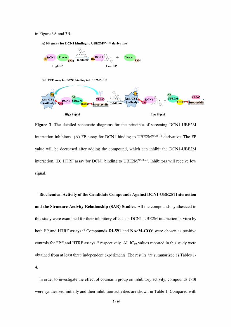
7 / 64
in Figure 3A and 3B.
Figure 3. The detailed schematic diagrams for the principle of screening DCN1-UBE2M
interaction inhibitors. (A) FP assay for DCN1 binding to UBE2MNAc1-12 derivative. The FP
value will be decreased after adding the compound, which can inhibit the DCN1-UBE2M
interaction. (B) HTRF assay for DCN1 binding to UBE2MNAc1-21. Inhibitors will receive low
signal.
Biochemical Activity of the Candidate Compounds Against DCN1-UBE2M Interaction
and the Structure-Activity Relationship (SAR) Studies. All the compounds synthesized in
this study were examined for their inhibitory effects on DCN1-UBE2M interaction in vitro by
both FP and HTRF assays.30 Compounds DI-591 and NAcM-COV were chosen as positive
controls for FP30 and HTRF assays,43 respectively. All IC50 values reported in this study were
obtained from at least three independent experiments. The results are summarized as Tables 1-
4.
In order to investigate the effect of coumarin group on inhibitory activity, compounds 7-10
were synthesized initially and their inhibition activities are shown in Table 1. Compared with
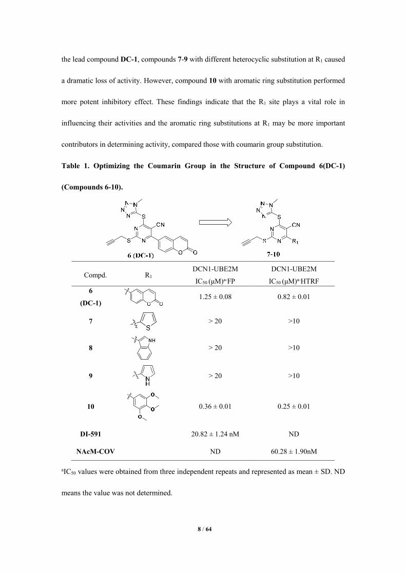
8 / 64
the lead compound DC-1, compounds 7-9 with different heterocyclic substitution at R1 caused
a dramatic loss of activity. However, compound 10 with aromatic ring substitution performed
more potent inhibitory effect. These findings indicate that the R1 site plays a vital role in
influencing their activities and the aromatic ring substitutions at R1 may be more important
contributors in determining activity, compared those with coumarin group substitution.
Table 1. Optimizing the Coumarin Group in the Structure of Compound 6(DC-1)
(Compounds 6-10).
Compd. R1 DCN1-UBE2M
IC50 (μM)a-FP
DCN1-UBE2M
IC50 (μM)a-HTRF 6
(DC-1) 1.25 ± 0.08 0.82 ± 0.01
7
> 20 >10
8
> 20 >10
9
> 20 >10
10
0.36 ± 0.01 0.25 ± 0.01
DI-591 20.82 ± 1.24 nM ND
NAcM-COV ND 60.28 ± 1.90nM
aIC50 values were obtained from three independent repeats and represented as mean ± SD. ND
means the value was not determined.
9 / 64
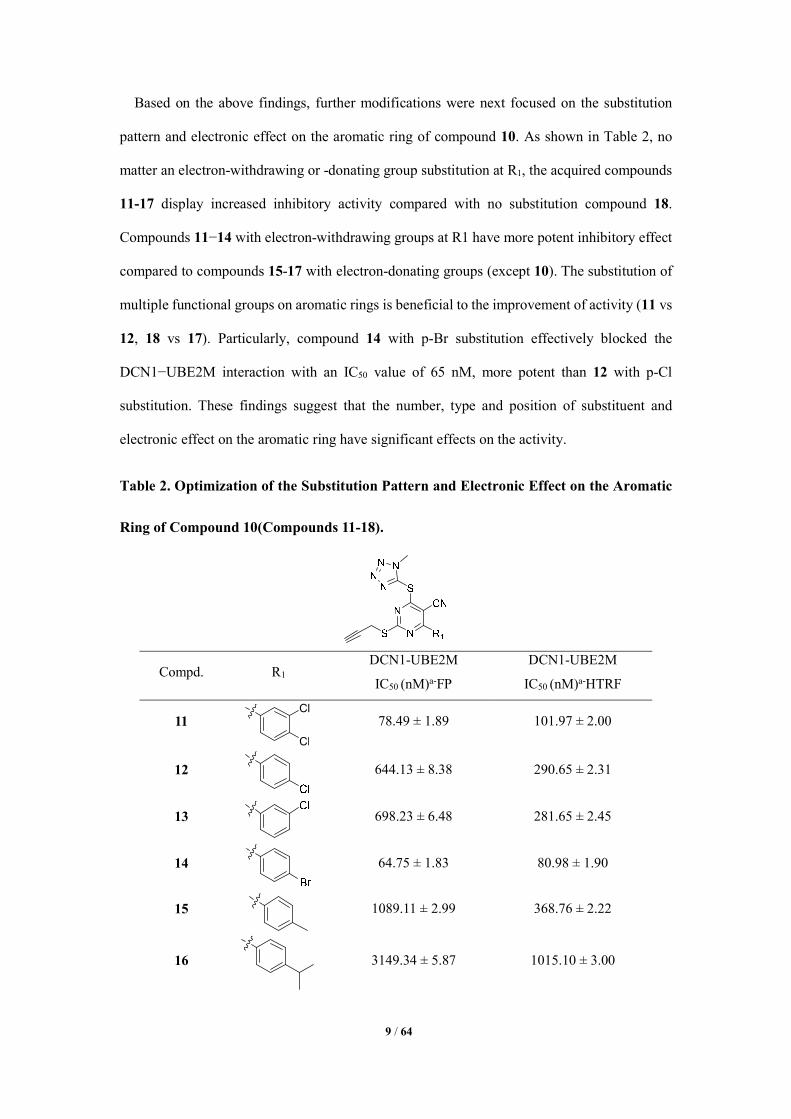
Based on the above findings, further modifications were next focused on the substitution
pattern and electronic effect on the aromatic ring of compound 10. As shown in Table 2, no
matter an electron-withdrawing or -donating group substitution at R1, the acquired compounds
11-17 display increased inhibitory activity compared with no substitution compound 18.
Compounds 11−14 with electron-withdrawing groups at R1 have more potent inhibitory effect
compared to compounds 15-17 with electron-donating groups (except 10). The substitution of
multiple functional groups on aromatic rings is beneficial to the improvement of activity (11 vs
12, 18 vs 17). Particularly, compound 14 with p-Br substitution effectively blocked the
DCN1−UBE2M interaction with an IC50 value of 65 nM, more potent than 12 with p-Cl
substitution. These findings suggest that the number, type and position of substituent and
electronic effect on the aromatic ring have significant effects on the activity.
Table 2. Optimization of the Substitution Pattern and Electronic Effect on the Aromatic
Ring of Compound 10(Compounds 11-18).
Compd. R1 DCN1-UBE2M
IC50 (nM)a-FP
DCN1-UBE2M
IC50 (nM)a-HTRF
11 78.49 ± 1.89 101.97 ± 2.00
12
644.13 ± 8.38 290.65 ± 2.31
13
698.23 ± 6.48 281.65 ± 2.45
14
64.75 ± 1.83 80.98 ± 1.90
15
1089.11 ± 2.99 368.76 ± 2.22
16
3149.34 ± 5.87 1015.10 ± 3.00
Cl
Cl
10 / 64
17
942.93 ± 6.93 338.83 ± 2.53
10
360.23 ± 3.64 251.13 ± 2.25
18
>20000 >20000
aIC50 values were obtained from three independent repeats and represented as mean ± SD. ND
means the value was not determined.
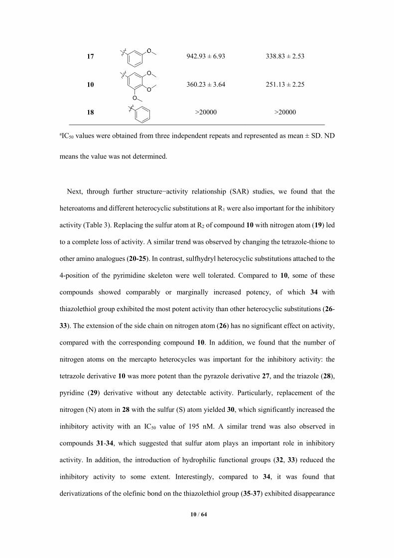
Next, through further structure−activity relationship (SAR) studies, we found that the
heteroatoms and different heterocyclic substitutions at R1 were also important for the inhibitory
activity (Table 3). Replacing the sulfur atom at R2 of compound 10 with nitrogen atom (19) led
to a complete loss of activity. A similar trend was observed by changing the tetrazole-thione to
other amino analogues (20-25). In contrast, sulfhydryl heterocyclic substitutions attached to the
4-position of the pyrimidine skeleton were well tolerated. Compared to 10, some of these
compounds showed comparably or marginally increased potency, of which 34 with
thiazolethiol group exhibited the most potent activity than other heterocyclic substitutions (26-
33). The extension of the side chain on nitrogen atom (26) has no significant effect on activity,
compared with the corresponding compound 10. In addition, we found that the number of
nitrogen atoms on the mercapto heterocycles was important for the inhibitory activity: the
tetrazole derivative 10 was more potent than the pyrazole derivative 27, and the triazole (28),
pyridine (29) derivative without any detectable activity. Particularly, replacement of the
nitrogen (N) atom in 28 with the sulfur (S) atom yielded 30, which significantly increased the
inhibitory activity with an IC50 value of 195 nM. A similar trend was also observed in
compounds 31-34, which suggested that sulfur atom plays an important role in inhibitory
activity. In addition, the introduction of hydrophilic functional groups (32, 33) reduced the
inhibitory activity to some extent. Interestingly, compared to 34, it was found that
derivatizations of the olefinic bond on the thiazolethiol group (35-37) exhibited disappearance
O
O
O
11 / 64
of activity, which indicated that the increase of steric hindrance at R2 was detrimental to the
activity.
Table 3. Optimization of the Heteroatoms and Different Heterocyclic Substitutions of
Compound 10(Compounds 19-37).
Compd. R2 DCN1-UBE2M
IC50 (nM)a-FP
DCN1-UBE2M
IC50 (nM)a-HTRF 19
>10000
>10000
20
>10000 >5000
21
>10000 >5000
22
>10000 >10000
23
>10000 >5000
24
>10000 >10000
25
>10000 >10000
26
591.11 ± 2.75 199.07 ± 2.29
27
1217.90 ± 1.32 >5000
28
>10000 >2500
29
>10000 >2500
30
195.66 ± 2.29 65.98 ± 1.81
31
152.82 ± 2.18 133.60 ± 2.12
32
820.78 ± 2.91 106.73 ± 2.02

12 / 64
33
413.55 ± 2.04 141.22 ± 2.15
34
141.22 ± 2.10 77.04 ± 1.88
35 (DC-2N)
>10000 >10000
36
>10000 >10000
37
>10000 >5000
10
360 ± 3.64 251.13 ± 2.25
aIC50 values were obtained from three independent repeats and represented as mean ± SD. ND
means the value was not determined.
In order to further investigate the importance of the two sites at R1 and R2 of 34, we
simultaneously changed them and synthesized compounds 38-50. Compared to 34, some of
these compounds showed comparable potency against DCN1, of which 42 (also named as DC-
2) showed the most potent inhibitory effect on the DCN1-UBE2M interaction with an IC50
value of 26 nM. In addition, compound 39 with chlorine substitution in para position at the
phenyl ring in R1 displayed more potent inhibitory activity compared to the meta- and di-
substituted compounds (38 and 41). A similar trend was also observed (46 vs 47, 48), which
suggested that the position of substituent at the phenyl ring in R1 plays an important role in the
inhibitory activity. Furthermore, replacement of the phenyl group at R1 with aromatic
heterocycle yielded 43-45, which significantly decreased the inhibitory activity, as well as 46-
49 vs 50, which further suggested that the position of substituent and electronic effect at R1
were important for the activity. Moreover, the importance of terminal alkyne moiety was also
evaluated. Due to the change of the propargyl group to ethene or ethyl group, compounds 51
and 52 exhibited no detectable (>10000nM) DCN1-UBE2M inhibitory effect compared to 39,
which may be related to the hydrophobic interactions by forming π−π stacking with DCN1-
UBE2M. In addition, replacing the propargyl group by triazole via click chemistry caused a

13 / 64
dramatic loss of activity (54 vs 39), which indicated that the increase of steric hindrance at R3
was detrimental to the activity. These modifications and SAR studies reveal that the terminal
alkyne group is also critical for their inhibitory activity, and the introduction of hydrophobic
functional groups of appropriate size at R3 may be beneficial to the improvement of activity.
Table 4. Optimization of the Aromatic and Heterocyclic Substitutions at R1, R2 and R3
of Compound 34(Compounds 38-54).
Compd. R1 R2 R3 DCN1-UBE2M
IC50 (nM)a-FP
DCN1-UBE2M
IC50 (nM)a-HTRF
34
141.22 ± 2.10 77.04 ± 1.88
38
361.69 ± 2.93 140.38 ± 2.14
39
36.59 ± 0.18 96.05 ± 1.98
40
551.87 ± 1.71 189.41 ± 1.95
41
62.99 ± 1.79 112.26 ± 2.05
42
(DC-2)
26.89 ± 0.24 15.71 ± 1.19
43
789.09 ± 2. 89 202.05 ± 2.00
44
1482.32 ± 1.39 1064.73±1.07
45
>20000 >10000
46
63.51 ± 1.80 122.65 ± 2.08
47
106.21 ± 2.02 379.91 ± 2.58

14 / 64
48
109.04 ± 2.03 119.80 ± 2.07
49
390.85 ± 2.56 172.29 ± 2.23
50
665.64 ± 2.74 814.60 ± 2.91
51
> 10000 > 10000
52
> 10000 > 10000
54
> 10000 > 10000
aIC50 values were obtained from three independent repeats and represented as mean ± SD. ND
means the value was not determined.
Figure 4. Compound DC-2(compound 42) combines with DCN1 and thus inhibits UBE2M-
DCN1 interaction in vitro. Compound DC-2 was added into the screening systems and its
inhibition rates (%) at different concentration were determined by both FP (A) and HTRF (B)
assays, respectively. Data are presented as means ± SD. Three individual experiments were
performed for each group.
In summary, starting from a potent pyrimidine-based DCN1-UBE2M interaction mediator
DC-1(compound 6) in our in-house structurally diverse molecular library (ca.1000 compounds),
DC-2(compound 42) containing 5-cyano-6-phenyl-pyrimidin was finally obtained with MW of
380, two IC50 values 26.89 ± 0.24 (Ki=20.83 ± 0.18nM) and 15.71 ± 1.19 nM (Ki=13.66 ±
15 / 64
1.03nM) from the FP and HTRF assays (Figure 4A and 4B), respectively, resulting in about 80-
fold improved potency. Furthermore, DC-2 has aqueous solubility in phosphate buffer (PBS,
pH 7.4) to some extent (Figure S2 in Supporting Information). Therefore, DC-2 was chosen for
the following target activity evaluation experiments. The compound 35 (DC-2N), which had
the similar structure with DC-2 but presented no obvious inhibitory effect on DCN1-UBE2M
interaction (IC50>10µM), was chosen as a negative control.
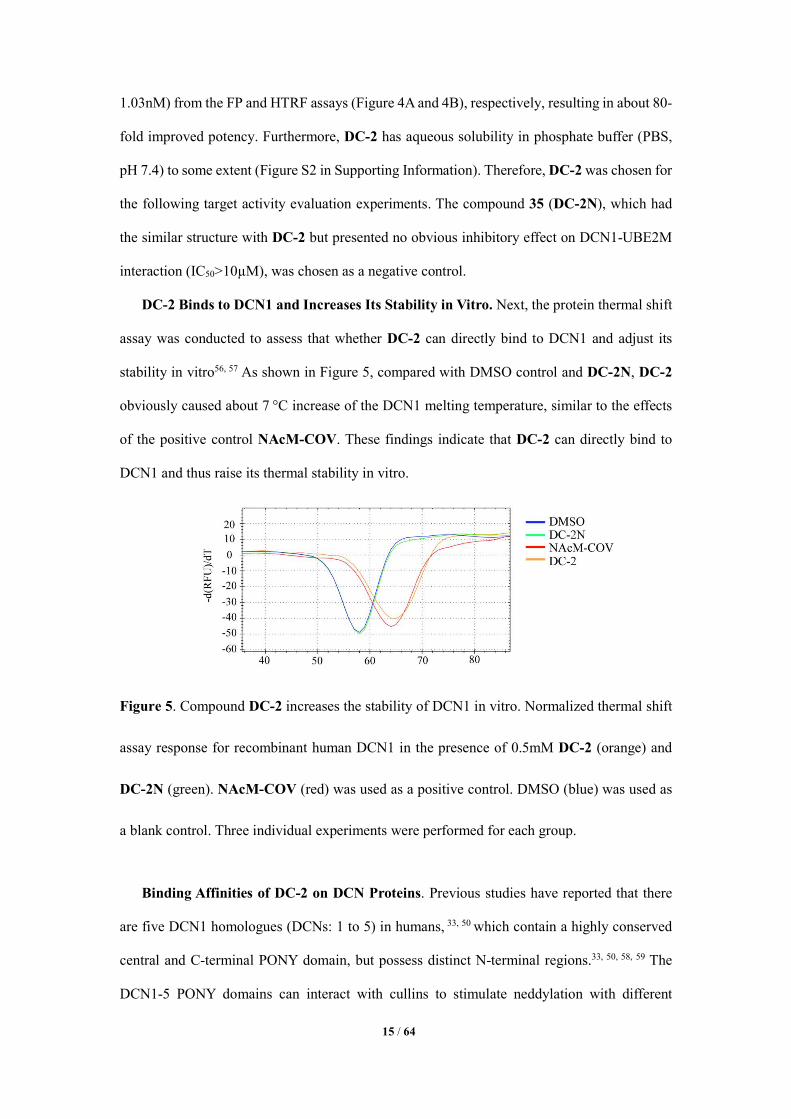
DC-2 Binds to DCN1 and Increases Its Stability in Vitro. Next, the protein thermal shift
assay was conducted to assess that whether DC-2 can directly bind to DCN1 and adjust its
stability in vitro56, 57 As shown in Figure 5, compared with DMSO control and DC-2N, DC-2
obviously caused about 7 °C increase of the DCN1 melting temperature, similar to the effects
of the positive control NAcM-COV. These findings indicate that DC-2 can directly bind to
DCN1 and thus raise its thermal stability in vitro.
Figure 5. Compound DC-2 increases the stability of DCN1 in vitro. Normalized thermal shift
assay response for recombinant human DCN1 in the presence of 0.5mM DC-2 (orange) and
DC-2N (green). NAcM-COV (red) was used as a positive control. DMSO (blue) was used as
a blank control. Three individual experiments were performed for each group.
Binding Affinities of DC-2 on DCN Proteins. Previous studies have reported that there
are five DCN1 homologues (DCNs: 1 to 5) in humans, 33, 50 which contain a highly conserved
central and C-terminal PONY domain, but possess distinct N-terminal regions.33, 50, 58, 59 The
DCN1-5 PONY domains can interact with cullins to stimulate neddylation with different
16 / 64
efficiencies.55, 60 Therefore, we further measured the binding affinities of DC-2 on DCN1 to 5
through competitive binding assay. As shown in Table 5, DC-2 also has high binding affinity
on DCN2, whose PONY domain in human shares 82% identity to that of DCN1.43 However, it
shows relatively weak binding effects (Ki >500nM) on DCN3-5, compared to that of DCN1.
These data suggest that DC-2 has specificity and selectivity for DCN1 and 2, similar to the
previously reported inhibitors (DI-591, NAcM-COV and DI-404).30, 32, 43
Table 5. Binding Affinities of Compound DC-2 on the Indicated DCN1-5 Recombinant
Proteins.
DCN1
Ki (nM)a
DCN2
Ki (nM)a
DCN3
Ki (nM)a
DCN4
Ki (nM)a
DCN5
Ki (µM)a
DC-2 13.66 ± 1.03 66.19 ± 1.72 591.72 ± 5.17 807.01 ± 9.42 2.14 ± 0.32
aKi values were obtained from three independent repeats and represented as mean ± SD. ND
means the value was not determined.
Furthermore, since the N-terminal domain of UBE2M has interaction with both DCN1 and
NEDD8 E1 subunit UBA3,52, 61 we intended to determine whether DC-2 also has inhibitory
effects on E1’s activity and UBE2M neddylation. As shown in Figure 6A and 6B, in the cell-
free NAE activity assay, even though MLN4924 significantly inhibits UBE2M neddylation,
compound DC-2 exhibits no obvious effect on NEDD8~UBE2M at concentration up to 100
μM, indicating that compound DC-2 has no effect on E1’s activity through ATP initiation and
UBE2M neddylation.
17 / 64
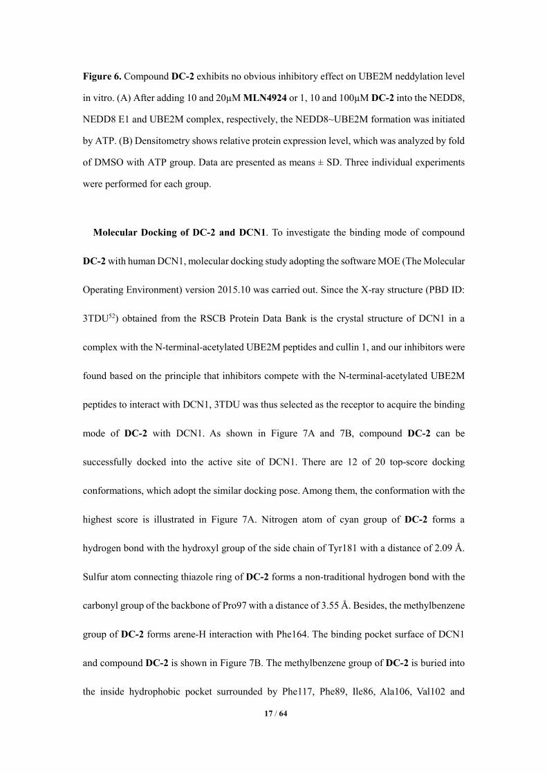
Figure 6. Compound DC-2 exhibits no obvious inhibitory effect on UBE2M neddylation level
in vitro. (A) After adding 10 and 20µM MLN4924 or 1, 10 and 100µM DC-2 into the NEDD8,
NEDD8 E1 and UBE2M complex, respectively, the NEDD8~UBE2M formation was initiated
by ATP. (B) Densitometry shows relative protein expression level, which was analyzed by fold
of DMSO with ATP group. Data are presented as means ± SD. Three individual experiments
were performed for each group.
Molecular Docking of DC-2 and DCN1. To investigate the binding mode of compound
DC-2 with human DCN1, molecular docking study adopting the software MOE (The Molecular
Operating Environment) version 2015.10 was carried out. Since the X-ray structure (PBD ID:
3TDU52) obtained from the RSCB Protein Data Bank is the crystal structure of DCN1 in a
complex with the N-terminal-acetylated UBE2M peptides and cullin 1, and our inhibitors were
found based on the principle that inhibitors compete with the N-terminal-acetylated UBE2M
peptides to interact with DCN1, 3TDU was thus selected as the receptor to acquire the binding
mode of DC-2 with DCN1. As shown in Figure 7A and 7B, compound DC-2 can be
successfully docked into the active site of DCN1. There are 12 of 20 top-score docking
conformations, which adopt the similar docking pose. Among them, the conformation with the
highest score is illustrated in Figure 7A. Nitrogen atom of cyan group of DC-2 forms a
hydrogen bond with the hydroxyl group of the side chain of Tyr181 with a distance of 2.09 Å.
Sulfur atom connecting thiazole ring of DC-2 forms a non-traditional hydrogen bond with the
carbonyl group of the backbone of Pro97 with a distance of 3.55 Å. Besides, the methylbenzene
group of DC-2 forms arene-H interaction with Phe164. The binding pocket surface of DCN1
and compound DC-2 is shown in Figure 7B. The methylbenzene group of DC-2 is buried into
the inside hydrophobic pocket surrounded by Phe117, Phe89, Ile86, Ala106, Val102 and

18 / 64
Leu103. The propargyl group of DC-2 is located in the hydrophobic regions, which formed by
Gln87, Ile83, Ile86 and Cys115. In addition, the thiazole ring of DC-2 forms hydrophobic
interactions with Tyr181, Leu184 and Met177. All these computer-based predicated
interactions indicate that compound DC-2 could be well and specifically docked into the
binding pocket of DCN1.
Figure 7. Predicted binding mode of compound DC-2 in DCN1 binding pocket (PDB ID:
3TDU). (A) Residues forming interactions in the docked complex. Compound DC-2 is shown
as cyan stick; the residues associated with the compound are shown in white lines. Hydrogen
bonds are shown in magenta dash lines and arene-H interaction is shown in red dash line. The
corresponding distances are given in Å. (B) The binding pocket surface of DCN1 and
compound DC-2. Green area represents hydrophobic region and red area represents exposed
solvent region. The mp values(C) and 665/615 ratios (D) were determined after adding
increased concentration of recombinant wild type (WT) DCN1 or its site specific mutations:
19 / 64
P97T, F164S and Y181I. Data are presented as means ± SD. At least three individual
experiments were performed for each group.
Site Specific Mutations in DCN1 Binding Pockets with DC-2. Previous studies have
reported that the Pro97, Phe164 and Tyr181 residues of human DCN1 are involved in the
interaction of DCN1 and UBE2M, especially Tyr181, which can clamp the UBE2M’s N-acetyl-
Met1 and Ile2, pressing UBE2M’s N-acetyl-Met into DCN1’s hydrophobic pocket.52, 55 In
addition, the reported DCN1-UBE2M interaction inhibitors all have interactions with these
three residues, either by forming hydrogen bonds or fitting into the hydrophobic pocket.30, 32, 43
Therefore, we hypothesize that these three residues may play an important role in DCN1-
UBE2M interaction. Furthermore, our above docking results also have confirmed that DC-2
has relatively strong interactions with Pro97, Phe164 and Tyr181 residues of DCN1 (Figure 7A
and 7B). Therefore, we speculate that the inhibitory effects of compound DC-2 on DCN1-
UBE2M interaction may be also related to these three amino acid residues. To prove our
hypothesis, the three residues of DCN1 (Pro97, Phe164 and Tyr181) were chosen to do the site
specific mutations to Thr97 (P97T), Ser164 (F164S) and Ile181 (Y181I), whose
physicochemical properties are totally different from their original amino acids, respectively
(Figure S1C in Supporting Information). In our in vitro FP and HTRF assays, we found the
complete loss of mp values and 665/615 ratios in DCN1 F164S and Y181I mutations, indicating
that there is no interaction between the DCN1 mutations and AcUBE2M1-21-biotin peptides.
However, these parameters in DCN1 P97T mutation remained almost the same as those in wild
type DCN1, indicating that P97T mutation did not affect its binding affinity with AcUBE2M1-
21-biotin (Figure 7C and 7D). To confirm this discovery, we further did the label-free BioLayer
Interferometry (BLI) assay. Consistent with our above findings, DCN1 F164S and Y181I
mutations lost the binding affinity with AcUBE2M1-21-biotin peptide rather than DCN1 P97T
mutation (Figure S3 in Supporting Information). All these findings suggest that both Phe164
and Tyr181 residues are crucial for DCN1-UBE2M interaction and the inhibition effect of DC-
2 may be related to its binding interaction with these two residues.
20 / 64
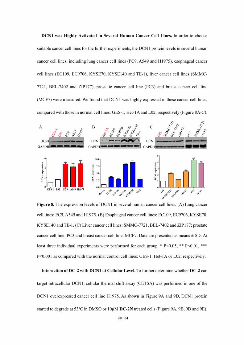
DCN1 was Highly Activated in Several Human Cancer Cell Lines. In order to choose
suitable cancer cell lines for the further experiments, the DCN1 protein levels in several human
cancer cell lines, including lung cancer cell lines (PC9, A549 and H1975), esophageal cancer
cell lines (EC109, EC9706, KYSE70, KYSE140 and TE-1), liver cancer cell lines (SMMC-
7721, BEL-7402 and ZIP177), prostatic cancer cell line (PC3) and breast cancer cell line
(MCF7) were measured. We found that DCN1 was highly expressed in these cancer cell lines,
compared with those in normal cell lines: GES-1, Het-1A and L02, respectively (Figure 8A-C).
Figure 8. The expression levels of DCN1 in several human cancer cell lines. (A) Lung cancer
cell lines: PC9, A549 and H1975. (B) Esophageal cancer cell lines: EC109, EC9706, KYSE70,
KYSE140 and TE-1. (C) Liver cancer cell lines: SMMC-7721, BEL-7402 and ZIP177; prostate
cancer cell line: PC3 and breast cancer cell line: MCF7. Data are presented as means ± SD. At
least three individual experiments were performed for each group. * P<0.05, ** P<0.01, ***
P<0.001 as compared with the normal control cell lines: GES-1, Het-1A or L02, respectively.
Interaction of DC-2 with DCN1 at Cellular Level. To further determine whether DC-2 can
target intracellular DCN1, cellular thermal shift assay (CETSA) was performed in one of the
DCN1 overexpressed cancer cell line H1975. As shown in Figure 9A and 9D, DCN1 protein
started to degrade at 53℃ in DMSO or 10µM DC-2N treated cells (Figure 9A, 9B, 9D and 9E).

21 / 64
While, it was very stable from 43 to 61℃ in the 10µM DC-2 treated cells, similar to that treated
by positive compound DI-591(Figure 9A and 9D).30 In addition, DC-2 increases DCN1 thermal
ability at concentration low to 1µM at 55℃ (Figure 9B and 9E). These findings indicate that
DC-2 can engage the cellular DCN1 protein and increase its thermal ability. Furthermore, the
DCN1 level, which was pulled down by UBE2M antibody, was obviously decreased in DC-2
treated cells, compared with those in DMSO or DC-2N treated groups (Figure 9C and 9F).
These findings further suggest that compound DC-2 can bind to DCN1 and inhibit the
association of UBE2M and DCN1 at cellular level.
Figure 9. Compound DC-2 specifically binds to DCN1 and blocks the interaction of DCN1-
UBE2M at cellular level. (A and B) H1975 cells were treated with 10µM compound DC-2, DI-
591 or DC-2N for 5h, respectively. Then, they were collected and heated from 43℃ to 61℃
(A). H1975 cells were treated with 0, 0.3, 1, 3, 10µM DC-2 or 10µM DC-2N before being
heated at 55℃. Then their DCN1 expression levels were determined by Western Blot (B). (C)
Compound DC-2, but not DC-2N blocks the association of DCN1 and UBE2M at cellular level.
After H1975 cells were treated with compound DC-2N, DC-2 or NAcM-COV, the protein
22 / 64
levels of UBE2M and DCN1, which were pulled down by UBE2M antibody, were determined
by Western Blot (right panel). The basal levels of DCN1 and UBE2M in cell lysates were
determined by Western Blot (left panel). (D-F) The bands intensities of proteins in figure A-C,
respectively. At least three individual experiments were performed for each group.
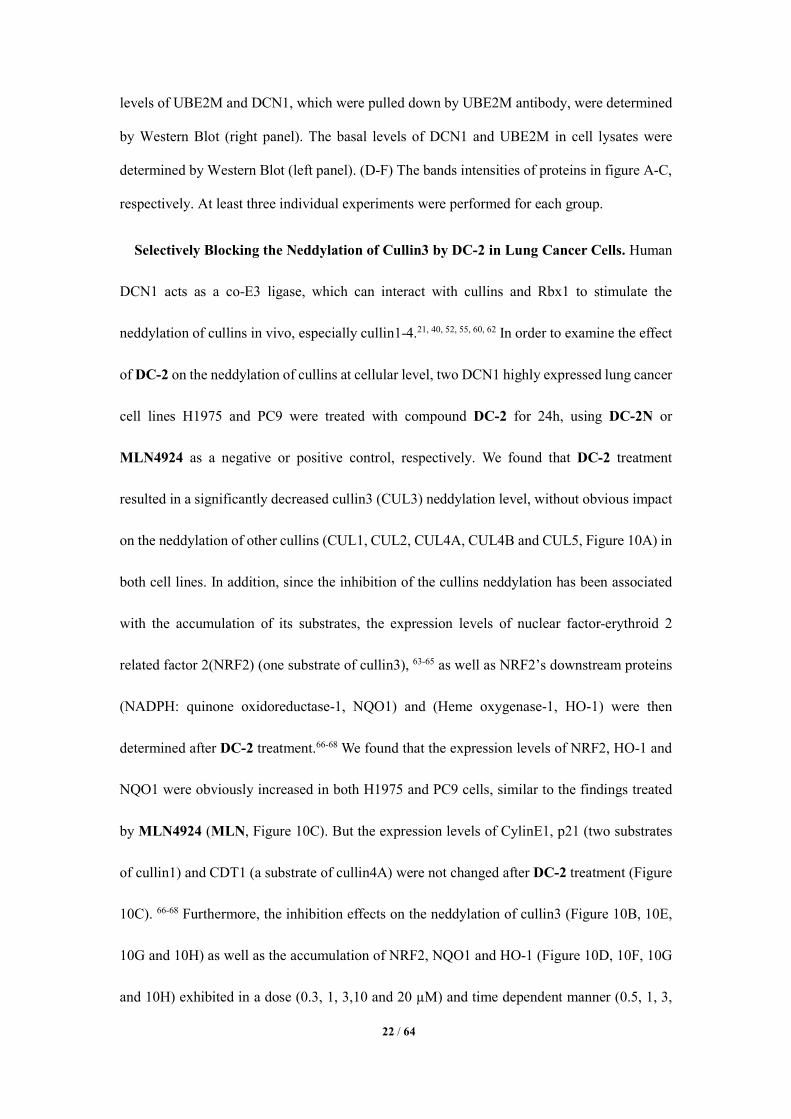
Selectively Blocking the Neddylation of Cullin3 by DC-2 in Lung Cancer Cells. Human
DCN1 acts as a co-E3 ligase, which can interact with cullins and Rbx1 to stimulate the
neddylation of cullins in vivo, especially cullin1-4.21, 40, 52, 55, 60, 62 In order to examine the effect
of DC-2 on the neddylation of cullins at cellular level, two DCN1 highly expressed lung cancer
cell lines H1975 and PC9 were treated with compound DC-2 for 24h, using DC-2N or
MLN4924 as a negative or positive control, respectively. We found that DC-2 treatment
resulted in a significantly decreased cullin3 (CUL3) neddylation level, without obvious impact
on the neddylation of other cullins (CUL1, CUL2, CUL4A, CUL4B and CUL5, Figure 10A) in
both cell lines. In addition, since the inhibition of the cullins neddylation has been associated
with the accumulation of its substrates, the expression levels of nuclear factor-erythroid 2
related factor 2(NRF2) (one substrate of cullin3), 63-65 as well as NRF2’s downstream proteins
(NADPH: quinone oxidoreductase-1, NQO1) and (Heme oxygenase-1, HO-1) were then
determined after DC-2 treatment.66-68 We found that the expression levels of NRF2, HO-1 and
NQO1 were obviously increased in both H1975 and PC9 cells, similar to the findings treated
by MLN4924 (MLN, Figure 10C). But the expression levels of CylinE1, p21 (two substrates
of cullin1) and CDT1 (a substrate of cullin4A) were not changed after DC-2 treatment (Figure
10C). 66-68 Furthermore, the inhibition effects on the neddylation of cullin3 (Figure 10B, 10E,
10G and 10H) as well as the accumulation of NRF2, NQO1 and HO-1 (Figure 10D, 10F, 10G
and 10H) exhibited in a dose (0.3, 1, 3,10 and 20 µM) and time dependent manner (0.5, 1, 3,
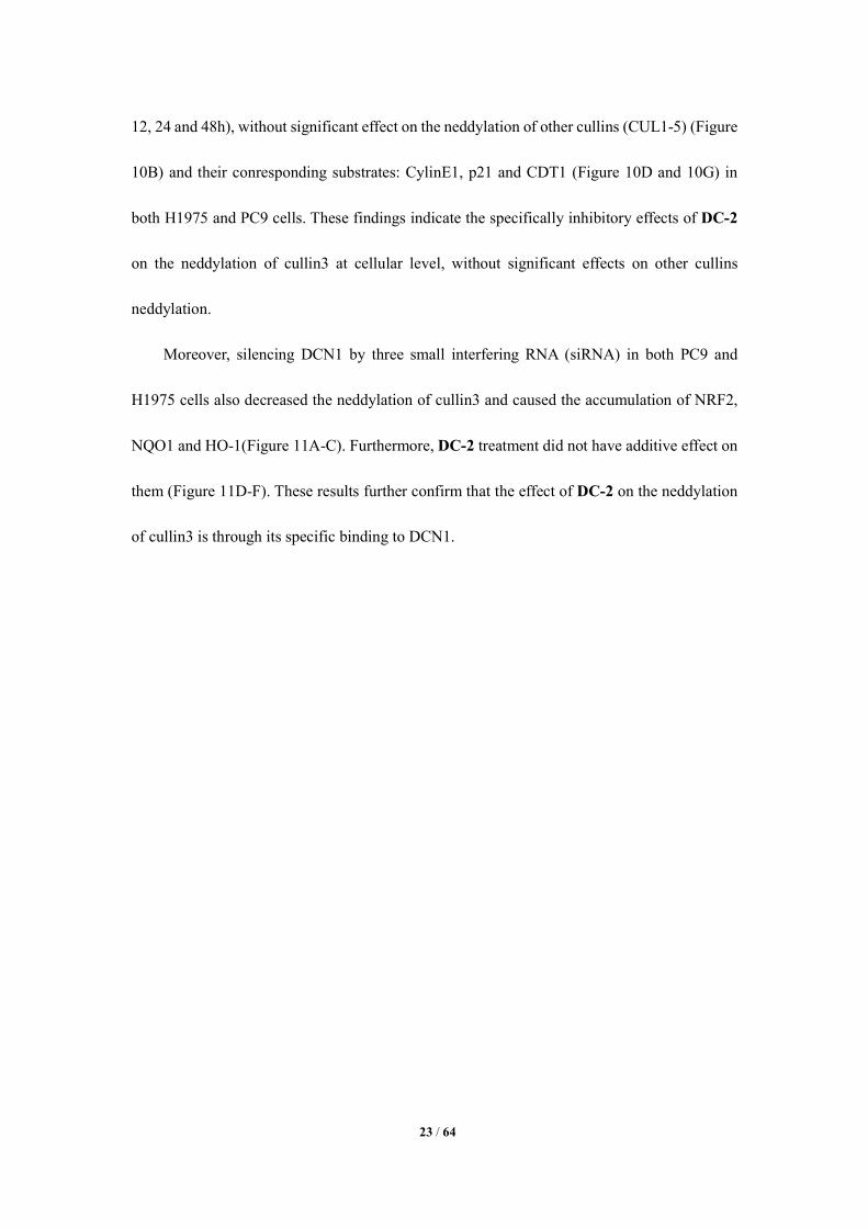
23 / 64
12, 24 and 48h), without significant effect on the neddylation of other cullins (CUL1-5) (Figure
10B) and their conresponding substrates: CylinE1, p21 and CDT1 (Figure 10D and 10G) in
both H1975 and PC9 cells. These findings indicate the specifically inhibitory effects of DC-2
on the neddylation of cullin3 at cellular level, without significant effects on other cullins
neddylation.
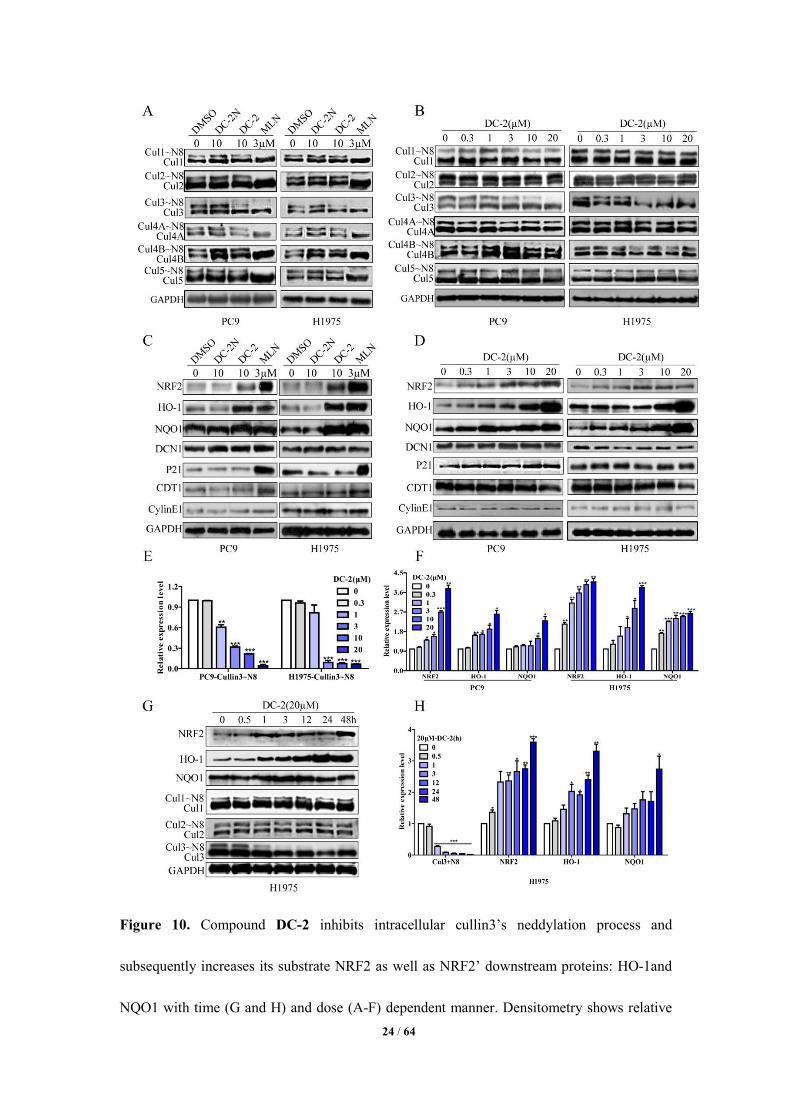
Moreover, silencing DCN1 by three small interfering RNA (siRNA) in both PC9 and
H1975 cells also decreased the neddylation of cullin3 and caused the accumulation of NRF2,
NQO1 and HO-1(Figure 11A-C). Furthermore, DC-2 treatment did not have additive effect on
them (Figure 11D-F). These results further confirm that the effect of DC-2 on the neddylation
of cullin3 is through its specific binding to DCN1.
24 / 64
Figure 10. Compound DC-2 inhibits intracellular cullin3’s neddylation process and
subsequently increases its substrate NRF2 as well as NRF2’ downstream proteins: HO-1and
NQO1 with time (G and H) and dose (A-F) dependent manner. Densitometry shows relative
25 / 64
protein expression level normalized by GAPDH. Data are presented as means ± SD. Three
individual experiments were performed for each group. * P<0.05, ** P<0.01, *** P<0.001 as
compared with the DMSO controls.
Figure 11. The protein levels of DCN1, NRF2, HO-1, NQO1, Cullin1 and Cullin3 were
measured after H1975 and PC9 cells were treated with three DCN1 siRNA (A). Non-targeting
siRNA (NT) treatment was used as control. (D) The protein levels of Cullin3, DCN1, NRF2,
HO-1 and NQO1 were determined after treating the H1975 and PC9 cells by siRNA#3 with or
without compound DC-2. (B, C, E and F) Densitometry shows relative protein expression level
normalized by GAPDH. Data are presented as means±SD. Three individual experiments were
performed for each group. * P<0.05, ** P<0.01, *** P<0.001 as compared with the controls

26 / 64
(NT).
Figure 12. Compound DC-2 inhibits cell proliferation and colony formation. (A) The inhibition
rates (%) of DC-2 on 8 different cancer cell lines and two normal cell lines: L02 and GES-1.
(B) The IC50 values were determined after treating the three lung cancer cell lines (PC9, H1975
and A549) with DC-2 for 24, 48 and 72h. (C) After the treatment of compound DC-2 at 0,
0.625, 1.25, 2.5, 5 and 10µM for 7 days, cells were stained by crystal violet and then imaged
27 / 64
by microscopy. (D) Subsequently, they were dissolved and then measured by a BioTek
microplate reader. (E and F) The apoptosis were determined by Flow cytometry after PC9,
H1975 and A549 cells were treated with compound DC-2 for 48h. Data are presented as means
± SD. Three individual experiments were performed for each group. * P<0.05, ** P<0.01, ***
P<0.001 as compared with the controls.
Effects of DC-2 on DCN1 Highly Expressed Cancer Cells. As DCN1 is highly expressed
in most cancers and has been discovered as an oncogene.10, 23, 33-39, 44-46, 69 In order to determine
whether DC-2 has the ability of inhibiting cancer cells’ proliferation, eight DCN1 amplified
cancer cell lines(KYSE70, PC9, SMMC-7721, PC3, MCF7, TE-1, KYSE140 and EC109) and
two normal cell lines (GES-1 and L02) were chosen to be treated with DC-2 for 72h. As shown
in Figure 12A, DC-2 shows strong anti-proliferation ability on these cancer cells rather than
normal cells. Furthermore, compound DC-2 could decrease three types of lung cancer cells’
viabilities (H1975, PC9 and A549) with time-dependent manner (Figure 12B), block their
colony formations (Figure 12C and 12D) and induce their apoptosis dose dependently (Figure
12E and 12F). These results suggest that compound DC-2 has the ability of blocking DCN1
highly expressed cancer cells’ proliferation. In addition, the cytotoxicities of DC-2N and the
compounds with very similar structures of DC-2N (27, 29, 36, 37 and 45) or DC-2 (33, 39, 40,
46 and 47) on five DCN1 highly expressed cancer cells (KYSE70, PC9, SMMC-7721, PC3 and
MCF7) were determined. As shown in Table 6, compounds 33, 39, 40, 46 and 47, which can
block the DCN1-UBE2M interaction at nanomole level in vitro, also have cytotoxicities on
those tested cancer cells with IC50 values ranging from 1 to 12µM. However, DC-2N, 27, 29,

28 / 64
36, 37 and 45, which have no inhibiting effect on DCN1-UBE2M interaction, exhibits less
cytotoxic activity (>20µM), indicating that the cytotoxicities of these compounds may be
related to their blocking effects on UBE2M-DCN1 interaction. However, the cytotoxic
activities of DI-591 on the five DCN1 highly expressed cancer cell lines were also measured.
The IC50 values were all above 20µM at 72h. Based on the current findings, whether DCN1 can
be recognized as a valuable anti-tumor target remains to be further investigated. In addition,
except the inhibitory effects of DC-2 on DCN1-2, DC-2 also exhibits relatively weak binding
affinities on DCN3-5 (Table 5), similar to the findings on NAcM-COV.43 Therefore, the
cytotoxicities of DC-2 may be also related to its inhibition on DCN3-5. However, the relative
less cytotoxicity of DI-591 indicates its higher selectivity on DCN1-2 over DCN3-5.30
Table 6. Cytotoxic activities (IC50s) of DC-2 and DC-2N derivatives against five DCN1
highly expressed cancer cell linesa.
Compd. IC50(μM)b DCN1-UBE2M
IC50(nM)-FP KYSE70 PC9 SMMC-7721 PC3 MCF7
27 >20 >20 >20 >20 >20 >10000
29 >10 >20 >20 >10 >10 >10000
DC-2N >20 >20 >20 >20 >20 >10000
36 >20 >20 >20 >20 >20 >10000
37 >20 >20 >20 >20 >20 >10000
45 >20 >20 >20 >20 >20 >20000
33 8.25 ± 0.91 7.96 ± 0.90 4.66 ± 0.66 10.28 ± 1.01 9.82 ± 0.99 195.66 ± 2.29
39 5.35 ± 0.72 4.13 ± 0.61 6.04 ± 0.78 3.62 ± 0.55 3.23 ± 0.51 36.59 ± 0.18
40 10.88 ± 1.03 12.11 ± 1.08 3.99 ± 0.60 8.53 ± 0.93 11.63 ± 1.06 551.87 ± 1.71
DC-2 4.30 ± 0.63 1.11 ± 0.04 2.45 ± 0.39 1.62 ± 0.32 2.20 ± 0.34 26.89 ± 0.24
46 6.56 ± 0.81 5.30 ± 0.72 7.34 ± 0.86 7.17 ± 0.85 6.18 ± 0.79 63.51 ± 1.80
47 5.32 ± 0.72 6.48 ± 0.81 8.64 ± 0.93 3.54 ± 0.55 4.12 ± 0.61 106.21 ± 2.02
NAcM-COV 17.89 ± 1.25 5.59 ± 0.74 16.24 ± 1.21 25.27 ± 1.40 10.17 ± 1.00 ND
a Cells were treated with different concentrations of the indicated compounds for 72h. Cell
viability was measured by MTT assay as described in the experimental section.

29 / 64
b IC50 values were indicated as the mean ± SD of three independent experiments. ND means the
value was not determined.
CONCLUSIONS
In this study, a novel series of compounds with a 5-cyano-6-phenylpyrimidin scaffold as
DCN1-UBE2M interaction modulators have been identified through structure-based
optimization, which enriches the structure types of DCN1 inhibitors. Among the inhibitors,
compound DC-2 exhibits the most potent inhibition effect on DCN1-UBE2M interaction at
molecule and cellular levels. Molecular docking results show that DC-2 can well dock into the
binding pocket of DCN1. Site specific mutations further verified its blocking effects.
Furthermore, unlike MLN4924, obliterating all cullins neddylation, DC-2 specifically
diminishes the neddylation of cullin3, which leads to the accumulation of cullin3’s substrate
NRF2 and NRF2’s downstream proteins: HO-1 and NQO1. Our findings indicate that the 5-
cyano-6-phenylpyrimidin based small molecules may serve as leading compounds specifically
targeting DCN1-UBE2M interaction.
EXPERIMENTAL SECTION
General Methods for Chemistry. Chemicals and solvents were obtained from standard
suppliers and used directly without further purification. Melting points were taken on an X-5
micromelting apparatus and were uncorrected. 1H and 13C NMR spectra were respectively
determined with a 400 and 100 MHz spectrometer. High resolution mass spectra (HRMS) were
obtained with a Water Q-TOF electrospray mass spectrometer (Water, Milford, MA). Final
products were of > 95% purity as analyzed by HPLC analysis (Phenomenex column, C-18, 5.0
µm, 4.6 mm × 150 mm) on Dionex UltiMate 3000 UHPLC instrument from ThermoFisher.
Besides, PAINS screening of the synthesized compounds was carried out by employing the
30 / 64
online program70 and all the tested compounds passed the filter.
General Procedure for the Synthesis of Compounds 6-52 and 54. To a well stirred
solution of the appropriate mercapto heterocyclics and anilines (5 mmol) in absolute ethanol
(10 mL), equimolar amount of a solution of compounds 5a-o or 53 (5 mmol) in absolute ethanol
(10 mL) was added. The reaction mixture was stirred and heated under reflux for 5 h. Upon
completion, the precipitated product was filtered off and washed with ethanol to afford the
crude product. The crude product was recrystallized from ethanol to yield the pure product 6-
52 and 54. The detailed information of synthesis and characterization of compounds 5a-o and
53 were reported in published articles.54, 71-73
4-((1-methyl-1H-tetrazol-5-yl)thio)-6-(2-oxo-2H-chromen-6-yl)-2-(prop-2-yn-1-
ylthio)pyrimidine-5-carbonitrile (6)
Yield 88.5%. White solid. Mp: 184–185°C. 1H NMR (400 MHz, DMSO-d6, ppm) δ 8.36
(d, J = 1.7 Hz, 1H, Ar-H), 8.22 (dd, J = 6.2, 4.0 Hz, 2H, Ar-H), 7.66 (d, J = 14.7 Hz, 1H, Ar-
H), 6.62 (d, J = 9.6 Hz, 1H, Ar-H), 4.14 (s, 3H, -CH3), 3.57 (d, 2H, -CH2-), 3.17 (s, 1H, J = 2.5
Hz, ≡C-H). 13C NMR (100 MHz, DMSO-d6, ppm) δ 172.31, 169.73, 165.20, 159.34, 155.73,
145.48, 143.73, 132.19, 130.26, 129.64, 119.06, 117.39, 117.10, 114.61, 98.65, 78.79, 73.56,
34.79, 19.12. HR-MS (ESI), calcd. C19H11N7NaO2S2, [M+Na]+ m/z: 456.0313, found: 456.0315.
4-((1-methyl-1H-tetrazol-5-yl)thio)-2-(prop-2-yn-1-ylthio)-6-(thiophen-3-yl)pyrimidine-
5-carbonitrile (7)
Yield 83.9%. White solid. Mp: 183–184°C. 1H NMR (400 MHz, DMSO-d6, ppm) δ 8.32
(s, 1H, Ar-H), 8.13 (d, J = 4.1 Hz, 1H, Ar-H), 7.39 (s, 1H, Ar-H), 4.11 (s, 3H, -CH3), 3.59 (d,
2H, -CH2-), 3.15 (t, 1H, ≡C-H). 13C NMR (100 MHz, DMSO-d6, ppm) δ 171.87, 169.80, 158.29,
145.45, 138.13, 135.80, 132.60, 129.72, 114.96, 94.27, 78.77, 73.52, 34.75, 19.06. HR-MS
(ESI), calcd. C14H9N7NaS3, [M+Na]+ m/z: 393.9979, found: 393.9982.
4-(1H-indol-3-yl)-6-((1-methyl-1H-tetrazol-5-yl)thio)-2-(prop-2-yn-1-ylthio)pyrimidine-
5-carbonitrile (8)
Yield 86.6%. White solid. Mp: 227–227°C. 1H NMR (400 MHz, DMSO-d6, ppm) δ 12.37
(s, 1H, NH, D2O exchangeable), 8.63 (d, J = 3.0 Hz, 1H, -CH=), 8.41 (d, J = 7.8 Hz, 1H, Ar-

31 / 64
H), 7.57 (d, J = 7.9 Hz, 1H, Ar-H), 7.29 (t, J = 7.3 Hz, 1H, Ar-H), 7.24 (t, J = 7.5 Hz, 1H, Ar-
H), 4.13 (s, 3H, -CH3), 3.59 (d, J = 2.3 Hz, 2H, -CH2-), 3.17 (t, J = 2.4 Hz, 1H, ≡C-H). 13C
NMR (100 MHz, DMSO-d6, ppm) δ 171.68, 168.60, 161.76, 136.52, 132.66, 125.44, 123.43,
122.29, 122.06, 116.14, 112.60, 110.30, 93.65, 79.15, 73.57, 34.75, 18.78. HR-MS (ESI), calcd.
C18H12N8NaS2, [M+Na]+ m/z: 427.0524, found: 427.0525.
4-((1-methyl-1H-tetrazol-5-yl)thio)-2-(prop-2-yn-1-ylthio)-6-(1H-pyrrol-2-yl)pyrimidine-
5-carbonitrile (9)
Yield 51.6%. White solid. Mp: 189–190°C. 1H NMR (400 MHz, DMSO-d6, ppm) δ 12.08
(s, 1H, NH, D2O exchangeable), 7.49 (s, 1H, -CH=), 7.29 (s, 1H, -CH=), 6.53 – 6.32 (m, 1H, -
CH=), 4.10 (s, 3H, -CH3), 3.69 (d, J = 2.6 Hz, 2H, -CH2-), 3.15 (t, J = 2.5 Hz, 1H, ≡C-H). 13C
NMR (100 MHz, DMSO-d6, ppm) δ 171.53, 168.77, 155.43, 145.62, 127.32, 125.45, 117.04,
115.44, 112.13, 91.76, 79.03, 73.67, 34.72, 18.86. HR-MS (ESI), calcd. C14H10N8NaS2,
[M+Na]+ m/z: 418.0521, found: 418.0521.
4-((1-methyl-1H-tetrazol-5-yl)thio)-2-(prop-2-yn-1-ylthio)-6-(3,4,5-
trimethoxyphenyl)pyrimidine-5-carbonitrile (10)
Yield 82.1%. White solid. Mp: 256–256°C. 1H NMR (400 MHz, DMSO-d6, ppm) δ 7.40
(s, 2H, Ar-H), 4.12 (s, 3H, -CH3), 3.87 (s, 6H, -CH3), 3.80 (s, 3H, -CH3), 3.65 (t, J = 12.7 Hz,
2H, -CH2-), 3.20 (t, J = 2.3 Hz, 1H, ≡C-H). 13C NMR (100 MHz, DMSO-d6, ppm) δ 172.55,
170.08, 166.35, 153.39, 146.06, 141.54, 129.54, 115.48, 107.34, 98.83, 79.69, 73.84, 60.78,
56.69, 35.24, 19.77. HR-MS (ESI), calcd. C19H17N7NaO3S2, [M+Na]+ m/z: 478.0732, found:
478.0733.
4-(4-chlorophenyl)-6-((1-methyl-1H-tetrazol-5-yl)thio)-2-(prop-2-yn-1-
ylthio)pyrimidine-5-carbonitrile (11)
Yield 87.7%. White solid. Mp: 163–164°C. 1H NMR (400 MHz, DMSO-d6, ppm) δ 8.02
(d, J = 8.4 Hz, 2H, Ar-H), 7.74 (t, J = 19.1 Hz, 2H, Ar-H), 4.13 (s, 3H, -CH3), 3.59 (s, 2H, -
CH2-), 3.15 (s, J = 2.5 Hz, 1H, ≡C-H). 13C NMR (100 MHz, DMSO-d6, ppm) δ 172.30, 169.69,
165.47, 145.47, 137.36, 132.92, 130.85, 129.06, 114.55, 98.72, 78.78, 73.51, 34.78, 19.10. HR-
MS (ESI), calcd. C16H10ClN7S2, [M + Na]+ m/z: 422.0025, found: 422.0025.
32 / 64
4-(3-chlorophenyl)-6-((1-methyl-1H-tetrazol-5-yl)thio)-2-(prop-2-yn-1-
ylthio)pyrimidine-5-carbonitrile (12)
Yield 89.7%. White solid. Mp: 163–165°C. 1H NMR (400 MHz, DMSO-d6, ppm) δ 8.00
(t, J = 1.8 Hz, 1H, Ar-H), 7.94 (d, J = 7.8 Hz, 1H, Ar-H), 7.78 – 7.73 (m, 1H, Ar-H), 7.67 (t, J
= 7.9 Hz, 1H, Ar-H), 4.13 (s, 3H, -CH3), 3.60 (d, J = 7.5 Hz, 2H, -CH2-), 3.16 (t, J = 2.5 Hz,
1H, ≡C-H). 13C NMR (100 MHz, DMSO-d6, ppm) δ 172.39, 169.63, 165.23, 145.46, 136.11,
133.55, 131.96, 130.87, 128.65, 127.65, 114.43, 99.11, 78.81, 73.46, 34.77, 19.12. HR-MS
(ESI), calcd. C16H10ClN7NaS2, [M+Na]+ m/z: 422.0025, found: 422.0025.
4-(4-bromophenyl)-6-((1-methyl-1H-tetrazol-5-yl)thio)-2-(prop-2-yn-1-
ylthio)pyrimidine-5-carbonitrile (13)
Yield 78.1%. Yellow solid. Mp: 238–239°C. 1H NMR (400 MHz, DMSO-d6, ppm) δ 8.23
(d, J = 3.3 Hz, 1H, -CH=), 8.13 (d, J = 3.3 Hz, 1H, -CH=), 7.97 – 7.91 (m, 2H, Ar-H), 7.88 –
7.82 (m, 2H, Ar-H), 3.79 (d, J = 2.6 Hz, 2H, -CH2-), 3.14 (t, J = 2.6 Hz, 1H, ≡C-H). 13C NMR
(100 MHz, DMSO-d6, ppm) δ 172.03, 171.48, 165.43, 150.29, 144.31, 133.50, 131.91, 131.04,
127.91, 126.18, 114.45, 98.20, 79.06, 73.86, 19.29. HR-MS (ESI), calcd. C16H10BrN7NaS2,
[M+Na]+ m/z: 466.9070, found: 466.9073.
4-((1-methyl-1H-tetrazol-5-yl)thio)-2-(prop-2-yn-1-ylthio)-6-(p-tolyl)pyrimidine-5-
carbonitrile (14)
Yield 83.1%. Yellow solid. Mp: 164–165°C. 1H NMR (400 MHz, DMSO-d6, ppm) δ 7.92
(d, J = 8.2 Hz, 2H, Ar-H), 7.44 (d, J = 8.1 Hz, 2H, Ar-H), 4.13 (s, 3H, -CH3), 3.56 (t, J = 20.3
Hz, 2H, -CH2-), 3.15 (t, J = 2.5 Hz, 1H, ≡C-H), 2.42 (s, 3H, -CH3). 13C NMR (100 MHz,
DMSO-d6, ppm) δ 172.13, 169.62, 166.39, 145.54, 142.88, 131.32, 129.49, 129.01, 114.82,
98.19, 78.87, 73.46, 34.76, 21.09, 19.05. HR-MS (ESI), calcd. C17H13N7NaS2, [M+Na]+ m/z:
402.0572, found: 402.0573.
5-(4-isopropylphenyl)-6-((1-methyl-1H-tetrazol-5-yl)thio)-2-(prop-2-yn-1-
ylthio)pyrimidine-5-carbonitrile (15)
Yield 83.4%. White solid. Mp: 141–142°C. 1H NMR (400 MHz, DMSO-d6, ppm) δ 7.95
(d, J = 3.9 Hz, 2H, Ar-H), 7.50 (d, J = 8.0 Hz, 2H, Ar-H), 4.14 (d, J = 4.0 Hz, 3H, -CH3), 3.60
33 / 64
(s, 2H, -CH2-), 3.15 (t, J = 2.5 Hz,1H, ≡C-H), 3.05 – 2.96 (m, 1H, -CH), 1.25 (d, J = 6.8 Hz,
6H, -CH3). 13C NMR (100 MHz, DMSO-d6, ppm) δ 172.16, 169.65, 166.36, 153.39, 145.54,
131.70, 129.19, 126.92, 114.85, 98.17, 78.89, 73.46, 34.77, 33.47, 23.47, 19.06. HR-MS (ESI),
calcd. C19H17N7NaS2, [M+Na]+ m/z: 430.0885, found: 430.0886.
4-(3-methoxyphenyl)-6-((1-methyl-1H-tetrazol-5-yl)thio)-2-(prop-2-yn-1-
ylthio)pyrimidine-5-carbonitrile (16)
Yield 89.9%. White solid. Mp: 189–192°C. 1H NMR (400 MHz, DMSO-d6, ppm) δ 7.56
(s, 1H, Ar-H), 7.54 (d, J = 3.7 Hz, 2H, Ar-H), 7.25 (dt, J = 6.9, 2.2 Hz, 1H, Ar-H), 4.12 (s, 3H,
-CH3), 3.84 (s, 3H, -CH3), 3.61 (d, J = 2.4 Hz, 2H, -CH2-), 3.17 (t, J = 2.5 Hz, 1H, ≡C-H). 13C
NMR (100 MHz, DMSO-d6, ppm) δ 172.22, 169.63, 166.35, 159.27, 145.52, 135.41, 130.15,
121.20, 118.09, 114.67, 114.17, 98.78, 78.95, 73.42, 55.43, 34.76, 19.11. HR-MS (ESI), calcd.
C17H13N7ONaS2, [M+Na]+ m/z: 418.0521, found: 418.0521.
4-(3,4-dichlorophenyl)-6-((1-methyl-1H-tetrazol-5-yl)thio)-2-(prop-2-yn-1-
ylthio)pyrimidine-5-carbonitrile (17)
Yield 89.8%. White solid. Mp: 151–152°C. 1H NMR (400 MHz, DMSO-d6, ppm) δ 8.21
(d, J = 1.8 Hz, 1H, Ar-H), 7.97 (dd, J = 8.4, 1.9 Hz, 1H, Ar-H), 7.93 (d, J = 8.4 Hz, 1H, Ar-H),
4.13 (s, 3H, -CH3), 3.61 (d, J = 2.3 Hz, 2H, -CH2-), 3.16 (t, J = 2.4 Hz, 1H, ≡C-H). 13C NMR
(100 MHz, DMSO-d6, ppm) δ 172.45, 169.65, 164.28, 145.42, 135.21, 134.56, 131.82, 131.26,
130.80, 129.05, 114.32, 99.14, 78.77, 73.52, 34.78, 19.18. HR-MS (ESI), calcd.
C16H9Cl2N7NaS2, [M+Na]+ m/z: 455.9636, found: 455.9637.
4-((1-methyl-1H-tetrazol-5-yl)thio)-6-phenyl-2-(prop-2-yn-1-ylthio)pyrimidine-5-
carbonitrile (18)
Yield 81.9%. White solid. Mp: 130–131°C. 1H NMR (400 MHz, DMSO-d6, ppm) δ 8.17
(d, J = 7.5 Hz, 2H, Ar-H), 8.07 (s, 1H, Ar-H), 7.59 – 7.55 (m, 2H, Ar-H), 4.10 (s, 3H, -CH3),
3.67 (d, 2H, -CH2-), 3.11 (t, J = 2.5 Hz,1H, ≡C-H). 13C NMR (100 MHz, DMSO-d6, ppm) δ
172.30, 169.69, 165.47, 145.47, 137.36, 132.92, 130.85, 129.06, 114.55, 98.72, 78.78, 73.51,
34.78, 19.10. HR-MS (ESI), calcd. C16H11N7NaS2, [M+Na]+ m/z: 388.0415, found: 388.0415.
34 / 64
4-((1-methyl-1H-tetrazol-5-yl)amino)-2-(prop-2-yn-1-ylthio)-6-(3,4,5-
trimethoxyphenyl)pyrimidine-5-carbonitrile (19)
Yield 94.1%. Yellow solid. Mp: 205–206°C. 1H NMR (400 MHz, DMSO-d6, ppm) δ 7.38
(s, 2H, Ar-H), 3.93 (s, 3H, -CH3), 3.87 (s, 6H, -CH3), 3.85 (d, J = 1.8 Hz, 2H, -CH2-), 3.78 (s,
3H, -CH3), 3.16 (t, J = 2.5 Hz, 1H, ≡C-H). 13C NMR (100 MHz, DMSO-d6, ppm) δ 171.96,
167.33, 161.41, 152.71, 150.04, 140.43, 130.11, 115.42, 106.68, 86.40, 79.95, 72.99, 60.22,
56.11, 33.61, 19.17. HR-MS (ESI), calcd. C19H18N8NaO3S, [M+Na]+ m/z: 461.1120, found:
461.1122.
4-(piperazin-1-yl)-2-(prop-2-yn-1-ylthio)-6-(3,4,5-trimethoxyphenyl)pyrimidine-5-
carbonitrile (20)
Yield 89.0%. White solid. Mp: 242–243°C. 1H NMR (400 MHz, DMSO-d6, ppm) δ 9.72
(s, 1H, NH, D2O exchangeable), 7.30 (s, 2H, Ar-H), 4.19 (s, 4H, -CH2-), 4.03 (d, J = 2.1 Hz,
2H, -CH2-), 3.86 (s, 6H, -CH3), 3.77 (s, 3H, -CH3), 3.34 (s, 4H, -CH2-), 3.21 (t, J = 2.3 Hz, 1H,
≡C-H). 13C NMR (100 MHz, DMSO-d6, ppm) δ 170.58, 169.15, 161.75, 152.60, 140.23, 130.88,
117.90, 107.18, 83.78, 80.45, 73.00, 60.18, 56.16, 45.75, 19.26. HR-MS (ESI), calcd.
C21H24N5O3S, [M+H]+ m/z: 426.1600, found: 426.1602.
2-(prop-2-yn-1-ylthio)-4-thiomorpholino-6-(3,4,5-trimethoxyphenyl)pyrimidine-5-
carbonitrile (21)
Yield 50.9%. Yellow solid. Mp: 152–153°C. 1H NMR (400 MHz, DMSO-d6, ppm) δ 7.28
(s, 2H, Ar-H), 4.19 (m, 4H, -CH2-), 3.98 (d, J = 2.4 Hz, 2H, -CH2-), 3.85 (s, 6H, -CH3), 3.77 (s,
3H, -CH3), 3.19 (t, J = 2.3 Hz, 1H, ≡C-H), 2.86 – 2.77 (m, 4H, -CH2-). 13C NMR (100 MHz,
DMSO-d6, ppm) δ 170.71, 169.42, 161.97, 152.56, 140.21, 130.93, 117.92, 107.23, 84.24,
80.46, 72.89, 60.17, 56.17, 49.70, 26.50, 19.22. HR-MS (ESI), calcd. C21H22N4O3NaS2,
[M+Na]+ m/z: 465.1031, found: 465.1032.
4-morpholino-2-(prop-2-yn-1-ylthio)-6-(3,4,5-trimethoxyphenyl)pyrimidine-5-
carbonitrile (22)
35 / 64
Yield 89.5%. White solid. Mp: 163–164°C. 1H NMR (400 MHz, DMSO-d6, ppm) δ 7.27
(s, 2H, Ar-H), 3.98 (d, J = 2.5 Hz, 2H, -CH2-), 3.97 – 3.93 (m, 4H, -CH2-), 3.85 (s, 6H, -CH3),
3.76 (s, 3H, -CH3), 3.74 (m, 4H, -CH2-), 3.19 (t, J = 2.5 Hz, 1H, ≡C-H). 13C NMR (100 MHz,
DMSO-d6, ppm) δ 170.70, 169.34, 161.84, 152.58, 140.24, 130.87, 117.86, 107.20, 83.94,
80.40, 72.91, 65.91, 60.17, 56.16, 47.17, 19.23. HR-MS (ESI), calcd. C21H23N4O4S, [M+H]+
m/z: 427.1440, found: 427.1441.
4-(piperidin-1-yl)-2-(prop-2-yn-1-ylthio)-6-(3,4,5-trimethoxyphenyl)pyrimidine-5-
carbonitrile (23)
Yield 90.0%. White solid. Mp: 120–121°C. 1H NMR (400 MHz, DMSO-d6, ppm) δ 7.27
(s, 2H, Ar-H), 3.97 (d, J = 2.5 Hz, 2H, -CH2-), 3.90 (m, 4H, -CH2-), 3.86 (s, 6H, -CH3), 3.77 (s,
3H, -CH3), 3.17 (t, J = 2.5 Hz, 1H, ≡C-H), 1.68 (s, 6H, -CH2-). 13C NMR (100 MHz, DMSO-
d6, ppm) δ 170.55, 169.37, 161.46, 152.54, 140.16, 131.04, 118.01, 107.19, 83.42, 80.48, 72.81,
60.15, 56.16, 47.99, 25.58, 23.64, 19.16. HR-MS (ESI), calcd. C22H25N4O3S, [M+H]+ m/z:
425.1647, found: 425.1648.
4-((1-(2-(dimethylamino)ethyl)-1H-tetrazol-5-yl)thio)-2-(prop-2-yn-1-ylthio)-6-(3,4,5-
trimethoxyphenyl)pyrimidine-5-carbonitrile (26)
Yield 75.9%. Yellow solid. Mp: 197–198°C. 1H NMR (400 MHz, DMSO-d6, ppm) δ 7.42
(s, 2H, Ar-H), 4.99 (t, J = 6.6 Hz, 2H, -CH2-), 3.87 (s, 6H, -CH3), 3.79 (s, 3H, -CH3), 3.72 (d,
J = 2.3 Hz, 2H, -CH2-), 3.69 (t, J = 6.6 Hz, 2H, -CH2-), 3.25 (t, J = 2.5 Hz, 1H, ≡C-H), 2.83 (s,
6H, -CH3). 13C NMR (100 MHz, DMSO-d6, ppm) δ 171.96, 169.87, 165.62, 152.90, 146.30,
141.06, 129.01, 115.03, 106.80, 98.30, 79.26, 73.45, 60.29, 56.19, 54.14, 42.69, 42.38, 19.36.
HR-MS (ESI), calcd. C22H25N8O3S2, [M+H]+ m/z: 513.1491, found: 513.1491.
4-((1H-1,2,4-triazol-3-yl)thio)-2-(prop-2-yn-1-ylthio)-6-(3,4,5-
trimethoxyphenyl)pyrimidine-5-carbonitrile (27)
Yield 80.9%. White solid. Mp: 234–235°C. 1H NMR (400 MHz, DMSO-d6, ppm) δ 8.85
(s, 1H, NH, D2O exchangeable), 7.41 (s, 2H, Ar-H), 3.87 (s, 6H, -CH3), 3.79 (s, 3H, -CH3), 3.78
– 3.78 (s, 2H, -CH2-), 3.15 (t, J = 2.4 Hz, 1H, ≡C-H). 13C NMR (100 MHz, DMSO-d6, ppm) δ
172.22, 169.63, 166.35, 159.27, 145.52, 135.41, 130.15, 121.20, 118.09, 114.67, 114.17, 98.78,
36 / 64
78.95, 73.42, 55.43, 34.76, 19.11. HR-MS (ESI), calcd. C19H16N6NaO3S2, [M+Na]+ m/z:
463.0623, found: 463.0622.
4-((1-methyl-1H-imidazol-2-yl)thio)-2-(prop-2-yn-1-ylthio)-6-(3,4,5-
trimethoxyphenyl)pyrimidine-5-carbonitrile (28)
Yield 32.8%. White solid. Mp: 176–177°C. 1H NMR (400 MHz, DMSO-d6, ppm) δ 7.62
(s, 1H, -CH=), 7.38 (s, 2H, Ar-H), 7.22 (s, 1H, -CH=), 3.87 (s, 6H, -CH3), 3.79 (s, 3H, -CH3),
3.68 (s, 2H, -CH2-), 3.67 (s, 3H, -CH3), 3.20 (t, J = 2.2 Hz, 1H, ≡C-H). 13C NMR (100 MHz,
DMSO-d6, ppm) δ 173.22, 171.82, 165.81, 152.83, 140.84, 130.39, 130.16, 129.27, 126.44,
115.07, 106.86, 97.48, 79.44, 73.36, 60.25, 56.18, 33.68, 19.22. HR-MS (ESI), calcd.
C21H19N5NaO3S2, [M+Na]+ m/z: 476.0827, found: 476.0828.
2-(prop-2-yn-1-ylthio)-4-(pyridin-2-ylthio)-6-(3,4,5-trimethoxyphenyl)pyrimidine-5-
carbonitrile (29)
Yield 80.2%. Yellow solid. Mp: 180–181°C. 1H NMR (400 MHz, DMSO-d6, ppm) δ 8.76
– 8.58 (m, 1H, Ar-H), 7.97 (td, J = 7.7, 1.7 Hz, 1H, Ar-H), 7.86 (d, J = 7.8 Hz, 1H, Ar-H), 7.55
(dt, J = 15.7, 7.8 Hz, 1H, Ar-H), 7.38 (s, 2H, Ar-H), 3.86 (s, 6H, -CH3), 3.78 (s, 3H, -CH3),
3.71 (d, J = 2.2 Hz, 2H, -CH2-), 3.14 (t, J = 2.3 Hz, 1H). 13C NMR (100 MHz, DMSO-d6, ppm)
δ 172.90, 171.53, 165.73, 152.80, 150.76, 149.32, 140.76, 138.08, 131.26, 129.36, 124.71,
115.22, 106.82, 98.15, 79.54, 73.35, 60.24, 56.14, 19.20. HR-MS (ESI), calcd.
C22H18N4NaO3S2, [M+Na]+ m/z: 473.0718, found: 473.0715.
4-((5-aminothiophen-2-yl)thio)-2-(prop-2-yn-1-ylthio)-6-(3,4,5-
trimethoxyphenyl)pyrimidine-5-carbonitrile (30)
Yield 89.3%. Yellow solid. Mp: 189–190°C. 1H NMR (400 MHz, DMSO-d6, ppm) δ 7.87
(s, 2H, -CH=), 7.42 (s, 2H, Ar-H), 3.95 (d, J = 2.1 Hz, 2H, -CH2-), 3.86 (s, 6H, -CH3), 3.79 (s,
3H, -CH3), 3.17 (d, J = 2.2 Hz, 1H, ≡C-H). 13C NMR (100 MHz, DMSO-d6, ppm) δ 173.78,
171.83, 171.36, 165.58, 152.84, 140.98, 129.19, 114.91, 106.95, 97.68, 79.43, 73.44, 60.27,
56.19, 19.55. HR-MS (ESI), calcd. C21H18N4NaO3S3, [M+Na]+ m/z: 493.0439, found: 493.0438.
4-((5-methyl-1,3,4-thiadiazol-2-yl)thio)-2-(prop-2-yn-1-ylthio)-6-(3,4,5-
trimethoxyphenyl)pyrimidine-5-carbonitrile (31)
37 / 64
Yield 76.5%. White solid. Mp: 186–187°C. 1H NMR (400 MHz, DMSO-d6, ppm) δ 7.44
(s, 2H, Ar-H), 3.93 (d, J = 2.4 Hz, 2H, -CH2-), 3.87 (s, 6H, -CH3), 3.80 (s, 3H, -CH3), 3.20 (t,
J = 2.5 Hz, 1H, ≡C-H), 2.86 (s, 1H, -CH3). 13C NMR (100 MHz, DMSO-d6, ppm) δ 171.88,
171.26, 169.59, 165.64, 154.06, 152.86, 141.07, 129.09, 114.85, 106.96, 98.14, 79.53, 73.43,
60.27, 56.19, 19.54, 15.53. HR-MS (ESI), calcd. C20H17N5NaO3S3, [M+Na]+ m/z: 494.0391,
found: 494.0389.
4-((5-amino-1,3,4-thiadiazol-2-yl)thio)-2-(prop-2-yn-1-ylthio)-6-(3,4,5-
trimethoxyphenyl)pyrimidine-5-carbonitrile (32)
Yield 65.1%. White solid. Mp: 181–182°C. 1H NMR (400 MHz, DMSO-d6, ppm) δ 7.83
(s, 2H, NH, D2O exchangeable), 7.43 (d, J = 0.6 Hz, 2H, Ar-H), 3.94 (t, J = 7.4 Hz, 2H, -CH2-),
3.87 (s, 6H, -CH3), 3.79 (d, J = 0.8 Hz, 3H, -CH3), 3.18 (dd, J = 2.5, 1.6 Hz, 1H, ≡C-H). 13C
NMR (100 MHz, DMSO-d6, ppm) δ 173.82, 171.81, 171.41, 165.59, 152.84, 140.96, 138.79,
129.21, 114.92, 106.95, 97.71, 79.44, 73.48, 60.26, 56.19, 19.55.
4-((1,3,4-thiadiazol-2-yl)thio)-2-(prop-2-yn-1-ylthio)-6-(3,4,5-
trimethoxyphenyl)pyrimidine-5-carbonitrile (33)
Yield 81.1%. White solid. Mp: 228–229°C. 1H NMR (400 MHz, DMSO-d6, ppm) δ 9.96
(s, 1H, -CH=), 7.45 (s, 2H, Ar-H), 3.94 (d, J = 2.5 Hz, 2H, -CH2-), 3.87 (s, 6H, -CH3), 3.79 (s,
3H, -CH3), 3.20 (t, J = 2.5 Hz, 1H, ≡C-H). 13C NMR (100 MHz, DMSO-d6, ppm) δ 171.83,
169.14, 165.64, 159.31, 155.09, 152.88, 141.10, 129.10, 114.85, 106.99, 98.24, 79.51, 73.53,
60.28, 56.20, 19.57.
2-(prop-2-yn-1-ylthio)-4-(thiazol-2-ylthio)-6-(3,4,5-trimethoxyphenyl)pyrimidine-5-
carbonitrile (34)
Yield 81.1%. White solid. Mp: 172–173°C. 1H NMR (400 MHz, DMSO-d6, ppm), 8.20
(d, J = 3.2 Hz, 1H, -CH=), 8.11 (d, J = 3.2 Hz, 1H, -CH=), 7.42 (s, 2H, Ar-H), 3.87 (s, 6H, -
CH3), 3.85 (s, 2H, -CH2-), 3.79 (s, 3H, -CH3), 3.18 (s, 1H, ≡C-H). 13C NMR (100 MHz, DMSO-
d6, ppm) δ 171.75, 171.21, 165.61, 152.84, 150.61, 144.13, 140.94, 129.20, 127.64, 114.88,
106.93, 97.72, 79.49, 79.11, 73.49, 60.26, 56.17, 19.41. HR-MS (ESI), calcd. C20H16N4NaO3S3,
[M+Na]+ m/z: 479.0282, found: 479.0284.
38 / 64
4-((4-methylthiazol-2-yl)thio)-2-(prop-2-yn-1-ylthio)-6-(3,4,5-
trimethoxyphenyl)pyrimidine-5-carbonitrile (35)
Yield 76.7%. White solid. Mp: 226–227°C. 1H NMR (400 MHz, DMSO-d6, ppm) δ 7.75
(d, J = 0.9 Hz, 1H, -CH=), 7.41 (s, 2H, Ar-H), 3.86 (s, 6H, -CH3), 3.86 (d, J = 2.8 Hz, 2H, -
CH2-), 3.79 (s, 3H, -CH3), 3.18 (t, J = 2.6 Hz, 1H), 2.46 (d, J = 0.8 Hz, 3H, -CH3). 13C NMR
(100 MHz, DMSO-d6, ppm) δ 171.72, 171.36, 165.67, 153.78, 152.84, 149.35, 140.94, 129.23,
121.97, 114.88, 106.95, 97.72, 79.51, 73.41, 60.26, 56.19, 19.38, 16.73. HR-MS (ESI), calcd.
C21H18N4NaO3S3, [M+Na]+ m/z: 493.0439, found: 493.0434.
4-((4,5-dimethylthiazol-2-yl)thio)-2-(prop-2-yn-1-ylthio)-6-(3,4,5-
trimethoxyphenyl)pyrimidine-5-carbonitrile (36)
Yield 82.9%. White solid. Mp: 192–193°C. 1H NMR (400 MHz, DMSO-d6, ppm) δ 7.23
(s, 2H, Ar-H), 3.70 (s, 6H, -CH3), 3.62 (s, 3H, -CH3), 3.22 (s, 2H, -CH2-), 3.02 (s, 1H, ≡C-H),
2.29 (s, 3H, -CH3), 2.19 (s, 3H, -CH3). 13C NMR (100 MHz, DMSO-d6, ppm) δ 171.72, 171.36,
165.67, 153.78, 152.84, 149.35, 140.94, 129.23, 121.97, 114.88, 106.95, 97.72, 79.51, 73.41,
60.26, 56.19, 19.38, 16.73, 19.24. HR-MS (ESI), calcd. C22H21N4O3S3, [M+H]+ m/z: 485.0776,
found: 485.0774.
4-(benzo[d]thiazol-2-ylthio)-2-(prop-2-yn-1-ylthio)-6-(3,4,5-
trimethoxyphenyl)pyrimidine-5-carbonitrile (37)
Yield 69.0%. White solid. Mp: 223–224°C. 1H NMR (400 MHz, DMSO-d6, ppm) δ 8.25
– 8.17 (m, 1H, Ar-H), 8.10 (d, J = 7.6 Hz, 1H, Ar-H), 7.58 (dtd, J = 16.4, 7.3, 1.3 Hz, 2H, Ar-
H), 7.47 (s, 2H, Ar-H), 3.92 (d, J = 2.4 Hz, 2H, -CH2-), 3.88 (s, 6H, -CH3), 3.80 (s, 3H, -CH3),
3.15 (t, J = 2.5 Hz, 1H, ≡C-H). 13C NMR (100 MHz, DMSO-d6, ppm) δ 171.72, 169.72, 165.59,
155.24, 152.86, 151.49, 141.09, 136.73, 129.15, 126.81, 126.23, 122.92, 122.16, 114.86,
107.01, 98.14, 79.40, 73.39, 60.27, 56.20, 19.52. HR-MS (ESI), calcd. C24H18N4NaO3S3,
[M+Na]+ m/z: 529.0439, found: 529.0439.
4-(3-chlorophenyl)-2-(prop-2-yn-1-ylthio)-6-(thiazol-2-ylthio)pyrimidine-5-carbonitrile
(38)
39 / 64
Yield 65.4%. White solid. Mp: 171–172°C. 1H NMR (400 MHz, DMSO-d6, ppm) δ 8.24
– 8.21 (m, 1H, Ar-H), 8.13 (d, J = 2.9 Hz, 1H, Ar-H), 8.03 (s, 1H, Ar-H), 7.96 (d, J = 7.6 Hz,
1H, Ar-H), 7.75 (d, J = 8.0 Hz, 1H, -CH=), 7.66 (t, J = 7.8 Hz, 1H, -CH=), 3.80 (s, 2H, -CH2-),
3.15 (s, 1H, ≡C-H). 13C NMR (100 MHz, DMSO-d6, ppm) δ 172.10, 171.43, 165.05, 150.21,
144.34, 136.33, 133.49, 131.82, 130.75, 128.75, 127.96, 127.72, 114.35, 98.60, 79.11, 73.82,
19.31. HR-MS (ESI), calcd. C17H9ClN4NaS3, [M+Na]+ m/z: 422.9576, found: 422.9577.
4-(4-chlorophenyl)-2-(prop-2-yn-1-ylthio)-6-(thiazol-2-ylthio)pyrimidine-5-carbonitrile
(39)
Yield 73.2%. White solid. Mp: 136–137°C. 1H NMR (400 MHz, DMSO-d6, ppm) δ 8.21
(t, J = 10.7 Hz, 1H, -CH=), 8.13 (d, J = 3.3 Hz, 1H, -CH=), 8.03 (d, J = 8.6 Hz, 2H, Ar-H), 7.71
(d, J = 8.6 Hz, 2H, Ar-H), 3.79 (d, J = 2.4 Hz, 2H, -CH2-), 3.15 (t, J = 2.5 Hz, 1H, ≡C-H). 13C
NMR (100 MHz, DMSO-d6, ppm) δ 172.00, 171.47, 165.31, 150.28, 144.31, 137.20, 133.13,
130.92, 128.97, 127.92, 114.47, 98.23, 79.08, 73.86, 19.27. HR-MS (ESI), calcd.
C17H9ClN4NaS3, [M+Na]+ m/z: 422.9576, found: 422.9578.
4-(4-bromophenyl)-2-(prop-2-yn-1-ylthio)-6-(thiazol-2-ylthio)pyrimidine-5-carbonitrile
(40)
Yield 78.1%. White solid. Mp: 238–239°C. 1H NMR (400 MHz, DMSO-d6, ppm) δ 8.23
(d, J = 3.3 Hz, 1H, -CH=), 8.13 (d, J = 3.3 Hz, 1H, -CH=), 7.97 – 7.91 (m, 2H, Ar-H), 7.88 –
7.82 (m, 2H, Ar-H), 3.79 (d, J = 2.6 Hz, 2H, -CH2-), 3.14 (t, J = 2.6 Hz, 1H, ≡C-H). 13C NMR
(100 MHz, DMSO-d6, ppm) δ 172.03, 171.48, 165.43, 150.29, 144.31, 133.50, 131.91, 131.04,
127.91, 126.18, 114.45, 98.20, 79.06, 73.86, 19.29. HR-MS (ESI), calcd. C17H9BrN4NaS3,
[M+Na]+ m/z: 466.9070, found: 466.9073.
4-(3,4-dichlorophenyl)-2-(prop-2-yn-1-ylthio)-6-(thiazol-2-ylthio)pyrimidine-5-
carbonitrile (41)
Yield 88.0%. White solid. Mp: 142–143°C. 1H NMR (400 MHz, DMSO-d6, ppm) δ 8.23
(d, J = 3.1 Hz, 2H, Ar-H), 8.13 (d, J = 3.3 Hz, 1H, Ar-H), 7.98 (dd, J = 8.4, 1.7 Hz, 1H, -CH=),
7.91 (d, J = 8.4 Hz, 1H, -CH=), 3.80 (d, J = 2.3 Hz, 2H, -CH2-), 3.15 (s, 1H, ≡C-H). 13C NMR
(100 MHz, DMSO-d6, ppm) δ 172.14, 171.46, 164.05, 150.12, 144.36, 135.06, 134.74, 131.74,

40 / 64
131.14, 130.89, 129.11, 127.99, 114.26, 98.58, 79.06, 73.84, 19.35. HR-MS (ESI), calcd.
C17H8Cl2N4NaS3, [M+Na]+ m/z: 456.9186, found: 456.9187.
2-(prop-2-yn-1-ylthio)-4-(thiazol-2-ylthio)-6-(p-tolyl)pyrimidine-5-carbonitrile (42)
Yield 72.8%. White solid. Mp: 156–157°C. 1H NMR (400 MHz, DMSO-d6, ppm) δ 8.21
(d, J = 3.3 Hz, 1H, -CH=), 8.12 (d, J = 3.3 Hz, 1H, -CH=), 7.93 (d, J = 8.1 Hz, 2H, Ar-H), 7.43
(d, J = 8.1 Hz, 2H, Ar-H), 3.80 (d, J = 2.5 Hz, 2H, -CH2-), 3.15 (t, J = 2.4 Hz, 1H, ≡C-H), 2.41
(d, J = 6.0 Hz, 3H, -CH3). 13C NMR (100 MHz, DMSO-d6, ppm) δ 172.22, 169.63, 166.35,
159.27, 145.52, 135.41, 130.15, 121.20, 118.09, 114.67, 114.17, 98.78, 78.95, 73.42, 55.43,
34.76, 19.11. HR-MS (ESI), calcd. C18H12N4NaS3, [M+Na]+ m/z: 403.0122, found: 403.0123.
4-(2-oxo-2H-chromen-6-yl)-2-(prop-2-yn-1-ylthio)-6-(thiazol-2-ylthio)pyrimidine-5-
carbonitrile (43)
Yield 90.8%. Yellow solid. Mp: 226–227°C. 1H NMR (400 MHz, DMSO-d6, ppm) δ 8.37
(d, J = 1.9 Hz, 1H, Ar-H), 8.23 (d, J = 3.3 Hz, 1H, Ar-H), 8.22 (d, J = 2.5 Hz, 1H, -CH=), 8.20
(s, 1H, -CH=), 8.13 (d, J = 3.3 Hz, 1H, -CH=), 7.64 (d, J = 8.7 Hz, 1H, -CH=), 6.62 (d, J = 9.6
Hz, 1H, Ar-H), 3.80 (d, J = 2.4 Hz, 2H, -CH2-), 3.16 (t, J = 2.4 Hz, 1H, ≡C-H). 13C NMR (100
MHz, DMSO-d6, ppm) δ 172.02, 171.52, 165.02, 159.35, 155.67, 150.23, 144.34, 143.75,
132.30, 130.47, 129.70, 127.97, 118.99, 117.34, 117.01, 114.53, 98.15, 79.08, 73.94, 19.32.
HR-MS (ESI), calcd. C20H10N4O2NaS3, [M+Na]+ m/z: 456.9864, found: 456.9862.
2-(prop-2-yn-1-ylthio)-4-(pyridin-4-yl)-6-(thiazol-2-ylthio)pyrimidine-5-carbonitrile (44)
Yield 76.3%. White solid. Mp: 189–190°C. 1H NMR (400 MHz, DMSO-d6, ppm) δ 8.87
(dd, J = 4.5, 1.5 Hz, 2H, Ar-H), 8.24 (d, J = 3.3 Hz, 1H, -CH=), 8.14 (d, J = 3.3 Hz, 1H, -CH=),
7.91 (dd, J = 4.5, 1.6 Hz, 2H, Ar-H), 3.79 (d, J = 2.5 Hz, 2H, -CH2-), 3.14 (t, J = 2.6 Hz, 1H,
≡C-H). 13C NMR (100 MHz, D MSO-d6, ppm) δ190.70, 176.34, 166.45, 165.53, 149.87, 142.64,
140.34, 121.31, 118.64, 117.01, 96.91, 76.90, 73.97, 25.02. HR-MS (ESI), calcd. C16H9N5NaS3,
[M+Na]+ m/z: 389.9918, found: 389.9919.
2-(prop-2-yn-1-ylthio)-4-(1H-pyrrol-2-yl)-6-(thiazol-2-ylthio)pyrimidine-5-carbonitrile
(45)
41 / 64
Yield 80.1%. White solid. Mp: 255–256°C. 1H NMR (400 MHz, DMSO-d6, ppm) δ 12.05
(s, 1H, NH, D2O exchangeable), 8.17 (d, J = 3.3 Hz, 1H, -CH=), 8.08 (d, J = 3.3 Hz, 1H, -CH=),
7.56 – 7.43 (m, 1H, -CH=), 7.30 (s, 1H, -CH=), 6.42 (dt, J = 4.2, 2.3 Hz, 1H, -CH=), 3.94 (d, J
= 2.5 Hz, 2H, -CH2-), 3.15 (t, J = 2.6 Hz, 1H, ≡C-H). 13C NMR (100 MHz, DMSO-d6, ppm) δ
171.25, 170.38, 155.31, 150.94, 143.92, 127.38, 127.03, 125.56, 116.89, 115.36, 112.01, 91.23,
79.30, 73.98, 19.04. HR-MS (ESI), calcd. C15H9N5NaS3, [M+Na]+ m/z: 377.9918, found:
377.9917.
4-(4-chlorophenyl)-6-((1-(2-(dimethylamino)ethyl)-1H-tetrazol-5-yl)thio)-2-(prop-2-yn-
1-ylthio)pyrimidine-5-carbonitrile (46)
Yield 89.0%. White solid. Mp: 194–196°C. 1H NMR (400 MHz, DMSO-d6, ppm) δ 8.00
(d, J = 8.5 Hz, 2H, Ar-H), 7.69 (d, J = 8.5 Hz, 2H, Ar-H), 4.94 (t, J = 6.1 Hz, 2H, -CH2-), 3.61
(s, 4H, -CH2-), 3.10 (s, 1H, ≡C-H), 2.85 (s, 6H, -CH3). 13C NMR (100 MHz, DMSO-d6, ppm)
δ 172.32, 169.75, 165.42, 146.12, 137.45, 132.85, 130.83, 129.10, 114.52, 98.73, 78.84, 73.55,
54.27, 42.58, 19.16. HR-MS (ESI), calcd. C19H18ClN8S2, [M+H]+ m/z: 457.0784, found:
457.0785.
4-(3-chlorophenyl)-6-((1-(2-(dimethylamino)ethyl)-1H-tetrazol-5-yl)thio)-2-(prop-2-yn-
1-ylthio)pyrimidine-5-carbonitrile (47)
Yield 75.2%. Yellow solid. Mp: 179–180°C. 1H NMR (400 MHz, DMSO-d6, ppm) δ 8.02
(s, 1H, Ar-H), 7.97 (d, J = 7.9 Hz, 1H, Ar-H), 7.81 – 7.75 (m, 1H, -CH=), 7.69 (t, J = 7.9 Hz,
1H, -CH=), 4.99 (t, J = 6.6 Hz, 2H, -CH2-), 3.69 (t, J = 5.1 Hz, 2H, -CH2-), 3.67 (d, J = 2.2 Hz,
2H, -CH2-), 3.21 (t, J = 2.4 Hz, 1H, ≡C-H), 2.84 (s, 6H, -CH3). 13C NMR (100 MHz, DMSO-
d6, ppm) δ 172.34, 169.87, 165.06, 146.17, 136.11, 133.61, 132.04, 130.93, 128.64, 127.64,
114.45, 99.20, 78.85, 73.69, 54.14, 42.72, 42.41, 19.24. HR-MS (ESI), calcd. C19H18ClN8S2,
[M+H]+ m/z: 457.0784, found: 457.0785.
4-((1-(2-(dimethylamino)ethyl)-1H-tetrazol-5-yl)thio)-2-(prop-2-yn-1-ylthio)-6-(p-
tolyl)pyrimidine-5-carbonitrile (48)
Yield 46.0%. White solid. Mp: 190–191°C. 1H NMR (400 MHz, DMSO-d6, ppm) δ 7.94
(d, J = 8.2 Hz, 2H, Ar-H), 7.45 (d, J = 8.2 Hz, 2H, Ar-H), 4.97 (t, J = 6.4 Hz, 2H, -CH2-), 3.65

42 / 64
(d, J = 2.3 Hz, 4H, -CH2-), 3.20 (t, J = 2.3 Hz, 1H, ≡C-H), 2.81 (s, 6H, -CH3), 2.43 (s, 3H, -
CH3). 13C NMR (100 MHz, DMSO-d6, ppm) δ 172.05, 169.89, 166.23, 146.24, 142.93, 131.32,
129.54, 129.01, 114.87, 98.23, 78.92, 73.67, 54.29, 42.83, 42.49, 21.11, 19.13. HR-MS (ESI),
calcd. C20H21N8S2, [M+H]+ m/z: 437.1331, found: 437.1330.
4-(3,4-dichlorophenyl)-6-((1-(2-(dimethylamino)ethyl)-1H-tetrazol-5-yl)thio)-2-(prop-2-
yn-1-ylthio)pyrimidine-5-carbonitrile (49)
Yield 46.0%. White solid. Mp: 174–175°C. 1H NMR (400 MHz, DMSO-d6, ppm) δ 8.23
(s, 1H, Ar-H), 7.97 (dd, J = 17.7, 8.2 Hz, 2H, Ar-H), 4.98 (t, J = 6.3 Hz, 2H, -CH2-), 3.69 (s,
2H, -CH2-), 3.68 (s, 2H, -CH2-), 3.21 (s, 1H, ≡C-H), 2.83 (s, 3H, -CH3). 13C NMR (100 MHz,
DMSO-d6, ppm) δ 172.40, 169.87, 164.10, 146.12, 135.29, 134.53, 131.88, 131.33, 130.77,
129.03, 114.35, 99.21, 78.82, 73.70, 54.15, 42.74, 42.44, 19.27. HR-MS (ESI), calcd.
C19H17Cl2N8S2, [M+H]+ m/z: 491.0395, found: 491.0395.
4-((1-(2-(dimethylamino)ethyl)-1H-tetrazol-5-yl)thio)-6-(2-oxo-2H-chromen-6-yl)-2-
(prop-2-yn-1-ylthio)pyrimidine-5-carbonitrile (50)
Yield 72.4%. Yellow solid. Mp: 197–198°C. 1H NMR (400 MHz, DMSO-d6, ppm) δ 8.39
(s, 1H, Ar-H), 8.27 – 8.19 (m, 2H, Ar-H), 7.66 (d, J = 8.7 Hz, 1H, Ar-H), 6.63 (d, J = 9.6 Hz,
1H, Ar-H), 5.00 (t, J = 6.5 Hz, 2H, -CH2-), 3.69 (d, J = 4.9 Hz, 2H, -CH2-), 3.66 (d, J = 1.9 Hz,
2H, -CH2-), 3.23 (t, J = 2.3 Hz, 1H, ≡C-H), 2.83 (s, 6H, -CH3). 13C NMR (100 MHz, DMSO-
d6, ppm) δ 172.25, 169.97, 165.07, 159.32, 155.78, 146.19, 143.74, 132.17, 130.26, 129.65,
119.12, 117.43, 117.17, 114.64, 98.72, 78.82, 73.79, 54.23, 42.81, 42.46, 42.34, 19.22. HR-MS
(ESI), calcd. C22H18N8NaO2S2, [M+Na]+ m/z: 513.0892, found: 513.0893.
2-(allylthio)-4-(4-chlorophenyl)-6-(thiazol-2-ylthio)pyrimidine-5-carbonitrile (51)
Yield 56.0%. White solid. Mp: 109-110°C. 1H NMR (400 MHz, CDCl3) δ 8.19 (d, J = 3.3
Hz, 1H, Ar-H), 8.11 (d, J = 3.3 Hz, 1H, Ar-H), 8.03 – 7.85 (m, 2H, Ar-H), 7.77 – 7.60 (m, 2H,
Ar-H), 5.70 (dd, J = 16.9, 10.1 Hz, 1H, -CH=), 5.28 – 4.98 (m, 2H, -CH2-), 3.56 (d, J = 6.8 Hz,
2H, -CH2-). 13C NMR (101 MHz, DMSO) δ 173.13, 171.37, 165.31, 150.35, 144.44, 137.05,
133.23, 132.71, 130.83, 128.94, 128.09, 118.36, 114.57, 97.88, 33.16. HR-MS (ESI), calcd.
C17H11ClN4NaS3, [M+Na]+ m/z: 424.9732. found: 424.9730.
43 / 64
4-(4-chlorophenyl)-2-(propylthio)-6-(thiazol-2-ylthio)pyrimidine-5-carbonitrile (52)
Yield 48.2%. White solid. Mp: 112-113°C. 1H NMR (400 MHz, DMSO) δ 8.25 (d, J = 3.3
Hz, 1H, Ar-H), 8.15 (d, J = 3.3 Hz, 1H, Ar-H), 8.02 – 7.89 (m, 2H, Ar-H), 7.70 (dd, J = 8.9, 2.1
Hz, 2H, Ar-H), 2.82 (t, J = 7.2 Hz, 2H, -CH2-), 1.46 (dd, J = 14.5, 7.3 Hz, 2H, -CH2-), 0.86 (t,
J = 7.3 Hz, 3H, -CH3). 13C NMR (101 MHz, DMSO) δ 173.90, 171.38, 165.19, 150.25, 144.56,
136.97, 133.29, 130.79, 128.93, 128.26, 114.65, 97.58, 32.44, 21.92, 13.05. HR-MS (ESI),
calcd. C17H13ClN4NaS3, [M+Na]+ m/z: 426.9889. found: 426.9890.
2-(((1-benzyl-1H-1,2,3-triazol-4-yl)methyl)thio)-4-(4-chlorophenyl)-6-(thiazol-2-
ylthio)pyrimidine-5-carbonitrile (54)
Yield 32.6%. White solid. Mp: 177–178°C. 1H NMR (400 MHz, DMSO-d6, ppm) δ 8.08
(d, J = 3.3 Hz, 1H, -CH=), 7.99 (d, J = 2.4 Hz, 2H, Ar-H), 7.97 (s, 1H, Ar-H), 7.87 (s, 1H, Ar-
H), 7.69 (d, J = 8.6 Hz, 2H, Ar-H), 7.36 (td, J = 8.8, 4.4 Hz,3H, Ar-H), 7.30 – 7.23 (m, 2H, Ar-
H), 5.53 (s, 2H, S-CH2-), 4.24 (s, 2H, -CH2-). 13C NMR (100 MHz, DMSO-d6, ppm) δ 190.71,
176.32, 166.40, 165.5, 142.63, 134.33, 133.78, 130.78, 129.37, 128.96, 128.63, 127.62, 125.79,
122.96, 118.62, 117.05, 96.95, 57.32, 35.20. HR-MS (ESI), calcd. C24H16ClN7NaS3, [M+Na]+
m/z: 556.0216, found: 556.0217.
Reagents. Rabbit monoclonal antibodies against DCN1 (ab181233), Cullin1 (ab75817),
Cullin2 (ab166917), Cullin3 (ab75851), UBE2M (ab109507), p21 (ab109520) and Cullin5
(ab184177) were purchased from Abcam Biotechnology (Cambridge, UK). Anti-DCN1 (GWB-
E3D700) antibody was from Genway Biotech (San Diego, CA). Mouse monoclonal antibody
against CyclinE1 (4129) was purchased from Cell Signaling Technologies. Rabbit polyclonal
antibodies against CDT1 (14382-1-AP), HO-1/HMOX1 (10701-1-AP), NQO1 (11451-1-AP),
NRF2 (16396-1-AP), CUL4A (14851-1-AP) and CUL4B (12916-1-AP) were purchased from
Proteintech (Wuhan, China).
E.coli DH5α and BL21(DE3) were obtained from Life technologies. E.coli BL21-AI was
44 / 64
purchased from Invitrogen. Kanamycin, isopropyl-β-D-galactopyranoside(IPTG) (I8070), L-
(+)-Arabinose(L8060), Bicinchoninic Acid (BCA) Protein Assay kit (PC0020), 3-(4,5-
dimethylthiazol-2-yl)-2,5-diphenyltetrazolium bromide (MTT) (M8180) and Glutathione were
purchased from Solarbio (China). Ni-beads and GST-trap column were purchased from
QIAGEN (USA). Annexin-V-FITC/PI apoptosis kit(KGA106) were purchased from Keygen
Biotech(China). Enhanced chemiluminescence (QL228436) and Lipofectamine® RNAiMAX
Reagent(13778030) were purchased from Thermo Fisher Scientific. HTRF detection buffer
(62SDBRDD), Streptavidin-XL665 (610SAXLA), Anti-GST-Cryptate Gold 61GSTKLA)
were purchased from Cisbio Bioassays. NEDD8 Conjugation Initiation Kit was obtained from
Boston BioChem (Cambridge, MA, USA). 5000Sypro Orange dye was purchased from
Invitrogen. Super Streptavidin (SSA) sensor was purchased from Fortebio.
Cell Lines. Cells were cultivated in corresponding medium supplemented with 10% FBS
and 5% CO2 at 37℃. Human esophageal carcinoma cell lines EC109, EC9706, TE-1, human
lung carcinoma cell lines PC9, A549 and H1975, prostate carcinoma cell line PC3 and liver
carcinoma cell lines BEL-7402, SMMC-7721 and ZIP177 were obtained from Cell Bank of the
Chinese Academy of Sciences (Shanghai, China). Human esophageal carcinoma cell lines
KYSE-70 and KYSE-140 and human immortalized normal esophageal epithelial cell Het-1A
were obtained as gifts from the First Affiliated Hospital of Zhengzhou University, which were
purchased from the American Type Culture Collection (Manassas, VA, USA). Human gastric
epithelial mucosa cell line GES-1 and human breast cancer cell line MCF-7 were purchased
from the State Key Laboratory of Molecular Oncology, Chinese Academy of Medical Sciences
(Beijing, China).
45 / 64
Expression and Purification of DCN1-5 Proteins. Human pDEST17-DCN1 (residues 58-
259) plasmid containing N-terminal His6 tag was a gift from Dr. Shaomeng Wang’s group in
University of Michigan Ann Arbor. DCN1 (residues 58-259), DCN2 (residues 62-259), DCN3
(residues 86-304), DCN4 (residues102-292) and DCN5 (residues 47-237) were cloned into an
N-terminal GST tag plasmid pGEX4T-1. The plasmids pDEST17-DCN1 and pGEX4T-1-
DCN1-5 were transformed into E. coli BL21-AI and BL21(DE3), respectively. A preculture
was grown in medium containing 50µg/ml ampicillin at 37℃. When the cells grew until
OD600>0.7 after about 5-8 hours, 4mg/mL L-(+)-Arabinose and 0.25mM isopropyl-β-D-
galactopyranoside (IPTG) were added to the culture medium to induce the protein synthesis for
8 h at 20℃. Subsequently, the cells were collected and disrupted in ice-cold washing buffer
(25mM Tris-HCl pH 7.5, 1mM DTT, 200mM NaCl) by sonication. The soluble and pellet
fractions were separated by centrifugation at 12,000g for 15 min. After filtration, cleared filtrate
containing N-terminal His6 tag protein was applied to a Ni-beads column and washed with
washing buffer containing different concentration of imidazole. The filtrate containing N-
terminal GST tag protein was then purified by GST-trap column and washed with washing
buffer containing 10mM glutathione. Afterwards, the molecular weights of these proteins were
confirmed by Commassie Blue staining and the concentration of the recombinant proteins were
determined, using a Bicinchoninic Acid (BCA) Protein Assay kit.
Competitive FP Binding Assay. The fluorescence polarization (FP) competitive binding
assays were performed similarly as described previously.30, 32 FP assays were carried out in 96-
well, black, round-bottom microtiter plates. The FAM-labed fluorescent probe (FAM-782) with
a Kd value of 120.43nM for DCN1 and the analyzing protocol35 were kindly provided by
46 / 64
Shaomeng Wang’s group in University of Michigan Ann Arbor for compounds screening.30
DCN1 containing N-terminal His6 tag was used for FP binding assay. DCN1 and the probe were
mixed to a final volume of 100µL in the assay buffer (100mM phosphate buffer, PH=6.5, 0.02%
Tween-20). After shaking at room temperature for 30min, the plates were measured by
PerkinElmer Envision microplate reader at an excitation wavelength of 485nm and an emission
wavelength of 530nm. The data were analyzed by SPSS 20 and GraphPad Prism 5 software. Ki
values were calculated based on the methods described earlier.32, 74 The equation is: Ki = [I]50 /
([L]50 / Kd + [P]0 / Kd + 1), where [I]50 is the concentration of the inhibitor at 50% inhibition, [L]50 is
the concentration of the ligand at 50% inhibition, Kd is the dissociation constant of the protein-ligand
complex, and [P]0 is the concentration of the protein at 0% inhibition.
HTRF Assay. HTRF assays were carried out in 384-well, white, round-bottom micro-titer
plates at a final volume of 20 μL per well. The assay cocktail was prepared as a mixture of
HTRF detection buffer(62SDBRDD), Streptavidin-XL665(610SAXLA), Anti-GST-Cryptate
Gold(61GSTKLA), 5nM GST-DCN1-5, 5nM AcUBE2M1-21-biotin, in assay buffer (100mM
phosphate buffer, PH=6.5, 0.02% Tween-20). The AcUBE2M1-21-biotin peptide was
synthetized by China peptide company (Shanghai, China) and its sequence is Ac-
MIKLFSLKQQKKEEESAGGTK-biotin. The cocktail was incubated for 30min at room
temperature. Compounds to be screened were added to assay plates and the control well were
added in the same volume of DMSO solution. The assay mixture was incubated for 30min at
room temperature before the measurement of the HTRF signal with a PerkinElmer Envision
microplate reader equipped with modules for excitation at 320nm and emission at 615 and
665nm. The data were analyzed by SPSS 20 and GraphPad Prism 5 software. Ki values were

47 / 64
calculated based on the methods described earlier.77
Normal Thermal Shift Assay. The assay was performed on C1000 Touch Thermal cycler-
CFX96TM Real-Time PCR (BIO-RAD), using the 96-well thin-wall PCR plate. The buffer
used in this experiment is 100mM phosphate buffer, PH=6.5, 0.02% Tween-20. A total volume
of 25 µl solution containing 0.2 mg/ml DCN1 protein, compounds (DC-2, DC-2N and NAcM-
COV) and 1 Sypro Orange dye was dispensed into the 96-well plate. The same volume of
DMSO was used as control. Then, the plates were heated in Cycler from 25 to 95 ℃ (71 heating
cycles in 35.5 min). The data were analyzed by BIO-RAD software.56
Cellular Thermal Shift Assay (CETSA). CETSA was performed to detect the ability of
compounds interacting with its targets at cellular level.75 Firstly, the cultured cells were treated
with cell media containing 1‰ DMSO or 10µM compounds DI-591, DC-2 and DC-2N for 3-
5h, respectively, the cells were collected, aliquoted into 7 PCR tubes and then heated for 10
minutes from 43℃ to 61℃. As for the dose dependent assay, 1‰ DMSO, 0.3, 1, 3, 10µM DC-
2 or 10µM DC-2N were added into the cells for 3-5h and then heated for 10min at 55℃. After
that, the cells were lysed by three repeated cycles of freeze-thawing, using liquid nitrogen.
Finally, after centrifuging at 12,000×g for 20 minutes, the supernates were used for Western
Blot analysis.
Molecular Docking. The crystal structure (PDB ID: 3TDU, resolution of 1.5 Å) of human
Cul1WHB-Dcn1P-acetylated Ubc12N complex was obtained from the RCSB protein data bank.
The preparation of protein structure was performed under Amber 10: EHT force field, using the
Quickprep module with the default parameters, which included the addition of hydrogen atoms,
the deletion of solvent, the repair of the missing residues and the setup of the protonation states
48 / 64
of the ionizable residues with pKa=7. The ligand was prepared under Amber 10: EHT force
field by energy minimization and conformational search. Afterwards, we got the
conformational database of ligand structure for further docking study. Then, all of the
conformations were docked into the binding pocket of human DCN1 with the default
parameters, using the method of molecular docking. The docking structure was scored by
GBVI/WSA dG and held 20 docking poses in the case of parameters by default.
Co-immunoprecipitation Assay. H1975 cells were treated with 1‰ DMSO, 10µM
compound DC-2N, DC-2 or NAcM-COV, respectively, for 5h. Then, the cells were collected
and lysed with RIPA buffer. The whole cell lysate was incubated by UBE2M antibody and
protein A/G PLUS-Agarose (sc-2003, Santa Cruz Biotech, CA). DCN1 and UBE2M protein
associated with beads was eluted by heating and detected by immunoblotting.
Cell-free NAE activity assay. NAE activity assay (in vitro NEDD8~UBE2M formation
assay) was conducted according to the manufacturer’s instructions. In brief, NEDD8 E1 was
mixed with NEDD8, UBE2M and 10 and 20µM MLN4924 or 1, 10 and 100µM DC-2 in the
reaction buffer and incubated at room temperature for 10 minutes. The reaction was initiated
by the addition of Mg-ATP solution. After 60 min’s incubation at 37℃, the reaction mixtures
were added with 1mM DTT and protein loading buffer (no DTT). The mixtures were then
separated by a 12%SDS-PAGE gel.76
Cell Viability Assay. 3-(4,5-dimethylthiazol-2-yl)-2,5-diphenyltetrazolium bromide (MTT)
was used to measure the cell viability, according to the manufacturer’s instructions. After
seeding the cells into 96-well plates at a concentration of 2~5×10³ cells per well, serial dilutions
of compounds were added and cultured for 72h. Then, MTT solution (5 mg/mL in PBS) was
49 / 64
added and incubated at 37°C for 4h. After adding 150 μL DMSO followed by removing the
MTT, the plate was measured at the absorbance of 490nm. The data were analyzed using SPSS
20 software.
Colony Formation Assay. Firstly, H1975, PC9 and A549 cells were seeded at a
concentration of 1×10³ cells per well and cultured overnight. Then, the fresh medium was added
with compound DC-2 into the wells. After 7 days’ incubation, the cells were fixed and stained
by 0.1% crystal violet solution and then imaged by microscopy (Nikon). Subsequently, the
crystal violet crystals were dissolved and measured by a BioTek microplate reader. The data
were analyzed using the GraphPad Prism 5 software.
Analysis of Cellular Apoptosis. For the analysis of apoptosis by flow cytometry. Firstly,
H1975, PC9 and A549 cells were seeded into 6-well plates (1~2×10⁶ cells/mL) and incubated
overnight. Then, compound DC-2 at 0, 2.5, 5 and 10μM were added. After treatment for 48h,
the cells were harvested and stained using the Annexin-V-FITC/PI apoptosis kit for 30 minutes.
Then, the cells were collected and measured by FACSCalibur flow cytometer (BD Biosciences).
All data were analyzed by FlowJo-V10 and GraphPad Prism 5 software.
Western Blot Analysis. H1975 and PC9 cells were seeded in 100mm2 plastic dishes(1×10⁶
cells/well) and incubated overnight prior to the addition of compound DC-2 at 0, 0.3, 1, 10 and
20μM for 24h. Then, the cells were harvested, lysed and centrifuged at 12,000×g for 20 minutes
at 4°C. The supernatant were determined by BCA Protein Assay kit, denatured at 100℃ for
10min and incubated at -20℃ for Western Blot. After separated by SDS−PAGE, the protein
were transferred to 0.22 μm nitrocellulose membranes and blocked by 5% skim milk. Then, the
membranes were probed with appropriate primary antibodies and incubated at 4°C overnight,

50 / 64
followed by the treatment of horseradish-peroxidase-conjugated secondary antibody (1:10000).
Subsequently, the membranes were washed with PBST and examined by enhanced
chemiluminescence. The data was determined and analyzed using Image J software and
GraphPad Prism 5 software.
Water Solubility Assay. Water solubility assay was performed as previous described.30, 73
Briefly, compound DC-2 was added as powder into an Eppendorf tube, which was full of
phosphate buffer (PBS, pH 7.4), until heterogeneous suspension was obtained. Then, the
suspension was sonicated in a water bath for 30 min and shaken for 24 h at room temperature
until reaching thermodynamic equilibrium. Suspensions were then centrifuged at 12000r for 15
min and the supernatants were filtered, using 0.45μm membrane. PBS was used as a control.
The filtrate was measured by UV-2700 spectroscopy (Shimadzu, Japan).
siRNA Experiments. Cells were seeded in 6-well plates for 16-24h before siRNA treatment.
Then, medium was taken away and the cells were transfected with siRNA using
Lipofectamine® RNAiMAX Reagent according to manufacturer’s instructions. Three days
later, the cells were collected and used for Western Blot assay. Three different siRNA duplexes
targeting three specific sequences of DCN1 and a negative control siRNA were synthesized by
GenePharma(Shanghai, China). The sense strand nucleotide sequence for DCN1 #1 siRNA was
5ʹ-GGAUAAAGUUCGUCAGUUUTT-3ʹ, and the anti-sense sequence was 5ʹ-
AAACUGACGAACUUUAUCCTT-3ʹ; DCN1 #2 siRNA was 5ʹ-
GGACAGGAAGAAGUUAGAATT-3ʹ, and the anti-sense sequence was 5ʹ-
UUCUAACUUCUUCCUGUCCTT-3ʹ; DCN1 #3 siRNA was 5ʹ-
GCCAUUGCCUACUGGAACUTT-3ʹ, and the anti-sense sequence was 5ʹ-
51 / 64
AGUUCCAGUAGGCAAUGGCTT-3ʹ. The negative control siRNA (siNeg) sense sequence
was 5ʹ-UUCUCCGAACGUGUCACGUTT-3ʹ, while the anti-sense sequence was 5ʹ-
ACGUGACACGUUCGGAGAATT-3ʹ.
Statistical Analysis. Data were expressed as mean ± SD. Statistical differences in two groups
were performed by studentʹs t-test using GraphPad Prism 5 software. One-way ANOVA
analysis was used for multiple group comparison by SPSS 20 software. P-values of 0.05 or less
were considered statistically significant.
AUTHOR INFORMATION
Corresponding Authors
*Wen Zhao, Email: [email protected], [email protected]; Phone, 86-15003898187
*Zhenhe Suo, Email: [email protected]; Phone, 47-98280093
*Hongmin Liu, Email: [email protected]; Phone, 86-371-67781739.
ORCID
Wen Zhao: 0000-0002-2530-7637
Hongmin Liu: 0000-0001-6771-9421
Author Contributions
#Wenjuan Zhou and Liying Ma contribute equally to this work.
Notes:
The authors declare no competing financial interest.
ACKNOWLEDGMENTS
We thank Dr. Brenda A Schulman (Department of Structural Biology and Howard Hughes
52 / 64
Medical Institute, St. Jude Children’s Research Hospital, Memphis, Tennessee, USA) and Dr.
R Kip Guy’s group (Department of Pharmaceutical Sciences, University of Kentucky,
Lexington, Kentucky, USA) for providing the compounds: NAcM-COV, NAcM-OPT and
NAcM-NEG.
This work was supported by National Key Grant from Chinese Ministry of Science and
Technology (2016YFA0501800 by Z.W), National Natural Science Foundation of China (NO.
81470524 and 81870297 by Z.W;NO. 81430085 by L.H-M; 81703328 by M. L-Y) and Henan
Scientific Innovation Talent Team, Department of Education (19IRTSTHN001 by Z.W); the
Starting Grant of Zhengzhou University (Grant 32210535 by M. L-Y); Scientific Program of
Henan Province (Grant 182102310070 by M. L-Y).
ABBREVIATIONS USED
DCN1, defective in cullin neddylation 1; UBE2M, NEDD8-conjugating enzyme Ubc12;
SCC, squamous cell carcinoma; CRL, cullin ring ligases; NEDD8, neural precursor cell
expressed, developmentally down-regulated 8; NAE, NEDD8-activating enzyme; RBX1, ring-
box 1; E3, ubiquitin protein ligase; CUL1, cullin 1; CUL2, cullin2; CUL3, cullin3; CUL4,
cullin4; CUL5, cullin5; MTT, 3-(4,5-dimethylthiazol-2-yl)-2,5-diphenyltetrazolium bromide;
CETSA, Cellular thermal shift assay; FP, fluorescence polarization; IPTG, isopropyl-β-D-
galactopyranoside; HTRF, Homogeneous Time-Resolved Fluorescence; NRF2, the nuclear
factor-erythroid 2 related factor 2; SAR, structure-activity relationship; NQO1, NADPH:
quinone oxidoreductase-1; HO-1, Heme oxygenase-1.
ASSOCIATED CONTENT
53 / 64
Supporting Information. The Supporting Information is available free of charge on the ACS
Publications website at DOI: XXXX
Molecular formula strings (CSV)
The purities and abundances of all the recombinant proteins; The direct interaction of
Biotin-AcUBE2M1-21 peptides with wild type DCN1 (GST-DCN1) and its mutants
(P97T, F164S and Y181I); Solubility of compound DC-2 and HPLC spectra of
representative compounds (PDF)
Coordinates of modeled structures of compound DC-2 in complex with DCN1 after
molecular dynamics simulations (PDB code: 3TDU). Authors will release the atomic
coordinates and experimental data upon article publication.
REFERENCES:
(1) Hicke, L. Protein regulation by monoubiquitin. Nat. Rev. Mol. Cell Biol. 2001, 2, 195-
201.
(2) Kerscher, O.; Felberbaum, R.; Hochstrasser, M. Modification of proteins by ubiquitin and
ubiquitin-like proteins. Annu. Rev. Cell Dev. Biol. 2006, 22, 159-180.
(3) Mukhopadhyay, D.; Riezman, H. Proteasome-independent functions of ubiquitin in
endocytosis and signaling. Science 2007, 315, 201-205.
(4) Aguilar, R. C.; Wendland, B. Ubiquitin: not just for proteasomes anymore. Curr. Opin.
Cell Biol. 2003, 15, 184-190.
(5) Nalepa, G.; Rolfe, M.; Harper, J. W. Drug discovery in the ubiquitin-proteasome system.
Nat. Rev. Drug Discov. 2006, 5, 596-613.
(6) Zhong, H. J.; Leung, K. H.; Lin, S.; Chan, D. S.; Han, Q. B.; Chan, S. L.; Ma, D. L.;
54 / 64
Leung, C. H. Discovery of deoxyvasicinone derivatives as inhibitors of NEDD8-activating
enzyme. Methods 2015, 71, 71-76.
(7) Brownell, J. E.; Sintchak, M. D.; Gavin, J. M.; Liao, H.; Bruzzese, F. J.; Bump, N. J.;
Soucy, T. A.; Milhollen, M. A.; Yang, X.; Burkhardt, A. L.; Ma, J.; Loke, H. K.; Lingaraj, T.;
Wu, D.; Hamman, K. B.; Spelman, J. J.; Cullis, C. A.; Langston, S. P.; Vyskocil, S.; Sells, T. B.;
Mallender, W. D.; Visiers, I.; Li, P.; Claiborne, C. F.; Rolfe, M.; Bolen, J. B.; Dick, L. R.
Substrate-assisted inhibition of ubiquitin-like protein-activating enzymes: the NEDD8 E1
inhibitor MLN4924 forms a NEDD8-AMP mimetic in situ. Mol. Cell 2010, 37, 102-111.
(8) Tanaka, K.; Suzuki, T.; Chiba, T. The ligation systems for ubiquitin and ubiquitin-like
proteins. Mol. Cell 1998, 8, 503-512.
(9) Zhao, Y.; Morgan, M. A.; Sun, Y. Targeting neddylation pathways to inactivate cullin-
RING ligases for anticancer therapy. Antioxid Redox Signal 2014, 21, 2383-2400.
(10) Soucy, T. A.; Smith, P. G.; Milhollen, M. A.; Berger, A. J.; Gavin, J. M.; Adhikari, S.;
Brownell, J. E.; Burke, K. E.; Cardin, D. P.; Critchley, S.; Cullis, C. A.; Doucette, A.; Garnsey,
J. J.; Gaulin, J. L.; Gershman, R. E.; Lublinsky, A. R.; McDonald, A.; Mizutani, H.; Narayanan,
U.; Olhava, E. J.; Peluso, S.; Rezaei, M.; Sintchak, M. D.; Talreja, T.; Thomas, M. P.; Traore,
T.; Vyskocil, S.; Weatherhead, G. S.; Yu, J.; Zhang, J.; Dick, L. R.; Claiborne, C. F.; Rolfe, M.;
Bolen, J. B.; Langston, S. P. An inhibitor of NEDD8-activating enzyme as a new approach to
treat cancer. Nature 2009, 458, 732-736.
(11) Osaka, F.; Saeki, M.; Katayama, S.; Aida, N.; Toh, E. A.; Kominami, K.; Toda, T.;
Suzuki, T.; Chiba, T.; Tanaka, K.; Kato, S. Covalent modifier NEDD8 is essential for SCF
ubiquitin-ligase in fission yeast. EMBO J. 2000, 19, 3475-3484.
55 / 64
(12) Duda, D. M.; Borg, L. A.; Scott, D. C.; Hunt, H. W.; Hammel, M.; Schulman, B. A.
Structural insights into NEDD8 activation of cullin-RING ligases: conformational control of
conjugation. Cell 2008, 134, 995-1006.
(13) Hori, T.; Osaka, F.; Chiba, T.; Miyamoto, C.; Okabayashi, K.; Shimbara, N.; Kato, S.;
Tanaka, K. Covalent modification of all members of human cullin family proteins by NEDD8.
Oncogene 1999, 18, 6829-6834.
(14) Kawakami, T.; Chiba, T.; Suzuki, T.; Iwai, K.; Yamanaka, K.; Minato, N.; Suzuki, H.;
Shimbara, N.; Hidaka, Y.; Osaka, F.; Omata, M.; Tanaka, K. NEDD8 recruits E2-ubiquitin to
SCF E3 ligase. EMBO J. 2001, 20, 4003-4012.
(15) Morimoto, M.; Nishida, T.; Honda, R.; Yasuda, H. Modification of cullin-1 by
ubiquitin-like protein Nedd8 enhances the activity of SCF(skp2) toward p27(kip1). Biochem.
Biophys. Res. Commun. 2000, 270, 1093-1096.
(16) Morimoto, M.; Nishida, T.; Nagayama, Y.; Yasuda, H. Nedd8-modification of Cul1 is
promoted by Roc1 as a Nedd8-E3 ligase and regulates its stability. Biochem. Biophys. Res.
Commun. 2003, 301, 392-398.
(17) Ohh, M.; Kim, W. Y.; Moslehi, J. J.; Chen, Y.; Chau, V.; Read, M. A.; Kaelin, W. G., Jr.
An intact NEDD8 pathway is required for Cullin-dependent ubiquitylation in mammalian cells.
Embo. Rep. 2002, 3, 177-182.
(18) Pan, Z. Q.; Kentsis, A.; Dias, D. C.; Yamoah, K.; Wu, K. Nedd8 on cullin: building an
expressway to protein destruction. Oncogene 2004, 23, 1985-1997.
(19) Parry, G.; Estelle, M. Regulation of cullin-based ubiquitin ligases by the Nedd8/RUB
ubiquitin-like proteins. Semin. Cell Dev. Biol. 2004, 15, 221-229.
56 / 64
(20) Saha, A.; Deshaies, R. J. Multimodal activation of the ubiquitin ligase SCF by Nedd8
conjugation. Mol. Cell 2008, 32, 21-31.
(21) Scott, D. C.; Sviderskiy, V. O.; Monda, J. K.; Lydeard, J. R.; Cho, S. E.; Harper, J. W.;
Schulman, B. A. Structure of a RING E3 trapped in action reveals ligation mechanism for the
ubiquitin-like protein NEDD8. Cell 2014, 157, 1671-1684.
(22) Nawrocki, S. T.; Griffin, P.; Kelly, K. R.; Carew, J. S. MLN4924: a novel first-in-class
inhibitor of NEDD8-activating enzyme for cancer therapy. Expert Opin. Investig. Drugs 2012,
21, 1563-1573.
(23) Ciechanover, A. Intracellular protein degradation: from a vague idea through the
lysosome and the ubiquitin-proteasome system and onto human diseases and drug targeting.
Bioorg. Med. Chem. 2013, 21, 3400-3410.
(24) Bassermann, F.; Eichner, R.; Pagano, M. The ubiquitin proteasome system -
implications for cell cycle control and the targeted treatment of cancer. Biochim. Biophys. Acta.
2014, 1843, 150-162.
(25) Wei, D.; Li, H.; Yu, J.; Sebolt, J. T.; Zhao, L.; Lawrence, T. S.; Smith, P. G.; Morgan,
M. A.; Sun, Y. Radiosensitization of human pancreatic cancer cells by MLN4924, an
investigational NEDD8-activating enzyme inhibitor. Cancer Res. 2012, 72, 282-293.
(26) Tanaka, T.; Nakatani, T.; Kamitani, T. Inhibition of NEDD8-conjugation pathway by
novel molecules: potential approaches to anticancer therapy. Mol. Oncol. 2012, 6, 267-275.
(27) Godbersen, J. C.; Humphries, L. A.; Danilova, O. V.; Kebbekus, P. E.; Brown, J. R.;
Eastman, A.; Danilov, A. V. The Nedd8-activating enzyme inhibitor MLN4924 thwarts
microenvironment-driven NF-kappaB activation and induces apoptosis in chronic lymphocytic
57 / 64
leukemia B cells. Clin. Cancer Res. 2014, 20, 1576-1589.
(28) Khalife, J.; Radomska, H. S.; Santhanam, R.; Huang, X.; Neviani, P.; Saultz, J.; Wang,
H.; Wu, Y. Z.; Alachkar, H.; Anghelina, M.; Dorrance, A.; Curfman, J.; Bloomfield, C. D.;
Medeiros, B. C.; Perrotti, D.; Lee, L. J.; Lee, R. J.; Caligiuri, M. A.; Pichiorri, F.; Croce, C. M.;
Garzon, R.; Guzman, M. L.; Mendler, J. H.; Marcucci, G. Pharmacological targeting of miR-
155 via the NEDD8-activating enzyme inhibitor MLN4924 (Pevonedistat) in FLT3-ITD acute
myeloid leukemia. Leukemia 2015, 29, 1981-1992.
(29) Swords, R. T.; Erba, H. P.; DeAngelo, D. J.; Bixby, D. L.; Altman, J. K.; Maris, M.;
Hua, Z.; Blakemore, S. J.; Faessel, H.; Sedarati, F.; Dezube, B. J.; Giles, F. J.; Medeiros, B. C.
Pevonedistat (MLN4924), a First-in-Class NEDD8-activating enzyme inhibitor, in patients
with acute myeloid leukaemia and myelodysplastic syndromes: a phase 1 study. Br. J. Haematol.
2015, 169, 534-543.
(30) Zhou, H.; Lu, J.; Liu, L.; Bernard, D.; Yang, C. Y.; Fernandez-Salas, E.; Chinnaswamy,
K.; Layton, S.; Stuckey, J.; Yu, Q.; Zhou, W.; Pan, Z.; Sun, Y.; Wang, S. A potent small-molecule
inhibitor of the DCN1-UBC12 interaction that selectively blocks cullin 3 neddylation. Nat.
Commun. 2017, 8, 1150.
(31) Hammill, J. T.; Scott, D. C.; Min, J.; Connelly, M. C.; Holbrook, G.; Zhu, F.; Matheny,
A.; Yang, L.; Singh, B.; Schulman, B. A.; Guy, R. K. Piperidinyl ureas chemically control
defective in cullin neddylation 1 (DCN1)-mediated cullin neddylation. J. Med. Chem. 2018, 61,
2680-2693.
(32) Zhou, H.; Zhou, W.; Zhou, B.; Liu, L.; Chern, T. R.; Chinnaswamy, K.; Lu, J.; Bernard,
D.; Yang, C. Y.; Li, S.; Wang, M.; Stuckey, J.; Sun, Y.; Wang, S. High-affinity peptidomimetic
58 / 64
inhibitors of the DCN1-UBC12 protein-protein interaction. J. Med. Chem. 2018, 61, 1934-1950.
(33) Sarkaria, I.; P, O. c.; Talbot, S. G.; Reddy, P. G.; Ngai, I.; Maghami, E.; Patel, K. N.;
Lee, B.; Yonekawa, Y.; Dudas, M.; Kaufman, A.; Ryan, R.; Ghossein, R.; Rao, P. H.; Stoffel,
A.; Ramanathan, Y.; Singh, B. Squamous cell carcinoma related oncogene/DCUN1D1 is highly
conserved and activated by amplification in squamous cell carcinomas. Cancer Res. 2006, 66,
9437-9444.
(34) Estilo, C. L.; P, O. C.; Ngai, I.; Patel, S. G.; Reddy, P. G.; Dao, S.; Shaha, A. R.; Kraus,
D. H.; Boyle, J. O.; Wong, R. J.; Pfister, D. G.; Huryn, J. M.; Zlotolow, I. M.; Shah, J. P.; Singh,
B. The role of novel oncogenes squamous cell carcinoma-related oncogene and
phosphatidylinositol 3-kinase p110alpha in squamous cell carcinoma of the oral tongue. Clin.
Cancer Res. 2003, 9, 2300-2306.
(35) P, O. c.; Sarkaria, I.; Talbot, S. G.; Reddy, P.; Dao, S.; Ngai, I.; Shaha, A.; Kraus, D.;
Shah, J.; Rusch, V.; Ramanathan, Y.; Singh, B. SCCRO (DCUN1D1) induces extracellular
matrix invasion by activating matrix metalloproteinase 2. Clin. Cancer Res. 2008, 14, 6780-
6789.
(36) Talbot, S. G.; P, O. c.; Sarkaria, I. S.; Ghossein, R.; Reddy, P.; Ngai, I.; Cordeiro, C. N.;
Wong, R. J.; Kris, M. G.; Rusch, V. W.; Singh, B. Squamous cell carcinoma related oncogene
regulates angiogenesis through vascular endothelial growth factor-A. Ann. Surg. Oncol. 2004,
11, 530-534.
(37) Huang, G.; Singh, B. Coamplification and cooperation: toward identifying biologically
relevant oncogenes. Clin. Cancer Res. 2013, 19, 5549-5551.
(38) Wang, J.; Qian, J.; Hoeksema, M. D.; Zou, Y.; Espinosa, A. V.; Rahman, S. M.; Zhang,
59 / 64
B.; Massion, P. P. Integrative genomics analysis identifies candidate drivers at 3q26-29
amplicon in squamous cell carcinoma of the lung. Clin. Cancer Res. 2013, 19, 5580-5590.
(39) Chen, Y.; McGee, J.; Chen, X.; Doman, T. N.; Gong, X.; Zhang, Y.; Hamm, N.; Ma, X.;
Higgs, R. E.; Bhagwat, S. V.; Buchanan, S.; Peng, S. B.; Staschke, K. A.; Yadav, V.; Yue, Y.;
Kouros-Mehr, H. Identification of druggable cancer driver genes amplified across TCGA
datasets. PLoS One 2014, 9, e98293.
(40) Kurz, T.; Chou, Y. C.; Willems, A. R.; Meyer-Schaller, N.; Hecht, M. L.; Tyers, M.;
Peter, M.; Sicheri, F. Dcn1 functions as a scaffold-type E3 ligase for cullin neddylation. Mol.
Cell 2008, 29, 23-35.
(41) Kurz, T.; Ozlu, N.; Rudolf, F.; O'Rourke, S. M.; Luke, B.; Hofmann, K.; Hyman, A. A.;
Bowerman, B.; Peter, M. The conserved protein DCN-1/Dcn1p is required for cullin
neddylation in C. elegans and S. cerevisiae. Nature 2005, 435, 1257-1261.
(42) Kim, A. Y.; Bommelje, C. C.; Lee, B. E.; Yonekawa, Y.; Choi, L.; Morris, L. G.; Huang,
G.; Kaufman, A.; Ryan, R. J.; Hao, B.; Ramanathan, Y.; Singh, B. SCCRO (DCUN1D1) is an
essential component of the E3 complex for neddylation. J. Biol. Chem. 2008, 283, 33211-33220.
(43) Scott, D. C.; Hammill, J. T.; Min, J.; Rhee, D. Y.; Connelly, M.; Sviderskiy, V. O.;
Bhasin, D.; Chen, Y.; Ong, S. S.; Chai, S. C.; Goktug, A. N.; Huang, G.; Monda, J. K.; Low, J.;
Kim, H. S.; Paulo, J. A.; Cannon, J. R.; Shelat, A. A.; Chen, T.; Kelsall, I. R.; Alpi, A. F.; Pagala,
V.; Wang, X.; Peng, J.; Singh, B.; Harper, J. W.; Schulman, B. A.; Guy, R. K. Blocking an N-
terminal acetylation-dependent protein interaction inhibits an E3 ligase. Nat. Chem. Biol. 2017,
13, 850-857.
(44) Sarkaria, I. S.; Pham, D.; Ghossein, R. A.; Talbot, S. G.; Hezel, M.; Dudas, M. E.;
60 / 64
Ebright, M. I.; Chuai, S.; Memoli, N.; Venkatraman, E. S.; Miller, V. A.; Kris, M. G.; Zakowski,
M. F.; Rusch, V. W.; Singh, B. SCCRO expression correlates with invasive progression in
bronchioloalveolar carcinoma. Ann. Thorac. Surg. 2004, 78, 1734-1741.
(45) Meyer-Schaller, N.; Chou, Y. C.; Sumara, I.; Martin, D. D.; Kurz, T.; Katheder, N.;
Hofmann, K.; Berthiaume, L. G.; Sicheri, F.; Peter, M. The human Dcn1-like protein DCNL3
promotes Cul3 neddylation at membranes. Proc. Natl. Acad. Sci. U S A 2009, 106, 12365-12370.
(46) Singh, B.; Gogineni, S. K.; Sacks, P. G.; Shaha, A. R.; Shah, J. P.; Stoffel, A.; Rao, P.
H. Molecular cytogenetic characterization of head and neck squamous cell carcinoma and
refinement of 3q amplification. Cancer Res. 2001, 61, 4506-4513.
(47) Zhang, Q.; Pi, J.; Woods, C. G.; Andersen, M. E. A systems biology perspective on
Nrf2-mediated antioxidant response. Toxicol. Appl. Pharmacol. 2010, 244, 84-97.
(48) Broderick, S. R.; Golas, B. J.; Pham, D.; Towe, C. W.; Talbot, S. G.; Kaufman, A.; Bains,
S.; Huryn, L. A.; Yonekawa, Y.; Carlson, D.; Hambardzumyan, D.; Ramanathan, Y.; Singh, B.
SCCRO promotes glioma formation and malignant progression in mice. Neoplasia 2010, 12,
476-484.
(49) Jiang, Z.; Song, Q.; Zeng, R.; Li, J.; Li, J.; Lin, X.; Chen, X.; Zhang, J.; Zheng, Y.
MicroRNA-218 inhibits EMT, migration and invasion by targeting SFMBT1 and DCUN1D1
in cervical cancer. Oncotarget 2016, 7, 45622-45636.
(50) Shuang, Y.; Li, C.; Zhou, X.; Huang, Y.; Zhang, L. MicroRNA-195 inhibits growth and
invasion of laryngeal carcinoma cells by directly targeting DCUN1D1. Oncol. Rep. 2017, 38,
2155-2165.
(51) Xiao, J.; Li, G.; Zhou, J.; Wang, S.; Liu, D.; Shu, G.; Zhou, J.; Ren, F. MicroRNA-520b

61 / 64
functions as a tumor suppressor in colorectal cancer by inhibiting defective in cullin
neddylation 1 domain containing 1 (DCUN1D1). Oncol. Res. 2018, 26, 593-604.
(52) Scott, D. C.; Monda, J. K.; Bennett, E. J.; Harper, J. W.; Schulman, B. A. N-terminal
acetylation acts as an avidity enhancer within an interconnected multiprotein complex. Science
2011, 334, 674-678.
(53) Fleury, L.; Faux, C.; Santos, C.; Ballereau, S.; Genisson, Y.; Ausseil, F. Development
of a CERT START domain-ceramide HTRF binding assay and application to pharmacological
studies and screening. J. Biomol. Screen 2015, 20, 779-787.
(54) Ma, L. Y.; Zheng, Y. C.; Wang, S. Q.; Wang, B.; Wang, Z. R.; Pang, L. P.; Zhang, M.;
Wang, J. W.; Ding, L.; Li, J.; Wang, C.; Hu, B.; Liu, Y.; Zhang, X. D.; Wang, J. J.; Wang, Z. J.;
Zhao, W.; Liu, H. M. Design, synthesis, and structure-activity relationship of novel LSD1
inhibitors based on pyrimidine-thiourea hybrids as potent, orally active antitumor agents. J.
Med. Chem. 2015, 58, 1705-1716.
(55) Monda, J. K.; Scott, D. C.; Miller, D. J.; Lydeard, J.; King, D.; Harper, J. W.; Bennett,
E. J.; Schulman, B. A. Structural conservation of distinctive N-terminal acetylation-dependent
interactions across a family of mammalian NEDD8 ligation enzymes. Structure 2013, 21, 42-
53.
(56) Almqvist, H.; Axelsson, H.; Jafari, R.; Dan, C.; Mateus, A.; Haraldsson, M.; Larsson,
A.; Martinez Molina, D.; Artursson, P.; Lundback, T.; Nordlund, P. CETSA screening identifies
known and novel thymidylate synthase inhibitors and slow intracellular activation of 5-
fluorouracil. Nat. Commun. 2016, 7, 11040.
(57) Fu, X.; Wang, Z.; Li, L.; Dong, S.; Li, Z.; Jiang, Z.; Wang, Y.; Shui, W. Novel chemical
62 / 64
ligands to ebola virus and marburg virus nucleoproteins identified by combining affinity mass
spectrometry and metabolomics approaches. Sci. Rep. 2016, 6, 29680.
(58) Villa, C.; Venturelli, E.; Fenoglio, C.; Clerici, F.; Marcone, A.; Benussi, L.; Gallone, S.;
Scalabrini, D.; Cortini, F.; Serpente, M.; Martinelli Boneschi, F.; Cappa, S.; Binetti, G.; Mariani,
C.; Rainero, I.; Giordana, M. T.; Bresolin, N.; Scarpini, E.; Galimberti, D. DCUN1D1 is a risk
factor for frontotemporal lobar degeneration. Eur. J. Neurol. 2009, 16, 870-873.
(59) Ma, T.; Shi, T.; Huang, J.; Wu, L.; Hu, F.; He, P.; Deng, W.; Gao, P.; Zhang, Y.; Song,
Q.; Ma, D.; Qiu, X. DCUN1D3, a novel UVC-responsive gene that is involved in cell cycle
progression and cell growth. Cancer Sci. 2008, 99, 2128-2135.
(60) Keuss, M. J.; Thomas, Y.; McArthur, R.; Wood, N. T.; Knebel, A.; Kurz, T.
Characterization of the mammalian family of DCN-type NEDD8 E3 ligases. J. Cell Sci. 2016,
129, 1441-1454.
(61) Huang, D. T.; Miller, D. W.; Mathew, R.; Cassell, R.; Holton, J. M.; Roussel, M. F.;
Schulman, B. A. A unique E1-E2 interaction required for optimal conjugation of the ubiquitin-
like protein NEDD8. Nat. Struct. Mol. Biol. 2004, 11, 927-935.
(62) Scott, D. C.; Monda, J. K.; Grace, C. R.; Duda, D. M.; Kriwacki, R. W.; Kurz, T.;
Schulman, B. A. A dual E3 mechanism for Rub1 ligation to Cdc53. Mol. Cell 2010, 39, 784-
796.
(63) Kobayashi, A.; Kang, M. I.; Okawa, H.; Ohtsuji, M.; Zenke, Y.; Chiba, T.; Igarashi, K.;
Yamamoto, M. Oxidative stress sensor Keap1 functions as an adaptor for Cul3-based E3 ligase
to regulate proteasomal degradation of Nrf2. Mol. Cell Biol. 2004, 24, 7130-7139.
(64) Cullinan, S. B.; Gordan, J. D.; Jin, J.; Harper, J. W.; Diehl, J. A. The Keap1-BTB protein
63 / 64
is an adaptor that bridges Nrf2 to a Cul3-based E3 ligase: oxidative stress sensing by a Cul3-
Keap1 ligase. Mol. Cell Biol. 2004, 24, 8477-8486.
(65) Venugopal, R.; Jaiswal, A. K. Nrf2 and Nrf1 in association with Jun proteins regulate
antioxidant response element-mediated expression and coordinated induction of genes
encoding detoxifying enzymes. Oncogene 1998, 17, 3145-3156.
(66) Nishitani, H.; Sugimoto, N.; Roukos, V.; Nakanishi, Y.; Saijo, M.; Obuse, C.; Tsurimoto,
T.; Nakayama, K. I.; Nakayama, K.; Fujita, M.; Lygerou, Z.; Nishimoto, T. Two E3 ubiquitin
ligases, SCF-Skp2 and DDB1-Cul4, target human Cdt1 for proteolysis. EMBO J. 2006, 25,
1126-1136.
(67) Gorrini, C.; Harris, I. S.; Mak, T. W. Modulation of oxidative stress as an anticancer
strategy. Nat. Rev. Drug Discov. 2013, 12, 931-947.
(68) Ma, Q. Role of nrf2 in oxidative stress and toxicity. Annu. Rev. Pharmacol. Toxicol.
2013, 53, 401-426.
(69) Lydeard, J. R.; Harper, J. W. Inhibitors for E3 ubiquitin ligases. Nat. Biotechnol. 2010,
28, 682-684.
(70) Baell, J. B.; Holloway, G. A. New substructure filters for removal of pan assay
interference compounds (PAINS) from screening libraries and for their exclusion in bioassays.
J. Med. Chem. 2010, 53, 2719-2740.
(71) Ma, L. Y.; Pang, L. P.; Wang, B.; Zhang, M.; Hu, B.; Xue, D. Q.; Shao, K. P.; Zhang, B.
L.; Liu, Y.; Zhang, E.; Liu, H. M. Design and synthesis of novel 1,2,3-triazole-pyrimidine
hybrids as potential anticancer agents. Eur. J. Med. Chem. 2014, 86, 368-380.
(72) Ma, L. Y.; Wang, B.; Pang, L. P.; Zhang, M.; Wang, S. Q.; Zheng, Y. C.; Shao, K. P.;
64 / 64
Xue, D. Q.; Liu, H. M. Design and synthesis of novel 1,2,3-triazole-pyrimidine-urea hybrids as
potential anticancer agents. Bioorg. Med. Chem. Lett. 2015, 25, 1124-1128.
(73) Wang, B.; Ma, L. Y.; Wang, J. Q.; Lei, Z. N.; Gupta, P.; Zhao, Y. D.; Li, Z. H.; Liu, Y.;
Zhang, X. H.; Li, Y. N.; Zhao, B.; Chen, Z. S.; Liu, H. M. Discovery of 5-cyano-6-
phenylpyrimidin derivatives containing an acylurea moiety as orally bioavailable reversal
agents against p-glycoprotein-mediated mutidrug resistance. J. Med. Chem. 2018, 61, 5988-
6001.
(74) Nikolovska-Coleska, Z.; Wang, R.; Fang, X.; Pan, H.; Tomita, Y.; Li, P.; Roller, P. P.;
Krajewski, K.; Saito, N. G.; Stuckey, J. A.; Wang, S. Development and optimization of a
binding assay for the XIAP BIR3 domain using fluorescence polarization. Anal Biochem. 2004,
332, 261-273.
(75) Martinez Molina, D.; Jafari, R.; Ignatushchenko, M.; Seki, T.; Larsson, E. A.; Dan, C.;
Sreekumar, L.; Cao, Y.; Nordlund, P. Monitoring drug target engagement in cells and tissues
using the cellular thermal shift assay. Science 2013, 341, 84-87.
(76) Zhong, H. J.; Wang, W.; Kang, T. S.; Yan, H.; Yang, Y.; Xu, L.; Wang, Y.; Ma, D. L.;
Leung, C. H. A rhodium(III) complex as an inhibitor of neural precursor cell expressed,
developmentally down-regulated 8-activating enzyme with in vivo activity against
inflammatory bowel disease. J. Med. Chem. 2017, 60, 497-503.
(77) Lee, M. M.; Peterson, B. R. Quantification of small molecule-protein interactions using
FRET between tryptophan and the pacific blue fluorophore. ACS Omega 2016, 1, 1266-1276.
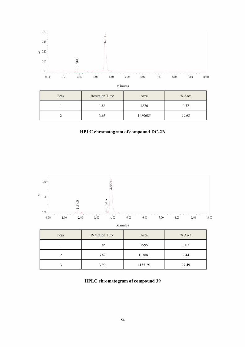
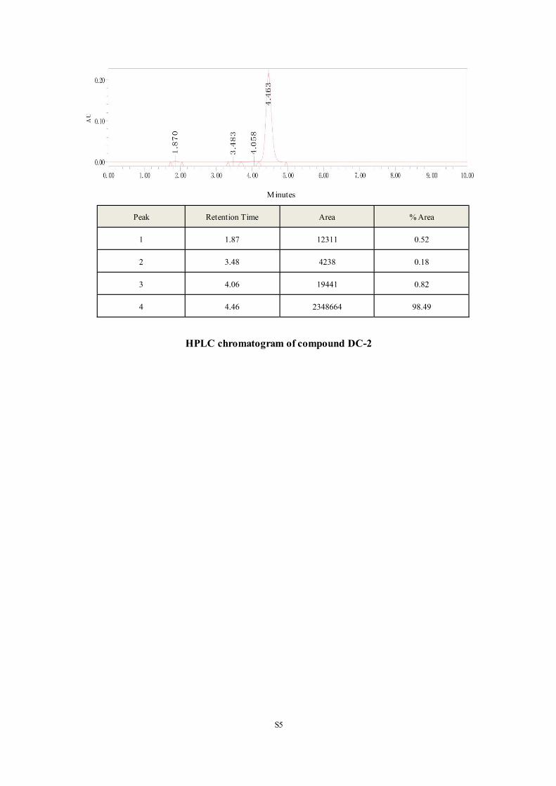
Copyright © 2022 FDOKUMEN


![Z )-3-Chloro-3-phenyl- N -[( S )-1-phenylethyl]prop-2-enamide](https://static.fdokumen.com/doc/165x107/63176320e88f2a90c801228d/z-3-chloro-3-phenyl-n-s-1-phenylethylprop-2-enamide.jpg)


![1-Phenyl-1 H -naphtho[1,2- e ][1,3]oxazin-3(2 H )-one](https://static.fdokumen.com/doc/165x107/6323a0da078ed8e56c0af332/1-phenyl-1-h-naphtho12-e-13oxazin-32-h-one.jpg)
![Molecular probes for the A 2A adenosine receptor based on a pyrazolo[4,3- e][1,2,4]triazolo[1,5- c]pyrimidin-5-amine scaffold](https://static.fdokumen.com/doc/165x107/631882dad93a162f9c0e927f/molecular-probes-for-the-a-2a-adenosine-receptor-based-on-a-pyrazolo43-e124triazolo15-.jpg)

![N-[2-Methyl-5-(triazol-1-yl)phenyl]pyrimidin-2-amine as a Scaffold for the Synthesis of Inhibitors of Bcr-Abl](https://static.fdokumen.com/doc/165x107/63359563b5f91cb18a0b780c/n-2-methyl-5-triazol-1-ylphenylpyrimidin-2-amine-as-a-scaffold-for-the-synthesis.jpg)


![Synthesis, anti- Toxoplasma gondii and antimicrobial activities of benzaldehyde 4-phenyl-3-thiosemicarbazones and 2-[(phenylmethylene)hydrazono]-4-oxo-3-phenyl-5-thiazolidineacetic](https://static.fdokumen.com/doc/165x107/63133f6fb22baff5c40f0921/synthesis-anti-toxoplasma-gondii-and-antimicrobial-activities-of-benzaldehyde.jpg)

![N -[4-( N -Cyclohexylsulfamoyl)phenyl]acetamide](https://static.fdokumen.com/doc/165x107/632f4f4de68feab59a0210b7/n-4-n-cyclohexylsulfamoylphenylacetamide.jpg)
![Microwave-Assisted Three-Component Synthesis and in vitro Antifungal Evaluation of 6-Cyano-5,8-dihydropyrido[2,3-d]pyrimidin-4(3H)-ones](https://static.fdokumen.com/doc/165x107/63206b11c5de3ed8a70db81f/microwave-assisted-three-component-synthesis-and-in-vitro-antifungal-evaluation.jpg)
![Structural requirements of pyrido[2,3-d]pyrimidin-7-one as CDK4/D inhibitors: 2D autocorrelation, CoMFA and CoMSIA analyses](https://static.fdokumen.com/doc/165x107/6333b4b7ce61be0ae50ec7c1/structural-requirements-of-pyrido23-dpyrimidin-7-one-as-cdk4d-inhibitors-2d.jpg)