Encapsulation of alpha-cyano-4-hydroxycinnamic acid into a NaY zeolite
6
Encapsulation of a-cyano-4-hydroxycinnamic acid into a NaY zeolite Nata ´lia Vilac ¸a • Ricardo Amorim • Olga Martinho • Rui M. Reis • Fa ´tima Baltazar • Anto ´nio M. Fonseca • Isabel C. Neves Received: 6 May 2011 / Accepted: 14 June 2011 / Published online: 29 June 2011 Ó Springer Science+Business Media, LLC 2011 Abstract The faujasite zeolite structure was studied to investigate its suitability for development of new drug delivery systems (DDS). The sodium form (NaY) of the zeolite was used for encapsulation of a-cyano-4-hydroxy- cinnamic acid (CHC), an experimental anticancer drug used in colorectal cancer therapy. The DDS was prepared by diffusion in liquid phase of CHC as a guest in the void space of the host zeolite structure at pH 7.0. The molecular integrity of CHC in the encapsulation process was evalu- ated by proton nuclear magnetic resonance spectroscopy ( 1 H NMR) and Ultraviolet–Visible spectroscopy (UV– Vis). The new drug delivery system, CHC@NaY, was characterized by Fourier transform infrared spectroscopy and UV–Vis, chemical analysis, powder X-ray diffraction, and Scanning electron microscopy. Analysis of the data of the drug alone and encapsulated in NaY show that CHC and the zeolite framework preserved their original struc- ture. The effect of the zeolite and DDS on HCT-15 human colon carcinoma cell line viability was evaluated. The encapsulation of CHC significantly increased its potency. Introduction Due to biological properties and stability in biological environments, zeolite structures have a great potential for medical use. Zeolites have been examined as carriers for slow or delayed release of anthelmintic and antitumoral drugs. The latest experimental studies, based on the results obtained in various tumor cells and in tumor bearing ani- mals, have shown that zeolites can be successfully used as adjuvants in anticancer therapy. Zeolite nanocrystals have been used in the enrichment of low abundance peptides/ proteins as well as in the immobilization of enzymes for biosensing and for processes by magnetic resonance imaging [1]. Zeolites are crystalline solids with very reg- ular microporous structures in which active chemical interesting compounds can be included. Zeolites have broad applications in heterogeneous catalysis, textile manufacturing, polymer catalytic degradation, electro- chemical properties, and have also attracted interest in material sciences for the development of functional mate- rials and in nanotechnology [2–15]. In this study, we report the use of the zeolite structure for drug delivery systems (DDS). The host zeolite has a faujasite (FAU) framework based on sodalite cages that are joined by oxygen bridges between the hexagonal faces. Eight sodalite cages are linked together, forming a large central cavity or supercage with a diameter of 1.18 nm. The supercages share a 12-membered ring with an open diameter of 0.74 nm [16–18] (Fig. 1). Encapsulation of a-cyano-4-hydroxycinnamic acid (CHC) was prepared by diffusion in liquid phase in the N. Vilac ¸a A. M. Fonseca I. C. Neves (&) Centre of Chemistry, Chemistry Department, University of Minho, Campus de Gualtar, 4710-057 Braga, Portugal e-mail: [email protected] R. Amorim O. Martinho R. M. Reis F. Baltazar Life and Health Sciences Research Institute (ICVS), School of Health Sciences, University of Minho, Braga, Portugal R. Amorim O. Martinho R. M. Reis F. Baltazar (&) ICVS/3B’s-PT Government Associate Laboratory, Braga/Guimara ˜es, Portugal e-mail: [email protected] R. M. Reis Molecular Oncology Research Center, Barretos Cancer Hospital, Barretos, SP, Brazil 123 J Mater Sci (2011) 46:7511–7516 DOI 10.1007/s10853-011-5722-2
-
Upload
independent -
Category
Documents
-
view
5 -
download
0
Transcript of Encapsulation of alpha-cyano-4-hydroxycinnamic acid into a NaY zeolite

Encapsulation of a-cyano-4-hydroxycinnamic acid into a NaYzeolite
Natalia Vilaca • Ricardo Amorim • Olga Martinho •
Rui M. Reis • Fatima Baltazar • Antonio M. Fonseca •
Isabel C. Neves
Received: 6 May 2011 / Accepted: 14 June 2011 / Published online: 29 June 2011
� Springer Science+Business Media, LLC 2011
Abstract The faujasite zeolite structure was studied to
investigate its suitability for development of new drug
delivery systems (DDS). The sodium form (NaY) of the
zeolite was used for encapsulation of a-cyano-4-hydroxy-
cinnamic acid (CHC), an experimental anticancer drug
used in colorectal cancer therapy. The DDS was prepared
by diffusion in liquid phase of CHC as a guest in the void
space of the host zeolite structure at pH 7.0. The molecular
integrity of CHC in the encapsulation process was evalu-
ated by proton nuclear magnetic resonance spectroscopy
(1H NMR) and Ultraviolet–Visible spectroscopy (UV–
Vis). The new drug delivery system, CHC@NaY, was
characterized by Fourier transform infrared spectroscopy
and UV–Vis, chemical analysis, powder X-ray diffraction,
and Scanning electron microscopy. Analysis of the data of
the drug alone and encapsulated in NaY show that CHC
and the zeolite framework preserved their original struc-
ture. The effect of the zeolite and DDS on HCT-15 human
colon carcinoma cell line viability was evaluated. The
encapsulation of CHC significantly increased its potency.
Introduction
Due to biological properties and stability in biological
environments, zeolite structures have a great potential for
medical use. Zeolites have been examined as carriers for
slow or delayed release of anthelmintic and antitumoral
drugs. The latest experimental studies, based on the results
obtained in various tumor cells and in tumor bearing ani-
mals, have shown that zeolites can be successfully used as
adjuvants in anticancer therapy. Zeolite nanocrystals have
been used in the enrichment of low abundance peptides/
proteins as well as in the immobilization of enzymes for
biosensing and for processes by magnetic resonance
imaging [1]. Zeolites are crystalline solids with very reg-
ular microporous structures in which active chemical
interesting compounds can be included. Zeolites have
broad applications in heterogeneous catalysis, textile
manufacturing, polymer catalytic degradation, electro-
chemical properties, and have also attracted interest in
material sciences for the development of functional mate-
rials and in nanotechnology [2–15].
In this study, we report the use of the zeolite structure
for drug delivery systems (DDS). The host zeolite has a
faujasite (FAU) framework based on sodalite cages that are
joined by oxygen bridges between the hexagonal faces.
Eight sodalite cages are linked together, forming a large
central cavity or supercage with a diameter of 1.18 nm.
The supercages share a 12-membered ring with an open
diameter of 0.74 nm [16–18] (Fig. 1).
Encapsulation of a-cyano-4-hydroxycinnamic acid
(CHC) was prepared by diffusion in liquid phase in the
N. Vilaca � A. M. Fonseca � I. C. Neves (&)
Centre of Chemistry, Chemistry Department, University
of Minho, Campus de Gualtar, 4710-057 Braga, Portugal
e-mail: [email protected]
R. Amorim � O. Martinho � R. M. Reis � F. Baltazar
Life and Health Sciences Research Institute (ICVS), School
of Health Sciences, University of Minho, Braga, Portugal
R. Amorim � O. Martinho � R. M. Reis � F. Baltazar (&)
ICVS/3B’s-PT Government Associate Laboratory,
Braga/Guimaraes, Portugal
e-mail: [email protected]
R. M. Reis
Molecular Oncology Research Center, Barretos Cancer Hospital,
Barretos, SP, Brazil
123
J Mater Sci (2011) 46:7511–7516
DOI 10.1007/s10853-011-5722-2

void space of the host zeolite NaY. The new drug delivery
system, CHC@NaY, was characterized by spectroscopic
techniques (Fourier transform infrared (FTIR), proton
nuclear magnetic resonance spectroscopy (1H NMR), and
Ultraviolet–Visible spectroscopy (UV–Vis)), chemical
analysis, powder X-ray diffraction (XRD), and scanning
electron microscopy (SEM). The experimental anticancer
drug a-cyano-4-hydroxycinnamic acid (CHC), here used as a
model for colon carcinoma treatment [19–22], was chosen as
the guest compound in the zeolite for drug delivery systems.
To the best of our knowledge, there are no studies using
zeolites as DDS for anticancer drugs [1, 23]. The new DDS
was evaluated on HCT-15 human colon carcinoma cell
viability. This study is a contribution for the development of
a new DDS based in zeolites in the cancer context.
Experimental
Materials and reagents
The zeolitic structure is commercially available; FAU zeo-
lite in powder form (CBV100) was obtained from Zeolyst
International. The zeolite in sodium form was previously
dehydrated at 120 �C overnight in an oven. CHC was pur-
chased from Sigma-Aldrich and used as received. Acetone,
the solvent used for encapsulation in this study, was pur-
chased from Merck (analytical grade). Human colon carci-
noma-derived cell line HCT-15 was kindly provided by
Dr. Raquel Seruca (IPATIMUP, Porto, Portugal).
Encapsulation of CHC in NaY
The encapsulation of CHC in NaY has been described
elsewhere [23]: 500 mg of NaY was reacted with a solution
of 49.20 mg CHC in acetone (15 mL), as a solvent. This
mixture was carried out by stirring at room temperature for
48 h. During this time, the original white color of NaY
changed to the characteristic color of the CHC, yellow,
indicating that the drug species was effectively entrapped
inside the host. This finding was also confirmed by the
disappearance of the yellow color of the starting CHC
solution after 3 h in contact with the zeolite and the effi-
ciency of the encapsulation was screened by HPLC. The
load of drug within the zeolite and on the surface was
determined by gravimetric analysis.
Characterization
Room temperature FTIR spectra of the samples in KBr
pellets were measured using a Bomem MB104 spectrom-
eter in the range 4000–500 cm-1 by averaging 20 scans at
a maximum resolution of 4 cm-1. Chemical analysis of C,
H, and N was carried out on a Leco CHNS-932 analyzer.1H NMR spectra were obtained on a Varian Unity Plus
Spectrometer at an operating frequency of 300 MHz using
the solvent peak as internal reference at 25 �C, chemical
shifts of protons being given in ppm using dH
Me4Si = 0 ppm as reference. The electronic UV–Vis
absorption spectrum of the residual solutions was obtained
in acetone. UV–Vis spectra of the drug molecule and the
suspension of NaY and CHC@NaY in Nujol were col-
lected in a Shimadzu UV/2501PC spectrophotometer using
quartz cells at room temperature. Phase analysis was per-
formed by XRD using a Philips PW1710 diffractometer.
Scans were taken at room temperature in a 2h range
between 5� and 60�, using Cu-Ka radiation. SEM was
collected on a LEICA Cambridge S360 Scanning Micro-
scope equipped with an EDX system (semi-quantitative
analysis with detection limits of *1.0 weight % for most
elements). In order to avoid surface charging, samples were
coated with gold in vacuum prior to analysis, by using a
Fisons Instruments SC502 sputter coater. Images of the
cells and the DDS during the drug bioactivity studies were
obtained in an optical inverted microscope (Olympus
IX51). The analysis was carried out by high performance
liquid chromatography (HPLC–JASCO 980-PU) using an
isocratic pump and a double on line detection including an
UV–Vis detector and refractometer.
Drug release studies
HCT-15 colon carcinoma cells were maintained in RPMI
1640 medium (Gibco), supplemented with 10% (v/v) fetal
bovine serum (FBS) (Gibco, Invitrogen, USA), and 1% (v/v)
penicillin–streptomycin solution (P/S) (Invitrogen, USA)
and incubated at 37 �C in a 5% CO2 humidified atmosphere.
Cells were subcultured approximately every 3 days and
maintained in a log-phase growth. Cell viability was assessed
using the In Vitro Toxicology Assay Kit, Sulforhodamine B
based (Sigma-Aldrich). HCT-15 cells were seeded in
96-well plates (5000 cells/100 lL/well) and incubated at
37 �C in a 5% CO2 humidified atmosphere for 24 h. In order
Fig. 1 Faujasite structure of NaY zeolite [16]
7512 J Mater Sci (2011) 46:7511–7516
123

to assess the effects of NaY and CHC@NaY, the spent media
were discarded and cells were incubated with increasing
concentrations of the systems in new culture medium.
Controls were performed with culture medium alone. After
an incubation period of 24 h, the spent media were removed
and the plate wells were washed with 19 phosphate buffered
saline, pH 7.4. After a fixation step with cold 10% Trichlo-
roacetic acid, cells were stained with 0.4% Sulforhodamine
B and the incorporated dye were solubilized with Sulforho-
damine B assay solubilization solution (10 mM Tris).
Absorbance was monitored with a microplate reader at
570 nm with a background absorbance of 655 nm. Cell
viability was determined as percent viability: (OD experi-
ment/OD control) 9 100 (%).
Results and discussion
An experimental anticancer drug (CHC) was used for
preparation of a new DDS based on a NaY zeolite which
was modified by trapping the drug molecule into its
framework in liquid phase at pH 7.
The length between the OH group from the aryl ring and
COOH is approximately 10.6 A in the planar and linear
CHC molecule. However, the aryl ring is 5.9 A, suggesting
that this drug can easily diffuse into the structure of the
NaY zeolite.
Loading of the CHC molecule into zeolite NaY was
determined by gravimetric analysis. The initial amount of
CHC used for encapsulation was 25.9 9 10-2 mmol. After
preparation of DDS, CHC loading in NaY was
22.6 9 10-2 mmol, indicating that 87.3% of the CHC
initially present has been retained inside the zeolite.
The yield of the CHC encapsulation was confirmed by
the analytical data of carbon and nitrogen content obtained
by elemental analysis (Table 1).
The C/N ratio of the encapsulated CHC, obtained by
chemical analysis, was similar to the theoretically expected
C/N ratio of CHC, indicating the presence of the molecular
drug structure inside the zeolite. The UV/Vis absorption
spectra of NaY zeolite and the DDS was used as a tool to
study the encapsulation of the drug molecule in the host
zeolite (Fig. 2).
The UV/Vis absorption spectrum of CHC@NaY display
bands which are not shown in the spectrum of the zeolite
host (Fig. 2a, c). The spectrum of CHC (Fig. 2b) measured
in Nujol at room temperature, exhibited two bands at 370
and 415 nm. The spectra of CHC@NaY exhibited similar
bands to CHC (Fig. 2b, d). However, the control in residual
solutions during the preparation, by 1H NMR and UV–Vis
spectroscopy, confirms that CHC after encapsulation in
NaY is in its neutral form.
The data obtained by vibrational spectroscopy can pro-
vide structural information of the host framework and the
encapsulated drug molecule. Figure 3 shows the infrared
spectra in the range 4000–600 cm-1 of NaY (a), CHC (b),
and CHC@NaY (c).
The FTIR spectra of the guest compound and the host
presented in Fig. 3 are dominated by the strong bands
assigned to the vibration of zeolite structure. The presence
of physisorbed water is detected by the m(O–H) stretching
vibration at 3450 cm-1 and the m(O–H) deformation band
at 1640 cm-1. The bands corresponding to the lattice
vibrations are observed in the spectral region between 1300
and 450 cm-1 [7, 8]. No shift or broadening of the zeolite
vibrations bands are observed upon inclusion of the CHC,
Table 1 Elemental composition of CHC and DDS
Samples C (%) N (%) C/N
CHC 63.49 7.40 8.6
CHC@NaY 11.45 1.31 8.7
Wavelength (nm)
Abs
orba
nce
(a.u
.)
(a)
(b)
(c)
(d)
Fig. 2 UV/vis spectra of the samples: (a) NaY, (b) CHC,
(c) CHC@NaY and (d) CHC@NaY without the matrix obtained in
Nujol suspensions
4000 3500 3000 2500 2000 1500 1000 500
0
10
20
30
40
50
60
Tra
nsm
ittan
ce (
%)
Wavenumber (cm-1)
(a)
(b)
(c)
Fig. 3 FTIR spectra of the samples: (a) NaY, (b) CHC and
(c) CHC@NaY
J Mater Sci (2011) 46:7511–7516 7513
123
which provides further evidence that the zeolite structure
remains unchanged after encapsulation.
In addition to these strong bands caused by the host
matrix, the FTIR spectrum of CHC@NaY show bands in
the 2300–2000 and 1600–1200 cm-1 regions where NaY
does not absorb and they are attributed to the presence of
the encapsulated drug. This means that the zeolite frame-
work does not interfere with the IR absorption of the
incorporated guest. Comparison of the spectra of the
encapsulated CHC with the parent drug molecule provides
evidence for the entrapping of CHC in NaY.
The spectrum of CHC shows the characteristic bands of
the drug: 3308 (mOH), 2224 (mCN), 1669 (mC=O), and
1567–1295 cm-1 attributed to C–C and C–H bands. The
spectra of the encapsulated drug as well as CHC alone
showed similar bands. The spectrum of CHC@NaY shows
bands due to the drug molecule at 2225, 1560, 1502, 1362,
and 1291 cm-1 indicating no host–guest interactions.
Furthermore, from elemental analysis, UV–Vis, and FTIR
data, we observed that the zeolitic environment preserves
the molecular structure of the drug.
FTIR analysis was an important tool also for obtaining
the framework Si/Al ratio of the FAU zeolite [24, 25]. The
most sensitive band in the zeolite Y structure can be
observed at 570 and 600 cm-1 and was used to calculate
the framework Si/Al ratio (Eq. 1):
x ¼ 3:857� 0:00621wDRðcm�1Þ; ð1Þ
where x = [1 ? (Si/Al)]-1, with 0.1 \ x \ 0.3, and wDR is
the zeolite specific double ring vibration mode in the range
570–600 cm-1. The obtained framework Si/Al ratio was
2.83 and 2.80 for NaY and DDS, respectively.
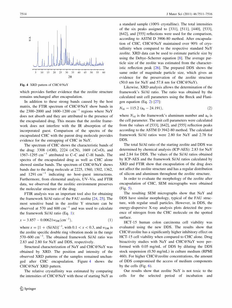
Structural characterization of NaY and CHC@NaY was
obtained by XRD. The position and intensity of the
observed XRD patterns of the samples remained unchan-
ged after CHC encapsulation. Figure 4 shows the
CHC@NaY XRD pattern.
The relative crystallinity was estimated by comparing
the intensities of CHC@NaY with those of starting NaY as
a standard sample (100% crystalline). The total intensities
of the six peaks assigned to [331], [511], [440], [533],
[642], and [555] reflections were used for the comparison,
according to ASTM D 3906-80 method. After encapsula-
tion of CHC, CHC@NaY maintained over 90% of crys-
tallinity when compared to the respective standard NaY
zeolite. XRD data can be used to estimate particle size by
using the Debye–Scherrer equation [8]. The average par-
ticle size of the zeolite was estimated from the character-
istic reflection peak [26]. The prepared DDS shows the
same order of magnitude particle size, which gives an
evidence for the preservation of the zeolite structure
(50.0 nm for NaY and 57.8 nm for CHC@NaY).
Likewise, XRD analysis allows the determination of the
framework’s Si/Al ratio. The ratio was obtained by the
calculated unit cell parameters using the Breck and Flani-
gen equation (Eq. 2) [27]:
NAl ¼ 115:2 (a0 � 24:191Þ; ð2Þ
where NAl is the framework’s aluminum number and a0 is
the cell parameter. The unit cell parameters were calculated
from the values of [533], [642], and [555] reflection peaks
according to the ASTM D 3942-80 method. The calculated
framework Si/Al ratios were 2.80 for NaY and 2.78 for
DDS.
The total Si/Al ratio of the starting zeolite and DDS was
determined by chemical analysis (ICP-AES): 2.83 for NaY
and 2.84 for DDS. The values of the Si/Al ratios obtained
by ICP-AES and the framework Si/Al ratios calculated by
XRD and FTIR show that encapsulation of the drug does
not affect the zeolite structure and has a regular distribution
of silicon and aluminum throughout the zeolite structure.
In order to evaluate the morphology of the zeolite after
encapsulation of CHC, SEM micrographs were obtained
(Fig. 5).
The resulting SEM micrographs show that NaY and
DDS have similar morphology, typical of the FAU struc-
ture, with regular small particles. However, in DDS, the
energy-dispersive X-ray analysis plots detected the pres-
ence of nitrogen from the CHC molecule on the spotted
surface.
HCT-15 human colon carcinoma cell viability was
evaluated using the new DDS. The results show that
CHC@zeolite has a significantly higher inhibitory effect on
HCT-15 cell viability when compared to CHC alone. Drug
bioactivity studies with NaY and CHC@NaY were per-
formed with 0.05 mg/mL of DDS by diluting the DDS
stock suspension (0.50 mg/mL) in culture medium (RPMI
460). For higher CHC@zeolite concentrations, the amount
of DDS compromised the access of medium components
by the cells (Fig. 6).
Our results show that zeolite NaY is not toxic to the
cells for the selected period of incubation and
2θ
u.a.
5 10 15 20 25 30 35 40 45 50 55 60
Fig. 4 XRD pattern of CHC@NaY
7514 J Mater Sci (2011) 46:7511–7516
123

concentrations. By comparing the results obtained when
treating HCT-15 cells with the non-encapsulated CHC with
the results obtained for CHC@NaY, there is an obvious
potentiation of the effect of the drug. About 6 mM of the
non-encapsulated drug are necessary for a 30% of inhibi-
tion (IC30) in cell viability. In contrast, only 0.054 mM of
the encapsulated drug produces the same magnitude of
inhibition, corresponding to a potentiation of the drug
effect of about 110-fold.
Conclusions
Combining spectroscopic techniques and chemical analysis
provides a powerful tool to reveal the unequivocal evi-
dence for the encapsulation of CHC in the framework
structure of zeolite NaY.
In the light of the obtained results, it can be concluded
that CHC molecule can be encapsulated in NaY zeolite
supercages, without structural modification or loss of
crystallinity of the zeolite framework, while the drug
molecule retains its molecular integrity.
Importantly, CHC@NaY led to an inhibition of cell
viability up to 110-fold when compared to the non-
encapsulated drug. These results indicate the potential of
the zeolites for drug loading and delivery to cancer cells. It
would be important in the near future to test other CHC/
zeolite ratios and also the encapsulation of CHC in dif-
ferent zeolite structures.
Acknowledgements The authors are thankful to Dr. K. Biernacki
for DFT calculations and Dr. A.S. Azevedo for collecting the powder
diffraction data. OM and RA are recipients of fellowships (SFRH/BD/
36463/2007, SFRH/BI/51118/2010) from Fundacao para a Ciencia e a
Tecnologia (FCT, Portugal).This study was supported by the Centre
of Chemistry and Life and Health Sciences Research Institute (ICVS),
University of Minho, Portugal, FCT (Portugal) through POCTI and
FEDER projects (ref. POCTI-SFA-3-686) and by the FCT grant ref.
PTDC/SAU-FCF/104347/2008, under the scope of ‘‘Programa Op-
eracional Tematico Factores de Competitividade’’ (COMPETE) of
‘‘Quadro Comunitario de Apoio III’’ and co-financed by Fundo
Comunitario Europeu FEDER.
References
1. Danilczuk PM, Dugopolska K, Ruman T, Pogocki D (2008) Mini
Rev Med Chem 8:1407
2. Corma A, Garcia H (2004) Eur J Inorg Chem 6:1143
3. Kuzniarska-Biernacka I, Biernacki K, Magalhaes AL, Fonseca
AM, Neves IC (2011) J Catal 278:102
4. Parpot P, Teixeira C, Almeida AM, Ribeiro C, Neves IC, Fonseca
AM (2009) Microporous Mesoporous Mater 117:297
5. Neves IC, Botelho G, Machado AV, Rebelo P, Ramoa S, Pereira
MFR, Ramanathan A, Pescarmona P (2007) Polym Degrad Stab
92:1513
6. Van Santen RA, Kramer GJ (1995) Chem Rev 95:637
5 μμm
5 μm (b)
(a)
Fig. 5 Scanning electron microscopy (SEM) images of NaY (a) and
CHC@NaY with same resolution (95000)
(a)
(b)
10 µm
10 µm
Fig. 6 Optical microscopy images taken in an inverted optical
microscope of (a) HCT-15 cells and 0.50 mg/mL of (b) DDS
J Mater Sci (2011) 46:7511–7516 7515
123
7. Neves IC, Cunha C, Pereira MR, Pereira MFR, Fonseca AM
(2010) J Phys Chem C 114:10719
8. Lopes AC, Silva MP, Goncalves R, Pereira MFR, Botelho G,
Fonseca AM, Lanceros-Mendez S, Neves IC (2010) J Phys Chem
C 114:14446
9. Abbo HS, Titinchi SJJ (2010) Top Catal 53:1401
10. Yang Y, Ding H, Hao S, Zhang Y, Kan Q (2011) Appl Or-
ganometal Chem 25:262
11. Perez E, Martin L, Rubio C, Urieta JS, Piera E, Caballero MA,
Tellez C, Coronas J (2010) Ind Eng Chem Res 49:8495
12. Talebi J, Halladj R, Askari S (2010) J Mater Sci 45:3318. doi:
10.1007/s10853-010-4349-z
13. Gomes R, Albuquerque RQ, Pina F, Parola J, De Cola L (2010)
Photochem Photobiol Sci 9:991
14. Lang JW, Kong LB, Wu WJ, Luo YC, Kang L (2009) J Mater Sci
44:4466. doi:10.1007/s10853-009-3677-3
15. Nethravathi BP, Mahendra KN, Reddy KRK (2011) J Porous
Mater 18:389
16. Database of zeolite structures from the International Zeolite
Association (IZA-SC). www.iza-structure.org/databases/. Acces-
sed 03 Apr 2011
17. Baerlocher Ch, McCusker LB, Olson DH (2007) Atlas of zeolite
framework types, sixth revised edition. Elsevier, Amsterdam
18. Subhash B (1990) Zeolite catalysis: principles, applications. CRC
Press, Inc., Boca Raton
19. Halestrap AP, Meredith D (2004) Eur J Physiol 447:619
20. Pinheiro C, Longatto-Filho A, Scapulatempo C, Ferreira L,
Martins S, Pellerin L, Rodrigues M, Alves VAF, Schmitt F,
Baltazar F (2008) Virchows Arch 452:139
21. Pinheiro C, Longatto-Filho A, Pereira SMM, Etlinger D, Moreira
MAR, Jube LF, Queiroz GS, Schmitt F, Baltazar F (2009) Dis
Marker 26:97
22. Pinheiro C, Albergaria A, Paredes J, Sousa B, Dufloth R, Vieira
D, Schmitt F, Baltazar F (2010) Histopathology 56:860
23. Rimoli MG, Rabaioli MR, Melisi D, Curcio A, Mondello S,
Mirabelli R, Abignente E (2007) J Biomed Mater Res A 87A:156
24. Ghesti GC, Macedo JL, Parente VCI, Dias JA, Dias SCL (2007)
Microporous Mesoporous Mater 100:27
25. Lutz W, Ruscher CH, Heidemann D (2002) Microporous Meso-
porous Mater 55:193
26. Morris R (2007) In: Cejka J, van Bekkum H, Corma A, Schuth F
(eds) Introduction to zeolite science and practice. Studies in
surface science and catalysis, vol 168. Elsevier, Amsterdam
27. Breck DW, Flanigen EM (1968) Molecular sieves. Society of
Chemical Industry, London, p 47
7516 J Mater Sci (2011) 46:7511–7516
123