characterization of mycobacteria spp. and antimycobacterial ...
Upload
independentCategory
view
2download
0
Published Ahead of Print 11 September 2006. 10.1128/AAC.00555-06.
2006, 50(11):3665. DOI:Antimicrob. Agents Chemother. and Kakoli MukherjeeXia Yang, Beat Weidmann, Kathryn Bracken, Thomas DickSchreiber, Samiul Hasan, Michael Cynamon, Neil S. Ryder, Duraiswamy, Sarah Liung, Veronique Dartois, MarkL. Yap, Mahesh Nanjundappa, Xinyi Ngew, Jeyaraj Jeanette W. P. Teo, Pamela Thayalan, David Beer, Amelia S. Antimycobacterial AgentsPeptide Deformylase Inhibitors as Potent
http://aac.asm.org/content/50/11/3665Updated information and services can be found at:
These include:
SUPPLEMENTAL MATERIAL Supplemental material
REFERENCEShttp://aac.asm.org/content/50/11/3665#ref-list-1at:
This article cites 63 articles, 24 of which can be accessed free
CONTENT ALERTS more»articles cite this article),
Receive: RSS Feeds, eTOCs, free email alerts (when new
http://journals.asm.org/site/misc/reprints.xhtmlInformation about commercial reprint orders: http://journals.asm.org/site/subscriptions/To subscribe to to another ASM Journal go to:
on March 7, 2014 by guest
http://aac.asm.org/
Dow
nloaded from
on March 7, 2014 by guest
http://aac.asm.org/
Dow
nloaded from

ANTIMICROBIAL AGENTS AND CHEMOTHERAPY, Nov. 2006, p. 3665–3673 Vol. 50, No. 110066-4804/06/$08.00�0 doi:10.1128/AAC.00555-06Copyright © 2006, American Society for Microbiology. All Rights Reserved.
Peptide Deformylase Inhibitors as Potent Antimycobacterial Agents�†Jeanette W. P. Teo,1‡ Pamela Thayalan,1 David Beer,1 Amelia S. L. Yap,1 Mahesh Nanjundappa,1
Xinyi Ngew,1 Jeyaraj Duraiswamy,1 Sarah Liung,1 Veronique Dartois,1 Mark Schreiber,1Samiul Hasan,1 Michael Cynamon,2 Neil S. Ryder,3 Xia Yang,3 Beat Weidmann,3
Kathryn Bracken,3 Thomas Dick,1 and Kakoli Mukherjee1*Novartis Institute for Tropical Diseases, 10 Biopolis Road, 05-01 Chromos, Singapore 138670, Republic of Singapore1;
Central New York Research Corporation, New York, New York2; and Novartis Institutes for Biomedical Research,Inc., Infectious Disease Area, 100 Technology Square, Cambridge, Massachusetts 021393
Received 4 May 2006/Returned for modification 6 June 2006/Accepted 17 August 2006
Peptide deformylase (PDF) catalyzes the hydrolytic removal of the N-terminal formyl group from nascentproteins. This is an essential step in bacterial protein synthesis, making PDF an attractive target for anti-bacterial drug development. Essentiality of the def gene, encoding PDF from Mycobacterium tuberculosis, wasdemonstrated through genetic knockout experiments with Mycobacterium bovis BCG. PDF from M. tuberculosisstrain H37Rv was cloned, expressed, and purified as an N-terminal histidine-tagged recombinant protein inEscherichia coli. A novel class of PDF inhibitors (PDF-I), the N-alkyl urea hydroxamic acids, were synthesizedand evaluated for their activities against the M. tuberculosis PDF enzyme as well as their antimycobacterialeffects. Several compounds from the new class had 50% inhibitory concentration (IC50) values of <100 nM.Some of the PDF-I displayed antibacterial activity against M. tuberculosis, including MDR strains with MIC90values of <1 �M. Pharmacokinetic studies of potential leads showed that the compounds were orally bio-available. Spontaneous resistance towards these inhibitors arose at a frequency of <5 � 10�7 in M. bovis BCG.DNA sequence analysis of several spontaneous PDF-I-resistant mutants revealed that half of the mutants hadacquired point mutations in their formyl methyltransferase gene (fmt), which formylated Met-tRNA. Theresults from this study validate M. tuberculosis PDF as a drug target and suggest that this class of compoundshave the potential to be developed as novel antimycobacterial agents.
Mycobacterium tuberculosis, a pathogen causing 2 milliondeaths and 8 million new cases of tuberculosis (TB) each year,is a leading health threat. This is further fueled by the humanimmunodeficiency virus epidemic and the appearance of mul-tidrug-resistant TB (17, 18, 46). Infections involving multidrug-resistant TB are harder to treat and have limited the efficacy ofTB control programs (29). This has necessitated the search fornew antituberculosis drugs with a novel mode of action.
In prokaryotes and in certain organelles, like plastids and mi-tochondria of eukaryotes, nascent proteins carry an N-formylatedmethionine (40). After translational initiation, peptide defor-mylase (PDF) (EC 3.5.1.27) removes the N-formyl group fromthe nascent proteins (42). Deformylated proteins undergo fur-ther N-terminal processing to give mature proteins (61). PDFis an attractive candidate for the discovery of antibacterialagents, as the essentiality of the gene (def) encoding PDF hasbeen shown for Escherichia coli and Streptococcus pneumoniae(8, 36). The enzyme has been characterized as a highly unsta-ble metallopeptidase that uses Fe2� as the catalytic metal (51).Oxidation of Fe2� inactivates the enzyme, and purification of
active bacterial PDF has been a challenge (51). So far, Ni2�,Co2�, and Mn2� have been described as cations that can stablyreplace Fe�2 while retaining high enzymatic activity (25, 49).
Naturally occurring antibiotics, like actinonin, inhibit theactivity of the PDF enzyme (2). Based on structural (7, 39, 41)and mechanistic design, as well as high-throughput screening,several classes of PDF inhibitors (PDF-I) have been studiedpreviously (64). Thus far, only compounds having hydroxamicacid-chelating or N-formyl hydroxylamine-chelating groupsshow potent enzyme inhibition, good antibacterial activity,and in vivo efficacy, including oral activity (9, 12, 27).
In this work, we validate def (gene encoding PDF) as a drugtarget for TB by proving its essentiality in Mycobacterium bovisBCG. PDF from M. tuberculosis strain H37Rv was cloned andexpressed heterologously in E. coli. Recombinant His-taggedM. tuberculosis PDF enzyme was purified, and biochemicalcharacterization was carried out. Activities of a novel class ofPDF-I, the N-alkyl urea reverse hydroxamates (27), were de-termined by both M. tuberculosis PDF enzyme and cell-basedassays for antimycobacterial activity. A potent lead, PDF-611(LBK-611) (15), was identified as a novel antimycobacterialagent.
MATERIALS AND METHODS
Enzymes, chemicals, and antibiotics. Chemicals used were of the highestgrade commercially available. Chemicals and antibiotics were purchased fromSigma (St. Louis, Mo.), unless otherwise indicated. Restriction enzymes andmodifying enzymes were purchased from New England Biolabs, Inc. (Beverly,Mass.), and were used according to the manufacturer’s recommendations.
Bacterial strains, plasmids, and media. Bacterial strains and plasmids used inthis study are listed in Table 1. E. coli strains were cultured in Luria-Bertani (LB)
* Corresponding author. Mailing address: Novartis Institute for Trop-ical Diseases, 10 Biopolis Road, 05-01 Chromos, Singapore 138670, Re-public of Singapore. Phone: 65-67222923. Fax: 65-67222917. E-mail:[email protected].
‡ Present address: National University Hospital, Department ofLaboratory Medicine, 5 Lower Kent Ridge Road, Main Building,Level 3, Singapore 119 074, Republic of Singapore.
† Supplemental material for this article may be found at http://aac.asm.org/.
� Published ahead of print on 11 September 2006.
3665
on March 7, 2014 by guest
http://aac.asm.org/
Dow
nloaded from

broth (Becton Dickinson, Franklin Lakes, NJ) and LB agar plates supplementedwith either 100 �g/ml of ampicillin or 50 �g/ml of kanamycin for cloning andmaintenance. Terrific broth (12 g tryptone, 24 g yeast extract, 4 ml glycerol, 2.3 gpotassium phosphate [monobasic], 9.4 g potassium phosphate [dibasic], pH 7.5,water to make 1 liter) was used as the growth medium for the expression of PDFfrom E. coli.
M. bovis BCG was cultured in Middlebrook 7H9 (catalog no. 271310; Difco)broth supplemented with 10% (vol/vol) albumin-dextrose saline (0.81% NaCl,5% bovine serum albumin fraction V [Roche, Mannheim, Germany], and 2%glucose), 0.2% glycerol, and 0.05% Tween 80 or on Middlebrook 7H10 (catalogno. 262710; Difco) agar plates supplemented with 10% (vol/vol) oleic acid-albumin-dextrose-catalase enrichment (catalog no. 212240; BBL), 0.5% glycerol,and 0.05% Tween 80. When required, hygromycin B (Roche) was used at theconcentration of 50 �g/ml. Sucrose was used in the 7H10 medium at a 4%concentration.
Buffers. Lysis buffer was composed of 50 mM HEPES (pH 7.5), 5 mM NiSO4,5 mM MgSO4, 10 �g/ml catalase, 0.25 mM Tris(2-carboxyethyl)phosphine hy-drochloride, 10 �g/ml DNase I, 1 ml protease cocktail inhibitor, 1 mg/ml ly-sozyme, and 1% Triton X-100. Buffer I was composed of 50 mM NaH2PO4, 300mM NaCl, and 10 mM imidazole, pH 8.0. Buffer II was composed of 50 mMNaH2PO4, 300 mM NaCl, and 20 mM imidazole, pH 8.0. Buffer III was com-posed of 50 mM NaH2PO4, 300 mM NaCl, and 250 mM imidazole, pH 8.0. Gelfiltration buffer was composed of 50 mM NaH2PO4, 1 M NaCl, and 5% glycerol.
Construction of the BCG def suicide plasmid and the def complementationplasmid. Gene regions flanking the def gene were PCR amplified and ligatedtogether to obtain a DNA fragment in which the def gene (636 bp) was excludedin frame (Fig. 1A). Primers BglF (5�-TGGTAAATAGAGATCTGTGTGCGACGTCATAGCCGAGTT-3�) and NdeR (5�-TGTCTTTTCATATGTAATGGTCTCGTGGCCGGGGC-3�) amplify an 1,150-bp fragment upstream of def (leftfragment). Primers NdeFF (5�-GAAGTGTACATATGGGGCTGAGGAGGCGGGCAAT-3�) and BglRR (5�-TTTAATTTTAGATCTGGCGGGCTTTGGCATAGCG-3�) amplify an 1,173-bp fragment downstream of def (right fragment).Primers BglF/BglRR and NdefR/NdeFF carry BglII and NdeI restriction sites,respectively, which enabled the left and right fragments to be ligated and clonedinto pCR-XL-TOPO (Table 1). The dimer fragment was then cloned into thesuicide vector pYUB657 (47), generating the def suicide plasmid pYUB657-dimer (Table 1). The def complementation vector was constructed by cloning thedef gene into the BamHI site of pMV262 (Table 1), generating pMV262-def.This vector was introduced into a def single-crossover strain to allow chromo-somal def to be deleted.
Genomic DNA isolation and Southern blot analysis. Genomic DNA from M.bovis BCG was isolated using the protocol described in a DNeasy tissue kit
(QIAGEN, Valencia, CA). For Southern blot analysis, DNA samples were di-gested with the appropriate restriction enzyme and fragments separated on a 1%agarose gel and transferred to Hybond-N� nylon membrane (Amersham).Southern hybridization was carried out using enhanced chemiluminescencedirect nucleic acid labeling and detection systems (Amersham).
Expression and purification of M. tuberculosis PDF. The M. tuberculosisH37Rv genomic DNA sequence (GenBank accession number NC_000962) wasused to design primers 5�-ATGGATCCATGGCAGTCGTACCCATCCG-3�and 5�-ATGGATCCGTGACCGAACGGGTCGGG-3�, which had BamHI sites(underlined) incorporated in them. The native TAA stop codon from the M.tuberculosis def gene was not included to utilize the vector-encoded stop codon,resulting in the fusion protein MRGSHHHHHHGS-M. tuberculosis PDF-GSACELGTPGRPAAKLN, with residues in italics originating from the vector. PCRwas carried out using a high-fidelity Platinum PCR Supermix (Invitrogen, Carls-bad, CA) according to the manufacturer’s protocol. PCR products were initiallycloned into the pCR2.1-TOPO cloning vector (Invitrogen), generating plasmidpCR2.1-PDF; from this, the def gene was subcloned into BamHI-linearized E.coli expression vector pQE30 (QIAGEN), generating plasmid pQE30-PDF (Ta-ble 1). Both pCR2.1-PDF and pQE30-PDF were sequenced. DNA sequenceswere analyzed using the software Vector NTI, version 7.0 (InforMax, Frederick,MD). Amino acid sequence alignments were prepared using CLUSTALW, ver-sion 1.83 (11). All DNA manipulations were performed under standard condi-tions as described previously (55).
E. coli M15(pREP4) cells carrying pQE30-PDF were grown in terrific brothcontaining 50 �g/ml of kanamycin and 100 �g/ml of ampicillin at 37°C until theoptical density at 600 nm reached �0.5. The culture was induced with 0.1 mMisopropyl-�-D-thiogalactopyranoside (IPTG) and incubated at 37°C for an addi-tional 4 h. Cells (from 1 liter) were harvested, and the cell pellet was washedtwice with sterile phosphate-buffered saline and suspended in 20 ml lysis buffer.The cells were lysed by incubation on ice for 30 min, followed by sonication. Celldebris was removed by centrifugation at 35,000 � g at 4°C for 15 min. Theproteins in the supernatant were precipitated by adjustment to 60% saturationwith ammonium sulfate. The pellet was dissolved in 10 ml of buffer I and addedto Ni-nitrilotriacetic acid (NTA) slurry (QIAGEN) for batch purification bymetal affinity chromatography. The resulting mixture was incubated for 1 h withgentle shaking at 4°C. The slurry was washed twice with buffer II, and the boundPDF was eluted with buffer III. Active fractions obtained by Ni-NTA batchpurification were further purified by gel filtration using a HiLoad Superdex 26/60prep grade column (Amersham) that had been equilibrated with gel filtrationbuffer. Elution was done with the same buffer at a flow rate of 1 ml/min. Theactive fractions were pooled and concentrated using a Vivaspin 20 centrifugal
TABLE 1. Strains and plasmids used in this work
Strain or plasmid Relevant characteristic(s) Source or reference
StrainsE. coli
XL1-Blue recA1 endA1 gyrA96 thi-1 hsdR17 supE44 relA1 lac �F� proAB lacIqZ�M15 Tn10 (Tetr);cloning strain
Stratagene
M15(pREP4) Nals Strs Rifs Thi Lac Ara� Gal� Mtl F RecA� Uvr� Lon�; expression strain forPT5-regulated gene expression
QIAGEN
M. bovis BCG Pasteur strain ATCC 35734 ATCCXO22 Single-crossover strain; Sucs Hygr This workXOdef Single-crossover strain XO22 carrying complementation plasmid pMV262-def This work
PlasmidspCR-XL-TOPO Ampr; TA-cloning vector for longer PCR products InvitrogenpCR2.1-TOPO Ampr; TA-cloning vector InvitrogenpQE30 Ampr; low-copy-number plasmid with inducible T5 promoter for production of N-
terminal His6 tag translational fusionQIAGEN
pCR2.1-PDF M. tuberculosis def gene cloned into pCR2.1-TOPO vector This workpQE30-PDF M. tuberculosis def gene cloned into BamHI site of pQE30 This workpYUB657 Ampr, Hygr; sacB counterselectable marker, E. coli-Mycobacterium shuttle vector 47pYUB657-dimer DNA fragment containing an in-frame def deletion cloned into pYUB657 suicide vector This workpMV262 E. coli-Mycobacterium shuttle plasmid, hsp60 promoter; Kanr ColE1 OriM 62pMV262-def pMV262 containing the def gene, expressed as a translational fusion with the first six
amino acids of hsp60This work
3666 TEO ET AL. ANTIMICROB. AGENTS CHEMOTHER.
on March 7, 2014 by guest
http://aac.asm.org/
Dow
nloaded from

concentrator (Sartorius, Hanover, Germany). Glycerol was added to a finalconcentration of 33% (vol/vol), and the enzyme was stored frozen at 80°C.
PDF enzyme assay. The PDF enzyme assay format was based on a publishedmethod (12), and the assay was carried out in a final volume of 50 �l in black,96-well, half-area plates (Corning Costar, Etobicoke, Ontario, Canada). Thereaction mixture contained 50 mM HEPES (pH 7.5) and �30 ng of E. coli-expressed M. tuberculosis PDF per well. The reaction was initiated by addingN-formyl-methionine-alanine-serine (fMAS) substrate to a final concentration of2 mM (Bachem, Germany). The reaction was done at room temperature for 2.5min and terminated by adding 25 �l of fluorescamine (0.02 mg/ml in dioxane).The microplate was further incubated for 5 min and read for fluorescence by aTecan Saffire plate reader using an excitation wavelength of 380 nm and anemission wavelength of 470 nm.
Biochemical characterization of M. tuberculosis PDF. Biochemical character-ization of M. tuberculosis PDF was performed using pure and batch-purifiedrecombinant enzyme. The effect of substrate concentrations on M. tuberculosisPDF activity was studied using two N-formylated peptide substrates, N-formyl-methionine-alanine (fMA) and fMAS (Bachem), with concentrations rangingfrom 0 to 10 mM. The effects of the divalent metal ions Ca2�, Mg2�, Mn2�,Co2�, Cu2�, Ni2�, and Zn2� on PDF activity were assessed by the addition ofdifferent ions at a final concentration of 1 mM to the assay buffer.
For inhibition studies of M. tuberculosis PDF, a 30-min preincubation of theinhibitor with the enzyme was included prior to initiating the reaction. Theconcentrations of the inhibitors varied from 0 to 10 �M. Fifty percent inhibitoryconcentration (IC50) values were obtained by fitting the data to a sigmoid dose-response equation using GraphPad Prism software, version 3.0 (GraphPad Soft-ware, Inc., San Diego, CA).
Antimicrobial agents and drug susceptibility testing. Streptomycin (STR),isoniazid (INH), rifampin (RIF), linezolid (LZD), and erythromycin (ERY)were obtained from Sigma (St. Louis, Mo.). Stock solutions of STR, LZD, andERY were prepared at 10 mM concentration in deionized water, while INH,RIF, and PDF-I were dissolved in 90% dimethyl sulfoxide. Drug susceptibilitytesting was based on a previously published method (20). Logarithmic-phase M.bovis BCG cultures were diluted in complete 7H9 broth to obtain an opticaldensity at 600 nm of �0.04, which corresponded to approximately 106 CFU/ml,before being added to the test plate. The plates were incubated at 37°C for 4days. On the fourth day, 50 �l of a freshly prepared 1:1 mixture of Alamar Blue(Serotec Ltd., Oxford, United Kingdom) and 10% Tween 80 was added to each
well. The plates were reincubated overnight at 37°C. STR was used as a referencedrug in each plate. The MICs were obtained by quantifying the fluorescence ofAlamar Blue, using excitation and emission wavelengths of 530 nm and 590 nm,respectively. Values below 150,000 relative fluorescence units were considered toreflect no growth.
Frequency of spontaneous resistant mutants. Log-phase BCG cultures at 109
CFU/ml were plated onto 7H10 plates containing drug at 10� MIC and ontodrug-free media. Plates were incubated at 37°C for approximately 6 weeks oruntil colonies appeared. The frequency of spontaneous resistance was calculatedas the ratio of the number of colonies that grew on drug-supplemented plates tothat of drug-free plates.
Mutational analyses of def and fmt genes. The def and fmt genes of PDF-I-resistant BCG mutants were analyzed for sequence variation. The def generegion was amplified using primers Updef (5�-GGTCCCGGTCTTGGTCTGCA-3�) and Dndef (5�-CGGCGATGATGCCCGCCG-3�), generating an 838-bpamplicon. The primers Upfmt (5�-TAGCGTGCCTTGCGTACCCA-3�) andDnfmt (5�-CAGCGCGGGCAACACCAG-3�) would amplify an 1,178-bp ampli-con. High-fidelity PCR was carried out using Proof Start DNA polymerase(QIAGEN). The PCR products were purified from agarose gels using aQIAquick gel extraction kit (QIAGEN). The purified PCR products were se-quenced at Research Biolabs Technologies (Singapore). Jalview software wasused for visualizing the alignment (11), and another software was used forbioinformatics analysis and secondary-structure prediction of the mutants (13).
In vivo pharmacokinetic studies. CD-1 female outbred mice (BioArc, India)were used for pharmacokinetic studies of selected PDF inhibitors. For intrave-nous (i.v.) and oral (p.o.) administration, the compounds were formulated in 4%and 10% polyethylene glycol 300, respectively. After 1 week of acclimation, micereceived a dose of 1 mg/kg of body weight i.v. via tail vein injection or a dose of5 mg/kg p.o. by gavage. Groups of four mice were euthanized by CO2 inhalationat different intervals from 5 min to 4 h following dosing. Blood samples werecollected by cardiac puncture into heparinized tubes. Blood was centrifuged, andplasma was recovered and stored frozen at 80°C. Plasma samples were ex-tracted with 1 ml ethyl acetate, analyzed, and quantified for drug content byliquid chromatography coupled with triple quadrupole mass spectrometry (X-Terra, C18, 4.6 by 50 mm, 5 �m), with carbamazepine as the internal standard.For quantitative calibration, standard curves were established using spiked com-pounds in plasma. The limit of detection was 10 ng/ml. A noncompartmentalmodel (WINNONLIN, version 4.1) was used to calculate pharmacokinetic pa-
FIG. 1. (A) Arrangement of def and neighboring genes in the M. tuberculosis genome. PCR fragments generated for the construction of the def�suicide vector are indicated by the solid arrows. See Materials and Methods for details. The genetic map is not drawn to scale. (B) Southern analysisof def� mutants. def� mutants (lanes 2 to 4) could be obtained only when the single-crossover strain was complemented, in trans, with def carriedon plasmid pMV262. Lane 1, wild-type BCG; lane 5, single-crossover strain; lane 6, single-crossover strain XOdef, harboring the def gene onplasmid pMV262-def; lane 7, plasmid pMV262-def; lane M, 1-kb DNA marker. Genomic DNA was digested with HindIII and probed with def.Wild-type BCG yields a 10.3-kb fragment, while the single-crossover strain yields an 8.3-kb fragment. Neither of these fragments was seen incomplemented def� mutants, indicating that the chromosomal copy of the def gene had been deleted.
VOL. 50, 2006 PDF INHIBITORS AS ANTIMYCOBACTERIALS 3667
on March 7, 2014 by guest
http://aac.asm.org/
Dow
nloaded from
rameters. The p.o. bioavailability was calculated as the ratio of the area under thecurve (AUC) for p.o. administration to the AUC for i.v. administration correctedfor the dose (F � AUCp.o. � dosei.v./AUCi.v. � dosep.o.).
RESULTS
Essentiality of def, the gene encoding peptide deformylase inM. bovis BCG. In order to determine the essentiality of peptidedeformylase in M. bovis BCG, a two-step allelic-exchangemethodology (47) was employed to generate def deletion mu-tants. A suicide plasmid, pYUB657-dimer (Table 1), was elec-troporated into BCG, and a single-crossover strain, XO22,confirmed by Southern hybridization (Table 1; Fig. 1B), wasobtained. This strain had a sucrose sensitivity and hygromycinresistance (Sucs Hygr) phenotype conferred through the inte-gration of the suicide vector into the chromosome. After thesecond recombination event, the selection markers were elim-inated and 75 revertant (Sucr Hygs) colonies were screened byPCR. All were positive for chromosomal def. The 28 coloniesthat were hyg negative by PCR were analyzed by Southernblotting by probing with def. Hybridization results showed thatall of the recombinants retained the wild-type def gene (3.8-kbband), while the single-crossover strain from which the second-ary recombinants were derived generated a larger, 4.6-kb frag-ment (data not shown), suggesting that the def gene is essentialand cannot be deleted.
A copy of def cloned into pMV262 (pMV262-def, Kanr [Ta-ble 1]) was introduced into the single-crossover strain XO22,resulting in transformant XOdef (Table 1). Double crossoverswere isolated from XOdef by culturing the strain in the ab-sence of hygromycin, thus allowing the integrated vector se-quence to be lost. All 28 Sucr Hygs Kmr colonies which wereselected had lost the integrated suicide vector. Of these, 25contained the wild-type def gene in the chromosome (data notshown). Southern hybridization of the remaining three isolates
confirmed that they contained a def deletion in the chromo-some (Fig. 1B). When these mutants were probed with def, thechromosomal copy of def could not be detected (Fig. 1B).
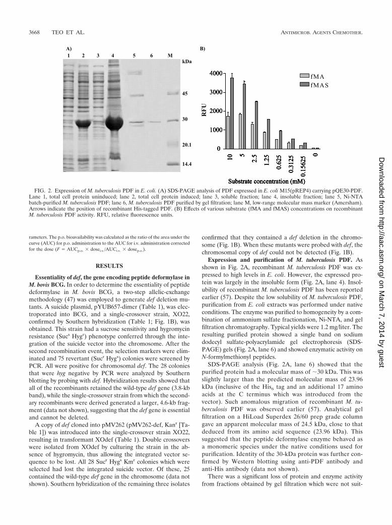
Expression and purification of M. tuberculosis PDF. Asshown in Fig. 2A, recombinant M. tuberculosis PDF was ex-pressed to high levels in E. coli. However, the expressed pro-tein was largely in the insoluble form (Fig. 2A, lane 4). Insol-ubility of recombinant M. tuberculosis PDF has been reportedearlier (57). Despite the low solubility of M. tuberculosis PDF,purification from E. coli extracts was performed under nativeconditions. The enzyme was purified to homogeneity by a com-bination of ammonium sulfate fractionation, Ni-NTA, and gelfiltration chromatography. Typical yields were 1.2 mg/liter. Theresulting purified protein showed a single band on sodiumdodecyl sulfate-polyacrylamide gel electrophoresis (SDS-PAGE) gels (Fig. 2A, lane 6) and showed enzymatic activity onN-formylmethionyl peptides.
SDS-PAGE analysis (Fig. 2A, lane 6) showed that thepurified protein had a molecular mass of �30 kDa. This wasslightly larger than the predicted molecular mass of 23.96kDa (inclusive of the His6 tag and an additional 17 aminoacids at the C terminus which was introduced from thevector). Such anomalous migration of recombinant M. tu-berculosis PDF was observed earlier (57). Analytical gelfiltration on a HiLoad Superdex 26/60 prep grade columngave an apparent molecular mass of 24.5 kDa, close to thatdeduced from its amino acid sequence (23.96 kDa). Thissuggested that the peptide deformylase enzyme behaved asa monomeric species under the native conditions used forpurification. Identity of the 30-kDa protein was further con-firmed by Western blotting using anti-PDF antibody andanti-His antibody (data not shown).
There was a significant loss of protein and enzyme activityfrom fractions obtained by gel filtration which were not suit-
FIG. 2. Expression of M. tuberculosis PDF in E. coli. (A) SDS-PAGE analysis of PDF expressed in E. coli M15(pREP4) carrying pQE30-PDF.Lane 1, total cell protein uninduced; lane 2, total cell protein induced; lane 3, soluble fraction; lane 4, insoluble fraction; lane 5, Ni-NTAbatch-purified M. tuberculosis PDF; lane 6, M. tuberculosis PDF purified by gel filtration; lane M, low-range molecular mass marker (Amersham).Arrows indicate the position of recombinant His-tagged PDF. (B) Effects of various substrate (fMA and fMAS) concentrations on recombinantM. tuberculosis PDF activity. RFU, relative fluorescence units.
3668 TEO ET AL. ANTIMICROB. AGENTS CHEMOTHER.
on March 7, 2014 by guest
http://aac.asm.org/
Dow
nloaded from

able for large-scale screens. Instead, as reported earlier with E.coli PDF (10), batch-purified M. tuberculosis PDF (Fig. 2A,lane 5) was used for the screening of the large library. After thebackground (induced E. coli M15 cell extracts transformedwith the pQE30 vector, treated in a similar way) was sub-tracted, the enzyme activity in the batch was very specific andcould be completely inhibited by PDF-I. The hits were recon-firmed using the purified enzyme.
Biochemical characterization of E. coli-expressed M. tuber-culosis PDF. Biochemical characterization was done with bothpure and batch-purified enzyme, and results were found to besimilar. During optimization of substrate concentrations offMA and fMAS, the latter was found to be a more optimalsubstrate for M. tuberculosis PDF (Fig. 2B). A prominent fea-ture of the kinetics observed for both substrates tested was thatthe enzyme showed no saturation kinetics up to �10 mMsubstrate, and concentrations above 12.5 mM fMAS were in-hibitory for M. tuberculosis PDF. This finding was similar tothat from an earlier report (57) (Fig. 2B). Subsequently, 2 mMfMAS was used for evaluating other parameters of the enzymeassays. Velocity versus substrate concentration plots typicallyproduced a straight line, indicating that the Km values for thesesubstrates are �5 mM (data not shown).
The enzyme displayed a pH optimum of 7.5, retaining lessthan 20% of its optimal activity at extreme pH, for example, atpH 5.5 or pH 10 (data not shown). Ionic strength had a sig-nificant detrimental effect on M. tuberculosis PDF activity atconcentrations greater than 0.1 M KCl (data not shown). Thisresult agreed with earlier results with M. tuberculosis PDF (57).Co2�, Ni2�, and Zn2� were partially inhibitory, while Cu2�
ions completely abolished PDF activity (data not shown). Ear-
lier reports had indicated that Co2�, Ni2�, and Zn2� had noeffect on M. tuberculosis PDF enzyme activity (57).
Enzyme inhibition studies. Several inhibitors from an avail-able PDF-I library (27) were tested in an M. tuberculosis PDFenzyme assay (Table 2). The most active compound, PDF-611,or LBK-611 (15), had an IC50 of 69.5 nM, while a similarcompound, PDF-709, had an IC50 of 90 nM. Two other knownantibacterial PDF-I, actinonin (10) and BB-3497 (12, 14),showed potent inhibition of the M. tuberculosis PDF enzyme,with IC50s of 50.5 and 23.9 nM, respectively.
In order to assess the issue of selectivity, the compoundswere also tested against purified Homo sapiens PDF andhuman matrilysin (MMP-7). Matrilysin belongs to the ma-trix metalloproteinase (MMP) family of zinc-containing en-zymes related to PDF (44). PDF-611 (LBK-611) and PDF-709 inhibited H. sapiens PDF with IC50s of 115 nM and 147nM, respectively (Table 2). BB-3497 had an IC50 of 8 nMwith H. sapiens PDF (45) and actinonin an IC50 of 43 nM(32). However, no compounds displayed significant inhibi-tion of MMP-7 with concentrations up to 10 �M or inhibitedthe growth of the K562 cell line with concentrations up to 10�M, except actinonin, which had an IC50 of 8.5 �M (datanot shown).
Antimycobacterial activities of PDF-611 (LBK-611) andPDF-709. PDF-611 (LBK-611) was the most potent in terms ofantimycobacterial activity, with a MIC90 value of 0.78 �M (0.25�g/ml) (Table 2). Both PDF-709 and BB-3497 displayed goodantimycobacterial activities against growing M. bovis BCG cul-tures (MIC90s of 6.25 �M and 3.125 �M, respectively); how-ever, actinonin showed a poor antimycobacterial effect, with aMIC90 of �25 �M (�16 �g/ml). Based on the results obtained
TABLE 2. Effects of PDF-I on M. tuberculosis PDF and H. sapiens PDF enzyme activities(IC50s) and antibacterial activities (MIC90s) in M. bovis BCG
Compound Structure
IC50 (nM)MIC90 (�M) inM. bovis BCGa Reference(s)M. tuberculosis
PDFaH. sapiens
Actinonin 50.5 7.7 43 �25 12, 32
PDF-709 90.0 4.2 147a 6.25 27
PDF-611 (LBK-611) 69.5 0.7 115a
0.78 15
BB-3497 23.9 7.5 8 3.125 12, 45
a This work.
VOL. 50, 2006 PDF INHIBITORS AS ANTIMYCOBACTERIALS 3669
on March 7, 2014 by guest
http://aac.asm.org/
Dow
nloaded from

from M. tuberculosis PDF inhibition and antimycobacterialstudies, the compounds PDF-709 and PDF-611 (LBK-611)were selected for further analyses.
The bactericidal effects of PDF-611 (LBK-611) and PDF-709 on M. bovis BCG over 4 days were determined throughtime-kill studies (data not shown). At both 1� and 10� MICfor each compound, there was �1 log unit decrease in viablecounts, demonstrating a bacteriostatic effect for the time pe-riod tested. However, on increasing incubation time with theinhibitor, higher bacterial killing was observed at the sameconcentrations (data not shown), indicating that the PDF-I areslow acting. PDF-611 (LBK-611) was also shown to be effectiveagainst several clinical isolates of M. tuberculosis. The com-pound demonstrated low MICs (�1 �g/ml) for several multi-drug-resistant clinical TB isolates (Table 3).
Spontaneous mutation rates and analysis of PDF-I-resis-tant M. bovis BCG isolates. The frequency of spontaneousresistance development was evaluated using a concentrationof 10� MIC. PDF-611- and PDF-709-resistant mutantsarose at frequencies of �5 � 107 in M. bovis BCG. Thesemutation frequencies were lower than the development ofresistance to INH, which occurred at a frequency of 7 �105, but higher than that for RIF, which was �1 � 108
(data not shown).The development of PDF-I resistance in other bacteria can
progress through at least two main mechanisms, mutations inthe def gene or mutations in the formyl methyltransferase gene(fmt), which catalyzes the formylation of methionylated initia-tor tRNA (12, 21, 34, 35). Twenty-nine spontaneous PDF-I-resistant M. bovis BCG mutants were characterized. The com-plete def and fmt genes from these isolates were sequenced.None of the isolates had mutations in the def gene. Of the 29isolates, 16 were found to have missense mutations in the fmtgene (Table 4). In the remaining 13 isolates, no changes weredetected in the def and fmt genes. It was noted that indepen-dently isolated mutants had the same amino acid mutation(fmt-32a, fmt-32b, and fmt-32c had the T32N mutation; fmt-222a and fmt-222b had the E222K mutation; and fmt-117a andfmt-117b had the G117C mutation). Nonconserved amino acidchanges were observed for half of the mutants. About half of
the formyl methyltransferase (FMT) mutants had mutationsthat mapped to regions around the active site, deduced from ahomology model of M. tuberculosis FMT based on E. coli FMTcrystal structure (Table 4; also see Fig. S3 in the supplementalmaterial).
For further studies of mutant strains, fmt-192, fmt-276, fmt-104a, and fmt-104b were characterized for growth and antimi-crobial susceptibility, as all of them were generated againstPDF-709. Two isolates, fmt-192 and fmt-104a, had slower dou-bling times (32 h and 35 h, respectively) than wild-type M. bovisBCG (23 h), while fmt-276 and fmt-104b had doubling times(22 h and 24 h, respectively) similar to that of wild-type M.bovis BCG (23 h). In MIC testing, all four FMT mutants werefound to have at least a 16-fold increase in MIC (�100 �M)against both PDF-611 (LBK-611) and PDF-709. However, themutants retained susceptibility to other TB drugs, like RIF andSTR, and unrelated drugs, like LZD and ERY. Target speci-ficity was further confirmed by expression of extra chromo-somal copies of the M. tuberculosis def gene in BCG, resultingin a 16-fold increase in the MIC for PDF-611 (LBK-611) (datanot shown). For comparative purposes, a mutant without amutation in the def or the fmt gene was included in the study.This mutant had a slightly slower doubling time (26 h) thanwild-type M. bovis BCG, with a susceptibility profile similar tothat of the fmt mutants. All fmt mutants regained susceptibilityto PDF-I when a wild-type fmt gene was supplied in trans,confirming that the resistance arose through a mutation in fmt(data not shown).
Pharmacokinetic studies with mice. Clearance of PDF-709and PDF-611 (LBK-611) in mouse serum was measured afterthe compounds were administered i.v. and p.o. The concentra-tions in serum at different times were used to calculate thecorresponding pharmacokinetic parameters presented in Table5. PDF-611 (LBK-611) had a lower half-life (0.5 h) than PDF-709 (1.6 h) after i.v. administration. However, PDF-611 (LBK-611) had a higher half-life (3.7 h) than PDF-709 (2.7 h) afterp.o. administration. Both compounds had similar bioavailabili-ties (�40%).
TABLE 3. MICs of PDF-611 (LBK-611) against variousM. tuberculosis isolates
StrainaMIC (�g/ml)b
PDF-611 BB-3497 Gatifloxacin
H37Rv (ATCC 27294) 0.125–0.5 0.125–0.5 0.03–0.06Beijing MDR 0.125 0.125–0.25 0.03–0.06W-Beijing MDR 0.125 0.125 0.25Beijing F29 MDR 0.03–0.25 0.03–0.5 0.015Inhr SDR 0.06–0.125 0.125 0.015Strr SDR 0.5 1.0 0.03Rifr SDR 0.25 1.2 0.015Pzar SDR 0.5 1.0 0.03Clinical MDR 0.06–1.0 0.06–2 �0.008–0.06
a MDR, multidrug resistance; SDR, single-drug resistance; Pza, pyrazinamide.The clinical MDR isolates are 10 clinical isolates which were described as MDR;however, a detailed susceptibility pattern for TB drugs is not available.
b All MIC determinations were done twice. Single MIC values represent iden-tical values in both experiments, while multiple values indicate different results intwo experiments. For clinical MDR, the multiple values indicate the range ofMIC values for 10 different clinical isolates.
TABLE 4. Bioinformatics analysis of fmt mutants
Mutant Mutation inFMT
Predicted structurallocationa
Predicted to be within8 Å of fMet-tRNA
binding sitea
fmt-100 P100L Loop Nofmt-222a E222K Helix Nofmt-53 A53V Helix Nofmt-32a T32N Loop Yesfmt-32b T32N Loop Yesfmt-230 P230S Loop Yesfmt-105 V105F Sheet Yesfmt-32c T32N Loop Yesfmt-192 R192P Loop Nofmt-276 T276A Loop Nofmt-104a W104L Loop Nofmt-104b W104L Loop Nofmt-117a G117C Sheet Yesfmt-124 A124G Helix Yesfmt-222b E222K Helix Nofmt-117b G117C Sheet Yes
a Deduced from a model of M. tuberculosis FMT, based on the E. coli FMTcrystal structure (59).
3670 TEO ET AL. ANTIMICROB. AGENTS CHEMOTHER.
on March 7, 2014 by guest
http://aac.asm.org/
Dow
nloaded from

DISCUSSION
M. tuberculosis PDF was earlier proposed as a potential drugtarget (14) since BB-3497, a PDF inhibitor, had potent in vitroactivity against M. tuberculosis. The gene encoding M. tubercu-losis PDF had also been shown to be one of the genes requiredfor optimal growth by M. tuberculosis through transposon sitehybridization (56). In this work, the failure to obtain def dele-tion mutants in M. bovis BCG strongly suggested that thedeletion of def was lethal. After the second recombinationevent, the chromosomal wild-type def gene could be deletedonly in the presence of an extra chromosomal complementingcopy of def. Taken together, these results show that the defgene is essential in M. bovis BCG.
It has been observed previously with E. coli PDF that themetal ions and reaction conditions (substrate and type of as-say) can dramatically change the activity and properties of theenzyme (10, 38, 49, 50). During biochemical characterizationof M. tuberculosis PDF, we observed some differences from anearlier published work (57, 58). These differences could possi-bly be attributed to the difference in assay protocol and ex-pression/purification conditions followed in this study com-pared to the earlier published work. In the earlier work, theinvestigators had not purified the enzyme in the presence ofexcess Ni�2 and enzyme activity was determined using fMA asthe substrate in a trinitrobenzenesulfonic acid assay (57).Moreover, the assay temperatures were also different (roomtemperature in this study versus 30°C in the previous study).
Structure-activity relationship studies of actinonin and otherN-alkyl urea hydroxamates (9, 27) indicate that efficaciousPDF-I share common structural features, the first being ametal-chelating group, usually a hydroxamate/reverse hydrox-amate, and the second being an n-alkyl butyl residue at the P1�position that mimics the methionine side chain (4). The PDF-Itested in this work were reverse hydroxamates, carrying ann-butyl residue at the P1� position (Table 2). This class ofcompounds inhibit M. tuberculosis PDF with IC50s of �100 nMand are bacteriostatic against M. bovis BCG, as was observedpreviously for other PDF-I against both Staphylococcus aureusand E. coli (3, 12, 35). The frequency of resistance to this classof inhibitors in M. bovis BCG is similar to what was previouslyreported for S. aureus (12, 35) and E. coli (3, 12).
Spontaneous PDF-I-resistant isolates in M. bovis BCG dem-onstrated elevated MICs specifically towards the PDF-I andnot towards other drug classes. In our work, the resistancearose through missense mutations in the fmt gene in 16 out of29 mutants, while the rest had no mutation in either the def orthe fmt gene. Several fmt mutants in this study were found to
have mutations in residues important for function (by use ofthe homology model with E. coli FMT crystal structure) (Table4; also see Fig. S3 in the supplemental material) (23, 26, 59).Three independently isolated mutants (fmt-32a, fmt-32b, andfmt-32c [with the T32N mutation]) had a Ser-to-Ala mutationin a region involved in the recognition of the initiator tRNA(53, 54). Mutant fmt-53 (with the A53V mutation) lies justoutside this region. Several mutants with mutations in theC-terminal region involved in the recognition of the tRNA byFMT were obtained (fmt-222a, fmt-222b, fmt-230, and fmt-276) (23). Three mutants, fmt-104a and fmt-104b (with theW104L mutation) and fmt-100 (with the P100L mutation),mapping close to conserved residues (Asn-106, His-108, andAsp-144) are predicted to participate in the catalytic steps (1,28) (see Fig. S3 in the supplemental material). Without for-mylation assays of Met-tRNAfMet, we are unsure about theextent to which different fmt mutations affect transformylaseactivity. Further studies are needed to understand the func-tional effect of the mutations.
Mutants that did not have mutations in the def or the fmtgene could be efflux pump mutants, as observed for E. coli andHaemophilus influenzae (12, 15). However, the actual mecha-nism of resistance for these mutants remains to be elucidated.Thus, in the case of mycobacteria, more than one mechanismis operative to confer resistance to PDF-I.
Recently, several eukaryotic PDF enzymes have been iden-tified and characterized, e.g., from Arabidopsis thaliana (22),Plasmodium falciparum (5), and mitochondria of H. sapiens(22, 32, 33, 45, 60). The mRNA for H. sapiens PDF has beenshown to be expressed at similar levels in all types of humantissues (22), with the enzyme being an active deformylase bothin vitro and in vivo (32, 33, 60). Actinonin has been found toinhibit cell growth in various human tumor cell lines and intumor models (32, 33, 63). In addition, two well-characterizedbacterial PDF inhibitors, BB-3497 and actinonin (also used inthis study), inhibit H. sapiens PDF at nanomolar concentra-tions similar to those at which its bacterial counterpart is in-hibited (32, 45).
However, despite the inhibition of cell-free H. sapiensPDF and antiproliferative effects on tumor cell lines, a num-ber of normal cell lines were found to be resistant to acti-nonin (33) and BB-3497 (45). Actinonin has been well tol-erated in mice at doses of up to 400 mg/kg (24), andsuccessors of BB-3497 (BB-83698) have been tested in hu-mans at levels of up to 475 mg without any clinically signif-icant adverse effects (52). Furthermore, recent biochemicalstudies with H. sapiens PDF showed that the enzyme had
TABLE 5. Pharmacokinetic parametersa of PDF-611 (LBK-611) and PDF-709 in mice
Compound Type ofadministration
Dose(mg/kg)
Cmax(ng/ml) Tmax (h) AUCinf
(ng · h/ml) t1/2 (h) CL(ml/min/kg)
Vss(liter/kg)
Bioavailability(%)
PDF-611(LBK-611) i.v. 1.000 603.6 0.083 253.5 0.500 65.8 3.0p.o. 5.000 194.3 0.250 566.0 3.70 45.0
PDF-709 i.v. 1.000 902.0 0.083 145.6 1.600 114.5 3.9p.o. 5.000 186.0 0.500 293.0 2.700 40.0
a Cmax, maximum concentration of drug in serum; Tmax, time to maximum concentration of drug in serum; AUCinf, area under the curve extrapolated to �; t1/2,half-life; CL, clearance; Vss, volume of distribution at steady state.
VOL. 50, 2006 PDF INHIBITORS AS ANTIMYCOBACTERIALS 3671
on March 7, 2014 by guest
http://aac.asm.org/
Dow
nloaded from

significantly lower activity (kcat/Km) than the bacterial en-zyme (E. coli PDF) (32, 33, 45, 52, 60).
In addition, the unambiguous localization of H. sapiens PDFin the mitochondria is important, as evidence suggests that themitochondria of tumor cells may be more sensitive to mito-chondrial insult (6, 48). There are examples of drugs selectivelytoxic to cancer cells working through the inhibition of mito-chondrial respiration (19, 43). Mitochondrial membranes areless permeable to small molecules than most other membranesand act as an efficient supplementary filter for many drugs. Forthis reason, a number of antibiotics (tetracycline, macrolides,etc.) that inhibit mitochondrial targets (16, 30, 31, 37) arenevertheless currently used in human medicine. Thus, acti-nonin’s inhibition of H. sapiens PDF, resulting in mitochon-drial disruption, could have a similar tumor specificity (33). Amajor, striking difference between H. sapiens PDF (also mousePDF) and bacterial PDF (E. coli PDF) is in the highly con-served EGCLS motif (motif III), where leucine is mutated intoglutamic acid in mammalian PDF (Glu-173 in H. sapiens PDF),which results in the low activity of H. sapiens PDF (60). Fromthe sequence similarity, M. tuberculosis PDF is 11% identicaloverall and has only 6% identity at the active site with H.sapiens PDF. These differences can be exploited to make com-pounds with decreased affinity for H. sapiens PDF withoutaffecting their potency with respect to M. tuberculosis PDF. Inthis study, the PDF-I do not show selectivity towards M. tuber-culosis PDF, but they were considered for further studies dueto their tolerability in mice, in addition to the lack of effect onMMP-7 and the K562 cell line up to 10 �M (�100-fold-higherconcentration than H. sapiens PDF IC50).
In order to ensure treatment compliance, antituberculosistherapy requires an oral dosing regimen of once a day or less.The oral bioavailabilities of PDF-611 (LBK-611) and PDF-709were in the range of 40% in the mouse, indicating potentialoral administration in humans. This was also reinforced by therelatively long half-lives of 3.7 h and 2.7 h, respectively, fol-lowing oral administration. Assuming interspecies scalingwithin normal range, such a half-life in the mouse would sup-port once-daily oral dosing in humans. These figures constitutean improvement over previous half-life reports for PDF inhib-itors (27). Oral bioavailabilities of PDF-611 (LBK-611) andPDF-709 are also a clear improvement over those reported forother PDF inhibitors of the class N-alkyl urea hydroxamic acids(0.1 to 3.2%) (27) and that for BB-83698 (i.v. application only)(52). The total clearance levels were moderate for both com-pounds, i.e., 66 and 114 ml/min/kg for PDF-611 (LBK-611) andPDF-709, respectively, which lie in the range of mouse liverblood flow (90 ml/min/kg). Thus, this group of PDF inhibitorshas the potential to be developed further as a new class ofantituberculosis agent.
ACKNOWLEDGMENTS
We thank Sabine Daugelat for providing advice on the knockout pro-cedure and Marty Pavelka for pYUB657. We thank Brigitte Gicquel forproviding pJEM15, which was used for overexpression studies.
REFERENCES
1. Almassy, R. J., C. A. Janson, C. C. Kan, and Z. Hostomska. 1992. Structuresof apo and complexed Escherichia coli glycinamide ribonucleotide trans-formylase. Proc. Natl. Acad. Sci. USA 89:6114–6118.
2. Apfel, C., D. W. Banner, D. Bur, M. Dietz, T. Hirata, C. Hubschwerlen, H.
Locher, M. G. Page, W. Pirson, G. Rosse, and J. L. Specklin. 2000. Hydrox-amic acid derivatives as potent peptide deformylase inhibitors and antibac-terial agents. J. Med. Chem. 43:2324–2331.
3. Apfel, C. M., H. Locher, S. Evers, B. Takacs, C. Hubschwerlen, W. Pirson,M. G. Page, and W. Keck. 2001. Peptide deformylase as an antibacterial drugtarget: target validation and resistance development. Antimicrob. AgentsChemother. 45:1058–1064.
4. Boularot, A., C. Giglione, I. Artaud, and T. Meinnel. 2004. Structure-activityrelationship analysis and therapeutic potential of peptide deformylase inhib-itors. Curr. Opin. Investig. Drugs 5:809–822.
5. Bracchi-Ricard, V., K. T. Nguyen, Y. Zhou, P. T. Rajagopalan, D.Chakrabarti, and D. Pei. 2001. Characterization of an eukaryotic peptidedeformylase from Plasmodium falciparum. Arch. Biochem. Biophys. 396:162–170.
6. Cavalli, L. R., and B. C. Liang. 1998. Mutagenesis, tumorigenicity, andapoptosis: are the mitochondria involved? Mutat. Res. 398:19–26.
7. Chan, M. K., W. Gong, P. T. Rajagopalan, B. Hao, C. M. Tsai, and D. Pei.1997. Crystal structure of the Escherichia coli peptide deformylase. Bio-chemistry 36:13904–13909.
8. Chan, P. F., K. M. O’Dwyer, L. M. Palmer, J. D. Ambrad, K. A. Ingraham,C. So, M. A. Lonetto, S. Biswas, M. Rosenberg, D. J. Holmes, and M.Zalacain. 2003. Characterization of a novel fucose-regulated promoter(PfcsK) suitable for gene essentiality and antibacterial mode-of-action studiesin Streptococcus pneumoniae. J. Bacteriol. 185:2051–2058.
9. Chen, D., C. Hackbarth, Z. J. Ni, C. Wu, W. Wang, R. Jain, Y. He, K.Bracken, B. Weidmann, D. V. Patel, J. Trias, R. J. White, and Z. Yuan. 2004.Peptide deformylase inhibitors as antibacterial agents: identification ofVRC3375, a proline-3-alkylsuccinyl hydroxamate derivative, by using an in-tegrated combinatorial and medicinal chemistry approach. Antimicrob.Agents Chemother. 48:250–261.
10. Chen, D. Z., D. V. Patel, C. J. Hackbarth, W. Wang, G. Dreyer, D. C. Young,P. S. Margolis, C. Wu, Z. J. Ni, J. Trias, R. J. White, and Z. Yuan. 2000.Actinonin, a naturally occurring antibacterial agent, is a potent deformylaseinhibitor. Biochemistry 39:1256–1262.
11. Clamp, M., J. Cuff, S. M. Searle, and G. J. Barton. 2004. The Jalview Javaalignment editor. Bioinformatics 20:426–427.
12. Clements, J. M., R. P. Beckett, A. Brown, G. Catlin, M. Lobell, S. Palan, W.Thomas, M. Whittaker, S. Wood, S. Salama, P. J. Baker, H. F. Rodgers, V.Barynin, D. W. Rice, and M. G. Hunter. 2001. Antibiotic activity and char-acterization of BB-3497, a novel peptide deformylase inhibitor. Antimicrob.Agents Chemother. 45:563–570.
13. Cuff, J. A., and G. J. Barton. 1999. Evaluation and improvement of multiplesequence methods for protein secondary structure prediction. Proteins 34:508–519.
14. Cynamon, M. H., E. Alvirez-Freites, and A. E. Yeo. 2004. BB-3497, a peptidedeformylase inhibitor, is active against Mycobacterium tuberculosis. J. An-timicrob. Chemother. 53:403–405.
15. Dean, C. R., S. Narayan, D. M. Daigle, J. L. Dzink-Fox, X. Puyang, K. R.Bracken, K. E. Dean, B. Weidmann, Z. Yuan, R. Jain, and N. S. Ryder. 2005.Role of the AcrAB-TolC efflux pump in determining susceptibility of Hae-mophilus influenzae to the novel peptide deformylase inhibitor LBM415.Antimicrob. Agents Chemother. 49:3129–3135.
16. de Vries, H., A. J. Arendzen, and A. M. Kroon. 1973. The interference of themacrolide antibiotics with mitochondrial protein synthesis. Biochim. Bio-phys. Acta 331:264–275.
17. Dye, C. 2006. Global epidemiology of tuberculosis. Lancet 367:938–940.18. Dye, C., C. J. Watt, D. M. Bleed, S. M. Hosseini, and M. C. Raviglione. 2005.
Evolution of tuberculosis control and prospects for reducing tuberculosisincidence, prevalence, and deaths globally. JAMA 293:2767–2775.
19. Fantin, V. R., M. J. Berardi, L. Scorrano, S. J. Korsmeyer, and P. Leder.2002. A novel mitochondriotoxic small molecule that selectively inhibitstumor cell growth. Cancer Cell 2:29–42.
20. Franzblau, S. G., R. S. Witzig, J. C. McLaughlin, P. Torres, G. Madico, A.Hernandez, M. T. Degnan, M. B. Cook, V. K. Quenzer, R. M. Ferguson, andR. H. Gilman. 1998. Rapid, low-technology MIC determination with clinicalMycobacterium tuberculosis isolates by using the microplate Alamar Blueassay. J. Clin. Microbiol. 36:362–366.
21. Giglione, C., and T. Meinnel. 2001. Resistance to anti-peptide deformylasedrugs. Expert Opin. Ther. Targets 5:415–418.
22. Giglione, C., A. Serero, M. Pierre, B. Boisson, and T. Meinnel. 2000. Iden-tification of eukaryotic peptide deformylases reveals universality of N-termi-nal protein processing mechanisms. EMBO J. 19:5916–5929.
23. Gite, S., Y. Li, V. Ramesh, and U. L. RajBhandary. 2000. Escherichia colimethionyl-tRNA formyltransferase: role of amino acids conserved in thelinker region and in the C-terminal domain on the specific recognition of theinitiator tRNA. Biochemistry 39:2218–2226.
24. Gordon, J. J., B. K. Kelly, and G. A. Miller. 1962. Actinonin: an antibioticsubstance produced by an actinomycete. Nature 195:701–702.
25. Groche, D., A. Becker, I. Schlichting, W. Kabsch, S. Schultz, and A. F.Wagner. 1998. Isolation and crystallization of functionally competent Esch-erichia coli peptide deformylase forms containing either iron or nickel in theactive site. Biochem. Biophys. Res. Commun. 246:342–346.
3672 TEO ET AL. ANTIMICROB. AGENTS CHEMOTHER.
on March 7, 2014 by guest
http://aac.asm.org/
Dow
nloaded from

26. Guillon, J.-M., Y. Mechulam, J.-M. Schmitter, S. Blanquet, and G. Fayat.1992. Disruption of the gene for Met-tRNAf
Metformyltransferase severelyimpairs growth of Escherichia coli. J. Bacteriol. 174:4294–4301.
27. Hackbarth, C. J., D. Z. Chen, J. G. Lewis, K. Clark, J. B. Mangold, J. A.Cramer, P. S. Margolis, W. Wang, J. Koehn, C. Wu, S. Lopez, G. Withers III,H. Gu, E. Dunn, R. Kulathila, S.-H. Pan, W. L. Porter, J. Jacobs, J. Trias,D. V. Patel, B. Weidmann, R. J. White, and Z. Yuan. 2002. N-Alkyl ureahydroxamic acids as a new class of peptide deformylase inhibitors withantibacterial activity. Antimicrob. Agents Chemother. 46:2752–2764.
28. Klein, C., P. Chen, J. H. Arevalo, E. A. Stura, A. Marolewski, M. S. Warren,S. J. Benkovic, and I. A. Wilson. 1995. Towards structure-based drug design:crystal structure of a multisubstrate adduct complex of glycinamide ribonu-cleotide transformylase at 1.96 A resolution. J. Mol. Biol. 249:153–175.
29. Kremer, L. S., and G. S. Besra. 2002. Current status and future developmentof antitubercular chemotherapy. Expert Opin. Investig. Drugs 11:1033–1049.
30. Kroon, A. M., B. H. Dontje, M. Holtrop, and C. Van den Bogert. 1984. Themitochondrial genetic system as a target for chemotherapy: tetracyclines ascytostatics. Cancer Lett. 25:33–40.
31. Kroon, A. M., and C. Van den Bogert. 1983. Antibacterial drugs and theirinterference with the biogenesis of mitochondria in animal and human cells.Pharm. Weekbl. Sci. 5:81–87.
32. Lee, M. D., C. Antczak, Y. Li, F. M. Sirotnak, W. G. Bornmann, and D. A.Scheinberg. 2003. A new human peptide deformylase inhibitable by acti-nonin. Biochem. Biophys. Res. Commun. 312:309–315.
33. Lee, M. D., Y. She, M. J. Soskis, C. P. Borella, J. R. Gardner, P. A. Hayes,B. M. Dy, M. L. Heaney, M. R. Philips, W. G. Bornmann, F. M. Sirotnak, andD. A. Scheinberg. 2004. Human mitochondrial peptide deformylase, a newanticancer target of actinonin-based antibiotics. J. Clin. Investig. 114:1107–1116.
34. Margolis, P., C. Hackbarth, S. Lopez, M. Maniar, W. Wang, Z. Yuan, R.White, and J. Trias. 2001. Resistance of Streptococcus pneumoniae todeformylase inhibitors is due to mutations in defB. Antimicrob. AgentsChemother. 45:2432–2435.
35. Margolis, P. S., C. J. Hackbarth, D. C. Young, W. Wang, D. Chen, Z. Yuan,R. White, and J. Trias. 2000. Peptide deformylase in Staphylococcus aureus:resistance to inhibition is mediated by mutations in the formyltransferasegene. Antimicrob. Agents Chemother. 44:1825–1831.
36. Mazel, D., S. Pochet, and P. Marliere. 1994. Genetic characterization ofpolypeptide deformylase, a distinctive enzyme of eubacterial translation.EMBO J. 13:914–923.
37. McKee, E. E., M. Ferguson, A. T. Bentley, and T. A. Marks. 2006. Inhibitionof mammalian mitochondrial protein synthesis by oxazolidinones. Antimi-crob. Agents Chemother. 50:2042–2049.
38. Meinnel, T., and S. Blanquet. 1995. Enzymatic properties of Escherichia colipeptide deformylase. J. Bacteriol. 177:1883–1887.
39. Meinnel, T., S. Blanquet, and F. Dardel. 1996. A new subclass of the zincmetalloproteases superfamily revealed by the solution structure of peptidedeformylase. J. Mol. Biol. 262:375–386.
40. Meinnel, T., C. Lazennec, and S. Blanquet. 1995. Mapping of the active sitezinc ligands of peptide deformylase. J. Mol. Biol. 254:175–183.
41. Meinnel, T., C. Lazennec, S. Villoing, and S. Blanquet. 1997. Structure-function relationships within the peptide deformylase family. Evidence for aconserved architecture of the active site involving three conserved motifs anda metal ion. J. Mol. Biol. 267:749–761.
42. Meinnel, T., Y. Mechulam, and S. Blanquet. 1993. Methionine as translationstart signal: a review of the enzymes of the pathway in Escherichia coli.Biochimie 75:1061–1075.
43. Modica-Napolitano, J. S., K. Koya, E. Weisberg, B. T. Brunelli, Y. Li, andL. B. Chen. 1996. Selective damage to carcinoma mitochondria by therhodacyanine MKT-077. Cancer Res. 56:544–550.
44. Muri, E. M., M. J. Nieto, R. D. Sindelar, and J. S. Williamson. 2002.
Hydroxamic acids as pharmacological agents. Curr. Med. Chem. 9:1631–1653.
45. Nguyen, K. T., X. Hu, C. Colton, R. Chakrabarti, M. X. Zhu, and D. Pei.2003. Characterization of a human peptide deformylase: implications forantibacterial drug design. Biochemistry 42:9952–9958.
46. Nunn, P., B. Williams, K. Floyd, C. Dye, G. Elzinga, and M. Raviglione.2005. Tuberculosis control in the era of HIV. Nat. Rev. Immunol. 5:819–826.
47. Pavelka, M. S., Jr., and W. R. Jacobs, Jr. 1999. Comparison of the construc-tion of unmarked deletion mutations in Mycobacterium smegmatis, Mycobac-terium bovis bacillus Calmette-Guerin, and Mycobacterium tuberculosisH37Rv by allelic exchange. J. Bacteriol. 181:4780–4789.
48. Penta, J. S., F. M. Johnson, J. T. Wachsman, and W. C. Copeland. 2001.Mitochondrial DNA in human malignancy. Mutat. Res. 488:119–133.
49. Ragusa, S., S. Blanquet, and T. Meinnel. 1998. Control of peptide deformylaseactivity by metal cations. J. Mol. Biol. 280:515–523.
50. Rajagopalan, P. T., A. Datta, and D. Pei. 1997. Purification, characterization,and inhibition of peptide deformylase from Escherichia coli. Biochemistry36:13910–13918.
51. Rajagopalan, P. T., and D. Pei. 1998. Oxygen-mediated inactivation of pep-tide deformylase. J. Biol. Chem. 273:22305–22310.
52. Ramanathan-Girish, S., J. McColm, J. M. Clements, P. Taupin, S. Barrowcliffe,J. Hevizi, S. Safrin, C. Moore, G. Patou, H. Moser, A. Gadd, U. Hoch, V. Jiang,D. Lofland, and K. W. Johnson. 2004. Pharmacokinetics in animals and humansof a first-in-class peptide deformylase inhibitor. Antimicrob. Agents Chemother.48:4835–4842.
53. Ramesh, V., S. Gite, Y. Li, and U. L. RajBhandary. 1997. Suppressor muta-tions in Escherichia coli methionyl-tRNA formyltransferase: role of a 16-amino acid insertion module in initiator tRNA recognition. Proc. Natl. Acad.Sci. USA 94:13524–13529.
54. Ramesh, V., S. Gite, and U. L. RajBhandary. 1998. Functional interaction ofan arginine conserved in the sixteen amino acid insertion module of Esch-erichia coli methionyl-tRNA formyltransferase with determinants for formyl-ation in the initiator tRNA. Biochemistry 37:15925–15932.
55. Sambrook, J., E. F. Fritsch, and T. Maniatis. 1989. Molecular cloning: alaboratory manual, 2nd ed. Cold Spring Harbor Laboratory Press, ColdSpring Harbor, N.Y.
56. Sassetti, C. M., D. H. Boyd, and E. J. Rubin. 2003. Genes required formycobacterial growth defined by high density mutagenesis. Mol. Microbiol.48:77–84.
57. Saxena, R., and P. K. Chakraborti. 2005. Identification of regions involvedin enzymatic stability of peptide deformylase of Mycobacterium tuberculosis.J. Bacteriol. 187:8216–8220.
58. Saxena, R., and P. K. Chakraborti. 2005. The carboxy-terminal end of thepeptide deformylase from Mycobacterium tuberculosis is indispensable forits enzymatic activity. Biochem. Biophys. Res. Commun. 332:418–425.
59. Schmitt, E., S. Blanquet, and Y. Mechulam. 1996. Structure of crystallineEscherichia coli methionyl-tRNA(f) Met formyltransferase: comparison withglycinamide ribonucleotide formyltransferase. EMBO J. 15:4749–4758.
60. Serero, A., C. Giglione, A. Sardini, J. Martinez-Sanz, and T. Meinnel. 2003.An unusual peptide deformylase features in the human mitochondrial N-terminal methionine excision pathway. J. Biol. Chem. 278:52953–52963.
61. Solbiati, J., A. Chapman-Smith, J. L. Miller, C. G. Miller, and J. E. Cronan,Jr. 1999. Processing of the N termini of nascent polypeptide chains requiresdeformylation prior to methionine removal. J. Mol. Biol. 290:607–614.
62. Stover, C. K., V. F. de la Cruz, T. R. Fuerst, J. E. Burlein, L. A. Benson, L. T.Bennett, G. P. Bansal, J. F. Young, M. H. Lee, G. F. Hatfull, et al. 1991. Newuse of BCG for recombinant vaccines. Nature 351:456–460.
63. Xu, Y., L. T. Lai, J. L. Gabrilove, and D. A. Scheinberg. 1998. Antitumoractivity of actinonin in vitro and in vivo. Clin. Cancer Res. 4:171–176.
64. Yuan, Z., J. Trias, and R. J. White. 2001. Deformylase as a novel antibac-terial target. Drug Discov. Today 6:954–961.
VOL. 50, 2006 PDF INHIBITORS AS ANTIMYCOBACTERIALS 3673
on March 7, 2014 by guest
http://aac.asm.org/
Dow
nloaded from
Copyright © 2022 FDOKUMEN